Prognostic markers in breast cancer analysed by lectin stainings, immunocytochemistry and flow cytometry Leena Krogerus Pathology Laboratory, Helsinki University Hospital, Helsinki, Finland Academic Dissertation Helsinki 2001 To be publicly discussed , with the permission of the Faculty of Medicine of the University of Helsinki, in the Small Lecture Hall of Haartman Institute, Haartmaninkatu 3, Helsinki, on August 31 st , at noon. HELSINKI 2001
Transcript
Prognostic markers in breast cancer analysed by lectin stainings,
immunocytochemistry and flow cytometry
Leena Krogerus
Pathology Laboratory,
Helsinki University Hospital, Helsinki, Finland
Academic Dissertation
Helsinki 2001
To be publicly discussed , with the permission of the Faculty of Medicine of the
University of Helsinki, in the Small Lecture Hall of Haartman Institute,
Haartmaninkatu 3, Helsinki, on August 31st , at noon.
HELSINKI 2001
2
3
This thesis was supervised by
Professor Leif Andersson
Pathology Laboratory
University of Helsinki and Helsinki University Hospital
Reviewed by
Docent Paula Martikainen
University of Tampere
and
Docent Ylermi Soini
University of Oulu
Opponent at the Dissertation
Professor Mårten Fernö
Onkology Unit
University of Lund
ISBN 952-91-3679-X (Printed version.)
ISBN 952-10-0083-X (PDF version, www.ethesis.fi)
4
Table of contents
TABLE OF CONTENTS.................................................................................................................4
1 LIST OF ORIGINAL PUBLICATIONS ....................................................................................5
4 REVIEW OF THE LITERATURE...........................................................................................10
4.1 BREAST CANCER INCIDENCE IN FINLAND................................ ................................ ................10 4.2 SCREENING FOR BREAST CANCER................................ ................................ ............................ 10 4.3 DIAGNOSIS OF BREAST CANCER ................................ ................................ .............................. 11
4.4 PROGNOSIS OF BREAST CANCER................................ ................................ .............................. 14 4.4.1 Classical prognostic markers................................ ................................ .......................... 14 4.4.2 Other histological criteria of breast cancer................................ ................................ ....17
4.5 PATIENT-RELATED PROGNOSTIC MARKERS ................................ ................................ .............20 4.5.1 Age of the patient ................................ ................................ ................................ ............20 4.5.2 Diet and life-style ................................ ................................ ................................ ............21
4.6 MOLECULAR PROGNOSIS MARKERS OF BREAST CANCER................................ ......................... 22 4.6.1 Genetics of breast cancer ................................ ................................ ................................ 22 4.6.2 Immunohistochemical characterisation of tumours ................................ ........................ 26 4.6.3 Lectin staining for tumour characterisation ................................ ................................ ...26 4.6.4 Hormone receptors as markers for differentiation and hormone dependency ................27 4.6.5 Kinetics of breast cancer................................ ................................ ................................ .29 4.6.6 Oncogen products in breast cancer ................................ ................................ ................31 4.6.7 Adhesion................................ ................................ ................................ .......................... 36 4.6.8 Metastasis ................................ ................................ ................................ ....................... 39
4.7 TREATMENT OF BREAST CANCER ................................ ................................ ............................ 40
5 AIMS OF THE STUDY..............................................................................................................41
6 MATERIALS AND METHODS ...............................................................................................42
This thesis is based on the following original publications:
I. Krogerus L, Andersson LC: Different lectin-binding patterns in
primary breast cancers and their metastases. Cancer 66:1802-9, 1990
II Leivonen M, Krogerus L, Nordling S: DNA analysis in advanced
breast cancer. Cancer Detection & Prevention 18:87-96, 1994
III. Krogerus LA, Railo M, Schoultz M, and Nordling S: Flow cytometric
DNA measurements in aspiration biopsies and surgical specimens of
breast cancer. Analytical & Quantitative Cytology & Histology
17:309-13, 1995
IV. Krogerus L, Leivonen M: HER-2/neu in advanced breast cancer.
Cancer Detection & Prevention 25:1-7, 2001
V. Krogerus L, Leivonen M, Hästö A-L: Expression patterns of biologic
markers in small breast cancers and preneoplastic breast lesions: The
Breast 9:281-5, 2000
6
2 Abbreviations
ADH Atypical ductal hyperplasia
CD Cluster of differentiation
CGH Comparative genomic hybridisation
CNB Core needle biopsy
ConA Concanavalin A
CV Coefficient of variation
DBA Dolichos biflorus agglutinin
DC Ductal carcinoma
DCIS Ductal carcinoma in situ
DI DNA index
EGFR Epidermal growth factor receptor
ER Oestrogen receptor
FISH Fluorecence in situ hybridisation
FITC Fluorescein isothiocyanate
FNA and FNAB Fine needle aspiration biopsy
HPA Helix pomatia agglutinin
IHC Immunohistochemistry
LC Lobular carcinoma
7
LCIS Lobular carcinoma in situ
LOH Loss of heterozygosity
mAb Monoclonal antibody
NPI Nottingham prognostic index
PNA Peanut agglutinin
PR Progesteron receptor
RCA Ricinus communis agglutinin
SLN Sentinel lymph node biopsy
SPF Synthesis phase fraction
UEAI Ulex europaeus agglutinin
WGA Wheat germ agglutinin
8
3 Introduction
Breast cancer is the most frequent malignancy of Finnish women leading to death
(Registry 1996). Once locally excised, some breast cancers are cured, while others
progress rapidly or leading to death even after staying dormant for many years.
This difference in the behaviour of the tumours can not be foreseen by
morphological criteria alone (Silvestrini et al. 1995). Reliable prediction of the
course of the disease has thus far not been possible, despite constant attempts
(McGuire and Clark 1992; Moss et al. 1994). With the advent of new investigative
methods based on molecular biology, the cancer cells can be more accurately
characterised, and perhaps targeted by new specific therapeutic agents (Neville et
al. 1992; Silvestrini et al. 1993; Silvestrini et al. 1994).
Cancer treatment has become more effective, but also more expensive. Besides
different kinds of surgical procedures, oncologists now use hormones and
antihormones (Harris et al. 1992), radiotherapy (Lavin et al. 1994; Meyn et al.
1996), many kinds of chemotherapy (Neville et al. 1992), and immunotherapy
(Voelker 2000). Profound knowledge of the specific properties of tumours
provides an opportunity to tailor individual cancer treatment for each patient
(Neville et al. 1992).
9
With the emergence of screening for breast cancer also premalignant diseases are
found (Murphy et al. 1995). Their malignant potential is variable, and the follow-
up of these patients may be problematic (Kerlikowske et al. 1995). Knowledge of
the recurrence risk in different diseases may save the patient undue anxiety and the
community unnecessary costs.
The established markers for a favourable prognosis in breast cancer are the
absence of lymph node metastasis (Toikkanen and Joensuu 1990), small tumour
size (Toikkanen and Joensuu 1990), and low histological grade (Blamey et al.
1979; Bloom and Richardson 1957; Toikkanen and Joensuu 1990). Some selected
histological types of breast cancer, such as mucinous carcinoma, have also been
found to behave in a more benign fashion than other types of cancer (Toikkanen
and Kujari 1989).
This study attempted to identify further characteristics of breast tumours useful for
the oncologists in their selection of treatment methods. The means were: Lectin
staining and flow cytometric analyses of advanced breast cancer, both primary
tumours and their metastases. Flow cytometry was done from fine needle
aspiration biopsies (FNAB) and tissue samples of malignant tumours, and the
accuracy of these different diagnostic methods was compared. Finally,
immunohistochemistry (IHC) of small,unpalpable breast cancers and known
premalignant lesions was done with a panel of seven antibodies related to cell
proliferation and cell death.
10
4 Review of the literature
4.1 Breast cancer incidence in Finland
The incidence of breast cancer in Finland has grown steadily during the 1980s, and
outnumbered the incidence of cancer of the digestive tract in the beginning of the
‘90s (Registry 1996). Although the cumulative five-year survival rate with modern
therapy (1985-1989 in Finland) was 79% (Registry 1996), this disease leads to
about 1870 deaths annually (1997). Of these women, 24% are still below 50 years
of age at the time of diagnosis (Registry 1996) making the loss for the society
even greater. Finding means for at least extending their survival is worthwhile.
4.2 Screening for breast cancer One of the most important prognostic markers is tumour size (Joensuu and
Toikkanen 1991; Rosen et al. 1992). Detection of the cancer at an early stage is
therefore believed to be essential. This is the philosophy behind the national breast
cancer screening programmes instituted in many Western countries. Efficient
screening has been claimed to reduce breast cancer mortality (Antman and Shea
1999; Kerlikowske et al. 1995; Miller et al. 2000; Nyström et al. 1993; Senie et al.
1994). Critics, however, have claimed that screening finds the wrong cancers, i.e.
those that would not be fatal anyway (Groenendijk et al. 2000; Kallioniemi et al.
1989; Klemi et al. 1992). The screening of large populations is associated with
socio-economic side effects, e.g. anxiety in the screened population . We therefore
have to know what we are looking for, and how to deal with the findings.
So far, the only method to find breast cancer when the tumour is smaller than 1cm
in diameter, and still not palpable, is mammography (Antman and Shea 1999).
11
Mammography is not an absolute tool, however. It may fail when the breast tissue
is very fibrotic (Lam et al. 2000; Mandelson et al. 2000), as it often is in young
women, and in those receiving hormonal replacement therapy (Lam et al. 2000). It
also fails if done too infrequently. Also, some types of tumours are difficult to see
on the mammograms (Porter et al. 2000; Silverstein et al. 1994). Young women
have to have screening mammograms taken at shorter intervals in order for the
screening to be effective. This has a negative psychological effect on the healthy
women targeted, and is one reason why some countries have not started screening
programmes for breast cancer (Cockburn et al. 1994). The death rate due to breast
cancer in such countries is nevertheless increasing (Antman and Shea 1999), while
it is not in countries with an effective screening programme (Nyström et al. 1993).
One conclusion to be drawn from the recent data is, that participation in screening
programmes is a favourable prognostic factor (Antman and Shea 1999).
The Canadian National Breast screening study has, however, shown that annual
screening with skilled physical examination alone, with the teaching of breast self-
examination is as effective as mammography in reducing breast cancer deaths
(Miller et al. 2000). This result is valid regardless of the fact that the tumours and
their axillary metastases are larger in size at the time of diagnosis than the tumours
detected by mammography.
4.3 Diagnosis of breast cancer
4.3.1 FNA and CNB
When a breast lump or parenchymal change is palpated or seen on a mammogram,
a tissue sample, either cytological (FNA) (Bondesson and Lindholm 1997;
Masood 1995; Wilkinson and Hendricks 1993) or histological (CNB) (Gajdos et
12
al. 1999; Sharifi et al. 1999) is taken. The pathologists estimate whether there are
malignant cells present or whether there is a benign process underlying the
findings. If only micro-calcifications are seen on a mammogram, histologic
specimens, CNB or a surgical biopsy, are needed (Tabar 1996). In a study on FNA
techniques, Kreula concluded that aspiration biopsy can rarely be used on tumours
smaller than 5 mm in diameter (Kreula 1990).
The decision of surgical treatment is based on the preoperative findings. When
radical treatment is decided on, the clinical picture, the mammogram and the
preoperative cytology/histology must be in concordance with each other. This is
called a triple diagnosis. When the three are in concordance, it is possible to
choose between the surgical methods in individual cases (Hermansen et al. 1984;
Morris et al. 1998; Salami et al. 1999). The concordance is best validated when the
diagnosticians meet with each other. If there are discrepancies or uncertainties in
the preoperative diagnostics, intra-operative frozen sections and/or imprint
cytology of the tumour are recommended before ablation and/or axillary
evacuation is done (Bianchi et al. 1995; Boerner and Sneige 1998; Ferreiro et al.
1995).
4.3.2 Radiography of tissue removed
As surgical treatment aims at radical removal of the cancer (White et al. 1995), the
tumours are excised with normal tissue around them. To ensure that the diagnosis
is made from the correct location of the tissue removed, unpalpable,
mammographically found lesions have to be tagged for the surgeon and the
pathologist to find them. This is best done by mammography before the operation,
and again of the removed tissue. The radiologist can also tell whether the tumour
13
has been radically removed by comparing the preoperative mammograms with the
specimen pictures (Lee and Carter 1995).
4.3.3 Frozen section
Frozen sections are prepared when a surgeon is uncertain about the nature of a
tumour, but wants to perform the surgical procedures in one session. The frozen
sections are done while the patient is still in narcosis. Tissue samples are snap
frozen, and sectioned in cryostats. Sections of the frozen tissue are rapidly stained,
and the pathologists have to make immediate decisions about the nature of the
changes. Small tumours at the margins of radial scars, small infiltrating processes
in large DCIS processes and very well differentiated tumours may not be reliably
diagnosed based on frozen sections (Ferreiro et al. 1995; Speights 1994).
Frozen sections are also used for investigating the margins of large tumours, and
DCIS changes (White et al. 1995). The surgeon is best able to select the critical
points of tumour growth to the margins, because fibrous septa between the tumour
and the central parts of the breast can be palpated when the tissues are cut (Malik
et al. 1999).
At centres giving cancer care and where experienced cytological knowledge is
thus available, frozen sections may be partially substituted by imprint cytology,
especially for the investigation of resection margins and the evaluation of sentinel
lymph nodes (Cox et al. 2000)
4.3.4 Final pathology reports
The final histopathology reports should contain information about all the factors
14
considered to have an impact on patient outcome, e.g. the established prognostic
markers and an evaluation of the radicality of the operation (Vicini et al. 1999;
Vicini et al. 2000). The reports are drawn up on the basis of measurements of the
freshly resected tissue and from formalin-fixed, paraffin-embedded material of the
operation specimens. Handling of the specimens should be standardised for the
results to be reliable (Luu et al. 1999; Sauer et al. 1992).
4.4 Prognosis of breast cancer
4.4.1 Classical prognostic markers
4.4.1.1 Stage of the disease
The stage of the disease has been shown to have an impact on patient outcome
(Palmer et al. 1982). The stage is defined by the pTNM classification which
includes tumour size, measured from histological sections (pT), extent of axillary
nodal involvement, number of involved lymph nodes investigated histologically
(pN), and the extent of distant metastases, verified histologically or cytologically
(pM) (Hermaneck et al. 1997; Spiessl et al. 1992a). There is ongoing discussion on
the incorporation of other prognostic factors into the staging system, but so far no
generally accepted recommendations have been made (Yarbro et al. 1999). The
prognostic impact of micro-metastases or occult metastases is being debated
(McGuckin et al. 1996). There is no agreement on the critical size of tumour cell
clusters that should be regarded as metastases (Cox et al. 2000).
4.4.1.2 Tumour grade
Already in the 1950s, Scarff, Bloom and Richardson introduced histologic grade
as a prognostic factor for breast cancer (Bloom and Richardson 1957), and this
15
grading has been validated (Elston 1984; Le Doussal et al. 1989; Toikkanen and
Joensuu 1990). Tumour grade consists of the ability of cancer cells to form
glandular structures, their nuclear morphology and mitotic counts (Bloom and
Richardson 1957). Elston and Ellis have refined and further stressed the
importance of using histologic grading. They have called their classification
system, with the inclusion of tumour size and of axillary nodal status, the
Nottingham Prognostic Index (NPI) (Galea et al. 1992).
In primary, operable breast cancer, NPI based on tumour size, lymph node
involvement and histological grade can identify three prognostic groups (PG) with
10-year survival rates of 83%, 52%, and 13% (Balslev et al. 1994). There are three
strong predictors of a good prognosis: 1) Small primary tumour size (Arriagada et
al. 1992; Reiss 1989; Skoog et al. 1987; Toikkanen and Joensuu 1990) . 2)
Absence of lymph node metastasis (Mann et al. 1999; Rosen et al. 1981; Shek and
Godolphin 1988; Sunderland and McGuire 1990; Toikkanen and Joensuu 1990) 3)
Low histological grade (Bloom and Richardson 1957; Pereira et al. 1995; Rank et
al. 1987; Schumacher et al. 1993; Toikkanen and Joensuu 1990). There are only
few studies opposing the strong adverse prognostic significance of lymph node
metastasis (Ciatto et al. 1992; Menard et al. 1994).
Tumour grade, nuclear morphology (Ciatto et al. 1992; le Doussal et al. 1989) and
mitotic counts are often considered as separate, independent prognostic markers
(Aaltomaa et al. 1992a), especially when analysed by morphometric methods
(Bacus et al. 1999; Wolberg et al. 1999).
The classical prognostic markers are well established and validated. They form the
cornerstone of breast cancer diagnostics, and all other prognosis indicators should
be tested against them. But not even these prognostic markers have proven
16
sufficiently reliable (Arriagada et al. 1992; Sears et al. 1982), and more powerful
predictors are still searched for (Blamey et al. 1979; Clark 1992b; Clark 1994;
Clark and McGuire 1983; Clark and McGuire 1989; Davis 1996).
4.4.1.3 Cancer type
Breast cancer is typed according to its morphology and named after the presumed
cellular origin in the terminal duct-lobular unit (TDLU) (Azzopardi et al. 1981).
The type of cancer has been shown to have an impact on survival. Breast cancer is
largely divided into ductal carcinomas comprising 70-90% of breast cancers; they
show morphological differention towards ductal epithelium (Elston and Ellis
1998). Lobular carcinomas, comprising 10-30% of breast cancers, resemble the
exocrine epithelium in the terminal lobules (Silverstein et al. 1994).
There are several histological types of ductal carcinoma, including small cell
ductal and large cell ductal carcinoma (Elston and Ellis 1998; Simpson and Page
1996). The ductal carcinoma of the small cell variety and lobular carcinoma
sometimes admix; a special variant of this mixture of low-grade malignancy is
called tubulo-lobular carcinoma (Elston and Ellis 1998). Small cell ductal
carcinoma may occur in special subtypes, including tubular, cribriform, mucinous
(Toikkanen and Kujari 1989) and certain papillary carcinomas. Also large cell
ductal breast carcinoma grows in several patterns, metaplastic, medullary and
infiltrating micropapillary carcinoma. An infiltrating ductal carcinoma usually
provokes the formation of a desmoplasic stroma and scarring, which make such
carcinomas tumorous and render them discernible in mammography quite early in
their progression. A ductal carcinoma frequently evokes an inflammatory reaction,
which is rarely seen in lobular carcinoma (Silverstein et al. 1994).
17
Lobular breast carcinomas are characterised by small cells with a scanty
cytoplasm. In the cytoplasm there are often perinuclear vacuoles, with small
periodic-acid-shiff positive dense granules containing glycodelin (Kamarainen et
al. 1997). The nuclei are pale staining, round, with wrinkling of the nuclear
membrane (Silverstein et al. 1994). The patients with lobular carcinoma have a
greater risk of developing cancer in the contra lateral breast than patients with a
ductal carcinoma (du Toit et al. 1991; Lesser et al. 1982). The majority of bilateral
cancers are however, of the ductal type (Engin 1994). The lobular carcinomas
often grow in a diffuse manner, invading most of the breast without forming
palpable tumours or destroying underlying structures. Due to its growth pattern it
may also be undetectable mammographically (Silverstein et al. 1994). From the
pathologist’s viewpoint lobular carcinoma is a great challenge in preoperative
diagnostics. It may be difficult to identify in FNA due to the small pale nuclei, and
may not be recognised in CNB and surgical margin specimens during surgery due
to its diffuse growth pattern. This may explain why lobular carcinomas more
frequently have local recurrences after breast-conserving therapy (du Toit et al.
1991). The long-term prognosis of patients with lobular carcinoma is nevertheless
still better than that of the average breast cancer patient (du Toit et al. 1991).
4.4.2 Other histological criteria of breast cancer
4.4.2.1 Vessel invasion and inflammation
Several studies have presented compelling evidence to support the prognostic
importance of the recognition of tumour cells invading lymphatic and blood
vessels (Pinder et al. 1994; Toikkanen and Joensuu 1990). This parameter appears
to be particularly valuable in the hands of experienced histo-pathologists who have
18
developed standardised criteria and expertise in vessel recognition. However, its
application is seriously hampered by inter-observer and intra-observer differences
in interpretation. A more uniform and objective approach, such as the use of
immunohistochemical techniques to recognise endothelial linings, may be helpful
in overcoming these obstacles. This may render lymphatic and blood vessel
invasion a reliably reproducible indicator that a pathologist can utilise to recognise
high-risk patients and recommend appropriate therapy (Lee et al. 1986; Marson et
al. 1999).
4.4.2.2 Tumour border
Pushing borders of tumours, instead of ragged infiltrating growth, are also
considered a sign of poor prognosis (Toikkanen and Joensuu 1990). On the other
hand, accumulation of lymphocytes at the tumour borders has been regarded a sign
of good prognosis (Toikkanen and Joensuu 1990).
4.4.2.3 Radial scars
The radial scar concept was born in the eighties (Linell et al. 1986). Radial scars
are very common. They appear to be remnants of scarring procedures that pull
tissue inwards, giving the appearance of a star, similar to that of a small ductal
cancer. At the centre of this still benign scar are elastic bundles that strangle ducts
and lobules (Linell et al. 1986). At the periphery, there are dilated ducts often with
different stages of proliferation in the epithelium. There may also be hyperplasia
and LCIS of the lobules. Linell thought originally that the strangled ductuli in the
centre were in fact already malignant (Linell et al. 1986). Nowadays radial scars
19
are considered to be normal scars, representing reparative processes that might
render the tissues more vulnerable to cancerous proliferation (Elston and Ellis
1998). In mammograms, radial scars appear as “black stars” with an empty centre,
as opposed to the “white stars” of overt cancers. T he “black stars” have longer
branches, and they are more slender than the “white stars” in mammograms (Tabar
1996).
4.4.2.4 ADH, DCIS and LCIS versus infiltrating cancer
A variety of proliferative lesions in the breasts have been recognised. Most of such
proliferative changes are associated with a higher incidence of breast cancer than
normal breast epithelium (Fisher et al. 1999; McDevitt et al. 1992; Raju and
Vertes 1996). The events that eventually turn such lesions into malignant growth
are still poorly understood. Patients with ADH have a twofold risk of developing
an invasive cancer in 5 years as compared to women with normal breast
epithelium (Dupont and Page 1989; McDevitt et al. 1992). Patients with LCIS and
with DCIS of the small cell variant have a similarly increased risk of developing
an infiltrative disease (Fisher et al. 1996; Wärnberg et al. 2000). This risk is
estimated to be 4-5 times that of average women (Wärnberg et al. 2000). Patients
with DCIS of the large cell variant will regularly get a cancer at some point in
their lives. The critical molecular events leading to malignancy are still to be
identified. Genetic changes, typical of an overt breast cancer, can also be found in
some of the DCIS and LCIS lesions (Visscher et al. 1996).
20
4.5 Patient-related prognostic markers
4.5.1 Age of the patient
Age influences the tissues and physiological processes in the body (Clark 1992a).
Hormone-producing tissues and female reproductive organs are affected in
particular. Even though the physiological proliferation of epithelia slows down
with age, the cumulated damage to the genome of epithelial cells increases with
time. This has an impact on breast cancer in a twofold manner. Although cancer is
more frequent in postmenopausal patients (Clark 1992a; Dhodapkar et al. 1996),
the cancers of premenopausal patients are usually more rapidly progressive
(Albain et al. 1994; Marcus et al. 1994). Clinical, but not anatomical, tumour size
is larger in young patients, suggesting higher stromal activity. The policy of
hormone replacement therapy given to ageing women may increase the risk of
neoplastic changes in oestrogen-responsive epithelium (Snedeker and Diaugustine
1996). However, the cancers developing during hormone replacement therapy are
often of low-grade malignancy (Bonnier et al. 1995b).
In univariate analyses of breast cancer, the following variables have been found to
correlate significantly with shortened recurrence-free survival in premenopausal
women: Young age, large tumour size, high number of metastatic lymph nodes in
the axilla, high histological grade, and negative ER and PR status of the tumour. In
multivariate analyses, young age is the most important adverse factor in pre-
menopausal patients, followed by tumour size and histological grade, whereas PR
status is of borderline significance. All of these variables should be included in
multivariate analyses testing the value of more recently introduced prognostic
21
factors (Davis 1996; de la Rochefordiere et al. 1993; Dhingra and Hortobagyi
1996; Mouridsen et al. 1992). Younger women have a higher risk of local
recurrence but, unlike older women, recurrence of the tumour does not worsen the
already unfavourable outcome (Bonnier et al. 1995a).
The effect of age as a prognostic factor in recurrent breast cancer was studied in
1,168 patients treated according to the Eastern Co-operative Oncology Group
(ECOG) protocols. Survival was significantly shorter in patients under 35 years of
age (P = .03). This was true even when other good prognostic factors were present.
Eighteen prognostic factors were analysed, and their power of predicting survival
was studied in each of the six age groups. Patients with a better performance
status, less than three sites of metastases, and without visceral or nodal metastases
had a longer survival time. A Cox proportional hazards model of survival showed
that younger age groups, irrespective of menopausal status, had shorter survival
times. The predicted median survival times after the first recurrence were 491 days
for patients under 35 years of age, 590 days for patients 36 to 45 years of age, and
700 days for those over 45 years of age (Falkson et al. 1986).
4.5.2 Diet and life-style
In Japan, breast cancer is a rare disease as compared to the Western Countries
(Tominaga and Kuroishi 1999). When Japanese women emigrate to the USA they
acquire the same risk for breast cancer as the main population of women in the
USA in a few generations’ time (Probst-Hensch et al. 2000). This is considered to
be due to environmental and dietary factors (Maskarinec 2000; Probst-Hensch et
al. 2000).
A high body mass index increases the risk for breast cancer (Lam et al. 2000), and
22
is a marker for poor prognosis according to some investigators (Greenberg et al.
1985), which is, however, denied by others (Obermair et al. 1995).
Nulliparous women are over-represented among patients with large tumours when
diagnosed. Women with a late first childbirth have tumours that are more
disseminated at the time of diagnosis than women with an early first childbirth.
However, such associations are not seen for women diagnosed with small tumous
or women with cancer that has not spread widely (Wohlfahrt et al. 1999).
The same factors that decrease the risk of developing breast cancer have been
shown to worsen the prognosis of developed breast cancer (Korzeniowski and
Dyba 1994). An adverse psychological reaction, like depression, related to the
disease has been reported to be a negative factor for patient outcome (Watson et
al. 1999).
4.6 Molecular prognosis markers of breast cancer
4.6.1 Genetics of breast cancer
Malignant cells are characterised by an unstable genome, making their behaviour
unpredictable (Gisselsson et al. 2000). Nicolson suggested already in the
beginning of the eighties that cell surface proteins play a decisive role in the
process of metastasis (Nicolson 1982). In the classical mouse melanoma
metastasizing experiments Fidler and Nicholson showed that cells with different
surface properties had a different propensity for metastasizing into selected organs
(Fidler 1973). They also found that primary tumours contained subclones of cells
having different cell surface properties. They thus proposed that this might be at
least partially caused by post-transcriptional heterogeneity, due to different
23
glycosylation of the same surface molecules. This again might be the result of
changed expression of the glycosylation enzymes (glycosyl transferases).
4.6.1.1 DNA analysis by flow cytometry
Flow cytometric DNA analysis of breast cancer yields information on the DNA
content of single cells, i.e. the ploidy and also of the fraction of cells in active
DNA synthesis, i.e. the proliferative activity (Feichter et al. 1988; Ferno et al.
1992; Gaglia et al. 1993; Witzig et al. 1994). Knowledge of the DNA synthesis
phase fraction gives seemingly more important prognostic information than
knowledge of the ploidy (Beerman et al. 1990; Fallenius et al. 1988; Ferno et al.
1992; Kallioniemi et al. 1987; Tubiana et al. 1984). Ploidy measured by FC gives
only crude information on lost or gained genetic material. The prognostic power of
DNA flow cytometry measurements has been enhanced by combining
proliferation activity and ploidy (Kallioniemi et al. 1988).
4.6.1.2 Oncogenes and tumour suppressor genes
Loss of heterozygosity from chromosomes 1, 3p, 4, 6q, 7q, 8p, 11, 13q, 16q, 17,
18q, and 22q is frequently seen in breast cancer tissues. LOH and chromosomal
deletions may lead to inactivation or loss of tumour suppressor genes (Bieche et al.
1999; Knuutila et al. 1999). Proto-oncogenes are normal human genes possessing
the potential to become oncogenic (Chan and McGee 1987). These genes are
mostly household genes that are involved in growth, differentiation or survival of
normal cells. When such genes become overactive, through e.g. DNA damage,
they may participate in the carcinogenesis (Elledge and Allred 1994). Genetic
24
abnormalities that are frequently observed in breast tumours are amplification of
the proto-oncogenes (myc and c-neu/erbB-2/her-2). Some protein products of
tumour suppressor genes in the normal cell arrest the cell cycle, e.g. p53. When
such a normal protein is absent or inactive, the proliferation of cells can be
unlimited.
There have been more or less fruitful attempts to correlate disturbances in
oncogene functions to the outcome of disease. Reports on the inverse effect of the
accumulation of HER2 (Lipponen et al. 1993a), bcl-2 (Lipponen et al. 1995), and
of p53 (Lipponen et al. 1993b) in cells on patient prognosis are numerous. But the
predictive power of the accumulation of these proteins has not been as strong as
grade or proliferation.
In the beginning of nineties, the search for specific genetic lesions in breast cancer
started anew from studies on hereditary cancers. Several genetic alterations were
found. Epidemiological studies had revealed a linkage between early-onset breast
cancer and ovarian cancer. A genetic marker was linked to chromosome 17q21
(Chamberlain et al. 1993; Eng and Ponder 1993; Friedman et al. 1995). The genes
involved, BRCA1 and 2, were originally found in Ashkenazi Jewish descendants
(Goldgar et al. 1993; Goldgar et al. 1994).
BRCA1 maps proximal to D17S579 on chromosome 17q21 as shown by genetic
analysis (Chamberlain et al. 1993). Recently it was shown that normal BRCA1 is a
zink finger protein which binds to introns of important cellular regulatory genes
(Li et al. 2000). Deletion of the BRCA1 gene in knockout mice is not compatible
with life (Cressman et al. 1999). In fibroblast cultures, lack of BRCA1 gave rapid
proliferation, which was further accentuated by a simultaneous lack of p53. Such
cells were, however, increasingly sensitive to DNA damaging agents, suggesting a
25
role for both gene products in DNA repair functions. After continued culture of
BRCA1 and p53 deficient cells, cell populations with still increased growth rates
could be isolated, which could mimic the events that occur during malignant
transformation in BRCA1 deficient epithelia (Cressman et al. 1999).
BRCA2 on chromosome 13q12-13, was cloned in 1995 (Goldgar et al. 1995). The
cells produce maximum levels of BRCA2 mRNA in late G1 and in S-phase.
Expression of BRCA2 has been shown to be independent of DNA synthesis. The
kinetics of up-regulation of BRCA2 mRNA appears to be similar to that of
BRCA1, suggesting that the two genes could be commonly controlled. The results
also imply that these two tumour suppressor genes are active during the growth of
normal epithelia, and may guard duplicating DNA (Vaughn et al. 1996a; Vaughn
et al. 1996b)
Mutations in BRCA2 are thought to account for as much as 35% of all inherited
breast cancer [Couch, 1996 #90]. The heterogeneity of the mutations found,
together with the large size of the gene, make clinical testing for BRCA1 and
BRCA2 mutations technically challenging (Abeliovich et al. 1997). In sporadic
breast cancer, LOH of BRCA1 or of BRCA2 does not add decisive prognostic
value, as stated for familial breast cancer (Bieche et al. 1999). Some investigators
have doubted the prognostic value of these genetic changes even in familial breast
cancers (Phillips et al. 1999).
Certain kinds of breast tumours have certain genetic aberrations. Well
differentiated ductal carcinomas often show loss of 16q, and a few other genetic
changes, whereas high grade ductal carcinomas have lots of genetic abnormalities
(Buerger et al. 1999; Garcia et al. 1999; Gonzalez et al. 1999) , among them, often
an expression of mutated BRCA2 (Bieche et al. 1999).
26
The gains and losses of genetic material in tumours have lately been extensively
investigated using CGH and FISH (Kallioniemi et al. 1994; Knuutila et al. 1999;
Tirkkonen et al. 1998). By DNA and tissue microarrays of tumours, information is
obtained on more discrete changes in gene structures and/or expression (Barlund
et al. 1997; Kononen et al. 1998).
4.6.2 Immunohistochemical characterisation of tumours
IHC methods are widely used in diagnostic pathology. The methodology is
relatively simple, and under stringent conditions fairly reliable (Battifora 1999).
As most archival material is formalin-fixed and embedded in paraffin, there is
frequently a need to retrieve antigenic epitopes. The procedure, with antigen
retrieval and signal enhancement-secondary antibodies, does not allow reliable
quantitation, but in most instances it is sufficient to show the expression of a
certain epitope. If quantification is essential, cell line specimens with known
amounts of the investigated protein may be added to the process for comparison
(Battifora 1999). The antibodies used must nevertheless be rigorously tested and
validated, and control slides must be included in every staining procedure
(Busmanis et al. 1994). Most genetic techniques are more complicated, time-
consuming, and also more prone to errors, and are therefore not as useful in
clinical pathology as demonstration of gene products by immunohistochemistry
(Martegani et al. 1999).
4.6.3 Lectin staining for tumour characterisation
Glycosylation means the modification of cell surface proteins after transcription,
27
thus multiplying the structural diversity of the proteins and also their functions
(Martegani et al. 1999). Lectins act, as nature’s own antibodies, which recognise
and bind to specific glycoconjugates. Most lectins are purified from plants.
Reactivity with some lectins like PNA have been shown to have some predictive
value by indicating ability for metastasis together with HER2 (Thomas et al.
1993). Other investigators claim that altered glycosylation has prognostic power in
itself. Fenlon showed that UEA1 reactivity of the tumour cell was related to the
disease-free interval and survival, and HPA reactivity was related to lymph node
stage, time to regional recurrence and to survival in breast cancer patients (Fenlon
et al. 1987). Paydas has suggested Con A reactivity to correlate with a low tumour
grade (Paydas et al. 1994).
4.6.4 Hormone receptors as markers for differentiation and hormone dependency
Given that the breast is a sex-steroid-dependent organ, the development and
growth of cancer in the breast is often dependent on sex steroids. The more
differentiated the cancer is, the more likely it is to depend on these hormones.
Hormone receptors, oestrogen receptors (ER) and progesterone receptors (PR)
mediate dependency on oestrogen and progesteron. ER- and PR-negative tumours
are rarely (<10% probability) dependent on sex hormones for growth (Pascual et
al. 1983; Reiner et al. 1987; Saez et al. 1984) .
Measuring the tumour content of ER and PR was first done either by radio-ligand
binding assay (ER-LBA) or enzyme immunoassay (ER-EIA) (Godolphin et al.
1981; Gotteland et al. 1994). Nowadays direct IHC demonstration of ER and/or
PR in tumour cells by mAbs have proven more reliable in predicting prognosis
and the response to anti-hormone therapy (Chariyalertsak et al. 1999; Cowen et al.
28
1990; Ellis et al. 1985). The presence of PR has turned out to be more reliable than
the presence of ER as a prognosticator and as an indicator of response to hormone
therapy (Mathiesen et al. 1991; Merkel and Osborne 1989). Also the impact of
IHC positivity for ER and PR is combined with other factors affecting patient
outcome, such as menopausal status and patient age (Mason et al. 1990; Moot et
al. 1987; Neville et al. 1992; Papatestas et al. 1986) .
Antibodies against ER were first available in 1985 (Ellis et al. 1985). Initially they
reacted only with fresh and frozen tissue. MAbs to PR were commercially
available in 1994, and useful mAbs that react also with formalin-fixed tissue are
now available (Chariyalertsak et al. 1999; Stierer et al. 1993). Hormone receptors
are labile proteins that start to degrade immediately after removal of tissue from
the patient. Prompt fixation or immediate snap freezing of the tissue is therefore
essential. Extended fixation may also destroy the receptor epitopes (Battifora
1999). Archival material is therefore not always reliable for immuno-staining of
hormone receptors. Still, archival material has shown a correlation between
positive receptor staining of cancers and good prognosis (Stierer et al. 1993). This
correlation is not independent of tumour grade or other classical prognostic
markers. ER reactivity shows no independent prognostic value, with the possible
exception of low grade node-negative, small cancers (Joensuu and Toikkanen
1992; McGuire et al. 1986). Some investigators have found that ER and PR
positivity is an independent predictor of good prognosis (Knight et al. 1977; Moot
et al. 1987). ER- and PR-positive tumours tend to be smaller and of lower grade
than hormone-receptor-negative tumours (Luna-More et al. 1996).
Some 40-60% of ER-positive tumours do not respond to hormonal therapy
(Osborne et al. 1980). This has been considered to reflect the occurrence of
29
alternatively spliced receptor proteins, some of which may be over-active, whereas
others may have lost their biologic activity. Even normal glandular epithelium in
the breast contains low amounts of variably spliced receptor proteins (Anandappa
et al. 2000).
In 1997 a second ER was cloned and mapped to chromosome 12. This ER was
named ER beta, and the original ER has been renamed ER alfa. These two ERs
bear substantial homology with each other (Macgregor and Jordan 1998). In breast
cancers both ERs are often coexpressed (Jarvinen et al. 2000b).The relative impact
of the two isotypes of ER on the prognostication and the therapy of breast cancer
remains to be established.
4.6.5 Kinetics of breast cancer
4.6.5.1 Proliferation rate
The rate of proliferation has been considered a more powerful prognostic factor
than tumour size. The estimation of proliferation rate has been done by counting
the frequencies of mitoses in the histological sections (Aaltomaa et al. 1991), by
3H-thymidine incorporation tests (Tubiana et al. 1984) or by means of proliferation
indexes measured by DNA cytometry (Witzig et al. 1994; Witzig et al. 1993).The
simplest way of measuring proliferation is IHC detection of different proteins
associated with proliferation. There are several proteins associated with cell
proliferation. The first antibodies that emerged were the anti-cyclins and
antibodies against PCNA. The results of immunostaining with these antisera
correlated with SPF and patient outcome (Aaltomaa et al. 1993; Visscher et al.
1992). MAbs that react with different epitopes of PCNA, Ki-67 and MIB are now
30
available (Cwikla et al. 1999; Depowski et al. 1999). The expression of
proliferation-associated antigens during the SPF varies, and so does the number of
positive cells in the tumours (Thor et al. 1999). The growth fraction plays a key
role in determining the prognosis of breast cancer patients (Courdi et al. 1989;
Lorenzato et al. 2000b; Pietilainen et al. 1996).
4.6.5.2 Apoptosis
Apoptosis is defined as programmed cell death. Apoptosis is energy-consuming,
and does not give rise to inflammation and scarring. Apoptosis appears as lumpy
condensation of the chromatin, and apoptotic chromatin particles are engulfed in
macrophages (Vakkala et al. 1999).
Several of the genes involved in the regulation of apoptosis are proto-oncogenes or
tumour suppresser genes. The study of Wang provides evidence that also the
physiological responses of breast epithelial cells to sex hormones involve control
of the apoptotic pathway (Wang and Phang 1995). This is also shown for
antioestrogens like Toremifene (Warri et al. 1993). Deregulation of apoptosis may
contribute to the pathogenesis of breast cancer, via an imbalance between anti-
apoptotic genes (such as bcl-2/bcl-x) and apoptosis-promoting genes like bax
(Bargou et al. 1995). Apoptosis and proliferation together define tumour kinetics,
and both are linked to the prognosis of the patient (de Jong et al. 2000; Nishimura
et al. 1999; Vakkala et al. 1999).
31
4.6.6 Oncogen products in breast cancer
4.6.6.1 Fas (CD95), the death receptor
The Fas receptor protein is normally expressed on most epithelial cells. It triggers
apoptosis when in contact with the Fas-ligand, expressed by activated T-cells. The
Fas-ligand is a protein homologous with tumour necrosis factor alfa. Down-
regulation of the Fas receptor has been seen in certain drug-resistant breast cancer
cell lines (Cai et al. 1996). Fas is a cell-surface receptor that exists in two forms,
transmembrane and soluble. The former induces apoptosis by ligation of FasL or
agonistic anti-Fas antibody, whereas the latter inhibits Fas-mediated apoptosis by
neutralising its ligand (Ueno et al. 1999).
4.6.6.2 p53
The gene for p53 consists of 11 exons encoding for a nuclear phosphoprotein. All
of the biological function(s) of p53 are still not evident, but substantial data
indicates that p53 is a transcription factor that regulates cell proliferation and
apoptosis (Harris 1996). Loss of p53 function eliminates growth arrest in response
to DNA-damage and facilitates the accumulation of mutations. The main role of
the p53 gene appears to include control of cell cycle checkpoint(s) and
maintenance of the integrity of the genome.
Changes in the p53 gene are the most frequently encountered genomic change in
human malignancies. Normal p53 protein is rapidly degraded. Most p53 mutations
result in a non-functional protein that accumulates in tumour cell nuclei, and is
detectable by IHC (Allred et al. 1993; Lucas et al. 2000). Initial IHC studies of
p53 in breast cancer focused on the association between cancer prognosis and p53
32
over-expression (Barbareschi 1996). Only about one-third of such studies reported
an association in the beginning, but differences in techniques and variability in the
frequency and intensity of immuno-reactivity obscured these early analyses
(Blazyk et al. 2000).
Cells lacking normal p53 function have a selective growth advantage and are more
resistant to ionising radiation and anti-cancer drugs (Aas et al. 1996). Cancers with
mutated p53 genes may therefore behave more aggressively than tumours with a
preserved normal function of p53.
The presence of p53 as detected by IHC has later been reported to predict the
response to certain apoptosis-inducing cytotoxic drugs (Aas et al. 1996). Despite
the strong correlation between accumulation of p53 protein and the rate of tumour
cell proliferation, both factors are independently associated with a poor prognosis.
This suggests that p53 may have other biological functions in addition to cell-
cycle regulation (Allred et al. 1993). Tissue immuno-reactivity for p53 is
significantly associated with the tumour grade and a negative ER status (Willsher
et al. 1996).
4.6.6.3 HER2
The neu/erbB-2/her-2 oncogene was first discovered by Weinberg and
collaborators in 1981 (Shih et al. 1981; Shih et al. 1979) in chemically induced rat
neuroblastomas. The human counterpart was independently cloned using cDNA
probes from parts of the epidermal growth factor receptor, with which HER2
shows homology. HER2 is a 185 kDa membrane-bound protein that belongs to the
tyrosine kinase family (Coussens et al. 1985). The gene is located on human
chromosome 17q21-22 (Coussens et al. 1985). No ligand to HER2 has been found,
33
but it forms heterodimers with other members of the HER-tyrosinkinase family to
potentiate the tyrosine kinase activity of, for example, c-erbB-3 and its ligand
(Graus-Porta et al. 1997).
HER2 is overexpressed in about 30% of breast cancers (Slamon et al. 1987),
mainly of the large cell ductal type.
Expression of HER2 is often more intensive in the DCIS component of cancers,
suggesting that the protein may play a role in the process of carcinogenesis. But it
seems that HER2 is no longer needed for the tumour invasion (Allred et al. 1992).
mAbs against the HER2 protein inhibit the proliferation of cancer cells over-
expressing the receptor (Hudziak et al. 1988). Multivariate analyses using
proportional hazard regression models have demonstrated that HER2 positivity
continued to predict a poor outcome even when accounting for the effects of other
prognostic factors (Anbazhagan et al. 1991). Even when only cases with
favourable (Stages I and II) nuclear grades were analysed, the overall survival and
disease-free survival were significantly shorter in HER2-positive cases, with a 9-
fold increase in risk of death and a 3-fold increase in risk of relapse. There is much
evidence suggesting that the demonstration of HER2 expression by IHC may help
to define breast cancer patients at greater risk of dying of the disease among
patients with low-stage/low-nuclear-grade tumours, as such patients have hitherto
been considered to have a good prognosis (Battifora et al. 1991).
Amplification of the gene for HER2 has also been shown to be an unfavourable
marker in inherited breast cancer (Xing et al. 1996).
Humanised antibodies against HER2 have not quite fulfilled the expectations put
in them (Piccart 2001; Schaller et al. 1999). But the co-amplification of
34
topoisomerase alfa with the gene for HER2 has changed the first-choice treatment
modalities of breast cancer (Hellemans et al. 1995; Jarvinen et al. 2000a; Sandri et
al. 1996)
4.6.6.4 Bcl-2
An important group of proteins influencing apoptosis is the bcl-2 family of
proteins, some of which, like Bax (Bargou et al. 1995; Krajewski et al. 1995),
promote, and others like bcl-2 inhibit apoptosis (Schorr et al. 1999). Bcl-2 is
normally expressed on the inner mitochondrial membranes in the cell. Bcl-2
counteracts the pro-apoptotic activity of p53 during tissue growth or repair. The
bcl-2 gene is located at 18q21 (Nathan et al. 1994). Translocation of the gene
(t14:18) to an active locus leads to the development of follicular lymphoma
(Tsujimoto et al. 1985). Via an alternative splicing, this gene can encode two
proteins of 26 and 22 kDa respectively. The larger protein is more abundant in all
tissues. A robust expression of bcl-2 protects cells from apoptosis (Lu et al. 1995).
Other biological functions of bcl-2 protein are not well known, but a role for bcl-2
in epithelial differentiation towards mesenchyme is suggested (Lu et al. 1995), like
the participation of bcl-2 in the process of tumorigenesis (Nathan et al. 1994).
Several studies have shown that a low expression of bcl-2 in breast cancer tissue is
associated with a poor outcome (Joensuu et al. 1994) and vice versa: High
expression of bcl-2 is associated with a good outcome for the patient (Lipponen et
al. 1995; Vakkala et al. 1999). A high level of bcl-2 expression is mostly found in
well-differentiated tumours and associates with a favourable prognosis. bcl-2
expression has not, however, proved to be an independent prognostic factor in
breast cancer (van Slooten et al. 1996), only in node-positive and recurring disease
35
(Vakkala et al. 1999).
4.6.6.5 p21ras (H-ras)
H-ras genes are rendered oncogenic either by mutation or by overexpression.
Using a mouse mammary tumour model, consisting of genetically related sister
sub-lines with variant metastatic capacities, a direct correlation between metastatic
behaviour and expression levels of normal H-ras was found (Pethe and Shekhar
1999). Although H-ras mutations are infrequent in breast cancer, occurring only in
about 5%, there is considerable evidence to suggest that H-ras signalling pathways
are deregulated in breast cancer cells. Elevated levels of normal H-ras have been
shown to play a crucial role in tumorigenesis. 50% of human breast cancers
express elevated levels of H-ras. Thus, it is possible that the aberrant function of
Ras or Ras-related proteins may contribute to breast cancer development and/or
progression. Over-expression of the H-ras gene has been postulated to result from
transcriptional deregulation. Also oestrogen-mediated regulation of H-ras
transcription takes place in mammary tumour cells (Pethe and Shekhar 1999).
The precence of H-ras, p21ras oncoprotein was claimed to be as powerful marker
for poor prognosis as axillary lymph node metastases (Watson et al. 1991).
Watson found no significant relationship between the levels of p21ras and the
menopausal status of the patient, tumour ER, grade or clinical stage. There was,
however, a significant trend for tumours to be associated with lymph node
involvement when p21ras was increasingly expressed. Elevated levels of p21ras
were also significantly related to early disease recurrence and death from the
tumour in early breast cancer (Watson et al. 1991)
36
4.6.7 Adhesion
The invasive and metastatic process is a series of events in which adhesion and
loss of adhesion are sequentially switched on and off. Loss of adhesion in normal
epithelial cells leads to cell death, often by apoptosis. Loss or alteration of
adhesion in malignant cells may lead to metastasis.
4.6.7.1 CD44
CD44 is a membrane-bound glycoprotein encoded by a gene composed of at least
20 exons with many alternatively spliced transcripts (Iida and Bourguignon 1995).
Different splicing variants are expressed on different epithelial cells (Iida and
Bourguignon 1995; Takeuchi et al. 1995). The gene for CD44 consists of multiple
domains. The glycosylation of the protein varies according to its surroundings or
enzymatic balance, rendering it a difficult target for IHC. Especially the variant
isoforms are frequently not recognised by their specific mAbs due to different
glycosylation. CD44 is thought to contribute to the interaction between cancer
cells and the matrix (Martegani et al. 1999). Expression of CD44 by cDNA
transfection to AU-565 breast cancer cells induced an up-regulated expression of
the intercellular adhesion molecule 1 (ICAM-1). The induction of ICAM-1 by
CD44 may affect the morphology, differentiation state, and metastatic propensity
of mammary tumour cells expressing HER2 (Bacus et al. 1993).
4.6.7.2 Integrins
The integrins belong to a family of transmembrane receptors that connect the cell
to the extracellular matrix and anchor it to the cytoskeleton. There are more than
37
20 integrin receptors formed by heterodimerization between different alfa and beta
subunits. Normal human breast epithelial cells express at least four alfa integrins
(1,2,3 and 6) and two beta integrins (beta 1 and beta 4) which dimerize to form
alfa-beta receptors. The integrin bridge is a bi-directional conduit for the transfer
of information between the surroundings and the cell. Both qualitative and
quantitative changes in integrin expression have been associated with breast
cancer (Hansen and Bissell 2000).
4.6.7.3 Cadherins and catenins
Cadherins form an intercellular zipper between homotypic cells. They are
transmembrane calcium-binding proteins with varying numbers of conserved
repeated amino acid sequences (Takeichi 1990).
E-cadherin (L-CAM, uvomorulin), with a mature protein product of 120 kDa, is
the epithelial cadherin. The gene for E-cadherin is located on chromosome 16q22
(Takeichi 1990). It is often lacking in invasive breast cancer cells (Frixen et al.
1991), especially in lobular cancer (Berx et al. 1995; Rasbridge et al. 1993). There
are also malignant cells with normal E-cadherin, but with defective catenins
(Pierceall et al. 1995). Catenins are intracellular proteins that form dimers and
heteromers between themselves and with cadherins (Nagafuchi et al. 1994).
38
Figure1. E-cadherin-catenin complex. E-cadherin binds to alpha-, beta- and gamma-catenin and other linkage proteins and is therefore linked to the cytoskeleton. The components of the complex bind to each other in a homophilic interaction and play a key role in cell-cell adhesion. This interaction is dependent on extracellular calcium levels. Catenins bound to E-cadherin may exchange with their intracellular pool. From: JIANG: Br J Surg, Vol 83,437-46
Figure1.
39
Many aggressive forms of breast cancer express the neural cadherin equivalent N-
cadherin (Nieman et al. 1999). When stimulated with fibroblast growth factor, N-
cadherin-containing cells can produce matrix metalloproteinase-9 with the ability
to digest components of the extracellular matrix (Hazan et al. 2000). HER2 has
also been shown to bind to catenins (Ochiai et al. 1994).
4.6.8 Metastasis
Metastasis is a complicated biological process. It comprises the detachment of
malignant cells from their original place of growth and their transport to a new
place of growth. At the new location the tumour cells have to be able to provide
themselves with nursing blood flow and a suitable matrix environment.
Metastasis, as a prognostic marker, has been discussed in the context of the stage
of the disease (Spiessl et al. 1992b).
Small tumour clusters have been found in the lymph nodes and bone marrow in
breast cancer patients already when the primary lesion has been only at the DCIS
stage (Cote et al. 1999). The consequence of this finding for the patient is still
under debate (Karrison et al. 1999; Spratt 2000), although some investigators have
shown that the prognosis of those patients with tumour cell clusters in their bone
marrow aspirates is worse than for those in whom they have not been found (Diel
et al. 1996; Diel et al. 1992).
40
4.7 Treatment of breast cancer
Surgical removal of the tumour is considered the treatment of choice in breast
cancer, even though some French radiotherapists have successfully used
radiotherapy alone (Bataini et al. 1978). This treatment, however, resulted in
fibrosis of the breast in 10% of the patients (Bataini et al. 1978). Still it appears
that postoperative radiotherapy adds significant benefit to the prognosis of most
breast cancer patients (Wallgren et al. 1986). Small tumours at the periphery of the
mammary gland are preferentially operated on by removal of only one segment of
the breast (Luu et al. 1999; Malik et al. 1999). Mastectomy is performed when the
tumours are large, adhere to the skin, or are centrally located. When preservation
of the breast is very important to the patient, preoperative medication with
chemotherapy may be given in order to reduce tumour size before the operation. In
such cases pre-treatment assessment of the known prognostic/predictive markers is
important, because pre-treated tumours may behave differently than untreated ones
in the prognostication tests used (Zambelli et al. 1999).
Evacuation of the lymph nodes from the arm pit is performed routinely today
when diagnosis of a malignant infiltrative cancer is made. The presence of tumour
spread to the axillary lymph nodes is the most powerful predictive factor known
(Jatoi 1999; Zurrida et al. 1999). In 1990 Umberto Veronesi showed that lymph
node drainage from a certain part of the mammary gland takes place in a certain
order, first to the sentinel lymph node (Veronesi et al. 1993; Veronesi et al. 1990).
This enables the staging of breast cancer disease without mutilating the patient
(Veronesi et al. 1999). The completion of the ongoing evaluation of the sentinel
lymph node (SNL) studies may alter the policy regarding the axillary evacuation
41
(Cox et al. 2000; McCready et al. 1999). The SLN procedure requires a multi-
disciplinary approach and is a learning process for the whole team (Guenther
1999). Some investigators also claim that tumours less than 1 cm in diameter have
such a low risk of lymph node metastasis that their lymph nodes can remain
uninvestigated altogether (Dimitrakakis et al. 1999).
The choices of post-operative treatment are made on the basis of established
prognostic/predictive markers (Winchester 1991). These choices include
radiotherapy (Lavin et al. 1994), chemotherapy (Sledge et al. 2000; Wood 1994;
Zambelli et al. 1999), immunotherapy (Schaller et al. 1999; Tokuda et al. 1999)
and antihormone therapy (Sledge et al. 2000; Teixeira et al. 1995).
5 Aims of the study
This study was undertaken to identify factors which might predict the behaviour of
breast cancer, and in particular to find out whether some IHC and/or cell kinetic
patterns of the cancer cells could predict the outcome to the patient. The specific
aims of the present study were:
1. To investigate whether metastatic cells differ from primary tumour cells
regarding their surface membrane glycoconjugates
2. To study the prognostic power of ploidy and SPF parameters defined by flow
cytometry in advanced breast cancer
3. To determine which of the prognostic factors in our use could be evaluated from
FNA material
4. To find out whether preneoplastic epithelium differs from invasive cancer
42
regarding oncogene activation, proliferation and apoptosis-related proteins
5. To correlate the tumour expression of HER2 and p53 with survival in advanced
breast cancer.
6 Materials and methods
Formalin-fixed and paraffin-embedded archival breast cancer material was used in
all the studies, except the FNAB study (III). Detailed information about the
numbers and kinds of tissue are given in Table 1 and the original publications (I-
V). For the FNAB study, freshly aspirated tumour cells were examined and
compared with fresh material from the surgical specimens.
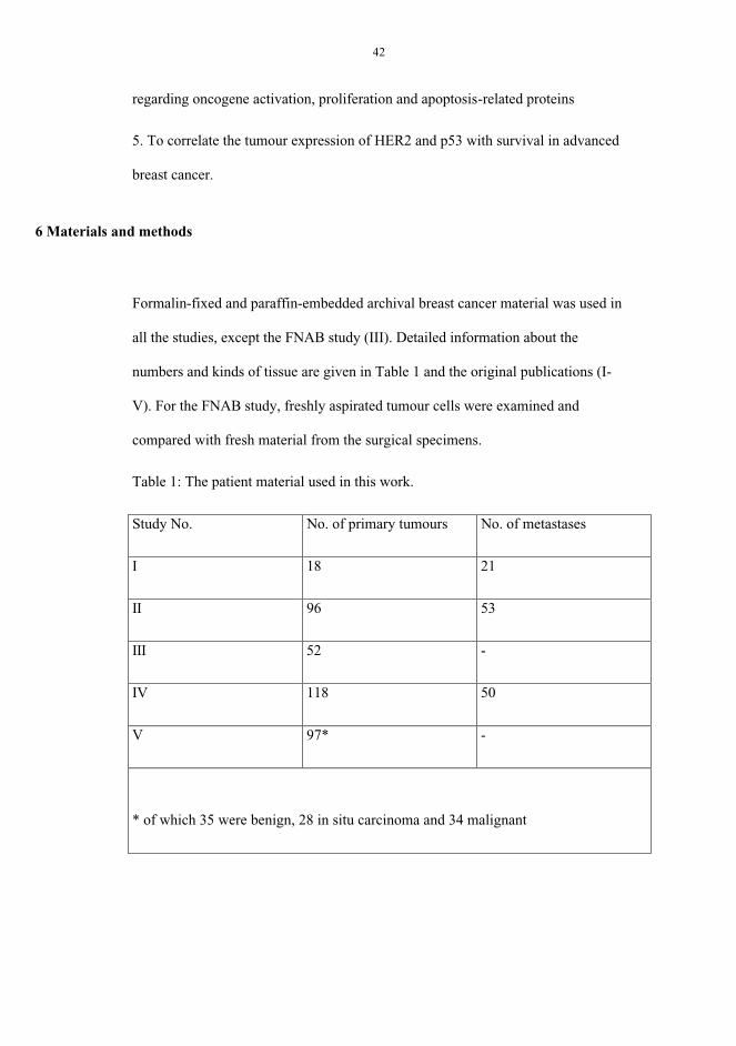
Table 1: The patient material used in this work.
Study No. No. of primary tumours No. of metastases
I 18 21
II 96 53
III 52 -
IV 118 50
V 97* -
* of which 35 were benign, 28 in situ carcinoma and 34 malignant
43
6.1 Tumour grading and typing
Tumour typing was performed according to WHO (Azzopardi et al. 1981). The
pTNM classification was done according to UICC 1992 (Spiessl et al. 1992b). The
cancers were graded according to Elston and Ellis’ (Elston 1984; Galea et al.
1992) modification of Bloom and Richardson’s original classification from 1957
(Bloom and Richardson 1957).
Benign breast lesions were first classified according to Dupont and Page (Dupont
and Page 1985) but as there were so few of them in each group, they were pooled
into atypical ductal hyperplasia (ADH) and papillomas. The ADH group included
four cases of atypical lobular hyperplasia, one fibroadenoma with unusually
proliferative epithelium with atypia, and two sclerosing adenomas with an
unusually florid appearance. Cysts and chronic cystic mastopathia without
epithelial atypia were excluded. Ductal cancer in situ was graded into
low/intermediate-grade, and high-grade types, DCIS1-2 and DCIS3. The DCIS1-2
was further subdivided into those without necrosis (DCIS1) and those with
necrosis (DCIS2), as described by Silverstein and co-workers (Silverstein et al.
1995).
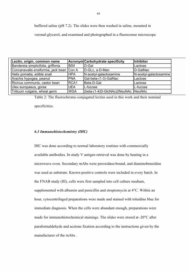
6.2 Lectin histochemistry
FITC-conjugated lectins were used on archival material of primary tumours and
their metastases. The lectins used and their specificities are given in Table 2 and
study I. They were all obtained desiccated from Sigma chemicals Co. Ltd. After
deparaffination and hydration, slides were incubated for 30 min in moist chambers
in the dark with the FITC-conjugated lectins diluted to 0.05mg/ml in phosphate-
44
buffered saline (pH 7.2). The slides were then washed in saline, mounted in
veronal-glycerol, and examined and photographed in a fluorecense microscope.
Table 2: The fluorochrome-conjugated lectins used in this work and their nominal
specificities.
6.3 Immunohistochemistry (IHC)
IHC was done according to normal laboratory routines with commercially
available antibodies. In study V antigen retrieval was done by heating in a
microwave oven. Secondary mAbs were peroxidase-bound, and diaminobenzidine
was used as substrate. Known positive controls were included in every batch. In
the FNAB study (III), cells were first sampled into cell culture medium,
supplemented with albumin and penicillin and streptomycin at 4°C. Within an
hour, cytocentrifuged preparations were made and stained with toluidine blue for
immediate diagnosis. When the cells were abundant enough, preparations were
made for immunohistochemical stainings. The slides were stored at -20°C after
paraformaldehyde and acetone fixation according to the instructions given by the
manufacturer of the mAbs .
Lectin, origin, common name Acronym Carbohydrate specificity InhibitorBandereia simplicifolia, griffonia BSII D-Gal LactoseConcanavalla ensiformia, jack bean Con A D-GLc, a-D-Man D-GalNacHelix pomatia, edible snail HPA N-acetyl-galactosamine N-acetyl-galactosamineArachis hypogea, peanut PNA Gal-beta-(1-3)-GalNac LactoseRicinus communis, castor bean RCA1 Beta-D-Gal LactoseUlex europaeus, gorse UEA L-fucose L-fucoseTriticum vulgaris, wheat germ WGA (beta-(1-4)D-GlcNAc)2NeuNAc NeuNAc
45
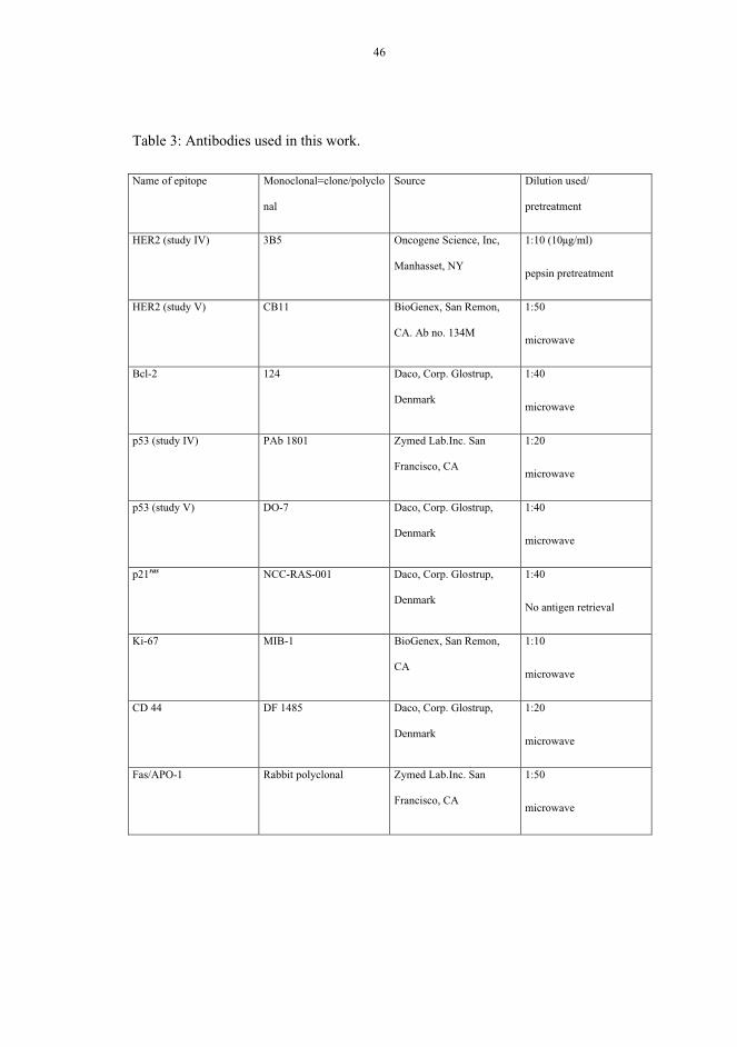
Immunoreactions were done on 4 µm sections of formalin-fixed, paraffin-
embedded tissue according to the manufacturer’s instructions. The antibodies
used, clone names and suppliers/manufacturers are listed in Table 3.
The immunoreaction in HER2 staining was considered positive when a brown
membrane positivity was seen in the cancer cells (Fig.1 B, C, E, F in study V).
Some tumours with very faint patchy staining were considered negative. In p53
staining, a brown staining of 10% or more of the nuclei of the cancer cells was
regarded as positive. The cut-off point was chosen according to current literature
on p53 (Ciesielski et al. 1995; Davidoff et al. 1991)
46
Table 3: Antibodies used in this work.
Name of epitope Monoclonal=clone/polyclo
nal
Source Dilution used/
pretreatment
HER2 (study IV) 3B5 Oncogene Science, Inc,
Manhasset, NY
1:10 (10µg/ml)
pepsin pretreatment
HER2 (study V) CB11 BioGenex, San Remon,
CA. Ab no. 134M
1:50
microwave
Bcl-2 124 Daco, Corp. Glostrup,
Denmark
1:40
microwave
p53 (study IV) PAb 1801 Zymed Lab.Inc. San
Francisco, CA
1:20
microwave
p53 (study V) DO-7 Daco, Corp. Glostrup,
Denmark
1:40
microwave
p21ras NCC-RAS-001 Daco, Corp. Glostrup,
Denmark
1:40
No antigen retrieval
Ki-67 MIB-1 BioGenex, San Remon,
CA
1:10
microwave
CD 44 DF 1485 Daco, Corp. Glostrup,
Denmark
1:20
microwave
Fas/APO-1 Rabbit polyclonal Zymed Lab.Inc. San
Francisco, CA
1:50
microwave
47
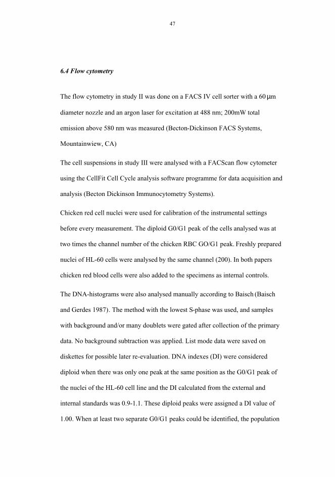
6.4 Flow cytometry
The flow cytometry in study II was done on a FACS IV cell sorter with a 60 µm
diameter nozzle and an argon laser for excitation at 488 nm; 200mW total
emission above 580 nm was measured (Becton-Dickinson FACS Systems,
Mountainwiew, CA)
The cell suspensions in study III were analysed with a FACScan flow cytometer
using the CellFit Cell Cycle analysis software programme for data acquisition and
Chicken red cell nuclei were used for calibration of the instrumental settings
before every measurement. The diploid G0/G1 peak of the cells analysed was at
two times the channel number of the chicken RBC GO/G1 peak. Freshly prepared
nuclei of HL-60 cells were analysed by the same channel (200). In both papers
chicken red blood cells were also added to the specimens as internal controls.
The DNA-histograms were also analysed manually according to Baisch (Baisch
and Gerdes 1987). The method with the lowest S-phase was used, and samples
with background and/or many doublets were gated after collection of the primary
data. No background subtraction was applied. List mode data were saved on
diskettes for possible later re-evaluation. DNA indexes (DI) were considered
diploid when there was only one peak at the same position as the G0/G1 peak of
the nuclei of the HL-60 cell line and the DI calculated from the external and
internal standards was 0.9-1.1. These diploid peaks were assigned a DI value of
1.00. When at least two separate G0/G1 peaks could be identified, the population
48
nearest to the channel of the G0/G1 peak of HL-60 nuclei was considered diploid,
the DI of the other populations was measured using this peak as a reference.
The quality of the histograms was estimated by the coefficients of variation (CV)
of the diploid G0/G1 peak. The manual model estimates the percent CV by
determining the peak width at the inflection point of the peak, which occurs at
approximately 60% of the peak height. The CVs of the aneuploid population's
G0/G1 peaks were used to compare the two methods. The percentage of cells in
SPF was estimated as the percentage of proliferating cells in the cell population
with the greatest DI. When theG0/G1 peaks were so close to each other that their
S-phases overlapped almost completely, a mean value was calculated for both
populations.
The FNA material was injected into an ampoule containing sterile RPMI 1461
(3ml) supplemented with 10% human serum. When there was sufficient material
in the FNAB, as measured from the firstly stained toluidine blue cytocentrifuged
preparations, additional cells were pelleted by centrifugation and resuspended in
50µg/ml of Ethidium bromide (Sigma Chemical Co, cat. no E8751) in 10mM
TRIS-EDTA buffer (pH 7.4) with 0.3% NP 40 and 1% RNAse (Sigma Chemical
Co) The sample was then passed through 50 m mesh nylon gauze and analysed by
a FACSscan4 flowcytometer.
Surgical specimens were immediately placed on ice, and frozen sections were
made within 30min. If the tumour was diagnosed as malignant, an adjacent tumour
section was snap frozen for later mechanical desegregation (mincing with a scalpel
in cell culture medium on a Petri dish), followed by staining and analysis as
described above for the FNAs.
49
Flow cytometry from archival material in study II was done on 50 µm thick
paraffin sections that were deparaffined, rehydrated and lysed with proteinase K.
The naked nuclei were stained with fluorescein-isothiocyanate as described above.
6.4 Statistical analysis
Statistical analysis comprised the Chi-square test, Fisher's exact test, and Mann-
Whitney rank-sum test, analysis of variance and Student's t-test. If the sample
distribution was skewed, an appropriate transformation was used before testing. If
there was a difference in group variance between the results for different
parameters, as determined by Lewene's test, Welch statistics were used. The life
table method and Mantel-Cox statistics estimated disease-free time and cumulative
survival rates. All computations were done using BMDP statistical programs and a
VAX 8600 computer (Dixon et al. 1983).
Differences between the groups were determined using Student's paired t-test.
Regression plots were used to study the correlation between the differences in the
SPF. Levene's test was also used to determine equality of the variances of the two
sample acquisition methods.
50
7 Results
7.1 FNA:StudyIII
The first aim of this study was to identify markers useful for preoperative
prognostication. Fifteen years ago CNBs were not in common use, and FNAB was
the leading method of preoperative diagnosis. A procedure was developed to make
cell blocks from FNAB material, allowing IHC to be done on consecutive sections
from the aspirated material (Krogerus and Andersson 1988). In many laboratories
it may be easier to make direct smears or multiple cyto-centrifuge preparations
than cell blocks for IHC, but also in such instances FNAB as well as CNB may be
used (Railo et al. 1996).
The quality of flow cytometric histograms was found to be better from FNAB
material than from tissue samples (III). There were more aneuploid peaks, on
average, in the FNABs than in the surgical specimens, 33 vs. 23 aneuploid peaks
out of 63 tumour samples. The correlation between SPA and the frequency of cells
staining positively for Ki-67 was better in the FNAB material than in the surgical
specimens.
The results of flow cytometry from the archival material largely confirmed what
has been claimed by other investigators. Flow cytometry gave reliable information
on cell kinetics and ploidy. This information was of prognostic value even in
advanced breast cancer, but the prognostic power did not exceed that of the stage
51
or grade of the tumour.
7.2 Lectin staining: Study I
The staining pattern as well as staining intensity were recorded. It was concluded
that there was more variability in the glucoconjugate composition of cells in the
primary tumour than in the cells of the metastases. Also metastases from the same
primary tumour could differ in their main lectin reactivity. Both the type and
intensity of staining apparently changed during the process of metastasis. This
may reflect clonal selection of the tumour cells to the metastatic site. Staining with
fluorescent lectins was seen in the cell membrane and cytoplasm or in the nuclei.
7.3 Proliferative epithelial lesions: Study V
The IHC staining results with seven different mAbs, given in Table 3, in
proliferative epithelial lesions were variable, and consistency was difficult to
obtain within the lesions or between the same category of lesions. It was found,
however, that the more atypical the lesion was, the more the results of the IHC
staining diverged from the staining patterns of morphologically normal epithelium.
A lower reactivity for HER2 was seen in ADH than in papillomas, while DCIS
stained more intensely than invasive cancer. In benign papillomas, the HER2
positivity associated significantly with a high percentage of staining for CD44.
The proliferative activity, as measured by MIB1 reactivity, was the highest in
invasive ductal carcinoma and the lowest in papillomas. There was, however, great
variability in proliferation activity among ductal carcinomas. A significant
52
difference in MIB1 stainings was seen only between ADH and DCIS3 (p<0.05).
In normal epithelium, CD44 staining was polarised, seen only in the baso-lateral
membranes, at the epithelial-myo-epithelial junction. Normal epithelium stained
more intensely than the malignant lesions, which showed a more haphazard
distribution of staining for CD44. No significant difference in the total lengths of
membranes staining positively was observed between the benign, pre-malignant
and malignant categories of the lesions.
A positive immuno-staining for Fas was found in about one third of the cells in all
types of lesions. The lowest intensity of staining was seen in papillomas.
The mAb for Ras p21 stained cells of both benign and malignant epithelium. The
p21 staining was difficult to interpret due to extensive background staining.
DCIS3 and ductal carcinoma of grade III had the lowest frequency of positive
cells (mean 25±31% (range 0-100) and 25±35% (range 0-100), respectively, and
lobular carcinoma had the highest percentage (59±34%, range 50-100).
In staining for bcl-2, the highest percentage of positive cells was seen in
papillomas and lobular carcinomas (71±33%, range 50-100 and 70±36%, range
50-100 , respectively) and the lowest in DCIS3 (34±47%, range 0-100). There was
a tendency towards an inverse correlation between the staining intensities of p53
and bcl-2 in all groups of lesions.
In benign papillomas, HER2 positivity was frequently seen in cells staining for
CD44.
53
7.4 Advanced breast cancer: Study II
It was found that only 44% of the HER2-positive primary cancers and 29% of the
HER2-negative primary cancers had HER2-positive metastases. This suggests that
expression of HER2 may associate with metastatic propensity. The histologic
grade of the primary cancer did not affect the HER2 status of the metastasis.
Positive staining for HER2 in the primary cancer did not correlate with the ploidy
or the SPF of the metastasis. HER2-positive metastases were more often (p<0.04)
aneuploid (DNA index 1.7) than negative (mean DNA index 1.3). The
proliferation activity was higher in the HER2-positive metastases (mean SPF
8.7%) than in the HER2-negative metastases (mean SPF of 5.6%), but this
difference was not significant.
There was no concordance between HER2-positivity and p53-positivity in this
material.
Patients with HER2-positive immunostaining of the primary tumours were free of
cancer for an average of 1.6 years, and HER2-negative cancer patients, on an
average of 2.0 years (p=0.8). When survival after recurrence was compared
between patients with either a HER2-positive or -negative primary cancer, there
was no significant difference between the two groups. HER2 did not correlate with
the clinical stage or size of the primary tumour. In histological grade I cancers (23
patients), 80% of the patients with HER2-positive cancer survived for five years,
but only 36% of the patients with HER2-negative cancer survived for five years.
54
The difference was, however, not quite significant (p=0.08), study V.
When compared with the primary tumours, it was more common for the
metastases to lose their positivity for HER2 than to gain it. In four cases a HER2-
negative primary tumour had HER2-positive metastases. One of them was on the
skin, one in the lung, and two in lymph nodes. The skin metastasis and one of the
lymph node metastases were lobular cancers; the lung metastasis was a ductal
cancer of grade II, and the other lymph node metastasis was a ductal cancer of
grade I (study IV).
8 Discussion
The prognosis of breast cancer patients has improved during the course of this
work (1996-2000) (Registry 1996; Rose'n et al. 2000). It is now apparent that
most breast cancer patients benefit from adjuvant therapy, regardless of the
presently used prognostic indexes ((EBCTCG). 1998a; (EBCTCG). 1998b) .
Patients with a favourable prognosis are nevertheless unnecessarily exposed to
prolonged medication. Therefore, there is still a demand for better prognostication
(Knorr et al. 1992; Rosen et al. 1992; Rosner and Lane 1993). The therapy has to
be tailored individually for each patient, and that is why also predictive measures
are needed (Klijn et al. 1993; Lavin et al. 1994; Rizzieri et al. 1999; Schaller et al.
1999).
Breast cancers are now found and treated at earlier stages, largely due to improved
mammography equipment (Rose'n et al. 2000). This places demands on the
diagnostic and therapeutic approaches. The ongoing discussion on the drawbacks
55
and benefits of FNAB contra CNB is one example of this change in attitudes
(Florentine et al. 1997; Masood 1995; Sharifi et al. 1999; Troncone et al. 1995) .
Palpable tumours can be easily diagnosed with almost any method. The small
tumour changes, targeted today by radiographic methods, need careful evaluation
for the best diagnostic approach. All doctors taking part in the decision making on
the treatment of breast cancer patients should therefore participate in the
discussion on the diagnosis, prognosis and treatment of the patients.
It is important that the community treating breast cancer recognises that it is
dealing with a disease of changing concepts. The measures taken to combat the
disease are modulating the behaviour of the disease (studies I and IV) (Zambelli et
al. 1999).
Several other investigators have evaluated some of the prognostic markers from
preoperative cytological specimens. Most of these authors have succeeded in
finding various cancer-related changes also in preneoplastic diseases (Gillett et al.
1998; Gupta et al. 1997; Lee 1995; Lucas et al. 2000; Pavelic et al. 1992;
Siziopikou et al. 1996). Thus, many changes occurring alone in the genome are
relatively innocent, but selection of a few critical changes probably initiates the
cancerous transformation (Minami et al. 1998; Moreno et al. 1997; Murphy et al.
1995).
Lectin staining has showen that the surface glycoconjugate composition of the
primary tumours was more heterogeneous than that of their metastases. This has
been shown also by other investigators by other methods (Gisselsson et al. 2000;
Könemann et al. 2000), but denied by others (Bonsing et al. 2000). Metastases
from the same primary tumour to different locations in the body can display
different patterns of lectin staining, implying that there are many clones in the
56
primary tumour, or that they can aquire different phenotypes in different locations
(study I). This suggests that the metastatic process may involve a selection for sub-
clones of tumour cells with better survival in a new environment.
Flow cytometry also showed that primary tumours frequently contained many cell
lines with different ploidy and proliferation kinetics (studies II and III) (Joensuu et
al. 1992). With FNAB, it is possible to enrich such populations for investigation,
as compared to conventional sectioning and suspending the tumour material. Other
investigators have confirmed this finding (Bach et al. 1991; Lorenzato et al.
2000a).
DI was strongly correlated with the grade, and probably therefore was not an
independent prognosticator in breast cancer patients, as also shown by others
(Blanco et al. 1990; Stanton et al. 1992; Toikkanen et al. 1989) . Though the SPF
gives reliable data on proliferation and has prognostic significance (Dressler et al.
1988; Toikkanen et al. 1989), proliferation can be measured equally reliably with
IHC (Aaltomaa et al. 1992b; Gaglia et al. 1993).
Arnelöv and Auer used image cytometry and reported a good and independent
correlation with DI and prognosis (Arnerlov et al. 1988; Fallenius et al. 1988;
Feichter 1991; Feichter et al. 1988). Beerman demonstrated improved prognostic
power of DI by grouping histograms into different prognostic classes (Beerman et
al. 1990). The concept of histogram type has been successfully used also by others
(Dieterich et al. 1995; Ferno et al. 1992). As in our studies, Tubiana found an
agreement on the limitations of ploidy analysis in advanced stages of breast cancer
(Tubiana et al. 1981).
The ICH obtained in this study from old archival material must be cautiously
57
interpreted. The fixation conditions of archival material are not always known.
Variability has been addressed using large series with simultaneous staining,
including negative and positive controls from the same batch. This makes reading
of the results more reliable, since it is possible to make background subtraction,
and subtraction for normal levels of staining (Battifora 1999).
Expression of HER2, and of many other proteins, may be lost or gained in the
process of metastasis (study IV). HER2-positive metastases displayed aneuploidy
more frequently and higher SPF than the HER2-negative metastases. It is possible
that the HER2-positivity of the primary cancer is not a feature that favours the
metastatic process; instead, the negative cell clones appeared to metastasise. This
is in line with the fact that HER2 was less often expressed in the infiltrating than
in the in situ component of a tumour. HER2 amplification is probably a sign,
among others, of the tumour-promoting DNA instability. This concept is
supported by the findings of a higher incidence of aneuploidy in the HER2-
positive tumours and their metastases.
HER2-positivity has been correlated with metastases in axillary lymph nodes and
with recurrence and visceral metastasis (Kallioniemi et al. 1991). An association
between the expression of HER2 and the tumour type and tumour diameter as
reported by Travis and his co-workers, could not be confirmed in this study
(Travis et al. 1996). This study did not demonstrate that HER2 adds new
prognostic information to breast cancer patient, as some investigators have shown
(Lipponen et al. 1993a). There are, however, recent reports on the use of HER2 as
a predictive marker of sensitivity to therapy (Cance and Liu 1995; Hudziak et al.
1988)
58
At advanced stages, grade 1, HER2-positive breast cancers are interesting. They
often lack apparent other criteria for high grade malignancy, such as high SPF,
aneuploidy, or p53 immunopositivity. However, in our material all of these (four
cases) had an additional adverse prognostic marker, besides HER2, either in the
primary tumour or in the metastasis.
Over-expression of HER2 has been studied mainly in primary tumours of the
breast. Therefore, little information is available on the HER2 status in metastatic
breast cancer. In the present study, 56% of the HER2-positive primary cancers had
negative metastases, and, conversely, 29% of the HER2-negative primary cancers
had positive metastases. This may be due to sampling error, since the study
included large tumours, and only selected areas of the cancer were studied.
Therefore HER2-positive clones in the primary HER2-negative cancers could be
missed. Otherwise the findings suggest that during the process of metastatic
spread, the cancer cells either lose HER2 over-expression or gain it.
Ductal cancers were more frequently p53- and CD44-positive and had higher
proliferative activity than the lobular cancers. The DCISs were more frequently
positive for p21, bcl-2 and HER2 (study V) than was LCIS. Among the invasive
cancers, lobular cancers were more often positive for HER2 than were the ductal
cancers. This might be a reflection of different behaviour in relation to the stroma
(du Toit et al. 1991). The expression of HER2 showed an inverse correlation with
the expression of adhesion molecule CD44. Thus, the HER2-positive tumours
were less positive for CD44 than were the HER2-negative cancers of the same
59
histological type.
According to my results and those of others (Lucas et al. 2000), aberrant protein
products are found in premalignant lesions as well as in overt cancers. There was
only a slight difference in the quantity and the distribution, not the quality of the
changes. The key difference between invasive and non-invasive tumour cell
populations remains to be defined. The possibility remains that the difference is
not inside the cells, but rather outside the cells, in the excreted enzymes and the
periductular stroma, as suggested by numerous investigators (Foekens et al. 1992;
Foekens et al. 1994; Foekens et al. 1993; Friedrichs et al. 1995; Frixen and
Nagamine 1993; Gasparini et al. 1997; Janicke et al. 1993; Joensuu et al. 1995) .
An interesting molecule is tenascin-C, which appears around micro-invasive cell
clusters (Jahkola et al. 1998; Jahkola et al. 1996).
The greatest challenge in prognostication, both for pre malignant and overtly
malignant disease, lies in the heterogeneity of the lesions. Even if the genetic
aberrations seems to go forward stepwise, the fenotypic diversity in different
tissues, and even from cell to cell, is endless. It is not the primary tumour, but
rather the metastatic disease that kills the patient. The ability to metastasise, to
adhere to vital organs and thrive there, are the key features of a killing disease, as
stated by Paget already at the end of the nineteenth century (Paget 1889).
First with the advent of oncogenes, then with more and more detailed
understanding of cell division and its regulation, I have come to the conclusion
that understanding cancer is like understanding chaos. It may be possible in
theory, but not in detail. Instead of taking single parameters as single prognostic
60
factors, we have to think of the tumour as a society, with lots of individuals, and
the more divergent the population is, the more potential it holds for destroying its
host. Nicholson, in his discussion on the process of metastasis, concluded that the
more aggressive metastatic clones had no means of emergin to something new,
and to send metastases to other locations, as had the heterogeneous primary
tumour (Nicolson 1982). It may of course be that chance alone decides how a
cancer cell behaves in its next division, what devious behaviour it will gain, and
that nothing in the cells predicts a malignant kind of behaviour.
61
9 Summary and conclusions
Breast cancer is the most significant malignancy of women in Western countries.
Biologically the disease is heterogeneous and therefore unpredictable. It has
unique features in its ability of dormancy and early metastasising. These features
make the disease difficult to cure, and a large number of women live their lives in
fear of the disease.
The purpose of this study was to find reasons for this unpredictability. In this
study, tumour material from advanced disease as well as from early and pre-
malignant disease were investigated with lectin staining, with flow cytometry and
with IHC for oncogen products and proliferation markers. Primary tumours were
compared with their metastases, and in situ components of the disease with the
invasive components. Furthermore, diagnostic procedures, FNAB and histology,
were compared with each other for diagnostic accuracy.
The primary tumours were more variable in their staining patterns than their
corresponding metastases. Single parameters, like SPF, DI, hormone receptors,
and single oncogene amplifications, did not predict the outcome in advanced
disease. Deviations from normal epithelium seen in breast cancers were also found
in pre-malignant disease. Additional markers for cellular changes, and probably
also for extra-cellular alterations are to be identified, in order to target the crucial
constellations of malignant behaviour.
FNAB proved to be better material for flow cytometry than surgically removed
tissue, and FNAB was also useful for IHC. The investigated parameters gave
useful information that may help in therapy decisions.
62
10 Acknowledgements
This work was carried out at the Department of Pathology, Haartman Institute, University of Helsinki. I want to thank the head of the department for the opportunity to complete this study and for the inspiring atmosphere prevailing in his laboratory and in all the laboratories in the Hartman Institute, especially the former transplantation laboratory. I warmly thank my supervisor, Professor Leif Andersson, for many challenging ideas and for coping with me despite my stubbornness and slowness. I want to thank the many wonderful women working in the laboratories, who taught me everything from cell culturing and immunohistochemistry to gel electrofhoresis and flow cytometry. Thank you Hannele L, Hilkka Toivonen, Tuula Halmesvaara, Monna Shoulz and Last but not least our ever efficient ward führer in Maria pathology laboratory Anna-Leena Hästö. I owe greatly to Docent Stig Nordling for arguing with me on hormone receptors and p-values and for having had the time and interest to read most of my papers before submission. I wish to thank my co-authors Marja Leivonen and Mikael Railo as well as Monna, Anna-Leena and Stig for fruitful co-operation and support. Docents Paula Martikainen and Ylermi Soini are thanked for fast and careful review of this thesis and expert advise. I want to give special thanks to my colleagues Terttu Toivonen and Raija Malmi for never ending interest in and support of my work. I also remember with warmth all other friends and pathology colleagues at the pathology laboratory at Hartman Institute, with whom many happy hours were spent during hard work. My heart beats for the pathology ward at Maria hospital, where the staff has always been with me in all my time consuming experimentation, never was there an aggressive woice despite enormous workloads. Everything I have asked for has always been done, so thank you Arja, Ulla, Bigi, Riitta, Tiina Anne, Irma, Gitta and Svenka. I also want to thank an other inspiring working group in which I have had the honour to be a part: The trust in me that I have encountered At the 4th floor investigation laboratory at the Surgical hospital in Helsinki, especially Irmeli Lautenschlager has rendered me some of the self-confidence needed to fulfil this task. Thank you dear Björn, Rasmus, Camilla, Niklas, Fanny, mother Maj-Lis, father Helge and family, neighbours and friends for giving me perspective to and enjoyment in life as a whole. This work was financially supported by Finska läkaresällskapet, The Finnish Breast Cancer Group, The Ida Montin Foundation and the Paulo Foundation. June 2001-06-27 LK
63
11 References
(EBCTCG)., E. B. C. T. C. G. (1998a). “Polychemotehrapy for early breast
cancer: an overview of the randomised trials.” Lancet, 352, 930-42.
(EBCTCG)., E. B. C. T. C. G. (1998b). “Tamoxifen for early breast cancer: an
owerview of the randomised trials.” Lancet, 351, 1451-67.
Aaltomaa, S., Lipponen, P., Eskelinen, M., Alhava, E., and Syrjanen, K. (1991).
“Nuclear morphometry and mitotic indexes as prognostic factors in breast
cancer.” Eur J Surg, 157(5), 319-24.
Aaltomaa, S., Lipponen, P., Papinaho, S., Klemi, P., Kosma, V. M., Marin, S.,
Eskelinen, M., Alhava, E., and Syrjanen, K. (1992a). “Nuclear
morphometry and DNA flow cytometry as prognostic factors in female
breast cancer.” Eur J Surg, 158(3), 135-41.
Aaltomaa, S., Lipponen, P., and Syrjanen, K. (1992b). “Prognostic value of cell
proliferation in breast cancer as determined by proliferating cell nuclear