ARTICLE Programmed hierarchical patterning of bacterial populations Christian R. Boehm 1,3 , Paul K. Grant 1,2 & Jim Haseloff 1 Modern genetic tools allow the dissection and emulation of fundamental mechanisms shaping morphogenesis in multicellular organisms. Several synthetic genetic circuits for control of multicellular patterning have been reported to date. However, hierarchical induc- tion of gene expression domains has received little attention from synthetic biologists, despite its importance in biological self-organization. Here we report a synthetic genetic system implementing population-based AND-logic for programmed autonomous induction of bacterial gene expression domains. We develop a ratiometric assay for bacteriophage T7 RNA polymerase activity and use it to systematically characterize different intact and split enzyme variants. We then utilize the best-performing variant to build a three-color patterning system responsive to two different homoserine lactones. We validate the AND gate-like behavior of this system both in cell suspension and in surface culture. Finally, we use the synthetic circuit in a membrane-based spatial assay to demonstrate programmed hierarchical patterning of gene expression across bacterial populations. DOI: 10.1038/s41467-018-03069-3 OPEN 1 Department of Plant Sciences, University of Cambridge, Downing Street, Cambridge, CB2 3EA, UK. 2 Microsoft Research, 21 Station Road, Cambridge, CB1 2FB, UK. 3 Present address: Max Planck Institute of Molecular Plant Physiology, Am Mühlenberg 1, 14476 Potsdam, Germany. Correspondence and requests for materials should be addressed to J.H. (email: [email protected]) NATURE COMMUNICATIONS | (2018)9:776 | DOI: 10.1038/s41467-018-03069-3 | www.nature.com/naturecommunications 1 1234567890():,;
Transcript
ARTICLE
Programmed hierarchical patterning of bacterialpopulationsChristian R. Boehm 1,3, Paul K. Grant1,2 & Jim Haseloff 1
Modern genetic tools allow the dissection and emulation of fundamental mechanisms
shaping morphogenesis in multicellular organisms. Several synthetic genetic circuits for
control of multicellular patterning have been reported to date. However, hierarchical induc-
tion of gene expression domains has received little attention from synthetic biologists,
despite its importance in biological self-organization. Here we report a synthetic genetic
system implementing population-based AND-logic for programmed autonomous induction of
bacterial gene expression domains. We develop a ratiometric assay for bacteriophage T7
RNA polymerase activity and use it to systematically characterize different intact and split
enzyme variants. We then utilize the best-performing variant to build a three-color patterning
system responsive to two different homoserine lactones. We validate the AND gate-like
behavior of this system both in cell suspension and in surface culture. Finally, we use the
synthetic circuit in a membrane-based spatial assay to demonstrate programmed hierarchical
patterning of gene expression across bacterial populations.
DOI: 10.1038/s41467-018-03069-3 OPEN
1 Department of Plant Sciences, University of Cambridge, Downing Street, Cambridge, CB2 3EA, UK. 2Microsoft Research, 21 Station Road, Cambridge, CB12FB, UK. 3Present address: Max Planck Institute of Molecular Plant Physiology, Am Mühlenberg 1, 14476 Potsdam, Germany. Correspondence and requestsfor materials should be addressed to J.H. (email: [email protected])
The hierarchical organization of multicellular organismsbuilds on mechanical and chemical interactions. Cellssense cues in their environment and modulate both their
metabolism and communication with neighbors. At the popula-tion level, the interplay of multiple processes across time andspace can lead to the emergence of self-organization throughmechanisms such as symmetry breaking, domain induction, andboundary formation1. Despite the advances made in the biolo-gical sciences over past decades, many of the mechanismsunderlying complex patterning and morphogenesis remainelusive.
In efforts to explain multicellular patterning, two types ofmodels have been pre-eminent to date: the reaction-diffusion(RD) model proposed by Alan Turing2, and the positionalinformation (PI) model (also known as the “French flag model”)originating from Lewis Wolpert3. RD-type systems are char-acterized by self-organized spatial patterns emerging from inter-acting positive- and negative-feedback loops in response to twodiffusible morphogens. In contrast, PI-type models employ asingle predefined morphogen gradient, which is interpreted byreceiving cells according to the local concentration of the mor-phogen. Though the two models are conceptually distinct, theyare not necessarily mutually exclusive4.
Empowered by modern genetic techniques, molecular biolo-gists are now striving to not only dissect developmental processes,but to exploit their modularity to customize the design of livingsystems5. Biological self-organization is a powerful tool for bio-processing and remediation in tailored microbial consortia, forsustainable bioproduction in novel plant compartments, or forapplications in regenerative medicine enabled by engineeredvertebrate tissues. A fundamental requirement for harnessing thispotential is control over the differentiation of cell types to createdomains of gene expression in spatially organized patterns. Todate, several synthetic biological circuits capable of multicellularpatterning have been reported, predominantly implementedeither by RD-type6,7 or by PI-type8–11 mechanisms.
However, mechanisms for hierarchical patterning havereceived little attention from synthetic biologists to date. In var-ious examples of morphogenesis, such as vulval induction innematodes12, the ABC model of flower development13, ormesoderm induction in vertebrates14, we can observe nesteddomains of gene expression. To emulate biological self-organization on this level of complexity, we require controlover hierarchical induction of new domains within existingpatterns.
Approaching this challenge, we report the implementation of asynthetic genetic circuit that controls emergence of a new domainof gene expression at the interface of existing bacterial popula-tions. To the best of our knowledge, this is the first description ofa synthetic genetic circuit implementing AND-logic for autono-mous hierarchical patterning at the population scale. A previouslyreported population-based edge detector has implemented AND(NOT (NOT)) logic in response to light projected through amask15. In contrast, the circuit introduced here is designed toestablish two layers of patterning in the absence of an externallydefined spatial input.
As intercellular signals mediating domain induction, we utilizethe diffusible small molecule signals N-(3-oxohexanoyl)-L-homoserine lactone (3OC6HSL) and N-(3-oxododecanoyl)-L-homoserine lactone (3OC12HSL). These compounds are derivedfrom the bacterial quorum-sensing systems from Vibrio fischeri16
and Pseudomonas aeruginosa17, respectively. Both systemsemploy a single biosynthetic enzyme to produce a diffusible signal(LuxI/LasI), and a single receiver protein (LuxR/LasR) to activatetranscription of a target gene controlled by their cognate pro-moters (PLux/PLas) upon signal binding18. The two signaling
systems have been previously combined as part of synthetic cir-cuits, but by default exhibit significant levels of crosstalk19–24.Overcoming this complication, we have recently reported anintercellular signaling system minimizing crosstalk between3OC6HSL and 3OC12HSL in the same cell24.
In this work, we combined this improved intercellular signalingsystem with the transcriptional output from an orthogonal splitbacteriophage T7 RNA polymerase (T7RNAP)25–29 to establishhierarchical induction of gene expression domains at the popu-lation scale. A first layer of patterning was established via pro-moters that respond to the two different homoserinelactones (HSLs) and induce production of cyan (CFP) and yellowfluorescent protein (YFP), respectively. In addition, each of thetwo HSLs induced expression of one half of the split T7RNAPprotein. This was used to generate a second layer of patterning,where RNA polymerase activity was limited to where the twodiffusible signals coincide, and was marked by expression of redfluorescent protein (RFP) from a T7 promoter. In order to con-struct this system, we characterized systematically the activity ofdifferent T7RNAP genes using a ratiometric strategy to identifythe split variant of highest dynamic range. We then implementeda synthetic three-color AND gate based on split T7RNAPresponsive to 3OC6HSL and 3OC12HSL in both cell suspensionsand surface cultures of Escherichia coli. Finally, we demonstratedthat the synthetic circuit autonomously mediated programmedemergence of a new gene expression domain at the interface oftwo bacterial populations (Fig. 1).
ResultsDesign of a ratiometric reporter for T7RNAP activity. Rationaldesign of a synthetic circuit generating desired multicellularbehavior requires a quantitative understanding of its core geneticcomponents. To increase robustness in measurements of activityfrom the T7 promoter, we extended a previously reported ratio-metric strategy30,31 to T7RNAP-driven gene expression: in ourratiometric reporter plasmids, the YFP variant mVenus32 servedas a primary reporter of T7RNAP activity. In addition, theratiometric reporter plasmids encoded a CFP variant mTur-quoise233 that was constitutively expressed under control ofreference promoter PJ23101 and reference RBSB0034 (MIT Registryof Standard Biological Parts). Relative promoter activity could beexpressed as the average YFP over CFP fluorescence intensity per
Design of a high-dynamic-range ratiometric reporter plasmid of T7RNAP activity
Ratiometric reporter plasmid
Ratiometric characterization of T7RNAP variants
Split T7RNAP
Design of a split T7RNAP-based three-color AND gate
Bicistronic controller plasmid
Controller tuning and validation using plate fluorometry
Improved bicistronic controller plasmid
Surface-based controller validation and application in programmed hierarchical patterning
Fig. 1 Design workflow of a genetic circuit for synthetic hierarchicalpatterning. The resulting synthetic circuit autonomously mediates theprogrammed emergence of a gene expression domain at the interface oftwo bacterial populations
cell during exponential growth phase30,31. This indicator waschosen in order to correct for variation in cellular gene expressioncapacity due to environmental conditions.
We designed a small library of plasmid reporters for T7RNAPactivity to identify a combination of promoter, 5′-UTR, and copynumber capable of supporting high dynamic range in reporterinduction. This library embraced all combinations of (i) the wild-type T7 promoter PT7 (refs. 34–36) or a mutant promoter PT7(-3G)(~20% activity of the consensus sequence)37, (ii) the 5′-UTR frombacteriophage T7 gene 10 RBST7g10 (ref. 38) or a synthetic 5′-UTRembracing the reference RBSB0034 (MIT Registry of StandardBiological Parts), and (iii) the high copy number backbonepSB1A3 (pMB1 ori, MIT Registry of Standard Biological Parts) orthe low copy number backbone pSB4A5 (pSC101 ori, MITRegistry of Standard Biological Parts). Notably, these plasmidsincluded a LacI generator cassette to reduce leaky expression ofT7RNAP from the genome of T7 Express E. coli.
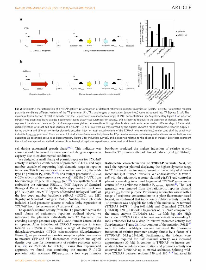
To quantify relative activity from the T7 promoter across thesmall library of ratiometric reporters outlined above, weintroduced the plasmids individually into T7 Express E. coliencoding a single genomic copy of T7RNAP under control of thelac operon. After induction of T7RNAP expression in trans-formed T7 Express E. coli using a range of isopropyl-β-D-thiogalactopyranoside (IPTG) concentrations (SupplementaryFigure 1), we performed ratiometric assays in a plate fluorometerto measure CFP and YFP fluorescence intensities and opticaldensity over time for measurement of relative promoter activity(Fig. 2a; see Methods for details). Taking this experimentalapproach, we found that combining the mutant PT7(-3G)promoter with reference RBSB0034 on a low copy number
backbone produced the highest induction of relative activityfrom the T7 promoter after addition of inducer (7.58± 0.08-fold).
Ratiometric characterization of T7RNAP variants. Next, weused the reporter plasmid displaying the highest dynamic rangein T7 Express E. coli for measurement of the activity of differentintact and split T7RNAP variants. We co-transformed TOP10 E.coli with the ratiometric reporter plasmid p4g3VT and controllerplasmids encoding intact and fragmented T7RNAP genes undercontrol of the arabinose-inducible PBAD/araC system26. The LacIgenerator was removed from the ratiometric reporter plasmidp4g3VTLacI for this purpose. Performing ratiometric assays with arange of arabinose concentrations in a plate fluorometer-basedformat, we confirmed that induction of relative activity from theT7 promoter was negligible for both of the individual N-terminal(T7RNAP[1–179]: 1.10± 0.01-fold) and C-terminal (T7RNAP[180-880]: 0.94± 0.05-fold) fragments of T7RNAP compared tothe intact enzyme (T7RNAP: 12.9± 0.3-fold; Fig. 2b). Highinduction of T7RNAP (i.e. at inducer concentrations exceeding 1mM arabinose) led to a drop in relative promoter activity (seeSupplementary Figure 2). Incorporation of the mutation R632S39
into the intact wild-type enzyme increased the maximuminduction of relative promoter activity almost by a factor of 4(T7*RNAP: 50.1± 0.9-fold). Furthermore, the inducer con-centration required for half maximal activity was increasedapproximately 30-fold. In contrast to T7RNAP, no inverse cor-relation between inducer concentration and promoter activity wasobserved for T7*RNAP up to 50mM arabinose. Splitting wild-type T7RNAP between residues 179 and 18025,26 increased its
Fig. 2 Ratiometric characterization of T7RNAP activity. a Comparison of different ratiometric reporter plasmids of T7RNAP activity. Ratiometric reporterplasmids combining different variants of the T7 promoter, 5′-UTRs, and origins of replication (underlined) were introduced into T7 Express E. coli. Themaximum fold-induction of relative activity from the T7 promoter in response to a range of IPTG concentrations (see Supplementary Figure 1 for inductioncurves) was quantified using a plate fluorometer-based assay (see Methods for details), and is reported relative to the absence of inducer. Error barsrepresent the standard deviation (s.d.) of average values yielded between three biological replicate experiments performed on different days. b Ratiometriccharacterization of intact and split variants of T7RNAP. TOP10 E. coli were co-transformed by the highest dynamic range ratiometric reporter p4g3VTtested under a and different controller plasmids encoding intact or fragmented variants of the T7RNAP gene (underlined) under control of the arabinose-inducible PBAD/araC promoter. The maximum fold-induction of relative activity from the T7 promoter in response to a range of arabinose concentrations wasquantified as described above (see Supplementary Figure 2 for induction curves), and is reported relative to the absence of inducer. Error bars representthe s.d. of average values yielded between three biological replicate experiments performed on different days
dynamic range by over a factor of 3 (split T7RNAP: 43± 2-fold).In contrast to the intact enzyme, incorporation of the R632Smutation decreased the maximum induction of relative promoteractivity in split T7RNAP (split T7*RNAP: 12.3± 0.3-fold).Adopting an approach previously taken27, we also tested whetherthe addition of SynZIP protein–protein interaction domains40,41
to wild type and mutant (R632S) variants of split T7RNAPincreased their dynamic range. Compared to enzyme modifica-tions described above, application of SynZIP domains furtherincreased the inducer concentration required for half maximalactivity (i.e. over 100-fold relative to T7RNAP; see SupplementaryFigure 2). However, neither modified variant exceeded wild-typesplit T7RNAP in maximum induction of activity from the T7promoter (split T7RNAP(+SynZIP): 16.3± 0.3-fold; split T7*RNAP(+SynZIP): 1.42± 0.04-fold). From the ratiometric assays per-formed, we concluded that split T7RNAP was the most promisingnicked variant to employ in our synthetic patterning circuit.
Design of an HSL-responsive three-color AND gate. Havingvalidated the activity of split T7RNAP, we utilized this enzyme todesign a three-color synthetic transcriptional AND gate. To thisend, we assembled a controller plasmid embracing two bicistronicoperons composed of T7RNAP[1–179] and mVenus, andT7RNAP[180–880] and mTurquoise2, respectively (Fig. 3). Thedownstream fluorescent proteins were included to serve as visualindicators of expression of the individual T7RNAP fragmentsin vivo. The two bicistronic operons were controlled by hybridpromoters responsive to either 3OC12HSL (PLas81*) or 3OC6HSL(PLux76*), respectively, alongside weak ribosomal binding sitesRBSB0033 (MIT Registry of Standard Biological Parts). The pro-moters in question were derived from our previously reportedsystem for orthogonal intercellular signaling24, and containedmutations shown to reduce basal activity from the Lux pro-moter42. Cognate receiver proteins LuxR and LasR were con-stitutively expressed from the vector backbone pR33S175. We
also constructed an RFP reporter plasmid of T7RNAP activityp4g3R based on the design principles proven earlier (see Fig. 2a):the mRFP1 gene was controlled by PT7(-3G) and RBSB0034 on alow copy number pSC101 backbone. In contrast to ratiometricreporter plasmids, p4g3R lacked the PJ23101-mTurquoise2 refer-ence operon. We validated the response of p4g3R to T7RNAP byintroduction into T7 Express E. coli and measuring RFP output invarying concentrations of IPTG (Supplementary Figure 3).
We began construction of the AND gate by testing thebehavior of the two individual controller half circuits. For thispurpose, we built half-circuit controller plasmids pCRBDRT7VTPLux*500CFP and pCRB DRT7VTPLux*500YFP, lack-ing either the bicistronic operon PLas81*-T7RNAP[1–179]-mVenusor PLux76*-T7RNAP[180-880]-mTurquoise2, respectively. Wetransformed TOP10 E. coli cells with each half-circuit controllerplasmid in addition to the RFP reporter plasmid p4g3R. Wemeasured CFP, YFP, and RFP fluorescence intensities over timeas a function of 3OC6HSL and 3OC12HSL concentrations using aplate fluorometer-based assay (see Methods for details). Asexpected, the half-circuit bicistronic controller plasmid pCRBDRT7VTPLux*500CFP responded to increasing concentrationsof 3OC6HSL with increased induction of corrected CFPfluorescence intensity. Similarly, the half-circuit bicistroniccontroller plasmid pCRB DRT7VTPLux*500YFP responded toincreasing concentrations of 3OC12HSL with increased inductionof corrected YFP fluorescence intensity (Table 1, SupplementaryFigure 4a, b). Induction of corrected RFP intensity from thereporter plasmid was negligible for both half-circuit bicistroniccontroller plasmids. Under the same assay conditions, thecomplete bicistronic controller plasmid pCRB DRT7VTPLux*500produced substantial levels of corrected RFP fluorescenceintensity only if exposed to both 3OC6HSL and 3OC12HSL(Supplementary Figure 4c). Notably, we observed a reduction incorrected YFP (up to 29± 7%) and CFP (up to 46± 9%)fluorescence intensities under conditions of high RFP induction.
Fig. 3 Design of a split T7RNAP-based three-color circuit responsive to HSLs. Schematic representations of the mRFP1-expressing reporter of T7RNAPactivity p4g3R (see Supplementary Figure 3 for induction curve) and the bicistronic controller plasmid pCRB DRT7VTPLux*500 encoding two bicistronicoperons responding to 3OC6HSL and 3OC12HSL by induction of T7RNAP[180-880] and mTurquoise2, or T7RNAP[1–179] and mVenus, respectively, areshown
Table 1 Comparison of a bicistronic controller plasmid and its constitutive half circuits
TOP10 E. coli were co-transformed with p4g3R, an mRFP1-expressing reporter of T7RNAP activity, and the bicistronic controller plasmid pCRB DRT7VTPLux*500 or one of its constitutive half circuits. Incontrast to pCRB DRT7VTPLux*500, pCRB DRT7VTPLux*500CFP and pCRB DRT7VTPLux*500YFP lacked either the bicistronic operon PLas81*-T7RNAP[1–179]-mVenus or PLux76*-T7RNAP[180-880]-mTurquoise2, respectively. The behavior of bicistronic controller plasmid variants was tested alongside the RFP reporter plasmid under a two-dimensional titration of 3OC6HSL and 3OC12HSL using aplate fluorometer-based assay (see Methods for details). The maximum observed fluorescence intensity, corrected for background signal present in the absence of externally supplied HSLs, is reported.Values shown are the mean of three biological replicate experiments performed on different days, plus or minus their s.d. Activity plots are shown in Supplementary Figure 4. Individual induction curvesare shown in Supplementary Figure 5.
While measurement of corrected RFP intensity under two-dimensional titration of HSLs showed AND gate-like behavior ofour circuit (Supplementary Figure 4c), we sought to furtherreduce background induction in the absence of 3OC12HSL.Corrected RFP fluorescence intensity in the absence of 3OC6HSLwas already low. We replaced RBSB0033 (relative strength 500arbitrary units) regulating expression of T7RNAP[1–179] inpCRB DRT7VTPLux*500 by a series of weaker ribosomal bindingsites (relative strength 250, 100, and 50 arbitrary units) designedby the Ribosome Binding Site Calculator43. We measured theresponse of the resulting bicistronic controller plasmid variants toincreasing concentrations of 3OC12HSL using plate fluorometry.pCRB DRT7VTPLux*250 demonstrated the greatest sensitivity to3OC12HSL (Supplementary Figures 6 and 7). When we inducedthe improved three-color circuit composed of bicistroniccontroller pCRB DRT7VTPLux*250 and reporter plasmidp4g3R with both HSLs, we observed corrected RFP fluorescenceintensity of over 8-fold of the maximum background detected inthe absence of either input signal (Supplementary Figure 8). Theimproved synthetic three-color circuit correctly exhibited thebehavior of a synthetic transcriptional AND gate responsive totwo different HSLs (Fig. 4).
HSL-mediated patterning of bacterial populations. To testwhether our three-color synthetic transcriptional AND gate wasresponding to HSLs in surface culture as well as in suspension, weemployed a previously reported spatial assay based on mem-branes printed with hydrophobic grids24. Bacterial populationswere confined to quadrants with well-defined geometry, separatedfrom one another while allowing HSL-mediated communicationacross the substrate. Following inoculation of membranes withdilute bacterial cultures, changes in CFP, YFP, and RFP fluores-cence intensities in response to environmental signals could bemonitored across the grid over time using a custom macroscopicimaging system (see Methods for details).
First, we uniformly inoculated 64 membrane quadrants withbacteria co-transformed by the RFP reporter plasmid p4g3R andthe improved bicistronic controller plasmid pCRBDRT7VTPLux*250. In this experiment, membranes were placedon minimal nutrient agar containing (i) no added HSLs, (ii) 25μM 3OC6HSL, (iii) 1 μM 3OC12HSL, or (iv) 25 μM 3OC6HSLand 1 μM 3OC12HSL. The concentrations of signaling moleculeswere chosen to match the condition of highest induction of RFPfluorescence intensity observed in suspension culture (see Fig. 4).Solid culture assays performed at 37 °C as outlined above
Fig. 4 An improved synthetic three-color circuit implementing AND gate-like behavior. The behavior of the high-dynamic-range bicistronic controllerplasmid pCRB DRT7VTPLux*250 was tested alongside the RFP reporter plasmid p4g3R under a two-dimensional titration of 3OC6HSL and 3OC12HSLusing a plate fluorometer-based assay (see Methods for details). Fluorescence intensity, corrected for background signal present in the absence ofexternally supplied HSLs, is reported for each condition. Plots show average values from three biological replicate experiments performed on different days.Corresponding s.d. values are shown alongside individual induction curves in Supplementary Figure 8
CFP YFP RFP Merge a
CFP
3OC6HSL
3OC12HSL
YFP
RFP
Merge
b
+ / +
– / +
+ / –
– / –
3OC6HSL / 3OC12HSL
Fig. 5 HSL-responsive surface-based patterning of bacterial gene expression. a Surface-based circuit behavior in response to HSLs present in the growthmedium at uniform concentration. TOP10 E. coli co-transformed by the RFP reporter plasmid p4g3R and the improved bicistronic controller plasmid pCRBDRT7VTPLux*250 (see Fig. 3) were incubated on membranes printed with hydrophobic ink, placed on minimal agar containing different combinations of3OC6HSL (25 μM) and 3OC12HSL (1 μM). Images shown were captured at t= 1500min (time relative to start of incubation). Corresponding correctedfluorescence intensities are shown in Supplementary Figure 9. b Surface-based AND gate behavior in response to HSL gradients. TOP10 E. coli cells co-transformed by controller and reporter plasmids as above were incubated on membranes placed on minimal agar lacking supplemented HSLs. Instead,aqueous solutions containing 500 μM 3OC6HSL or 200 μM 3OC12HSL, respectively, were spotted next to the cells on either side and left to diffuse intothe bacterial population from opposite directions. Images shown were captured at t= 3000min (time relative to start of incubation). Correctedfluorescence intensities recorded over time are shown in Supplementary Figure 10
confirmed that presence of both HSLs was required forsubstantial induction of corrected RFP fluorescence intensity,suggesting AND gate-like behavior (Fig. 5a). Corrected CFP andYFP fluorescence intensities were reduced to background levelsunder the condition of co-induction compared to presence ofeither signaling molecule alone.
Next, we sought to test whether the same synthetic three-colorcircuit could implement surface-based spatial patterning across abacterial population in response to gradients of diffusing HSLs.For this purpose, we inoculated rows of quadrants with double-transformed cells on agar lacking supplemented HSLs. At a
distance of one quadrant from either end of the inoculated rows,we then spotted solutions of 3OC6HSL (500 μM) or 3OC12HSL(200 μM), and allowed the inducers to diffuse into the bacterialpopulation from opposite directions. Over a course of 50 h, weobserved the emergence of a three-color pattern across thebacterial population: with fluorescence of CFP and YFP mirroringthe opposite gradients of 3OC6HSL and 3OC12HSL, RFPfluorescence was found to peak near the center of the inoculatedrows where the two gradients overlap (Fig. 5b).
Programmed emergence of a bacterial gene expression domain.Finally, we implemented a greater degree of autonomy in oursynthetic three-color patterning system by enabling bacterial cellsto produce and secrete their own inducers. The improved bicis-tronic controller plasmid pCRB DRT7VTPLux*250 and the RFPreporter p4g3R had been characterized both in suspension (seeFig. 4) and in surface culture (see Fig. 5). We introduced a thirdplasmid into E. coli: the sender plasmids pSB1C3 I0500 (LuxI/LasI) contained either luxI (encoding an enzyme producing3OC6HSL; ref.16) or lasI (encoding an enzyme producing3OC12HSL; ref.17) under control of the PBAD/araC promotersystem.
Utilizing solid culture assays, we inoculated membrane gridswith two adjacent populations of the different triple-transformedcell types: each population contained (i) the RFP reporter, (ii) theimproved bicistronic controller, and (iii) either the LuxI or theLasI sender plasmid. In the absence of arabinose, fluorescenceintensity was low in all three channels. In the presence of 25 mMarabinose, corrected CFP and YFP fluorescence intensities weresubstantially induced in cells expressing LuxI and LasI,respectively (Fig. 6). By comparing arabinose-induced popula-tions to HSL standard curves prepared under the same conditions(Supplementary Figure 12), we were able to estimate the effectiveconcentration of HSLs those populations were experiencing. Weestimated the effective concentrations of 3OC6HSL and3OC12HSL in their respective domains of synthesis to be roughly0.2–1 μM and 1 μM, respectively, at quadrants farthest from theinterface. As intended, a symmetrical domain of high RFP activityspontaneously emerged at the interface of 3OC6HSL- and3OC12HSL-sending cell populations. Our synthetic three-colorpatterning circuit generated programmed emergence of this newgene expression domain in absence of externally applied signalinggradients, implementing an autonomous mechanism for hier-archical patterning.
To assess the robustness of this behavior, we subjected thesuboptimal bicistronic controller plasmids pCRBDRT7VTPLux*500 and pCRB DRT7VTPLux*50 (see Supple-mentary Figure 6) to the same solid culture experiment. The useof the stronger-than-optimal RBSB0033 (pCRBDRT7VTPLux*500) led to slightly increased corrected YFPintensity and reduction of corrected CFP fluorescence intensityby approximately two-thirds compared to the optimal plasmid.Peak corrected RFP fluorescence intensity in this context wasone-third higher than that observed using the optimal plasmidbut RFP fluorescence in the LuxI-expressing domain (measuredin quadrants farthest from the interface) was more than doublethat of the optimal system (Fig. 6 and Supplementary Figure 11).
Use of the weaker-than-optimal RBS50 (pCRBDRT7VTPLux*50) resulted in a one-third reduction in correctedYFP fluorescence intensity and a doubling of corrected CFPfluorescence intensity compared to optimal. Peak corrected RFPfluorescence intensity was reduced almost to half of its originalvalue, with high background activity across the luxI-expressingdomain (Fig. 6 and Supplementary Figure 11). Comparing thebehavior of circuit variants in solid and liquid culture reveals that
0
1000
2000
3000
4000
5000
6000
1 4 7 10 13 16 19 22
0
1000
2000
3000
4000
5000
6000
1 4 7 10
Grid square (from left)
13 16 19 22
Cor
rect
ed fl
uore
scen
ce in
tens
ity [R
FU
]
CFP
YFP
RFP
RBS250
RBS50
0
1000
2000
3000
4000
5000
6000
1 4 7 10 13 16 19 22
+PBAD/ara-/asl +PBAD/ara-luxI
RBSB0033
Fig. 6 Programmed emergence of an RFP-expressing domain acrossbacterial populations. TOP10 E. coli were co-transformed by the RFPreporter plasmid p4g3R, a bicistronic controller plasmid pCRBDRT7VTPLux*(500/250/50), and a sender plasmid pSB1C3 I0500 (LuxI/LasI) encoding either luxI or lasI under control of the arabinose-induciblePBAD/araC promoter. Adjacent populations of the different triple-transformed cell types were incubated on membranes placed on minimalnutrient agar which has or has not been supplemented with 25mMarabinose. Fluorescence intensity of each quadrant at t= 3000min (timerelative to start of incubation), corrected for background signal present inthe absence of arabinose, is plotted against position (genotype boundarybetween quadrants 12 and 13). Error bars represent the s.d. of averagevalues between the 12 equidistant quadrants on each membrane.Corresponding images captured at t= 3000min are shown inSupplementary Figure 11
the variants shown to be suboptimal in plate fluorometer assays(see Supplementary Figure 6) are also suboptimal in membrane-based assays. This highlights the power of modular empiricalcomponent optimization in implementing circuits with complexspatial behavior on solid surfaces.
DiscussionHierarchical induction of gene expression domains is a keymechanism for patterning in living organisms as they establishtheir body plan and physical shape12,14,44. However, thismechanism has received little attention from synthetic biologistsin efforts to emulate developmental processes fundamental tomorphogenesis5–11,24. In this work, we implemented pro-grammed spatial patterning resembling hierarchical induction inpopulations of bacterial cells. Our approach was based on asynthetic three-color genetic circuit embracing a split T7RNAP.This enzyme was controlled by two different signaling moleculesderived from bacterial quorum sensing (3OC6HSL and3OC12HSL)16–18. The HSLs shaped a first layer of patterning andspatially controlled expression of split T7RNAP, which thenestablished a second layer of patterning by transcriptional activityfrom its cognate T7 promoter.
In developing our synthetic circuit, we first sought to identify avariant of split T7RNAP exhibiting the highest dynamic rangeamong a range of candidates. To this end, we extended a pre-viously reported ratiometric strategy for robust quantification ofpromoter activity30,31 to T7RNAP-driven gene expression: in ourratiometric reporter plasmids, the T7 promoter controlledexpression of mVenus YFP32 as primary output of T7RNAPactivity. Simultaneous monitoring of constitutive mTurquoise2CFP33 expression from the same plasmid allows correction of theprimary output for the overall gene expression capacity of the cellunder given environmental conditions.
Comparing different ratiometric reporter plasmids forT7RNAP activity, we found that a variant combining a weakmutant T7 promoter PT7(-3G)37 with a low copy number originof replication pSC101 (ref. 45) exhibited the highest dynamicrange (see Fig. 2a). This observation reflects the well-establishedfact that high cellular activity of T7RNAP can result in toxicity46,likely due to decoupling of T7RNAP-driven transcription andhost translation47–49 and/or depletion of cellular resources suchas nucleotides or amino acids50. Indeed, the mutation R632S,thought to reduce the processivity of T7RNAP, has been shown toalleviate toxicity associated with this expression system in E.coli39. In our ratiometric assays, presence of the R632S mutationin T7RNAP increased the maximum induction from the T7promoter by almost 4-fold (see Fig. 2b). Splitting T7RNAPbetween residues 179 and 180 (ref. 25) is another modification tothe enzyme which has been shown to both reduce its processiv-ity51 and to alleviate toxicity in E. coli26. In our experiments, (i)introducing the R632S mutation into intact T7RNAP and (ii)splitting the enzyme increased the maximum induction of activityfrom the T7 promoter to a similar extent. The inducer con-centration required for half maximal activity from the T7 pro-moter was also similarly increased in T7*RNAP and splitT7RNAP (~30-fold) compared to T7RNAP (see SupplementaryFigure 2). Our results confirm an earlier report of split T7RNAPresolving loss of activity from the T7 promoter upon high enzymeinduction26.
We also tested variants of split T7RNAP modified by either theR632S mutation, the addition of SynZIP protein–protein inter-action domains40,41, or both of these features. Interestingly, withsplit T7RNAP, the individual modifications decreased the max-imum induction of activity from the T7 promoter to approxi-mately a third of its original value, with combination of the R632S
mutation and SynZIP domains reducing it another 10-fold. Theinducer concentration required for half maximal activity was alsomarkedly increased for these variants (over 100-fold compared toT7RNAP; see Supplementary Figure 2). We concluded thatsplitting T7RNAP sufficiently reduced its processivity to alleviatetoxic effects and further modification of this variant unnecessarilycompromised its activity.
As the next step towards a synthetic patterning circuit, wesought to build on the proven capacity of split T7RNAP toimplement AND-logic26–28, and to make this behavior dependenton spatial coincidence of the diffusible signaling molecules3OC6HSL and 3OC12HSL. In our bicistronic controller plasmidpCRB DRT7VTPLux*500 (see Fig. 3), the N- and C-terminalfragments of split T7RNAP were controlled by promotersinduced by either of the two HSLs. Those promoters were derivedfrom our previously reported system for orthogonal intercellularsignaling24, but modified by mutations shown to reduce basalactivity from the Lux promoter by approximately 6-fold42. Asvisual indicators of expression of the individual T7RNAP frag-ments in vivo, we chose to include the fluorescent reporter genesmTurquoise2 (downstream of the 3OC6HSL-responsive PLux76*promoter and T7RNAP[180-880]) and mVenus (downstream ofthe 3OC12HSL-responsive PLas81* promoter and T7RNAP[1–179]) in the bicistronic controller plasmid. Both were correctlyinduced by their cognate HSLs in suspension (see Table 1, Sup-plementary Figure 4). However, a notable level of activity fromthe T7 promoter as indicated by the RFP reporter plasmid wasobserved for the bicistronic controller system in the absence ofsupplemented 3OC12HSL (up to approximately a third of max-imum circuit induction). This suggested that the two constitutivehalf circuits were not perfectly orthogonal, but the promoterPLas81* was induced by 3OC6HSL to some extent. To alleviate thiseffect, we tested several weak ribosomal binding sites designed bythe Ribosome Binding Site Calculator43 to control expression ofT7RNAP[1–179] downstream of PLas81* (see Supplementary Fig-ure 6). We identified a variant RBS250 capable of enhancing themaximum circuit induction in the presence of both HSLs to over8-fold of the maximum background detected in the absence ofeither input signal (see Fig. 4, Supplementary Figure 8). Inter-estingly, we observed that corrected fluorescence intensity in boththe CFP and the YFP channels was reduced by over 25% underconditions exhibiting high levels of RFP fluorescence compared toconditions inducing either controller half circuit alone. This maybe a consequence of rapid transcription of the mRFP1 gene byT7RNAP relative to mVenus and mTurquoise2 transcribed byendogenous E. coli RNA polymerase46–49, leading to the lattertwo transgenes being partially out-competed for cellular geneexpression capacity. Post-transcriptional limitation of fluorescentprotein expression is further suggested by the inverse correlationbetween CFP expression and RBS strength controlling the YFPoperon observed in solid culture assays (Fig. 6). Alternatively,increased expression of certain fluorophores could result inquenching of the others.
Our synthetic three-color patterning circuit also behaved in anAND gate-like manner in surface culture (see Fig. 5), albeit with areduced dynamic range (see Supplementary Figure 9). The pre-viously mentioned reduction in CFP and YFP fluorescenceintensities under conditions of high RFP expression was morepronounced in the surface-based experiment than in suspensionculture. One possible explanation for this observation lies incomparatively slow bacterial growth rates during surface cul-ture52, which can lead to intracellular accumulation of proteins53:in the case of T7RNAP components, protein build-up may pro-mote leaky expression of mRFP1 from the T7 promoter andincrease out-competition of mVenus and mTurquoise2 for cellulargene expression capacity, as discussed above. Intracellular
accumulation of fluorescent proteins may also promote quench-ing due to molecular crowding effects.
Both in suspension (see Fig. 4) and on minimal nutrient agarcontaining a uniform concentration of HSLs (see Fig. 5a, Sup-plementary Figure 9), we observed induction of RFP fluorescenceto be primarily limited by 3OC12HSL. This is consistent withlow-level response of PLas81* to 3OC6HSL. In an earlier report, wehave shown fluorescent proteins directly controlled by PLux76 andPLas81 to be induced by their cognate homoserine lactones3OC6HSL and 3OC12HSL in an effectively orthogonal manner24.In this work, T7RNAP serves as a potent amplifier of primarypromoter activity, magnifying a previously undetectable level ofcrosstalk.
Despite this effect, our synthetic three-color circuit was capableof generating the emergence of a new pattern of gene expressionin response to opposite inducer gradients applied externally (seeFig. 5b) or produced autonomously (see Fig. 6). Under the formercondition, we observed a comparatively flat peak in correctedRFP fluorescence across the isogenic bacterial population, likelyshaped by the high concentration of externally applied signalingmolecules. By contrast, a sharp domain strongly expressing RFPwas generated at the interface of 3OC6HSL- and 3OC12HSL-sending cell populations.
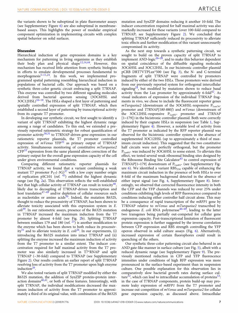
This demonstration opens a door to programming hierarchicalpatterning behavior across populations of cells. For example,replacing the mRFP1 gene in our synthetic three-color circuit by abiosynthetic enzyme producing a third diffusible signal ortho-gonal to 3OC6HSL and 3OC12HSL would allow programmedpatterning of a multicellular population into five differentdomains across three hierarchical levels (Fig. 7). Candidateorthogonal signaling systems for this purpose have already beendescribed22,23,54. In principle, the population-based AND gatecan be iterated with n orthogonal signaling systems to generate(2 × n) – 1 different domains.
The implementation of higher-order hierarchical patterningprocesses may become limited by insufficient signal-to-noise ratioin effector expression. In this respect, our approach to imple-mentation of two-layer synthetic hierarchical patterning under-lines the importance of empirically characterizing individualgenetic components prior to their application in a complex syn-thetic circuit. As illustrated by the performance of suboptimalbicistronic controller plasmid variants in solid culture assay (seeFig. 6), modulation of a single ribosomal binding site was capableof qualitatively changing circuit behavior: exchange of RBS250 forthe stronger RBSB0033 or the weaker RBS50 upstream of T7RNAP[1–179] both shifted the balance between CFP and YFP expres-sion in the first layer of patterning and affected the signal-to-noise ratio in RFP expression in the second layer of patterning.Our success in designing a synthetic genetic circuit implementingthe desired behavior was dependent on empirical selection of ahigh-dynamic-range variant of split T7RNAP through ratiometricassays (see Fig. 2) and on translational tuning of the resultingAND gate through plate fluorometer assays (see SupplementaryFigure 6).
In nature, morphogen gradients mediate developmental pat-terning at a high level of precision in face of various perturba-tions, supported by sophisticated genetic circuits and layers offeedback mechanisms55–57. Likewise, the robustness of synthetichierarchical patterning could be increased by supplementarymechanisms implementing positive feedback or morphogendegradation, for example. In terms of the circuit introducedherein, positive feedback could be implemented by expression ofa T7RNAP gene under control of the T7 promoter, renderingmaintenance of the second layer of patterning independent fromthe first layer inducers 3OC6HSL and 3OC12HSL. To alleviatepotential propagation of signaling crosstalk to higher levels ofpatterning, the first layer inducers 3OC6HSL and 3OC12HSLcould further be specifically degraded at the second layer ofpatterning. This could be achieved by expression of a quorum-quenching enzyme—such as the lactonase aiiA58—under controlof the T7 promoter. Feedback mechanisms like the ones outlinedcan be used to consolidate induced domains and to promotecorrect transmission of spatial information across layers ofcomplexity. As a test-bed for the development of new syntheticfeedback circuits, our system for synthetic hierarchical patterningpromises to facilitate future efforts at creating custom multi-cellular organisms for applications in bioproduction, remediation,or medicine.
MethodsPlasmid construction. All plasmids (listed in Supplementary Table 4) were con-structed using Gibson assembly59 with parts obtained from the MIT Registry ofStandard Biological Parts (http://partsregistry.org), from Addgene (www.addgene.org), or synthesized by Integrated DNA Technologies (Coralville, IA, USA), andare available on Addgene. Identities and source of backbone vectors, genes, andregulatory elements used in this work are summarized in Supplementary Tables 1–3. Sequences are available on Genbank (accession numbers KY643824 andKX986152 to KX986173). Controller devices encoding intact and split variants ofT7RNAP were based on plasmids acquired from Shis and Bennett26. Bicistroniccontroller devices were based on a double receiver plasmid previously reported24.All cloning was performed in TOP10 E. coli (Invitrogen, Waltham, MA). Withexception of ratiometric reporter plasmids for T7RNAP activity (T7 Express E. coli;New England Biolabs, Ipswich, MA, USA) all analysis was also carried out inTOP10 E. coli.
Plate fluorometer assays. Overnight cultures were diluted 1:100 in M9 mediumsupplemented with 0.2%w/v casamino acids, 0.4%w/v glucose, and inducers to theconcentrations described, and loaded in a final volume of 200 μL per well onto aclear-bottom 96-well microplate (Greiner, Kremsmünster, Austria). Measurementsof CFP (excitation 430/10 nm, emission 480/10 nm), YFP (excitation 500/10 nm,emission 530/10 nm), and RFP (excitation 550/10 nm, emission 610/20 nm)fluorescence intensities, and OD600 were taken approximately every 12 min for 100cycles (approximately 19 h) in a BMG FLUOstar Omega plate fluorometer (BMGLabtech, Ortenberg, Germany) at 37 °C, under shaking at 200 rpm between
a
a
a
(i)
(ii)
(iii)
(iv)
(v)
dc
eb
c
ba AND b
a AND c
c AND b
b
Fig. 7 A general framework for hierarchical patterning of bacterialpopulations. (i) An initial asymmetry splits a bacterial population into twodifferent gene expression domains (yellow boxes and cyan boxes). Theyellow domain produces a diffusible morphogen a (yellow background), thecyan domain a diffusible morphogen b (cyan background). (ii) Morphogensa and b coincide at the boundary of the yellow and cyan domains (greenbackground). (iii) Population-based logic induces a red gene expressiondomain in response to a AND b (red boxes). The red domain produces anadditional diffusible morphogen c (red background). (iv) Morphogens a andc coincide at the boundary of the yellow and red domains (orangebackground), morphogens c and b at the boundary of red and cyan domains(purple background). (v) Population-based logic induces an orange geneexpression domain (orange boxes) in response to a AND c, and a purplegene expression domain (purple boxes) in response to c AND b. Theorange and purple domains produce additional diffusible morphogens d ande, respectively. This scheme may be iterated to generate (2 × n) – 1 differentdomains with n orthogonal signaling systems
readings. Data analysis was performed using the Genetic Engineering of Cells(GEC) modeling and design environment (version 6.12.2014)30.
Solid culture assays. Single colonies were picked from LB agar plates and grownovernight in supplemented M9 medium with appropriate antibiotics (50 μg/mLcarbenicillin and 50 μg/mL kanamycin for double-transformed cells; 50 μg/mLcarbenicillin, 50 μg/mL kanamycin, and 25 μg/mL chloramphenicol for triple-transformed cells). Cultures were diluted 1:100, then grown into exponential phaseto an optical density at 600 nm of 0.3. This dilute culture was spotted onto Iso-Gridmembranes (40 × 40 quadrants of 1 mm per side; Neogen, Lansing, MI, USA)placed on 1.5%w/v agar plates containing the same supplemented M9 growthmedium. The culture was plated at a volume of 0.5 μL per quadrant. Plates wereincubated and imaged in a custom imaging device which has been described indetail elsewhere24. For quantification of fluorescence intensities, mean pixel grayvalues localized to individual Iso-Grid quadrants across the CFP, YFP, and RFPchannels were extrapolated using the open-source Fiji distribution of ImageJ60.
Data availability. The sequences of 23 plasmids used in this study have beensubmitted to the GenBank nucleotide database under accession codes KY643824and KX986152 to KX986173. The authors declare that all data supporting thefindings of this study are available within the paper and its SupplementaryInformation files or are available from the corresponding author on request.
Received: 23 November 2016 Accepted: 17 January 2018
References1. Davies, J. Mechanisms of Morphogenesis (Academic Press, 2013).2. Turing, A. M. The chemical basis of morphogenesis. Philos. Trans. R. Soc.
Lond. B Biol. Sci. 237, 37–42 (1952).3. Wolpert, L. Positional information and the spatial pattern of cellular
differentiation. J. Theor. Biol. 25, 1–47 (1969).4. Green, J. B. A. & Sharpe, J. Positional information and reaction-diffusion: two big
ideas in developmental biology combine. Development 142, 1203–1211 (2015).5. Teague, B. P., Guye, P. & Weiss, R. Synthetic morphogenesis. Cold Spring
Harb. Perspect. Biol. 8, a023929 (2016).6. Isalan, M., Lemerle, C. & Serrano, L. Engineering gene networks to emulate
Drosophila embryonic pattern formation. PLoS Biol. 3, e64 (2005).7. Payne, S. et al. Temporal control of self-organized pattern formation without
morphogen gradients in bacteria. Mol. Syst. Biol. 9, 697–697 (2014).8. Basu, S., Gerchman, Y., Collins, C. H., Arnold, F. H. & Weiss, R. A synthetic
multicellular system for programmed pattern formation. Nature 434,1130–1134 (2005).
9. Sohka, T., Heins, R. A. & Ostermeier, M. Morphogen-defined patterning ofEscherichia coli enabled by an externally tunable band-pass filter. J. Biol. Eng.3, 10 (2009).
10. Greber, D. & Fussenegger, M. An engineered mammalian band-pass network.Nucleic Acids Res. 38, e174 (2010).
11. Schaerli, Y. et al. A unified design space of synthetic stripe-forming networks.Nat. Commun. 5, 4905 (2014).
12. Sternberg, P. W. & Horvitz, H. R. The combined action of two intercellularsignaling pathways specifies three cell fates during vulval induction in C.elegans. Cell 58, 679–893 (1989).
13. Coen, E. S. & Meyerowitz, E. M. The war of the whorls: genetic interactionscontrolling flower development. Nature 353, 31–37 (1991).
14. Kimelman, D. Mesoderm induction: from caps to chips. Nat. Rev. Genet. 7,360–372 (2006).
15. Tabor, J. J. et al. A synthetic genetic edge detection program. Cell 137,1272–1281 (2009).
16. Stevens, A. M. & Greenberg, E. P. Quorum sensing in Vibrio fischeri: essentialelements for activation of the luminescence genes. J. Bacteriol. 179, 557–562(1997).
17. Schuster, M., Urbanowski, M. L. & Greenberg, E. P. Promoter specificity inPseudomonas aeruginosa quorum sensing revealed by DNA binding ofpurified LasR. Proc. Natl. Acad. Sci. USA 101, 15833–15839 (2004).
18. Ng, W.-L. & Bassler, B. L. Bacterial quorum-sensing network architectures.Annu. Rev. Genet. 43, 197–222 (2009).
19. Brenner, K., Karig, D. K., Weiss, R. & Arnold, F. H. Engineered bidirectionalcommunication mediates a consensus in a microbial biofilm consortium. Proc.Natl. Acad. Sci. USA 104, 17300–17304 (2007).
20. Balagaddé, F. K. et al. A synthetic Escherichia coli predator-prey ecosystem.Mol. Syst. Biol. 4, 187 (2008).
21. Wu, F., Menn, D. J. & Wang, X. Quorum-sensing crosstalk-driven syntheticcircuits: from unimodality to trimodality. Chem. Biol. 21, 1629–1638 (2014).
22. Davis, R. M., Muller, R. Y. & Haynes, K. A. Can the natural diversity ofquorum-sensing advance synthetic biology? Front. Bioeng. Biotechnol. 3, 30(2015).
23. Scott, S. R. & Hasty, J. Quorum sensing communication modules for microbialconsortia. ACS Synth. Biol. 5, 969–977 (2016).
24. Grant, P. K. et al. Orthogonal intercellular signaling for programmed spatialbehavior. Mol. Syst. Biol. 12, 849 (2016).
25. Ikeda, R. A. & Richardson, C. C. Enzymatic properties of a proteolyticallynicked RNA polymerase of bacteriophage T7. J. Biol. Chem. 262, 3790–3799(1987).
26. Shis, D. L. & Bennett, M. R. Library of synthetic transcriptional AND gatesbuilt with split T7 RNA polymerase mutants. Proc. Natl. Acad. Sci. USA 110,5028–5033 (2013).
27. Segall-Shapiro, T. H., Meyer, A. J., Ellington, A. D., Sontag, E. D. & Voigt, C.A. A ‘resource allocator’ for transcription based on a highly fragmented T7RNA polymerase. Mol. Syst. Biol. 10, 742 (2014).
28. Schaerli, Y., Gili, M. & Isalan, M. A split intein T7 RNA polymerase fortranscriptional AND-logic. Nucleic Acids Res. 42, 12322–12328 (2014).
29. Han, T., Chen, Q. & Liu, H. Engineered photoactivatable genetic switchesbased on the bacterium phage T7 RNA polymerase. ACS Synth. Biol. 6,357–366 (2017).
30. Yordanov, B. et al. A computational method for automated characterization ofgenetic components. ACS Synth. Biol. 3, 578–588 (2014).
31. Rudge, T. J. et al. Characterization of intrinsic properties of promoters. ACSSynth. Biol. 5, 89–98 (2016).
32. Nagai, T. et al. A variant of yellow fluorescent protein with fast and efficientmaturation for cell-biological applications. Nat. Biotechnol. 20, 87–90 (2002).
33. Goedhart, J. et al. Structure-guided evolution of cyan fluorescent proteinstowards a quantum yield of 93%. Nat. Commun. 3, 751 (2012).
34. Rosa, M. D. Four T7 RNA polymerase promoters contain an identical 23 bpsequence. Cell 16, 815–825 (1979).
35. Panayotatos, N. & Wells, R. D. Recognition and initiation site for four latepromoters of phage T7 is a 22-base pair DNA sequence. Nature 280, 35–39(1979).
36. Dunn, J. J., Studier, F. W. & Gottesman, M. Complete nucleotide sequence ofbacteriophage T7 DNA and the locations of T7 genetic elements. J. Mol. Biol.166, 477–535 (1983).
37. Imburgio, D., Rong, M., Ma, K. & McAllister, W. T. Studies of promoterrecognition and start site selection by T7 RNA polymerase using acomprehensive collection of promoter variants. Biochemistry 39, 10419–10430(2000).
38. Rosenberg, A. H. et al. Vectors for selective expression of cloned DNAs by T7RNA polymerase. Gene 56, 125–135 (1987).
39. Temme, K., Hill, R., Segall-Shapiro, T. H., Moser, F. & Voigt, C. A. Modularcontrol of multiple pathways using engineered orthogonal T7 polymerases.Nucleic Acids Res. 40, 8773–8781 (2012).
40. Reinke, A. W., Grant, R. A. & Keating, A. E. A synthetic coiled-coilinteractome provides heterospecific modules for molecular engineering. J. Am.Chem. Soc. 132, 6025–6031 (2010).
41. Thompson, K. E., Bashor, C. J., Lim, W. A. & Keating, A. E. SYNZIP ProteinInteraction Toolbox: in vitro and in vivo specifications of heterospecificcoiled-coil interaction domains. ACS Synth. Biol. 1, 118–129 (2012).
42. Moon, T. S., Lou, C., Tamsir, A., Stanton, B. C. & Voigt, C. A. Geneticprograms constructed from layered logic gates in single cells. Nature 491,249–253 (2012).
43. Salis, H. M. The ribosome binding site calculator. Methods Enzymol. 498,19–42 (2011).
44. Haughn, G. W. & Somerville, C. R. Genetic control of morphogenesis inArabidopsis. Dev. Genet. 9, 73–89 (1988).
45. Cohen, S. N. & Chang, A. C. Revised interpretation of the origin of thepSC101 plasmid. J. Bacteriol. 132, 734–737 (1977).
46. Studier, F. W. & Moffatt, B. A. Use of bacteriophage T7 RNA polymerase to directselective high-level expression of cloned genes. J. Mol. Biol. 189, 113–130 (1986).
47. Iost, I., Guillerez, J. & Dreyfus, M. Bacteriophage T7 RNA polymerase travelsfar ahead of ribosomes in vivo. J. Bacteriol. 174, 619–622 (1992).
48. Makarova, O. V., Makarov, E. M., Sousa, R. & Dreyfus, M. Transcribing ofEscherichia coli genes with mutant T7 RNA polymerases: stability of lacZmRNA inversely correlates with polymerase speed. Proc. Natl. Acad. Sci. USA92, 12250–12254 (1995).
49. Miroux, B. & Walker, J. E. Over-production of proteins in Escherichia coli:mutant hosts that allow synthesis of some membrane proteins and globularproteins at high levels. J. Mol. Biol. 260, 289–298 (1996).
50. Dong, H., Nilsson, L. & Kurland, C. G. Gratuitous overexpression of genes inEscherichia coli leads to growth inhibition and ribosome destruction. J.Bacteriol. 177, 1497–1504 (1995).
51. Muller, D. K., Martin, C. T. & Coleman, J. E. Processivity of proteolyticallymodified forms of T7 RNA polymerase. Biochemistry 27, 5763–5771 (1988).
52. Wang, L., Fan, D., Chen, W. & Terentjev, E. M. Bacterial growth, detachmentand cell size control on polyethylene terephthalate surfaces. Sci. Rep. 5, 15159(2015).
53. Klumpp, S., Zhang, Z. & Hwa, T. Growth rate-dependent global effects ongene expression in bacteria. Cell 139, 1366–1375 (2009).
54. Chen, Y., Kim, J. K., Hirning, A. J., Josi, K. & Bennett, M. R. Emergent geneticoscillations in a synthetic microbial consortium. Science 349, 986–989 (2015).
55. Bollenbach, T., Kruse, K., Pantazis, P., González-Gaitán, M. & Jülicher, F.Robust formation of morphogen gradients. Phys. Rev. Lett. 94, 18103 (2005).
56. Barkai, N. & Shilo, B.-Z. Robust generation and decoding of morphogengradients. Cold Spring Harb. Perspect. Biol. 1, a001990 (2009).
57. Lo, W.-C., Zhou, S., Wan, F. Y.-M., Lander, A. D. & Nie, Q. Robust andprecise morphogen-mediated patterning: trade-offs, constraints andmechanisms. J. R. Soc. Interface 12, 20141041 (2015).
58. Wang, L. H., Weng, L. X., Dong, Y. H. & Zhang, L. H. Specificity and enzymekinetics of the quorum-quenching N-acyl homoserine lactone lactonase(AHL-lactonase). J. Biol. Chem. 279, 13645–13651 (2004).
59. Gibson, D. G. et al. Enzymatic assembly of DNA molecules up to severalhundred kilobases. Nat. Methods 6, 343–345 (2009).
60. Schindelin, J. et al. Fiji: an open-source platform for biological-image analysis.Nat. Methods 9, 676–682 (2012).
AcknowledgementsC.R.B. acknowledges support from the Gates Cambridge Trust. P.K.G. acknowledgessupport from the John Templeton Foundation (Grant No. 15619: “Mind, Mechanism andMathematics: Turing Centenary Research Project”). J.H. acknowledges support from theBiotechnology and Biological Sciences Research Council and the Engineering and Phy-sical Sciences Research Council (OpenPlant Grant No. BB/L014130/1) and EC FP7project no. 612146 (PLASWIRES).
Author contributionC.R.B., P.G., and J.H. conceived and contributed key ideas to the development of theproject; C.R.B., and P.G. designed and performed the experiments; C.R.B. analyzed thedata; J.H. supervised the project. All authors contributed to the manuscript.
Additional informationSupplementary Information accompanies this paper at https://doi.org/10.1038/s41467-018-03069-3.
Competing financial interests: The authors declare no competing financial interests.
Reprints and permission information is available online at http://npg.nature.com/reprintsandpermissions/
Publisher's note: Springer Nature remains neutral with regard to jurisdictional claims inpublished maps and institutional affiliations.
Open Access This article is licensed under a Creative CommonsAttribution 4.0 International License, which permits use, sharing,
adaptation, distribution and reproduction in any medium or format, as long as you giveappropriate credit to the original author(s) and the source, provide a link to the CreativeCommons license, and indicate if changes were made. The images or other third partymaterial in this article are included in the article’s Creative Commons license, unlessindicated otherwise in a credit line to the material. If material is not included in thearticle’s Creative Commons license and your intended use is not permitted by statutoryregulation or exceeds the permitted use, you will need to obtain permission directly fromthe copyright holder. To view a copy of this license, visit http://creativecommons.org/licenses/by/4.0/.