Progress towards Development of a Vaccine for AmebiasisSAMUEL L. STANLEY, JR.*
Department of Medicine and Molecular Microbiology, Washington UniversitySchool of Medicine, St. Louis, Missouri 63110
INTRODUCTION .......................................................................................................................................................637E. HISTOLYTICA INFECTION.................................................................................................................................637FEASIBILITY OF A VACCINE TO PREVENT AMEBIASIS..............................................................................637PARENTERALLY ADMINISTERED RECOMBINANT ANTIGEN-BASED AMEBIASIS VACCINES ........639
Serine-rich E. histolytica Protein...........................................................................................................................639E. histolytica Gal/GalNAc-Specific Lectin ............................................................................................................64129-kDa Cysteine-Rich E. histolytica Antigen........................................................................................................642Future Vaccine Candidates....................................................................................................................................643Limitations of Current Vaccine Studies ..............................................................................................................643
DEVELOPMENT OF AN ORAL VACCINE TO PREVENT AMEBIASIS.........................................................643Delivery of Recombinant E. histolytica Antigens with Cholera Toxin or Cholera Toxin B Subunit ...........644Attenuated Salmonella spp. as Delivery Systems for the Recombinant SREHP Molecule ...........................645Limitations of Current Oral Vaccine Studies.....................................................................................................646
The protozoan parasite Entamoeba histolytica is the caus-ative agent of amebic dysentery and amebic liver abscess. It hasbeen estimated that 50,000,000 people worldwide suffer fromdiarrhea secondary to E. histolytica infection and that amebicdysentery and amebic liver abscess kill at least 50,000 individ-uals yearly (117). The disease is far more prevalent in devel-oping countries, and there appear to be regional hot spots forE. histolytica infection where morbidity and mortality fromamebiasis seem especially high. Such regions include Mexico,South Africa, and India (117). The magnitude of the problemin Mexico has been illustrated by a recent nationwide serologicsurvey that showed that more than 8% of the Mexican popu-lation have had an episode of E. histolytica infection (10).While effective therapy for amebiasis is available in the form ofthe drug metronidazole, the use of this agent does not appearto have had any significant effect on the prevalence of amebicinfection. The main mode of transmission of amebiasis is theingestion of fecally contaminated food or water containingE. histolytica cysts; therefore, improved sanitary conditions areclearly the long-term approach to reducing or eliminating ame-biasis from areas of endemic infection. However, it is unlikelythat this will be accomplished in the near future. For thisreason, several laboratories have initiated efforts towards de-veloping a vaccine to prevent amebic infection. In this review,I will discuss the scientific rationale for an amebiasis vaccine,identify candidate E. histolytica antigens for inclusion in a vac-cine, and summarize the recent progress made in developingand testing both parenteral and oral recombinant E. histolyticaantigen-based vaccines for amebiasis.
E. HISTOLYTICA INFECTION
E. histolytica is a parasitic ameba of the human gastrointes-tinal tract. It has a simple life cycle, existing only as the motile
trophozoite or the hardy, infective, nonmotile cyst stage (Fig.1) (reviewed in reference 51). E. histolytica naturally infectsonly humans and perhaps some higher nonhuman primates.Infection is established by ingestion of the cyst stage of theparasite. After cysts pass through the stomach and the proxi-mal small bowel, they excyst to the motile trophozoite form,which colonizes and can penetrate the colonic mucosa. Understill undefined conditions within the colon, trophozoites encyst,and the infective cysts are passed out in the stool, where theycan infect new hosts. Trophozoites may be passed in the stoolas well, but they are noninfectious. Invasion of the colonicmucosa by E. histolytica trophozoites results in diarrhea, whichis usually bloody (amebic dysentery). In roughly 5 to 10% ofindividuals with intestinal amebiasis, the E. histolytica tropho-zoites invade through the colonic mucosa and reach the portalcirculation, which carries them to the liver. There they canestablish amebic liver abscess, a major cause of liver abscessworldwide.
Until recently, one unexplained aspect of the epidemiologyof amebiasis was the presence of a large number of asymptom-atic individuals who passed cysts but had no evidence for in-vasive disease. Pioneering work involving isoenzyme analysis(85–87) and, more recently, molecular techniques (9, 14, 108,111) showed conclusively that there are two genetically distinctbut morphologically identical species of what was formerlyreferred to as Entamoeba histolytica. One species, which isclearly associated with disease in humans, is still referred to asEntamoeba histolytica. The second species, which is more prev-alent but does not appear to be associated with disease inhumans, is called Entamoeba dispar. The vaccine work re-viewed here deals solely with E. histolytica, the agent of amebicdysentery and amebic liver abscess.
FEASIBILITY OF A VACCINE TOPREVENT AMEBIASIS
The challenges in developing an effective vaccine against anyinfectious disease are daunting, and parasitic diseases offer anumber of additional problems. In many cases, protozoan par-asites have complex life cycles with multiple hosts and stages.
* Mailing address: Department of Medicine, Campus Box 8051, 660S. Euclid Ave., St. Louis, MO 63110. Phone: (314) 362-1070. E-mail:[email protected].
These create problems both in the logistics of working with theparasite and in the identification of protective antigens, sincetarget antigens identified in one stage of the parasite may notbe expressed in another stage, thus limiting their effectivenessas vaccine candidates. The presence of animal hosts for manyparasites also limits the possibility of eradicating the disease byvaccination, since these alternative hosts may provide a con-stant reservoir for infection of humans. Most of the clinicallyimportant protozoan parasites are predominantly intracellularparasites, thus providing limited opportunities for antibody-mediated protection, a critical correlate of much vaccine-me-diated immunity. Fortunately, as noted above, most of theseproblems do not apply to amebiasis. There are no insect vec-tors or secondary hosts involved in the transmission of amebi-asis, and a vaccine that eliminated colonization and infectionof humans with E. histolytica could eventually eradicate thedisease and the organism. Finally, E. histolytica is an extracel-lular parasite, thus providing a potential role for antibody invaccine-induced immunity to amebic infection.
An important consideration in evaluating the feasibility of avaccine for amebiasis is whether immunity to subsequent in-fection develops following a bout of amebiasis. Those diseasesfor which protective and long-lived immunity develops follow-ing infection, such as mumps and smallpox, have been veryeffectively prevented by vaccines, while diseases for which thehost immune response to initial infection is only minimallyeffective or ineffective in preventing subsequent infection or
chronic infection, such as malaria, appear to pose particularchallenges for successful vaccine development (70). Data onthe acquisition of immunity following infection with E. histo-lytica are limited. In one epidemiologic study, individuals witha prior episode of amebic liver abscess had a much lower rate(0.2% during the study) of contracting a liver abscess than didpreviously uninfected patients based on historical controls(22), suggesting that prior infection may result in protectiveimmunity. The experience of clinicians in areas of endemicinfection appears to support this concept (92), but carefullycontrolled prospective studies are simply not available.
If protective immunity can develop after amebic infection inhumans, how is it mediated? One possible mechanism involvesthe development of anti-amebic antibodies. Individuals withamebic liver abscess develop anti-amebic antibodies, which areusually detectable within 1 week of the onset of symptoms (38,43). These anti-amebic antibodies form the basis of serologictests used to diagnose amebic liver abscess. The developmentof anti-amebic antibodies appears to have no effect on thecourse of amebic liver abscess, which has a high fatality ratewithout appropriate therapy (16). However, the failure of an-tibodies to alter the course of established amebic liver abscessdoes not mean that preexisting anti-amebic antibodies couldnot provide protection against subsequent infection (78). Thelower inoculum associated with new infection, and antibodycontact with trophozoites in the bloodstream rather than inabscess tissue, might explain the difference in efficacy. Thereare experimental studies to support this concept. Hamsterspassively immunized with human immune serum and then in-trahepatically challenged with E. histolytica trophozoites werepartially protected from amebic liver abscess (91). Recently,we demonstrated that human anti-amebic antibodies, obtainedfrom patients with amebic liver abscess, could protect micewith severe combined immunodeficiency (SCID mice) fromdeveloping an amebic liver abscess when the antibodies wereadministered before an intrahepatic challenge with virulentE. histolytica trophozoites (93). Because SCID mice lack func-tional B or T lymphocytes, these studies clearly indicate thatpreexisting human anti-amebic antibody alone can protectagainst amebic liver abscess in this model.
The production of mucosal immunoglobulin A (IgA) anti-amebic antibodies after invasive intestinal amebiasis could alsoplay a role in protecting against subsequent intestinal infectionwith E. histolytica. Anti-amebic antibodies of the IgA subclasshave been detected in saliva from patients with intestinal ame-biasis (11), and it has been demonstrated that human salivaryIgA anti-amebic antibodies can block E. histolytica adherenceto a mammalian cell line, suggesting that they could play a rolein inhibiting amebic infection/colonization in vivo (11). Thesefindings speak directly to the feasibility of developing an oralvaccine for amebiasis that is capable of stimulating mucosalIgA anti-amebic antibodies that could block amebic adherenceto human intestinal epithelial cells.
A role for cell-mediated immunity in protection againstamebiasis has also been investigated. Individuals with amebicliver abscess and intestinal amebiasis develop T-cell prolifera-tive responses to amebic antigens following infection (81, 82,88, 115). When activated, macrophages from uninfected andpreviously infected individuals can kill amebic trophozoites invitro (82, 83). It has also been reported that lectin-stimulatedCD81 T cells or amebic antigen-stimulated CD81 T cells frompatients treated for amebic liver abscess have cytotoxic activityagainst amebic trophozoites in vitro (81, 82); but how suchkilling occurs in the absence of class I antigen expression onthe surface of E. histolytica trophozoites is unexplained. Thestrongest evidence for a role of cell-mediated immunity in
FIG. 1. Life cycle of E. histolytica. Disease begins when the cyst form of theparasite is ingested, excystation into the trophozoite form probably occurs in thelower small bowel, and the trophozoites are capable of penetrating into colonicmucosa as indicated. Trophozoites can encyst in the colon, and these cysts areexcreted in the stool, releasing infectious cysts into the environment to renew thelife cycle. In some cases, trophozoites invade through the colonic mucosa andreach the portal circulation, where they penetrate into liver tissue and create anamebic liver abscess.
protection from amebiasis comes from anectodal reports ofworsening complications of amebiasis in patients receiving cor-ticosteroids (37, 57). However, arguing against a critical rolefor cell-mediated immunity in protection against amebiasis hasbeen the absence to date of reports of any significant increasein the incidence of intestinal amebiasis or amebic liver abscessin patients with AIDS in areas where both diseases are en-demic. Thus, while findings from patients indicate that cell-mediated immune responses to amebic antigens develop fol-lowing infection, the role of these responses in protectionagainst amebiasis in humans remain unknown, and whether avaccine to prevent amebiasis must stimulate cell-mediated im-mune responses for protective efficacy is unclear.
Data obtained from animal studies of protective immunity toamebiasis indicate that immunity to amebiasis can be producedby prior infection or vaccination. Dogs reportedly develop im-munity to reinfection after experimentally induced intestinalamebiasis, and passive immunization of naive dogs with serumfrom E. histolytica-immune animals refractory to amebic infec-tion resulted in a decrease in susceptibility to intestinal ame-biasis in the naive group (103). Following a drug-terminatedbout of amebic liver abscess, hamsters were protected from asecond infection (114). Guinea pigs infected intracecally with anoninvasive strain of E. histolytica were protected against sub-sequent challenge with a virulent strain of E. histolytica (34). Ina number of different studies, rats, guinea pigs, hamsters, ormonkeys vaccinated with either intact amebae or various formsof complex amebic antigens have been protected against ame-bic liver abscess, or, in the case of guinea pigs, intracecal infec-tion with E. histolytica trophozoites (31, 32, 35, 44, 56, 90, 116).
There are a number of potential approaches to an amebiasisvaccine. To date, work has focused on the trophozoite stage ofthe parasite, since it can be cultivated in vitro, while no at-tempts to incorporate any antigens derived from the cyst stage(which cannot be obtained in culture) have yet been at-tempted. One approach, which has been successful for otherpathogens, is to attempt to use killed or inactivated E. histo-lytica trophozoites as the vaccine components. This strategyhas been successful in some of the animal studies cited above.However, it is relatively expensive to grow E. histolytica inculture, and the cost of producing E. histolytica trophozoites orantigens on the scale necessary for vaccine use could be pro-hibitive. In addition, there are always concerns about reacto-genicity when using killed or inactivated whole organisms invaccine preparations. For these reasons, much of the recentprogress in a vaccine for amebiasis has come from the use ofrecombinant E. histolytica antigens. Recombinant techniquesoffer the possibility of large-scale production of structurallydefined E. histolytica antigens through prokaryotic or yeastexpression systems. In addition, they provide the opportunityto genetically engineer multiple antigen vaccines or to use at-tenuated viral or bacterial vectors as carriers for amebic anti-gens. There are now three different E. histolytica proteins thathave formed the basis for recombinant antigen vaccines, theserine-rich E. histolytica protein (SREHP), the Gal/GalNAcbinding lectin, and a 29-kDa antigen. The three subsequentsections will review what is known about the structure, func-tion, and immunogenicity of each of these amebic antigens andthe vaccine-related studies associated with them.
Serine-rich E. histolytica Protein
A partial cDNA clone encoding the SREHP was first iso-lated by differential screening with E. histolytica HM1:IMSSmRNA or mRNA from Entamoeba moshkovskii Laredo, anonpathogenic species, of duplicate filters of a cDNA libraryderived from the virulent E. histolytica HM1:IMSS strain. Thisstrategy was designed to isolate E. histolytica-specific genesthat could play a role in virulence (97). Those E. histolyticacDNA clones hybridizing only with E. histolytica mRNA andnot with both E. histolytica and E. moshkovskii mRNA wereselected for further analysis. The full-length SREHP cDNAclone encodes a peptide of approximately 25 kDa, but thenative SREHP molecule was found to migrate at 47 and 52kDa on sodium dodecyl sulfate-polyacrylamide gel electro-phoresis (SDS-PAGE). This discrepancy is due both to post-translational modifications of the SREHP molecule (101) andto the high hydrophilicity and multiple proline residues in theSREHP peptide which cause anomalous migration in SDS-PAGE (97). The protein was designated serine-rich E. histo-lytica protein due to the large number of serine residues (52 of233 amino acids) (97). The other striking feature of the struc-ture was the presence of multiple tandemly repeated octapep-tides and dodecapeptides. Subsequently, a similar clone wasindependently isolated by antibody screening of an E. histo-lytica cDNA library, and it was found that E. dispar has a highlyhomologous gene with nucleotide differences between theE. histolytica and E. dispar SREHP cDNA sequences confinedprimarily to the tandem repeats (42). The structural motif ofthe SREHP molecule is somewhat reminiscent of the circum-sporozoite proteins of malaria, with a hydrophobic N-terminalregion (signal sequence) followed by a region of highly chargedamino acids consisting primarily of lysine, glutamic acid, andaspartic acid, leading to a series of tandemly repeated hydro-philic dodecapeptide or octapeptide sequences composed ofserine, aspartic acid, threonine, proline, lysine, glutamic acid,asparagine, and alanine (Fig. 2). This region is followed by aC-terminal hydrophobic domain consistent with a membraneinsertion region. SREHP is a surface membrane protein, but ithas posttranslational modifications including phosphorylation
FIG. 2. Model of the native SREHP. Pertinent features of the SREHP mol-ecule are indicated, including a region of charged amino acids at the N terminus,the presence of multiple tandem repeats, posttranslational modifications whichinclude multiple O-linked phosphate residues on serines, O-linked glucosamineresidues, and the presence of a hydrophobic domain at the C terminus, whichprobably is anchored in the plasma membrane by an ester-linked lipid group.
of serine residues and glycosylation with O-linked N-acetylglu-cosamine residues, which are more commonly seen in nuclearor cytoplasmic proteins (101). It appears to be anchored in themembrane by a C-terminal covalently linked lipid (101). Thefunction of SREHP remains unclear. Antibodies to SREHPblock amebic adherence to mammalian cells in vitro, but theisolated SREHP molecule shows no obvious adhesin proper-ties (97, 121). Recently, we demonstrated that SREHP canserve as a chemoattractant for E. histolytica trophozoites, butthe physiologic relevance of this property is unknown (101).
SREHP is highly immunogenic and appears to possess anumber of conserved epitopes. Among individuals with amebicliver abscess, more than 80% have antibodies that recognizethe SREHP molecule (100). SREHP appears to be the productof a single-copy gene (50), and while the SREHP genes fromdifferent isolates of E. histolytica can differ in the number ofdodecapeptide and octapeptide repeats encoded (this propertyis the basis of PCR tests that can differentiate among strains ofE. histolytica [15]), no strain completely lacking sequences en-coding the dodecapeptide repeat of SREHP has been de-scribed. Consistent with this finding, when eight different clin-ical and laboratory isolates of E. histolytica and E. dispar wereanalyzed by immunoblotting with a monoclonal antibody thatrecognizes the dodecapeptide repeat of SREHP, all the strainsanalyzed possessed a protein product that reacted with themonoclonal antibody, although there was size variation in thisprotein among the strains (96). Epitope mapping with serumfrom patients with amebic liver abscess suggests that mostantibodies to SREHP bind epitopes within the dodecapeptideand octapeptide repeats (118).
The aforementioned properties of surface location, the abil-ity of SREHP immunization to induce amebic adherenceblocking antibodies, and the presence of conserved SREHPepitopes in geographically diverse isolates of E. histolytica allsuggested that SREHP could be a viable vaccine candidate.Further support for testing SREHP as a parenteral vaccine toprevent amebic liver abscess came from passive immunizationstudies. SCID mice were passively immunized with rabbit poly-clonal antiserum to recombinant SREHP-TrpE and then in-trahepatically challenged with virulent E. histolytica HM1:IMSS trophozoites. None of the SCID mice receiving the anti-SREHP antibodies developed an amebic liver abscess, while allthe SCID mice receiving preimmune serum developed an ab-scess (121). These data established the efficacy of anti-SREHPantibodies in protecting against amebic liver abscess and pro-vided some of the strongest evidence for a protective role ofpreexisting anti-amebic antibodies in preventing the develop-ment of amebic liver abscess.
The SREHP cDNA has been expressed as TrpE, glutathi-one-S-transferase (GST), and maltose binding protein (MBP)fusion proteins in Escherichia coli (62, 97). Expression ofSREHP in E. coli without a fusion partner has not beenachieved, suggesting that SREHP alone may be toxic for bac-terial cells. SREHP has been expressed without any fusionpartner in baculovirus (101), where recombinant SREHPreaches the surface of virally infected insect cells and is phos-phorylated and glycosylated but does not appear to have a lipidanchor (101). The highest yield of soluble recombinantSREHP was obtained with MBP as the fusion partner, and thiswas the preparation used for parenteral vaccine studies (62).
To test the safety and protective efficacy of SREHP-MBPvaccination, a gerbil model of amebic liver abscess was used.Gerbils develop amebic liver abscess after direct intrahepaticinoculation with virulent amebic trophozoites, and if un-treated, they will usually die of the infection in 21 days (12). Intwo separate trials, gerbils were vaccinated with three doses of
SREHP-MBP intraperitoneally, while in another trial, a singledose of SREHP-MBP was administered intradermally. Controlgroups received equivalent doses of MBP or phosphate-buff-ered saline (PBS) (122, 124). Vaccination was well tolerated byall groups. Approximately 3 weeks after vaccination, all thegerbils were challenged intrahepatically with virulent E. histo-lytica HM1:IMSS trophozoites, and 7 days later the animalswere sacrificed and liver abscess formation was assessed. Thereadout in these studies was whether an abscess could bedetected at 7 days and the percentage (based on weight) of theliver occupied by an amebic abscess in animals developingdisease. Among gerbils vaccinated with SREHP-MBP in thethree trials, the protective efficacy of vaccination (no amebicliver abscess detectable) was 64% of vaccinated animals in trial1, 100% of vaccinated animals in trial 2 (intradermal vaccina-tion), and 100% of vaccinated animals in trial 3 (122, 124).In contrast, all MBP- or PBS-vaccinated gerbils developedamebic liver abscess (protective efficacy of 0%). SREHP-MBP-vaccinated gerbils developed antibody and cell-mediated im-mune responses (as measured by delayed-type hypersensitiv-ity) to SREHP and ameba. Quantitatively similar anti-amebicantibody responses were seen in gerbils that were protectedfrom amebic liver abscess and in the few SREHP-MBP vaccinefailures seen in these studies, making it difficult to ascribeprotective immunity solely to the magnitude of the anti-SREHP antibody response. Whether there are differences inthe epitopes recognized in the anti-SREHP response in vac-cine successes and failures was not examined in this study.
The high level of complete protection from amebic liverabscess seen in gerbils vaccinated with recombinant SREHP-MBP demonstrated the potential of SREHP as a vaccine can-didate and illustrated the feasibility of using recombinantE. histolytica antigens in amebiasis vaccines. Subsequently, apreliminary analysis of the safety and efficacy of the recombi-nant SREHP-MBP vaccine in nonhuman primates was per-formed (98). Two African green monkeys were vaccinated withSREHP-MBP in three doses on days 0, 30, and 53 by subcu-taneous administration; a single control monkey received PBSand adjuvant alone at the same time points. The SREHP-MBPvaccine was well tolerated, with no systemic side effects de-tected in the vaccinated monkeys and with no abnormalities inliver function tests, complete blood count, or renal functionseen in the vaccinated monkeys or the single control animal.Some local induration at the inoculation site was seen in boththe control monkey and the SREHP-MBP-vaccinated animals;this was believed to be secondary to the Freund adjuvant usedin these experiments. At 10 days following the first boostervaccine dose, anti-SREHP and anti-amebic antibodies weredetected in SREHP-MBP-vaccinated monkeys; anti-SREHPand anti-amebic antibody titers did not significantly increasefollowing the second booster dose of SREHP-MBP. The singlecontrol monkey had no anti-amebic or anti-SREHP antibodies.Importantly, serum from SREHP-MBP-vaccinated animals ob-tained after the first or second booster immunization couldsignificantly inhibit amebic adherence to Chinese hamsterovary (CHO) cells in vitro (98). The ability of anti-amebicserum to inhibit amebic adherence is the property that corre-lated with protection from amebic liver abscess in the passive-immunization studies in SCID mice (121). A major disappoint-ment in this trial, and the reason why the study was limited toa few monkeys, was the inability to test the protective efficacyof SREHP-MBP vaccination in African green monkeys (98).Contrary to a published report, in this study amebic liver ab-scesses could not be established in E. histolytica-naive (basedon amebic serology and stool examination for amebic tropho-zoites) African green monkeys by direct hepatic inoculation
with virulent amebic trophozoites, eliminating the possibility ofstudies of protective efficacy in this primate species. Despitethis limitation, demonstrating the safety and immunogenicityof the SREHP-MBP preparation in nonhuman primates rep-resented an important step in the development of a recombi-nant SREHP-based amebiasis vaccine.
E. histolytica Gal/GalNAc-Specific Lectin
One of the best-studied molecules of E. histolytica is a sur-face adhesin with specificity for galactose and N-acetylgalac-tosamine residues, designated the Gal/GalNAc lectin (re-viewed in reference 58). The adhesin is large (260 kDa) andcan be separated into component heavy (170-kDa) and light(35- or 31-kDa) subunits by SDS-PAGE under reducing con-ditions (66). The primary structures of both the heavy and lightchain subunits have been obtained from the nucleotide se-quence from cDNA clones and genomic sequences (55, 59, 71,109, 110). The derived amino acid sequence of the heavy (170-kDa) subunit contains a signal sequence, an approximately370-amino-acid cysteine-poor region, an approximately 275-amino-acid region of pseudorepeats of 30 amino acids contain-ing four conserved cysteine residues, an approximately 550-amino-acid cysteine-rich (10.9% cysteine residues) domain, a26- to 28-amino-acid putative hydrophobic transmembrane do-main, and a cytoplasmic tail of approximately 40 amino acids(55, 109). The 170-kDa subunit is encoded by at least threegenes, so that multiple isoforms of the 170-kDa molecule areprobably present in amebae. Sequence homology among thegenes is high (between 89.2 and 95.2% at the derived aminoacid level), and there are probably multiple epitopes conservedamong different isoforms. The light-subunit cDNA encodes ahydrophilic protein with a predicted mass of 32 kDa, whichmay be anchored to the membrane by a glycosylphosphatidy-linositol anchor (59, 110). At least two light-subunit genes arepresent and encode proteins with derived amino acid se-quences that are 80.6% identical (59, 60, 110).
The Gal/GalNAc lectin appears to play an important role inamebic binding to mammalian cells. The native adhesin bindsto galactose and can be affinity purified by galactose-Sepharose(68). E. histolytica binding to CHO cells can be blocked bygalactose, implicating the Gal/GalNAc lectin in amebic adher-ence to target cells (72). In addition, studies with mutant CHOcells have shown that E. histolytica trophozoites show greatlyreduced binding to mutant cells lacking terminal galactose orN-acetyllactosamine residues (48, 75). Amebic cytolysis ofmammalian cells is contact dependent, and the mutant CHOcells lacking terminal N-acetyllactosamine residues are par-tially resistant to killing by E. histolytica trophozoites (49).Monoclonal antibodies directed against certain regions of theGal/GalNAc lectin can inhibit amebic adherence to CHO cells(74), while monoclonal antibodies to other epitopes actuallyenhance amebic adhesion to the same cells (69). All monoclo-nal antibodies to the Gal/GalNAc lectin that affect adherenceappear to bind to the cysteine-rich region of the heavy subunit(69, 74). The Gal/GalNAc lectin has also been implicated inthe ability of E. histolytica trophozoites to resist killing byhuman complement. An anti-amebic monoclonal antibody thatrenders E. histolytica trophozoites susceptible to killing by hu-man complement components C5b9 was found to bind epi-topes within the cysteine-rich region of the 170-kDa subunit ofthe Gal/GalNAc lectin (7). This region of the 170-kDa subunitshowed some homology to the human complement regulatoryprotein CD59, a molecule which can block assembly of theC5b-9 attack complex, thus providing a potential mechanismfor complement resistance by amebae. What remains unclear is
whether naturally occurring complement-resistant and com-plement-susceptible E. histolytica isolates differ in either theprimary structure in the CD59-like region of the 170-kDa mol-ecule or the surface expression of the Gal/GalNAc lectin.
The Gal/GalNAc lectin is highly immunogenic, with morethan 90% of patients with amebic liver abscess developingantibodies capable of binding to the native molecule (73).Antibodies appear to be directed almost entirely against the170-kDa subunit (73). There are significant differences in theimmunogenicity of the three structural domains of the 170-kDa subunit. Using recombinant GST fusion proteins contain-ing the entire 170-kDa sequence (except the signal sequenceand the putative transmembrane and cytoplasmic domains),the cysteine-poor region alone (designated 170CP), the pseu-dorepeat region alone (designated 170PR), and the cysteine-rich region alone (designated 170CR), we found that all thepatients with amebic liver abscess, who came from severaldifferent geographic regions, had antibodies to the entire 170-kDa molecule and that they all had antibodies that bound tothe cysteine-rich region (170CR fusion protein) (127). Approxi-mately 89% of the patients with amebic liver abscess had an-tibodies to the cysteine-poor domain of the molecule (170CPfusion protein), while only 9% had antibodies that bound tothe pseudorepeat region (170PR fusion protein). The reasonfor the poor immunogenicity of the 170PR region is unknownbut could reflect MHC restrictions or the induction of anti-bodies that recognize conformational epitopes and hence donot react with the denatured 170PR fusion protein. The im-munogenic properties of the cysteine-rich region of the 170-kDa subunit were confirmed and extended by recent studiesdemonstrating that more than 95% of sera from patients withamebic liver abscess have IgA antibodies that recognize a re-combinant protein derived from the cysteine-rich domain ofthe 170-kDa subunit (94) and that more than 80% of individ-uals with amebic colitis develop mucosal IgA antibodies thatbind this recombinant protein (1).
The function of the Gal/GalNAc lectin in amebic adherence,the potential role of this molecule in mediating E. histolyticaresistance to complement, the immunogenicity of the heavysubunit, and the presence of highly conserved epitopes madethis molecule an obvious vaccine candidate. The first vaccinestudies were performed with the purified native 220-kDa Gal/GalNAc lectin and the gerbil model of amebic liver abscess.The purified Gal/GalNAc lectin was administered as a primarydose and two booster doses with Freund’s adjuvant (67). Ger-bils vaccinated with the purified lectin developed antibodiesthat recognized the 170-kDa subunit of the molecule; antibod-ies to the light subunit were not detected. Serum from vacci-nated gerbils blocked amebic adherence to CHO cells whenused at a 1/10 dilution but enhanced adherence when used ata 1/1,000 dilution (67). Among control animals receiving onlythe adjuvant, 81% developed an amebic liver abscess, while inthe Gal/GalNAc-vaccinated gerbils, only 27% developed anamebic liver abscess, representing a 67% reduction in the in-cidence of liver abscess in the vaccinated gerbils (67). Oneunexpected finding was that the amebic liver abscesses de-tected in gerbils vaccinated with the Gal/GalNAc lectin weresignificantly larger than those seen in the control gerbils. Thisstudy established the vaccine potential of the Gal/GalNAclectin and demonstrated that vaccination with a purified ame-bic antigen could provide protection against amebic liver ab-scess. It also suggested that some component of the immuneresponse to vaccination with the Gal/GalNAc lectin could, in asubgroup of gerbils, exacerbate amebic liver abscess.
The success of the native Gal/GalNAc lectin in vaccine stud-ies provided a rationale for exploring the efficacy of recombi-
nant Gal/GalNAc lectin antigens in protection against amebicliver abscess. In addition, it was anticipated that the ability touse only the heavy subunit of the lectin, and even specificdomains of that molecule, might allow one to develop a re-combinant Gal/GalNAc-based vaccine that could mimic theprotective effects of the native molecule without any exacer-bative effects on amebic liver abscess. Based on the serologicmapping studies, which showed that all patients with amebicliver abscess had antibodies that recognized epitopes on thecysteine-rich domain of the 170-kDa subunit, and the studieswhich suggested that both the carbohydrate-binding domainsof the molecule and the region of the molecule involved incomplement resistance map to the cysteine-rich region, weselected a GST fusion protein containing this domain (170CR)for the initial vaccine studies (124). Gerbils received a primaryimmunization and two booster immunizations with the 170CRprotein (which contained amino acids 649 to 1202 of the 170-kDa subunit of the Gal/GalNAc lectin fused to GST) inFreund’s adjuvant and were challenged intrahepatically withvirulent E. histolytica trophozoites 21 days after the secondbooster dose. Control gerbils were immunized with the GSTfusion protein alone and adjuvant or with PBS and adjuvant.Among gerbils vaccinated with 170CR, only 17% developed anamebic liver abscess compared with 100% of PBS- and adju-vant-vaccinated gerbils and 90% of GST- and adjuvant-vacci-nated gerbils. The protective efficacy of the recombinant170CR vaccine was 81%, a figure that compares well with the67% efficacy seen with vaccination with the native Gal/GalNAclectin (67). In addition, the few vaccine “failures” in this study,i.e., gerbils that were vaccinated with the 170CR protein butdid develop an amebic liver abscess, did not have abscessesthat were larger than those in either the GST-vaccinated orPBS-vaccinated control groups (124). These data indicatedthat the recombinant 170CR vaccine could perform as well asor better than the native Gal/GalNAc lectin-based vaccine inprotection against amebic liver abscess and might offer a sig-nificant advantage by not exacerbating disease in vaccine fail-ures. Independent confirmation of these findings came from asubsequent study in which another cysteine-rich region-basedrecombinant protein (this time containing amino acids 758 to1134 of the 170-kDa subunit of the Gal/GalNAc lectin fused tosix histidine residues) was used to vaccinate gerbils (94). In thisstudy, only 19% of the gerbils vaccinated with the fusion pro-tein (designated LC3) and the adjuvant Titermax developed anamebic liver abscess after direct hepatic inoculation with E. his-tolytica trophozoites, compared with 59% of gerbils receivingTitermax alone, for a vaccine protective efficacy of 68%. Im-portantly, in this study as well, the size of the amebic liverabscess in recombinant vaccine failures was not significantlylarger than that seen in control animals (94).
The protective efficacy of the 170CR vaccine and theSREHP-MBP vaccine in the previously described trials wasalready high (64 to 100%); however, one possible way to im-prove protection would be to combine the recombinant pro-teins into a single vaccine preparation. In this case, individualswho fail to respond to one antigen might develop a protectiveresponse to the other component of the vaccine, or the im-mune response to both antigens might provide better protec-tive efficacy than the response to a single antigen (i.e. syner-gistic effects of anti-adherence antibodies directed against twodifferent targets). To test this hypothesis, gerbils were vacci-nated with the 170CR and SREHP-MBP vaccine preparationssimultaneously, in the same individual dosage used for singleadministration (124). We performed this trial at the same timeas trials of the SREHP-MBP and 170CR vaccine preparationsalone, and control gerbils received the MBP or GST fusion
partner with adjuvant. Vaccinated gerbils developed antibod-ies that recognized both the 170-kDa subunit of the lectin andthe native SREHP molecule. Gerbils receiving the combina-tion vaccine were protected against amebic liver abscess, withonly 14% of vaccinated gerbils developing amebic liver abscessversus 100 and 90% of MBP- and GST-vaccinated gerbils,respectively (124). Comparison with SREHP-MBP alone or170CR alone did not show increased efficacy for the combina-tion vaccine, however, and if trials were continued under sim-ilar conditions, it was estimated that a prohibitive number ofanimals would have to be used to show a difference in protec-tive efficacy among the three different vaccine preparations(124). Whether the SREHP-MBP–170CR combination vac-cine would prove superior to the single-vaccine preparations ifa higher challenge dose of amebae was used or would allowreduced dosing of each of the vaccine components remainsuntested. However, these data indicate that simultaneous ad-ministration of the SREHP-MBP and 170CR vaccines does notappear to diminish the immune response to either antigen, andthey suggest that a combination vaccine containing those twoantigens is feasible.
29-kDa Cysteine-Rich E. histolytica Antigen
A partial cDNA clone encoding a 29-kDa cysteine-rich ame-bic antigen was first identified by anti-amebic antibody screen-ing of an E. histolytica strain H-302:NIH cDNA library (113).The derived amino acid sequence showed 7% cysteine residueswith multiple cysteine doublets (113). Analysis of a full-lengthcDNA and genomic clones showed that the derived amino acidsequence of the 29-kDa protein has significant relatedness tothe sequences of hydrogen peroxide-inactivating proteins thatare found in gut-living prokaryotes (8). These findings were ofinterest because it had been previously shown that E. histolyticastrains lack catalase and peroxidase but possess superoxidedismutase, raising the question of how H2O2 is detoxified byamebae (8, 107). Attempts to localize the 29-kDa cysteine-richprotein in E. histolytica have given differing results. The puta-tive function of this molecule, the absence of a signal sequence,and indirect immunofluorescence studies have been used toargue that the 29-kDa antigen is located in the cytoplasm andnucleus of E. histolytica trophozoites (8, 104). However, sur-face labeling, indirect immunofluorescence, and transmissionelectron microscopy with immunogold suggest that the 29-kDaantigen can be found on the surface membrane of amebae (28,77, 113). In this regard, it should be noted that the E. histolyticaalcohol dehydrogenase 2 (EhADH2) molecule of E. histolytica,a critical enzyme in the amebic fermentation pathway, alsolacks a signal sequence but that it (or immunologically identi-cal isoforms) appears to be present both in the cytoplasm andon the surface membrane of E. histolytica trophozoites (30).
The 29-kDa molecule is immunogenic. Approximately 80%of individuals with amebic liver abscess develop antibodies thatbind to the native or recombinant 29-kDa protein (29, 95). Arecombinant 29-kDa fusion protein derived from the sequenceof a full-length cDNA clone fused to sequences encoding sixhistidine residues was used for the first vaccine trials of thisantigen (95). Gerbils received three immunizations with therecombinant protein and Titermax adjuvant at 2-week intervalsand then were challenged with an intrahepatic inoculationof E. histolytica trophozoites some time following the finalbooster dose. Control groups of gerbils received three immu-nizations with Titermax or saline alone. Gerbils vaccinatedwith the recombinant 29-kDa fusion protein developed anti-bodies that recognized the native 29-kDa protein in amebiclysates, while sera from control gerbils showed no reactivity
with the native molecule. Among gerbils vaccinated with therecombinant 29-kDa protein, 30% developed an amebic liverabscess, while in the Titermax control group, 59% developedan abscess, for a calculated vaccine efficacy of 49%.
The protective efficacy of 49% seen with the recombinant29-kDa antigen is lower than that reported with the recombi-nant SREHP-MBP, 170CR, or LC3 antigen preparations (94,122, 124). However, differences in the number of E. histolyticatrophozoites used for liver inoculation, the virulence of theameba used, and the dose of vaccine administered make com-parisons between the SREHP-MBP and 170CR trials and therecombinant LC3 and 29-kDa antigen trials problematic. How-ever, the LC3 and 29-kDa antigen trials were performed underidentical conditions, and the results with their control groupswere identical (or the same control group was used for eachtrial), making the comparisons valid. When the results aredirectly compared, there is no statistically significant differencein protective efficacy between the recombinant 29-kDa antigenand the LC3 preparation (P . 0.5).
Future Vaccine Candidates
The recombinant SREHP, 170-kDa subunit of the Gal/GalNAc lectin, and 29-kDa antigen preparations and the na-tive 220-kDa Gal/GalNAc protein are the only defined amebicantigens tested as vaccines in animal models of amebic liverabscess. However, there are a number of other E. histolyticaantigens that represent potential candidates for inclusion in anamebiasis vaccine. The neutral amebic cysteine proteinase is arecognized virulence factor of E. histolytica and appears to playa critical role in amebic invasion and destruction of host ex-tracellular matrix tissues (39, 76, 112). Patients who have hadamebic liver abscess make antibodies that recognize amebiccysteine proteinase (65), and neutralization of the effects ofcysteine proteinase by proteinase inhibitors can reduce the sizeof amebic liver abscess in SCID mice (102). A recombinant ver-sion of the 27-kDa E. histolytica cysteine proteinase I (EhCP1)has been produced. However, the recombinant EhCP1 proteinlacked proteinase activity, probably because it failed to achievethe conformation of the native proteinase (102). Whether thepresumed conformational difference between the recombinantprotein and the native cysteine proteinase would compromisethe efficacy of the recombinant EhCP1 molecule as a vaccineremains to be tested.
Another potential vaccine candidate is amebapore, a pep-tide of 77 amino acids that appears to play an important role inE. histolytica pathogenesis (reviewed in reference 47). Thispore-forming protein forms ion channels in lipid bilayers andprobably plays a critical role in the cytolytic activity of E. his-tolytica trophozoites (47). Synthetic peptides derived from thesequence of amebapore have been produced, and a peptidethat can reproduce the cytolytic and pore-forming propertiesof the native protein have been identified (2, 46). Chemicallyproduced isoforms of amebapore, which lack cytolytic andpore-forming activity based on the modification of histidine orlysine residues, have been generated (3); if these compoundsare immunogenic and if antibodies to them neutralize thecytolytic activity of the native amebapore, the inactive isoformscould serve as the basis for an amebapore-derived vaccine.Other potential candidates include the 96-kDa E. histolyticaEhADH2, a bifunctional enzyme with alcohol dehydrogenaseand acetaldehyde dehydrogenase activities that plays a key rolein the amebic fermentation pathway and is implicated in ame-bic adherence to extracellular matrix proteins (30, 119). The96-kDa EhADH2 enzyme or an isoform can be detected on thesurface of amebic trophozoites and can bind to extracellular
matrix proteins such as fibronectin and laminin (119). A re-combinant version of the EhADH2 protein has been producedin E. coli and did not require a fusion partner (120). Theamebic glycoconjugate is a highly abundant surface moleculethat structurally resembles the Leishmania lipophosphoglycanmolecule (99). The amebic glycoconjugate is highly immuno-genic, and antibodies to this molecule can block amebic ad-herence to mammalian cells (99). However, there has not yetbeen an identifiable peptide component to this molecule, andthe immunogenic epitopes are probably carbohydrates, indi-cating that the native molecule would probably be required forany vaccine studies.
Limitations of Current Vaccine Studies
The studies cited above have demonstrated the feasibility ofdeveloping recombinant parenteral vaccines to prevent amebicliver abscess and have identified three recombinant amebicantigens that can protect in the gerbil model of disease. How-ever, there are still a number of important issues to be resolvedin the recombinant amebic antigen vaccine studies. First, allthe studies to assess protection from amebic liver abscess aftervaccination have been done in one animal model, the gerbil,and use an artificial form of establishing amebic liver abscess,direct hepatic inoculation of trophozoites. While it could beargued that this is a more stringent challenge than the naturalroute in humans (i.e., hematogenous spread of what is proba-bly significantly fewer amebae into the liver), the predictivevalue of the gerbil model for vaccine success in humans re-mains unknown. Second, the cDNA sequences for the recom-binant antigens used in these studies were derived from theHM1:IMSS laboratory strain of E. histolytica, and the samestrain, HM1:IMSS, was used as the challenge organism in allstudies. While there is ample serologic evidence to suggest thatthere are highly conserved epitopes in the SREHP, 170-kDaGal/GalNAc binding lectin, and 29-kDa antigens (29, 73, 94,95, 100, 127), no data have yet been presented to demonstratethat the HM1:IMSS-derived recombinant antigens can protectgerbils against “wild-type” E. histolytica isolates. Also unre-solved is the question of the duration of protective immunity inrecombinant amebic antigen-vaccinated gerbils. The amebicchallenges have been performed at time points (1 to 3 weeksfollowing booster immunizations) when circulating anti-ame-bic antibodies are detectable; whether protection would beseen at much later time points (e.g., 6 months) after vaccina-tion needs to be studied. Questions regarding the optimumvaccine dosing and interval, as well as the most effective adju-vants, also remain unanswered. The critical question whetherparenteral immunization with any of the candidate vaccineantigens provides protection against intestinal amebiasis hasalso not been approached, due to the lack of reliable animalmodels for intestinal disease. Finally, how protection is medi-ated in the gerbil model of amebic liver abscess remains un-clear. A protective role for anti-amebic antibodies could beassessed by measuring the protective efficacy of passive immu-nization of naive gerbils with antibodies from recombinantamebic antigen-vaccinated gerbils. However, the lack of gerbil-specific monoclonal antibody reagents for T-cell markers orcytokines makes an assessment of the contribution of cell-mediated immunity to vaccine-induced protection difficult.
DEVELOPMENT OF AN ORAL VACCINETO PREVENT AMEBIASIS
Amebiasis begins as an intestinal infection, and the ability ofE. histolytica trophozoites to adhere to intestinal epithelial cells
may be critical in maintaining colonization and initiating dis-ease (reviewed in reference 51). Mucosal anti-amebic antibod-ies could play a critical role in host defense, by blocking theability of E. histolytica trophozoites to attach to the intestinalsurface. Thus, a vaccine capable of stimulating mucosal IgAanti-amebic antibodies which block amebic adherence to in-testinal epithelial cells could provide protection against bothE. histolytica colonization and disease. Experimental supportfor this hypothesis has come from the aforementioned exper-iments showing that IgA antibodies isolated from the salivaof patients with amebic intestinal disease can block amebicadherence to target cells (11). In addition, it has been dem-onstrated that rats vaccinated with the native 220-kDa Gal/GalNAc lectin and the B subunit of cholera toxin, adminis-tered by direct inoculation into the rat Peyer’s patches, developmucosal IgA anti-Gal/GalNAc antibodies capable of partiallyinhibiting amebic adherence to mammalian cells (41).
The best studied approach to stimulating mucosal IgA re-sponses has been oral immunization. In addition to the advan-tage of potentially stimulating mucosal immune responses, oralvaccines may have significant advantages in acceptability,based on lower costs and ease of administration. To date, twodifferent approaches to an oral vaccine for amebiasis havebeen used. The first approach is based on the oral administra-tion of recombinant amebic antigens in conjunction with chol-era toxin or the B subunit of cholera toxin, and the secondapproach is centered on the expression and delivery of recom-binant amebic antigens by attenuated Salmonella or Vibriocholerae carriers.
Delivery of Recombinant E. histolytica Antigens withCholera Toxin or Cholera Toxin B Subunit
The rationale for combining amebic antigens with choleratoxin for oral immunization derives from the recognized adju-vant properties of cholera toxin. Cholera toxin is composed ofone A subunit containing the active toxin domain (A1) and ashort sequence (A2) which serves to link the A subunit non-covalently to five B subunits (reviewed in reference 5). The fiveB subunits mediate the binding of the toxin to GM1 gangliosideon the surface of intestinal cells; this binding permits internal-ization of the A subunit into cells with resultant intoxication(17). Cholera toxin is a potent mucosal immunogen, inducingsecretory IgA and serum IgA anti-toxin antibodies as well asserum IgG responses in animals when administered orally (27).Cholera toxin can also serve as a potent mucosal adjuvant.Proteins such as keyhole limpet hemocyanin (KLH) are nor-mally not immunogenic when administered orally. However,when KLH is administered orally with cholera toxin, bothanti-cholera toxin and anti-KLH antibodies are induced (26,27, 53). The mechanism underlying the adjuvant properties ofcholera toxin has been controversial, with some studies sug-gesting that toxin activity is required for adjuvant activity andothers suggesting that the nontoxic B subunit can provide ad-juvant activity (4, 20, 23, 33, 45, 52, 54, 61, 79, 106). There maybe interspecies differences in the cholera toxin response, withmice requiring the toxin moiety for adjuvant activity while inhumans the B subunit alone may serve as an adjuvant (21, 84).These points bear directly on the clinical utility of cholera toxinas an adjuvant, since administration of the nontoxic B subunitas adjuvant is clearly preferable to a formulation with toxinactivity.
Two recent studies have used cholera toxin B (CtxB) or theholotoxin as an adjuvant to facilitate the mucosal response torecombinant amebic antigens administered by the oral route.Using constructs developed by Dertzbaugh et al. (24, 25), we
engineered a fusion protein (CtxB–SREHP-12) that containedthe dodecapeptide repeat of SREHP fused to the cholera toxinB subunit (Fig. 3) (123). The CtxB–SREHP-12 protein couldbe purified from E. coli lysates by a two-step procedure, and,importantly, the CtxB moiety maintained the ability to bind toGM1 ganglioside. In fact, inhibition studies showed that theCtxB–SREHP-12 molecule was as potent as CtxB alone inblocking the binding of labeled CtxB to GM1 ganglioside.These results confirmed that small peptide molecules of atleast 12 amino acids can be fused to the CtxB molecule withoutinhibiting the ability of CtxB to pentamerize and bind to GM1ganglioside. Mice were orally immunized with CtxB–SREHP-12 and a subclinical dose of cholera toxin on days 0, 14, and 28and were sacrificed 7 days following the final boost, whilecontrol mice were orally vaccinated with CtxB and the subclin-ical dose of cholera toxin with the same schedule. At the timeof sacrifice, the serum, bile, and stool were assayed for bothIgG and IgA anti-amebic and anti-CtxB antibodies. In addi-tion, the number of cells secreting IgA or IgG antibodiesagainst SREHP, amebic lysates, cholera toxin, and choleratoxin B was assayed in samples obtained from the mesentericlymph nodes and spleen. CtxB–SREHP-12-vaccinated micedeveloped IgA anti-ameba antibodies in stool and in bile. Theyalso had significant numbers of antibody-secreting cells pro-ducing anti-SREHP and anti-amebic antibodies in mesentericlymph nodes and spleen. In contrast, CtxB-vaccinated miceshowed no level of anti-SREHP or anti-amebic antibodies orantibody-secreting cells that differed from background levels inany assay. Both CtxB- and CtxB–SREHP-12-vaccinated miceproduced anti-CtxB antibodies, suggesting that coupling ofSREHP to the CtxB protein did not significantly alter theimmunogenicity of CtxB (123). These results confirmed thefeasibility of generating mucosal immune responses to immu-nogenic peptides through their fusion to CtxB and establishedCtxB–SREHP-12 as one of the first candidates for an oralvaccine to prevent amebiasis.
An alternative approach was recently used by Beving et al.,who orally immunized mice by simultaneous administration ofthe recombinant LC3 protein (which contains amino acids 758to 1134 of the 170-kDa subunit of the Gal/GalNAc lectin fusedto six histidine residues) and cholera toxin (6). Mice receivingthis combination developed mucosal IgA anti-LC3 and anti-lectin antibody responses based on the analysis of intestinal
FIG. 3. Model of the CtxB–SREHP-12 fusion protein (123). In this depictionof the assembled CtxB–SREHP-12 fusion protein, five cholera toxin B molecules,each fused to the dodecapeptide repeat of SREHP, have formed the naturalpentameric structure.
secretions derived from intestinal rinsing and homogenizationof intestinal tissue. Importantly, the intestinal secretions de-rived from LC3-cholera toxin-immunized mice significantly in-hibited the adherence of amebic trophozoites to mammaliancells in an in vitro assay, establishing that oral vaccination witha recombinant amebic antigen can induce adherence-blockingantibodies in mice.
The challenge now will be to demonstrate that both theCtxB–SREHP-12 fusion protein and some form of the 170-kDa subunit of the Gal/GalNAc lectin can be mucosally im-munogenic in humans without the need for whole cholera toxinmolecule as an adjuvant. One approach to this problem maycome from work by Jobling and Holmes, who have developedconstructs that allow for bacterial expression of an antigensuch as MBP fused to the A2 (the CtxB-binding portion) seg-ment of the cholera toxin A moiety (36). Expression of CtxB onthe same plasmid allows the formation of “pseudotoxin” mol-ecules, where assembly of the toxin molecule occurs but wherethe toxic CtxA domain is replaced by the antigen (in thisexample, MBP) of interest. We are currently testing a pseudo-toxin where SREHP-MBP is fused to CtxA2 (Fig. 4) and com-paring its mucosal immunogenicity with that of the CtxB–SREHP-12 protein given without cholera toxin in mice (96).As a second approach, in a recent collaborative effort, theCtxB–SREHP-12 molecule was expressed in the attenuatedPeru2 strain of V. cholerae. Neonatal mice orally vaccinatedwith the Peru2 strain expressing CtxB–SREHP-12 developedboth anti-amebic and vibriocidal antibodies (80). The success-ful expression of CtxB–SREHP-12 in an attenuated V. choleraestrain and the induction of both anti-amebic and anti-Vibrioantibodies in vaccinated animals provide concrete support forthe feasibility of a combined vaccine designed to prevent bothamebiasis and cholera.
Attenuated Salmonella spp. as Delivery Systems for theRecombinant SREHP Molecule
We have also explored the use of attenuated strains of Sal-monella as carriers to deliver the SREHP antigen to inducemucosal and cell-mediated anti-amebic immune responses. Sal-monella spp. are particularly attractive as vectors to deliveramebic antigens because oral vaccination with salmonellae ex-pressing heterologous antigens can induce mucosal IgA, serumIgG, and cell-mediated immune responses to the foreign anti-
gens (19). Since amebiasis begins as an intestinal infection,there is a clear rationale for attempting to induce a mucosalIgA response; however, once E. histolytica trophozoites invadethrough colonic mucosae, serum antibody or cell-mediatedimmune responses may become much more important in hostdefense. As noted above, protection from amebic liver abscessmay be dependent on anti-amebic antibodies and cell-medi-ated immune responses; therefore, the wide range of hostresponses induced by salmonellae may be especially valuable inprotection against amebiasis.
Curtiss and colleagues have developed strains of salmonel-lae that have engineered deletions in cyclic AMP receptor (crp)and adenyl cyclase (cya) genes, resulting in significant attenu-ation (18). In addition, the strains have an engineered deletionin the asd gene, which results in a requirement for diamin-opimelic acid for bacterial growth (63). The asd gene deletioncan be complemented by a plasmid copy, thus providing anon-antibiotic resistance-selectable marker for transformationwhich is extremely desirable in strains destined for human oranimal vaccine use (63).
In initial studies, low-level expression of the SREHP-MBPfusion protein was achieved in Salmonella typhimurium x3987(Dcya Dcrp Dasd), and this strain was used to orally vaccinateboth mice and gerbils (13). While colonization of vaccinatedanimals with S. typhimurium expressing SREHP-MBP wasdemonstrated and the vaccine strain could be recovered fromthe spleens of vaccinated animals, indicating that the vaccinestrain could reach lymphoid tissue, no anti-SREHP or anti-amebic antibodies were detected in vaccinated mice or gerbils(13). One potential explanation for this failure was simplyinadequate levels of expression of the SREHP-MBP protein inthese strains. Therefore, to improve the level of SREHP-MBPexpression, a new SREHP-MBP plasmid with a pUC18 originof replication was constructed that was capable of reachinghigh copy numbers in S. typhimurium (125). We found thatS. typhimurium x4550 (Dcya Dcrp Dasd) transformed with thisplasmid (designated Stm-SREHP) produced high levels of theSREHP-MBP protein (15 to 20% of the total protein detectedin SDS-PAGE-separated lysates of the vaccine strain). A three-dose oral vaccination of mice with Stm-SREHP induced mu-cosal IgA anti-amebic antibodies in the saliva and stool ofvaccinated mice and IgG anti-amebic antibodies in the serumof vaccinated mice. Gerbils vaccinated with Stm-SREHP alsodeveloped mucosal IgA anti-amebic antibodies in stool andIgG anti-amebic antibodies in serum (125). Our ability to ob-tain mucosal and serum anti-amebic responses in animals afteroral immunization with the new construct clearly indicatedthat, at least in the case of SREHP-MBP, the immunogenicityof a heterologous antigen delivered by S. typhimurium is di-rectly dependent on the quantity of antigen produced by thevaccine carrier. Control mice and gerbils that were vaccinatedwith S. typhimurium x4550 (Dcya Dcrp Dasd) containing theplasmid without the SREHP-MBP coding sequence (Stm-Ctrl)did not produce any detectable anti-amebic antibodies. Miceand gerbils vaccinated with Stm-SREHP or Stm-Ctrl devel-oped mucosal and serum antibodies to S. typhimurium LPS,indicating that expression of the SREHP-MBP molecule didnot alter the immunogenicity of the S. typhimurium carrier(125).
Because oral vaccination with Stm-SREHP had induced se-rum IgG responses to SREHP and amebae, we explored wheth-er vaccination with Stm-SREHP protected gerbils against ame-bic liver abscess. Strikingly, of the gerbils vaccinated with Stm-SREHP, only 22% developed an amebic liver abscess afterintrahepatic challenge with virulent E. histolytica trophozoites,
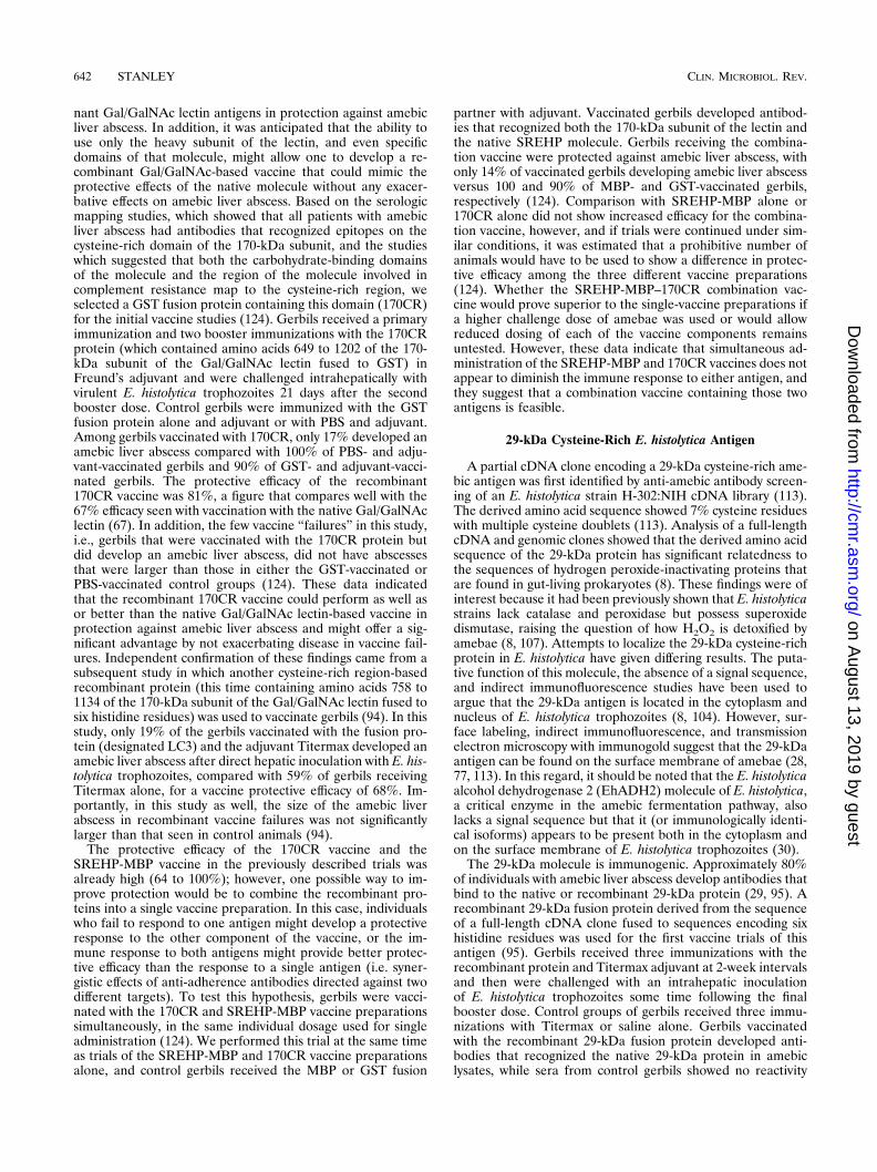
FIG. 4. Model of the CtxA2-SREHP-MBP–CtxB and CtxA2-MBP–CtxB fu-sion proteins. The CtxA2-MBP–CtxB complex, engineered by Jobling andHolmes (36), contains the MBP fused to the A2 domain of cholera toxin. Thisprotein can bind to five cholera toxin B subunits to create the MBP-pseudotoxinmolecule shown on the right. In collaboration with Jobling and Holmes, I havecreated a new version of this protein, where a SREHP-MBP fusion protein isfused (at the carboxy terminus of SREHP) to the A2 domain of cholera toxin(96). This protein in turn can bind to pentameric cholera toxin B to create theSREHP-MBP-pseudotoxin molecule shown on the left.
while 100% of Stm-Ctrl and nonvaccinated gerbils developedan amebic liver abscess after intrahepatic challenge (125).
These studies established that S. typhimurium could serve asan effective delivery system for the amebic SREHP moleculeand demonstrated that vaccination by the oral route couldprovide protection against amebic liver abscess. A logical nextstep was to ask whether similar results could be obtained withexpression of SREHP in attenuated strains of S. typhi, thusproviding the possibility of a combination amebiasis-typhoidfever vaccine. The SREHP-MBP fusion protein has now beensuccessfully expressed in S. typhi TY2 x4297 (Dcya Dcrp Dasd)(126). High-level expression of SREHP-MBP was achieved (10to 15% of the total protein detected in Coomassie staining ofSDS-PAGE-separated lysates), expression of the SREHP-MBP fusion protein did not alter the attenuation of the S. typhivaccine strain based on its LD50 in mice, and SREHP re-mained immunogenic, since mice parenterally immunized withS. typhi TY2 x4297 (Dcya Dcrp Dasd) expressing SREHP-MBPdeveloped serum anti-amebic antibodies. While the inability ofS. typhi to colonize mice by the oral route precluded studies ofthe mucosal immunogenicity of this construct, the data ob-tained suggest that the S. typhi TY2 x4297 (Dcya Dcrp Dasd)strain expressing SREHP-MBP represents a potential candi-date for human trials.
Limitations of Current Oral Vaccine Studies
While the results of these recent studies are promising, someimportant caveats apply. The S. typhi TY2 x4297 (Dcya DcrpDasd) strain was studied in a previous clinical trial in humansand was found to be immunogenic (in terms of the induction ofanti-lipopolysaccharide antibodies) and attenuated, but severalindividuals experienced fever and/or diarrhea with vaccination(105). A new S. typhi strain which includes a deletion mutationof the cdt gene (implicated in colonization of deep tissue),S. typhi TY2 x4632 (Dcya Dcrp-cdt Dasd), has recently beendeveloped and has been used to express the pre-S1 protein ofthe hepatitis B virus (64). In a recent clinical trial in femaleadult volunteers, this strain retained immunogenicity (anti-lipopolysaccharide antibodies) when administered by the oralor rectal route and showed decreased reactogenicity. However,only one vaccinated individual showed seroconversion to thepre-S1 protein, a particularly disappointing result since, in aprevious study, vaccination of mice with S. typhimurium ex-pressing the same protein had resulted in seroconversion topre-S1 protein in eight of nine orally vaccinated mice (89).These data emphasize that murine immune responses to het-erologous antigens expressed in S. typhimurium may not pre-dict the human immune response to the same antigen ex-pressed in S. typhi and indicate that further modifications tothe S. typhi (Dcya Dcrp-cdt Dasd)-based system may be neces-sary before it can be used to reliably induce immune responsesto heterologous antigens in humans.
Another unresolved problem is the aforementioned lack of agood animal model for intestinal amebiasis. This obviouslyaffects the ability to study the efficacy of any of these oralvaccine constructs in preventing intestinal disease. How oneselects the best oral vaccine candidates for testing in humans inthe absence of efficacy testing in animal models remains adifficult question. Finally, even if these vaccines prove success-ful in stimulating a secretory IgA response to protective ame-bic antigens in humans, there are at present no data that allowus to predict the efficacy of such a response in protectingagainst amebiasis and the duration of any protective responsethat develops. In this regard, the report that E. histolytica candegrade human secretory IgA in vitro raises as yet unanswered
questions about the efficacy of E. histolytica-specific secretoryIgA in preventing amebic invasion in vivo (40).
CONCLUSIONS
The application of molecular biologic techniques to thestudy of amebiasis has produced a tremendous increase in ourknowledge of the biology of E. histolytica and has enabled therapid development of several candidate vaccines to preventamebiasis. Studies with the gerbil model of amebic liver ab-scess have now identified three recombinant E. histolytica an-tigens that can protect animals from amebic liver abscess. Suc-cessful oral delivery of recombinant amebic antigens has beenachieved, with the induction of mucosal immune responses inanimals and successful protection of gerbils from amebic liverabscess. However, no data are available on the efficacy of anyof the candidate vaccines in preventing intestinal amebiasis.This is problematic, and in the absence of any reliable animalmodel for intestinal amebiasis, it may be impossible to ade-quately assess the protective efficacy of any candidate vaccinein preventing intestinal amebiasis before human trials can beperformed. Despite these difficulties, there is reason for opti-mism. In addition to their value as potential vaccines to pre-vent amebiasis, constructs such as the CtxB–SREHP-12 pro-tein are providing important information about the design ofenteric vaccines and new insights into the requirements formucosal immunogenicity. The recent demonstration that therecombinant amebic SREHP antigen can be mucosally immu-nogenic when delivered by attenuated V. cholerae or S. typhivectors provides additional evidence for the feasibility of com-bination vaccines, designed to be effective against multipleenteric pathogens. This is an especially welcome development,given the profound limitations in health care resources in manyregions where enteric infections are endemic. Given these fi-nancial constraints, “bundling” an E. histolytica vaccine into acombination enteric vaccine may represent the most cost-ef-fective method of delivering protection against amebiasis intothe regions of the world where the vaccine is needed most. Thefirst steps in the process of developing an oral combinationenteric vaccine that could prevent amebiasis have now beenachieved, but the major question of the safety and efficacy ofthese candidate vaccines in humans remains unanswered.
REFERENCES
1. Abou-El-Magd, I., C. J. G. Soong, A. M. El-Hawey, and J. I. Ravdin. 1996.Humoral and mucosal IgA antibody response to a recombinant 52-kDacysteine-rich portion of the Entamoeba histolytica galactose-inhibitable lec-tin correlates with detection of native 170-kDa lectin antigen in serum ofpatients with amebic colitis. J. Infect. Dis. 174:157–162.
2. Andra, J., O. Berninghausen, J. Wulfken, and M. Leippe. 1996. Shortenedamoebapore analogs with enhanced antibacterial and cytolytic activity.FEBS Lett. 385:96–100.
3. Andra, J., and M. Leippe. 1994. Pore-forming peptide of Entamoeba his-tolytica: significance of positively charged amino acid residues for its modeof action. FEBS Lett. 354:97–102.
4. Bessen, D., and V. Fischetti. 1988. Influence of intranasal immunizationwith synthetic peptides corresponding to conserved epitopes of M proteinon mucosal colonization by group A streptococci. Infect. Immun. 56:2666–2672.
5. Betley, M., V. Miller, and J. Mekalanos. 1989. Genetics of bacterial ente-rotoxins. Annu. Rev. Microbiol. 40:577–605.
6. Beving, D. E., C.-J. G. Soong, and J. I. Ravdin. 1996. Oral immunizationwith a recombinant cysteine-rich section of the Entamoeba histolytica ga-lactose-inhibitable lectin elicits an intestinal secretory immunoglobulin Aresponse that has in vitro adherence inhibition activity. Infect. Immun.64:1473–1476.
7. Braga, L. L., H. Ninomiya, J. J. McCoy, S. Eacker, T. Wiedmer, C. Pham,S. Wood, P. J. Sims, and W. A. Petri, Jr. 1992. Inhibition of the complementmembrane attack complex by the galactose-specific adhesin of Entamoebahistolytica. J. Clin. Invest. 90:1131–1137.
8. Bruchhaus, I., and E. Tannich. 1993. Analysis of the genomic sequence
encoding the 29 kDa cysteine-rich protein of Entamoeba histolytica. Trop.Med. Parasitol. 44:116–118.
9. Burch, D. J., E. Li, S. Reed, T. F. H. G. Jackson, and S. L. Stanley, Jr. 1991.Isolation of a strain-specific Entamoeba histolytica cDNA clone. J. Clin.Microbiol. 29:696–701.
10. Caballero-Salcedo, A., M. Viveros-Rogel, B. Salvatierra, R. Tapia-Conyer,J. Sepulveda-Amor, G. Gutierrez, and L. Ortiz-Ortiz. 1994. Seroepidemi-ology of amebiasis in Mexico. Am. J. Trop. Med. Hyg. 4:412–419.
11. Carrero, J. C., M. Yadira Dıaz, M. Viveros, B. Espinoza, E. Acosta, and L.Ortiz-Ortiz. 1994. Human secretory immunoglobulin A anti-Entamoebahistolytica antibodies inhibit adherence of amebae to MDCK cells. Infect.Immun. 62:764–767.
12. Chadee, K., and E. Meerovitch. 1984. The pathogenesis of experimentallyinduced amebic liver abscess in the gerbil (Meriones Unguiculatus). Am. J.Pathol. 117:71–80.
13. Cieslak, P. R., T. Zhang, and S. L. Stanley, Jr. 1993. Expression of arecombinant Entamoeba histolytica antigen in a Salmonella typhimuriumvaccine strain. Vaccine 11:773–776.
14. Clark, C. G., and L. S. Diamond. 1991. Ribosomal RNA genes of ‘patho-genic’ and ‘nonpathogenic’ Entamoeba histolytica are distinct. Mol. Bio-chem. Parasitol. 49:297–302.
15. Clark, C. G., and L. S. Diamond. 1993. Entamoeba histolytica: a method forisolate identification. Exp. Parasitol. 77:450–455.
16. Councilman, W. T., and H. A. LaFleur. 1891. Amoebic dysentery. JohnsHopkins Hosp. Rep. 2:395–548.
17. Cuatrecasas, P. 1973. Gangliosides and membrane receptors for choleratoxin. Biochemistry 12:3558–3566.
18. Curtiss, R., III, and S. M. Kelly. 1987. Salmonella typhimurium deletionmutants lacking adenylate cyclase and cyclic AMP receptor protein areavirulent and immunogenic. Infect. Immun. 55:3035–3043.
19. Curtiss, R., III, S. M. Kelly, P. A. Gulig, and K. Nakayama. 1989. Selectivedelivery of antigens by recombinant bacteria. Cur. Top. Microb. Immunol.146:35–49.
20. Czerkinsky, C., M. W. Russell, N. Lycke, M. Lindblad, and J. Holmgren.1989. Oral administration of a streptococcal antigen coupled to choleratoxin B subunit evokes strong antibody responses in salivary glands andextramucosal tissues. Infect. Immun. 57:1072–1077.
21. Czerkinsky, C., A.-M. Svennerholm, and J. Holmgren. 1993. Induction andassessment of immunity at enteromucosal surfaces in humans: Implicationsfor vaccine development. Clin. Infect. Dis. 16(Suppl. 2):S106–S116.
22. deLeon, A. 1970. Pronostico tardio en el absceso hepatico amibiano. ArchInvest. Med. Mexico 1:s205–s206.
23. Dertzbaugh, M. T., and C. O. Elson. 1993. Comparative effectiveness of thecholera toxin B subunit and alkaline phosphatase as carriers for oral vac-cines. Infect. Immun. 61:48–55.
24. Dertzbaugh, M. T., and F. L. Macrina. 1989. Plasmid vectors for construct-ing translational fusions to the B subunit of cholera toxin. Gene 82:335–342.
25. Dertzbaugh, M. T., D. L. Peterson, and F. L. Macrina. 1990. Cholera toxinB-subunit gene fusion: structural and functional analysis of the chimericprotein. Infect. Immun. 58:70–79.
26. Elson, C. O., and W. Ealding. 1984. Cholera toxin feeding did not induceoral tolerance in mice and abrogated oral tolerance to an unrelated proteinantigen. J. Immunol. 133:2892–2897.
27. Elson, C. O., and W. Ealding. 1984. Generalized systemic and mucosalimmunity in mice after mucosal stimulation with cholera toxin. J. Immunol.132:2736–2741.
28. Flores, B. M., M. A. Batzer, M. A. Stein, C. Petersen, D. L. Diedrich, andB. E. Torian. 1993. Structural analysis and demonstration of the 29-kDaantigen of pathogenic Entamoeba histolytica as the major accessible freethiol-containing surface antigen. Mol. Microbiol. 7:755–763.
29. Flores, B. M., S. L. Reed, J. I. Ravdin, and B. E. Torian. 1993. Serologicreactivity to purified recombinant and native 29-kilodalton peripheral mem-brane protein of pathogenic Entamoeba histolytica. J. Clin. Microbiol. 31:1403–1407.
30. Flores, B. M., S. L. Stanley, Jr., T. S. Yong, M. Ali, W. G. Yang, D. L.Diedrich, and B. E. Torian. 1996. Surface localization, regulation, andbiologic properties of the 96-kDa alcohol aldehyde dehydrogenase(EhADH2) of pathogenic Entamoeba histolytica. J. Infect. Dis. 173:226–231.
31. Ghadirian, E., and E. Meerovitch. 1978. Vaccination against hepatic ame-biasis in hamsters. J. Parasitol. 64:742–743.
32. Ghadirian, E., E. Meerovitch, and D.-P. Hartmann. 1980. Protectionagainst amebic liver abscess in hamsters by means of immunization withamebic antigen and some of its fractions. Am. J. Trop. Med. Hyg. 29:779–784.
33. Gonzalez, R. A., J. Sanchez, J. Holmgren, S. Lopez, and C. F. Arias. 1993.Immunological characterization of a rotavirus-neutralizing epitope fused tothe cholera toxin B subunit. Gene 133:227–232.
34. Jain, P., S. Sawhney, and V. K. Vinayak. 1980. Experimental amoebicinfection in guinea pigs immunized with low grade amoebic infection.Trans. R. Soc. Trop. Med. Hyg. 74:347–355.
35. Jiminez-Cardoso, J. M., E. Jiminez, M. de Jesus-Bernal, and J. Kumate.
1989. Induccion de inmunidad protectora antiamibiana en hamsters conantigenos heterologos. Rev. Invest. Clin. 41:133–140.
36. Jobling, M. G., and R. K. Holmes. 1992. Fusion proteins containing the A2domain of cholera toxin assemble with B polypeptides of cholera toxin toform immunoreactive and functional holotoxin-like chimeras. Infect. Im-mun. 60:4915–4924.
37. Kanai, S. R., and R. Knight. 1969. Relapsing amoebic colitis of 12 years’standing exacerbated by corticosteroids. Br. Med. J. 2:613–614.
38. Katzenstein, D., V. Rickerson, and A. Braude. 1982. New concepts ofamebic liver abscess derived from hepatic imaging, serodiagnosis, and he-patic enzymes in 67 consecutive cases in San Diego. Medicine 61:237–246.
39. Keene, W. E., M. E. Hidalgo, E. Orozco, and J. H. McKerrow. 1990.Entamoeba histolytica: correlation of the cytopathic effect of virulent tro-phozoites with secretion of a cysteine proteinase. Exp. Parasitol. 71:199–206.
40. Kelsall, B. L., and J. I. Ravdin. 1993. Degradation of human IgA byEntamoeba histolytica. J. Infect. Dis. 168:1319–1322.
41. Kelsall, B. L., and J. I. Ravdin. 1995. Immunization of rats with the 260-kilodalton Entamoeba histolytica galactose-inhibitable lectin elicits an intes-tinal secretory immunoglobulin A response that has in vitro adherence-inhibitory activity. Infect. Immun. 63:686–689.
42. Kohler, S., and E. Tannich. 1993. A family of transcripts (K2) of Entamoebahistolytica contains polymorphic repetitive regions with highly conservedelements. Mol. Biochem. Parasitol. 59:49–58.
43. Krupp, I. M. 1970. Antibody response in intestinal and extraintestinalamebiasis. Am. J. Trop. Med. Hyg. 19:57–62.
44. Krupp, I. M. 1974. Protective immunity to amebic infection demonstratedin guinea pigs. Am. J. Trop. Med. Hyg. 23:355–362.
45. Lee, A., and M. Chen. 1994. Successful immunization against gastric infec-tion with Helicobacter species: use of a cholera toxin B-subunit whole-cellvaccine. Infect. Immun. 62:3594–3597.
46. Leippe, M., J. Andra, and H. J. Muller-Eberhard. 1994. Cytolytic andantibacterial activity of synthetic peptides derived from amoebapore, thepore-forming peptide of Entamoeba histolytica. Proc. Natl. Acad. Sci. USA91:2602–2606.
47. Leippe, M., and H. J. Muller-Eberhard. 1994. The pore-forming peptide ofEntamoeba histolytica, the protozoan parasite causing human amoebiasis.Toxicology 87:5–18.
48. Li, E., A. Becker, and S. L. Stanley, Jr. 1988. Use of Chinese hamster ovarycells with altered glycosylation patterns to define the carbohydrate speci-ficity of Entamoeba histolytica adhesion. J. Exp. Med. 167:1725–1730.
49. Li, E., A. Becker, and S. L. Stanley, Jr. 1989. Chinese hamster ovary cellsdeficient in N-acetylglycosaminyltransferase I activity are resistant to Ent-amoeba histolytica-mediated cytotoxicity. Infect. Immun. 57:8–12.
50. Li, E., C. Kunz-Jenkins, and S. L. Stanley, Jr. 1992. Isolation and charac-terization of genomic clones encoding a serine-rich Entamoeba histolyticaprotein. Mol. Biochem. Parasitol. 50:355–358.
51. Li, E., and S. L. Stanley, Jr. 1996. Protozoa: amebiasis. Gastroenterol. Clin.North Am. 25:471–492.
52. Liang, X., M. Lamm, and J. Nedrud. 1988. Oral immunization of choleratoxin-Sendai virus conjugates potentiates gut and respiratory immunityagainst sendai virus. J. Immunol. 141:1495–1501.
53. Lycke, N., and J. Holmgren. 1986. Strong adjuvant properties of choleratoxin on gut mucosal immune responses to orally presented antigens. J. Im-munol. 59:301–308.
54. Lycke, N., T. Tsuji, and J. Holmgren. 1992. The adjuvant effect of Vibriocholera and Escherichia coli heat-labile enterotoxins is linked to their ADP-ribosyltransferase activity. Eur. J. Immunol. 22:2277–2281.
55. Mann, B. J., B. E. Torian, T. S. Vedvick, and W. A. Petri, Jr. 1991. Sequenceof a cysteine-rich galactose-specific lectin of Entamoeba histolytica. Proc.Natl. Acad. Sci. USA 88:3248–3252.
56. Martinez-Reyes, R., I. Gonzalez-Pacheco, I. Gomez-Reyes, M. de la Torre,and B. Sepulveda. 1980. Induccion de inmunidad antimiabisica en primatessubhumanos. Con antigeno lisosoma de Entamoeba histolytica. VI. Inocu-lacion intrahepatica de E. histolytica virulenta en monos. Arch. Invest. Med.Mexico 11:267–274.
57. McAllister, T. A. 1962. Diagnosis of amebic colitis on routine biopsies fromrectum and sigmoid colon. Br. Med. J. 1:362–364.
58. McCoy, J. J., B. J. Mann, and W. A. Petri, Jr. 1994. Adherence andcytotoxicity of Entamoeba histolytica, or how lectins let parasites stickaround. Infect. Immun. 62:3045–3050.
59. McCoy, J. J., B. J. Mann, T. S. Vedvick, Y. Pak, D. B. Heimark, and W. A.Petri, Jr. 1993. Structural analysis of the light subunit of the Entamoebahistolytica galactose-specific adherence lectin. J. Biol. Chem. 268:24223–24231.
60. McCoy, J. J., B. J. Mann, T. S. Vedvick, and W. A. Petri, Jr. 1993. Sequenceanalysis of genes encoding the light subunit of the Entamoeba histolyticagalactose-specific adhesin. Mol. Biochem. Parasitol. 61:325–328.
61. McKenzie, S. J., and J. F. Halsey. 1984. Cholera toxin B subunit as a carrierprotein to stimulate a mucosal immune response. J. Immunol. 133:1818–1824.
62. Myung, K., D. Burch, T. F. H. G. Jackson, and S. L. Stanley, Jr. 1992.
Serodiagnosis of invasive amebiasis using a recombinant Entamoeba histo-lytica antigen-based ELISA. Arch. Med. Res. 23:285–288.
63. Nakayama, K., S. M. Kelly, and R. Curtiss III. 1988. Construction of anasd1 expression-cloning vector: stable maintenance and high level expres-sion of cloned genes in a Salmonella vaccine strain. Bio/Technology 6:693–697.
64. Nardelli-Haefliger, D., J.-P. Kraehenbuhl, R. Curtiss III, F. Schodel, A.Potts, S. M. Kelly, and P. De Grandi. 1996. Oral and rectal immunizationof adult female volunteers with a recombinant attenuated Salmonella typhivaccine strain. Infect. Immun. 64:5219–5224.
65. Osorio, L. M., T. Pico, and A. Luaces. 1992. Circulating antibodies tohistolysain, the major cysteine proteinase of Entamoeba histolytica, in amoe-bic liver abscess patients. Parasitology 105:207–210.
66. Petri, W. A., Jr., M. D. Chapman, T. Snodgrass, B. J. Mann, J. Broman,and J. I. Ravdin. 1989. Subunit structure of the galactose and N-acetyl-D-galactosamine-inhibitable adherence lectin of Entamoeba histolytica. J. Biol.Chem. 264:3007–3012.
67. Petri, W. A., Jr., and J. I. Ravdin. 1990. Protection of gerbils from amebicliver abscess by immunization with the galactose-specific adherence lectinof Entamoeba histolytica. Infect. Immun. 59:97–101.
68. Petri, W. A., Jr., R. D. Smith, P. H. Schlesinger, C. F. Murphy, and J. I.Ravdin. 1987. Isolation of the galactose binding lectin that mediates the invitro adherence of Entamoeba histolytica. J. Clin. Invest. 80:1238–1244.
69. Petri, W. A., Jr., T. L. Snodgrass, T. F. H. G. Jackson, V. Gathiram,A. E. Simjee, K. Chadee, and M. D. Chapman. 1990. Monoclonal antibodiesdirected against the galactose-binding lectin of Entamoeba histolytica en-hance adherence. J. Immunol. 144:4803–4809.
70. Phillips, R. S. 1992. Vaccination against malaria. Immunobiology 184:240–262.
71. Purdy, J. E., B. J. Mann, E. C. Shugart, and W. A. Petri, Jr. 1993. Analysisof the gene family encoding the Entamoeba histolytica galactose-specificadhesin 170-kDa subunit. Mol. Biochem. Parasitol. 62:53–60.
72. Ravdin, J. I., and R. L. Guerrant. 1981. Role of adherence in cytopatho-genic mechanisms of Entamoeba histolytica: studies with mammalian tissueculture cells and human erythrocytes. J. Clin. Invest. 68:1305–1313.
73. Ravdin, J. I., T. F. H. G. Jackson, W. A. Petri, Jr., C. F. Murphy, B. L.Ungar, V. Gathiram, J. Skilogiannis, and A. E. Simjee. 1990. Association ofserum antibodies to adherence lectin with invasive amebiasis and asymp-tomatic infection with pathogenic Entamoeba histolytica. J. Infect. Dis.162:768–772.
74. Ravdin, J. I., W. A. Petri, Jr., C. F. Murphy, and R. D. Smith. 1986.Production of mouse monoclonal antibodies which inhibit in vitro adher-ence of Entamoeba histolytica trophozoites. Infect. Immun. 53:1–5.
75. Ravdin, J. I., P. Stanley, C. F. Murphy, and W. A. Petri, Jr. 1989. Charac-terization of cell surface carbohydrate receptors for Entamoeba histolyticaadherence lectin. Infect. Immun. 57:2179–2186.
76. Reed, S., J. Bouvier, A. S. Pollack, J. C. Engel, M. Brown, K. Hirata, X.Que, A. Eakin, P. Hagblom, F. Gillin, and J. H. McKerrow. 1993. Cloningof a virulence factor of Entamoeba histolytica. Pathogenic strains possess aunique cysteine proteinase gene. J. Clin. Invest. 91:1532–1540.
77. Reed, S. L., B. M. Flores, M. A. Batzer, M. A. Stein, V. L. Stroeher,J. E. Carlton, D. L. Diedrich, and B. E. Torian. 1992. Molecular and cellularcharacterization of the 29-kilodalton peripheral membrane protein of Ent-amoeba histolytica: differentiation between pathogenic and nonpathogenicisolates. Infect. Immun. 60:542–549.
78. Robbins, J. B., R. Schneerson, and S. C. Szu. 1995. Hypothesis: Serum IgGantibody is sufficient to confer protection against infectious diseases byinactivating the inoculum. J. Infect. Dis. 171:1387–1398.
79. Russell, M. W., and H. Wu. 1991. Distribution, persistence, and recall ofserum and salivary antibody responses to peroral immunization with pro-tein antigen I/II of Streptococcus mutans coupled to the cholera toxin Bsubunit. Infect. Immun. 59:4061–4070.
80. Ryan, E. T., J. R. Butterton, T. Zhang, S. L. Stanley, Jr., and S. B. Calder-wood. 1997. Oral immunization with attenuated vaccine strains of Vibriocholerae expressing a dodecapeptide repeat of the serine-rich Entamoebahistolytica protein fused to the cholera toxin B subunit induces systemic andmucosal anti-amebic and anti-V. cholerae antibody responses in mice. In-fect. Immun. 65:3118–3125.
81. Salata, R. A., J. G. Cox, and J. I. Ravdin. 1987. The interaction of humanT-lymphocytes and Entamoeba histolytica: killing of virulent amoebae bylectin-dependent lymphocytes. Parasite Immunol. 9:249–261.
82. Salata, R. A., A. Martinez-Palomo, H. W. Murray, L. Conales, N. Trevino,E. Segovia, C. F. Murphy, and J. I. Ravdin. 1986. Patients treated foramebic liver abscess develop cell-mediated immune responses effective invitro against Entamoeba histolytica. J. Immunol. 136:2633–2639.
83. Salata, R. A., R. D. Pearson, and J. I. Ravdin. 1985. Interaction of humanleukocytes and Entamoeba histolytica. Killing of virulent amebae by theactivated macrophage. J. Clin. Invest. 76:491–499.
84. Sanchez, J. L., A. F. Trofa, D. N. Taylor, R. A. Kuschner, R. F. DeFraites,S. C. Craig, M. R. Rao, J. D. Clemens, A.-M. Svennerholm, J. C. Sadoff, andJ. Holmgren. 1993. Safety and immunogenicity of the oral, whole cell/
recombinant B subunit cholera vaccine in North American volunteers.J. Infect. Dis. 167:1446–1449.
85. Sargeaunt, P. G., and J. E. Williams. 1978. The differentiation of invasiveand non-invasive Entamoeba histolytica by isoenzyme electrophoresis.Trans. R. Soc. Trop. Med. Hyg. 72:519–521.
86. Sargeaunt, P. G., and J. E. Williams. 1978. Electrophoretic isoenzymepatterns of Entamoeba histolytica and Entamoeba coli. Trans. R. Soc. Trop.Med. Hyg. 72:164–166.
87. Sargeaunt, P. G., and J. E. Williams. 1979. Electrophoretic isoenzymepatterns of the pathogenic and non-pathogenic intestinal amoebae of man.Trans. R. Soc. Trop. Med. Hyg. 73:225–227.
88. Schain, D. C., R. A. Salata, and J. I. Ravdin. 1992. Human T-lymphocyteproliferation, lymphokine production, and amebicidal activity elicited bythe galactose-inhibitable adherence protein of Entamoeba histolytica. In-fect. Immun. 60:2143–2146.
89. Schodel, F., S. M. Kelly, D. L. Peterson, D. R. Milich, and R. Curtiss III.1994. Hybrid hepatitis B virus core-pre-S proteins synthesized in avirulentSalmonella typhimurium and Salmonella typhi for oral vaccination. Infect.Immun. 62:1669–1676.
90. Sepulveda, B. 1980. Induccion de immunidad protectora antiamibiasica enprimates subhumanos con antigeno lisosomal de Entamoeba histolytica.Comentarios y conclusiones. Arch. Invest. Med. Mexico 11:275–286.
91. Sepulveda, B., M. Tanimoto-Weki, P. Calderon, and M. de la Torre. 1974.Neutralizacion de la virulencia en cultivos de e. histolytica con suero im-mune. Arch Invest. Med. Mexico 5:447–450.
92. Sepulveda, B., and A. Martınez-Palomo. 1982. Immunology of amoebiasisby Entamoeba histolytica, p. 170–191. In S. Cohen and K. S. Warren (ed.),Immunology of parasitic infections. Blackwell Scientific Publications, Ox-ford, United Kingdom.
93. Seydel, K. B., K. Braun, T. Zhang, T. F. H. G. Jackson, and S. L. Stanley,Jr. 1996. Human anti-amebic antibodies provide protection against amebicliver abscess formation in the SCID mouse. Am. J. Trop. Med. Hyg. 55:330–332.
94. Soong, C.-J. G., K. C. Kain, M. Abd-Alla, T. F. H. G. Jackson, and J. I.Ravdin. 1995. A recombinant-cysteine rich section of the Entamoeba his-tolytica galactose-inhibitable lectin is efficacious as a subunit vaccine in thegerbil model of amebic liver abscess. J. Infect. Dis. 171:645–651.
95. Soong, C. G., B. E. Torian, M. Abd-Alla, T. F. H. G. Jackson, V. Gatharim,and J. I. Ravdin. 1995. Protection of gerbils from amebic liver abscess byimmunization with recombinant Entamoeba histolytica 29-kilodalton anti-gen. Infect. Immun. 63:472–477.
96. Stanley, S. L., Jr. 1997. Unpublished observations.97. Stanley, S. L., Jr., A. Becker, C. Kunz-Jenkins, L. Foster, and E. Li. 1990.
Cloning and expression of a membrane antigen of Entamoeba histolyticapossessing multiple tandem repeats. Proc. Natl. Acad. Sci. USA 87:4976–4980.
98. Stanley, S. L., Jr., J. L. Blanchard, N. Johnson, L. Foster, C. Kunz-Jenkins,T. Zhang, K. Tian, and F. B. Cogswell. 1995. Immunogenicity of the re-combinant serine rich Entamoeba histolytica protein (SREHP) in Africangreen monkeys. Vaccine 13:947–951.
99. Stanley, S. L., Jr., H. Huizenga, and E. Li. 1992. Isolation and partialcharacterization of a surface glycoconjugate of Entamoeba histolytica. Mol.Biochem. Parasitol. 50:127–138.
100. Stanley, S. L., Jr., T. F. H. G. Jackson, S. L. Reed, J. Calderon, C. Kunz-Jenkins, V. Gathiram, and E. Li. 1991. Serodiagnosis of invasive amebiasisusing a recombinant Entamoeba histolytica protein. JAMA 266:1984–1986.
101. Stanley, S. L., Jr., K. Tian, J. P. Koester, and E. Li. 1995. The serine richEntamoeba histolytica protein (SREHP) is a phosphorylated membraneprotein containing O-linked terminal N-acetylglucosamine (O-GlcNAc)residues. J. Biol. Chem. 270:4121–4126.
102. Stanley, S. L., Jr., T. Zhang, D. Rubin, and E. Li. 1995. Role of theEntamoeba histolytica cysteine proteinase in amebic liver abscess formationin severe combined immunodeficient (SCID) mice. Infect. Immun. 63:1587–1590.
103. Swartzwelder, J. C., and W. H. Avant. 1952. Immunity to amebic infectionin dogs. Am. J. Trop. Med. Hyg. 1:567–575.
104. Tachibana, H., S. Kobayashi, Y. Kato, K. Nagakura, Y. Kaneda, and T.Takeuchi. 1990. Identification of a pathogenic isolate-specific 30,000 Mrantigen of Entamoeba histolytica using a monoclonal antibody. Infect. Im-mun. 58:955–960.
105. Tacket, C. O., D. M. Hone, R. Curtiss III, S. M. Kelly, G. Losonsky, L.Guers, A. M. Harris, R. Edelman, and M. M. Levine. 1992. Comparison ofthe safety and immunogenicity of DaroC DaroD and Dcya Dcrp Salmonellatyphi strains in adult volunteers. Infect. Immun. 60:536–541.
106. Tamura, S., Y. Samegai, H. Kurata, T. Nagamine, C. Aizawa, and T.Kurata. 1988. Protection against influenza virus infection by vaccine inoc-ulated intranasally with cholera toxin B subunit. Vaccine 6:409–413.
107. Tannich, E., I. Bruchhaus, R. D. Walter, and R. D. Horstmann. 1991.Pathogenic and nonpathogenic Entamoeba histolytica: identification andmolecular cloning of an iron-containing superoxide dismutase. Mol. Bio-chem. Parasitol. 49:61–72.
108. Tannich, E., and G. D. Burchard. 1991. Differentiation of pathogenic from
nonpathogenic Entamoeba histolytica by restriction fragment analysis of asingle gene amplified in vitro. J. Clin. Microb. 29:250–255.
109. Tannich, E., F. Ebert, and R. D. Horstmann. 1991. Primary structure of the170-kDa surface lectin of pathogenic Entamoeba histolytica. Proc. Natl.Acad. Sci. USA 88:1849–1853.
110. Tannich, E., F. Ebert, and R. D. Horstmann. 1992. Molecular cloning ofcDNA and genomic sequences coding for the 35-kilodalton subunit of thegalactose-inhibitable lectin of pathogenic Entamoeba histolytica. Mol. Bio-chem. Parasitol. 55:225–228.
111. Tannich, E., R. D. Horstmann, J. Knobloch, and H. H. Arnold. 1989.Genomic DNA differences between pathogenic and nonpathogenic Ent-amoeba histolytica. Proc. Natl. Acad. Sci. USA 86:5118–5122.
112. Tannich, E., H. Scholze, R. Nickel, and R. D. Horstmann. 1991. Homolo-gous cysteine proteinases of pathogenic and nonpathogenic Entamoebahistolytica. Differences in structure and expression. J. Biol. Chem. 266:4798–4803.
113. Torian, B. E., B. M. Flores, V. L. Stroeher, F. S. Hagen, and W. E. Stamm.1990. cDNA sequence analysis of a 29-kDa cysteine-rich surface antigen ofpathogenic Entamoeba histolytica. Proc. Natl. Acad. Sci. USA 87:6358–6362.
114. Vazquez-Saavedra, J. A., M. Tanimoto-Weki, J. I. Olivera-Lopez, F. Cara-cheo-Reyes, and A. Cortes-Arcos. 1973. Immunity after cured amebic in-fection in the hamster. Arch. Invest. Med. Suppl. 1:155–158. (In Spanish.)
115. Velazquez, C., I. Valette, M. Cruz, M.-L. Labra, J. Montes, S. L. Stanley,Jr., and J. Calderon. 1995. Identification of immunogenic epitopes of the170-kDa subunit adhesin of Entamoeba histolytica in patients with invasiveamebiasis. J. Eukaryot. Microbiol. 42:636–641.
116. Vinayak, V. K., S. Sawhney, P. Jain, and R. N. Chakravarti. 1980. Protec-tive effects of crude and chromatographic fractions of axenic Entamoebahistolytica in guinea pigs. Trans. R. Soc. Trop. Med. Hyg. 74:483–487.
117. Walsh, J. A. 1986. Problems in recognition and diagnosis of amebiasis:estimation of the global magnitude of morbidity and mortality. Rev. Infect.Dis. 8:228–238.
118. Wang, L., J. Calderon, and S. L. Stanley, Jr. Identification of B cell epi-topes in the serine rich Entamoeba histolytica protein. Am. J. Trop. Med.Hyg., in press.
119. Yang, W., E. Li, T. Kairong, and S. L. Stanley, Jr. 1994. Entamoeba histo-
lytica has an alcohol dehydrogenase homologous to the multifunctionaladhE gene product of Escherichia coli. Mol. Biochem. Parasitol. 64:253–260.
120. Yong, T., E. Li, D. Clark, and S. L. Stanley, Jr. 1996. Complementation ofa Escherichia coli adhE mutant by the Entamoeba histolytica EhADH2 geneprovides a method for the identification of new anti-amebic drugs. Proc.Natl. Acad. Sci. USA 93:6464–6469.
121. Zhang, T., P. R. Cieslak, L. Foster, C. Kunz-Jenkins, and S. L. Stanley, Jr.1994. Antibodies to the serine rich Entamoeba histolytica protein (SREHP)prevent amebic liver abscess in severe combined immunodeficient (SCID)mice. Parasite Immunol. 16:225–230.
122. Zhang, T., P. R. Cieslak, and S. L. Stanley, Jr. 1994. Protection of gerbilsfrom amebic liver abscess by immunization with a recombinant Entamoebahistolytica antigen. Infect. Immun. 62:1166–1170.
123. Zhang, T., E. Li, and S. L. Stanley, Jr. 1995. Oral immunization with thedodecapeptide repeat of the serine rich Entamoeba histolytica protein(SREHP) fused to the cholera toxin B subunit induces a mucosal andsystemic anti-SREHP antibody response. Infect. Immun. 63:1349–1355.
124. Zhang, T., and S. L. Stanley, Jr. 1994. Protection of gerbils from amebicliver abscess by immunization with a recombinant protein derived from the170-kilodalton surface adhesin of Entamoeba histolytica. Infect. Immun.62:2605–2608.
125. Zhang, T., and S. L. Stanley, Jr. 1996. Oral immunization with an attenu-ated vaccine strain of Salmonella typhimurium expressing the serine-richEntamoeba histolytica protein (SREHP) induces an anti-amebic immuneresponse and protects gerbils from amebic liver disease. Infect. Immun.64:1526–1531.
126. Zhang, T., and S. L. Stanley, Jr. 1997. Expression of the serine rich Ent-amoeba histolytica protein (SREHP) in the avirulent vaccine strain Salmo-nella typhi TY2 4297 (Dcya Dcrp Dasd): safety and immunogenicity in mice.Vaccine, in press.
127. Zhang, Y., E. Li, T. F. H. G. Jackson, T. Zhang, V. Gathiram, and S. L.Stanley, Jr. 1992. Use of a recombinant 170-kilodalton surface antigen ofEntamoeba histolytica for serodiagnosis of amebiasis and identification ofimmunodominant domains of the native molecule. J. Clin. Microbiol. 30:2788–2792.