Project: Ghana Emergency Medicine Collaborative Document Title: A Potpourri of Wound Care Issues Author(s): Alexander J. Rogers, MD (University of Michigan) 2010 License: Unless otherwise noted, this material is made available under the terms of the Creative Commons Attribution Share Alike-3.0 License: http://creativecommons.org/licenses/by-sa/3.0/ We have reviewed this material in accordance with U.S. Copyright Law and have tried to maximize your ability to use, share, and adapt it. These lectures have been modified in the process of making a publicly shareable version. The citation key on the following slide provides information about how you may share and adapt this material. Copyright holders of content included in this material should contact [email protected]with any questions, corrections, or clarification regarding the use of content. For more information about how to cite these materials visit http://open.umich.edu/privacy-and-terms-use. Any medical information in this material is intended to inform and educate and is not a tool for self-diagnosis or a replacement for medical evaluation, advice, diagnosis or treatment by a healthcare professional. Please speak to your physician if you have questions about your medical condition. Viewer discretion is advised: Some medical content is graphic and may not be suitable for all viewers. 1
Transcript
Project: Ghana Emergency Medicine Collaborative
Document Title: A Potpourri of Wound Care Issues
Author(s): Alexander J. Rogers, MD (University of Michigan) 2010
License: Unless otherwise noted, this material is made available under the terms of the Creative Commons Attribution Share Alike-3.0 License: http://creativecommons.org/licenses/by-sa/3.0/
We have reviewed this material in accordance with U.S. Copyright Law and have tried to maximize your ability to use, share, and adapt it. These lectures have been modified in the process of making a publicly shareable version. The citation key on the following slide provides information about how you may share and adapt this material.
Copyright holders of content included in this material should contact [email protected] with any questions, corrections, or clarification regarding the use of content.
For more information about how to cite these materials visit http://open.umich.edu/privacy-and-terms-use.
Any medical information in this material is intended to inform and educate and is not a tool for self-diagnosis or a replacement for medical evaluation, advice, diagnosis or treatment by a healthcare professional. Please speak to your physician if you have questions about your medical condition.
Viewer discretion is advised: Some medical content is graphic and may not be suitable for all viewers.
1
Attribution Key
for more information see: http://open.umich.edu/wiki/AttributionPolicy
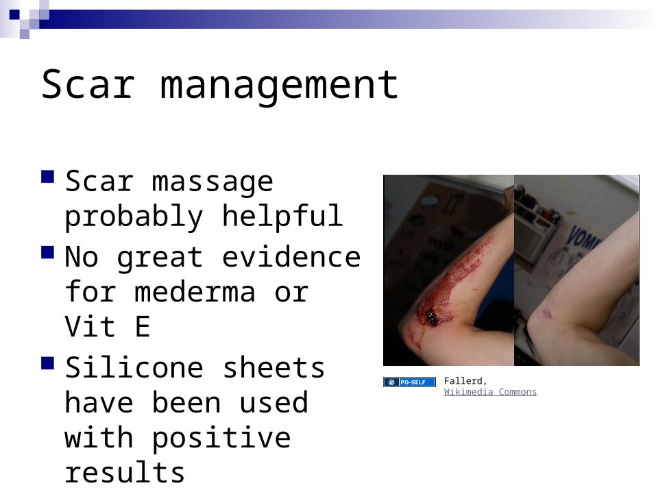
Shear is caused by a small total energy applied symmetrically to a very small volume of tissue, e.g. a scalpel wound. Shear-type wounds have a low potential for infection
Compression involves energy distributed over a greater volume of tissue and requires the absorption of a far greater total energy in order to produce tissue failure. There is, therefore, much greater tissue injury, and a much higher potential for infection
stellate laceration caused by a blunt object striking the skin at 90 degrees
Tension type wounds are intermediate between shear and compression in terms of the energy required to produce tissue failure and the degree of tissue damage. The entirety of tissue damage caused by a tension injury is often not immediately evident
Biology of Wound Healing 4 classic stages of wound healing
Topical Less reliable Time consuming End arteriolar issues
Regional Less reliable Technically difficult Specific locations
How to make anesthesia…less painful Application of topical anesthesia at triage is
feasible and effective in reducing pain of injection, and saves time (Singer and Stark, AEM, 1999)
There is at least some evidence for: Buffered Lidocaine Warm solution Infiltrate within wound Slow infiltration Subcutaneous (vs. intradermal)
Nerve Blocks…A different talk
BUT MCP vs. block along digit (standard digital)
In study of 30 volunteers (ouch) MCP block less reliable (23% vs. 3% failure) and
slower (6.35 vs. 2.82 minutes) Knoop and Trott, Annals Emergency Medicine, 1994
Toxic DosesDrug Adult max
dosePeds max dose
Lidocaine 300 mg 4 mg/kg
Lidocaine with epi
500 mg 7 mg/kg
Bupivicaine 175 mg 1.5 mg/kg
Bupivicaine with epi
225 3 mg/kg
Alternative Anesthetic Agents
For those with ‘caine’ allergies Diphenhydramine
more painful, but works about as well as lidocaine Benzyl alcohol
found as preservative in multidose saline not very painful to inject Short acting – can mix with epi Add 0.2 ml epi 1:1000 to 20 mL vial of normal saline
with 0.9% benyzl alcohol
What about epi in the finger?
Finger Injection with High-Dose (1:1,000) Epinephrine: Does it Cause Finger Necrosis and Should it be Treated?
Colleen Fitzcharles-Bowe & Keith Denkler & Don Lalonde
Documented 59 cases of accidental epi injection with NO cases of tissue necrosis
High dose epi led to about 10 weeks of neuropraxia
“One of the authors of this paper (DL) had three of his own fingers injected with epinephrine on July 21, 2005, to carefully and accurately document the outcome.”
So to treat or not to treat?
For high dose (1:1000) epi (epi-pen) use of phentolamine did decrease the length of time to reperfusion
Unclear (but possible) if treatment could prevent neuropraxia
Again, no cases of tissue necrosis
Wound Closure
A little history Ancient Egyptians used form of tape to close
eyebrow wounds in 2500 BC Oldest known sutures – 1100 BC – on mummy Ancient Hindus used ant mandibles to close wounds In middle ages – pus was believed necessary for
healing
Classifications of closures
Primary closure (primary intention) – clean, minimally contaminated woundsThis is most of what we do
Secondary closure (secondary intention) – not closed and allowed to heal gradually
Tertiary closure (delayed primary closure) – initially cleaned, and then closed after 4 or 5 days – consider for highly contaminated wounds
Wound Closure Techniques
Tissue Adhesives Sutures Tapes Staple Other mechanisms Important to know when to use each one!
Tissue Adhesives
Octylcyanoacrylate(Dermabond)
Butylcyanoacrylate(Indermil)
Carbon side chains 8 4
Breaking strength Moderate Low
Flexibility Great Poor
Microbial Barrier Yes Some
Sutures - AbsorbableSuture Type 50% Strength
RetentionReactivity Use Filament
Plain Gut 5-7 days Moderate Intraoral* Mono
Chronic Gut 10-14 days Moderate Intraoral Mono
Vicryl 3 weeks Minimal Deep sutures Braided
Vicryl Rapide 5 days Minimal Skin approximation
Braided
Monocryl 1.5 weeks Minimal Deep sutures Mono
Fast Gut <5-7 days Percutanious Mono
Prolene ___ Least Skin Approximation
Mono
Needle Selection
Size Common types
Conventional cutting Reverse cutting
Ethicon Needles FS (for skin) – lower
quality PS (for plastic skin) P (for precision point) PC (for precision
A Randomized, Controlled Trial Comparing Long-tern Cosmetic Outcomes of Traumatic Pediatric Lacerations Repaired with Absorbable Plain Gut Vs. Nonabsorbable Nylon Suture (Karounis et al. AEM 2003)
Randomized trial of patients 1-18 years old with lacerations presenting to Peds ED Excluded bites, crush, gross contaminated, crossing
joints, diabetes, tendon/nerve/cartilage, scalp Re-evaluated in 10 days by research nurse, and 4-5
months by plastic surgeon
Plain Gut vs. Nylon
147 eligible, 95 enrolled At 10 days, optimal score (no significant
difference) 63% for absorbable 49% for non-absorbable
No difference in dehiscence or infection At 4 month follow up
No significant difference (trend towards better results with absorbable)
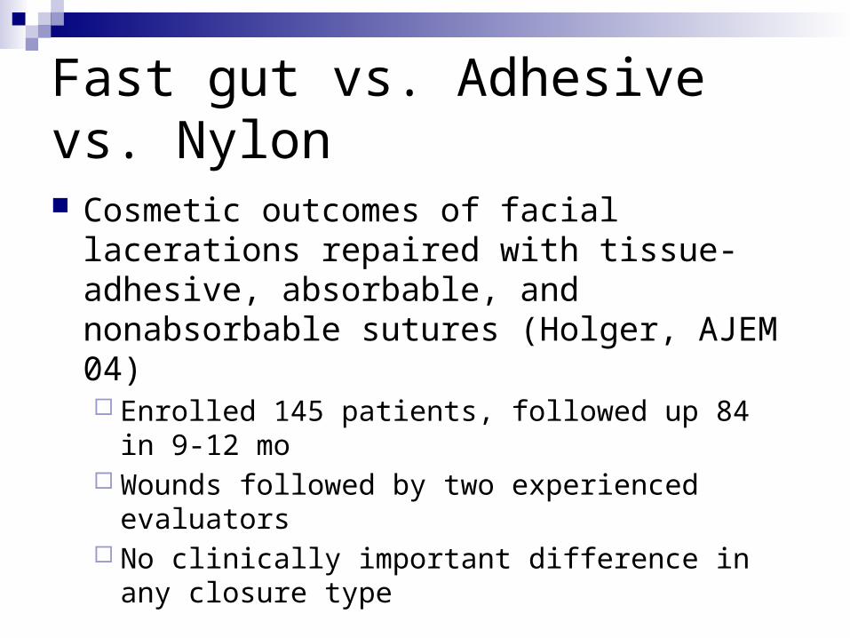
Fast gut vs. Adhesive vs. Nylon
Cosmetic outcomes of facial lacerations repaired with tissue-adhesive, absorbable, and nonabsorbable sutures (Holger, AJEM 04) Enrolled 145 patients, followed up 84 in 9-12 mo Wounds followed by two experienced evaluators No clinically important difference in any closure type
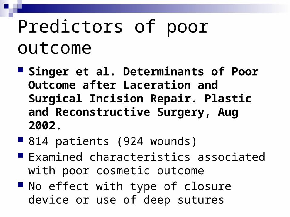
Predictors of poor outcome
Singer et al. Determinants of Poor Outcome after Laceration and Surgical Incision Repair. Plastic and Reconstructive Surgery, Aug 2002.
814 patients (924 wounds) Examined characteristics associated with poor
cosmetic outcome No effect with type of closure device or use of
Poor antibiotic penetration into abscess with fibrous wall Prophylactic antibiotics before I/D?
Consider endocarditis risk/immunocompromised Needle vs. formal I/D?
Needle I/D is generally diagnostic for pus, but inadequately therapeutic and definitely painful
Consider ultrasound to identify pus pocket
Abscesses Performance of I/D
Local infiltration of lidocaine notorious for only superficial effect
Consider field block if possible Incision with tension lines ? Culture in the new microbiological
climate BEWARE THE PULSATILE ‘ABSCESS’
Abscesses
I/D continued… Incision should be kept open with wick, but
not necessary to tightly “pack” abscess cavityRemove in 48 hours If continues with purulent drainage, may need
to re-explore, re-irrigate and re-pack
Source UndeterminedSource Undetermined
Antibiotics with MRSA Abscess
Lee et al, Pediatric Infectious Disease Journal. 23(2):123-127, February 2004 Followed 69 patients with MRSA abscesses
96% drained, 65% packed All got antibiotics, but only 7% were sensitive Only predictor of hospitalization (4 patients) was abscess >
5cm Receipt of effective antibiotic not predictive of treatment
failure Incision and drainage without adjunctive antibiotic therapy
was effective management of CA-MRSA skin and soft tissue abscesses with a diameter of <5 cm in immunocompetent children.
Antibiotics post I/D
Probably wise to use antibiotics if signs of systemic illness, significant overlying cellulitis, or high-risk area/host
Consider local resistance patterns for antibiotic choice…
Some evidence that use of Bactrim may effect subsequent lesions (Randomized Controlled Trial of
TMP-SMX for Uncomplicated Skin Abscesses in Patients at Risk for Community-Associated Methicillin-Resistant Staphylococcus aureus Infection, Schmitz, AEM 2010)
Our Staph resistance
Data applicable to U of M Hospital in-patients onlyClindamycin – 41%Doxy – 33%Methacillin – 50%Bactrim – 4%
Questions to consider
When to start antibiotics?
Is cephalexin an orphan?
What about inducible resistance?
Now for some specific situations…
Perianal Abscess
Infection arising in the crypto-glandular epithelium lining the anal canal Secondary to obstructed glands Bacteria can travel through crypts to inter-sphincteric
space Common in infants, then peaks in 3rd-4th decade
of life, male predominance E. Coli, Enterococcus, Bacteroides common
Treatment is incision and drainage Pack with Iodophor Culture material as multiple organisms
may be involved Often does not need antibiotics Large abscesses should be evaluated by
surgery
Pilonidal Cyst/Sinus
Historical perspectiveDescribed by Herbert Mayo in 1830Named by Hodges (pilus=hair, nidal=nest)Also known as Jeep riders disease, led to
80,000 soldiers hospitalized in WW2, and 4.2 million sick days
Initially thought to be infected congenital hair containing sinus tract
Pilonidal Cyst/Sinus
Pathophysiology Acquired condition – enlarged and deformed hair
follicles in natal cleft Bacteria enter, cause local inflammation sealing
mouth and creating abscess When abscess breaks into subcutaneous fatty tissue,
leads to pilonidal disease Staph Aureus most common Bacteroides most common anaerobe
Pilonidal Cyst/Sinus
Average age of presentation – 21 years Risk factors
Male sex Family predisposition Obesity Sedentary lifestyle Repeated trauma Occupation requiring prolonged sitting
Pilonidal Cyst/Sinus
History: Progressive tenderness after physical activity or a
period of prolonged sitting, such as during a long drive.
Acute purulent drainage, pain, and/or swelling may be present.
Systemic manifestations are rare, but patients may have malaise and fever.
Eighty percent of symptomatic presentations are exacerbations or manifestations of chronic disease.
Pilonidal Cyst/Sinus
Physical exam Presacral midline edema and/or nodule Fluctuance, warmth, tenderness Purulent discharge from one or more lesions Induration and/or cellulitis (usually minimal) Visible or palpable tracts of 2-5 cm in length in chronic
or recurrent disease Fever (infrequent) Nontenderness and/or nonfluctuance at rectal
examination
Pilonidal Cyst/Sinus
Treatment I/D with incision lateral to midlineEvacuate all materialBreak up loculationsCopious irrigationPackingSurgical follow up in 1 week40% recurrence rates
Emergency treatmentDigital block (+/-)Elevate lateral nail fold Irrigate with isotonic saline In severe or horseshoe paronychia, may
use a wick for 24 hoursSubungal abscess required removal of
nail plateAntibiotics if cellulitis present
Felon Infection in pulp of finger Can lead to compartment syndrome, tissue
necrosis, tenosynovitis Midline incision Blunt dissection to avoid trauma to nerve or
vessels Irrigation/packing
Plantar Puncture Wounds
Problems with plantar puncture woundsFrequent debris pushed into woundComplex bacteriologyForce inflicting punctureBones/joints close to skin
Plantar Puncture Wounds
Fitzgerald and Cowan – dated study of 887 plantar puncture wounds (mostly kids), 98% caused by nails3% had retained FB In early presenters 8.4% had/got cellulitisLate presenters 57% with cellulitis4% overall with serious infectionsStaph and Pseudomonas most common
Plantar Puncture Wounds
Management Blind probing dangerous Soaking probably not effective Irrigation may be futile Options
Conservative management Enlarging wound edges Coring out Trimming of epidermal edges Lack of data for best practice


![Action-Items XCI [Potpourri]](https://static.documents.pub/doc/80x56/577ccf061a28ab9e788eb048/action-items-xci-potpourri.jpg)


