REVIEW Propionibacterium (Cutibacterium) acnes Bacteriophage Therapy in Acne: Current Evidence and Future Perspectives David E. Castillo . Sonali Nanda . Jonette E. Keri Received: August 14, 2018 / Published online: December 11, 2018 Ó The Author(s) 2018 ABSTRACT Acne vulgaris is the most common dermato- logical disorder worldwide. It is a multifactorial disease that involves increased sebum produc- tion, hyperkeratinization of the pilosebaceous unit, Propionibacterium acnes (Cutibacterium acnes) colonization, and inflammation. The human skin microbiome hosts a wide variety of microorganisms, including bacteria, viruses, and fungi. A delicate balance of these microor- ganisms is essential for the barrier function of the skin. Propionibacterium acnes represents nearly 90% of the human skin microbiome of healthy adults. Acne is a chronic recurrent dis- ease that requires long-lasting treatment, which has led to the emergence of antibiotic resis- tance. New alternatives to traditional therapy are emerging, including antimicrobial peptides, natural engineered antibodies, and bacterio- phages. Bacteriophages have been shown to play a role in human skin health and disease. There is evidence supporting phage therapy in many types of skin infections. P. acnes bacte- riophages have been isolated and characterized. However, only a few in vitro studies have tested the ability of bacteriophages to kill P. acnes. Furthermore, there is no evidence on bacterio- phage therapy in the treatment of acne in humans. In this review, we summarize the most recent evidence regarding P. acnes bacterio- phages and the potential role of these bacte- riophages in the treatment of acne. Further research on this field will provide the evidence to use phage therapy to decrease rates of antibiotic resistance and restore antibiotic sus- ceptibility of P. acnes. Keywords: Acne; Antibiotic resistance; Bacteriophages; Microbiome; Phage therapy; Phages; Propionibacterium acnes INTRODUCTION Acne vulgaris is the most common dermato- logical disorder worldwide. It affects around 50 million people each year in the USA, with an estimated annual cost of $2.5 billion [1]. The worldwide prevalence of acne is estimated to be around 9% [2], accounting for 0.3% of the glo- bal disease burden [3]. Although acne affects people of all ages, 85% of all affected individuals Enhanced Digital Features To view enhanced digital features for this article go to https://doi.org/10.6084/ m9.figshare.7376021. D. E. Castillo Á S. Nanda Á J. E. Keri (&) Department of Dermatology and Cutaneous Surgery, University of Miami Miller School of Medicine, Miami, FL, USA e-mail: [email protected]J. E. Keri Veterans Affairs Miami Health Care System, Miami, FL, USA Dermatol Ther (Heidelb) (2019) 9:19–31 https://doi.org/10.1007/s13555-018-0275-9
Transcript
REVIEW
Propionibacterium (Cutibacterium) acnesBacteriophage Therapy in Acne: Current Evidenceand Future Perspectives
David E. Castillo . Sonali Nanda . Jonette E. Keri
Received: August 14, 2018 / Published online: December 11, 2018� The Author(s) 2018
ABSTRACT
Acne vulgaris is the most common dermato-logical disorder worldwide. It is a multifactorialdisease that involves increased sebum produc-tion, hyperkeratinization of the pilosebaceousunit, Propionibacterium acnes (Cutibacteriumacnes) colonization, and inflammation. Thehuman skin microbiome hosts a wide variety ofmicroorganisms, including bacteria, viruses,and fungi. A delicate balance of these microor-ganisms is essential for the barrier function ofthe skin. Propionibacterium acnes representsnearly 90% of the human skin microbiome ofhealthy adults. Acne is a chronic recurrent dis-ease that requires long-lasting treatment, whichhas led to the emergence of antibiotic resis-tance. New alternatives to traditional therapyare emerging, including antimicrobial peptides,natural engineered antibodies, and bacterio-phages. Bacteriophages have been shown to
play a role in human skin health and disease.There is evidence supporting phage therapy inmany types of skin infections. P. acnes bacte-riophages have been isolated and characterized.However, only a few in vitro studies have testedthe ability of bacteriophages to kill P. acnes.Furthermore, there is no evidence on bacterio-phage therapy in the treatment of acne inhumans. In this review, we summarize the mostrecent evidence regarding P. acnes bacterio-phages and the potential role of these bacte-riophages in the treatment of acne. Furtherresearch on this field will provide the evidenceto use phage therapy to decrease rates ofantibiotic resistance and restore antibiotic sus-ceptibility of P. acnes.
Acne vulgaris is the most common dermato-logical disorder worldwide. It affects around 50million people each year in the USA, with anestimated annual cost of $2.5 billion [1]. Theworldwide prevalence of acne is estimated to bearound 9% [2], accounting for 0.3% of the glo-bal disease burden [3]. Although acne affectspeople of all ages, 85% of all affected individuals
Enhanced Digital Features To view enhanced digitalfeatures for this article go to https://doi.org/10.6084/m9.figshare.7376021.
D. E. Castillo � S. Nanda � J. E. Keri (&)Department of Dermatology and CutaneousSurgery, University of Miami Miller School ofMedicine, Miami, FL, USAe-mail: [email protected]
J. E. KeriVeterans Affairs Miami Health Care System, Miami,FL, USA
are 12–24 years old [4–7]. Severe acne also car-ries a high social and psychological impact,affecting emotions, self-esteem, and increasingthe risk of depression and suicide [7].
The role of Propionibacterium acnes in thepathophysiology of acne is still under debate. P.acnes is the predominant commensal microor-ganism of the human skin microbiome. A deli-cate balance within the skin microbiota isessential for the barrier function of the skin andprevention of pathogen colonization [8].
The emergence of antibiotic resistance hasbecome a public health problem worldwide [9].The long-term use of topical and systemicantibiotics has led to high rates of antibiotic-resistant P. acnes strains [10]. As research onnew antibiotic agents is decreasing due to costand difficulty, the development of new, natural,and non-conventional alternatives—such asantimicrobial peptides, natural engineeredantibodies, and bacteriophages—is becomingcritical. Bacteriophage therapy seems to be apromising alternative. Its advantages are hostspecificity and simplicity of isolation and pro-duction. Although both in vitro and in vivostudies have shown the potential of targetedbacteriophage therapy in skin infections,research is lacking on bacteriophage therapytargeting P. acnes-associated infections. It hasrecently been proposed that the species P. acnesbe reclassified to Cutibacterium acnes and othergenera [11]. Here, we use the old nomenclature(P. acnes) throughout because it is still used bymost of the evidence presented in this review.In this review, we summarize the most recentevidence on P. acnes bacteriophages and itspotential role in the treatment of acne. Thisarticle is based on previously conducted studiesand does not contain any studies with humanparticipants or animals performed by any of theauthors.
ACNE VULGARISPATHOPHYSIOLOGY
Acne is a multifactorial disease. Increasedsebum production by androgen stimulation,abnormal hyperkeratinization of the piloseba-ceous duct, and subsequent bacterial
colonization and inflammation all contribute tothe disease [12, 13]. It is proposed that P. acnescolonization plays a pivotal role in the patho-genesis of acne since antimicrobial therapy hasbeen effective in treating acne for many years.However, its contribution to acne developmentis controversial [14].
Propionibacterium acnes is a Gram-positive,anaerobic/microaerophilic, fat-splitting, rod-shaped bacterium found on the skin; it repre-sents nearly 90% of the skin microbiome ofhealthy adults [1, 14, 15]. The concentration ofP. acnes depends on the abundance of sebaceousfollicles and the age of the individual[13, 16, 17]. Accordingly, its concentration ishigher on sebaceous areas such as the face,scalp, and back [13, 18], and various studieshave reported an association between P. acneslevels and sebum production [16]. There is amarked increase in P. acnes colonization duringpuberty [15], which correlates with the timewhen sebaceous glands mature [13]. P. acnesmay disrupt keratinocyte differentiation in thefollicle, thereby contributing to the formationof comedones and inflammatory acne lesionsby triggering a host inflammatory response[12, 19]. P. acnes produces enzymes that degradeskin components as well as chemotactic factorsthat stimulate keratinocytes and inflammatorycells to release pro-inflammatory cytokines(e.g., interleukin [IL]-8, IL-12, IL-1a, IL1-b,tumor necrosis factor alpha) and reactive oxy-gen species [1, 13, 20–23]. Although P. acnes is acommensal organism in humans, not all heal-thy adolescents or adults develop acne, indi-cating that differences in the pathogenicity of P.acnes strains must exist [1]. It has been proposedthat certain strains play a pathogenic role andothers act as bystanders [24–27]. Studies havealso shown that specific genes in the P. acnesgenome contribute to bacterium virulence andhence to acne pathophysiology [14, 26, 28].
EMERGING ANTIBIOTICRESISTANCE
Over 2 million Americans become infectedevery year with antibiotic-resistant bacteria,resulting in about 23,000 deaths [29, 30]. The
20 Dermatol Ther (Heidelb) (2019) 9:19–31
post-antibiotic era is approaching as antibioticeffectiveness steadily declines, and multiplecommon infections become resistant to treat-ment [14, 31]. The excessive use of antibiotics inagriculture and humans, the evolutionary pres-sure inherent to antibiotics [31], and the lack ofresearch on new antibiotic agents are some ofthe reasons behind antibiotic resistance[14, 29, 31, 32].
Antibiotic resistance is among the maincauses of treatment failure in acne vulgaris [33].The mechanism of antibiotic resistance to P.acnes is explained by the remarkable geneticplasticity of bacteria [34]. Two major geneticstrategies permit antimicrobial resistance: (1)gene mutation, and (2) foreign DNA codingacquisition through horizontal gene transfer[34]. Gene mutation is the predominant mech-anism leading to P. acnes antibiotic resistance[12, 35].
Acne therapy warrants long-term treatmentwith topical and systemic antibiotics, whichcontributes to resistant P. acnes strains. Glob-ally, P. acnes resistance to antimicrobials hasincreased almost 40% between the 1980s and2000s worldwide [10]. Erythromycin/clin-damycin-resistant P. acnes seems to be the mostcommon pattern of resistance based on reportsfrom the USA, Europe, and Asia [36–38]. How-ever, resistance rates vary by region. Europe,Singapore, and Hong-Kong have high preva-lence rates of erythromycin/clindamycin-resis-tant P. acnes (45–91%) and tetracycline-resistantP. acnes (2–26%) [36, 37, 39–42], but countriesthat practice conservative use of antibiotics,such as Japan and Korea, report much lowerresistance rates (2–4%) [36, 42, 43].
The long-term use of antibiotics can promotethe formation of an antibiotic-resistant biofilmthat protects the bacterium against host defen-ses and can alter the natural microbiota of theskin [12, 44]. Studies have shown colonizationby antibiotic-resistant coagulase-negative Sta-phylococci and Streptococcus pyogenes in acnepatients who have used both topical and oralantibiotic therapy [45, 46]. Thereby, it is rec-ommended to limit monotherapy with topicalantibiotics and instead to combine them withother topical agents such as retinoids or benzoylperoxide to decrease the risk of resistance [47].
HUMAN SKIN MICROBIOME
The human skin microbiome is home to avariety of microorganisms, including bacteria,fungi, viruses, and arthropods. A delicate bal-ance between the microorganisms is essentialfor local immunity and barrier function of theskin [8]. Imbalances in this system have beenlinked to dermatologic diseases, such as acne,atopic dermatitis, psoriasis, and rosacea [48].
The dominant bacterial species found onadult skin are Propionibacterium, Corynebac-terium, and Staphylococcus [48]. The skin can bedivided into dry, moist, or sebaceous microen-vironments, and each of these microclimateshost varying proportions of these commonbacterial flora [48].
Age-related shifts in bacterial communitiescould explain why certain skin diseases areprevalent at different stages of life. For example,the microbiome in children, who are moresusceptible to atopic dermatitis, is composed ofmostly Streptococcaceae, Bacteroides, and Pro-teobacteria [49]. Pubertal spikes in androgenslead to a more susceptible environment for thedevelopment of acne and a shift in microbiomeconcentration to greater levels of Propionibac-terium and Corynebacterium.
BACTERIOPHAGES
Delving deeper into the bacteria of the micro-biome brings us to bacteriophages. Bacterio-phages, or viruses that infect bacteria, can befound throughout the biosphere, are essentialmembers of the human microbiome, and mayplay an important regulatory role in humanskin health and disease [8, 50–52]. Little isknown about bacteriophage interaction withskin microbiota. Bacteriophages are obligatoryintracellular parasites; thereby, their distribu-tion depends on their host organisms [50].There are over 6000 well-known bacteriophages[53], and these bacteriophages are estimated tobe at least tenfold more common than bacteria[14]. There is wide diversity in the structure ofthese phages (e.g., tailed, polyhedral, pleomor-phic, filamentous), and they are usually classi-fied based on their genetic content [53].
Dermatol Ther (Heidelb) (2019) 9:19–31 21
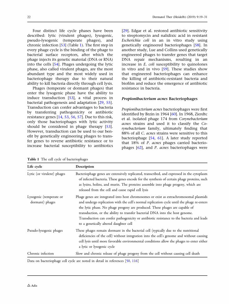
Four distinct life cycle phases have beendescribed: lytic (virulent phages), lysogenic,pseudo-lysogenic (temperate phages), andchronic infection [53] (Table 1). The first step inevery phage cycle is the binding of the phage tobacterial surface receptors, after which thephage injects its genetic material (DNA or RNA)into the cells [54]. Phages undergoing the lyticphase, also called virulent phages, are the mostabundant type and the most widely used inbacteriophage therapy due to their naturalability to kill bacteria directly through cell lysis.
Phages (temperate or dormant phages) thatenter the lysogenic phase have the ability toinduce transduction [53], a vital process inbacterial pathogenesis and adaptation [29, 55].Transduction can confer advantages to bacteriaby transferring pathogenicity or antibioticresistance genes [14, 53, 56, 57]. Due to this risk,only those bacteriophages with lytic activityshould be considered in phage therapy [53].However, transduction can be used to our ben-efit by genetically engineering phages to trans-fer genes to reverse antibiotic resistance or toincrease bacterial susceptibility to antibiotics
[29]. Edgar et al. restored antibiotic sensitivityto streptomycin and nalidixic acid in resistantEscherichia coli in an in vitro study usinggenetically engineered bacteriophages [58]. Inanother study, Lue and Collins used geneticallyengineered phages to transfer genes that targetDNA repair mechanisms, resulting in anincrease in E. coli susceptibility to quinolonesin vitro and in vivo [59]. These studies showthat engineered bacteriophages can enhancethe killing of antibiotic-resistant bacteria andbiofilm and reduce the emergence of antibioticresistance in bacteria.
Propionibacterium acnes Bacteriophages
Propionibacterium acnes bacteriophages were firstidentified by Brzin in 1964 [60]. In 1968, Zierdtcet al. isolated phage 174 from Corynebacteriumacnes strains and used it to classify the Co-rynebacterium family, ultimately finding that88% of all C. acnes strains were sensitive to thisbacteriophage [54, 61]. A later study reportedthat 18% of P. acnes phages carried bacterio-phages [62], and P. acnes bacteriophages were
Table 1 The cell cycle of bacteriophages
Life cycle Description
Lytic (or virulent) phages Bacteriophage genes are extensively replicated, transcribed, and expressed in the cytoplasm
of infected bacteria. These genes encode for the synthesis of certain phage proteins, such
as lysins, holins, and murin. The proteins assemble into phage progeny, which are
released from the cell and cause rapid cell lysis
Lysogenic (temperate or
dormant) phages
Phage genes are integrated into host chromosomes or exist as extrachromosomal plasmids
and undergo replication with the cell’s normal replication cycle until the phage re-enters
the lytic phase. No phage progeny are produced. These phages are capable of
transduction, or the ability to transfer bacterial DNA into the host genome.
Transduction can confer pathogenicity or antibiotic resistance to the bacteria and leads
to a genetically altered daughter cell
Pseudo-lysogenic phages These phages remain dormant in the bacterial cell (typically due to the nutritional
deficiencies of the cell) without integration into the cell’s genome and without causing
cell lysis until more favorable environmental conditions allow the phages to enter either
a lytic or lysogenic cycle
Chronic infection Slow and chronic release of phage progeny from the cell without causing cell death
Data on bacteriophage cell cycle are noted in detail in references [50, 116]
22 Dermatol Ther (Heidelb) (2019) 9:19–31
subsequently used to classify Corynebacteriumand Propionibacterium [63, 64].
P. acnes bacteriophages are relatively moreabundant in lipid-rich areas of the skin, corre-lating with the distribution of P. acnes in theskin [65–67]. These bacteriophages are thedominant phages in the pilosebaceous unit.Fitz-Gibbon et al. reported a 1:120 bacterio-phages:P. acnes ratio in pilosebaceous units inhealthy skin samples [26, 68, 69]. Most P. acnesbacteriophages possess a siphoviral morphology(i.e., isometric head and long flexible tail)[8, 15, 70, 71] and have a pseudolysogenic lifecycle (Table 1) [8, 15, 72, 73]. P. bacteriophagesdisplaying a lytic life cycle have also beencharacterized [70, 74].
Interestingly, despite the isolation of P. acnesbacteriophages over a varied temporal andgeographical range, their genome is preservedwith very limited genetic diversity[8, 15, 71, 72]. Marinelli et al. investigated thediversity of bacteriophages that infect P. acnesand isolated 11 P. acnes bacteriophages whichlacked the genetic diversity seen in other phagepopulations [15]. A recent study by Liu et al.sequenced 48 P. acnes bacteriophages fromhuman skin follicles and found a sequenceidentity of between 85 and 100% betweenstrains, suggesting that the P. acnes bacterio-phage population in the skin microbiota isdominated by one strain [8]. The authors testedthe P. acnes–bacteriophage interaction andfound that the 74 P. acnes strains were suscep-tible to the 15 tested P. acnes bacteriophages.They suggested multiple reasons for the lack ofphage diversity, including a bottle-neckhypothesis leaving one dominant genotype, orthe evolutionary constrains imposed on phagesand bacteria to maintain a single phage, thuslimiting the spreading of phage resistance [8].Another possible explanation for the limiteddiversity of P. acnes bacteriophages could be dueto the niche in which they live, as P. acnesmakes up 90% of the microbiota of the pilose-baceous unit, thereby limiting horizontal genetransfer and increased diversity between phagesin the pilosebaceous unit [14].
Liu et al. also found that some individualsshared the same bacteriophage strains in theskin microbiota, suggesting the existence of a
pool of common bacteriophages among humanpopulations [8]. They further discovered iden-tical bacteriophages strains between closelyrelated individuals (siblings), which makeshuman to human virus transmission a possi-bility. As seen in other studies [15], the resis-tance of certain P. acnes strains tobacteriophages was an issue [8]. Two possibleresistance mechanisms are described by theseauthors, namely, restriction modification andclustered regularly interspaced short palin-dromic repeat (CRISPR), both of which targetviral DNA integration into the host genome [8].The findings of this study led the authors toconclude that the ability of P. acnes bacterio-phages to lyse only susceptible strains may alterthe bacteria population, as different strains willgrow at different rates, thereby modulating thecomposition and dynamics of the skinmicrobiota.
The apparent lack of genetic diversity of P.acnes bacteriophages and their broad host rangemake them ideal candidates for phage therapyin acne [14]. Moreover, lytic bacteriophagesengineered to target P. acnes strains in thespecific microbiome of individuals will increasethe success rate of acne treatments.
Propionibacterium acnes BacteriophageTherapy
The potential role of bacteriophage therapy inacne vulgaris has recently attracted the interestof researchers and clinicians. Phages activeagainst P. acnes have been isolated from theskin, oral cavity, and gastrointestinal tract [14].It is important to note that only phages withproven lytic activity should be used in phagetherapy because lysogenic or temperate phagescarry the risk for transduction of antibioticresistance or pathogenicity genes and may leadto delayed cell lysis [14, 53].
Bacteriophage therapy has been used inhumans for several types of infections withgood results [75–78]. However, no trials on P.acnes bacteriophage therapy have been con-ducted in humans. Brown et al. isolated tenbacteriophages capable of lysing P. acnes fromhuman skin microbiota and tested their
Dermatol Ther (Heidelb) (2019) 9:19–31 23
therapeutic potential [72]. These authors cre-ated a suspension for each bacteriophage at afinal concentration of 2.5 9 108 PFU/g using anaqueous cetomacrogol cream that showed thatthese bacteriophage formulations effectivelylysed P. acnes cells in agar lawn culture platesand remained active in the cream for up to90 days when stored at 4 �C in light-protectedbottles. The bacteriophage was specific to P.acnes strains and did not lyse other bacteria ofthe Propionibacterium family. Cells that regrewfrom the areas within the P. acnes plaquesshowed phage resistance [72]. Although someauthors have suggested that a cocktail of phagescould be used to decrease the risk of phageresistance [8, 15], P. acnes bacteriophage vari-ability is relatively low, which may limit thisapproach. This important limitation needs to beexplored in further studies. However, the resultsusing the cream formulation of Brown et al. [72]suggest that P. acnes bacteriophage therapy is asimple and realistic therapeutic option for thetreatment of acne. Another in vitro studyshowed effective eradication of P. acnes strainswhen P. acnes bacteriophages were isolated fromhuman skin microbiota and applied in dropsonto agar plates [79]. In this study, P. acnesbacteriophages were unable to kill other bacte-ria, such as Staphylococcus aureus, S. epidermidis,and Corynebacterium xerosis, confirming thespecificity of these bacteriophages [79]. Formu-lations such as oil–base cream, water–oilnanoemulsion, biodegradable polyester matrix,antiseptic gel, and paraffin-oil-based lotion,have proven to be effective strategies to deliverbacteriophages [80].
P. acnes bacteriophage genomes encodeendolysins involved in bacteria cell-wall degra-dation (muramidases, amidases, endopepti-dases, glucosaminidases, and transglycosylases)[15, 70, 81]. These endolysins are implicated inthe release of progeny following phage assem-bly by targeting peptidoglycan in the bacterialwall [70]. Marinelli et al. suggested that endo-lysins are a potential therapeutic option in acnetherapy [15]. Phage endolysins are highly con-served in different P. acnes bacteriophage strains(95% at the amino acid level) [15], whichimplies that endolysins from any P. acnes bac-teriophage could be active against most P. acnes
strains. Phage endolysins have been used asantimicrobials both in vitro and in vivo withpromising results [82]. Furthermore, no resis-tance to phage endolysins has been reported[82]. It has been shown that even bacteria thatbecome phage resistant may remain endolysinsensitive [14]. This introduces yet another wayto treat acne through the genetic engineering ofenzymes to target bacteria cell walls.
These possibilities carry important thera-peutic implications in the management of acne.Antibiotics, in combination with bacteriophagecocktails, could be used to decrease antibioticresistance and to treat antibiotic-resistant P.acnes. However, further research is needed toevaluate P. acnes bacteriophage therapy inhuman subjects, both as monotherapy and incombination with conventional therapies.
Advantages of Phage Therapy
Bacteriophages have a low environmentalimpact compared to chemical antibiotics due totheir natural origin [53, 83]. They target bothGram-positive and Gram-negative bacteria[84–92], and many in vitro and in vivo modelshave shown that bacteriophages are effectiveagainst multidrug-resistant bacteria [85–88]. Asantibiotic resistance grows, phages retain theability to kill antibiotic-resistant bacteria due totheir differing mechanisms of action [53]. Bac-teriophages are specific to their bacterial hosts(species), and only replicate locally, limiting thepressure on normal non-targeted flora of theskin and other organs [75, 93, 94]. Bacterio-phages have also been shown to distribute ingood concentrations all over the body, includ-ing the central nervous system [53, 91, 95](Table 2).
Another potential benefit of bacteriophagetherapy is the ability to decrease biofilm for-mation [83, 96–101]. Many in vitro and in vivostudies have proven that the combinationtherapy of antibiotics and lytic bacteriophagesdisplays synergism by improving the efficacy ofbacteria and biofilm eradication and preventingthe emergence of resistant bacteria[92, 102–111]. Antibiotics could be conjugatedwith bacteriophages to deliver antibiotics to
24 Dermatol Ther (Heidelb) (2019) 9:19–31
specific bacteria and at higher concentrations[14]. Furthermore, engineered bacteriophagescould be used to improve efficacy through thetransfer of susceptibility or sensitizing genes bymeans of genetic engineering.
The identification of bacteria and bacterio-phage isolation for therapeutic purposes is arapid and affordable process compared to thedevelopment of new antibiotics [93]. Moreover,the cost of bacteriophage therapy seems to belower than that of traditional antibiotic therapy[112]; however, more studies are needed toestablish the real short- or long-term costs ofbacteriophage therapy.
Finally, bacteriophages are safe and well-tol-erated, and no significant adverse events havebeen reported [75, 77, 78, 94, 113, 114].
Limitations of Bacteriophage Therapy
Although the prospect of using bacteriophagetherapy to treat acne in a world with increasingantibiotic resistance is promising, this noveltherapeutic endeavor comes with limitations.The first of these is our evolving understandingof phages and their life cycles. The newest datasuggests that phages exist on a continuumbetween lytic and lysogenic life cycles [115].
This creates challenges when using phages astherapeutic vehicles since conventional phagetherapy requires phages to undergo lytic cyclesand rapidly kill their hosts. Most P. acnes phagescharacterized thus far, however, display pseu-dolysogeny (Table 1).
In addition, CRISPR protects bacteria fromviral DNA integration, and P. acnes may becomeresistant to phage therapy through this mech-anism [54]. This issue of resistance becomeseven more likely with the knowledge that bac-teriophages targeting P. acnes are highlyhomogeneous [26]. Therefore, the acquisitionof resistance to one phage may confer P. acneswith resistance to many of its bacteriophages[14]. However, the risk is low compared toantibiotics, partially because bacteriophages canmutate and bypass bacteriophage resistancemechanisms [93]. Resistance can also be pre-vented by using multiple bacteriophages (cock-tails) or synergistic combinations ofbacteriophages ? antibiotics [93].
In addition to these limitations, the morepractical aspects of establishing optimal thera-peutic doses, treatment frequency, and durationhave not been established [54]. A more long-term risk with this therapy includes theunknown consequences to the cutaneous
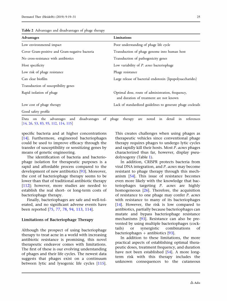
Table 2 Advantages and disadvantages of phage therapy
Advantages Limitations
Low environmental impact Poor understanding of phage life cycle
Cover Gram-positive and Gram-negative bacteria Transduction of phage genome into human host
No cross-resistance with antibiotics Transduction of pathogenicity genes
Host specificity Low variability of P. acnes bacteriophage
Low risk of phage resistance Phage resistance
Can clear biofilm Large release of bacterial endotoxin (lipopolysaccharides)
Transduction of susceptibility genes
Rapid isolation of phage Optimal dose, route of administration, frequency,
and duration of treatment are not known
Low cost of phage therapy Lack of standardized guidelines to generate phage cocktails
Good safety profile
Data on the advantages and disadvantages of phage therapy are noted in detail in references[14, 26, 53, 83, 93, 112, 114, 115]
Dermatol Ther (Heidelb) (2019) 9:19–31 25
microbiome if P. acnes, a vital member to thiscommunity, is temporarily eradicated throughacne treatment. We do not fully understand therepercussions of altering the natural micro-ecosystem of the skin.
Future therapeutic option
As the antibiotic resistance era approaches,research on new alternative antimicrobialagents is becoming critical. Bacteriophages,ubiquitous microorganisms of the human skinmicrobiome, contain distinct advantages thatmark them as promising alternatives to con-ventional antimicrobial therapy. Among theseadvantages are host specificity, limited crossresistance, ease of isolation, low cost, and afavorable safety profile compared to antibiotics.
The potential clinical application of bacte-riophage therapy for acne vulgaris is promising.Limitations to phage therapy, such as the risk oftransduction of pathogenicity genes and thelow P. acnes bacteriophage variability, can beovercome by a more thorough understanding ofthe bacteria–bacteriophage interaction in thehuman skin microbiome. Phage resistance isanother limitation that must be considered andwarrants further study.
As the field develops, more data is neededbefore phage therapy in human subjects isintroduced. Developing targeted phage therapy,engineered bacteriophages, and enzyme-basedtherapies, either alone or as an adjuvant toantibiotics, may lead to decreasing rates of P.acnes resistance to antibiotics and the restora-tion of antibiotic susceptibility to P. acnes.
ACKNOWLEDGEMENTS
Funding. No funding sources were used forthe development of this review.
Authorship. All named authors meet theInternational Committee of Medical JournalEditors (ICMJE) criteria for authorship for thisarticle, take responsibility for the integrity of
the work as a whole, and have given theirapproval for this version to be published.
Disclosures. David E. Castillo, Sonali Nanda,and Jonette E. Keri declare have nothing todisclose.
Compliance with Ethics Guidelines. Thisarticle is based on previously conducted studiesand does not contain any studies with humanparticipants or animals performed by any of theauthors.
Open Access. This article is distributedunder the terms of the Creative CommonsAttribution-NonCommercial 4.0 InternationalLicense (http://creativecommons.org/licenses/by-nc/4.0/), which permits any non-commercial use, distribution, and reproductionin any medium, provided you give appropriatecredit to the original author(s) and the source,provide a link to the Creative Commons license,and indicate if changes were made.
REFERENCES
1. Zaenglein A, Thiboutot D. Acne vulgaris. In:Bolognia J, Schaffer J, Cerroni L, eds. Dermatology.4th edn. Amsterdam: Elsevier; 2012:588–603.
2. Hay R, Johns N, Williams H, et al. The global burdenof skin disease in 2010: an analysis of the prevalenceand impact of skin conditions. J Investig Dermatol.2013;134:1527–34.
3. Karimkhani C, Dellavalle RP, Coffeng LE, et al.Global skin disease morbidity and mortality: anupdate from the global burden of disease study2013. JAMA Dermatol. 2017;153(5):406–12.
4. Lynn DD, Umari T, Dunnick CA, Dellavalle RP. Theepidemiology of acne vulgaris in late adolescence.Adolesc Health Med Ther. 2016;7:13–25.
5. Sidbury R, Paller AS. The diagnosis and manage-ment of acne. Pediatr Ann. 2000;29(1):17–24.
6. White GM. Recent findings in the epidemiologicevidence, classification, and subtypes of acne vul-garis. J Am Acad Dermatol. 1998;39:S34–7.
7. Halvorsen JA, Stern RS, Dalgard F, Thoresen M,Bjertness E, Lien L. Suicidal ideation, mental health
problems, and social impairment are increased inadolescents with acne: a population-based study.J Investig Dermatol. 2011;131(2):363–70.
8. Liu J, Yan R, Zhong Q, et al. The diversity and hostinteractions of Propionibacterium acnes bacterio-phages on human skin. ISME J. 2015;9(9):2078–93.
9. World Health Organization (WHO). Antimicrobialresistance: global report on surveillance. 2014.Available online at: http://apps.who.int/iris/bitstream/10665/112642/1/9789241564748_eng.pdf. Accessed 30 June 2018.
10. Coates P, Vyakrnam S, Eady EA, Jones CE, Cove JH,Cunliffe WJ. Prevalence of antibiotic-resistant pro-pionibacteria on the skin of acne patients: 10-yearsurveillance data and snapshot distribution study.Br J Dermatol. 2002;146(5):840–8.
11. Scholz CF, Kilian M. The natural history of cuta-neous propionibacteria, and reclassification ofselected species within the genus Propionibacteriumto the proposed novel genera Acidipropionibacteriumgen. nov., Cutibacterium gen. nov. and Pseudopropi-onibacterium gen. nov. Int J Syst Evol Microbiol.2016;66(11):4422–32.
12. Dessinioti C, Katsambas A. Propionibacterium acnesand antimicrobial resistance in acne. Clin Derma-tol. 2017;35(2):163–7.
13. Liu PF, Hsieh YD, Lin YC, Two A, Shu CW, HuangCM. Propionibacterium acnes in the pathogenesis andimmunotherapy of acne vulgaris. Curr Drug Metab.2015;16(4):245–54.
14. Jonczyk-Matysiak E, Weber-Dabrowska B, Zaczek M,et al. Prospects of phage application in the treat-ment of acne caused by Propionibacterium acnes.Front Microbiol. 2017;8:164.
15. Marinelli LJ, Fitz-Gibbon S, Hayes C, et al. Propi-onibacterium acnes bacteriophages display limitedgenetic diversity and broad killing activity againstbacterial skin isolates. MBio. 2012;3(5):e00279.https://doi.org/10.1128/mBio.00279-12
16. McGinley KJ, Webster GF, Ruggieri MR, Leyden JJ.Regional variations in density of cutaneous propi-onibacteria: correlation of Propionibacterium acnespopulations with sebaceous secretion. J ClinMicrobiol. 1980;12(5):672–5.
17. Brzuszkiewicz E, Weiner J, Wollherr A, et al. Com-parative genomics and transcriptomics of Propioni-bacterium acnes. PLoS One. 2011;6(6):e21581.
19. Tyner H, Patel R. Propionibacterium acnes biofilm—asanctuary for Staphylococcus aureus? Anaerobe.2016;40:63–7.
20. Kim J, Ochoa MT, Krutzik SR, et al. Activation oftoll-like receptor 2 in acne triggers inflammatorycytokine responses. J Immunol.2002;169(3):1535–41.
21. Jeremy AH, Holland DB, Roberts SG, Thomson KF,Cunliffe WJ. Inflammatory events are involved inacne lesion initiation. J Investig Dermatol.2003;121(1):20–7.
22. Vowels BR, Yang S, Leyden JJ. Induction of proin-flammatory cytokines by a soluble factor of Propi-onibacterium acnes: implications for chronicinflammatory acne. Infect Immun.1995;63(8):3158–65.
23. Nagy I, Pivarcsi A, Kis K, et al. Propionibacteriumacnes and lipopolysaccharide induce the expressionof antimicrobial peptides and proinflammatorycytokines/chemokines in human sebocytes.Microbes Infect. 2006;8(8):2195–205.
24. Kwon H, Suh D. Recent progress in the researchabout Propionibacterium acnes strain diversity andacne: pathogen or bystander? Int J Dermatol.2016;55(11):1196–204.
25. Jahns A, Lundskog B, Dahlberg I, Tamayo N,McDowell A, Patrick S, et al. No link between rosa-cea and Propionibacterium acnes. APMIS.2012;120(11):922–5.
26. Fitz-Gibbon S, Tomida S, Chiu BH, et al. Propioni-bacterium acnes strain populations in the humanskin microbiome associated with acne. J InvestigDermatol. 2013;133(9):2152–60.
27. Lomholt HB, Kilian M. Population genetic analysisof Propionibacterium acnes identifies a subpopulationand epidemic clones associated with acne. PLoSOne. 2010;5(8):e12277.
28. Lodes MJ, Secrist H, Benson DR, et al. Variableexpression of immunoreactive surface proteins ofPropionibacterium acnes. Microbiology. 2006;152(Pt12):3667–81.
29. Jassim A, Limoges R. Natural solution to antibioticresistance: bacteriophages ‘‘The Living Drugs’’.World J Microbiol Biotechnol. 2014;30:2153–70.
30. Centers for Disease Control and Prevention (CDC).Antibiotic use in the United States, 2017: progressand opportunities. Atlanta: US Department ofHealth and Human Services; 2017.
31. Zucca M, Savoia D. The post-antibiotic era:promising developments in the therapy of infec-tious diseases. Int J Biomed Sci. 2010;6(2):77–86.
32. Clarke T. Drug companies snub antibiotics as pipe-line threatens to run dry. Nature. 2003;425:225.
33. Sadhasivam S, Sinha M, Saini S, et al. Heterogeneityand antibiotic resistance in Propionibacterium acnesisolates and its therapeutic implications: blurringthe lines between commensal and pathogenicphylotypes. Dermatol Ther. 2016;29(6):451–4.
34. Munita JM, Arias CA. Mechanisms of antibioticresistance. Microbiol Spectr. 2016;4(2). https://doi.org/10.1128/microbiolspec.VMBF-0016-2015
35. Mendoza N, Hernandez PO, Tyring SK, Haitz KA,Motta A. Antimicrobial susceptibility of Propioni-bacterium acnes isolates from acne patients inColombia. Int J Dermatol. 2013;52(6):688–92.
36. Luk N, Hui M, Lee H, et al. Antibiotic-resistantPropionibacterium acnes among acne patients in aregional skin centre in Hong Kong. J Eur AcadDermatol Venereol. 2013;27(1):31–6.
37. Dumont-Wallon G, Moyse D, Blouin E, Dreno B.Bacterial resistance in French acne patients. Int JDermatol. 2010;49(3):283–8.
38. Eady EA, Gloor M, Leyden JJ. Propionibacterium acnesresistance: a worldwide problem. Dermatology.2003;206(1):54–6.
39. Dreno B, Reynaud A, Moyse D, Habert H, Richet H.Erythromycin-resistance of cutaneous bacterial florain acne. Eur J Dermatol. 2001;11(6):549–53.
40. Ross JI, Snelling AM, Carnegie E, et al. Antibiotic-resistant acne: lessons from Europe. Br J Dermatol.2003;148(3):467–78.
41. Tan HH, Tan AW, Barkham T, Yan XY, Zhu M.Community-based study of acne vulgaris in ado-lescents in Singapore. Br J Dermatol.2007;157(3):547–51.
42. Kurokawa I, Nishijima S, Kawabata S. Antimicrobialsusceptibility of Propionibacterium acnes isolatedfrom acne vulgaris. Eur J Dermatol. 1999;9(1):25–8.
43. Nishijima S, Kurokawa I, Katoh N, Watanabe K. Thebacteriology of acne vulgaris and antimicrobialsusceptibility of Propionibacterium acnes and Staphy-lococcus epidermidis isolated from acne lesions.J Dermatol. 2000;27(5):318–23.
44. Burkhart C, Burkhart C. Microbiology’s principle ofbiofilms as a major factor in the pathogenesis ofacne vulgaris. Int J Dermatol. 2003;42:925–7.
45. Levy R, Huang E, Roling D, Leyden J, Margolis D.Effect of antibiotics on the oropharyngeal flora inpatients with acne. Arch Dermatol.2003;139:467–71.
46. Mills O Jr, Thornsberry C, Cardin CW, Smiles KA,Leyden JJ. Bacterial resistance and therapeutic out-come following three months of topical acne ther-apy with 2% erythromycin gel versus its vehicle.Acta Derm Venereol. 2002;82(4):260–5.
47. Thiboutot D, Gollnick H, Bettoli V, et al. Newinsights into the management of acne: an updatefrom the Global Alliance to Improve Outcomes inAcne group. J Am Acad Dermatol. 2009;60:1–50.
48. Grice EA. The skin microbiome: potential for noveldiagnostic and therapeutic approaches to cutaneousdisease. Semin Cutan Med Surg. 2014;33(2):98–103.
49. Oh J, Conlan S, Polley EC, Segre JA, Kong HH. Shiftsin human skin and nares microbiota of healthychildren and adults. Genome Med. 2012;4(10):77.
51. Lin L, Hong W, Ji X, Han J, Huang L, Wei Y. Isola-tion and characterization of an extremely long tailThermus bacteriophage from Tengchong hotsprings in China. J Basic Microbiol.2010;50(5):452–6.
52. Prigent M, Leroy M, Confalonieri F, Dutertre M,DuBow M. A diversity of bacteriophage forms andgenomes can be isolated from the surface sands ofthe Sahara Desert. Extremophiles.2005;9(4):289–96.
53. Wittebole X, De Roock S, Opal S. A historical over-view of bacteriophage therapy as an alternative toantibiotics for the treatment of bacterial pathogens.Virulence. 2014;5(1):226–35.
54. Bruggemann H, Lood R. Bacteriophages infectingPropionibacterium acnes. Biomed Res Int.2013;2013:705741.
55. Abedon S. Phages. In: Hyman P, Abedon S, eds.Bacteriophages in health and disease. AMCM 24advances in molecular and cellular microbiology.Wallingford:CABI; 2012. Retrieved from https://ebookcentral.proquest.com. Accessed 7 July 2018.
56. O’Shea YA, Boyd EF. Mobilization of the Vibriopathogenicity island between Vibrio cholerae isolatesmediated by CP-T1 generalized transduction. FEMSMicrobiol Lett. 2002;214(2):153–7.
57. Maiques E, Ubeda C, Tormo MA, et al. Role ofstaphylococcal phage and SaPI integrase in intra-
and interspecies SaPI transfer. J Bacteriol.2007;189(15):5608–16.
58. Edgar R, Friedman N, Molshanski-Mor S, Qimron U.Reversing bacterial resistance to antibiotics byphage-mediated delivery of dominant sensitivegenes. Appl Environ Microbiol. 2012;78(3):744–51.
59. Lu T, Collins J. Engineered bacteriophage targetinggene networks as adjuvants for antibiotic therapy.Proc Natl Acad Sci USA. 2009;106(12):4629–34.
60. Brzin B. Studies on the Corynebacterium acnes. ActaPathol Microbiol Scand. 1964;60:599–608.
61. Zierdt C, Webster C, RudeW. Study of the anaerobiccorynebacteria. Int J Syst Evol Microbiol.1968;18(1):33–47.
62. Webster G, Cummins C. Use of bacteriophage typ-ing to distinguish Propionibacterium acne types I andII. J Clin Microbiol. 1978;7(1):84–90.
63. Voss JG. Differentiation of two groups of Co-rynebacterium acnes. J Bacteriol. 1970;101(2):392–7.
64. Whiteside JA, Voss JG. Incidence and lipolyticactivity of Propionibacterium acnes (Corynebacteriumacnes group I) and P. granulosum (C. acnes group II)in acne and in normal skin. J Investig Dermatol.1973;60(2):94–7.
65. Marples RR, Leyden JJ, Stewart RN, Mills OH Jr,Kligman AM. The skin microflora in acne vulgaris.J Investig Dermatol. 1974;62(1):37–41.
67. Puhvel SM, Amirian DA. Bacterial flora of come-dones. Br J Dermatol. 1979;101(5):543–8.
68. Willner D, Furlan M, Schmieder R, et al. Metage-nomic detection of phage-encoded platelet-bindingfactors in the human oral cavity. Proc Natl Acad SciUSA. 2011;108(Suppl 1):4547–53.
69. Sharon I, Morowitz MJ, Thomas BC, Costello EK,Relman DA, Banfield JF. Time series communitygenomics analysis reveals rapid shifts in bacterialspecies, strains, and phage during infant gut colo-nization. Genome Res. 2013;23(1):111–20.
70. Farrar MD, Howson KM, Bojar RA, et al. Genomesequence and analysis of a Propionibacterium acnesbacteriophage. J Bacteriol. 2007;189(11):4161–7.
71. Lood R, Collin M. Characterization and genomesequencing of two Propionibacterium acnes phagesdisplaying pseudolysogeny. BMC Genom.2011;12:198.
72. Brown TL, Petrovski S, Dyson ZA, Seviour R, Tucci J.The formulation of bacteriophage in a semi solidpreparation for control of Propionibacterium acnesgrowth. PLoS One. 2016;11(3):e0151184.
73. Lood R, Morgelin M, Holmberg A, Rasmussen M,Collin M. Inducible siphoviruses in superficial anddeep tissue isolates of Propionibacterium acnes. BMCMicrobiol. 2008;8:139.
74. Zierdt CH. Properties of Corynebacterium acnes bac-teriophage and description of an interference phe-nomenon. J Virol. 1974;14(5):1268–73.
75. Bruttin A, Brussow H. Human volunteers receivingEscherichia coli phage T4 orally: a safety test of phagetherapy. Antimicrob Agents Chemother.2005;49(7):2874–8.
76. Kutter E, De Vos D, Gvasalia G, et al. Phage therapyin clinical practice: treatment of human infections.Curr Pharm Biotechnol. 2010;11(1):69–86.
77. Rhoads DD, Wolcott RD, Kuskowski MA, WolcottBM, Ward LS, Sulakvelidze A. Bacteriophage therapyof venous leg ulcers in humans: results of a phase Isafety trial. J Wound Care. 2009;18(6):237–8, 240–3.
78. Wright A, Hawkins C, Anggard E, Harper D. Acontrolled clinical trial of a therapeutic bacterio-phage preparation in chronic otitis due to antibi-otic-resistant Pseudomonas aeruginosa; a preliminaryreport of efficacy. Clin Otolaryngol.2009;34(4):349–57.
79. Neely K, Albright B, Zurowski M, Davis M. Devel-opment of bacteriophage therapy for the skin dis-ease acne. In: The 108th General Meeting of theAmerican Society for Microbiology. Boston; 2008.
80. O’Flaherty S, Ross R, Meaney W, Fitzgerald G,Elbreki M, Coffey A. Potential of the polyvalentanti-Staphylococcus bacteriophage K for control ofantibiotic-resistant staphylococci fromhospitals.Appl Environ Microbiol. 2005;71:1836–42.
81. Schmelcher M, Donovan D, Loessner M. Bacterio-phage endolysins as novel antimicrobials. FutureMicrobiol. 2012;7(10):1147–71.
82. Nelson D, Schmelcher M, Rodriguez-Rubio L, et al.Endolysins as antimicrobials. Adv Virus Res.2012;83:299.
83. Loc-Carrillo C, Abedon ST. Pros and cons of phagetherapy. Bacteriophage. 2011;1(2):111–4.
84. Matsuzaki S, Yasuda M, Nishikawa H, et al. Experi-mental protection of mice against lethal Staphylo-coccus aureus infection by novel bacteriophage phiMR11. J Infect Dis. 2003;187(4):613–24.
Dermatol Ther (Heidelb) (2019) 9:19–31 29
85. Biswas B, Adhya S, Washart P, et al. Bacteriophagetherapy rescues mice bacteremic from a clinicalisolate of vancomycin-resistant Enterococcus faecium.Infect Immun. 2002;70(1):204–10.
86. Vinodkumar CS, Neelagund YF, Kalsurmath S. Bac-teriophage in the treatment of experimental sep-ticemic mice from a clinical isolate of multidrugresistant Klebsiella pneumoniae. J Commun Dis.2005;37(1):18–29.
87. Wang J, Hu B, Xu M, et al. Use of bacteriophage inthe treatment of experimental animal bacteremiafrom imipenem-resistant Pseudomonas aeruginosa.Int J Mol Med. 2006;17:309–17.
88. Wang J, Hu B, Xu M, et al. Therapeutic effectivenessof bacteriophages in the rescue of mice withextended spectrum beta-lactamase-producingEscherichia coli bacteremia. Int J Mol Med.2006;17(2):347–55.
89. Chibani-Chennoufi S, Sidoti J, Bruttin A, Kutter E,Sarker S, Brussow H. In vitro and in vivo bacteri-olytic activities of Escherichia coli phages: implica-tions for phage therapy. Antimicrob AgentsChemother. 2004;48(7):2558–69.
90. Merabishvili M, De Vos D, Verbeken G, et al.Selection and characterization of a candidate ther-apeutic bacteriophage that lyses the Escherichia coliO104:H4 strain from the 2011 outbreak in Ger-many. PLoS One. 2012;7:e52709.
91. Pouillot F, Chomton M, Blois H, et al. Efficacy ofbacteriophage therapy in experimental sepsis andmeningitis caused by a clone O25b:H4-ST131 Escher-ichia coli strain producing CTX-M-15. AntimicrobAgents Chemother. 2012;56(7):3568–75.
92. Chhibber S, Kaur T, Sandeep K. Co-therapy usinglytic bacteriophage and linezolid: effective treat-ment in eliminating methicillin resistant Staphylo-coccus aureus (MRSA) from diabetic foot infections.PLoS One. 2013;8(2):e56022.
93. Golkar Z, Bagasra O, Pace DG. Bacteriophage ther-apy: a potential solution for the antibiotic resis-tance crisis. J Infect Dev Ctries. 2014;8(2):129–36.
94. Sarker SA, McCallin S, Barretto C, et al. Oral T4-likephage cocktail application to healthy adult volun-teers from Bangladesh. Virology.2012;434(2):222–32.
95. Gorski A, Miedzybrodzki R, Borysowski J, et al.Bacteriophage therapy for the treatment of infec-tions. Curr Opin Investig Drugs. 2009;10(8):766–74.
96. Azeredo J, Sutherland I. The use of phages for theremoval of infectious biofilm. Curr Pharm Biotech-nol. 2008;9:261–6.
97. Alemayehu D, Casey P, McAuliffe O, et al. Bacte-riophages /MR299-2 and /NH-4 can eliminatePseudomonas aeruginosa in the murine lung andon cystic fibrosis lung airway cells. mBio.2012;3(2):e00029.
98. Alves DR, Perez-Esteban P, Kot W, et al. A novelbacteriophage cocktail reduces and disperses Pseu-domonas aeruginosa biofilms under static and flowconditions. Microb Biotechnol. 2016;9(1):61–74.
99. Curtin J, Donlanm R. Using bacteriophages toreduce formation of catheter-associated biofilms byStaphylococcus epidermidis. Antimicrob Agents Che-mother. 2006;50(4):1268–75.
100. Fong SA, Drilling A, Morales S, et al. Activity ofbacteriophages in removing biofilms of Pseu-domonas aeruginosa isolates from chronic rhinosi-nusitis patients. Front Cell Infect Microbiol.2017;7:418.
101. Lehman SM, Donlan RM. Bacteriophage-mediatedcontrol of a two-species biofilm formed bymicroorganisms causing catheter-associated urinarytract infections in an in vitro urinary cathetermodel. Antimicrob Agents Chemother.2015;59(2):1127–37.
103. Coulter LB, McLean RJ, Rohde RE, Aron GM. Effectof bacteriophage infection in combination withtobramycin on the emergence of resistance inEscherichia coli and Pseudomonas aeruginosa biofilms.Viruses. 2014;6(10):3778–86.
104. Hagens S, Habel A, Blasi U. Augmentation of theantimicrobial efficacy of antibiotics by filamentousphage. Microbial Drug Resist (Larchmt, NY).2006;12(3):164–8.
105. Kamal F, Dennis JJ. Burkholderia cepacia complexphage-antibiotic synergy (PAS): antibiotics stimu-late lytic phage activity. Appl Environ Microbiol.2015;81(3):1132–8.
106. Kirby AE. Synergistic action of gentamicin andbacteriophage in a continuous culture populationof Staphylococcus aureus. PLoS One.2012;7(11):e51017.
107. Knezevic P, Curcin S, Aleksic V, Petrusic M, Vlaski L.Phage-antibiotic synergism: a possible approach tocombatting Pseudomonas aeruginosa. Res Microbiol.2013;164(1):55–60.
108. Torres-Barcelo C, Arias-Sanchez FI, Vasse M, Ram-sayer J, Kaltz O, Hochberg ME. A window of
30 Dermatol Ther (Heidelb) (2019) 9:19–31
opportunity to control the bacterial pathogenPseudomonas aeruginosa combining antibiotics andphages. PLoS One. 2014;9(9):e106628.
109. Torres-Barcelo C, Hochberg M. Evolutionary ratio-nale for phages as complements of antibiotics.Trends Microbiol. 2016;24(4):249–56.
110. Verma V, Harjai K, Chhibber S. Restricting cipro-floxacin-induced resistant variant formation inbiofilm of Klebsiella pneumoniae B5055 by comple-mentary bacteriophage treatment. J AntimicrobChemother. 2009;64(6):1212–8.
111. Zhang QG, Buckling A. Phages limit the evolutionof bacterial antibiotic resistance in experimentalmicrocosms. Evol Appl. 2012;5(6):575–82.
112. Miedzybrodzki R, Fortuna W, Weber-Dabrowska B,Gorski A. Phage therapy of staphylococcal infec-tions (including MRSA) may be less expensive thanantibiotic treatment. Postepy Hig Med Dosw.2007;3:461–5.
113. Denou E, Bruttin A, Barretto C, Ngom-Bru C, Brus-sow H, Zuber S. T4 phages against Escherichia colidiarrhea: potential and problems. Virology.2009;388(1):21–30.
114. Miedzybrodzki R, Borysowski J, Weber-DabrowskaB, et al. Clinical aspects of phage therapy. Adv VirusRes. 2012;83:73–121.
115. Lin DM, Koskella B, Lin HC. Phage therapy: analternative to antibiotics in the age of multi-drugresistance. World J Gastrointest Pharmacol Ther.2017;8(3):162–73.
116. Drulis-Kawa Z, Majkowska-Skrobek G, MaciejewskaB, Delattre AS, Lavigne R. Learning from bacterio-phages—advantages and limitations of phage andphage-encoded protein applications. Curr ProteinPept Sci. 2012;13(8):699–722.