8760 Biochemistry 1990, 29, 8760-877 1 Prostaglandin H Synthase: Spectroscopic Studies of the Interaction with Hydroperoxides and with Indomethacin? Richard J. Kulmacz* and Yong Ren Department of Biochemistry, University of Illinois at Chicago, Chicago, Illinois 6061 2 Ah-Lim Tsai Division of Hematology and Oncology, UTHSC-Houston, Houston, Texas 77030 Graham Palmer Department of Biochemistry and Cell Biology, Rice University, Houston, Texas 77251 Received January 29, 1990; Revised Manuscript Received May 8, I990 ABSTRACT: Prostaglandin H synthase has both a heme-dependent peroxidase activity and a cyclooxygenase activity. A current hypothesis considers the cyclooxygenase reaction to be a free radical chain reaction, initiated by an interaction of the synthase peroxidase with hydroperoxides leading to the production of a tyrosyl free radical [Stubbe, J. A. (1989) Annu. Rev. Biochem. 58,257-2851. We have examined the kinetics of radical formation with both ethyl hydroperoxide (EtOOH) and 15-hydroperoxyeicosatetraenoic acid (1 5-HPETE) and have analyzed the effects of indomethacin (a selective cyclooxygenase inhibitor) and tetranitromethane (TNM; a selective agent for nitration of tyrosyl residues) on the synthase. At -14 "C both EtOOH and 15-HPETE generated within 5 s a free radical species whose electron paramagnetic resonance spectrum was dominated by a doublet centered at g = 2.005 (splitting of -16 G; overall peak-to-trough width of 35 G) that has been attributed to a tyrosyl radical. The doublet subsequently gave way to a singlet with a similar peak-to-trough width; the doublet-to-singlet transition was complete in 20-60 s. The intensity of the doublet/singlet combination peaked at 0.6 spins/heme after 120 s with EtOOH and at about 0.3 spins/heme after 20 s with 15-HPETE; the radical intensity declined slowly with EtOOH but more rapidly with 15-HPETE. Reaction of the indomethacin-synthase complex with EtOOH resulted in a narrower (peak-to-trough width of 24 G) singlet free radical signal, with no evidence of an earlier doublet; the intensity of the singlet peaked at 0.45 spins/heme after about 300 s. Reaction of TNM-treated synthase with EtOOH resulted in a singlet almost identical with that seen for the indomethacin-synthase complex. Reaction of the synthase holoenzyme with TNM at pH 8.0 led to inactivation of both cyclooxygenase and peroxidase activity, with the former being lost rapidly and completely while the latter was lost slowly and to about 50%. Ibuprofen, a competitive cyclooxygenase inhibitor, slowed the rate of inactivation of the cyclooxygenase by about 20-fold. The rate of inactivation of the cyclooxygenase activity in synthase apoenzyme by TNM was also about 20-fold less than that observed with the holoenzyme. Amino acid analyses revealed that TNM-reacted holoenzyme with C 10% residual activity contained 1.8 nitrotyrosines/subunit; apoenzyme reacted under the same conditions had >80% of the original activity and contained 0.7 nitro- tyrosine/subunit. Reaction of the synthase with hydroperoxides thus appears to lead to the sequential generation of two tyrosyl radicals, with one or both of the radicals required for cyclooxygenase catalysis. The electronic absorbance and magnetic circular dichroism spectra of the synthase holoenzyme before and after addition of heme ligands (cyanide, azide, and fluoride) and the electron paramagnetic spectrum of the resting synthase were little changed from the corresponding spectra of the indomethacinsynthase complex. Indomethacin thus does not appear to greatly alter the heme environment in the synthase; inhibition of the cyclooxygenase by this agent is more likely due to a perturbation of the hydroperoxide-induced radical species. Rostaglandin H synthase catalyzes the first committed step in the biosynthesis of a series of physiologically important compounds, namely prostaglandins, thromboxanes, and prostacyclins. The synthase has two distinct catalytic activities: a cyclooxygenase that converts arachidonic acid to prosta- glandin G2 and a peroxidase that reduces hydroperoxides to the corresponding alcohols at the expense of a reducing co- substrate. Accumulating circumstantial evidence indicates that the cyclooxygenase reaction proceeds via a free radical branched-chain mechanism (Schreiber et al., 1986). The This work was supported in part by National Institutes of Health Grants GM 30509 (to R.J.K.) and GM 21337 (to G.P.) and Welch Foundation Grant C636 (to G.P.). *To whom correspondence should be addressed at the Division of Hematology and Oncology, UTHSC-Houston, P.O. Box 20708, Houston. TX 77225. initiation step in the reaction requires hydroperoxide (Smith & Lands, 1972), and the initiation process is suspected to involve oxidized, heme-containing intermediates from the peroxidase catalytic cycle (Kulmacz et al., 1985; Kulmacz, 1986; Karthein et al., 1987). The heme-dependent peroxidase activity of the synthase shares many spectroscopic and kinetic features with cytochrome c peroxidase and horseradish per- oxidase, with the heme cycling between a resting ferric state and higher oxidized states containing perferryl and ferry1 species (Lambeir et al., 1985; Kulmacz, 1986; Karthein et al., 1987). Hydroperoxides reversibly generate free radical species in the synthase with a concomitant decrease in the amount of resting ferric heme (Karthein et al., 1987; Kulmacz et al., 1987). On the basis of its spectroscopic similarity to the better-studied radical in ribonucleotide reductase, one of the peroxide-generated radical species in the synthase has been tentatively identified as a tyrosine radical (Karthein et al., 0006-2960/90/0429-8760$02.50/0 0 1990 American Chemical Society
Transcript
8760 Biochemistry 1990, 29, 8760-877 1
Prostaglandin H Synthase: Spectroscopic Studies of the Interaction with Hydroperoxides and with Indomethacin?
Richard J. Kulmacz* and Yong Ren Department of Biochemistry, University of Illinois at Chicago, Chicago, Illinois 6061 2
Ah-Lim Tsai Division of Hematology and Oncology, UTHSC-Houston, Houston, Texas 77030
Graham Palmer Department of Biochemistry and Cell Biology, Rice University, Houston, Texas 77251
Received January 29, 1990; Revised Manuscript Received May 8, I990
ABSTRACT: Prostaglandin H synthase has both a heme-dependent peroxidase activity and a cyclooxygenase activity. A current hypothesis considers the cyclooxygenase reaction to be a free radical chain reaction, initiated by an interaction of the synthase peroxidase with hydroperoxides leading to the production of a tyrosyl free radical [Stubbe, J. A. (1989) Annu. Rev. Biochem. 58,257-2851. We have examined the kinetics of radical formation with both ethyl hydroperoxide (EtOOH) and 15-hydroperoxyeicosatetraenoic acid (1 5-HPETE) and have analyzed the effects of indomethacin (a selective cyclooxygenase inhibitor) and tetranitromethane (TNM; a selective agent for nitration of tyrosyl residues) on the synthase. At -14 "C both EtOOH and 15-HPETE generated within 5 s a free radical species whose electron paramagnetic resonance spectrum was dominated by a doublet centered at g = 2.005 (splitting of -16 G; overall peak-to-trough width of 35 G) that has been attributed to a tyrosyl radical. The doublet subsequently gave way to a singlet with a similar peak-to-trough width; the doublet-to-singlet transition was complete in 20-60 s. The intensity of the doublet/singlet combination peaked at 0.6 spins/heme after 120 s with EtOOH and at about 0.3 spins/heme after 20 s with 15-HPETE; the radical intensity declined slowly with EtOOH but more rapidly with 15-HPETE. Reaction of the indomethacin-synthase complex with EtOOH resulted in a narrower (peak-to-trough width of 24 G) singlet free radical signal, with no evidence of an earlier doublet; the intensity of the singlet peaked at 0.45 spins/heme after about 300 s. Reaction of TNM-treated synthase with EtOOH resulted in a singlet almost identical with that seen for the indomethacin-synthase complex. Reaction of the synthase holoenzyme with T N M at pH 8.0 led to inactivation of both cyclooxygenase and peroxidase activity, with the former being lost rapidly and completely while the latter was lost slowly and to about 50%. Ibuprofen, a competitive cyclooxygenase inhibitor, slowed the rate of inactivation of the cyclooxygenase by about 20-fold. The rate of inactivation of the cyclooxygenase activity in synthase apoenzyme by T N M was also about 20-fold less than that observed with the holoenzyme. Amino acid analyses revealed that TNM-reacted holoenzyme with C 10% residual activity contained 1.8 nitrotyrosines/subunit; apoenzyme reacted under the same conditions had >80% of the original activity and contained 0.7 nitro- tyrosine/subunit. Reaction of the synthase with hydroperoxides thus appears to lead to the sequential generation of two tyrosyl radicals, with one or both of the radicals required for cyclooxygenase catalysis. The electronic absorbance and magnetic circular dichroism spectra of the synthase holoenzyme before and after addition of heme ligands (cyanide, azide, and fluoride) and the electron paramagnetic spectrum of the resting synthase were little changed from the corresponding spectra of the indomethacinsynthase complex. Indomethacin thus does not appear to greatly alter the heme environment in the synthase; inhibition of the cyclooxygenase by this agent is more likely due to a perturbation of the hydroperoxide-induced radical species.
Rostaglandin H synthase catalyzes the first committed step in the biosynthesis of a series of physiologically important compounds, namely prostaglandins, thromboxanes, and prostacyclins. The synthase has two distinct catalytic activities: a cyclooxygenase that converts arachidonic acid to prosta- glandin G2 and a peroxidase that reduces hydroperoxides to the corresponding alcohols at the expense of a reducing co- substrate.
Accumulating circumstantial evidence indicates that the cyclooxygenase reaction proceeds via a free radical branched-chain mechanism (Schreiber et al., 1986). The
This work was supported in part by National Institutes of Health Grants GM 30509 (to R.J.K.) and GM 21337 (to G.P.) and Welch Foundation Grant C636 (to G.P.) .
*To whom correspondence should be addressed at the Division of Hematology and Oncology, UTHSC-Houston, P.O. Box 20708, Houston. TX 77225.
initiation step in the reaction requires hydroperoxide (Smith & Lands, 1972), and the initiation process is suspected to involve oxidized, heme-containing intermediates from the peroxidase catalytic cycle (Kulmacz et al., 1985; Kulmacz, 1986; Karthein et al., 1987). The heme-dependent peroxidase activity of the synthase shares many spectroscopic and kinetic features with cytochrome c peroxidase and horseradish per- oxidase, with the heme cycling between a resting ferric state and higher oxidized states containing perferryl and ferry1 species (Lambeir et al., 1985; Kulmacz, 1986; Karthein et al., 1987). Hydroperoxides reversibly generate free radical species in the synthase with a concomitant decrease in the amount of resting ferric heme (Karthein et al., 1987; Kulmacz et al., 1987). On the basis of its spectroscopic similarity to the better-studied radical in ribonucleotide reductase, one of the peroxide-generated radical species in the synthase has been tentatively identified as a tyrosine radical (Karthein et al.,
0006-2960/90/0429-8760$02.50/0 0 1990 American Chemical Society
PGH Synthase, Hydroperoxides, and Indomethacin
1987). The potential function of a synthase radical species as an oxidant in cyclooxygenase catalysis has attracted con- siderable attention (Stubbe, 1989). The present report de- scribes studies of the kinetics of the appearance and disap- pearance of the peroxide-induced radical signals in the synthase that support the presence of a tyrosyl radical and a functional role for the radical in cyclooxygenase catalysis.
Indomethacin, a nonsteroidal antiinflammatory agent, rapidly and stoichiometrically converts the synthase to a stable form that retains all of the peroxidase activity but only 4% of the cyclooxygenase activity (Rome & Lands, 1975; Mizuno et al., 1982; Kulmacz & Lands, 1985). Because of this se- lective impairment, the indomethacin-treated synthase provides a good system to look for perturbations of the heme environ- ment or of the peroxide-induced radical species that might accompany inhibition of cyclooxygenase function, thus shed- ding light both on the mechanism of inhibition by indo- methacin and on the role of the heme in propagation of the cyclooxygenase reaction. The results of the EPR,' MCD, and electronic absorbance spectroscopy studies described in this report show that binding of indomethacin leads to subtle but definite changes in the heme environment and to rather dra- matic changes in the hydroperoxide-generated radical species.
MATERIALS AND METHODS Heme, Rose Bengal, tetranitromethane, 3-nitrotyrosine,
phenyl isothiocyanate, and indomethacin were obtained from Sigma Chemical Co., St. Louis, MO, and arachidonic acid was from NuChek Preps Inc., Elysian, MN. A mixture of standard amino acids (Standard H), constant-boiling HCl, and tri- ethylamine were from Pierce Chemical Co., Rockford, IL. Hydrogen peroxide (30%) was purchased from Fisher Scien- tific, Itasca, 1L; fresh stocks were prepared by dilution each day. Ethyl hydrogen peroxide was purchased as a 5% aqueous solution from Polysciences, Inc., Warrington, PA. 15-HPETE was prepared by the method of Graff (1982). The other positional isomers of HPETE were prepared by photooxidation of arachidonic acid (Porter et al., 1979) with Rose Bengal as the initiator instead of methylene blue. Lipid hydroperoxides were purified by high-pressure liquid chromatography on a silica gel column as described (Porter et al., 1979). The purity of the lipid hydroperoxides was assessed by thin-layer chro- matography; their concentration was determined spectropho- tometrically by reduction in the presence of TMPD with an excess of prostaglandin H synthase, assuming an extinction coefficient of 13.5 (mM TMPD oxidized)-' cm-' and a stoi- chiometry of 2 mol of TMPD oxidized/mol of hydroperoxide reduced (Kulmacz, 1987). 15-HETE was prepared by re- duction of 15-HPETE with triphenylphosphine (Graff, 1982) followed by purification by HPLC (Porter et al., 1979). The concentration of 15-HETE was calculated from its UV ab- sorbance (Schewe et al., 1987).
Prostaglandin H synthase was purified to electrophoretic homogeneity from sheep seminal vesicles as described previ- ously and stored at -70 OC with 30% glycerol as cryoprotectant (Kulmacz & Lands, 1987). Addition of glycerol has been observed to have no effect on the cyclooxygenase activity; this indicates that the glycerol did not contain appreciable levels of hydroperoxides, because the enzyme is known to be ex- tremely sensitive to inactivation by hydroperoxides (Smith &
I Abbreviations: PG, prostaglandin; TMPD, N,N,N',N'-tetra- methyl-p-phenylenediamine; 15-HPETE, 15-hydroperoxyeicosatetraenoic acid; 15-HETE, 15-hydroxyeicosatetraenoic acid; EtOOH, ethyl hydro- gen peroxide; MCD, magnetic circular dichroism; EPR, electron para- magnetic resonance; TNM, tetranitromethane.
Biochemistry, Vol. 29, No. 37, 1990 8761
Lands, 1972). The detergent-solubilized synthase is a dimer, with two 70-kDa subunits (van der Ouderaa et al., 1977). Because of the recognized batch-to-batch variability in the characteristics of the synthase (e.g., the heme binding stoi- chiometry and the fraction of nonfunctional synthase protein; Kulmacz & Lands, 1984; Kulmacz, 1989a), we believe it is important to provide considerable details about the handling of the enzyme. Two batches of the synthase were used for the spectroscopic studies described here.
Batch A had a cyclooxygenase specific activity of 125 000 units/mg of protein when assayed in the presence of 1 pM heme (the heme prosthetic group is removed to a variable extent during purification; addition of heme to the assay converts any apoenzyme to holoenzyme). When assayed in the absence of heme, the specific activity was 93 000 units/mg of protein; thus 74% of the synthase was considered to be present initially as holoenzyme. No further heme was added to this batch of the synthase except where indicated. The protein concentration was 3.0 mg/mL (43 pM 70-kDa sub- unit), and the heme concentration was 18.8 pM; the calculated heme/subunit ratio at 100% holoenzyme was thus 0.59. The presence of full cyclooxygenase activity with substoichiometric heme has been described previously and interpreted to mean that only one heme is needed per synthase dimer for full activity (Kulmacz & Lands, 1984). The extinction coefficient of Batch A at 41 1 nm was 160 (mM heme)-' cm-' and at 280 nm was 113 (mM subunit)-' cm-I.
Batch B of synthase, which as isolated was 85% in the apoenzyme form and had about 0.1 heme/subunit, was re- constituted with heme as follows. The synthase was mixed with heme (1 mol/mol of subunit) and incubated at room temperature for 30 min. Unbound heme was removed by addition of 0.07 volume of DEAE-cellulose (equilibrated with 50 mM potassium phosphate buffer, pH 7.2) and agitation for 10 min, followed by centrifugation to sediment the ion- exchange resin. The supernatant liquid was removed and the buffer was exchanged by chromatography on Bio-Rad lODG desalting columns that had been equilibrated with 50 mM potassium phosphate buffer, pH 7.2/0.02% octyl glucoside/ 30% glycerol. The resulting enzyme had a cyclooxygenase specific activity of 88 000 units/mg in the presence of 1 pM heme and 86 000 units/mg without added heme, indicating that 98% of the synthase was present as holoenzyme. The protein concentration was 1.6 mg/mL (22.9 pM subunit), and the heme concentration was 16.3 pM. The calculated heme/subunit ratio at 100% holoenzyme for this batch was thus 0.73, somewhat different from the value of 0.59 found with Batch A above; such variability in heme stoichiometry has been observed routinely (Kulmacz & Lands, 1984). For Batch B, the extinction coefficient at 41 1 nm was 157 (mM heme)-' cm-' and at 280 nm was 120 (mM subunit)-' cm-I.
Cyclooxygenase activity was assayed with an oxygen elec- trode as described earlier (Kulmacz & Lands, 1987). The reaction was conducted in 3 mL of 0.1 M potassium phosphate buffer, pH 7.2, containing 1 mM phenol and 0.1 mM ara- chidonic acid, with 1 pM heme (to assay cyclooxygenase present as either apoenzyme or holoenzyme) or without heme (to assay only that portion of cyclooxygenase present as ho- loenzyme), and was thermostated at 30 "C. The steepest slope in the plot of oxygen concentration versus time was used to calculate the velocity in each reaction. One unit of cyclo- oxygenase activity resulted in a velocity of 1 nmol of oxy- gen/min under the standard conditions.
The cyclooxygenase reaction requires a hydroperoxide-de- pendent initiation (Hemler et al., 1978). To assess the ini-
8762
tiation of the cyclooxygenase reaction by EtOOH and HOOH, the cyclooxygenase reaction of synthase holoenzyme (17 nM subunit) was monitored with an oxygen electrode (Kulmacz & Lands, 1987) in vessels containing 0.1 M potassium phosphate buffer (pH 7.2), 5 mM NaCN, 1 mM phenol, 0.1 mM arachidonate, and various levels of EtOOH or HOOH at 25 OC. Each reaction was started by injection of enzyme, and the time required for the oxygen consumption to accelerate to the highest velocity (a measure of the effectiveness of cy- clooxygenase initiation; Marshall et al., 1985) was determined from the digitized record of oxygen consumption (Kulmacz & Lands, 1987).
Peroxidase activity was assayed spectrophotometrically in stirred cuvettes thermostated at 23 OC, as described previously (Marshall & Kulmacz, 1988); the assay mixture contained 0.1 M Tris buffer, pH 8.5, and 85 pM TMPD as the cosub- strate. The reactions were started by injection of the hydro- peroxide and were monitored by the increase in absorbance at 61 1 nm due to oxidized TMPD.
Protein was routinely assayed by a modified Lowry method (Peterson, 1979) using bovine serum albumin as the standard. The protein concentration was used to calculate the concen- tration of the 70-kDa synthase subunit. As a check on the accuracy of the Lowry assay, the tryptophan concentration of one synthase solution was determined from the intensity of its positive MCD band near 293 nm, with a solution of N-acetyltryptophanamide as a standard (Holmquist & Vallee, 1973); the concentration of the standard was determined from its absorbance at 280 nm and an extinction coefficient of 5.52 mM-I cm-' (Edelhoch et al., 1968). The synthase concen- tration calculated from the tryptophan concentration by using a tryptophan content of nine residues per monomer (Merlie et al., 1988) was only 1.5% higher than the value from the Lowry assay. Heme was assayed spectrophotometrically as the reduced pyridine hemochrome (Kulmacz et al., 1987).
Electronic absorbance spectra were obtained with an IBM Model 9430 or a Cary Model 219 spectrophotometer. MCD spectra were obtained with a Jasco 5-500 spectropolarimeter with a 1.3-T electromagnet and a Jasco DP-500 data pro- cessing system. EPR spectra were recorded with a Varian E-6 spectrometer equipped with an Air Products flexible transfer line for cooling with liquid helium. The EPR and MCD instruments were interfaced with laboratory data systems for data storage, manipulation, and display.
For the first series of reactions of the synthase with various hydroperoxides, samples of Batch A of the synthase in quartz EPR tubes were cooled to -12 to -13 OC in a salt/ice bath. The hydroperoxide, in a few microliters of either water (for HOOH or EtOOH) or ethanol (for lipid hydroperoxides), was placed in the looped tip of a length of nichrome wire and rapidly mixed into the sample. The outside of the tube was then quickly wiped clean with paper tissue and the tube was immersed in a dry ice/acetone bath to freeze the reaction mixture before it was transferred to liquid nitrogen for storage until analysis. About 3 s elapsed between mixing and im- mersion in dry ice/acetone and an additional few seconds before the sample was frozen solid.
Batch B of the synthase was used for the more detailed examination of the kinetics of the appearance of the radical species during reaction with EtOOH and 15-HPETE. The synthase was placed in an EPR tube and cooled by immersion in a water/ethylene glycol bath thermostated at -14 O C . The hydroperoxide, dissolved in 2-5 FL of either water or 2- propanol, was injected into the sample tube with a long-needle microsyringe, and the solutions was immediately mixed with
Biochemistry, Vol. 29, No. 37, 1990 Kulmacz et al.
a nichrome wire loop that had been poised in the tube. After the desired incubation at -14 "C, the tube was quickly transferred to a dry ice/acetone bath to freeze the sample and then to liquid nitrogen to await EPR analysis.
To prepare a sample of TNM-treated enzyme for EPR experiments, the synthase holoenzyme (Batch B; 23 pM subunit in 50 mM potassium phosphate buffer, pH 7.2, with 30% glycerol and 0.02% octyl glucoside) was incubated at room temperature with 1 mM TNM. Similar conditions have been found to give selective nitration of tyrosyl residues in other proteins (Sokolovsky et al., 1966). Aliquots were removed periodically for assay of cyclooxygenase activity. The cyclo- oxygenase activity was found to be inactivated in a first-order process with an apparent rate constant of 0.073 min-I. In a parallel incubation of the synthase with 0.5 mM TNM, the apparent rate constant for the inactivation of the cyclo- oxygenase activity was 0.033 min-]. After 47 min of incu- bation with 1 mM TNM, the synthase was separated from the reagent by gel filtration on a Bio-Rad lODG column. The heme concentration of the TNM-treated synthase was de- termined to be 7.4 pM from the absorbance at 41 l nm, and the subunit concentration was found to be 13.6 pM by protein assay. From the increase in absorbance at 430 nm, it was estimated (Sokolovsky et al., 1966) that three tyrosine residues had been nitrated per synthase subunit. No cyclooxygenase activity was detected in the TNM-treated synthase after gel filtration: the peroxidase specific activity was about half that before TNM treatment. Reaction of the TNM-treated syn- thase with EtOOH was performed as described above for the native synthase.
Hydrolysis of the synthase to its constituent amino acids and analysis of the amino acids as the phenylthiocarbamyl derivatives were accomplished as described by Bergman et al. (1986). The phenylthiocarbamyl derivative of authentic 3- nitrotyrosine was found to elute from the C18 reversed-phase HPLC column immediately after the corresponding leucine derivative; the extinction coefficient of the nitrotyrosyl de- rivative was determined by analyzing simultaneously a mixture of standard amino acid derivatives and the standard nitro- tyrosyl derivative. The number of nitrotyrosyl residues per synthase subunit was estimated from amino acid analyses of the hydrolyzed synthase by using the 36 endogenous phen- ylalanine residues as the internal standard, Le., the measured ratio of nitrotyrosine/phenylalanine in a hydrolyzed sample was multiplied by 36, the number of phenylalanine residues in the synthase subunit (Merlie et al., 1988).
RESULTS Demonstration of the Formation of an Indomethacin-
Synthase Complex. When a preparation of the synthase that was 98% in holoenzyme form was titrated with [l-14C]indo- methacin, it was found that maximal binding of indomethacin to the synthase and maximum inhibition of the cyclooxygenase (96%) both occurred when about 0.5 equiv of indomethacin were added/mol of synthase protein subunit (Figure 1). The titration was performed under conditions that lead to rapid inactivation of cyclooxygenase capacity in holoenzyme (Kulmacz & Lands, 1985) and to somewhat slower inactiva- tion of the capacity in apoenzyme (Kulmacz, 1989b). The interaction with the synthase does not covalently modify the indomethacin (Kulmacz & Lands, 1985). Thus inhibition of the cyclooxygenase by indomethacin was accompanied by the formation of a stoichiometric and relatively stable noncovalent complex between the synthase and the drug; the binding was sufficiently tight that essentially no free indomethacin was present until all the binding sites were occupied.
PGH Synthase, Hydroperoxides, and Indomethacin
1 .o
0.0
8 0.6
.$ 0.4
5
4
I4 d
0.2 -
0.4
0.3 i I I I I I I I -0.2 1 ' -0.1
-0.2 0.0 0.2 0.4 0.6 0.8 1.0 1.2 1.4
Indomethacin added (mol/mol subunit) FIGURE 1 : Formation of indomethacin-synthase complex. Holoenzyme (Batch B) was diluted 1:5 with 50 mM potassium phosphate buffer, pH 7.2/0.02% octyl glucoside/30% glycerol. Separate aliquots were mixed with [14C]indomethacin (New England Nuclear, 40 pCi/pmol) to give the indicated ratios of agent/subunit and then left at room temperature for 30 min. The fraction of initial cyclooxygenase activity (open circles) was determined before, and the ratio of bound agent/subunit (filled circles) was determined after, gel filtration on a desalting column to remove unbound indomethacin.
Electronic Absorbance Spectra of Synthase and Indo- methacin-Synthase Complex. Absorbance spectra were re- corded for a 1:3 dilution of the holoenzyme with and without the addition of 0.1 M levels of the heme ligands fluoride, cyanide, and azide. The same series of spectra were also recorded for holoenzyme that had been previously incubated with indomethacin (1 mol/mol of subunit) for several hours at room temperature to ensure complete formation of the indomethacin-synthase complex. The resting synthase had an optical spectrum very similar to that seen for earlier preparations (Kulmacz et al., 1987); the Soret peak was lo- cated at 41 1 nm and had an extinction coefficient of 166 (mM heme)-' cm-I. Formation of the indomethacin-synthase complex had little effect on the resting spectrum: there was a 2% increase in the Soret absorbance at 41 1 nm and a more pronounced increase in absorbance below about 380 nm. Most of this increase is due to the indomethacin chromophore, which has an absorbance maximum at 320 nm; at 380 nm its ex- tinction coefficient is about 3 mM-' cm-'.
Ligation of the heme by cyanide shifted the Soret peak of the resting synthase to 426 nm, as observed previously (Kulmacz et al., 1987), and reduced the extinction coefficient to 124 (mM heme)-' cm-'. By comparison, the Soret peak of the cyanide complex of the indomethacin-synthase was slightly blue-shifted, to 424 nm, and its intensity was not reduced as severely: the extinction coefficient was 131 (mM heme)-' cm-I. This apparently reflects differences in the heme-cyanide complex when indomethacin is bound and is not simply a consequence of a decreased affinity of the syn- thase heme for cyanide in the presence of indomethacin (see below).
Addition of 0.1 M azide did not shift the position of the Soret peak and only slightly decreased the absorbance in the synthase; the charge-transfer band at 630 nm was also unaffected. Although these data suggest that azide does not bind to the heme in the synthase to any appreciable extent, MCD spectra (to be described below) reveal that this is not the case. Addition of 0.1 M fluoride slightly blue-shifted (<1 nm) the Soret peak of the synthase but otherwise had a neg-
Biochemistry, Vol. 29, No. 37, 1990 8763
ligible effect on the spectra. Essentially identical results were obtained with the indomethacin-synthase complex after ad- dition of azide and fluoride.
Anaerobic reduction of the indomethacin-synthase with dithionite resulted in a shift of the Soret peak to 432 nm, the appearance of a peak in the visible at 557 nm, and the dis- appearance of the charge-transfer band at 630 nm, much as was seen previously for the synthase itself (Kulmacz & Lands, 1987). Interestingly, the intensity of the ferrous Soret peak relative to the ferric Soret peak was lower for the indo- methacin-synthase complex (ratio of 0.79) than it was for the synthase alone (ratio of 0.94; Kulmacz & Lands, 1987).
MCD Spectra of Synthase and Indomethacin-Synthase Complex. The MCD spectrum of the resting form of the synthase was essentially the same as that published previously (Kulmacz et al., 1987). The spectrum of the indomethacin- synthase complex was almost identical with that of the syn- thase alone in the Soret region and differed mainly by vari- ations in the intensity of bands in the visible region but not in the position of these bands.
The MCD spectra of the cyanide adducts of the synthase and the indomethacinsynthase complex exhibited the typical MCD C-term expected for ferric low-spin hemes. The zero- crossing in the Soret region for the cyanide adduct of the native enzyme was at 426 nm, in agreement with the maximum in the absorption spectrum. Likewise, the zero-crossing of the MCD spectrum of the indomethacin-synthase was blue- shifted, to 424 nm, as would be expected from the similar shift in the absorbance maximum. The parallel shifts in the MCD zero-crossing and in the Soret absorbance maximum in the two samples provide good evidence that the presence of bound indomethacin results in a distinct low-spin species.
The intensity of the Soret MCD increased over 4-fold upon addition of cyanide for both samples, yielding a final value comparable with that found with other low-spin heme cyanide complexes (Vickery et al., 1976; Nozawa et al., 1976). The visible MCD spectra of the synthase and its indomethacin complex differed only slightly in the intensity, not in the position, of the extrema present.
The MCD spectra of the synthase and the indomethacin- synthase complex after addition of 0.1 M fluoride did not exhibit a dramatic decrease in the Soret MCD intensity. As fluoride is expected to generate the high-spin form of the heme, this is evidence that most of the resting, ferric synthase and the indomethacin-synthase complex were already in the high-spin state. The addition of fluoride to the indo- methacin-synthase resulted in a 25% less intense peak at 408 nm than did addition of fluoride to the synthase alone, though the intensity of the trough at 423 nm was almost identical for these two species. Furthermore, the intensity of the near-in- frared feature characteristic of high-spin heme was of similar amplitude in the ligated enzyme.
The ligation of azide, like cyanide, would be expected to convert the heme to a low-spin state, but the intensity of the Soret MCD of both the synthase and the indomethacinsyn- thase complex was increased by only 50-75% by the addition of azide, in contrast to the 4-fold increase seen after the ad- dition of cyanide. The increase in intensity was slightly larger for azide addition to the indomethacin-synthase complex. Thus, although it is clear that azide did bind to the heme iron, either (i) only a fraction of the enzyme was involved or (ii) the heme-azide derivative was mixed-spin or (iii) the low-spin MCD of azide has only about one-sixth the MCD activity of the cyanide derivative. In this connection it should be noted that the MCD C-term of the myoglobin-azide complex is
8764 Biochemistry, Vol. 29, No. 37, 1990
about half the size of the myoglobin-yanide complex (Vickery et al., 1976).
EPR Spectra of Synthase, Indomethacin-Synthase Com- plex, and TNM-Treated Synthase. The EPR spectrum of the resting holoenzyme (Figure 2A, spectrum a) exhibited a prominent peak at g = 6.8 and derivative feature a t g = 5 . 5 , which arise from a single high-spin species with rhombic symmetry; the very small feature a t g = 6.2 represents minor high-spin heme with axial symmetry. The derivative feature a t g = 4.4 is due to a small amount of “adventitious” iron in the sample. The origin of a small trough at g = 2.2 (not shown) is unknown, whereas a feature a t g = 2.0 (not shown) arises in part from g, of the high-spin heme species. There was no evidence for the g = 3 signal, corresponding to low-spin ferric heme, that we observed in previous samples of the ho- loenzyme (Kulmacz et al., 1987). Thus, EPR and MCD spectroscopy both indicate that the heme was almost entirely in the high-spin state in the present preparations.
The presence of indomethacin changed the appearance of the EPR spectrum little, except that the intensity of the contribution from the rhombic high-spin species (at g = 6.8, 5 .5 , and 2.0) decreased by about one-third (Figure 2A, spectrum c). If the rhombic component arises from specifically bound heme as suspected (Ruf et al., 1984; Kulmacz et al., 1987), formation of the indomethacinsynthase complex may have dislodged some of the heme from its native binding ar- rangement.
A portion of the synthase holoenzyme was incubated with 1 mM T N M at pH 7.2 under conditions reported to give selective nitration of tyrosine residues in other proteins (Sokolovsky et al., 1966; see Materials and Methods for de- tails). The EPR spectrum of this TNM-treated synthase is presented in Figure 2A, spectrum e. The proportion of the rhombic high-spin component was greatly decreased from that in the native synthase and was only slightly more intense than the axial component.
For the synthase, the indomethacin-synthase complex, and the TNM-treated synthase, addition of 10-20 equiv (based on heme concentration) of ethyl hydroperoxide generated an intense radical signal at g = 2 that could be trapped by freezing the sample at -70 OC following a few seconds of incubation at -12 to -14 OC (Figure 2B). Double integration of the radical signals indicated the presence of about 0.22 spins/heme for the synthase, 0.18 spins/heme for the indomethacinsynthase complex, and 0.1 0 spins/heme for the TNM-treated synthase. In each case the increase in the signal a t g = 2 was accom- panied by a large, ca. 80’36, decrease in the amplitude of both the rhombic and the axial high-spin signals (rhombic only for TNM-treated synthase) but not of the signal from “adventitious” iron at g = 4.3 (Figure 2A, spectra b, d, and 0. Thus, a considerable portion of the high-spin heme species could be converted to an EPR-silent form upon formation of the radical. This conversion was reversible, as the high-spin signals reappeared when the samples were allowed to stand at room temperature for a few minutes (data not shown), just as was seen previously (Kulmacz et al., 1987; Karthein et al., 1988). The presence of indomethacin or prior treatment of the synthase with T N M thus did not prevent the interaction of the synthase heme with hydroperoxide to form the radical species, nor did they prevent the eventual return of the radical species to the resting state.
The expanded spectra of the radical species produced by addition of ethyl hydroperoxide to the synthase, the indo- methacin-synthase complex, and the TNM-treated synthase (Figure 2B) revealed marked differences in the line shapes of
Kulmacz et al.
A
-
d
IA n
i 0 750 1500 2250 3000
Magnetic Field/gauss
2048 I A
a TI 3
-Q
3
a € 0 4
L a W
-2048 1 3264 3289 3314 3339 3364
Magnetic Field/gauss
FIGURE 2: EPR spectra of the synthase, the indomethacinsynthase complex, the TNM-treated synthase, and their adducts with EtOOH. Panel A: High-spin region. (a) The spectrum of the synthase (Batch A; 18.8 pM heme) was recorded at a power level of 4 mW, a mod- ulation amplitude of 20 G, a frequency of 9.29 GHz, and a temperature of 9.7 K. A 20-fold excess of EtOOH was added at -15 “C, the mixture was rapidly frozen in dry ice/acetone, and spectrum b was recorded with power set at 4 mW, a modulation amplitude of 10 G, a frequency of 9.29 GHz, and a temperature of 10.6 K. The spectrum of the indomethacin-synthase complex (Batch A; 1 mol of indo- methacin added/mol of subunit; 43 pM subunit; 18.8 pM heme) was recorded before (c) and after (d) addition of 190 pM EtOOH. For both spectra the power level was 4 mW, the modulation amplitude was 10 G, the frequency was 9.29 GHz, and the temperature was 1 1 . 1 K. The spectrum of the TNM-treated synthase (7.4 pM heme, 13.6 pM subunit; see Materials and Methods) was recorded before (e) and after ( f ) the addition of 73 pM EtOOH. For both spectra the power level was 4 mW, the modulation amplitude was 20 G, the frequency was 9.28 GHz, and the temperature was 10.6 K. The amplitudes in the six spectra are scaled to represent contributions from an equal concentration of heme, except for (a) and (c), where the effective heme concentration is 0.625 times that in the other spectra. Panel B: Detailed spectra of the g = 2 region after addition of EtOOH to the synthase (a), to the TNM-treated synthase (b), or to the indo- methacin-synthase complex (c). For these spectra the power level was 0.01 mW, the modulation amplitude was 2 G, the frequency was 9.29 GHz, and the temperature was 1 1 K. The amplitudes in the three spectra are scaled to represent contributions from an equal concentration of heme.
PGH Synthase, Hydroperoxides, and Indomethacin
the radical species. In the case of the synthase, the per- oxide-induced radical signal consisted of a sharp doublet centered at g = 2.005 and split by about 16 G . For the indomethacin-synthase complex and the TNM-treated syn- thase, the radical signals consisted of a single derivative line centered at g = 2.005 with a line width of about 26 G and bore clear evidence of unresolved hyperfine structure. Power sat- uration studies indicated a Pl12 value of about 0.2 mW for all three samples at a temperature of 10-1 1 K.
Kinetics and Specificity of Inactivation of the Cyclo- oxygenase during Reaction with TNM. When the synthase holoenzyme (44 pM subunit in 35 mM Tris buffer, pH 8.0/70 mM NaC1/70 pM EDTA/30% glycerol/0.07% octyl gluco- side/0.007% sodium azide) was incubated with 1 mM TNM at room temperature, the cyclooxygenase activity was lost in an exponential fashion with a decay constant of 0.090 min-I. The peroxidase activity of the synthase under the same con- ditions was inactivated considerably more slowly, with half of the initial peroxidase activity remaining after 30 min, compared with less than 10% of the cyclooxygenase activity. Thus, TNM under these conditions differentially inactivated the cyclooxygenase and peroxidase activities of the synthase.
The effects of the heme prosthetic group and of nonsteroidal antiinflammatory agents on the inactivation of the cyclo- oxygenase activity by TNM were also examined. In these experiments, the synthase apoenzyme was diluted with buffer to give a final concentration of 6-16 pM subunit in 90 mM Tris buffer, pH 8.0/14 mM NaC1/14 pM EDTA/6% gly- cerol/0.05% octyl glucoside/0.001% sodium azide and incu- bated at room temperature with 1 mM TNM. The cyclo- oxygenase activity was lost in an exponential fashion with a decay constant of 0.018 min-I. When heme was added (1 mol/subunit) to reconstitute the holoenzyme before addition of TNM, the cyclooxygenase activity was lost more quickly (k = 0.30 min-I). Addition of 200 pM ibuprofen as well as heme before the TNM resulted in a slower loss of cyclo- oxygenase activity (k = 0.013 min-I). The observation of a severalfold faster loss of cyclooxygenase activity from holo- enzyme after dilution of the stock solution (in 30% glycerol) has been reproducible and may be due to the different prop- erties of the solvent accompanying the decrease in glycerol concentration. In similar experiments, the residual cyclo- oxygenase activity in synthase holoenzyme treated with the irreversible cyclooxygenase inhibitors indomethacin and me- clofenamic acid (Kulmacz & Lands, 1985) was also found to be resistant to destruction by TNM. Thus, it appears that the presence of the heme prosthetic group is necessary for the reaction with TNM that leads to loss of cyclooxygenase activity and that both reversible (ibuprofen) and irreversible (indo- methacin and meclofenamate) cyclooxygenase inhibitors block the reaction of holoenzyme with TNM that leads to destruction of the cyclooxygenase activity.
The extent of tyrosine nitration in the synthase during re- action with TNM was examined by incubating portions of diluted apoenzyme (synthase depleted of heme) and holo- enzyme (synthase supplemented with heme) with 1 mM TNM at room temperature under the conditions described above for 10 min, conditions that destroyed over 90% of the cyclo- oxygenase activity of the holoenzyme but only about 20% of the activity in the apoenzyme. Both samples were quickly chromatographed to remove the TNM and further purified by reversed-phase HPLC on a 3-cm Brownlee C8 cartridge, and then duplicate samples of each were hydrolyzed with 6 N HCl. The 3-nitrotyrosine content was then determined from amino acid analysis, as described under Materials and
Biochemistry, Vol. 29, No. 37, 1990 8765
I I
PI P
t 4
a c L 0
W
I W I
I~ I
3264 3289 33'1 4 3339 3364
Magnetic Field/gausr
FIGURE 3: Kinetics of appearance of EtOOH-induced radical species in native synthase. Synthase holoenzyme (Batch B; 16.3 pM heme, 23 pM subunit) was cooled to -14 OC, mixed with 162 pM EtOOH, and incubated at -14 OC for the indicated number of seconds (number next to each spectrum) before the sample was frozen for EPR analysis, as described under Materials and Methods. The spectra were recorded at a gain of 12 500, the power level was 0.01 mW, the modulation amplitude was 2 G, the frequency was 9.29 GHz, and the temperature was 10.6 K.
Methods. The apoenzyme reacted with TNM was found to have 0.7 nitrotyrosine/protein subunit, whereas the holoenzyme reacted with TNM had 1.8 nitrotyrosine/protein subunit. The simplest interpretation is that one tyrosine on each subunit of the apoenzyme was nitrated without loss of cyclooxygenase activity and an additional tyrosine on each subunit of the holoenzyme was nitrated, with derivatization of the second class of tyrosine leading to loss of cyclooxygenase activity. Although the TNM-treated holoenzyme used for the EPR experiments above (Figure 2) appeared to have a higher amount of nitration (3/subunit), it had been reacted for an extended length of time at a different pH, and the spectro- photometric method used to estimate nitrotyrosine in the EPR sample is less precise than amino acid analysis. In any event, as few as one or two of the 27 tyrosines on each subunit of holoenzyme (Merlie et al., 1988) are all that need be nitrated during the course of cyclooxygenase inactivation.
Kinetics of Generation of Radical Signals by Hydroper- oxides. The kinetics of the appearance of the EtOOH-induced radical species were studied by incubating the synthase with a 10-fold excess (over heme) of EtOOH for 2-1200 s at -14 OC. The enlarged EPR spectra for the g = 2 region are presented in Figure 3. The native synthase reacted with EtOOH initially exhibited a doublet pattern, similar to that shown in Figure 2B, with the spectrum gradually changing during the first 60 s to a singlet with the same peak-to-trough separation. This singlet increased in intensity, reaching a maximum of about 0.60 spins/heme after 600 s, and declined slowly thereafter (Figure 4). Power saturation studies of the singlet signal indicated a value of 0.2 mW, the same as the initial doublet. For the indomethacin-synthase complex reacted with EtOOH under the same conditions, there was no indication of a doublet signal even at short times of incubation (Figure 5). Instead, a singlet resonance, with clear evidence
Biochemistry, Vol. 29, No. 37, 1990
' O 1 - 7 60
50
40
10
20
IO
Ot 1 -10 -F------ 300 800 1000 1200
100 200
Time (sec)
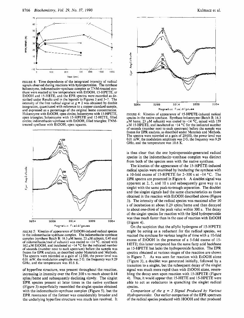
FIGURE 4: Time dependence of the integrated intensity of radical signals observed during reactions with hydroperoxides. The synthase holoenzyme, indomethacin-synthase complex or TNM-treated syn- thase were reacted at low temperature with EtOOH, 15-HPETE, or EtOOH and 15-HETE, and the EPR spectra were recorded as de- scribed under Results and in the legends to Figures 3 and 5-7. The intensity of the free radical signal at g = 2 was obtained by double integration, quantitated with reference to a copper standard sample, and expressed as a percentage of the original heme concentration. Holoenzyme with EtOOH, open circles; holoenzyme with 15-HPETE, open triangles: holoenzyme with 1 5-HPETE and 1 5-HETE, filled circles; indomethacinsynthase with EtOOH, filled triangles; TNM- treated synthase with EtOOH, open squares.
u -0
4
A
a a
a L W
3264 3289 33'1 4 3339 3364
Magnetic Field/gauss
FIGURE 5: Kinetics of appearance of EtOOH-induced radical species in the indomethacin-synthase complex. The indomethacinsynthase complex (synthase Batch B; 16.3 pM heme, 23 pM subunit, 0.45 mol of indomethacin/mol of subunit) was cooled to -14 O C , mixed with 162 pM EtOOH, and incubated at -14 O C for the indicated number of seconds (number next to each spectrum) before the sample was frozen for EPR analysis, as described under Materials and Methods. The spectra were recorded at a gain of 12 500, the power level was 0.01 mW, the modulation amplitude was 2 G, the frequency was 9.29 GHz, and the temperature was 10.6 K.
of hyperfine structure, was present throughout the reaction, increasing in intensity over the first 300 s to reach about 0.44 spins/heme and subsequently declining slowly. The singlet EPR species present a t later times in the native synthase (Figure 3) superficially resembled the singlet species obtained with the indomethacin-synthase complex (Figure 5), but the EPR resonance of the former was considerably broader and the underlying hyperfine structure was much less resolved. It
Kulmacz et al.
s -u
v, + a < L a
W
3264 3289 3314 3339 3364
Magnetic Field/gauss
FIGURE 6: Kinetics of appearance of 15-HPETE-induced radical species in the native synthase. Synthase holoenzyme (Batch B; 16.3 pM heme, 23 pM subunit) was cooled to -14 OC, mixed with 159 pM 15-HPETE, and incubated at -14 O C for the indicated number of seconds (number next to each spectrum) before the sample was frozen for EPR analysis, as described under Materials and Methods. The spectra were recorded at a gain of 20 000, the power level was 0.01 mW, the modulation amplitude was 2 G, the frequency was 9.29 GHz, and the temperature was 10.6 K.
is thus clear that the one hydroperoxide-generated radical species in the indomethacin-synthase complex was distinct from both of the species seen with the native synthase.
The kinetics of the appearance of the 15-HPETE-induced radical species were examined by incubating the synthase with a 10-fold excess of 15-HPETE for 2-300 s at -14 OC. The EPR spectra are presented in Figure 6. A doublet signal was apparent a t 2, 5 , and 10 s and subsequently gave way to a singlet with the same peak-to-trough separation. The doublet and the singlet signals had the same characteristics as those obtained in the reaction with EtOOH described above (Figure 3). The intensity of the radical species was maximal after 10 s of incubation a t about 0.29 spinslheme and then decayed to about one-third of the peak value within 300 s. The decay of the singlet species for reaction with the lipid hydroperoxide was thus much faster than in the case of reaction with EtOOH (Figure 4).
On the suspicion that the allylic hydrogens of 15-HPETE might be acting as a reductant for the radical species, we reacted the synthase for various lengths of time with a 10-fold excess of EtOOH in the presence of a 5-fold excess of 15- HETE; this latter compound has the same fatty acid backbone as 15-HPETE but lacks the hydroperoxide function. The EPR spectra obtained a t various stages of the reaction are shown in Figure 7 . As was seen for reaction with EtOOH alone (Figure 3), a doublet was generated initially, followed by a transition to a singlet, but the subsequent decay of the singlet signal was much more rapid than with EtOOH alone, resem- bling the decay seen upon reaction with 15-HPETE (Figure 4). Thus, it would appear that 15-HETE and 15-HPETE were able to act as reductants in quenching the singlet radical species.
Comparison of the g = 2 Signal Produced by Various Hydroperoxides. Our earlier comparison of the EPR spectrum of the radical species produced with HOOH and that produced
PGH Synthase, Hydroperoxides, and Indomethacin
T 1
PI D
$ 4
a < L
W e
3264 3289 331 4 3339 3364
Magnetic Field/gouso
FIGURE 7: Kinetics of appearance of EtOOH-induced radical species in the native synthase in the presence of 15-HETE. Synthase holo- enzyme (Batch B; 16.3 pM heme, 23 pM subunit) was cooled to -14 O C , mixed with 80 pM 15-HETE and then with 162 pM EtOOH, and incubated at -14 O C for the indicated number of seconds (number next to each spectrum) before the sample was frozen for EPR analysis, as described under Materials and Methods. The spectra were recorded at a gain of 20000, the power level was 0.01 mW, the modulation amplitude was 2 G, the frequency was 9.29 GHz, and the temperature was 10.6 K.
with 15-HPETE (Kulmacz et al., 1987) suggested that the structure of the hydroperoxide influenced the nature of the radical. To examine this matter in more detail, we reacted one preparation of the synthase with a series of different hy- droperoxides (HOOH, EtOOH, 9-HPETE, 1 1-HPETE, 15- HPETE, and PGGJ and with arachidonic acid, which can generate PGGz in situ. The samples were reacted in a salt/ice bath and then frozen to trap the transient radical species as rapidly as possible. The g = 2 regions of these spectra are shown in Figure 8. The most intense radical signals were obtained with ethyl hydrogen peroxide (spectrum A) and hydrogen peroxide (spectrum B). The fatty acid hydroper- oxides gave much weaker radical signals in the order 15- HPETE > 11-HPETE > 9-HPETE (spectra C-E). The doublet character of the radical signal was clearly apparent with both ethyl hydrogen peroxide and 15-HPETE (see also Figure 6), but its presence was ambiguous for the adducts with HOOH and 1 I-HPETE; the radical signal obtained by using 9-HPETE was too weak to allow any conclusions. The peak-to-trough value for the radial signal was quite similar in each case: 35, 36, 37, and 33 G for ethyl hydrogen peroxide, hydrogen peroxide, 1 SHPETE, and 1 1 -HPETE, respectively. In addition a broad feature, with a peak near g = 2.08 and a trough near g = 1.97, was quite apparent in the spectra of the EtOOH and HOOH adducts but less so in the spectra of the 15-HPETE and 1 1 -HPETE adducts. Double integration of the g = 2 region of the spectrum of the EtOOH adduct indicated a spin concentration equal to 0.36 spins/heme, with about two-thirds contributed by the doublet feature and the rest by the broad feature. For the HOOH adduct, double integration indicated a spin concentration corresponding to 0.32 spins/heme, again with about two-thirds contributed by the narrow central signal.
Biochemistry, Vol. 29, No. 37, 1990 8767
1'1
3
CL
a L L
W
3100 3200 3300 3400 3500
Magnetic Field/gauss
FIGURE 8: EPR spectra of the adducts of the synthase with various hydroperoxides. The synthase (Batch A; 18.8 pM heme) was rapidly mixed with a 10-fold excess of each of the indicated hydroperoxides at about -14 OC as described under Materials and Methods. The gain was 20000 (except 16 OOO for arachidonate), the power level was 0.1 mW, the modulation amplitude was 2 G, the frequency was 9.29 GHz, and the temperature was between 9.9 and 11.4 K. The spectra correspond to reaction of the synthase with (A) EtOOH, (B) HOOH, (C) 9-HPETE, (D) 11-HPETE, (E) 15-HPETE, and (F) arachidonic acid.
The EPR spectra of the EtOOH and HOOH adducts of the indomethacin-synthase complex in the g = 2 region were almost superimposable (data not shown), suggesting that the different hydroperoxides were generating the same radical species with the indomethacinsynthase complex. Both spectra exhibited a singlet with clear hyperfine structure, with a peak-to-trough value of about 26 G, and also exhibited the weak broad resonance with a peak at g = 2.08 and a trough at g = 1.97 noted above. The radical species induced by HOOH and EtOOH in the indomethacin-synthase complex were clearly narrower than any of the peroxide-induced radical species seen in the native synthase, each of which had a peak-to-trough separation of about 35 G (Figure 8).
Evaluation of EtOOH as Peroxidase Substrate and Cy- clooxygenase Initiator. To compare EtOOH as a peroxidatic substrate for the synthase with previously studied hydroper- oxides, the peroxidase activity was assayed spectrophoto- metrically with 85 NM TMPD as the chromogenic cosubstrate (Marshall & Kulmacz, 1988) at several levels of EtOOH between 0.2 and 1.2 mM. The initial velocity data were fitted to the standard Michaelis-Menten equation by using a non- linear least-squares algorithm yielding estimates of the ap- parent V , (570 f 30 EtOOH s-l subunit-') and the apparent K, (0.39 f 0.05 mM EtOOH). These values are presented in Table I along with the corresponding values for HOOH and 15-HPETE (Kulmacz, 1986). Comparison of the V,,,/K, ratios indicates that EtOOH was about 16-fold better as a substrate for the synthase peroxidase than HOOH but about 50-fold poorer than 15-HPETE. The kinetics of the EtOOH-dependent inactivation of the peroxidase activity during catalysis were also examined by analysis of the decrease in peroxidase activity as a function of time, as described previously (Marshall & Kulmacz, 1988). The estimated rate constant for inactivation of the peroxidase with EtOOH as the
8768 Biochemistry, Vol. 29, No. 37, 1990 Kulmacz et al.
Table I: Peroxidase Activitv and Cvclooxvaenase Initiation peroxidase activity
EtOOH 0.39 f 0.05 570 f 30 180 7 HOOH 3.6 f 0.5 320 f 20 33 35 15-HPETE 0.012 f 0.002 790 f 70 17000 0.024
substrate was 180 M-' s-I. Comparison of this value with those obtained for other hydroperoxides (Table I) indicates that with EtOOH peroxidase activity was lost about 6-fold more quickly than with HOOH but 90-fold more slowly than with 15- HPETE (Marshall & Kulmacz, 1988).
The ability of EtOOH to initiate the cyclooxygenase activity of the synthase was assessed as described previously (Marshall et al., 1985), with the cyclooxygenase partially inhibited by the addition of 5 mM NaCN to the standard assay buffer, and at a slightly decreased temperature (25 "C). Under such conditions, the time required for the cyclooxygenase to ac- celerate to its highest velocity (the lag time) decreases as the concentration of added hydroperoxide is increased (Marshall et al., 1985). The lag time with no added hydroperoxide was 48 s; it decreased to 39 s with 25 pM HOOH and 24 s with 67 pM HOOH and reached a minimum of 20 s for HOOH above 83 pM. In a parallel series of reactions, the lag time decreased to 30 s with 7 pM EtOOH and reached a minimum of about IO s at levels above 50 pM. From these results it was possible to estimate that the level needed for half-maximal effect was about 35 pM for HOOH and 7 pM for EtOOH. Comparison of these values with that obtained for 15-HPETE (Table I ) indicates that EtOOH is about 5-fold better as a cyclooxygenase initiator than HOOH but about 300-fold poorer than 15-HPETE, which had a half-maximal effect at only 24 nM (Marshall & Kulmacz, 1988).
DISCUSSION Sequential Generation of Two Radical Species by Hydro-
peroxides. Two distinct radical species were apparent in the reaction of the pure synthase with either EtOOH or 15- HPETE (Figures 3 and 6). The earliest intermediate species trapped by the manual mixing technique used gave rise to an EPR doublet signal with a splitting of about 16 G and a peak-to-trough width of about 35 G. This doublet signal is similar to the narrow central feature of the radical signal found earlier by us for reaction with HOOH (Kulmacz et al., 1987) and the doublet observed after reaction with a variety of hy- droperoxides by Karthein et al. (1988). After slightly longer reaction times at low temperature, the doublet signal was replaced by a singlet EPR signal (Figures 3 and 6). This singlet species was relatively long-lived in the case of reaction with EtOOH but decayed more quickly when a polyunsatu- rated fatty acid was present (Figure 4).
The similarities in the g = 2 signals obtained from the adducts of the synthase with various hydroperoxides (Figures 2, 3, 6 , and 8) suggest that very similar radical species were produced in each case, with the spectral differences consistent with differences in the rate of generating the initial adduct, the rate of the doublet-to-singlet transition, or the rate of decay of the singlet. The current studies differ from those that we published earlier (Kulmacz et al., 1987) in two obvious ways: (i) in the present experiments we have used a 10-fold excess of peroxide rather than the 2-fold excess used originally and (ii) the present incubations with substrate were conducted at or below -12 "C, rather than at 4 "C. Given the transient nature of the doublet signal (Figures 3 and 6 ) and the greater
effectiveness in accumulating the signal with EtOOH and HOOH than with 15-HPETE (Figures 3,6, and 8), it is likely that the differences between the spectra of the HOOH and 15-HPETE adducts reported previously [Figure 9 of Kulmacz et al. (1987)l are due to the samples studied having been trapped at different stages of reaction and decay (though after the same incubation period) rather than to the presence of different hydroperoxide fragments, as had been concluded earlier (Kulmacz et al., 1987).
Karthein et al. (1988) did observe a singlet signal after incubation of a PGG,-synthase adduct at higher temperature (0 "C), but it was distinctly narrower than the singlet we observed, with a peak-to-trough width of only 16 G instead of 35 G. The similarity in power saturation behavior between the doublet and the singlet we observed for both was 0.2 mW) contrasts with the almost 100-fold difference in P I l 2 values for the doublet and singlet observed by Karthein et al. (1988). It is thus quite probable that the two singlet signals come from very different free radicals; Karthein et al. (1988) in fact ascribed their singlet to inactivated synthase appearing after relatively long incubation with the peroxide. It is not clear why Karthein et al. (1988) did not observe the wider singlet that we did, particularly because their reaction con- ditions were quite similar to ours. One possibility is that the singlet is more rapidly quenched by the presence of the PGG2/PGH2 that they used than it is by 15-HPETE/15- HETE that we used.
After reaction of the synthase with hydroperoxides, the fractional decrease of the high-spin heme signal (about 80%) was considerably more extensive than could be accounted for by the intensity of the free radical species (about 0.2 spins/ heme; Figure 2). This probably reflects the presence of sig- nificant amounts of EPR-silent species without a free radical, perhaps peroxidase Compound 11. Reliable assignment of particular heme redox states to the species giving rise to the EPR doublet and singlet signals and any EPR-silent species will require alternative spectral measurements (UV-vis, MCD) under the same low-temperature conditions used for prepa- ration of the EPR samples.
The principal features of the doublet EPR signal could be reproduced by a simple model involving hyperfine coupling to two protons, each having an isotropic coupling constant defined by the angle-dependent McConnell relationship, and using the value of 162 MHz for B1 given by Bender et al. (1989). The best simulation was obtained when the dihedral angle between the pz orbital of C-1 of the tyrosine ring and the C-H bond of one of the methylene protons was 40". In- cluding the ring protons in the calculation did not change this value but did produce the slight asymmetry seen at both the low- and high-field ends of the spectrum. Increasing the dihedral angle to 50" converted the calculated spectrum to a singlet with overall spectral width somewhat smaller than the doublet (the intrinsic line width was held constant). Thus, the observed transition from the doublet to the singlet species need not involve any structural change at the tyrosine other than a small rotation of the methylene group relative to the plane of the phenol ring, and it is consequently not required that the second radical species be a different amino acid residue.
The synthase preparations used for our earlier studies had a significant proportion of heme in the low-spin state, as ev- idenced by characteristic features in their EPR and MCD spectra (Kulmacz et al., 1987). We have subsequently ob- served that the proportion of low-spin heme is quite variable from preparation to preparation, with some preparations
PGH Synthase, Hydroperoxides, and Indomethacin
showing no evidence of any low-spin signal in their EPR spectra (Figure 2). The reconstitution process used to convert synthase from apoenzyme to holoenzyme, which involves ad- dition of exogenous heme and treatment with DEAE-cellulose, did not induce the g = 3 EPR signal in a preparation of the synthase initially lacking low-spin heme (Batch A, data not shown). However, samples that were concentrated by ul- trafiltration (to >50 pM heme) consistently have shown considerable low-spin signal (relative to the high-spin signal) in the EPR, whereas samples that remained more dilute generally had little low-spin signal (data not shown), suggesting that the spin-state transition has a concentration-dependent aspect.
Evidence That the Hydroperoxide-Induced Radical Is a Tyrosyl Free Radical and That I t Has a Functional Role in Cyclooxygenase Catalysis. Karthein et al. (1988), noting the similarities between the EPR spectrum of the hydroper- oxide-induced synthase EPR doublet signal and that of the stable tyrosyl free radical in ribonucleotide reductase, assigned the synthase doublet signal to a tyrosyl radical. The present finding that TNM, a selective agent for nitration of tyrosyl residues (Sokolovsky et al., 1966), did nitrate a small number of tyrosyl residues on the synthase and markedly altered the spectrum of the hydroperoxide-induced radical (Figure 2B) is strong circumstantial evidence that the doublet signal indeed arises from a tyrosyl radical. Our further findings that cy- clooxygenase activity was preferentially destroyed by reaction of the synthase with TNM and that one more tyrosine was nitrated in holoenzyme (which became inactivated by TNM) than in apoenzyme (which retained most activity; see Results) suggests that a particular tyrosine residue in the synthase has a functional role in cyclooxygenase catalysis. It is possible that the effects of TNM on the synthase arise from modification of amino acids other than tyrosine: cysteine and tryptophan have been found to react with TNM in other proteins under some conditions (Muhlrad et al., 1968). However, it is most unlikely that the heme modification by TNM reported by Atassi (1969) has occured in the synthase because no change in the position of the Soret maximum was observed. The parallels between TNM and indomethacin as to differential loss of cyclooxygenase activity (Mizuno et al., 1982) and modification of the EPR signal of the hydroperoxide-induced radical species (Figure 2B) testify to the importance of the "native" tyrosyl radical to cyclooxygenase activity.
Smith et al. (1990) have also found that treatment of the synthase with TNM abolished the cyclooxygenase activity and that the presence of the competitive cyclooxygenase inhibitor ibuprofen slowed inactivation by TNM. We have confirmed their results with ibuprofen (see Results) and found that two irreversible inhibitors of the cyclooxygenase, indomethacin and meclofenamic acid, also protected the cyclooxygenase from TNM (see Results). The protective effect of the anticyclo- oxygenase agents and the observation that heme is required for rapid inactivation by TNM (see Results) suggest that the functional tyrosine residue nitrated by TNM is physically close to the fatty acid and heme binding sites.
The synthase tyrosyl radical has been postulated (Karthein et al., 1988) to function by abstracting an allylic hydrogen from the substrate polyunsaturated fatty acid, thus setting in motion the cyclooxygenase reaction by producing a fatty acyl radical capable of reacting with molecular oxygen. The plausibility of this scheme is demonstrated by the ability of the polyun- saturated fatty acid 15-HETE to quench the tyrosyl radical (Figure 4), presumably by donation of an allylic hydrogen. Lambeir et al. (1985) earlier reported that 5-phenyl-4-pentenyl
Biochemistry, Vol. 29, No. 37, 1990 8769
hydroperoxide, which has allylic hydrogens, appeared to act as a reductant of oxidized peroxidase intermediates.
Effectiveness of Hydroperoxides as Cyclooxygenase Ini- tiators. The proposed (Kulmacz et al., 1985; Kulmacz, 1986; Karthein et al., 1988) initiation of the cyclooxygenase reaction via oxidized intermediates of the peroxidase catalytic cycle, specifically peroxidase Compound I, predicts that the ability of a hydroperoxide to initiate the cyclooxygenase reaction depends upon its ability to react as a peroxidase substrate. A previous comparison of lipid hydroperoxides and HOOH as cyclooxygenase initiators and peroxidase substrates (Kulmacz et al., 1985; Kulmacz, 1986) was consistent with this pre- diction, as the lipid hydroperoxide was about 1000-fold more effective in both regards. The correlation is strengthened by the present observation that EtOOH ranks between HOOH and fatty acid hydroperoxides like 15-HPETE both in its ability to act as a peroxidase substrate (Table I; Kulmacz, 1986) and in its effectiveness in initiating the cyclooxygenase reaction (Table I; Marshall et al., 1985).
The hydroperoxide-induced radical species are presumably generated via peroxidase intermediates. If one of the radical species represents the initiated, cyclooxygenase-competent enzyme intermediate, then the ability of a hydroperoxide to generate the radical should parallel its effectiveness as an initiator of the cyclooxygenase reaction. The water-soluble hydroperoxides (HOOH and EtOOH) did appear (Figures 3, 6, and 8) to be more effective than the HPETEs at accumu- lating radical species, even though they were much less efficient as substrates for the peroxidase activity (Table I; Lambeir et al., 1985; Kulmacz, 1986) and as initiators of the cyclo- oxygenase reaction (Table I; Kulmacz et al., 1985). This apparent discrepancy can be resolved. Under the conditions used here, the formation of peroxidase Compound I is rapid enough to be complete within a few seconds even for the slowest-reacting hydroperoxide (Lambeir et al., 1985; Kul- macz, 1986; Karthein et al., 1987), and the formation of the doublet radical species, presumably from Compound I, is also complete in the earliest sample obtained by our hand mixing technique (Figure 3A). Thus, the rate at which the radical is dissipated will determine the size of the EPR signal observed in the time frame we examined. Because the polyunsaturated fatty acid is a good reductant (Figure 4), the level of radical accumulated would be expected to be lower with the HPETEs than with EtOOH or HOOH, which lack reducing functions.
Nature of Cyclooxygenase Impairment by Indomethacin and TNM. The demonstration of a stable complex between the synthase and indomethacin, with a stoichiometry of 0.5 mol/mol of subunit (Figure l) , makes it likely that the changes observed in the characteristics of the hydroperoxide-induced synthase radical species (Figure 2B) were due to the presence of stably bound indomethacin rather than to stable changes in the synthase induced by transient binding of indomethacin. Maximal indomethacin binding coincided with maximum in- hibition of the cyclooxygenase (Figure I ) , and the stoichiom- etry of binding (0.5 mol/protein subunit) agreed with that found in earlier titrations with indomethacin (Kulmacz & Lands, 1985) and with heme (Kulmacz & Lands, 1984) and may indicate a functional inequivalence of the two synthase subunits. The rather small differences in the electronic and MCD spectra of the synthase and the synthase-indomethacin complex in the presence of various ligands (see Results) sug- gests that indomethacin does not grossly alter the heme en- vironment, although the decrease in the rhombic high-spin species in the resting EPR spectra (Figure 2A) indicates that definite changes occur upon indomethacin binding. The
8770 Biochemistry, Vol. 29, No. 37, 1990 Kulmacz et al.
Eds.) pp 45-55, Springer-Verlag, Berlin, FRG.
Chem. 243, 4799-4805. Edelhoch, H., Lippoldt, R. E., & Wilchek, M. (1968) J . Biol.
Graff, G. (1982) Methods Enzymol. 86, 386-392. Hemler, M. E., Graff, G., & Lands, W. E. M. (1978) Bio-
chem. Biophys. Res. Commun. 85, 1325-1 33 1. Holmquist, B., & Vallee, B. L. (1973) Biochemistry 12,
Karthein, R., Nastainczyk, W., & Ruf, H. H. (1987) Eur. J .
Karthein, R., Dietz, R., Nastainczyk, W., & Ruf, H. H.
Kulmacz, R. J. (1986) Arch. Biochem. Biophys. 249,273-285. Kulmacz, R. J . (1987) Prostaglandins 34, 225-240. Kulmacz, R. J . (1989a) Prostaglandins 38, 277-288. Kulmacz, R. J. (1989b) J . Biol. Chem. 264, 14136-14144. Kulmacz, R. J., & Lands, W. E. M. (1984) J . Biol. Chem.
Kulmacz, R. J., & Lands, W. E. M. (1985) J . Biol. Chem.
Kulmacz, R. J., & Lands, W. E. M. (1987) in Prostaglandins and Related Substances: A Practical Approach (Benedetto, C., McDonald-Gibson, R. G., Nigam, S., & Slater, T. F., Eds.) pp 209-227, IRL Press, Washington, DC.
Kulmacz, R. J., Miller, J. F., Jr., & Lands, W. E. M. (1985) Biochem. Biophys. Res. Commun. 130, 918-923.
Kulmacz, R. J., Tsai, A.-L., & Palmer, G. (1987) J . Biol. Chem. 262, 10524-10531.
Lambeir, A.-M., Markey, C. M., Dunford, H. B., & Marnett, L. J. (1985) J . Biol. Chem. 260, 14894-14896.
Marshall, P. J., & Kulmacz, R. J. (1988) Arch. Biochem. Biophys. 266, 162-170.
Marshall, P. J., Warso, M. A,, & Lands, W. E. M. (1985) Anal. Biochem. 145, 192-199.
Merlie, J. P., Fagan, D., Mudd, J., & Needleman, P. (1 988) J . Biol. Chem. 263, 3550-3553.
Mizuno, K., Yamamoto, S., & Lands, W. E. M. (1982) Prostaglandins 23, 743-757.
Muhlrad, A,, Corsi, A., & Granata, A. L. (1968) Biochim. Biophys. Acta 162, 435-443.
Peterson, G. L. (1979) Anal. Biochem. 100, 201-220. Porter, N. A., Logan, J., & Kontoyiannidou, V. (1 979) J . Org.
Chem. 44, 3 177-3 18 1. Rome, L. H., & Lands, W. E. M. (1975) Proc. Natl. Acad.
Sci. U.S.A. 72, 4863-4865. Ruf, H. H., Schuhn, D., & Nastainczyk, W. (1984) FEBS
Lett. 165, 293-296. Sahlin, M., Graslund, A., Ehrenberg, A., & Sjoberg, B.-M.
(1982) J . Biol. Chem. 257, 366-369. Schewe, T., Kuhn, H., & Rapoport, S. M. (1987) in Prosta-
glandins and Related Substances: A Practical Approach (Benedetto, C., McDonald-Gibson, R. G., Nigam, S., & Slater, T. F., Eds.) pp 229-242, IRL Press, Washington, DC.
Schreiber, J., Eling, T. E., & Mason, R. P. (1986) Arch. Biochem. Biophys. 249, 126-1 36.
Smith, W. L., & Lands, W. E. M. (1972) Biochemistry 11, 3276-3285.
Smith, W. L., DeWitt, D. L., Kraemer, S . A., Andrews, M. J., Hla, T., Maciag, T., & Shimokawa, T. (1990) in Ad- vances in Prostaglandin, Thromboxane, and Leukotriene Research (Hedqvist, P., & Dahlen, S.-E., Eds.), Raven Press, New York (in press).
4409-44 17.
Biochem. 166, 173-1 80.
(1988) Eur. J . Biochem. 171, 313-320.
259, 6358-6363.
260, 12572-12578.
parallels between the formation of the indomethacinsynthase complex, the inhibition of cyclooxygenase activity, and the alteration of the tyrosyl radical line shape suggest that the inhibitory effect of indomethacin is a consequence of its perturbation of the tyrosyl radical.
Neither indomethacin nor TNM treatment abolished per- oxidase activity or much affected the ability to accumulate a large portion of the synthase in the singlet radical form (Figure 4). Thus, an unaltered tyrosyl radical does not appear necessary to peroxidase catalysis, and the difficulties in cy- clooxygenase catalysis in the indomethacin or TNM-treated synthase may be due to an electronic arrangement in the altered tyrosyl radical that is unsuitable for proper interaction with the fatty acid. If the analysis of the tyrosine radical in ribonucleotide reductase (Sahlin et al., 1982) is applicable to the synthase radical, the much narrower splitting seen in the hydroperoxide adducts of the indomethacinsynthase complex and the TNM-treated synthase than in the native synthase (Figure 2B) suggests that either the tilt of the tyrosine ring relative to the methylene protons or the distribution of the unpaired electron in the ring, or both, have been altered by the presence of the indomethacin or by nitration. Both a change in ring conformation and a decrease in charge density (especially at C-1 in the ring) can be envisioned as conse- quences of attachment of the bulky, electron-withdrawing nitro group to the tyrosyl ring; in the case of the noncovalent in- domethacin-synthase complex, binding of the agent in the vicinity of the tyrosine bearing the unpaired electron could also lead to conformational or electronic perturbations. The in- herent uncertainties about the oxidation and spin states of the iron in the radical species make it difficult to estimate reliably the distance between the radical and the heme from the power saturation behavior of the radical. However, the simplest interpretation of the observation that the power saturation value was very similar (0.2 mW, see Results) for the doublet and singlet signals in the native synthase and the singlet signals in the indomethacin- and TNM-treated synthases would be that the heme-radical distance is the same in each species.
Accumulating evidence thus indicates that reaction of the synthase peroxidase with hydroperoxides leads to generation of a tyrosyl radical that has an important functional role in cyclooxygenase catalysis by the protein. In this light, the synthase might be envisioned as a heme-dependent peroxidase that evolved the ability to use oxidized peroxidase intermediates to generate a tyrosyl radical and thereby propagate a spe- cialized fatty acid oxygenase reaction. Further, the inhibition of the cyclooxygenase by nonsteroidal antiinflammatory agents like indomethacin can be considered in terms of their per- turbation of the proper function of the hydroperoxide-induced tyrosyl radical.
ACKNOWLEDGMENTS We thank Carol Williams, Yoshihiro Oh-Hashi, Akira
Ohta, Robert Pendleton, Qinghai Pan, and James F. Miller, Jr., for their help in purification of the synthase and William L. Smith for a preprint of some of his group’s work.
REFERENCES Atassi, M. Z. (1969) Biochim. Biophys. Acta 177,663-665. Bender, C. J., Sahlin, M., Babcock, G. T., Barry, B. A.,
Chandrashekar, T. K., Salowe, S. P., Stubbe, J. A,, Lind- strom, B., Petersson, L., Ehrenberg, A., & Sjoberg, B.-M. (1989) J . Am. Chem. SOC. 111, 8076-8083.
Bergman, T., Carlquist, M., & Jornvall, H. (1986) in Ad- vanced Methods in Protein Microsequence Analysis (Wittmann-Liebold, B., Salnikow, J., & Erdmann, V. A.,
Biochemistry 1990, 29, 8771-8779 8771
van Dorp, D. A. (1977) Biochim. Biophys. Acta 487, 31 5-33 1 .
van der Ouderaa, F. J., Buytenhek, M., Nugteren, D. H., &
Determination of the Secondary Structure Content of Proteins in Aqueous Solutions from Their Amide I and Amide I1 Infrared Bands. Comparison between
Classical and Partial Least-Squares Methods? FranGoise Dousseau and Michel P&zolet*
Centre de Recherche en Sciences et Ingenierie des Macromolecules, DPpartement de Chimie, UniversitP Laval, CitP Universitaire, Quebec, Canada GIK 7P4
Received March 14, 1990; Revised Manuscript Received June 4, I990
ABSTRACT: A method for estimating protein secondary structure from infrared spectra has been developed. The infrared spectra of H 2 0 solutions of 13 proteins of known crystal structure have been recorded and corrected for the spectral contribution of water in the amide I and I1 region by using the algorithm of Dousseau et al. [Dousseau, F., Therrien, M., & Pizolet, M. (1989) Appl. Spectrosc. 43, 538-5421. This calibration set of proteins has been analyzed by using either a classical least-squares (CLS) method or the partial least-squares (PLS) method. The pure-structure spectra calculated by the classical least-squares method are in good agreement with spectra of poly(L-lysine) in the a-helix, 6-sheet, and undefined conformations. The results show that the best agreement between the secondary structure determined by X-ray crystal- lography and that predicted by infrared spectroscopy is obtained when both the amide I and I1 bands are used to generate the calibration set, when the PLS method is used, and when it is assumed that the secondary structure of proteins is composed of only four types of structure: ordered and disordered a-helices, 6-sheet, and undefined conformation. Attempts to include turns in the secondary structure estimation have led to a loss of accuracy. The standard deviation of the difference between X-ray and infrared secondary structure estimates with this method is 4.8% for the a-helix, 3.7% for the 6-sheet, and 5.1% for the undefined structure, whereas the regression coefficients are 0.95, 0.96, and 0.56, respectively. The spectra of the calibration proteins were also recorded in 2H20 solution. After correction for the contribution of the combination band of 2 H 2 0 in the amide I’ band region, the spectra were analyzed with PLS, but the results were not as good as for the spectra obtained in H 2 0 , especially for the a-helical conformation.
%e complete tertiary structure of proteins is currently ac- cessible only by X-ray crystallography and a few closely related diffraction techniques. All these techniques require that the molecule can form a well-ordered crystalline array, which is only attainable for a small fraction of proteins. Indeed, some proteins are disordered by their very nature and cannot be studied by high-resolution diffraction techniques. Two-di- mensional NMR] spectroscopy offers a viable alternative to diffraction techniques but, so far, it has been limited to low molecular weight proteins. Therefore, methods for the de- termination of the secondary structure of proteins in solution by spectroscopic techniques like circular dichroism (CD) and vibrational spectroscopy have been developed and provide a valuable average picture of the structure of proteins in an environment that is close to their native one.
The marked sensitivity of infrared amide bands to the conformation of the peptide backbone of proteins is well es- tablished. In their pioneering paper, Elliot and Ambrose (1950) were the first to demonstrate the existence of empirical correlations between the amide I and amide I1 infrared bands of polypeptides and their conformation. Later, Miyazawa and Blout (1961) and Krimm and Bandekar (1986 and earlier
papers) have refined these observations by making detailed vibrational analyses of the amide bands in order to establish correlations between the frequency of these bands and types of secondary structure of polypeptides such as a-helix, @sheet, turns, and undefined structure.
Since the polypeptide backbone of globular proteins is normally folded in more than one conformation, the amide bands of these proteins result from the superimposition of bands corresponding to the different types of structure. Byler and Susi (1986) and Surewicz and Mantsch (1988) have ex- tracted information from the infrared spectra of proteins in 2H,0 solution by resolution enhancement of the broad amide I’ band. These band-narrowing methods allow the decom- position of the amide I’ band into its underlying components. Although this methodology provides a basis for the qualitative estimation of the secondary structure of proteins, it is not a routine procedure since there are still difficulties associated with obtaining artifact-free resolution-enhanced spectra and with the band-fitting procedure. On the other hand, Eckert et al. (1977) have calculated from the amide I’ bands of proteins of known conformational composition the charac-
‘This research was supported in part by the Natural Sciences and Engineering Research Council of Canada (M.P.) and the Fonds FCAR of the Province of Quebec (M.P.).