CONTINUING EDUCATION Prostate Cancer: PET with 18 F-FDG, 18 F- or 11 C-Acetate, and 18 F- or 11 C-Choline Hossein Jadvar Department of Radiology, Keck School of Medicine, University of Southern California, Los Angeles, California Learning Objectives: On successful completion of this activity, participants should be able to describe (1) the potential role of imaging in prostate cancer and (2) the current evidence on the use of 18 F-FDG, 18 F- or 11 C-acetate, and 18 F- or 11 C-choline in the imaging evaluation of prostate cancer. Financial Disclosure: The authors of this article have indicated no relevant relationships that could be perceived as a real or apparent conflict of interest. CME Credit: SNM is accredited by the Accreditation Council for Continuing Medical Education (ACCME) to sponsor continuing education for physicians. SNM designates each JNM continuing education article for a maximum of 1.0 AMA PRA Category 1 Credit. Physicians should claim only credit commensurate with the extent of their participation in the activity. For CE credit, participants can access this activity through the SNM Web site (http://www.snm.org/ce_online) through January 2012. Prostate cancer is biologically and clinically a heterogeneous disease that makes imaging evaluation challenging. The role of imaging in prostate cancer should include diagnosis, local- ization, and characterization (indolent vs. lethal) of the primary tumor, determination of extracapsular spread, guidance and evaluation of local therapy in organ-confined disease, staging of locoregional lymph nodes, detection of locally recurrent and metastatic disease in biochemical relapse, planning of radiation treatment, prediction and assessment of tumor response to salvage and systemic therapy, monitoring of active surveillance and definition of a trigger for definitive therapy, and prognos- tication of time to hormone refractoriness in castrate disease and overall survival. To address these tasks effectively, imaging needs to be tailored to the specific phases of the disease in a patient-specific, risk-adjusted manner. In this article, I review the preclinical and clinical evidence on the potential and emerg- ing role of PET with the 3 most commonly studied radiotracers in prostate cancer, namely 18 F-FDG, 18 F- or 11 C-acetate, and 18 F- or 11 C-choline. Key Words: genitourinary; molecular imaging; PET; PET/CT; acetate; choline; FDG; prostate J Nucl Med 2011; 52:81–89 DOI: 10.2967/jnumed.110.077941 Among men in the United States, prostate cancer is the second most common cancer (exceeded only by nonmela- noma skin cancers) and the second leading cause of cancer death (exceeded only by lung cancer). In 2009, the inci- dence of and deaths from this disease were 192,280 cases and 27,360 cases, respectively (1). As life expectancy in- creases, so will the incidence of this disease, creating what will become an epidemic male health problem. Prostate cancer is clinically a heterogeneous disease characterized by an overall long natural history in comparison to the other solid tumors, with a wide spectrum of biologic behavior that ranges from indolent to aggressive (2). Prostate-specific antigen (PSA) screening has resulted in increased detection of clinically insignificant prostate cancers through repeated standard and occasionally satu- ration biopsies (overdiagnosis and stage migration), which have inevitably led to early unnecessary therapy in many patients (overtreatment). In the post-PSA era, at the time of initial presentation, 80% of patients present with local disease, 12% with regional disease, and 4% with meta- static disease, with the remaining 4% classified as un- known (1). Despite highly successful treatments for localized pros- tate cancer, approximately 15%–40% of men will experi- ence a detectable rise in the serum PSA level (biochemical failure) within 10 y from the primary treatment, suggesting that prostate cancer can metastasize relatively early in the course of the disease, probably as a result of genetic insta- bility, including loss of metastasis-suppressor genes (3). About 25%–35% of men with an increasing serum PSA level will develop locally recurrent disease only, 20%– 25% will develop metastatic disease only, and 45%–55% will develop both local recurrence and metastatic disease (1). Pound et al., in their landmark article, documented the natural history of progression to metastatic disease and death after PSA elevation after radical prostatectomy and no adjuvant hormonal therapy (4). A detectable serum PSA level of at least 0.2 ng/mL was considered evidence of biochemical recurrence. The actuarial metastasis-free sur- vival for all men was 82% at 15 y after surgery. The median Received Jul. 3, 2010; revision accepted Nov. 18, 2010. For correspondence or reprints contact: Hossein Jadvar, Keck School of Medicine, University of Southern California, 2250 Alcazar St., CSC 102, Los Angeles, CA 90033. E-mail: [email protected]COPYRIGHT ª 2011 by the Society of Nuclear Medicine, Inc. PET IN PROSTATE CANCER • Jadvar 81 by on May 19, 2018. For personal use only. jnm.snmjournals.org Downloaded from
Transcript
C O N T I N U I N G E D U C A T I O N
Prostate Cancer: PET with 18F-FDG, 18F- or 11C-Acetate,and 18F- or 11C-Choline
Hossein Jadvar
Department of Radiology, Keck School of Medicine, University of Southern California, Los Angeles, California
Learning Objectives: On successful completion of this activity, participants should be able to describe (1) the potential role of imaging in prostate cancerand (2) the current evidence on the use of 18F-FDG, 18F- or 11C-acetate, and 18F- or 11C-choline in the imaging evaluation of prostate cancer.
Financial Disclosure: The authors of this article have indicated no relevant relationships that could be perceived as a real or apparent conflict of interest.
CME Credit: SNM is accredited by the Accreditation Council for Continuing Medical Education (ACCME) to sponsor continuing education for physicians.SNM designates each JNM continuing education article for a maximum of 1.0 AMA PRA Category 1 Credit. Physicians should claim only creditcommensurate with the extent of their participation in the activity.
For CE credit, participants can access this activity through the SNM Web site (http://www.snm.org/ce_online) through January 2012.
Prostate cancer is biologically and clinically a heterogeneousdisease that makes imaging evaluation challenging. The role ofimaging in prostate cancer should include diagnosis, local-ization, and characterization (indolent vs. lethal) of the primarytumor, determination of extracapsular spread, guidance andevaluation of local therapy in organ-confined disease, staging oflocoregional lymph nodes, detection of locally recurrent andmetastatic disease in biochemical relapse, planning of radiationtreatment, prediction and assessment of tumor response tosalvage and systemic therapy, monitoring of active surveillanceand definition of a trigger for definitive therapy, and prognos-tication of time to hormone refractoriness in castrate diseaseand overall survival. To address these tasks effectively, imagingneeds to be tailored to the specific phases of the disease in apatient-specific, risk-adjusted manner. In this article, I reviewthe preclinical and clinical evidence on the potential and emerg-ing role of PET with the 3 most commonly studied radiotracersin prostate cancer, namely 18F-FDG, 18F- or 11C-acetate, and18F- or 11C-choline.
J Nucl Med 2011; 52:81–89DOI: 10.2967/jnumed.110.077941
Among men in the United States, prostate cancer is thesecond most common cancer (exceeded only by nonmela-noma skin cancers) and the second leading cause of cancerdeath (exceeded only by lung cancer). In 2009, the inci-
dence of and deaths from this disease were 192,280 casesand 27,360 cases, respectively (1). As life expectancy in-creases, so will the incidence of this disease, creating whatwill become an epidemic male health problem. Prostatecancer is clinically a heterogeneous disease characterizedby an overall long natural history in comparison to the othersolid tumors, with a wide spectrum of biologic behaviorthat ranges from indolent to aggressive (2).
Prostate-specific antigen (PSA) screening has resultedin increased detection of clinically insignificant prostatecancers through repeated standard and occasionally satu-ration biopsies (overdiagnosis and stage migration), whichhave inevitably led to early unnecessary therapy in manypatients (overtreatment). In the post-PSA era, at the timeof initial presentation, 80% of patients present with localdisease, 12% with regional disease, and 4% with meta-static disease, with the remaining 4% classified as un-known (1).
Despite highly successful treatments for localized pros-tate cancer, approximately 15%–40% of men will experi-ence a detectable rise in the serum PSA level (biochemicalfailure) within 10 y from the primary treatment, suggestingthat prostate cancer can metastasize relatively early in thecourse of the disease, probably as a result of genetic insta-bility, including loss of metastasis-suppressor genes (3).About 25%–35% of men with an increasing serum PSAlevel will develop locally recurrent disease only, 20%–25% will develop metastatic disease only, and 45%–55%will develop both local recurrence and metastatic disease(1). Pound et al., in their landmark article, documented thenatural history of progression to metastatic disease anddeath after PSA elevation after radical prostatectomy andno adjuvant hormonal therapy (4). A detectable serum PSAlevel of at least 0.2 ng/mL was considered evidence ofbiochemical recurrence. The actuarial metastasis-free sur-vival for all men was 82% at 15 y after surgery. The median
Received Jul. 3, 2010; revision accepted Nov. 18, 2010.For correspondence or reprints contact: Hossein Jadvar, Keck School of
Medicine, University of Southern California, 2250 Alcazar St., CSC 102, LosAngeles, CA 90033.E-mail: [email protected] ª 2011 by the Society of Nuclear Medicine, Inc.
PET IN PROSTATE CANCER • Jadvar 81
by on May 19, 2018. For personal use only. jnm.snmjournals.org Downloaded from
actuarial time to metastases was 8 y from the time of PSArelapse. Once men developed metastatic disease, themedian survival time to death was 5 y. The time to bio-chemical progression, PSA doubling time, and Gleasonscore were predictive of the probability and time to thedevelopment of metastatic disease. The interval from sur-gery to the appearance of metastatic disease was predictiveof time until death. Once men develop castrate-resistantmetastatic disease, the 1-y survival is about 24%, with amedian survival of only 8–18 mo (4). The hormone-refrac-tory state is believed to occur via bypassing or sensitizingthe androgen receptor signaling pathway. The factors in-volved may be androgen receptor mutation such that thereceptor either is activated promiscuously by different ste-roids or is activated in a ligand-independent manner. Otherfactors include amplification of coactivators, activation ofoncogenes, and autocrine growth factor stimulation (5).
Diagnostic Imaging Evaluation of Prostate Cancer
Imaging evaluation of prostate cancer remains challeng-ing (6). The overall role of imaging in prostate cancershould include diagnosis, localization and characterization(indolent vs. lethal) of the primary tumor, determination ofextracapsular spread, guidance and evaluation of local ther-apy in organ-confined disease, staging of locoregionallymph nodes, detection of locally recurrent and metastaticdisease in biochemical relapse, planning of radiation treat-ment, prediction and assessment of tumor response to sal-vage and systemic therapy, monitoring of active surveillanceand definition of a trigger for definitive therapy, and prog-nostication of time to hormone refractoriness in castrate dis-ease and overall survival.Initial imaging diagnosis may be made with ultrasound
or MRI using endorectal probes and image-guided biopsieswhen disease is suspected on the basis of a high serum PSAlevel or abnormal findings on digital rectal examination.Because prostate cancer is often multifocal, and standard10- to 12-core biopsy may miss 38% of cancers or un-derrepresent higher-grade tumor foci (which probably drivethe overall cancer biologic behavior and outcome), theimportant role of imaging in localization and character-ization of primary tumors becomes clear (7). Accuratedepiction of the primary tumor foci may guide and evaluatethe response to focal therapies (“male lumpectomy”) ofaggressive cancers (;15% of tumors) and avoid early treat-ment of indolent cancers, which can then be followed byactive surveillance (8).Imaging also provides important information on the
local extent of disease and examines for potential regionaland distant metastatic disease in high-risk patients. Theoptimal method for imaging evaluation of men with PSArelapse (biochemical failure) has not been determined, butthe goal of imaging is to determine whether there is recur-rence in the treated prostate bed or whether distant diseaseis present (or both), because such a determination affectstherapeutic management, including consideration for sal-
vage therapy for local recurrence and systemic treatment formetastatic disease. Despite their overall utility, currentimaging tests, including ultrasound, CT, MRI, 99mTc-basedbone scintigraphy, and 111In-capromab pendetide scintigra-phy, are not sufficiently accurate in detecting and character-izing disease in prostate cancer (9).
In this article, I review the use of the 3 most studied PETradiotracers in prostate cancer: 18F-FDG, 18F- or 11C-labeled acetate, and 18F- or 11C-labeled choline. The dis-cussion is organized by radiotracer, allowing the reader tofocus on the information for a particular radiotracer ofinterest independently of the discussion of other radio-tracers. Nested within each radiotracer category is anattempt to present the available information on the basisof disease phases or imaging tasks. However, many studiesused patients with a mixture of clinical phases, and henceclear separation of patient categories was often challenging.Moreover, the data on 11C and 18F labels of acetate andcholine are presented separately for 2 reasons: first, thereare substantially more published data on the 11C labels, andsecond, it would be helpful to review the 18F label informa-tion separately because the pertinent data are rapidly ex-panding, with results that may differ from those of the 11Clabel, as does the overall relevance to the clinical settingin view of the longer half-life (110 min) and potential sup-ply availability through regional distribution centers (sim-ilar to 18F-FDG). I first briefly review the biologic basis ofthe relevant radiotracer uptake in prostate tumor and thenpresent the available clinical evidence.
18F-FDG and Prostate Cancer
Molecular Biology Correlates of Tumor Uptake. Theability of 18F-FDG PET to detect cancer is based on ele-vated glucose metabolism in the malignant tissue in com-parison to the normal tissue (Warburg effect) as a result ofincreased expression of cellular membrane glucose trans-porters (mainly transporter 1) and enhanced hexokinase IIenzymatic activity in tumors (10,11).
Few studies have reported on expression of glucosetransporters in human prostate cancer. In 1 investigation,the glucose transporter 1 messenger RNA expression wasassessed by Northern blot analysis in the androgen-independent cell lines DU145 and PC3 and the androgen-sensitive LNCaP prostate cancer cell line (12). Althoughglucose transporter 1 expression was detected in all 3 celllines, the level of expression was higher in the poorly dif-ferentiated cell lines DU145 and PC3 than in the well-dif-ferentiated hormone-sensitive LNCaP cell line, suggestingthat the level of glucose transporter 1 expression increaseswith progression of malignancy grade. Recently, Britishinvestigators evaluated the expression of several hypoxia-associated genes within benign prostatic hyperplasia andprostate cancer (Gleason score 5–10) human tissue speci-mens (13). GLUT1 gene expression was significantly higherin prostate cancer than in benign prostatic hyperplasia andcorrelated directly with Gleason score (R 5 0.274, P 5
82 THE JOURNAL OF NUCLEAR MEDICINE • Vol. 52 • No. 1 • January 2011
by on May 19, 2018. For personal use only. jnm.snmjournals.org Downloaded from
0.026). These findings may explain not only the observationof higher 18F-FDG accumulation in castration-resistant(androgen-independent) tumors than in castration-sensitivetumors but also the modulatory effect of androgen on theglucose metabolism of castration-sensitive tumors (14).Normal Prostate Tissue. The glucose metabolism and CT
density of the normal prostate gland in relation to age andprostate size have been assessed using 18F-FDG PET/CT in145 men who had indications unrelated to prostate pathol-ogy (15). The average prostate size was 4.36 0.5 cm (mean6 SD), with a range of 2.9–5.5 cm. Mean and maximumCT densities, in Hounsfield units, were 36.0 6 5.1 (range,23–57) and 91.7 6 20.1 (range, 62–211), respectively,whereas mean and maximum standardized uptake values(SUVs) were 1.3 6 0.4 (range, 0.1–2.7) and 1.6 6 0.4(range, 1.1–3.7), respectively. The mean SUV tended todecrease as the prostate size increased (r 5 –0.16, P 50.058), whereas the prostate size tended to increase withincreasing age (r 5 0.32, P , 0.001).Primary Tumor and Staging. Initial analysis of the data
of the National Oncologic PET Registry clearly indicatesthat 18F-FDG PET can influence the clinical managementof men with prostate cancer (from nontreatment to treat-ment in 25.3% of cases and from treatment to nontreatmentin 9.7% of cases), although the influence is lower than forother cancers (16). Nevertheless, the overall clinical expe-rience with 18F-FDG PET in prostate cancer suffers fromheterogeneity in published studies with regard to the clin-ical phases of disease, relatively small numbers of patients,and variability and limitations in the validation criteria.The level of 18F-FDG accumulation can overlap in normal
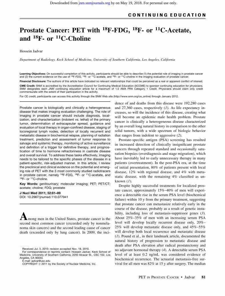
prostate tissue, benign prostatic hyperplasia, and prostatecancer tissues, all of which often coexist (17). 18F-FDGPET might not be useful in the diagnosis or staging of clin-ically organ-confined disease or in the detection of locallyrecurrent disease because of the relatively similar uptake of18F-FDG by the posttherapy changes and tumor cells andbecause of the high level of excreted radiotracer in the adja-cent urinary bladder that may mask any lesions in the vicin-ity (18). False-positive results may occur with prostatitis(19). Despite the drawbacks and the overall heterogeneityof published studies with relatively small numbers of sub-jects, several animal-based translational and human-basedclinical studies have demonstrated that 18F-FDG PET canbe useful in certain clinical circumstances in prostate cancer.18F-FDG uptake is higher in poorly differentiated primarytumors (Gleason sum score. 7) and higher PSA values thanin tumors with lower Gleason scores, a more localized clin-ical stage, and lower serum PSA values (Fig. 1) (20).
18F-FDG PET was less sensitive than 99mTc-based bonescintigraphy at identifying bone metastases, and detection ofpelvic lymph node metastases was limited because of blad-der urine activity (21). In patients with known osseous meta-static disease, however, 18F-FDG PET might distinguish themetabolically active lesions from the metabolically dormantlesions (22). Furthermore, data from our laboratory suggest
that the rate of concordance of 18F-FDG PET with otherimaging studies may depend on the phase of disease (cas-trate-resistant vs. castrate-sensitive), time of imaging in rela-tion to therapy (before or during), and type of lesions (lymphnode and visceral vs. osseous) (23).
Biochemical Failure and Restaging. 18F-FDG PET maybe useful in detecting disease in a fraction of the largeproportion of men who present with PSA relapse, in whom,by definition, there is no standard imaging evidence of dis-ease. In this group of men, detection of disease by non-standard imaging can direct appropriate treatment, suchas salvage radiation therapy for local recurrence in theprostate bed or systemic therapy for metastatic disease. Ina study of 24 patients who had rising serum PSA levelsafter treatment for localized prostate tumors, 18F-FDGPET was performed before pelvic lymph nodes were dis-sected (24). In none of the patients did pelvic CT yieldpositive findings. The histology of the pelvic lymph nodesobtained from surgery confirmed the presence of metastasesin 67% of patients. Increased 18F-FDG uptake was shown atthe sites of histopathologically proven metastases in 75% ofthese patients. The sensitivity, specificity, accuracy, positivepredictive value, and negative predictive value of 18F-FDGPET in detecting metastatic pelvic lymph nodes were75.0%, 100%, 83.3%, 100%, and 67.7%, respectively. Ina similar retrospective study of 91 patients with PSArelapse after prostatectomy and validation of tumor pres-ence by biopsy or clinical and imaging follow-up, meanserum PSA levels were higher in 18F-FDG PET–positivepatients than in 18F-FDG PET–negative patients (9.5 62.2 ng/mL vs. 2.1 6 3.3 ng/mL) (25). A PSA level of 2.4ng/mL and PSA velocity of 1.3 ng/mL/y provided the bestcompromise between sensitivity (80% for 18F-FDG PET–positive and 71% for 18F-FDG PET–negative patients) andspecificity (73% for 18F-FDG PET–positive and 77% for18F-FDG PET–negative patients) in a receiver-operating-
FIGURE 1. A 67-y-old man with biopsy-confirmed prostate cancer
characteristic curve analysis. Overall, 18F-FDG PET detectedlocal or systemic disease in 31% of patients with PSArelapse. However, confidence in the accuracy and relevanceof this figure is somewhat limited in view of the heteroge-neity and limitation of the validation criteria—an issue withother similar studies. 18F-FDG PET may also be particularlyuseful in staging of advanced prostate cancer in patients whohave a rising PSA level despite treatment (26). Moreover, inthis clinical setting, 18F-FDG PET is advantageous over111In-capromab pendetide scintigraphy in the detection ofmetastatic disease in patients with high PSA levels or highPSA velocity (27).
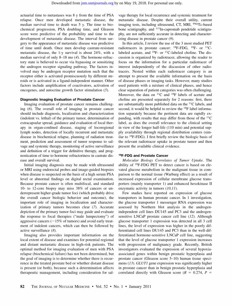
Therapy Response Assessment. In 1 report, 18F-FDGaccumulation in the primary prostate cancer and metastaticsites decreased over a period of 1–5 mo after initiation ofandrogen deprivation therapy, as was consistent with resultsfrom animal xenograft studies (28). However, an earlierstudy of prostate cancer in rats showed that the global18F-FDG SUV was unchanged after treatment with gemci-tabine (29). Our preliminary results show that tumor 18F-FDG uptake decreases with successful treatment (usingandrogen deprivation or various chemotherapy regimens),in general concordance with other measures of response,such as a decline in serum PSA level (Fig. 2) (30).Prognostication. The level and extent of 18F-FDG accu-
mulation in metastatic lesions may provide information onprognosis. An increase of over 33% in the average maxi-mum SUV measurement from up to 5 lesions, or theappearance of new lesions, was reported to be able tocategorize castrate-sensitive metastatic prostate cancer pa-tients treated with antimicrotubule chemotherapy into pro-gressors or nonprogressors (31). Similarly, another group
reported that patients with primary prostate tumors withhigh SUVs had a poorer prognosis than did those withlow SUVs (32). Moreover, because 18F-FDG uptake inprostate tumors appears to depend on the presence and ac-tivity of androgen, 18F-FDG PET might also be useful inpredicting the time to reach the androgen-refractory state(e.g., by an early increase in castrate tumor 18F-FDG up-take), which might facilitate earlier therapeutic modifica-tion to avert or delay this clinical state in order to improveoverall outcome.
18F- or 11C-Acetate and Prostate Cancer
Molecular Biology Correlates of Tumor Acetate Uptake.Acetate participates in cytoplasmic lipid synthesis, which isbelieved to be increased in tumors. The cellular retention ofradiolabeled acetate in prostate cancer cell lines is primarilydue to incorporation of the radiocarbon into phosphatidyl-choline and neutral lipids of the cells (33). It has beensuggested that fatty acid metabolism rather than glycolysismay be dominant in prostate cancer in view of alterations inseveral enzymes involved in the metabolism of fatty acidsand an enhanced b-oxidation pathway (34). Recent in vitroand animal model in vivo studies by the group at Washing-ton University in St. Louis confirmed the extensive involve-ment of the fatty acid synthesis pathway in 11C-acetateuptake in prostate tumors as an imaging marker for fattyacid synthase expression (35). Fatty acid synthase is themajor enzyme required for converting carbohydrates tofatty acids, and its upregulation plays a role in tumorigen-esis of the prostate in the transgenic adenocarcinoma of themouse prostate model (36).
Primary Tumor and Staging. Normal biodistribution of11C-acetate demonstrates high accumulation in the pancreas,
FIGURE 2. Serial 18F-FDG PET/CT andbone scans of 63-y-old man with castrate-
resistant metastatic prostate cancer with
original primary cancer Gleason score of 9.Rows from top to bottom are scans at base-
line (before chemotherapy) and at 4, 8, and 12
mo after initiation of chemotherapy. Columns
from left to right are axial CT scans (bonewindow level), 18F-FDG PET scan, fused
PET/CT scans, mid sagittal CT scan (bone
window level), PET maximum-intensity-
projection images, and 99mTc-methylenediphosphonate bone scans. Concordant
decline in overall metabolic activity of meta-
static lesions and PSA level is seen withtreatment. Sclerosis of osseous lesions in-
creases as corresponding metabolic activity
declines with treatment.
84 THE JOURNAL OF NUCLEAR MEDICINE • Vol. 52 • No. 1 • January 2011
by on May 19, 2018. For personal use only. jnm.snmjournals.org Downloaded from
variable uptake in the liver and bowel, and some renaluptake, with little urinary excretion (37). The lack of accu-mulation of 11C-acetate in urine is advantageous to imagingprostate cancer in particular, because the prostate bedremains unobstructed by the adjacent high levels of radio-activity in the urinary bladder, potentially a problem with18F-FDG. Although there can be a considerable overlapbetween the uptake level in primary cancer, benign prostatichyperplasia, and the normal prostate gland, tracer uptakegenerally appears to be greater in the tumor than in normaland benign prostatic hyperplasia tissue (38). Another findingfrom this study was an age-related physiologic accumulationof 11C-acetate (SUV, 3.4 6 0.7 in age , 50 y, 2.3 6 0.7 inage $ 50 y). In another study, from Japan, 11C-acetate wascompared with 18F-FDG for the detection of primary tumors(39). The tumors demonstrated a variable uptake of 11C-ace-tate (in all 18 patients), with SUVs ranging from 3.3 to 9.9(measured at 10–20 min after tracer administration), in com-parison to 18F-FDG accumulation (in 15/18 patients), whichhad SUVs ranging from 1.97 to 6.34 (measured at 40–60 minafter tracer administration). The authors concluded that 11C-acetate is more sensitive than 18F-FDG in the detection ofprimary prostate cancer.Biochemical Failure and Restaging. 11C-acetate may also
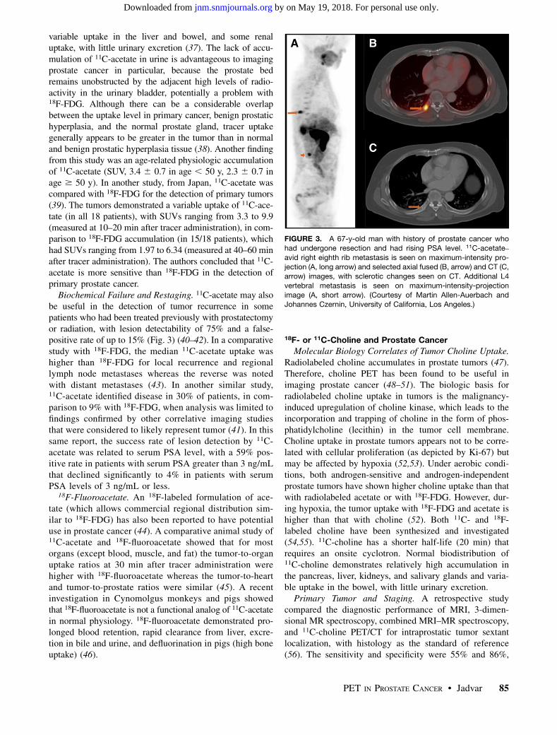
be useful in the detection of tumor recurrence in somepatients who had been treated previously with prostatectomyor radiation, with lesion detectability of 75% and a false-positive rate of up to 15% (Fig. 3) (40–42). In a comparativestudy with 18F-FDG, the median 11C-acetate uptake washigher than 18F-FDG for local recurrence and regionallymph node metastases whereas the reverse was notedwith distant metastases (43). In another similar study,11C-acetate identified disease in 30% of patients, in com-parison to 9% with 18F-FDG, when analysis was limited tofindings confirmed by other correlative imaging studiesthat were considered to likely represent tumor (41). In thissame report, the success rate of lesion detection by 11C-acetate was related to serum PSA level, with a 59% pos-itive rate in patients with serum PSA greater than 3 ng/mLthat declined significantly to 4% in patients with serumPSA levels of 3 ng/mL or less.
18F-Fluoroacetate. An 18F-labeled formulation of ace-tate (which allows commercial regional distribution sim-ilar to 18F-FDG) has also been reported to have potentialuse in prostate cancer (44). A comparative animal study of11C-acetate and 18F-fluoroacetate showed that for mostorgans (except blood, muscle, and fat) the tumor-to-organuptake ratios at 30 min after tracer administration werehigher with 18F-fluoroacetate whereas the tumor-to-heartand tumor-to-prostate ratios were similar (45). A recentinvestigation in Cynomolgus monkeys and pigs showedthat 18F-fluoroacetate is not a functional analog of 11C-acetatein normal physiology. 18F-fluoroacetate demonstrated pro-longed blood retention, rapid clearance from liver, excre-tion in bile and urine, and defluorination in pigs (high boneuptake) (46).
18F- or 11C-Choline and Prostate Cancer
Molecular Biology Correlates of Tumor Choline Uptake.Radiolabeled choline accumulates in prostate tumors (47).Therefore, choline PET has been found to be useful inimaging prostate cancer (48–51). The biologic basis forradiolabeled choline uptake in tumors is the malignancy-induced upregulation of choline kinase, which leads to theincorporation and trapping of choline in the form of phos-phatidylcholine (lecithin) in the tumor cell membrane.Choline uptake in prostate tumors appears not to be corre-lated with cellular proliferation (as depicted by Ki-67) butmay be affected by hypoxia (52,53). Under aerobic condi-tions, both androgen-sensitive and androgen-independentprostate tumors have shown higher choline uptake than thatwith radiolabeled acetate or with 18F-FDG. However, dur-ing hypoxia, the tumor uptake with 18F-FDG and acetate ishigher than that with choline (52). Both 11C- and 18F-labeled choline have been synthesized and investigated(54,55). 11C-choline has a shorter half-life (20 min) thatrequires an onsite cyclotron. Normal biodistribution of11C-choline demonstrates relatively high accumulation inthe pancreas, liver, kidneys, and salivary glands and varia-ble uptake in the bowel, with little urinary excretion.
Primary Tumor and Staging. A retrospective studycompared the diagnostic performance of MRI, 3-dimen-sional MR spectroscopy, combined MRI–MR spectroscopy,and 11C-choline PET/CT for intraprostatic tumor sextantlocalization, with histology as the standard of reference(56). The sensitivity and specificity were 55% and 86%,
FIGURE 3. A 67-y-old man with history of prostate cancer who
had undergone resection and had rising PSA level. 11C-acetate–
avid right eighth rib metastasis is seen on maximum-intensity pro-jection (A, long arrow) and selected axial fused (B, arrow) and CT (C,
arrow) images, with sclerotic changes seen on CT. Additional L4
vertebral metastasis is seen on maximum-intensity-projection
image (A, short arrow). (Courtesy of Martin Allen-Auerbach andJohannes Czernin, University of California, Los Angeles.)
PET IN PROSTATE CANCER • Jadvar 85
by on May 19, 2018. For personal use only. jnm.snmjournals.org Downloaded from
respectively, for PET/CT, 54% and 75%, respectively, forMRI, and 81% and 67%, respectively, for MR spectroscopy.Therefore, in this study, 11C-choline PET/CT demonstrated alower sensitivity relative to MR spectroscopy alone or com-bined withMRI. Opposite findings have been reported by theJapanese investigators in that 11C-choline PET was moresensitive than MRI and MR spectroscopy for detection ofprimary prostate lesions (100% for PET vs. 60% for MRIvs. 65% forMR spectroscopy) (57). These conflicting resultsmay be due to differing methodology in data collection oranalysis and in patient populations. Research is under wayto provide more sophisticated image fusion software foraccurate registration of anatomic MRI, diffusion MRI, 11C-choline PET, and histologic sections of the prostate gland—fusion that may be facilitated by the emergence of the hybridPET/MRI systems (58).German investigators compared 11C-choline PET/CT
with whole-body MRI retrospectively for staging of pros-tate cancer (59). Diagnostic validation was by histology,follow-up, or consensus reading. Overall sensitivity andspecificity were 97% and 77%, respectively, for 11C-cholinePET and 79% and 94%, respectively, for whole-body MRI.Therefore the 2 imaging modalities were complementary. Astudy similar to the MRI reports, comparing transrectalultrasonography and 11C-choline PET/CT in patients withclinically localized prostate cancer, showed that both PETand transrectal ultrasonography tended to understage pros-tate cancer. The authors therefore suggested that reliableclinical decision making (e.g., with regard to decisions onnerve-sparing radical prostatectomy) based on the findingson these imaging modalities might not be possible (60).Despite these reports, other investigations have found arelatively good diagnostic performance for 11C-cholinePET and PET/CT in the detection of primary prostate can-cer. For example, Scher et al. reported a sensitivity of 87%and specificity of 62% for the detection primary prostatecancer, with histopathologic examination of resection spec-imens or biopsy as the reference standard (61). Interestingly,the group from Italy reported nearly the reverse values, withsensitivity of 66% and specificity of 81% for localization ofprimary prostate cancer on a sextant histopathologic analysis(62). Martorana et al. assessed the diagnostic performance of11C-choline PET/CT for nodules 5 mm or larger in 43patients with known prostate cancer before the initial 12-coretransrectal biopsy (63). PET demonstrated a sensitivity of83% in this setting but had lower sensitivity than MRI forthe assessment of extraprostatic extension (22% vs. 63%,respectively, P , 0.001). Therefore, although 11C-cholinePET may be helpful in detecting primary prostate cancer,the sensitivity may depend on several factors that will needto be defined (e.g., tumor grade, size, and location).Biochemical Failure and Restaging. 11C-choline PET has
been evaluated for detecting local, regional, and metastaticprostate cancer (64). The tracer uptake was noted todecrease both in the primary tumor and in metastases afterhormonal therapy, although this finding has been disputed
in other studies (47,65). In another study, 11C-choline wasdetermined to localize recurrence in a higher percentage ofmen after primary radiation therapy than after radical pros-tatectomy (78% vs. 38%, respectively) (49). The reason forsuch a difference based on the type of primary therapy isunclear. Potential utility for 11C-choline PET/CT in thedetection of local recurrence after radical prostatectomyhas also been demonstrated with a sensitivity of 73% andspecificity of 88% (51). The Italian researchers reported asensitivity and specificity of 64% and 90%, respectively, forthe detection of nodal metastases in men with PSA failureafter radical retropubic prostatectomy (66).
A positive correlation between 11C-choline PET/CTlesion detection rate and PSA level has been reported,although there are also reports of no significant correlation(patient-based analysis) between lesion maximum SUV,PSA level, Gleason score, and pathologic stage at the timeof initial diagnosis (67,68). In the study by Krause et al.,from Germany, the detection rate of 11C-choline PET/CTwas 36% for PSA, 1 ng/mL, 43% for 1# PSA, 2 ng/mL,62% for 2# PSA, 3 ng/mL, and 73% for PSA$ 3 ng/mL(65). Castellucci et al., from Italy, evaluated the likelihoodof lesion detection in 190 men after radical prostatectomywho presented with PSA relapse (defined as PSA . 0.2 ng/mL; range, 0.2–25.4 ng/mL; mean, 4.2 ng/mL) (69). Theauthors found that the likelihood of lesion detection by thenonstandard imaging evaluation with 11C-choline PET wasincreased when PSA was higher than 2.4 ng/mL or whenPSA was less than 2.4 ng/mL but that the PSA doublingtime was lower than 3.4 mo or PSA velocity was higherthan 1 ng/mL/y. However, this study included some patientswith abnormal standard imaging evidence of disease thatdid not satisfy the pure-definition requirement of PSArelapse-only disease with negative standard imaging results.
In a more recent investigation, dual-tracer 11C-cholineand 18F-FDG PET were investigated in detecting diseasein 73 men with PSA relapse after radical prostatectomy(67.1% of patients), radiation therapy (32.9% of patients),and adjuvant hormonal therapy (20.5% of patients) (70).The PSA range and median were 0.87–5.4 and 2.4 ng/mL,respectively, for the radical prostatectomy group and 2.1–5.57 and 3.5, respectively, for the radiation therapy group.The Gleason score for the primary disease was 2–7 (well tomoderately differentiated) in 69.9% of patients and 8–10(poorly differentiated) for 27.4% of patients. The standardof reference was biopsy, increase in PSA without therapy,and decrease in PSA after therapy. The authors reported asensitivity of 60.6% for 11C-choline and 31% for 18F-FDGat all PSA levels. Additionally they noted that 18F-FDGcorrelated better with Gleason score than did 11C-choline.They concluded that although 11C-choline appears to bemore sensitive than 18F-FDG for the detection of diseasein PSA relapse, 18F-FDG was better in discriminating theproliferative character of the disease.
In summary, despite some mixed results, it appears thatthe sensitivity of PET may generally depend directly on
86 THE JOURNAL OF NUCLEAR MEDICINE • Vol. 52 • No. 1 • January 2011
by on May 19, 2018. For personal use only. jnm.snmjournals.org Downloaded from
serum PSA level, with the expectation that at higher PSAlevels, the probability of lesion localization increases.Moreover, 11C-acetate and 11C-choline appear to be aboutequally useful in imaging prostate cancer in individualpatients (71).
18F-Fluorocholine. An 18F-labeled formulation of cholinehas also been developed and preliminarily tested in menwith prostate cancer (Fig. 4). Price et al. showed thatmurine xenografts of prostate cancer accumulated higher18F-FDG than 18F-fluorocholine whereas, interestingly, inhumans the 18F-fluorocholine uptake in lesions was higherthan the 18F-FDG uptake (72). The exact reason for suchan observation is unclear but may be due to the biologicdifferences between an implanted tumor and a nativetumor. A recent animal study from our group showed thatuptake interval and castration do not significantly affectthe level of choline uptake in prostate tumors (47).The normal biodistribution of 18F-fluorocholine demon-
strates relatively high accumulation in the pancreas, liver,spleen, and kidneys; variable uptake in the bowel; andexcretion into urine. 18F-fluorocholine uptake overlapsamong normal, benign, and malignant prostate tissues (sim-ilar to 11C-acetate and 11C-choline) (73). In a recent report,disease was missed in a significant number of patients(#75%) with elevated PSA (73), although in another reportthe results were more encouraging (74).Beheshti et al. examined the potential utility of 18F-fluo-
rocholine PET/CT in men who had clinical organ-confinedtumor but were at intermediate (PSA 5 10–20 ng/mL,Gleason score 5 7) and high (PSA . 20 ng/mL, Gleasonscore $ 8) risk for extracapsular extension before under-going radical prostatectomy with extended pelvic lymphnode dissection (75). The sensitivity, specificity, positivepredictive value, and negative predictive value of 18F-fluo-rocholine for detection of pelvic lymphadenopathy were45%, 96%, 82%, and 83% for all lymph node sizes, respec-tively, and 66%, 96%, 82%, and 92% for those lymph nodesgreater than or equal to 5 mm. Not surprisingly, the diag-nostic performance improved when nodes smaller than thePET spatial resolution were excluded.Another study from the same group of investigators
correlated the uptake of 18F-fluorocholine in bone metasta-ses with the morphologic changes on CT in 70 men withprostate cancer (76). The standard of reference was otherimaging and clinical follow-up. The overall sensitivity andspecificity of 18F-fluorocholine for the detection of bonemetastases were 79% and 97%, respectively. Lytic lesionsdemonstrated higher metabolism than blastic lesions (aver-age maximum SUV of 11 6 3.2 for lytic lesions vs. 7.8 63.0 for blastic lesions). No statistically significant differ-ence was found in the maximum SUVof lesions in relationto the presence or absence of hormonal therapy. Conversely,the bone lesion CT density was significantly higher inpatients receiving hormonal therapy. The authors identified3 correlative PET/CT patterns for bone metastases: lesionswith 18F-fluorocholine uptake only, probably representing
bone marrow infiltration without morphologic changes onCT; lesions with both 18F-fluorocholine uptake and CTmorphologic changes; and lesions with no 18F-fluorocho-line uptake but displaying dense sclerosis on CT (Houns-field units. 825), probably indicating nonviable tumor. Wehave observed similar findings with 18F-FDG PET/CT inbone metastases of prostate cancer (23). The same group ofresearchers also compared 18F-fluorocholine and 18F-fluo-ride PET/CT in the detection of bone metastases (77). Thisstudy revealed that 18F-fluorocholine might be superior forearly detection (i.e., bone marrow involvement) of meta-static bone disease and that in patients with 18F-fluorocho-line–negative suggestive sclerotic lesions, 18F-fluoride canbe helpful, with the caveat that 18F-fluoride PET could alsobe negative in highly dense sclerotic lesions, presumablyreflecting treated disease. Therefore, metabolic and morpho-logic changes of bone metastases are dynamic processes, andcombined imaging is best suited to capture the natural courseof these changes to allow for management decisions andaccurate assessment of treatment response.
In relation to men with biochemical failure, the Italianinvestigators showed that the detection rate of 18F-fluoro-choline PET/CT in patients with biochemical relapse pos-itively correlates with the serum PSA level, similarly to theresults of 11C-choline studies (78). In this study, 18F-fluo-rocholine PET demonstrated a detection rate of 20% forPSA # 1 ng/mL, 44% for 1 , PSA # 5 ng/mL, and
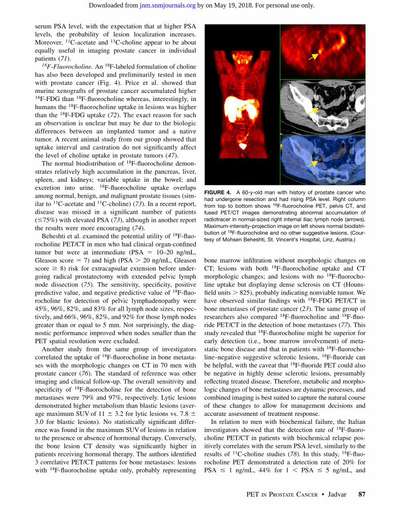
FIGURE 4. A 60-y-old man with history of prostate cancer who
had undergone resection and had rising PSA level. Right column
from top to bottom shows 18F-fluorocholine PET, pelvis CT, andfused PET/CT images demonstrating abnormal accumulation of
radiotracer in normal-sized right internal iliac lymph node (arrows).
Maximum-intensity-projection image on left shows normal biodistri-
bution of 18F-fluorocholine and no other suggestive lesions. (Cour-tesy of Mohsen Beheshti, St. Vincent’s Hospital, Linz, Austria.)
PET IN PROSTATE CANCER • Jadvar 87
by on May 19, 2018. For personal use only. jnm.snmjournals.org Downloaded from
82% for PSA . 5 ng/mL. Another study, from Austria,demonstrated a 41% true-positive rate in restaging patientswith a PSA level of less than 5 ng/mL (79). Thus, thedecision to use, and expectation of outcome for, 18F-fluoro-choline (or 11C-choline) PET in an individual patient withPSA relapse may be adapted to the relevant PSA level (andprobably other PSA-derived parameters) (80).
SUMMARY
Biologically and clinically, prostate cancer is a hetero-geneous disease that is characterized by states ranging fromindolent to aggressive. The use of PET in prostate cancershould be considered in the context of the limitations andchallenges associated with other imaging modalities inprostate cancer.Current evidence indicates that 18F-FDG PET might be
useful in diagnosis and staging of primary tumors that areknown or suspected to have a high Gleason score, in detec-tion of metastatic disease in a fraction of men with bio-chemical failure with scan sensitivity that increases withincreasing PSA level, in assessment of the extent of meta-bolically active castrate-resistant disease, in monitoringresponse to androgen deprivation and other therapies, andin prognostication. 18F-FDG PET has limited use in thediagnosis and staging of clinically organ-confined diseaseand can give false-negative results because of the uptake of18F-FDG by normal tissue, benign prostatic hyperplasia,and posttherapy changes, as well as false-positive resultsin the setting of inflammation and infection.Both 11C-acetate and 11C-choline appear to be some-
what equally useful in imaging prostate cancer in individ-ual patients, although more comparative data are needed.11C-choline and, more recently, 18F-fluorocholine areincreasingly used in many centers in Europe and Japanfor the detection of locally recurrent or metastatic diseasein men with biochemical failure, with scan sensitivity thatcorrelates positively with serum PSA level. Like 18F-FDG,choline and acetate cannot differentiate between malignantand benign prostate disease. 11C-choline PET may be helpfulin detecting primary prostate cancer, but the sensitivity maydepend on several factors that will need to be defined (e.g.,PSA level, tumor grade, size, and location). There are alsomixed findings about the effect of androgen deprivation ther-apy on choline uptake in prostate tumors, probably due to theheterogeneity of androgen receptor function.It is clear that prospective clinical imaging trials using
various PET tracers, singly or in combination, in differentclinical-state–specific patient cohorts with well-definedendpoints, will be needed to decipher the optimal use ofPET in prostate cancer.
ACKNOWLEDGMENT
This work was supported by National Institutes ofHealth–National Cancer Institute grants R01-CA111613and R21-CA142426.
REFERENCES
1. SEER Stat Fact Sheets: Prostate. Available at: http://seer.cancer.gov/statfacts/
html/prost.html. Accessed November 19, 2010.
2. Kessler B, Albertsen P. The natural history of prostate cancer. Urol Clin North
Am. 2003;30:219–226.
3. Dong JT, Rinker-Schaeffer CW, Ichikawa T, et al. Prostate cancer: biology of
metastasis and its clinical implications. World J Urol. 1996;14:182–189.
4. Pound CR, Partin AW, Eisenberger MA, et al. Natural history of progression
after PSA elevation following radical prostatectomy. JAMA. 1999;281:1591–
1597.
5. Jenster G. The role of the androgen receptor in the development and progression
of prostate cancer. Semin Oncol. 1999;26:407–421.
6. Jadvar H, Alavi A. Role of imaging in prostate cancer. PET Clin. 2009;4:135–
138.
7. Patel AR, Jones JS, Rabets J, et al. Parasagittal biopsies add minimal information
in repeat saturation prostate biopsy. Urology. 2004;63:87–89.
8. Mazzucchelli R, Scarpelli M, Cheng L, et al. Pathology of prostate cancer and
tomography can detect local recurrence of prostate cancer. Eur J Nucl Med Mol
Imaging. 2002;29:1380–1384.
41. Oyama N, Miller TR, Dehdashti F, et al. 11C-acetate PET imaging of prostate cancer:
detection of recurrent disease at PSA relapse. J Nucl Med. 2003;44:549–555.
42. Sandblom G, Sorensen J, Lundin N, et al. Positron emission tomography with11C-acetate for tumor detection and localization in patients with prostate specific
antigen relapse after radical prostatectomy. Urology. 2006;67:996–1000.
43. Fricke E, Machtens S, Hofmann M, et al. Positron emission tomography with 11C-
acetate and 18F-FDG in prostate cancer patients. Eur J Nucl Med Mol Imaging.
2003;30:607–611.
44. Matthies A, Ezziddin S, Ulrich EM, et al. Imaging of prostate cancer metastases
with 18F-fluoroacetate using PET/CT. Eur J Nucl Med Mol Imaging. 2004;31:797.
45. Ponde DE, Dence CS, Oyama N, et al. 18F-fluoroacetate: a potential acetate
analog for prostate tumors imaging—in vivo evaluation of 18F-fluoroacetate
versus 11C-acetate. J Nucl Med. 2007;48:420–428.
46. Lindhe O, Sun A, Ulin J, et al. [18F]fluoroacetate is not a functional analogue of
[11C]acetate in normal physiology. Eur J Nucl Med Mol Imaging. 2009;36:1453–
1459.
47. Jadvar H, Gurbuz A, Li X, et al. Choline autoradiography of human prostate
cancer xenograft: effect of castration. Mol Imaging. 2008;7:147–152.
48. Kotzerke J, Prang J, Neumaier B, et al. Experience with carbon-11 choline
49. de Jong IJ, Pruim J, Elsinga PH, et al. 11C-choline positron emission tomography
for the evaluation after treatment of localized prostate cancer. Eur Urol.
2003;44:38–38.
50. Reske SN. [11C]choline uptake with PET/CT for the initial diagnosis of prostate
cancer: relation to PSA levels, tumor stage and anti-androgenic therapy. Eur J
Nucl Med Mol Imaging. 2008;35:1740–1741.
51. Reske SN, Blumstein NM, Glatting G. [11C]choline PET/CT imaging in occult
local relapse of prostate cancer after radical prostatectomy. Eur J Nucl Med Mol
Imaging. 2008;35:9–17.
52. Hara T, Bansal A, DeGrado TR. Effect of hypoxia on the uptake of [methyl-3H]
choline, [1-14C]acetate and [18F]FDG in cultured prostate cancer cells. Nucl Med
Biol. 2006;33:977–984.
53. Breeuwsma AJ, Pruim J, Jongen MM, et al. In vivo uptake of [11C]choline does
not correlate with cell proliferation in human prostate cancer. Eur J Nucl Med
Mol Imaging. 2005;32:668–673.
54. Reischl G, Bieg C, Schmiedl O, et al. Highly efficient automated synthesis of
[11C]choline for multi dose utilization. Appl Radiat Isot. 2004;60:835–838.
55. DeGrado TR, Coleman RE, Wang S, et al. Synthesis and evaluation of 18F-
labeled choline as an oncologic tracer for positron emission tomography: initial
findings in prostate cancer. Cancer Res. 2001;61:110–117.
56. Testa C, Schiavina R, Lodi R, et al. Prostate cancer: sextant localization with MR
imaging, MR spectroscopy, and 11C-choline PET-CT. Radiology. 2007;244:797–806.
57. Yamaguchi T, Lee J, Uemura H, et al. Prostate cancer: a comparative study of11C-choline PET and MR imaging combined with proton MR spectroscopy. Eur
J Nucl Med Mol Imaging. 2005;32:742–748.
58. Park H, Piert MR, Khan A, et al. Registration methodology for histological sections
and in vivo imaging of human prostate. Acad Radiol. 2008;15:1027–1039.
59. Eschmann SM, Pfannenberg AC, Rieger A, et al. Comparison of 11C-choline
PET/CT and whole body-MRI for staging of prostate cancer. Nuklearmedizin.
Information about subscriptions to JNM can be found at:
http://jnm.snmjournals.org/site/misc/permission.xhtmlInformation about reproducing figures, tables, or other portions of this article can be found online at:
(Print ISSN: 0161-5505, Online ISSN: 2159-662X)1850 Samuel Morse Drive, Reston, VA 20190.SNMMI | Society of Nuclear Medicine and Molecular Imaging
is published monthly.The Journal of Nuclear Medicine


![[18F]FDG uptake of bone marrow on PET/CT for predicting ......BLR ≥ 0.91 had a distant recurrence rate of 40.7%. Conclusions: BLR on pretreatment [18F]FDG PET/CT were significant](https://static.documents.pub/doc/80x56/60de3dd8893f706a1901a451/18ffdg-uptake-of-bone-marrow-on-petct-for-predicting-blr-a-091-had.jpg)


![Radiomics analysis of pre-treatment [18F]FDG PET/CT for patients … · 2018. 10. 26. · ORIGINAL ARTICLE Radiomics analysis of pre-treatment [18F]FDG PET/CT for patientswith metastatic](https://static.documents.pub/doc/80x56/5fcdb0e68fed49190433314d/radiomics-analysis-of-pre-treatment-18ffdg-petct-for-patients-2018-10-26.jpg)


