Protective effect of substituted biguanidine on rat diabetic nephropaty – histological study ORIGINAL ARTICLE Eur. J. Anat. 19 (4): 371– 379 (2015) Sohair A. Eltony 1 , Sanaa A.M. Elgayar 1 , Shaaban K. Mohamed 2 1 Histology Dept., Faculty of Medicine, Assiut University, Egypt 2 Chemistry and Environmental Division, Manchester Metropolitan University, England SUMMARY Nephropathy remains an important complication of diabetes. This work was carried out to evaluate the protective effects of N-thioacetylbiguanidine on the diabetic rat kidney. Fifteen adult male Wistar rats were divided into two groups: Group I, control (n=5) and Group II, diabetic group (n=10). The lat- ter was equally divided into two subgroups: IIa (diabetic control) and IIb (diabetic N- thioacetylbiguanidine treated). Specimens were taken from the renal cortex, processed and exam- ined using light, immunohistochemical, ultrastruc- tural and morphometric techniques. The renal cortices from diabetic animals showed dilated glomeruli and tubules and thickening of the glomerular capillary basement membranes. The proximal tubules revealed partial loss of brush bor- der and deposition of PAS-positive material in some distal tubules. iNOS-immunoreactivity was strongly expressed in the renal tubules and glo- meruli. The juxtaglomerular (JG) cells revealed no intra-cytoplasmic secretory granules. The histologi- cal changes in renal glomeruli and tubules were improved in N-thioacetylbiguanidine treated group. Use of N-thioacetylbiguanidine characteristically reduced iNOS expression in kidney and renin se- cretion in JG cells. In conclusion, N-thioacetylbiguanidine is effective in attenuating the histological changes of diabetic nephropathy reaching healing features, which re- semble that of a normal kidney. Key words: Diabetic nephropathy – N-thioacetylbiguanidine – Histopathology INTRODUCTION Diabetes mellitus (DM) is one of the most com- mon endocrine diseases in the world characterized by the state of hyperglycemia. Diabetes mellitus patients are prone to some long-term complica- tions like nephropathy, retinopathy and neuropathy (Nathan, 1993). The kidney is the main affected target organ, and diabetic nephropathy is one of the serious secondary consequences of diabetes resulting in end-stage renal disease (Schena and Gesualdo, 2005). Multiple biochemical mechanisms have emerged to explain the adverse effect of hyperglycemia and the genesis of oxidative stress in both diabetic pa- tients and diabetic experimental animals. These include glucose autooxidation, protein glycation, formation of advanced glycation endproducts (AGEs) and the polyol pathway (West, 2000). As a consequence of increased substrate (glucose) availability, AGEs accumulate at an accelerated rate in patients with diabetes, where they have been postulated to play a major role in the patho- genesis of the microvascular complications of dia- betes (Kelly et al., 2001). The anti-glycation agent, aminoguanidine, inhibits AGEs formation both in vitro and in vivo (Ihm et al., 1999; Kousar et al., 2009) and received the widest interest from the perspective of clinical trial. However, a study by Bolton et al. (2004) indicated that aminoguanidines may have some toxicity when administered for dia- betic nephropathy. This led scientists to search for some other guanidine compounds with anti- glycation activity. Biguanidines and cyclic guanidines are one of 371 Submitted: 14 August, 2015. Accepted: 3 September, 2015. Corresponding author: Sanaa A. Elgayar. Histology Dept., Faculty of Medicine, Assiut University, Assiut, Egypt. Phone: +02(01273636763); Fax: +2(088)2343703. E-mail: [email protected]
Transcript
Protective effect of substituted biguanidine on rat diabetic
nephropaty – histological study
ORIGINAL ARTICLE Eur. J. Anat. 19 (4): 371– 379 (2015)
Sohair A. Eltony 1, Sanaa A.M. Elgayar 1, Shaaban K. Mohamed 2 1Histology Dept., Faculty of Medicine, Assiut University, Egypt
2Chemistry and Environmental Division, Manchester Metropolitan University, England
SUMMARY
Nephropathy remains an important complication of diabetes. This work was carried out to evaluate the protective effects of N-thioacetylbiguanidine on the diabetic rat kidney. Fifteen adult male Wistar rats were divided into two groups: Group I, control (n=5) and Group II, diabetic group (n=10). The lat-ter was equally divided into two subgroups: IIa (diabetic control) and IIb (diabetic N-thioacetylbiguanidine treated). Specimens were taken from the renal cortex, processed and exam-ined using light, immunohistochemical, ultrastruc-tural and morphometric techniques.
The renal cortices from diabetic animals showed dilated glomeruli and tubules and thickening of the glomerular capillary basement membranes. The proximal tubules revealed partial loss of brush bor-der and deposition of PAS-positive material in some distal tubules. iNOS-immunoreactivity was strongly expressed in the renal tubules and glo-meruli. The juxtaglomerular (JG) cells revealed no intra-cytoplasmic secretory granules. The histologi-cal changes in renal glomeruli and tubules were improved in N-thioacetylbiguanidine treated group. Use of N-thioacetylbiguanidine characteristically reduced iNOS expression in kidney and renin se-cretion in JG cells.
In conclusion, N-thioacetylbiguanidine is effective in attenuating the histological changes of diabetic nephropathy reaching healing features, which re-semble that of a normal kidney.
mon endocrine diseases in the world characterized by the state of hyperglycemia. Diabetes mellitus patients are prone to some long-term complica-tions like nephropathy, retinopathy and neuropathy (Nathan, 1993). The kidney is the main affected target organ, and diabetic nephropathy is one of the serious secondary consequences of diabetes resulting in end-stage renal disease (Schena and Gesualdo, 2005).
Multiple biochemical mechanisms have emerged to explain the adverse effect of hyperglycemia and the genesis of oxidative stress in both diabetic pa-tients and diabetic experimental animals. These include glucose autooxidation, protein glycation, formation of advanced glycation endproducts (AGEs) and the polyol pathway (West, 2000). As a consequence of increased substrate (glucose) availability, AGEs accumulate at an accelerated rate in patients with diabetes, where they have been postulated to play a major role in the patho-genesis of the microvascular complications of dia-betes (Kelly et al., 2001). The anti-glycation agent, aminoguanidine, inhibits AGEs formation both in vitro and in vivo (Ihm et al., 1999; Kousar et al., 2009) and received the widest interest from the perspective of clinical trial. However, a study by Bolton et al. (2004) indicated that aminoguanidines may have some toxicity when administered for dia-betic nephropathy. This led scientists to search for some other guanidine compounds with anti-glycation activity.
most active diabetic inhibitors and showed a wide range of biological activities. For example, it not only lowers blood glucose (Vaillancourt et al., 2001) and inhibits dihydrofolate reductase of op-portunistic microorganisms but also acts as an anti-inflammatory (Sutherland et al., 2008). Besides, in an in vitro glycation reactions, biguanidines deriva-tives in aqueous medium, demonstrated marked antiglycation activity (Mohamed et al., in progress).
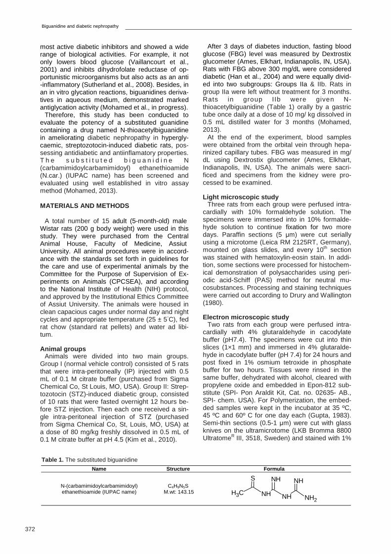
Therefore, this study has been conducted to evaluate the potency of a substituted guanidine containing a drug named N-thioacetylbiguanidine in ameliorating diabetic nephropathy in hypergly-caemic, streptozotocin-induced diabetic rats, pos-sessing antidiabetic and antiinflamatory properties. T h e s u b s t i t u t e d b i g u a n i d i n e N (carbamimidoylcarbamimidoyl) ethanethioamide (N.car.) (IUPAC name) has been screened and evaluated using well established in vitro assay method (Mohamed, 2013).
MATERIALS AND METHODS
A total number of 15 adult (5-month-old) male Wistar rats (200 g body weight) were used in this study. They were purchased from the Central Animal House, Faculty of Medicine, Assiut University. All animal procedures were in accord-ance with the standards set forth in guidelines for the care and use of experimental animals by the Committee for the Purpose of Supervision of Ex-periments on Animals (CPCSEA), and according to the National Institute of Health (NIH) protocol, and approved by the Institutional Ethics Committee of Assiut University. The animals were housed in clean capacious cages under normal day and night cycles and appropriate temperature (25 ± 5◦C), fed rat chow (standard rat pellets) and water ad libi-tum.
Animal groups
Animals were divided into two main groups. Group I (normal vehicle control) consisted of 5 rats that were intra-peritoneally (IP) injected with 0.5 mL of 0.1 M citrate buffer (purchased from Sigma Chemical Co, St Louis, MO, USA). Group II: Strep-tozotocin (STZ)-induced diabetic group, consisted of 10 rats that were fasted overnight 12 hours be-fore STZ injection. Then each one received a sin-gle intra-peritoneal injection of STZ (purchased from Sigma Chemical Co, St, Louis, MO, USA) at a dose of 80 mg/kg freshly dissolved in 0.5 mL of 0.1 M citrate buffer at pH 4.5 (Kim et al., 2010).
After 3 days of diabetes induction, fasting blood glucose (FBG) level was measured by Dextrostix glucometer (Ames, Elkhart, Indianapolis, IN, USA). Rats with FBG above 300 mg/dL were considered diabetic (Han et al., 2004) and were equally divid-ed into two subgroups: Groups IIa & IIb. Rats in group IIa were left without treatment for 3 months. Rats in group IIb were given N-thioacetylbiguanidine (Table 1) orally by a gastric tube once daily at a dose of 10 mg/ kg dissolved in 0.5 mL distilled water for 3 months (Mohamed, 2013).
At the end of the experiment, blood samples were obtained from the orbital vein through hepa-rinized capillary tubes. FBG was measured in mg/dL using Dextrostix glucometer (Ames, Elkhart, Indianapolis, IN, USA). The animals were sacri-ficed and specimens from the kidney were pro-cessed to be examined.
Light microscopic study
Three rats from each group were perfused intra-cardially with 10% formaldehyde solution. The specimens were immersed into in 10% formalde-hyde solution to continue fixation for two more days. Paraffin sections (5 µm) were cut serially using a microtome (Leica RM 2125RT, Germany), mounted on glass slides, and every 10th section was stained with hematoxylin-eosin stain. In addi-tion, some sections were processed for histochem-ical demonstration of polysaccharides using peri-odic acid-Schiff (PAS) method for neutral mu-cosubstances. Processing and staining techniques were carried out according to Drury and Wallington (1980).
Electron microscopic study
Two rats from each group were perfused intra-cardially with 4% glutaraldehyde in cacodylate buffer (pH7.4). The specimens were cut into thin slices (1×1 mm) and immersed in 4% glutaralde-hyde in cacodylate buffer (pH 7.4) for 24 hours and post fixed in 1% osmium tetroxide in phosphate buffer for two hours. Tissues were rinsed in the same buffer, dehydrated with alcohol, cleared with propylene oxide and embedded in Epon-812 sub-stitute (SPI- Pon Araldit Kit, Cat. no. 02635- AB., SPI- chem. USA). For Polymerization, the embed-ded samples were kept in the incubator at 35 ºC, 45 ºC and 60º C for one day each (Gupta, 1983). Semi-thin sections (0.5-1 µm) were cut with glass knives on the ultramicrotome (LKB Bromma 8800 UltratomeR III, 3518, Sweden) and stained with 1%
toluidine blue (pH 7.3) for examination on a light microscope (Olympus, Bx50. Model Bx50F-3, SC09160, Tokyo, Japan). Ultrathin sections (50-80 nm) were cut from selected areas of the blocks on a Reichert ultramicrotome (Leica WILDM3Z, 89386, Austria), placed on copper grids (G300, 3.05 mm, Polaron Equipment Ltd. Watford. Eng-land) and contrasted with uranyl acetate and lead citrate. These sections were examined using the transmission electron microscope (Jeol EM-100 CX11; Japanese electron optic laboratory, Tokyo, Japan) and photographed at 80 kV.
Immunohistochemical study
Expression of inducible nitric oxide synthase (iNOS) was detected in formalin- fixed paraffin- embedded sections (Yabuki et al., 2006). Sections (5 µm) were deparaffinized in xylene and rehydrat-ed in alcohol. The (iNOS) Rabbit Polyclonal Anti-body (Thermo Fisher Scientific, Fremont, CA 94538-6406, USA) was used at 1:100 dilution for 20 minutes at room temperature. Sections were boiled in 10mM citrate buffer, pH 6.0 for 15 minutes followed by cooling at room temperature for 20 minutes. Sections were processed accord-ing to the manufacture instructions using the uni-versal kit (Ultra Vision LP system, HRP polymer & DAB plus chromogen, Thermo Fisher Scientific, Fremont, CA 94538-6406, USA). After completion of the reaction, counterstaining was done using Mayer’s haematoxylin, dehydrated and cover-slipped using DPX (Oxford laboratory reagents, Bombay, India).
Morphometric study
Using computer-assisted image analysis (Soft Imaging System, Analysis-2004, Olympus Compa-ny, Tokyo, Japan), the arbitary area of the glomer-uli (renal corpuscles) was measured on H&E-stained sections (Kiran et al., 2012). Thirty glomer-uli per animal group were measured using X40 objective lens.
Statistical analysis
The morphometric data of each animal group were statistically analyzed using the computer sta-tistics Prism-5.0 package (GraphPad Software, Inc., San Diego, CA, USA). One-way analysis of variance (ANOVA) followed by Newman-Kelus test as a post-test was employed to compare the stud-ied animal group. The results were expressed as mean ± standard deviation (SD). P-value < 0.05 was considered significant.
RESULTS
Histological Examinations Group I
Renal histological sections from control animals exhibited normal architecture. The proximal convo-
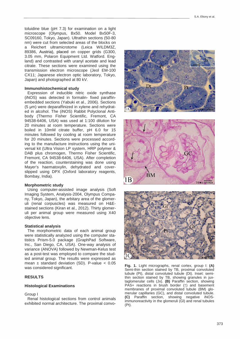
Fig. 1. Light micrographs, renal cortex, group I: (A) Semi-thin section stained by TB, proximal convoluted tubule (Pt), distal convoluted tubule (Dt). Inset: semi-thin section stained by TB, showing granules in jux-taglomerular cells (Jx). (B) Paraffin section, showing PAS+ reactions in brush border (↑) and basement membranes of proximal convoluted tubule (BM) glo-merular capillaries (GC), and distal convoluted tubule. (C) Paraffin section, showing negative iNOS-immunoreactivity in the glomeruli (Gl) and renal tubules (Pt).
luted tubules (PTs) had an intact brush border, vesicular round nuclei and granular cytoplasm (Fig. 1A). The distal convoluted tubules (DTs) showed wider lumen with ill-defined brush border (Fig. 1A). The juxtaglomerular cells (JGCs) of the afferent arteriole revealed renin secretory granules in their cytoplasm (Fig. 1A, inset).
PAS stain demonstrated + reactions in the base-ment membranes of the parietal layer lining Bow-man's capsule, the glomerular capillary loops, the mesangial matrix and the renal tubules, and the brush border of the PTs (Fig. 1B). Using immune-reactivity, no expression of iNOS was observed in the renal tissue (Fig. 1C).
In ultrastructure, the cells of the PTs showed characteristic numerous apical microvilli, ovoid eu-chromatic nuclei and basal in-foldings of plasma membrane with many basally located elongated mitochondria (Fig. 2A). The apical cytoplasm con-tained a few vacuoles (Fig. 2A). The lining cells of the DTs had a few luminal microvilli, and the nuclei were more apically located with less basal in-foldings of plasma membrane and elongated mito-
chondria (Fig. 2B). The podocytes which lined the visceral layer of Bowman's capsule embraced the glomerular capillaries. They exhibited large and irregular nuclei (Fig. 2C), several primary cytoplas-mic processes which arose from their body, and in turn gave secondary processes or pedicles cover-ing the glomerular capillary basement membrane (Fig. 2C). The JGCs of the afferent arteriole re-vealed intra-cytoplasmic dark granules (Fig. 2D).
Group IIa
Renal sections of group IIa demonstrated chang-es consistent with early diabetic nephropathy, including tubular dilation and exfoliated epithelial casts in the lumina. The epithelial cells lining PT were distorted with vacuolated cytoplasm, numerous dark granules and loss of brush border(Fig. 3A). The DTs exhibited dark nuclei, vacuolated cytoplasm, disrupted and dilated capil-laries (Fig. 3A). PAS reaction revealed a strong positive reaction in the basement membranes of the parietal layer lining Bowman's capsule, glomer-ular capillary loops, the mesangial matrix and the
S.A. Eltony et al.
375
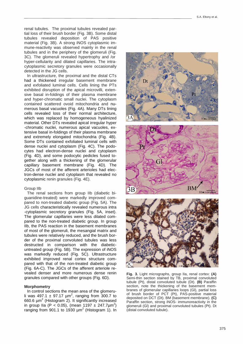
Fig. 3. Light micrographs, group IIa, renal cortex: (A) Semi-thin section stained by TB, proximal convoluted tubule (Pt), distal convoluted tubule (Dt). (B) Paraffin section, note the thickening of the basement mem-branes of glomerular capillaries loops (Gl), partial loss of brush border of PCT (Pt), PAS-positive material deposited on DCT (Dt). BM (basement membrane). (C) Paraffin section, strong iNOS- immunoreactivity in the glomeruli (Gl) and proximal convoluted tubules (Pt). Dt (distal convoluted tubule).
renal tubules. The proximal tubules revealed par-tial loss of their brush border (Fig. 3B). Some distal tubules revealed deposition of PAS positive material (Fig. 3B). A strong iNOS cytoplasmic im-mune-reactivity was observed mainly in the renal tubules and in the periphery of the glomeruli (Fig. 3C). The glomeruli revealed hypertrophy and /or hyper-cellularity and dilated capillaries. The intra-cytoplasmic secretory granules were occasionally detected in the JG cells.
In ultrastructure, the proximal and the distal CTs had a thickened irregular basement membrane and exfoliated luminal cells. Cells lining the PTs exhibited disruption of the apical microvilli, exten-sive basal in-foldings of their plasma membrane and hyper-chromatic small nuclei. The cytoplasm contained scattered ovoid mitochondria and nu-merous basal vacuoles (Fig. 4A). Many DTs lining cells revealed loss of their normal architecture, which was replaced by homogeneous hyalinized material. Other DTs revealed apical irregular hyper-chromatic nuclei, numerous apical vacuoles, ex-tensive basal in-foldings of their plasma membrane and extremely elongated mitochondria (Fig. 4B). Some DTs contained exfoliated luminal cells with dense nuclei and cytoplasm (Fig. 4C). The podo-cytes had electron-dense nuclei and cytoplasm (Fig. 4D), and some podocytic pedicles fused to-gether along with a thickening of the glomerular capillary basement membrane (Fig. 4D). The JGCs of most of the afferent arterioles had elec-tron-dense nuclei and cytoplasm that revealed no cytoplasmic renin granules (Fig. 4E).
Group IIb
The renal sections from group IIb (diabetic bi-guanidine-treated) were markedly improved com-pared to non-treated diabetic group (Fig. 5A). The JG cells characteristically revealed numerous intra-cytoplasmic secretory granules (Fig. 5A, inset). The glomerular capillaries were less dilated com-pared to the non-treated diabetic group. In group IIb, the PAS reaction in the basement membranes of most of the glomeruli, the mesangial matrix and tubules were relatively reduced, and the brush bor-der of the proximal convoluted tubules was less destructed in comparison with the diabetic-untreated group (Fig. 5B). The expression of iNOS was markedly reduced (Fig. 5C). Ultrastructure exhibited improved renal cortex structure com-pared with that of the non-treated diabetic group (Fig. 6A-C). The JGCs of the afferent arteriole re-vealed denser and more numerous dense renin granules compared with other groups (Fig. 6D).
Morphometry
In control sections the mean area of the glomeru-li was 497.1 ± 97.17 µm2, ranging from 300.7 to 660.6 µm2 (Histogram 2). It significantly increased in group IIa (P < 0.05), (mean 1197 ± 247.7µm2) ranging from 901.1 to 1930 µm2 (Histogram 1). In
Biguanidine and diabetic nephropathy
376
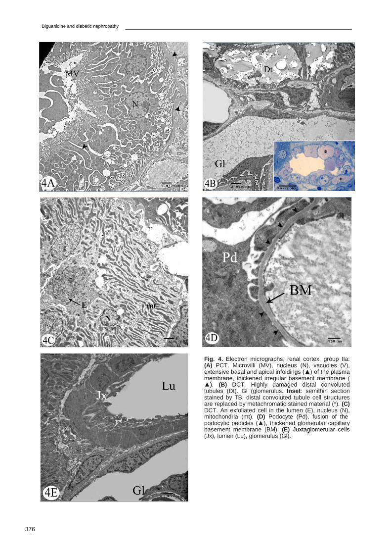
Fig. 4. Electron micrographs, renal cortex, group IIa: (A) PCT. Microvilli (MV), nucleus (N), vacuoles (V), extensive basal and apical infoldings (▲) of the plasma membrane, thickened irregular basement membrane (▲). (B) DCT. Highly damaged distal convoluted tubules (Dt). Gl (glomerulus. Inset : semithin section stained by TB, distal convoluted tubule cell structures are replaced by metachromatic stained material (*). (C) DCT. An exfoliated cell in the lumen (E), nucleus (N), mitochondria (mt). (D) Podocyte (Pd), fusion of the podocytic pedicles (▲), thickened glomerular capillary basement membrane (BM). (E) Juxtaglomerular cells (Jx), lumen (Lu), glomerulus (Gl).
S.A. Eltony et al.
377
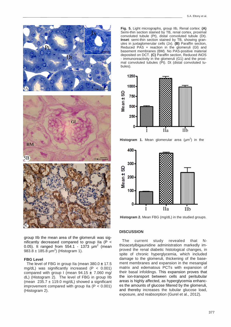
group IIb the mean area of the glomeruli was sig-nificantly decreased compared to group IIa (P < 0.05). It ranged from 554.1 - 1373 µm2 (mean 983.8 ± 185.8 µm2) (Histogram 1).
FBG Level
The level of FBG in group IIa (mean 380.0 ± 17.5 mg/dL) was significantly increased (P < 0.001) compared with group I (mean 94.15 ± 7.060 mg/dL) (Histogram 2). The level of FBG in group IIb (mean 235.7 ± 119.0 mg/dL) showed a significant improvement compared with group IIa (P < 0.001) (Histogram 2).
Fig. 5. Light micrographs, group IIb, Renal cortex: (A) Semi-thin section stained by TB, renal cortex, proximal convoluted tubule (Pt), distal convoluted tubule (Dt). Inset : semi-thin section stained by TB, showing gran-ules in juxtaglomerular cells (Jx). (B) Paraffin section, Reduced PAS + reaction in the glomeruli (Gl) and basement membranes (BM). No PAS-positive material deposited on DCT. (C) Paraffin section, Reduced iNOS- immunoreactivity in the glomeruli (G1) and the proxi-mal convoluted tubules (Pt). Dt (distal convoluted tu-bules).
Histogram 2. Mean FBG (mg/dL) in the studied groups.
Histogram 1. Mean glomerular area (µm2) in the
DISCUSSION
The current study revealed that N-thioacetylbigaunidine administration markedly im-proved the renal diabetic histological changes, in spite of chronic hyperglycemia, which included damage to the glomeruli, thickening of the base-ment membranes and expansion in the mesangial matrix and edematous PCTs with expansion of their basal infoldings. This expansion proves that the ion-transport between cells and peritubular areas is highly affected, as hyperglycemia enhanc-es the amounts of glucose filtered by the glomeruli, and thereby increases the tubular glucose load, exposure, and reabsorption (Gurel et al., 2012).
Biguanidine and diabetic nephropathy
378
The immune-reactivity for the enzyme iNOS in the normal kidneys was low or undetectable, whereas a strong expression of iNOS in diabetic rat-proximal tubules is in accordance with several studies (Bank et al., 1996; Heeringa et al., 1998; Chou et al., 2002), although some studies denied its expression in diabetic kidney Veelken et al., 2000; Chen et al., 2005). In support of our find-ings, a critical role of iNOS is suggested in the pro-gressive renal dysfunction and structural injury. Gurel et al. (2012) reported that increased iNOS caused kidney tissue degeneration, whereas inhi-bition of iNOS ameliorated renal damage.
The use of N-thioacetylbiguanidine suppressed iNOS immune-reaction in the renal tissue. Use of N-thioacetylbiguanidine demonstrated marked an-tiglycation activity in an in vitro glycation reactions (Mohamed et al., in progress). It is a nucleophilic hydrazine compound like AG, which possesses multiple biological effects including prevention of AGEs formation (Vlassara, 1994), inhibition of dia-mino oxidase (Sessa and Perin, 1994) and selec-
tive inhibition of iNOS (Corbett et al., 1992; Misko et al., 1993). In support of our hypothesis, some investigators suggested that AG inhibits a patho-physiologic function of iNOS independent of free radical-mediated lipid peroxidation or significant effects on AGEs deposition (Reckelhoff et al., 1999).
Suppression of renin in JGCs of a diabetic kidney, detected in his work, was frequently ob-served (Christlieb et al., 1976; Perez et al., 1977). The low renin state has been attributed to sclerotic renal arterioles and glomeruli, possibly amplified by functional factors such as autonomic neuropa-thy and sodium retention. The use of the substitut-ed biguanidin produced a remarkable decrease in renin release reflected by the presence of numer-ous granules in JGCs.
A permissive role for nitric oxide in renin re-lease has been suggested (Beierwaltes, 1994, 1995; Reid and Chou, 1995). According to Kurtz (2011), nitric oxide appears as a tonic enhancer of renin secretion, acting via inhibition of cAMP deg-radation through the action of cGMP.
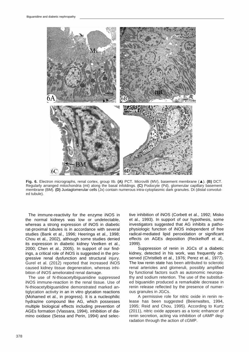
Fig. 6. Electron micrographs, renal cortex, group IIb. (A) PCT. Microvilli (MV), basement membrane (▲). (B) DCT. Regularly arranged mitochondria (mt) along the basal infoldings. (C) Podocyte (Pd), glomerular capillary basement membrane (BM). (D) Juxtaglomerular cells (Jx) contain numerous intra-cytoplasmic dark granules. Dt (distal convolut-ed tubule).
S.A. Eltony et al.
379
Conclusion
In this study we report that N-thioacetylbiguanidine attenuated the histological changes of diabetic nephropathy reaching healing features, which resembled that of a normal kidney, reduced iNOS tissue reactivity and increased renin secretory granules in the JGCs.
BOLTON WK, CATTRAN DC, WILLIAMS ME, ADLER SG, APPEL GB, CARTWRIGHT K, FOILES PG, FREEDMAN BI, RASKIN P, RATNER RE, SPI-NOWITZ BS, WHITTIER FC, WUERTH JP (2004) Action I Investigator Group: Randomized trial of an inhibitor of formation of advanced glycation end prod-ucts in diabetic nephropathy. Am J Nephrol, 24: 32-40.
CHEN H, BRAHMBHATT S, GUPTA A, SHARMA AC (2005) Duration of streptozotocin-induced diabetes differentially affect sp38-Mitogen-Activated Protein Kinase (MAPK) phosphorylation in renal and vascular dysfunction. Cardiovasc Diabetol, 4: 3.
CHOU DE, CAI H, JAYADEVAPPA D, PORUSH JG (2002) Regional expression of inducible nitric oxide synthase in the kidney stimulated by D. lipopolysac-charide in the rat. Exp Physiol, 87: 153-162.
CHRISTLIEB AR, KALDANY A, D’ELIA JA (1976) Plas-ma renin activity and hypertension in diabetes mellitus. Diabetes, 25: 969-974.
CORBETT JA, TILTON RG, CHANG K, HASAN KS, IDO Y, WANG JL, SWEETLAND MA, LANCASTER JR Jr, WILLIAMSON JR, McDANIEL ML (1992) Ami-noguanidine: A novel inhibitor of nitric oxide formation prevents diabetic vascular dysfunction. Diabetes, 41: 552-556.
CORBETT JA, MC DANIEL ML (1996) The use of ami-noguanidine, a selective iNOS inhibitor, to evaluate the role of nitric oxide in the development of autoimmune diabetes. Methods, 10: 21-30.
DRURY RAB, WALLINGTON EA (1980) Carelton’s His-tology Technique, 5th edit. Oxford University Press, Oxford, New York, Toronto.
GUPTA PD (1983) Ultrastructural study on semithin sections. Science Tools, 30: 6-7.
GUREL E, YILMAZER N, DEMIRCI-TANSEL C (2012) The effect of aminoguanidine on the kidney of diabetic albino Balb/c mice. IUFS J Biol, 71: 17-29.
HAN SY, SO G, JEE YH, HAN KH, KANG YS, KIM HK, KANG SW, HAN DS, HAN JY, CHA DR (2004) Effect
of retinoic acid in experimental diabetic nephropathy. Immunol Cell Biol, 82: 568-576.
HEERINGA P, VAN GOOR H, MOSHAGE H, KLOK PA, HUITEMA MG, DE JAGER A, SCHEP AJ, KALLEN-BERG CG (1998) Expression of iNOS, eNOS and per-oxynitrite-modified proteins in experimental anti-myeloperoxidase associated crescentic glomerulone-phritis. Kidney Internat, 53: 382-393.
IHM SH, YOO HJ, PARK SW, IHM J (1999) Effect of aminoguanidine on lipid peroxidation in streptozotocin-induced diabetic rats. J Metabol, 48: 1141-1145.
KELLY DJ, GILBERT RE, COX AJ, SOULIS T, JERUMS G, COOPER ME (2001) Aminoguanidine ameliorates overexpression of prosclerotic growth factors and col-lagen deposition in experimental diabetic nephropathy. J Am Soc Nephrol, 12: 2098-2107.
KIM JB, SONG BW, PARK S, HWANG KC, CHA BS, JANG Y, LEE HC, LEE MH (2010) Alagebrium chlo-ride, a novel advanced glycation end-product cross linkage breaker, inhibits neointimal proliferation in a diabetic rat carotid balloon injury model. Korean Circ J, 40: 520-526.
KIRAN G, NANDINI CD, RAMESH HP, SALIMATH PV (2012) Progression of early phase diabetic nephropa-thy in streptozotocin-induced diabetic rats: Evaluation of various kidney-related parameters. Indian J Exp Biol, 50: 133-140.
KURTZ A (2011) Renin release: sites, mechanisms, and control. Ann Rev Physiol, 17: 377-399.
MISKO TP, MOORE WM, KASTEN TP, NICKOLS GA, CORBETT JA, TILTON RG, MCDANIEL ML, WIL-LIAMSON JR, CURRIE MG (1993) Selective inhibition of the inducible nitric oxide synthase by aminoguani-dine. Eur J Pharmacol, 233: 119-125.
MOHAMED S (2013) Effect of guanidine compounds on glycation of proteins in vitro. Master Thesis, Manches-ter Metropolitan University.
NATHAN DM (1993) Long-term complications of diabe-tes mellitus. New Engl J Med, 328: 1676-1685.
PEREZ GO, LESPIER L, JACOBI J (1977) Hyporeninemia and hypoaldosteronism in diabetes mellitus. Archieve Internat J Med, 137: 852-855.
PONGSUKWETCHAKUL A, KULAPUTANA O, PA-TUMRAJ S, UDAYACHALERM W (2007) Genistein improves blood sugar and glycated hemoglobin levels in diabetic rats. Thai J Physiol Science, 20: 31-36.
RECKELHOFF JF, HENNINGTON BS, KANJI V, RA-CUSEN LC, SCHMIDT AM, YAN SD, MORROW J, ROBERTS LJ 2nd, SALAHUDEEN AK (1999) Chronic aminoguanidine attenuates renal dysfunction and inju-ry in aging rats. Am J Hypertens, 12: 492-498.
REID IA, CHOU L (1995) Effect of blockade of nitric oxide synthesis on the renin secretory response to furosemide in conscious rabbits. Clin Sci, 88: 657-663.
KOUSAR S, SHEIKH MA, ASGHAR M, SARWAR M (2009) Effect of aminoguanidine on advanced gly-cation end products (AGEs) using normal and diabetic plasma. J Chem Soc Pakistan, 31: 109-114.
SAKAMOTO I, ITO Y, MIZUNO M, SUZUKI Y, SAWAI A, TANAKA A, MARUYAMA S, TAKEI Y, YUZAWA Y,
Biguanidine and diabetic nephropathy
380
MATSUO S (2009) Lymphatic vessels develop during tubulointerstitial fibrosis. Kidney Int, 75: 828-838.
SESSA A, PERIN A (1994) Diamine oxidase in relation to diamine and polyamine metabolism. Agents Actions, 43: 69-77.
SCHENA FP, GESUALDO L (2005) Pathogenetic mech-anisms of diabetic nephropathy. J Am Soc Nephrol, 16: S30-S33.
SUTHERLAND CJ, LAUNDY M, PRICE N, BURKE M, FIVELMAN QL, PASVOL G, JOHN L, KLEIN JL, CHI-ODINI PL(2008) Mutations in the Plasmodium falcipa-rum cytochrom eb gene are associated with delayed parasite recrudescence in malaria patients treated with atovaquone-proguanill. Malaria J, 7: 240.
VEELKEN R, HILGERS KF, HARTNER A, HAAS A, BÖHMER KP, STERZEL B (2000) Nitric oxide syn-thase isoforms and glomerular hyperfiltration in early diabetic nephropathy. J Am Soc Nephrol, 11: 71-79.
VLASSARA H (1994) Recent progress on the bio-logic and clinical significance of advanced glyco-sylation end products. J Lab Clin Med, 124: 19-30.
VAILLANCOURT VA, LARSEN SD, TANIS SP, BURR J E, CONNELL M A, CUDAHY MM, EV-ANS BR, FISHER P V, MAY P D, MEGLASSON MD, ROBINSON D D, STEVENS FC, TUCKER J A, VIDMAR TJ, YU JH (2001) Synthesis and bio-logical activity of aminoguanidine and diamino-guanidine analogues of the antidiabetic/antiobesity agent 3-guanidinopropionic acid. J Medicinal Chem, 44: 1231-1248.
WEST IC (2000) Radicals and oxidative stress in diabe-tes. Diabetic Med, 17: 171-180.
YABUKI A, TAHARA T, TANIGUCHI K, MATSUMOTO M, SUZUKI S (2006) Neuronal nitric oxide synthase and cyclooxygenase-2 in diabetic nephropathy of type 2 diabetic OLETF rats. Exp Animals, 55: 17-25.


![Hypericum Perforatum L. Protective Effects on Fat- and ... · levels of vitamins decreased in DMBA-treated rat group, but levels of vitamins increased in . HP-treated rat group [3].](https://static.documents.pub/doc/80x56/611e4e1f525d6d085d3d5532/hypericum-perforatum-l-protective-effects-on-fat-and-levels-of-vitamins-decreased.jpg)

