20
Protein Crystallography Part I Tim Grüne Dept. of Structural Chemistry Prof. G. Sheldrick University of Göttingen [email protected]

Protein CrystallographyPart I
Tim GrüneDept. of Structural Chemistry
Prof. G. SheldrickUniversity of Göttingen

Overview
• Crystal Lattice and Symmetry
• Growing Protein Crystals
• X-ray diffraction
• Bragg’s Law
• Resolution
• Electron Density
Molecular Biology 1 Protein Crystallography I

Examples of Protein Crystals
Kirsten Böttcher et al.
Molecular Biology 2 Protein Crystallography I

Crystals
A crystal consists of an infinite number of copies of one object. This object can be as small as a singleatom or ion ( e.g. NaCl or diamond crystals) or as big as a protein-RNA complex as the ribosome.
The objects must form a regular pattern, the “crystal lattice”.
The main characteristic of a lat-tice is its translational invari-ance: If you move from onelattice point to another one butkeep looking in the same direc-tion, you can tell no difference.
Molecular Biology 3 Protein Crystallography I

Seven Lattice Types
The lattice is an infinite repetition of one “box”, the unit cell. It is defined by the lengths of three edges a,b, c, and the angles α, β, and γ. There are 7 different lattice types that allow to fill an infinite space.
a 6= b 6= c
α 6= β 6= γ
triclinic
a 6= b 6= c
α = γ = 90◦ 6= β
a = b 6= c
tetragonal
a 6= b 6= c
orthorhombic
a = b = c
α = β = 90◦
γ = 120◦
hexagonal monoclinic
α=
β=
γ=
90◦
cubic
trigonal
a = b 6= c
α = β = 90◦
γ = 120◦
b
a
a = b = c
γ
βcα
Molecular Biology 4 Protein Crystallography I

Crystal Symmetry
Only a limited number of “operations” are allowed to go from one object to the next one. These operationscan be translations by a vector (h · a, k · b, l · c) with h, k, l integers, but also symmetry operations, e.g.
• rotations (only 2-, 3-, 4-,and 6-fold axes possible)
2-fold 3-fold 4-fold 6-fold
• mirrors and inversion cen-tres
centre of inversionmirror plane
Molecular Biology 5 Protein Crystallography I

Space Groups
In a three dimensional lattice, symmetry operations cannot be combined arbitrarily. Together with transla-tions, there are 230 allowed combinations, the 230 space groups. Of those, only 65 are chiral, i.e. suitablefor macromolecules like proteins, RNA, or DNA.
An important example of combining two symmetry elements is the screw axis: rotation with simultaneoustranslation along an axis.
The example shows a 41 axis. A rotation of 1/4 ·360◦ accompanied by a translation of 1/4 of thelength of the axis.
Molecular Biology 6 Protein Crystallography I

Unit cell and asymmetric unit
In every crystal there is always a smallest box, defined by its edges a, b, and c, and the angles theyenclose, α, β, and γ, from which one can create the whole crystal solely by integer translations along itssides. This is called the unit cell.
(0,b)
b
a(2a,0)
(a,2b)
A unit cell consists itself of a smallest unit from which one can create the unit cell by applying all symmetryoperators that belong to the crystals space group. This is called the asymmetric unit.
Molecular Biology 7 Protein Crystallography I

Growing Crystals
Protein crystals are usually grown from solution. The probably most common way is to apply the “vapourdiffusion” method.
The protein must be very pure ( ≥ 90–95% ) andhighly concentrated ( several mg/ml). The reservoircontains a buffer, a precipitant ( salts, PEG’s withM∅
r = 400–20,000, organic solvents) and additives(small molecules that help packing, e.g. MgCl2).
equilibration
reservoirsolution
before equilibration
protein/reservoir
after equilibration
This method is very fast and one can perform a large number of experiments in a rather short time. Robotsare used in some laboratories, especially in companies, that can set up 100 drops in 1/2 hour.
Other techniques include
batch dialysis agarose
oil seal membraneagarose
Molecular Biology 8 Protein Crystallography I
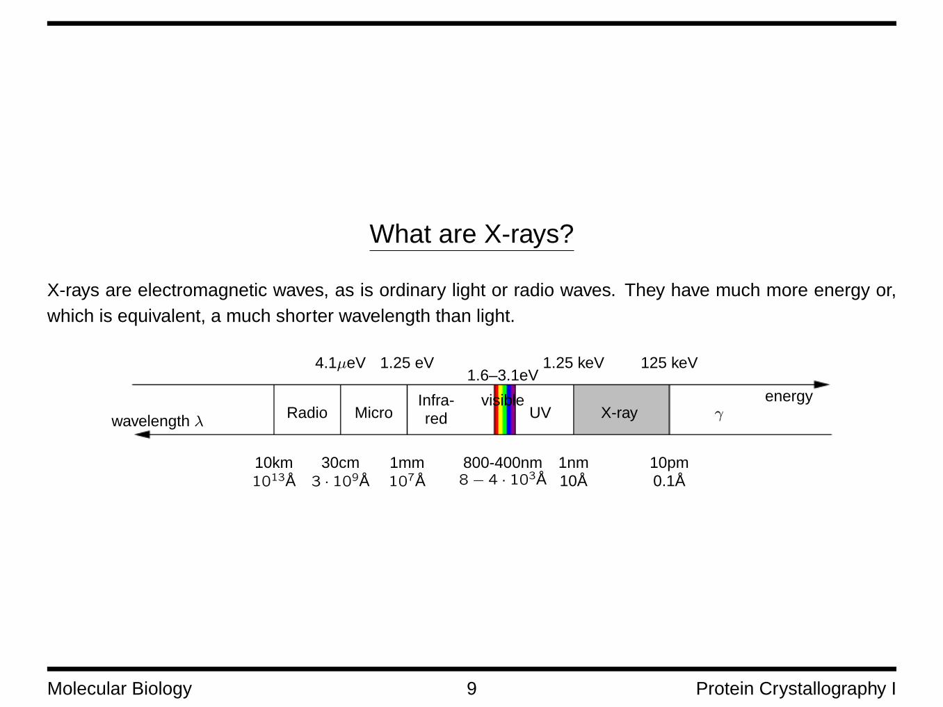
What are X-rays?
X-rays are electromagnetic waves, as is ordinary light or radio waves. They have much more energy or,which is equivalent, a much shorter wavelength than light.
wavelength λRadio Micro
visibleUV X-ray γ
Infra-red
10km1013Å
energy
125 keV1.25 keV1.25 eV4.1µeV1.6–3.1eV
30cm3 · 109Å
1mm107Å
800-400nm8− 4 · 103Å
1nm10Å
10pm0.1Å
Molecular Biology 9 Protein Crystallography I

X-ray diffraction
X-ray beam
λ ≈ 1Å(0.1nm)
crystal ≈ (0.2mm)3
Diffraction pattern on CCDor image plate
≈ 1013 unit cells
T. Schneider
Molecular Biology 10 Protein Crystallography I
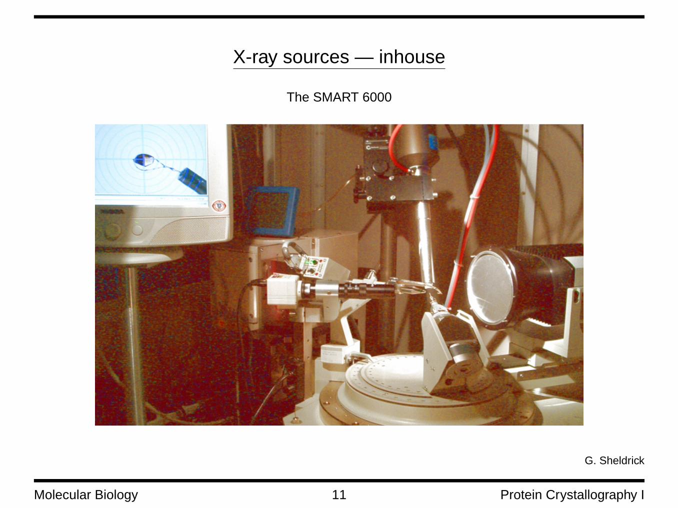
X-ray sources — inhouse
The SMART 6000
G. Sheldrick
Molecular Biology 11 Protein Crystallography I
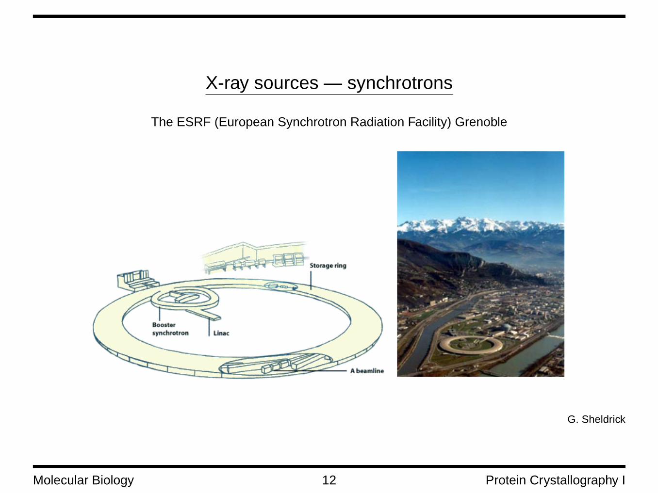
X-ray sources — synchrotrons
The ESRF (European Synchrotron Radiation Facility) Grenoble
G. Sheldrick
Molecular Biology 12 Protein Crystallography I

Bragg’s Law — Crystal Planes
The lattice points in the crystal define sets of parallel planes. In 3 dimensions the planes are defined bythree integers (h, k, l), the so-called “Miller indices”. h denotes the number of intersections of the a-axisof the crystal’s unit cell by the set, etc.
Each set of parallel planes runs through lattice points. The distance d between two planes is given by
1
d2=
h2
a2+
k2
b2+
l2
c2
Molecular Biology 13 Protein Crystallography I

Bragg Reflections
The X-ray beam is reflected at sets of planes in the crystal. Dueto interference, a plane does only produce a reflection spot, ifBragg’s law is met:
λ = 2d sin θ
λ is the wavelength of the beam, d the distance between theplanes of one set.Every spot that can be seen on a diffraction image originatesfrom one set of planes that is in diffracting orientation accordingto Bragg’s law. The spot can be indexed with the same (h, k, l)
values that describe the set of planes.
Θ Θ
} d
(Bragg’s law is actually nλ = 2d sin θ, but n > 1 refers to higher order reflections that are too weak to be recorded for “normal”
crystals.)
Molecular Biology 14 Protein Crystallography I
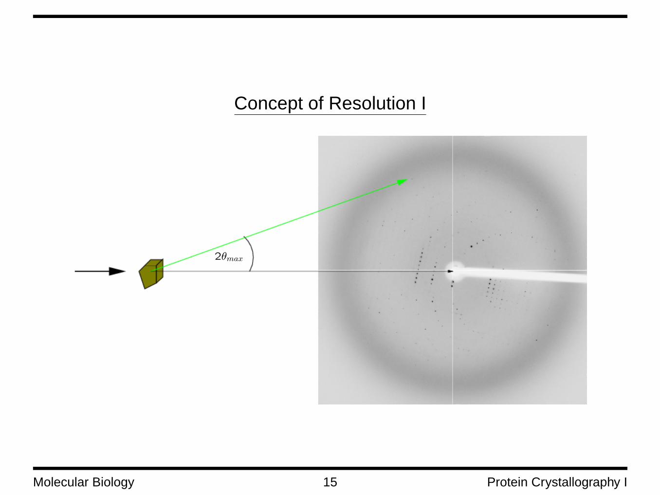
Concept of Resolution I
2θmax
Molecular Biology 15 Protein Crystallography I

The Concept of Resolution II
The resolution of data collected by X-ray diffraction is a measure for how much detail can be seen. It isrelated with the plane distance d through Bragg’s law by
d =λ
2 sin θmax
θmax is the maximum angle to which data could be collected.
The resolution corresponds quite well to the minimum distance between two atoms that can still be re-solved in the electron density map.
high resolution
d
low resolution
d’
Molecular Biology 16 Protein Crystallography I

Resolution — Examples
G. Sheldrick
Molecular Biology 17 Protein Crystallography I

X–ray diffraction → Electron Density → Model !?
The intensities of the reflections measured by an X-ray diffraction experiment are proportional to thesquare modulus of the structure factors.
Every reflection marked by (h, k, l) has its origin in a structure factor F (h, k, l). The structure factors arerelated to the electron density distribution within the unit cell by an expression called Fourier transforma-tion:
ρ(x, y, z) =1
Vunitcell
h,k,l=∞∑h,k,l=−∞
F (h, k, l) · e−2πi(hx+ky+lz)
and its inversion
F (h, k, l) =∫Vunitcell
d3x ρ(x, y, z)e2πi(hx+ky+lz)
If we knew all structure factors, we could calculate the electron density in the whole unit cell.
A major effort of crystallography lies in the determination of as many and as accurate structure factors aspossible.
Molecular Biology 18 Protein Crystallography I

Limitations of Data Collection
Every X-ray data collection has a limited resolution. There are various reasons for this:
1. it is not possible to determine an infinite number of reflections⇒ resolution limit, truncation errors in Fourier transform
2. due to Bragg’s law, even a perfect crystal has a resolution limit
3. crystals have limited size, i.e. there are only a finite number of planes. This becomes an issue forsmall crystals with a large unit cell
4. Crystal imperfections, mostly mosaicity — it leads to a broadening of the signal so that weak reflec-tions vanish in the background noise
Molecular Biology 19 Protein Crystallography I


![Protein Crystallography - instruct.uwo.ca · Protein Crystallization • Principles of protein solubility [PPt] [protein] Undersaturated solubility Supersaturated Precipitation Nucleation](https://static.documents.pub/doc/80x56/5e18b58cfac19c6065246f42/protein-crystallography-protein-crystallization-a-principles-of-protein-solubility.jpg)

![Protein structure determination. Tertiary protein structure: protein folding Three main approaches: [1] experimental determination (X-ray crystallography,](https://static.documents.pub/doc/80x56/56649d3e5503460f94a17891/protein-structure-determination-tertiary-protein-structure-protein-folding.jpg)


