Page 1
Sensors 2008, 8, 4296-4307; DOI: 10.3390/s8074296
sensors ISSN 1424-8220
www.mdpi.org/sensors Review
Protein Detection with Aptamer Biosensors
Beate Strehlitz *, Nadia Nikolaus and Regina Stoltenburg
UFZ - Helmholtz Centre for Environmental Research, UBZ, Permoserstr. 15, 04318 Leipzig, Germany;
E-Mails: [email protected] ; [email protected]
* Author to whom correspondence should be addressed; E-mail: [email protected] ;
Tel.: +49-341-235-1764; Fax: +49-341-235-451764
Received: 15 June 2008; in revised form: 7 July 2008 / Accepted: 21 July 2008 /
Published: 23 July 2008
Abstract: Aptamers have been developed for different applications. Their use as new
biological recognition elements in biosensors promises progress for fast and easy detection
of proteins. This new generation of biosensor (aptasensors) will be more stable and well
adapted to the conditions of real samples because of the specific properties of aptamers.
Keywords: aptamer, protein, biosensor, SELEX
1. Introduction
There is a high demand for convenient methodologies for detecting and measuring the levels of
specific proteins in biological and environmental samples because their detection, identification and
quantification can be very complex, expensive and time consuming. Biosensors are interesting tools
offering certain operational advantages over standard photometric methods, notably with respect to
rapidity, ease-of-use, cost, simplicity, portability, and ease of mass manufacture. Biosensors have been
developed for more than 25 years now, and have been commercialized for some special applications
like blood glucose and lactate measurement or bioprocess control, amongst others. However, they have
not entered the market as much as expected, which is caused by several reasons. One reason is the
instability of the biological recognition element of the biosensor (e.g. enzymes, cells or antibodies).
Aptamers, which are ssDNA or RNA oligonucleotides, can bind to their targets due to their specific
three dimensional structures; they offer specific properties which favor them as new biorecognition
elements for biosensors. In particular their outstanding and modifiable stability and their regenerative
target binding promise the development of a new biosensor generation. Aptasensors [1] open up new
OPEN ACCESS
Page 2
Sensors 2008, 8
4297
vistas for the detection of analytes which are not accessible to easy and fast detection methods until
now.
Until now, proteins are detected mostly by antibodies in analytical formats like ELISA,
immunobead assay, western blotting, microarrays and also biosensors. Aptamers are equal to
monoclonal antibodies concerning their binding affinities, but furthermore, they provide decisive
advantages. They are more resistant to denaturation and degradation, their binding affinities and
specificities can easily be manipulated and improved by rational design or by techniques of molecular
evolution, and they can be modified with functional groups or tags that allow covalent, directed
immobilization on biochips, resulting in highly ordered receptor layers [2]. Aptamers can distinguish
between chiral molecules and are able to recognize a distinct epitope of a target molecule [3, 4]. In
principle, aptamers can be selected for virtually any desired target, even non-immunogenic or toxic
proteins, because they are produced in vitro by an evolutionary method called SELEX (systematic
evolution of ligands by exponential enrichment) [5, 6], without the constraints imposed by having to be
selected or produced in a living organism. The selection of ligands beyond natural systems emanates
from a chemically produced oligonucleotide library with the big variety of, e.g., 1015 different
oligonucleotides. The number of variation depends on the length of the variable region. With a variable
region of 25 oligonucleotides, there are, theoretically, 425 (≅ 1015) different oligonucleotide sequences
possible. The big variety of the oligonucleotide library and the amplification steps of target-binding
oligonucleotides during the selection process considerably facilitates the selection of ligands with
highest affinity compared to natural selection [7]. Moreover, the SELEX process can be carried out
under conditions akin to those used in the assay for which the aptamer is being developed. As a
consequence, the aptamer will maintain its structure and will function in the final assay. Especially, the
aptamer will not dissociate or otherwise change its characteristics, which can be a problem with
antibodies [8]. The SELEX conditions can be further modified to direct the selection to aptamers with
desired features. This is in contrast to the classical production of antibodies, where it is not possible to
influence such parameters which therefore leaves the resulting bioreceptors (antibodies) limited to
physiological conditions [9]. Another advantage of using aptamers instead of antibodies for biosensing
applications is the fact that non-specific adsorption phenomena are usually less pronounced on nucleic
acid derivated surfaces as compared to protein derivated ones [10].
Although aptamers have been developed for all classes of targets ranging from small molecules to
large proteins and even cells, proteins seem to be the biggest group of target molecules. In principle, it
should be possible to generate aptamers for virtually every protein target. However, it is striking that
there is only a small range of proteins that are detected using aptasensors (cf. Table 1). This review
gives an overview of recent developments and applications of aptamer biosensors for protein detection.
2. Biosensor
As per definition of IUPAC, a biosensor is an integrated receptor-transducer device, which is
capable of providing selective quantitative or semi-quantitative analytical information. The biosensor
consists, on the one hand, of a biological recognition element, which acts upon a biochemical
mechanism, and, on the other hand, of a transducer relying on electrochemical, mass, optical or thermal
principles (Figure (1)). The characteristic trait of a biosensor is the direct spatial contact between the
Page 3
Sensors 2008, 8
4298
biological recognition element (or bioreceptor) and the transducing element [11]. Typical bioreceptors
in biosensors are enzymes, antibodies, microorganisms, and nucleic acids. Aptamers are a new
promising group of bioreceptors, because of their outstanding selectivity, sensitivity and stability, the
reproducibility of the target binding reaction, their production by chemical synthesis ensuring a
constant lot-to-lot quality, and the ease of regeneration of aptamer derivated surfaces.
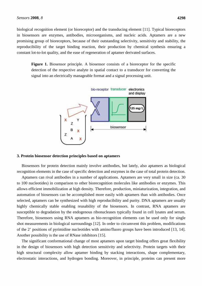
Figure 1. Biosensor principle. A biosensor consists of a bioreceptor for the specific
detection of the respective analyte in spatial contact to a transducer for converting the
signal into an electrically manageable format and a signal processing unit.
3. Protein biosensor detection principles based on aptamers
Biosensors for protein detection mainly involve antibodies, but lately, also aptamers as biological
recognition elements in the case of specific detection and enzymes in the case of total protein detection.
Aptamers can rival antibodies in a number of applications. Aptamers are very small in size (ca. 30
to 100 nucleotides) in comparison to other biorecognition molecules like antibodies or enzymes. This
allows efficient immobilization at high density. Therefore, production, miniaturization, integration, and
automation of biosensors can be accomplished more easily with aptamers than with antibodies. Once
selected, aptamers can be synthesized with high reproducibility and purity. DNA aptamers are usually
highly chemically stable enabling reusability of the biosensors. In contrast, RNA aptamers are
susceptible to degradation by the endogenous ribonucleases typically found in cell lysates and serum.
Therefore, biosensors using RNA aptamers as bio-recognition elements can be used only for single
shot measurements in biological surroundings [12]. In order to circumvent this problem, modifications
of the 2’ positions of pyrimidine nucleotides with amino/fluoro groups have been introduced [13, 14].
Another possibility is the use of RNase inhibitors [15].
The significant conformational change of most aptamers upon target binding offers great flexibility
in the design of biosensors with high detection sensitivity and selectivity. Protein targets with their
high structural complexity allow aptamer binding by stacking interactions, shape complementary,
electrostatic interactions, and hydrogen bonding. Moreover, in principle, proteins can present more
Page 4
Sensors 2008, 8
4299
than one binding site for aptamers, allowing the selection of a pair of aptamers binding to different
regions of the target and enabling sandwich-assay based biosensors.
3.1. Electrochemical aptasensors
Electrochemical transduction of biosensors using aptamers as bioreceptors include methods like
Faradaic Impedance Spectroscopy (FIS), differential pulse voltammetry, alternating current
voltammetry, square wave voltammetry, potentiometry or amperometry.
In principle, it can be differentiated between either a positive or negative readout signal, i.e. an
increase or a decrease of response following upon receptor-target interaction, cf. [10].
Xu et al. demonstrated an electrochemical impedance spectroscopy detection method for aptamer-
modified array electrodes as a promising label-free detection method for IgE [16]. They compared
DNA aptamer based electrodes with anti-human IgE antibody based electrodes and found lower
background noise, decreased nonspecific adsorption, and larger differences in the impedance signals
due to the small size and simple structure of the aptamers in comparison to the antibody [16].
Impedance sensors allow the real-time monitoring of the sensor signal and can give rise to kinetic
aspects of the ligand-analyte interaction [17]. Schlecht et al. have compared an RNA aptamer and an
antibody for thrombin detection by use of a nanometer gap-sized impedance biosensor. They found that
both ligands showed equal suitability for the highly specific detection of their analyte. Their device has
a multiplexer-approach enabling the parallel readout of five sensor elements. This opens up the
possibility to use reference sensors for the elimination of background signals and simultaneous
detection of different analytes by immobilizing their respective ligands on separate electrodes [17].
For impedance methods, usually a negative readout signal can be found in consequence of an
increase in electron transfer resistance. However, Rodriguez et al., 2005 described the set-up of an
impedance-based method exhibiting a positive readout signal [18] making use of the change of surface
charge from negative to positive upon target protein binding (at proper pH).
A very similar approach, also depending on electrostatic interactions, was made by Cheng et al.,
2007. A DNA aptamer for lysozyme was immobilized on gold surfaces by means of self-assembly and
[Ru(NH3)6]3+ bound to the DNA phosphate backbone via electrostatic interaction. The surface density
of aptamers can be determined by measuring the [Ru(NH3)6]3+ reduction peak height in the cyclic
voltammogram. Upon target binding of lysozyme to the aptamers, the surface bound [Ru(NH3)6]3+
cations are released. This can be detected as a decrease in the integrated charge of the reduction peak
[19].
The hindrance of the redox reaction of K3Fe(CN)6 on a gold surface due to an increased density of
the covering layer by binding of the immobilized DNA aptamer with its target thrombin [20] was used
as signal for the binding reaction. The signal was measured by cyclic voltammetry. The aptasensor for
thrombin is reusable and allows measurements in the relevant analytical range for clinical applications
(cf. Table 1) [20].
Another label free method is to use intercalators that bind to double stranded regions of the aptamer.
If these regions are close enough to the electrode, the intercalators can serve as reporters. Upon binding
and the consecutive conformational changes, the intercalator can be released producing a negative
response. An example is described in [21] where an aptamer for thrombin was immobilized on a gold
Page 5
Sensors 2008, 8
4300
electrode. Methylene blue (MB) intercalates into a double strand region and will be released upon
target binding because of the conformational change of the aptamer. The MB cathodic peak current in
the differential pulse voltammogram decreases with increasing thrombin concentration.
These techniques described above are label-free, that is, neither the bioreceptor nor the target has to
be covalently labeled with indicator molecules and this therefore omits a further step in the production
process of the sensor. In contrast, many electrochemical aptasensors rely on the labeling of the
bioreceptor with a reporter unit.
For example, aptamers can be labeled at both ends. At one end, a moiety for immobilization at the
surface is tethered to the aptamer and at the other end, the reporter. The electrode surface is then
covered with a layer of those aptamers. Upon target binding, the mobility of the aptamer and/or the
density of the layer are altered due to beacon-like conformational changes. This results in a smaller or
greater distance of the reporter unit from the electrode leading to an increased or decreased electron
transfer, respectively [22, 23].
Sandwich assays rely on the possibility that more than one aptamer can be generated for one protein
target. One aptamer, attached to the sensor surface, binds the target at one epitope. The second
aptamer, directed to a different epitope is labeled with the reporter, e.g., (PQQ) glucose dehydrogenase.
Binding of the second aptamers to the target brings the reporter in proximity to the sensor surface.
After a washing step, the binding is detected (in this case by amperometry after addition of glucose as a
substrate for (PQQ) glucose dehydrogenase) leading to a positive readout signal via the redox mediator
1-methoxyphenazine methosulfate [24].
3.2. Optical aptasensors
Optical transduction methods in aptasensors comprise, for example, the utilization of surface
plasmon resonance, evanescent wave spectroscopy, as well as fluorescence anisotropy and
luminescence detection.
Surface plasmon resonance (SPR) and evanescent wave based biosensors rely on the change of
optical parameters upon changes in the layer closest to the sensitive surface. Since the binding of, for
example, proteins to a receptor layer of those biosensors changes the refractive index of the layer, the
event of binding can be detected and quantified in a label free way.
Examples for the use of surface plasmon resonance biosensor detection of the respective target
binding to the bioreceptor – the aptamer (in most cases thiolated for the immobilization at gold
surfaces by self-assembly) – can be found in [25], [26] and [27]. Thrombin was captured by a DNA
aptamer immobilized at Biacore chips. Several parameters like incubation time, incubation
temperature effect of immobilization orientation etc. were extensively studied and optimized [25]. IgE
was captured by a DNA aptamer with a detection limit of 2 nM and a linear range of detection from 8.4
to 84 nM using a combination of the methods of SPR and fixed-angle imaging [26]. HIV-1 Tat protein
was captured by an RNA aptamer with a linear detection range from 0 to 2.5 ppm using a Biacore X
instrument. Due to the inherent sensitivity of RNA to nucleases, all instrumentation was freed from
RNases prior to preparation of the sensor chips and measurements [27].
We have constructed a thrombin aptasensor (unpublished results) by immobilisation of the anti-
thrombin aptamer, selected by Bock et al. 1992 [28], via biotin on a streptavidin modified surface of
Page 6
Sensors 2008, 8
4301
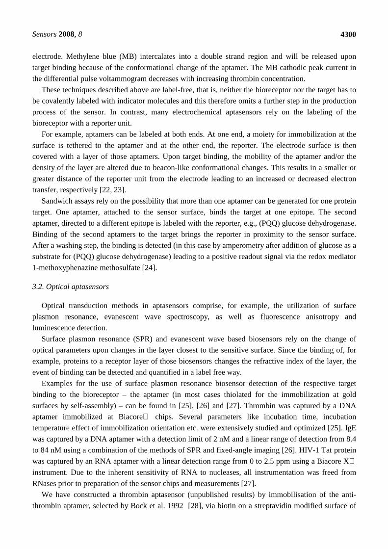
an IAsys cuvette. IAsys (Neosensors Ltd., UK) is a real time evanescent wave biosensor. The binding
of different concentrations of thrombin (0,5 nM – 75 nM) in TA-buffer (20 mM TRIS-HCl, pH 7,4,
140 mM NaCl, 5 mM KCl, 1 mM CaCl2, 1 mM MgCl2) was assayed. Elastase and HSA (25 nM each)
were used as negative controls. The results (binding curves) are shown in Figure (2). The saturation
curve was constructed from the binding curves (measuring time 5 min, Figure (3)). The dissociation
constant Kd was determined by nonlinear regression analysis (Kd = 11.06 nM) and is in good
accordance with published results in the range of 5 to 300 nM detected with different methods [25, 28,
29].
Figure 2. Binding of increasing amounts of human thrombin (0.5 … 75 nM) to the
immobilized 3' biotinylated anti-thrombin aptamer (15 nt, G-quartet), measured by use
of the IAsys-system. Conditions: measurement in TA-buffer (20 mM TRIS-HCl, pH 7.4,
140 mM NaCl, 5 mM KCl, 1 mM CaCl2, 1 mM MgCl2), time 5 min, Negative controls:
Elastase and HSA (25 nM each).
Abrin toxin is highly toxic to eukaryotic cells with possible applications as an immunotoxin in
cancer chemotherapy and as a potential biological warfare agent. A promising rapid and specific
detection method is a DNA aptamer biosensor based on luminescence change detection caused by a
molecular light switching intercalator [Ru(phen)2(dppz)]2+, which binds into duplex nucleic acid
domains of the folded aptamer, emitting luminescence. Conformation changes of the aptamer upon
target binding result in a significant target-dependent luminescence change [30].
An aptamer array sensor was developed for the parallel detection of four analytes (thrombin and the
cancer associated targets inosine monophosphate dehydrogenase II – IMPDH, vascular endothelial
growth factor – VEGF, basic fibroblast growth factor – bFGF). The transduction principle here is based
0 50 100 150 200 250 300 350
0
20
40
60
80
100
120
0,5 nM1 nM
3,5 nM
5 nM
7,5 nM
10 nM
20 nM
35 nM
50 nM
75 nM
Bin
ding
[arc
sec
]
Time [sec]
Elastase 25 nM
HSA 25 nM
Page 7
Sensors 2008, 8
4302
on fluorescence polarization. All four immobilized aptamers (DNA for thrombin, RNA for the others)
showed highly specific responses to their protein targets, even in a complex biological solution [12].
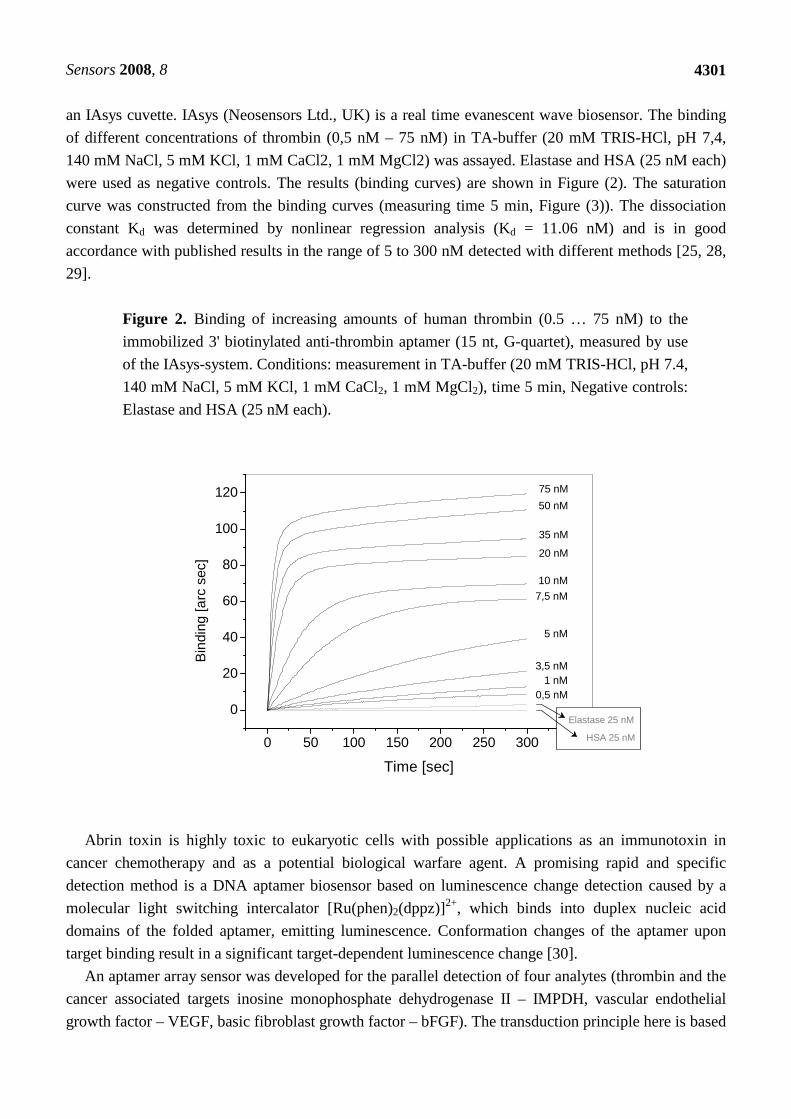
Figure 3. Saturation curves generated from results in Fig.2. Each point represents the
measuring signal for one thrombin concentration after 5 min measuring time. The fitted
curve was used for the determination of Kd by nonlinear regression analysis (Kd = 11.06
nM).
3.3. Mass sensitive aptasensors
Microgravimetric methods on piezoelectric quartz crystals base on the change of the oscillation
frequency of the crystal upon mass change at its surface due to receptor-target binding (quartz crystal
microbalance, QCM). This change of oscillation frequency is the signal that is detected. With this
method, a label-free detection of the target is possible. However, the use of “weight labels” – e.g.
aptamer functionalized Au nanoparticles – for the amplification of the binding reaction on the QCM
surface seems useful [31].
Quartz crystals were coated with gold layers and streptavidin was subsequently immobilized.
Biotinylated aptamers were then added and used as the receptor layer. DNA aptamers were used for the
detection of IgE with a detection limit of 100 µg/L and a linear detection range from 0 to 10 mg/L.
HIV-1 Tat protein was detected using RNA aptamers as receptors. Detection limits of 0.25 ppm and
0.65 ppm with linear detection ranges of 0 – 1.25 ppm and 0 – 2.5 ppm, respectively, were achieved
[15, 27].
0 10 20 30 40 50 60 70 80
0
20
40
60
80
100
120
Bin
ding
[arc
sec
]
Thrombin [nM]
Page 8
Sensors 2008, 8
4303
3.4. Potentiometric aptasensors
Potentiometric sensors are based on the measurement of a difference in potential between working
and reference electrode caused by a difference in analyte concentration. Field effect transistors belong
to the class of potentiometric sensors. Carbon nanotube field-effect transistors (CNT-FETs) are among
the most promising candidates to possibly succeed to CMOS (complementary metal–oxide–
semiconductor) technology by further miniaturization. The semiconducting behavior of CNTs is the
main reason for the endeavor to build CNT-FETs.
Aptamer-modified CNT-FETs for the detection of IgE were constructed and compared to CNT-FET
biosensors based on a monoclonal antibody (mAb) against IgE [32]. 5’-amino-modified 45-mer
aptamers and IgE-mAb were immobilized on the CNT channels, respectively. The amount of the net
source-drain current increased in dependence of the IgE concentration after IgE introduction on the
aptamer-modified CNT-FETs. The detection limit of 250 pM and linear dynamic range of 250 pM to
20 nM was determined. The IgE-mAb sensor showed only a small change of the net source-drain
current at 0.2 and 1.8 nM IgE. The aptamer-modified CNT-FETs displayed a better performance for
IgE detection under similar conditions than the monoclonal antibody based CNT-FET [32].
4. Aptamer biosensors for protein detection
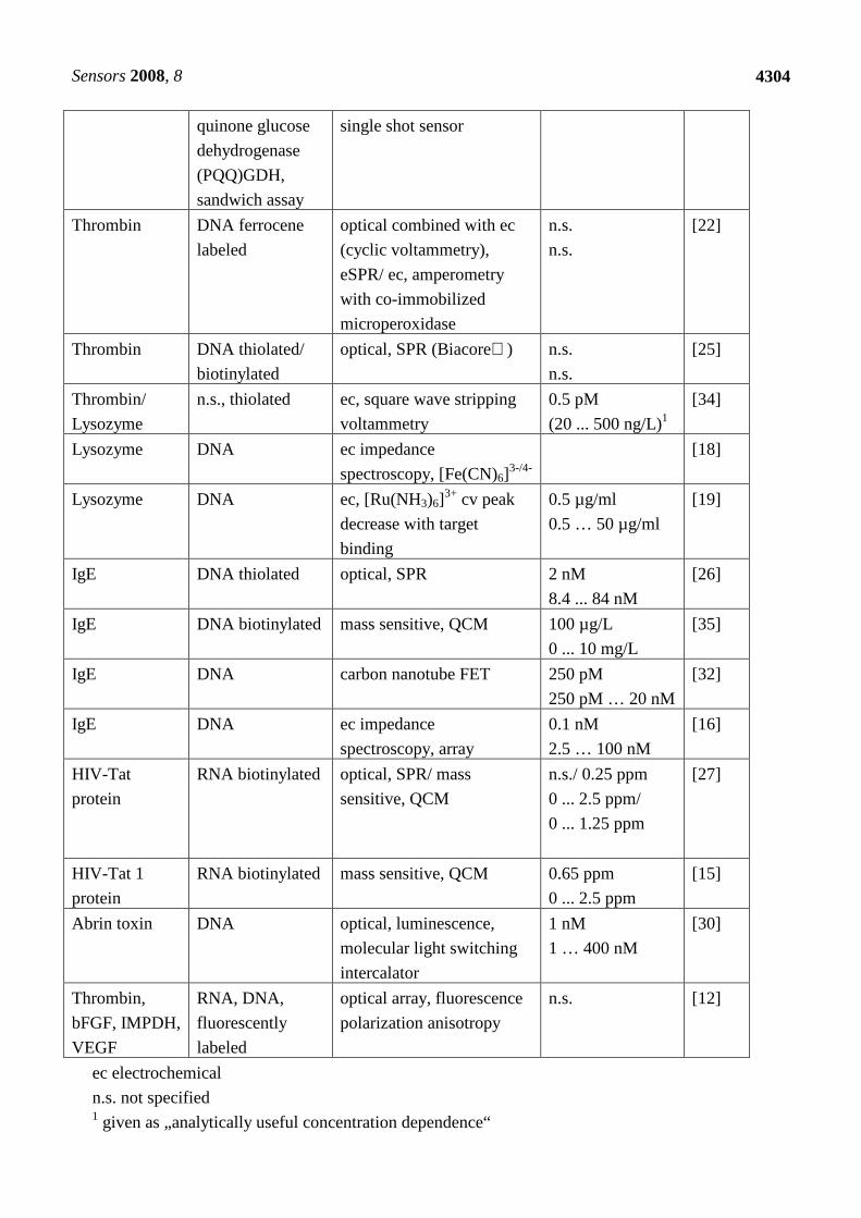
In the following table (Table 1), aptamer biosensors for different protein targets are presented and
listed according to the kind of nucleic acid of the aptamer (DNA or RNA), the transduction mode and
their reporter units (mediators, enzymes, dyes, etc.). Also, the achieved detection limits and linear
detection ranges are listed.
Table 1. Aptamer biosensors for proteins.
Target Protein Aptamer Type of Sensor, Reporter
Unit
Detect. Limit,
Linear Range
Ref
Thrombin DNA beacon ec, differential pulse
voltammetry, methylene
blue intercalator
11 nM
0 … 50.8 nM
[21]
Thrombin DNA ec, impedance
spectroscopy, [Fe(CN)6]3-/4-
2 nM
5 … 35 nM
[20]
Thrombin DNA thiolated/
biotinylated
ec, differential pulse
polarography,
p-nitroaniline/ peroxidase/
HRP
80 nM/ 3.5 nM
n.s.
[33]
Thrombin DNA labeled with
methylene blue
ec, alternating current
voltammetry, methylene
blue
n.s.
n.s. (logarithmic
dependence)
[23]
Thrombin DNA labeled with
pyrroquinoline
ec, amperometry,
glucose;
10 nM
40 ... 100 nM
[24]
Page 9
Sensors 2008, 8
4304
quinone glucose
dehydrogenase
(PQQ)GDH,
sandwich assay
single shot sensor
Thrombin DNA ferrocene
labeled
optical combined with ec
(cyclic voltammetry),
eSPR/ ec, amperometry
with co-immobilized
microperoxidase
n.s.
n.s.
[22]
Thrombin DNA thiolated/
biotinylated
optical, SPR (Biacore) n.s.
n.s.
[25]
Thrombin/
Lysozyme
n.s., thiolated ec, square wave stripping
voltammetry
0.5 pM
(20 ... 500 ng/L)1
[34]
Lysozyme DNA ec impedance
spectroscopy, [Fe(CN)6]3-/4-
[18]
Lysozyme DNA ec, [Ru(NH3)6]3+ cv peak
decrease with target
binding
0.5 µg/ml
0.5 … 50 µg/ml
[19]
IgE DNA thiolated optical, SPR 2 nM
8.4 ... 84 nM
[26]
IgE DNA biotinylated mass sensitive, QCM 100 µg/L
0 ... 10 mg/L
[35]
IgE DNA carbon nanotube FET 250 pM
250 pM … 20 nM
[32]
IgE DNA ec impedance
spectroscopy, array
0.1 nM
2.5 … 100 nM
[16]
HIV-Tat
protein
RNA biotinylated optical, SPR/ mass
sensitive, QCM
n.s./ 0.25 ppm
0 ... 2.5 ppm/
0 ... 1.25 ppm
[27]
HIV-Tat 1
protein
RNA biotinylated mass sensitive, QCM 0.65 ppm
0 ... 2.5 ppm
[15]
Abrin toxin DNA optical, luminescence,
molecular light switching
intercalator
1 nM
1 … 400 nM
[30]
Thrombin,
bFGF, IMPDH,
VEGF
RNA, DNA,
fluorescently
labeled
optical array, fluorescence
polarization anisotropy
n.s. [12]
ec electrochemical
n.s. not specified 1 given as „analytically useful concentration dependence“
Page 10
Sensors 2008, 8
4305
5. Conclusions
The use of aptamers as new biological receptors can accelerate the development of biosensors of
practical relevance. Because of their exceptionally high stability, selectivity and sensitivity, aptasensors
have the potential to overcome the lacking functional and storage stability of most biosensors (besides
some exceptions like glucose and lactate biosensors very well established on the market). This review
shows that a big variety of biosensor principles (e.g. electrochemical, optical, mass sensitive) is
available for the use of aptamers as biological receptors. However, only for a few proteins (thrombin,
lysozyme, IgE and some others) aptasensors were described. The more aptamers for proteins will be
developed and characterized, the more aptasensors will be developed in the future.
Acknowledgements
We thank Doerthe Mann for the preparation of the drawing and Christine Reinemann for helpful
discussions.
Abbreviations
bFGF Basic fibroblast growth factor
CNT-FET Carbon nanotube field-effect transistor
CMOS Complementary metal–oxide–semiconductor
DNA, ssDNA Desoxyribonucleic acid, single stranded desoxyribonucleic acid
ELISA Enzyme linked immunosorbent assay
FET Field effect transistor
FIS Faradaic Impedance Spectroscopy
HSA Human serum albumin
IMPDH Inosine monophosphate dehydrogenase
IUPAC International Union of Pure and Applied Chemistry
Kd Dissociation constant
mAb Monoclonal Antibody
MB Methylene Blue
QCM Quartz crystal microbalance
RNA Ribonucleic acid
RNAse Ribonuclease
SELEX Systematic evolution of ligands by exponential enrichment
SPR Surface plasmon resonance
VEGF Vascular endothelial growth factor
References and Notes
1. O'Sullivan, C.K. Aptasensors - the future of biosensing. Anal. Bioanal. Chem. 2002, 372, 44-48.
2. Stadtherr, K.; Wolf, H.; Lindner, P. An aptamer-based protein biochip. Anal. Chem. 2005, 77,
3437-3443.
Page 11
Sensors 2008, 8
4306
3. Jenison, R.D.; Gill, S.C.; Pardi, A.; Polisky, B. High-resolution molecular discrimination by
RNA. Science. 1994, 263, 1425-1429.
4. Michaud, M.; Jourdan, E.; Villet, A.; Ravel, A.; Grosset, C.; Peyrin, E. A DNA aptamer as a new
target-specific chiral selector for HPLC. J. Am. Chem. Soc. 2003, 125, 8672-8679.
5. Ellington, A.D.; Szostak, J.W. In vitro selection of RNA molecules that bind specific ligands.
Nature. 1990, 346, 818-822.
6. Tuerk, C.; Gold, L. Systematic evolution of ligands by exponential enrichment: RNA ligands to
bacteriophage T4 DNA polymerase. Science. 1990, 249, 505-510.
7. Stoltenburg, R.; Reinemann, C.; Strehlitz, B. SELEX-A (r)evolutionary method to generate high-
affinity nucleic acid ligands. Biomol. Eng. 2007, 24, 381-403.
8. Mukhopadhyay, R. Aptamers are ready for the spotlight. Anal. Chem. 2005, 77, 114A-118A.
9. Jayasena, S.D. Aptamers: an emerging class of molecules that rival antibodies in diagnostics.
Clin. Chem. 1999, 45, 1628-1650.
10. Willner, I.; Zayats, M. Electronic Aptamer-Based Sensors. Angew. Chem. Int. Ed Engl. 2007, 46,
6408-6418.
11. Thevenot, D.R.; Toth, K.; Durst, R.A.; Wilson, G.S. Electrochemical Biosensors: Recommended
Definitions and Classification. Pure Appl. Chem. 1999, 71, 2333-2348.
12. McCauley, T.G.; Hamaguchi, N.; Stanton, M. Aptamer-based biosensor arrays for detection and
quantification of biological macromolecules. Anal. Biochem. 2003, 319, 244-250.
13. Kubik, M.F.; Bell, C.; Fitzwater, T.; Watson, S.R.; Tasset, D.M. Isolation and characterization of
2'-fluoro-, 2'-amino-, and 2'-fluoro/amino-modified RNA ligands to human IFN-gamma that
inhibit receptor binding. J. Immunol. 1997, 159, 259-267.
14. Kujau, M.J.; Wolfl, S. Intramolecular derivatization of 2'-amino-pyrimidine modified RNA with
functional groups that is compatible with re-amplification. Nucleic Acids Res. 1998, 26, 1851-
1853.
15. Minunni, M.; Tombelli, S.; Gullotto, A.; Luzi, E.; Mascini, M. Development of biosensors with
aptamers as bio-recognition element: the case of HIV-1 Tat protein. Biosens. Bioelectron. 2004,
20, 1149-1156.
16. Xu, D.K.; Xu, D.W.; Yu, X.B.; Liu, Z.H.; He, W.; Ma, Z.Q. Label-free electrochemical detection
for aptamer-based array electrodes. Anal. Chem. 2005, 77, 5107-5113.
17. Schlecht, U.; Malave, A.; Gronewold, T.; Tewes, M.; Lohndorf, M. Comparison of antibody and
aptamer receptors for the specific detection of thrombin with a nanometer gap-sized impedance
biosensor. Anal. Chim. Acta. 2006, 573, 65-68.
18. Rodriguez, M.C.; Kawde, A.N.; Wang, J. Aptamer biosensor for label-free impedance
spectroscopy detection of proteins based on recognition-induced switching of the surface charge.
Chem. Commun. 2005, 4267-4269.
19. Cheng, A.K.; Ge, B.; Yu, H.Z. Aptamer-based biosensors for label-free voltammetric detection
of lysozyme. Anal. Chem. 2007, 79, 5158-5164.
20. Radi, A.E.; Sanchez, J.L.A.; Baldrich, E.; O'Sullivan, C.K. Reusable impedimetric aptasensor.
Anal. Chem. 2005, 77, 6320-6323.
Page 12
Sensors 2008, 8
4307
21. Bang, G.S.; Cho, S.; Kim, B.G. A novel electrochemical detection method for aptamer
biosensors. Biosens. Bioelectron. 2005, 21, 863-870.
22. Mir, M.; Katakis, I. Aptamers as elements of bioelectronic devices. Mol. Biosyst. 2007, 3, 620-
622.
23. Xiao, Y.; Lubin, A.A.; Heeger, A.J.; Plaxco, K.W. Label-free electronic detection of thrombin in
blood serum by using an aptamer-based sensor. Angew. Chem. Int. Ed Engl. 2005, 44, 5456-
5459.
24. Ikebukuro, K.; Kiyohara, C.; Sode, K. Novel electrochemical sensor system for protein using the
aptamers in sandwich manner. Biosens. Bioelectron. 2005, 20, 2168-2172.
25. Baldrich, E.; Restrepo, A.; O'Sullivan, C.K. Aptasensor development: Elucidation of critical
parameters for optimal aptamer performance. Anal. Chem. 2004, 76, 7053-7063.
26. Wang, Z.; Wilkop, T.; Xu, D.; Dong, Y.; Ma, G.; Cheng, Q. Surface plasmon resonance imaging
for affinity analysis of aptamer-protein interactions with PDMS microfluidic chips. Anal.
Bioanal. Chem. 2007, 389, 819-825.
27. Tombelli, S.; Minunni, A.; Luzi, E.; Mascini, M. Aptamer-based biosensors for the detection of
HIV-1 Tat protein. Bioelectrochemistry. 2005, 67, 135-141.
28. Bock, L.C.; Griffin, L.C.; Latham, J.A.; Vermaas, E.H.; Toole, J.J. Selection of single-stranded
DNA molecules that bind and inhibit human thrombin. Nature. 1992, 355, 564-566.
29. Potyrailo, R.A.; Conrad, R.C.; Ellington, A.D.; Hieftje, G.M. Adapting selected nucleic acid
ligands (aptamers) to biosensors. Anal. Chem. 1998, 70, 3419-3425.
30. Tang, J.; Yu, T.; Guo, L.; Xie, J.; Shao, N.; He, Z. In vitro selection of DNA aptamer against
abrin toxin and aptamer-based abrin direct detection. Biosens. Bioelectron. 2007, 22, 2456-2463.
31. Pavlov, V.; Xiao, Y.; Shlyahovsky, B.; Willner, I. Aptamer-functionalized Au nanoparticles for
the amplified optical detection of thrombin. J. Am. Chem. Soc. 2004, 126, 11768-11769.
32. Maehashi, K.; Katsura, T.; Kerman, K.; Takamura, Y.; Matsumoto, K.; Tamiya, E. Label-free
protein biosensor based on aptamer-modified carbon nanotube field-effect transistors. Anal.
Chem. 2007, 79, 782-787.
33. Mir, M.; Vreeke, M.; Katakis, L. Different strategies to develop an electrochemical thrombin
aptasensor. Electrochem. Commun. 2006, 8, 505-511.
34. Hansen, J.A.; Wang, J.; Kawde, A.N.; Xiang, Y.; Gothelf, K.V.; Collins, G. Quantum-
dot/aptamer-based ultrasensitive multi-analyte electrochemical biosensor. J. Am. Chem. Soc.
2006, 128, 2228-2229.
35. Liss, M.; Petersen, B.; Wolf, H.; Prohaska, E. An aptamer-based quartz crystal protein biosensor.
Anal. Chem. 2002, 74, 4488-4495.
© 2008 by the authors; licensee Molecular Diversity Preservation International, Basel, Switzerland.
This article is an open-access article distributed under the terms and conditions of the Creative
Commons Attribution license (http://creativecommons.org/licenses/by/3.0/).