PROTEIN ENGINEERING B.Tech Biotechnology SBT1206 UNIT-I THE THREE-DIMENSIONAL STRUCTURE OF PROTEINS The covalent backbone of proteins is made up of hundreds of individual bonds. If free rotation were possible around even a fraction of these bonds, proteins could assume an almost infinite number of three dimensional structures. Each protein has a specific chemical or structural function; however, strongly suggesting that each protein has a unique three- dimensional structure The simple fact that proteins can be crystallized provides strong evidence that this is the case. The ordered arrays of molecules in a crystal can generally form only if the molecular units making up the crystal are identical. The enzyme urease (Mr 483,000) was among the first proteins crystallized, by James Sumner in 1926. This accomplishment demonstrated dramatically that even very large proteins are discrete chemical entities with unique structures, and it revolutionized thinking about proteins. 1. OVERVIEW OF PROTEIN STRUCTURE The spatial arrangement of atoms in a protein is called a conformation. The term conformation refers to a structural state that can, without breaking any covalent bonds, interconvert with other structural states. A change in conformation could occur, for example, by rotation about single bonds. Of the innumerable conformations that are theoretically possible in a protein containing hundreds of single bonds, one generally predominates. This is usually the conformation that is thermodynamically the most stable, having the lowest Gibbs' free energy (G). Proteins in their functional conformation are called native proteins.
Transcript
PROTEIN ENGINEERING B.Tech Biotechnology SBT1206
UNIT-I
THE THREE-DIMENSIONAL STRUCTURE OF PROTEINS
The covalent backbone of proteins is made up of hundreds of individual bonds. If free
rotation were possible around even a fraction of these bonds, proteins could assume an almost
infinite number of three dimensional structures. Each protein has a specific chemical or
structural function; however, strongly suggesting that each protein has a unique three-
dimensional structure The simple fact that proteins can be crystallized provides strong
evidence that this is the case. The ordered arrays of molecules in a crystal can generally form
only if the molecular units making up the crystal are identical. The enzyme urease (Mr
483,000) was among the first proteins crystallized, by James Sumner in 1926. This
accomplishment demonstrated dramatically that even very large proteins are discrete
chemical entities with unique structures, and it revolutionized thinking about proteins.
1. OVERVIEW OF PROTEIN STRUCTURE
The spatial arrangement of atoms in a protein is called a conformation. The term
conformation refers to a structural state that can, without breaking any covalent bonds,
interconvert with other structural states. A change in conformation could occur, for example,
by rotation about single bonds. Of the innumerable conformations that are theoretically
possible in a protein containing hundreds of single bonds, one generally predominates. This is
usually the conformation that is thermodynamically the most stable, having the lowest Gibbs'
free energy (G). Proteins in their functional conformation are called native proteins.
PROTEIN ENGINEERING B.Tech Biotechnology SBT1206
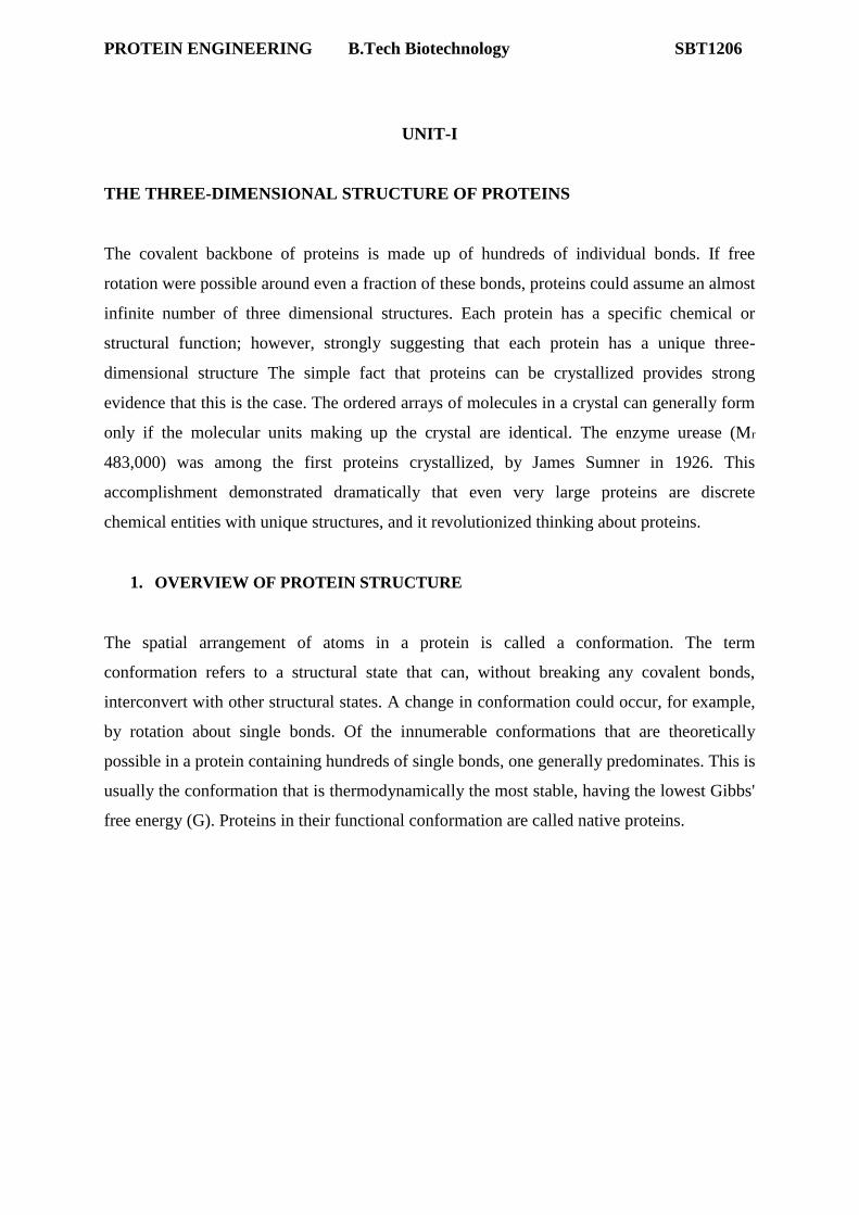
Four Levels of Architecture in Proteins
Figure 1 Levels of structure in proteins
Conceptually, protein structure can be considered at four levels (Fig. 1). Primary structure
includes all the covalent bonds between amino acids and is normally defined by the sequence
of peptide-bonded amino acids and locations of disulfide bonds. The relative spatial
arrangement of the linked amino acids is unspecified. Polypeptide chains are not free to take
up any three-dimensional structure at random. Steric constraints and many weak interactions
stipulate that some arrangements will be more stable than others.
Secondary structure refers to regular, recurring arrangements in space of adjacent amino
acid residues in a polypeptide chain. There are a few common types of secondary structure,
the most prominent being the a helix and the β conformation.
PROTEIN ENGINEERING B.Tech Biotechnology SBT1206
Tertiary structure refers to the spatial relationship among all amino acids in a polypeptide;
it is the complete three-dimensional structure of the polypeptide. The boundary between
secondary and tertiary structure is not always clear. Several different types of secondary
structure are often found within the three-dimensional structure of a large protein. Proteins
with several polypeptide chains have one more level of structure: quaternary structure,
which refers to the spatial relationship of the polypeptides, or subunits, within the protein.
1.1.Protein Secondary Structure
Several types of secondary structure are particularly stable and occur widely in proteins. The
most prominent are the α helix and β conformations. Using fundamental chemical principles
and a few experimental observations, Linus Pauling and Robert Corey predicted the existence
of these secondary structures in 1951, several years before the first complete protein structure
was elucidated. In considering secondary structure, it is useful to classify proteins into two major groups:
fibrous proteins, having polypeptide chains arranged in long strands or sheets, and globular
proteins, with polypeptide chains folded into a spherical or globular shape. Fibrous proteins
play important structural roles in the anatomy and physiology of vertebrates, providing
external protection, support, shape, and form. They may constitute one-half or more of the
total body protein in larger animals. Most enzymes and peptide hormones are globular
proteins. Globular proteins tend to be structurally complex, often containing several types of
secondary structure; fibrous proteins usually consist largely of a single type of secondary
structure. Because of this structural simplicity, certain fibrous proteins played a key role in
the development of the modern understanding of protein structure and provide particularly
clear examples of the relationship between structure and function; they are considered in
some detail after the general discussion of secondary structure.
The Peptide Bond Is Rigid and Planar
In the peptide bond, the π-electrons from the carbonyl are delocalized between the oxygen
and the nitrogen. This means that the peptide bond has ~40% double bond character. This
partial double bond character is evident in the shortened bond length of the C–N bond. The
length of a normal C–N single bond is 1.45 Å and a C=N double bond is 1.25 Å, while the
peptide C–N bond length is 1.33 Å.
PROTEIN ENGINEERING B.Tech Biotechnology SBT1206
Because of its partial double bond character, rotation around the N–C bond is severely
restricted. The peptide bond allows rotation about the bonds from the α- carbon, but not the
amide C–N bond. Only the Φ and Ψ torsion angles (see below) can vary reasonably freely. In
addition, the six atoms in the peptide bond (the two α-carbons, the amide O, and the amide N
and H) are coplanar. Finally, the peptide bond has a dipole, with the O having a partial
negative charge, and the Namide having a partial positive charge.
This allows the peptide bond to participate in electrostatic interactions, and contributes to the
hydrogen bond strength between the backbone carbonyl and the Namide proton.
Peptide bond and protein structure
The peptide bond contains three sets of torsion angles (also known as dihedral angles). The
least variable of these torsion angles is the ω angle, which is the dihedral angle around the
amide bond. As discussed above, this angle is fixed by the requirement for orbital overlap
between the carbonyl double bond and the Namide lone pair orbital. Steric considerations
strongly favor the trans configuration (i.e. an ω angle of 180°), because of steric hindrance
between the alpha carbons of adjacent amino acid residues. This means that nearly all peptide
bonds in a protein will have an ω angle of 180°.
PROTEIN ENGINEERING B.Tech Biotechnology SBT1206
In considering peptide structures, it is usually much more important to look at the backbone
angles that can vary more widely. These angles are the Φ (= phi, Cα–Namide) and Ψ (= psi,
Cα–Camide) angles. By definition, the fully extended conformation corresponds to 180° for
both Φ and Ψ. (Note that 180° = –180°). Numeric values of angles increase in the clockwise
direction when looking away from the α-carbon
By definition, Φ = 0° when the Camide-Namide and Camide-Cα bonds are in the same plane,
and Ψ = 0° when the Namide-Camide and Namide-Cα bonds are in the same plane. The (+)
direction is clockwise while looking away from the Cα. The torsion angles that the atoms of
the peptide bond can assume are limited by steric constraints. Some Φ / Ψ pairs will result in
atoms being closer than allowed by the van der Waals radii of the atoms, and are therefore
sterically forbidden (for example: 0°:0°, 180°:0°, and 0°:180° are forbidden because of
backbone atom clashes). For tetrahedral carbons, the substituents are typically found in staggered conformations (see
figure, above). Peptide bonds are more complicated, because while the α-carbon is tetrahedral,
the two other backbone atom types are not. However, the same principle applies: the preferred
conformations for peptide bond atoms have the substituent atoms at maximal distances from
one another. A Ψ angle of 180° results in an alignment of the Namide with the carbonyl oxygen from the
same residue. This is allowed, although not especially favored. A Ψ angle of 0° places the
PROTEIN ENGINEERING B.Tech Biotechnology SBT1206
Namide from one residue very close to the Namide from the previous residue; this results in a
steric clash (as well as an unfavorable electrostatic interaction, because both Namide have
partial positive charges). The residue side-chains also impose steric constraints. Glycine,
because of its small side chain, has a much large ranger of possible Φ / Ψ pairs than any other
residue. Proline has a very limited range of Φ angles because its side-chain is covalently
bonded to its Namide. Most other residues are limited to relatively few Φ / Ψ pairs (although
more than proline). This is especially true for the β-branched residues threonine, valine, and
isoleucine, which are the most restricted, because these residues have more steric bulk due to
the presence of two groups attached their β- carbon. Allowed values for Φ and Ψ are
graphically revealed when Ψ is plotted versus Φ in a Ramachandran plot, introduced by G.
N. Ramachandran .
The Ramachandran Plot
In a polypeptide the main chain N-Calpha and Calpha-C bonds relatively are free to rotate.
These rotations are represented by the torsion angles phi and psi, respectively.
G N Ramachandran used computer models of small polypeptides to systematically vary phi
and psi with the objective of finding stable conformations. For each conformation, the
structure was examined for close contacts between atoms. Atoms were treated as hard
spheres with dimensions corresponding to their van der Waals radii. Therefore, phi and psi
angles which cause spheres to collide correspond to sterically disallowed conformations of
the polypeptide backbone.
PROTEIN ENGINEERING B.Tech Biotechnology SBT1206
In the diagram above the white areas correspond to conformations where atoms in the
polypeptide come closer than the sum of their van der Waals radi. These regions are sterically
disallowed for all amino acids except glycine which is unique in that it lacks a side chain.
The red regions correspond to conformations where there are no steric clashes, ie these are
the allowed regions namely the alpha-helical and beta-sheet conformations. The yellow areas
show the allowed regions if slightly shorter van der Waals radi are used in the calculation, ie
the atoms are allowed to come a little closer together. This brings out an additional region
which corresponds to the left-handed alpha-helix.
L-amino acids cannot form extended regions of left-handed helix but occassionally individual
residues adopt this conformation. These residues are usually glycine but can also be
asparagine or aspartate where the side chain forms a hydrogen bond with the main chain and
therefore stabilises this otherwise unfavourable conformation. The 3(10) helix occurs close to
the upper right of the alpha-helical region and is on the edge of allowed region indicating
lower stability.
PROTEIN ENGINEERING B.Tech Biotechnology SBT1206
Disallowed regions generally involve steric hindrance between the side chain C-beta
methylene group and main chain atoms. Glycine has no side chain and therefore can adopt
PROTEIN ENGINEERING B.Tech Biotechnology SBT1206
phi and psi angles in all four quadrants of the Ramachandran plot. Hence it frequently occurs
in turn regions of proteins where any other residue would be sterically hindered.
Secondary structure
The term secondary structure refers to the local conformation of some part of a polypeptide.
The discussion of secondary structure most usefully focuses on common regular folding
patterns of the polypeptide backbone. A few types of secondary structure are particularly
stable and occur widely in proteins. The most prominent are the α-helix and β-sheet. Using
fundamental chemical principles and a few experimental observations, Pauling and Corey
predicted the existence of these secondary structures in 1951, several years before the first
complete protein structure was elucidated.
Alpha helix (α-helix)
The alpha helix (α-helix) is a common secondary structure of proteins and is a right hand-
coiled or spiral conformation (helix) in which every backbone N-H group donates a hydrogen bond to the backbone C=O group of the amino acid four residues earlier ( hydrogen bonding). This secondary structure is also sometimes called a classic
Pauling–Corey–Branson alpha helix (see below). The name 3.613-helix is also used for this
type of helix, denoting the number of residues per helical turn, and 13 atoms being involved
in the ring formed by the hydrogen bond. Among types of local structure in proteins, the α-
helix is the most regular and the most predictable from sequence, as well as the most
The pitch of the alpha-helix (the vertical distance between consecutive turns of the helix) is
5.4 Å (0.54 nm), which is the product of 1.5 and 3.6. What is most important is that the N-H
group of an amino acid forms a hydrogen bond with the C=O group of the amino acid four residues earlier; this repeated hydrogen bonding is the most prominent
characteristic of an α-helix. Similar structures include the 310 helix ( hydrogen bonding) and the π-
helix ( hydrogen bonding). The α helix can be described as a 3.613 helix, since the i +
4 spacing adds 3 more atoms to the H-bonded loop compared to the tighter 310 helix, and on
average, 3.6 amino acids are involved in one ring of α helix. The subscripts refer to the number of
atoms (including the hydrogen) in the closed loop formed by the hydrogen bond.
Residues in α-helices typically adopt backbone (φ, ψ) dihedral angles around (-60°, -45°), as
shown in the image at right. In more general terms, they adopt dihedral angles such that the ψ
dihedral angle of one residue and the φ dihedral angle of the next residue sum to roughly -
105°. As a consequence, α-helical dihedral angles, in general, fall on a diagonal stripe on the
Ramachandran diagram (of slope -1), ranging from (-90°, -15°) to (-35°, -70°). For
comparison, the sum of the dihedral angles for a 310 helix is roughly -75°, whereas that for
the π-helix is roughly -130°.
Structural features of the three major forms of protein helices
Geometry attribute α-helix 310 helix π-helix
Residues per turn 3.6 3.0 4.4
Translation per residue 1.5 Å (0.15 nm) 2.0 Å (0.20 nm) 1.1 Å (0.11 nm)
Radius of helix 2.3 Å (0.23 nm) 1.9 Å (0.19 nm) 2.8 Å (0.28 nm)
Pitch 5.4 Å (0.54 nm) 6.0 Å (0.60 nm) 4.8 Å (0.48 nm)
An α-helix has a dipole, with the partial positive charge toward N-terminus. This is true
because all of the partial charges of the peptide bonds are in alignment.
The backbone of the helix is ~6 Å in diameter (ignoring side chains).
Two-dimensional representations of α-helices
Drawing a three-dimensional helix on paper is difficult. Two types of two dimensional
representations (helical wheel and helical net diagrams) are commonly used to simplify the
analysis of helical segments of proteins. The two-dimensional representations are somewhat
stylized, but show the major features more clearly than attempting to draw a three-
dimensional structure accurately in two dimensions. The first type of representation is a Helical Wheel diagram. In this diagram, the
representation involves looking down the helix axis, and plotting the rotational angle around
the helix for each residue. This representation is conceptually easily grasped, but tends to
obscure the distance along the helix; residues 0 and 18 are exactly aligned on this diagram,
but are actually separated in space by 27 Å.
Helical Wheel Residue #0 = 0° (by definition)
#1 = 100°
#2 = 200°
#3 = 300°
#4 = 400° = 40°
#5 = 140°
#6 = 240° #7 = 340°
#8 = 440° = 80°
9# = 900° (from first) = 180°
These angles can be plotted on a circle.
PROTEIN ENGINEERING B.Tech Biotechnology SBT1206
Doing so results in a representation that corresponds to the view looking down the long axis of the helix. (Note that the rotation is clockwise as the residue number increases.)
Note that residues 0, 3, 4, 7, and 8 are all located on one face of the helix
A helix that has its axis along the border of this region would be expected to have a
corresponding, amphipathic, distribution of polar and non-polar residues. (Amphipathic,
meaning “hating both” refers to the presence of both polar and non-polar groups in the helix.)
PROTEIN ENGINEERING B.Tech Biotechnology SBT1206
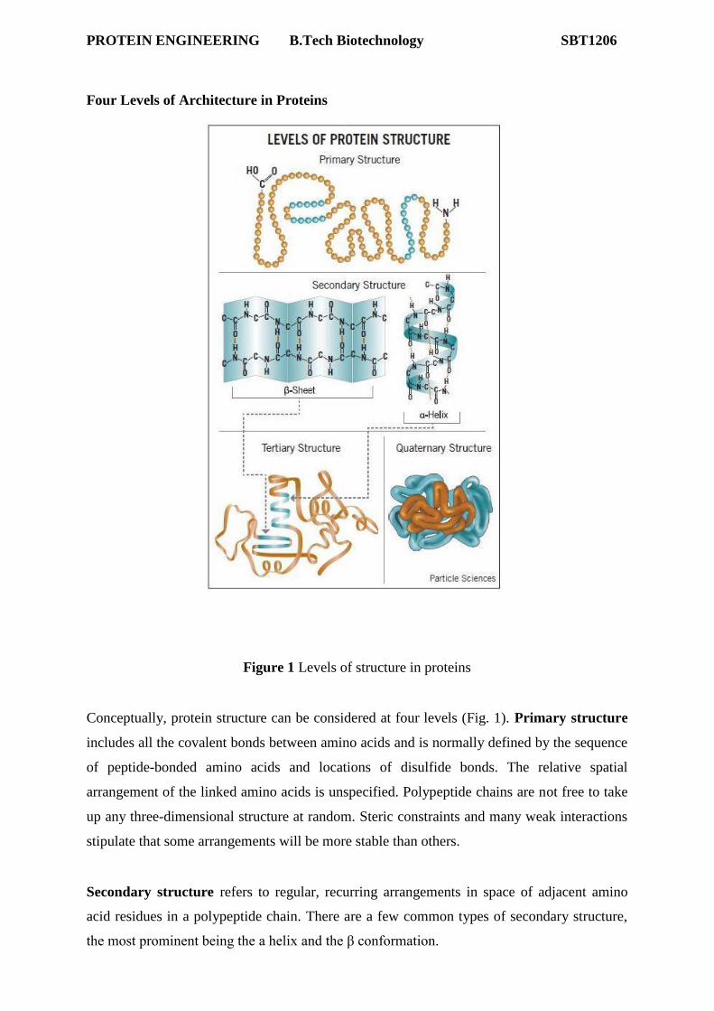
The βß Conformation Organizes Polypeptide Chains into Sheets
Pauling and Corey predicted a second type of repetitive structure, the β
conformation. an extended state for which angles phi = -135o and psi = +135o; the
polypeptide chain alternates in direction, resulting in a zig-zag structure for the
peptide chain. Note the shaded circle around R; the extended strand arrangement
also allows the maximum space and freedom of movement for a side chain. The
repeat between identically oriented R-groups is 7.0 Å, with 3.5 Å per amino acid,
matching the fiber diffraction data for beta-keratins.
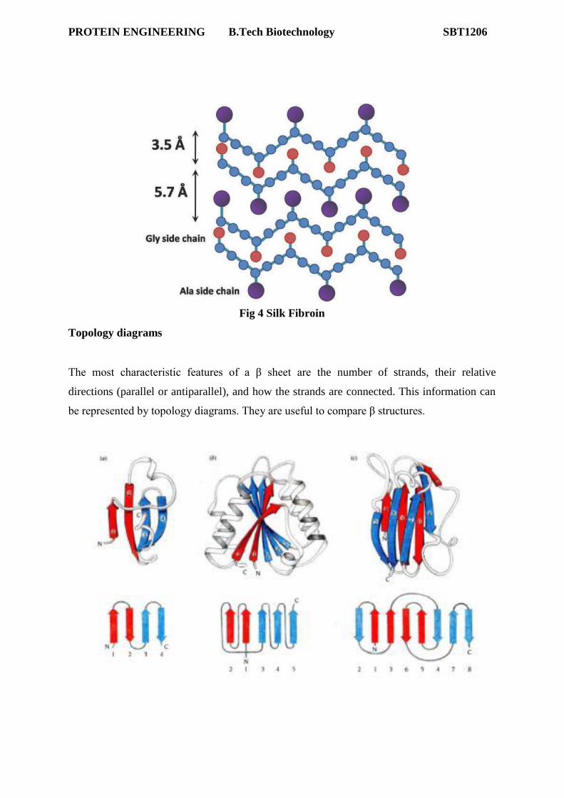
PROTEIN ENGINEERING B.Tech Biotechnology SBT1206
Pauling's extended state model matched the spacing of fibroin exactly (3.5 and 7.0
Å). In the extended state, H-bonding NH and CO groups point out at 90o to the
strand. If extended strands are lined up side by side, H-bonds bridge from strand to
strand. Identical or opposed strand alignments make up parallel or antiparallel beta
sheets (named for beta keratin). Antiparallel beta-sheet is significantly more stable
due to the well aligned H-bonds.
Amino acid preferences for different secondary structure
Alpha helix may be considered the default state for secondary structure. Although
the potential energy is not as low as for beta sheet, H-bond formation is intra-strand,
so there is an entropic advantage over beta sheet, where H-bonds must form
PROTEIN ENGINEERING B.Tech Biotechnology SBT1206
from strand to strand, with strand segments that may be quite distant in the
polypeptide sequence.
The main criterion for alpha helix preference is that the amino acid side chain
should cover and protect the backbone H-bonds in the core of the helix. Most
The most characteristic features of a β sheet are the number of strands, their relative
directions (parallel or antiparallel), and how the strands are connected. This information can
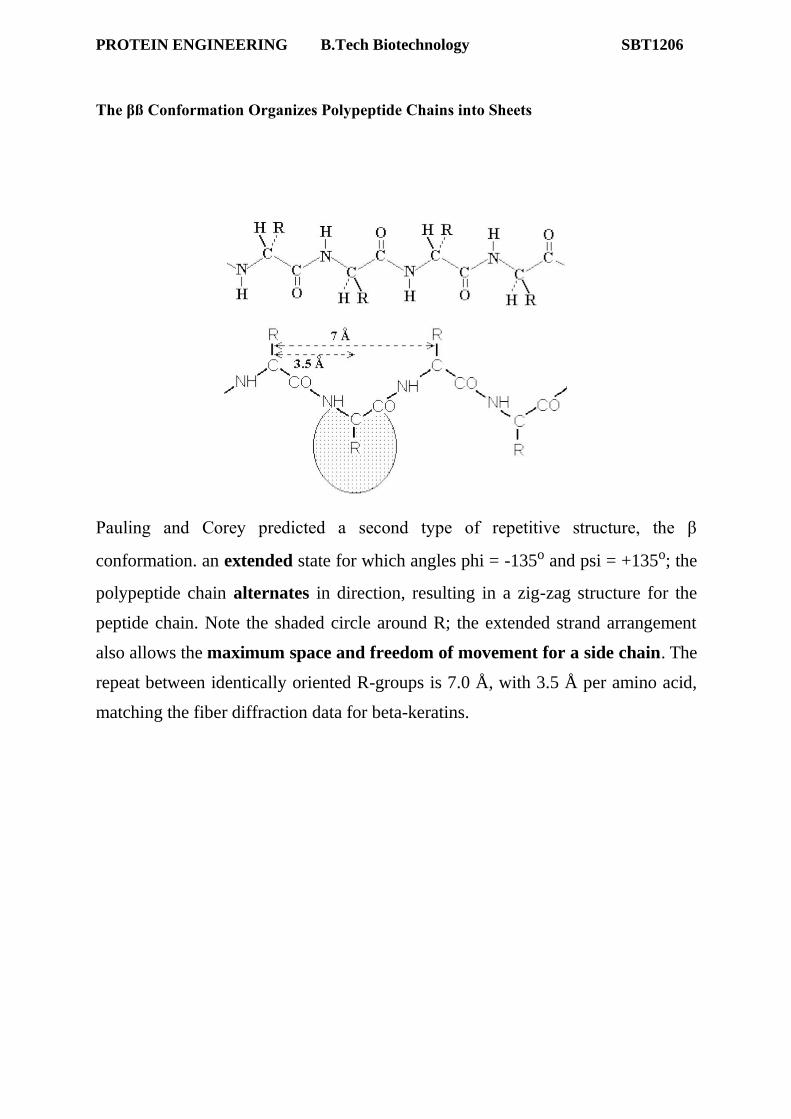
be represented by topology diagrams. They are useful to compare β structures.
PROTEIN ENGINEERING B.Tech Biotechnology SBT1206
1.2.Protein Tertiary Structure
Tertiary structure refers to the three-dimensional arrangement of all atoms in a protein.
Tertiary structure is formed by the folding in three dimensions of the secondary structure
elements of a protein. While the α helical secondary structure is held together by interactions
between the carbonyl and amide groups within the backbone, tertiary structure is held
together by interactions between R-groups of residues brought together by folding. Disulfide
bonds are also counted under the category of tertiary structure interactions. Proteins that are
compact are known as globular proteins.
Examination of protein structures resolved by X-ray diffraction and NMR has revealed a
variety of folding patterns common to many different proteins. However, even within these
folds, distinct substructures or structural motifs, i.e. distinctive arrangements of elements of
secondary structure, have been described. The term supersecondary structure has been
coined to describe this level of organisation, which is intermediate between secondary and
tertiary.
Motifs or folds, are particularly stable arrangements of several elements of the secondary
structure. • Supersecondary structures are usually produced by packing side chains from
adjacent secondary structural elements close to each other.
Rules for secondary structure.
• Hydrophobic side groups must be buried inside the folds, therefore, layers must be created
(β−α−β; α− α). • α-helix and β-sheet, if occur together, are found in different structural layers. • Adjacent polypeptide segments are stacked together. • Connections between secondary structures do not form knots. • The β-sheet is the most stable.
Motif
• Secondary structure composition, e.g. all α, all β, segregated α+β, mixed α/β • Motif = small, specific combinations of secondary structure elements, e.g. β-α-β loop
PROTEIN ENGINEERING B.Tech Biotechnology SBT1206
1. Helix super secondary structures
Helix-Turn-Helix Motif
Also called the alpha-alpha type (αα-type). The motif is compromised of two antiparallel
helices connected by a turn. The helix-turn-helix is a functional motif and is usually identified
in proteins that bind to DNA minor and major grooves, and Calcium-binding proteins.
DNA binding Helix-turn-Helix motif
Calcium binding (EF Hand- Calcium binding) motif
PROTEIN ENGINEERING B.Tech Biotechnology SBT1206
Helix-hairpin-helix: Involved in DNA binding
Alpha-alpha corner
Short loop regions connecting helices which are roughly perpendicular to one another
2. Sheet super secondary structures
All beta tertiary structural domains can occur in proteins with one domain (eg.
concanavalin A, superoxide dismutase), and occurs at least once in proteins with two domains (eg. chymotrypsin), or three domains (eg. OmpF).
The beta strands making up these domains are all essentially antiparallel and form structures to achieve stable packing arrangements within the protein.
There are presently (as of version 1.39) about 70 subclasses listed in SCOP for this domain, and some examples of these are outlined below.
This is the most abundant beta-domain structure and as the name suggests the domain forms a 'barrel-like' structure. The beta barrels are not geometrically perfect and can be rather distorted.
There are three main types:
1. Up-and-down barrels
2. Greek key barrels
3. Jelly roll (Swiss roll) barrels
Up-and-down beta-sheets or beta-barrels
The simple topology of an up-and-down barrel (named because the beta strands follow each other in sequence in an up-and-down fashion).
Usually, the loops joining the beta strands do not crossover the 'ends' of the barrel.
Greek key barrels
These are barrels formed from two, or more, Greek Key motifs.
It is a stable structure
The Greek key barrel consists of four anti-parallel Beta strands where one strand changes the
topology direction. Hydrogen bonding occurs between strands 1:4, and strands 2:3. Strand 2
then folds over to form the structural motif.
PROTEIN ENGINEERING B.Tech Biotechnology SBT1206
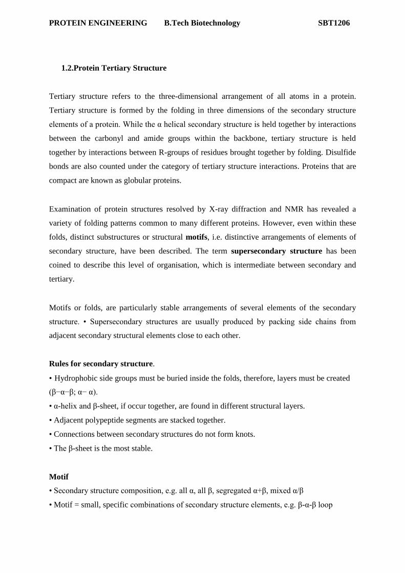
Jelly roll barrels
These barrels are formed from a 'Greek Key-like' structure called a jelly roll. Supposedly named because the polypeptide chain is wrapped around a barrel core like a jelly roll (swiss roll).
It is a stable structure
This structure is found in coat proteins of spherical viruses, plant lectin concanavalin A, and hemagglutinin protein from influenza virus.
The essential features of a jelly roll barrel are that:
it is like an inverted 'U' (which is often seen twisted and distorted in proteins)
it is usually divided into two beta sheets which are packed against each other most jelly roll barrels have eight strands although any even number greater than 8 can
form a jelly roll barrel it folds such that hydrogen bonds exist between strands 1 and 8; 2 and 7; 3 and 6; and
4 and 5
PROTEIN ENGINEERING B.Tech Biotechnology SBT1206
Beta sandwich
A beta sandwich is essentially a 'flattened' beta barrel with the two sheets packing closely together (like a sandwich!). The first and last strands of the sandwich do not hydrogen bond to each other to complete a 'barrel' structure.
Beta sandwich in beta 2 microglobulin.
Aligned or Orthogonal beta strands
Beta strands in barrels or sandwich structures can be orientated in two general ways:
where the strands in two sheets are almost aligned, and in the same orientation, to each other and form an 'aligned beta' structure (eg. gamma crystallin)
PROTEIN ENGINEERING B.Tech Biotechnology SBT1206
where the strands, in at least two sheets, are roughly perpendicular to each other and form an 'orthogonal beta' structure.
Beta-hairpin: two antiparallel beta strands connected by a “hairpin” bend, i.e. beta-turn 2 x
Two antiparallel beta strands which form a beta hairpin can change direction abruptly. The angle of the change of direction is about 90 degrees and so the structure is known as a 'beta corner'
The abrupt angle change is achieved by one strand having a glycine residue (so there is no steric hindrance from a side chain) and the other strand having a beta bulge (where the hydrogen bond is broken).
An important and widespread supersecondary structural motif in proteins is known as the β-
α-β motif (Beta-Alpha-Beta motif). The motif consists of two parallel Beta strands that is
connected via an alpha helix (with two turns). The motif is found in most proteins that
contain parallel beta strands, and the axis of the Helix and the Strands are roughly parallel to
each other with all three elements forming a hydrophobic core due to shielding. The β-α-β
motif may be structurally or functionally involved. The Loop that connects the C-terminal of
first Beta strand and N-terminal of Helix is frequently involved in ligand binding functions,
and the motif itself is frequently found in ion channels.
The β - α - β - α - β subunit, often present in nucleotide-binding proteins, is named
the Rossman Fold, after Michael Rossman
PROTEIN ENGINEERING B.Tech Biotechnology SBT1206
α/β horseshoe
17-stranded parallel b sheet curved into an open horseshoe shape, with 16 a-helices packed against the outer surface. It doesn't form a barrel although it looks as though it should. The strands are only very slightly slanted, being nearly parallel to the central `axis'.
placental ribonuclease inhibitor takes the concept of the repeating α/β unit to extremes.
α/β barrels
Consider a sequence of eight α/β motifs:
If the first strand hydrogen bonds to the last, then the structure closes on itself forming a barrel-like structure. This is shown in the picture of triose phosphate isomerase.
PROTEIN ENGINEERING B.Tech Biotechnology SBT1206
Note that the "staves" of the barrel are slanted, due to the twist of the b sheet. Also notice that there are effectively four layers to this structure. The direction of the sheet does not change (it
is anticlockwise in the diagram). Such a structure may therefore be described as singly
wound.
In a structure which is open rather than closed like the barrel, helices would be situated on only one side of the b sheet if the sheet direction did not reverse. Therefore open a/b structures must be doubly wound to cover both sides of the sheet.
The chain starts in the middle of the sheet and travels outwards, then returns to the centre via a loop and travels outwards to the opposite edge:
PROTEIN ENGINEERING B.Tech Biotechnology SBT1206
Doubly-wound topologies where the sheet begins at the edge and works inwards are rarely observed.
PROTEIN ENGINEERING B.Tech Biotechnology SBT1206
Alpha+Beta Topologies
This is where we collect together all those folds which include significant alpha and beta
secondary structural elements, but for which those elements are `mixed', in the sense that
they do NOT exhibit the wound alpha-beta topology. This class of folds is therefore referred
to as α+ β
Domains
stable, independently folded, globular units, often consisting of combinations of motifs
vary from 25 to 300 amino acids, average length – 100.
large globular proteins may consist of several domains linked by stretches of polypeptide. Separate domain may have distinct functions (eg G3P dehydrogenase). In many cases binding site formed by cleft between 2 domains
frequently correspond to exon in gene
Some examples of domains:
1. in volving α-helix 4-helix bundle globin fold
PROTEIN ENGINEERING B.Tech Biotechnology SBT1206
The globin fold is found in its namesake globin protein families: hemoglobins and myoglobins, as well as in phycocyanins. Because myoglobin was the first protein whose
structure was solved, the globin fold was thus the first protein fold discovered.
2. parallel β-sheets
hydrophobic residues on both sides, therefore must be buried. barrel: 8 β strands each flanked by an antiparallel α-helix eg triose phosphate
isomerase.)
3. antiparallel β -sheet
hydrophobic residues on one side, one side can be exposed to environment, minimum
The immunoglobulin domain is a type of protein domain that consists of a 2-layer sandwich
of 7-9 antiparallel β-strands arranged in two β-sheets with a Greek keytopology, consisting of
about 80 amino acids. The backbone switches repeatedly between the two β-sheets. Typically, the pattern is (N-
terminal β-hairpin in sheet 1)-(β-hairpin in sheet 2)-(β-strand in sheet 1)-(C-terminal β-
hairpin in sheet 2). The cross-overs between sheets form an "X", so that the N- and C-
terminal hairpins are facing each other. Members of the immunoglobulin superfamily are found in hundreds of proteins of different
functions. Examples include antibodies, the giant muscle kinase titin, andreceptor tyrosine
kinases. Immunoglobulin-like domains may be involved in protein–protein and protein–
ligand interactions.
Example of Tertiary Structure: Myoglobin and Hemoglobin
Myoglobin and hemoglobin are hemeproteins whose physiological importance is principally related to their ability to bind molecular oxygen. Myoglobin Single polypeptide chain (153 amino acids) No disulfide bonds 8 right handed alpha helices form a hydrophobic pocket which contains
Myoglobin is a monomeric heme protein found mainly in muscle tissue where it serves as an
intracellular storage site for oxygen During periods of oxygen deprivation oxymyoglobin releases
its bound oxygen which is then used for metabolic purposes The tertiary structure of myoglobin
is that of a typical water soluble globular protein Its secondary structure is unusual in that it
contains a very high proportion (75%) of α-helical secondary structure A myoglobin polypeptide
is comprised of 8 separate right handed a-helices, designated A through H, that are connected by
short non helical regions Amino acid R-groups packed into the interior of the molecule are
predominantly hydrophobic in character while those exposed on the surface of the molecule are
generally hydrophilic, thus making the molecule relatively water soluble
Each myoglobin molecule contains one heme prosthetic group inserted into a hydrophobic
cleft in the protein Each heme residue contains one central coordinately bound iron atom that
is normally in the Fe 2+ , or ferrous, oxidation state The oxygen carried by hemeproteins is
bound directly to the ferrous iron atom of the heme prosthetic group
The heme group is located in a crevice Except for one edge, non polar side chains surround
the heme Fe 2+ is octahedrally coordinated Fe 2+ covalently bonded to the imidazole group
of histidine 93 (F8) O 2 held on the other side by histidine 64 (E7)
Hydrophobic interactions between the tetrapyrrole ring and hydrophobic amino acid R groups
on the interior of the cleft in the protein strongly stabilize the heme protein conjugate. In
addition a nitrogen atom from a histidine R group located above the plane of the heme ring is
coordinated with the iron atom further stabilizing the interaction between the heme and the
protein. In oxymyoglobin the remaining bonding site on the iron atom (the 6th coordinate
position) is occupied by the oxygen, whose binding is stabilized by a second histidine residue
Carbon monoxide also binds coordinately to heme iron atoms in a manner similar to that of
oxygen, but the binding of carbon monoxide to heme is much stronger than that of oxygen.
PROTEIN ENGINEERING B.Tech Biotechnology SBT1206
The preferential binding of carbon monoxide to heme iron is largely responsible for the
asphyxiation that results from carbon monoxide poisoning.
Hemoglobin
Oxygen transporter Four polypeptide chains Tetramer Each chain has a heme group Hence
four O 2 can bind to each Hb Two alpha (141 amino acids) and two beta (146 amino acids)
chains
Hemoglobin is an [α(2):β(2)] tetrameric hemeprotein found in erythrocytes where it is
responsible for binding oxygen in the lung and transporting the bound oxygen throughout the
body where it is used in aerobic metabolic pathways Each subunit of a hemoglobin tetramer
has a heme prosthetic group identical to that described for myoglobin. Although the
secondary and tertiary structure of various hemoglobin subunits are similar, reflecting
extensive homology in amino acid composition, the variations in amino acid composition that
do exist impart marked differences in hemoglobin's oxygen carrying properties In addition,
the quaternary structure of hemoglobin leads to physiologically important allosteric
interactions between the subunits, a property lacking in monomeric myoglobin which is
otherwise very similar to the α-subunit of haemoglobin
1.3. Quaternary structure
• 3-dimensional relationship of the different polypeptide chains (subunits) in a multimeric
protein, the way the subunits fit together and their symmetry relationships
PROTEIN ENGINEERING B.Tech Biotechnology SBT1206
• only in proteins with more than one polypeptide chain; proteins with only one chain have no
quaternary structure.)
Terminology
• Each polypeptide chain in a multichain protein = a subunit • 2-subunit protein = a dimer, 3
subunits = trimeric protein, 4 = tetrameric • homo(dimer or trimer etc.): identical subunits • hetero(dimer or trimer etc.): more than one kind of subunit (chains with different amino acid
sequences) • different subunits designated with Greek letters – e.g., subunits of a
heterodimeric protein = the "α subunit" and the "β subunit".
– NOTE: This use of the Greek letters to differentiate different polypeptide chains in a
multimeric protein has nothing to do with the names for the secondary structures α helix and
β conformation.
• Some protein structures have very complex quaternary arrangements; e.g., mitochondrial
ATP synthase, viral capsids….
PROTEIN ENGINEERING B.Tech Biotechnology SBT1206
Symmetry in quaternary structures
• simplest kind of symmetry = rotational symmetry • Individual subunits can be superimposed on other identical subunits (brought into
coincidence) by rotation about one or more rotational axes.
• If the required rotation = 180° (360°/2), protein has a 2-fold axis of symmetry (e.g., Cro
repressor protein above).
• If the rotation = 120° (360°/3), e.g., for a homotrimer, the protein has a 3-fold symmetry
axis. Rotational symmetry in proteins: Cyclic symmetry: all subunits are related by rotation
about a single n-fold rotation axis (C2 symmetry has a 2-fold axis, 2 identical subunits; C3
symmetry has a 3-fold axis, 3 identical subunits, etc.)
Example: Protein Capsid
Viral genomes are surrounded by protein shells known as capsids. One interesting question is
how capsid proteins recognize viral, but not cellular RNA or DNA. The answer is that there
is often some type of "packaging" signal (sequence) on the viral genome that is recognized by
the capsid proteins. A capsid is almost always made up of repeating structural subunits that
are arranged in one of two symmetrical structures, a helix or an icosahedron. In the simplest
case, these "subunits" consist of a single polypeptide. In many cases, however, these
structural subunits (also called protomers) are made up of several polypeptides. Both
helical and icosahedral structures are described in more detail below.
PROTEIN ENGINEERING B.Tech Biotechnology SBT1206
1) Helical Capsids: The first and best studied example is the plant tobacco mosaic virus
(TMV), which contains a SS RNA genome and a protein coat made up of a single, 17.5 kd
protein. This protein is arranged in a helix around the viral RNA, with 3 nt of RNA fitting
into a groove in each subunit. Helical capsids can also be more complex, and involve more
than one protein subunit.
A helix can be defined by two parameters, its amplitude (diameter) and pitch, where pitch is
defined as the distance covered by each turn of the helix. P = m x p, where m is the number
of subunits per turn and p is the axial rise per subunit. For TMV, m = 16.3 and p= 0.14 nm,
so P=2.28 nm. This structure is very stable, and can be dissociated and re-associated readily
by changing ionic strength, pH, temperature, etc. The interactions that hold these molecules
together are non-covalent, and involve H-bonds, salt bridges, hydrophobic interactions, and
vander Waals forces.
Several families of animal virus contain helical nucleocapsids, including the
Orthomyxoviridae (influenza), the Paramyxoviridae (bovine respiratory syncytial virus), and
the Rhabdoviridae (rabies). All of these are enveloped viruses (see below).
2) Icosahedral Capsids: In these structures, the subunits are arranged in the form of a hollow,
quasi spherical structure, with the genome within. An icosahedron is defined as being made
up of 20 equilateral triangular faces arranged around the surface of a sphere. They display
2-3-5 fold symmetry as follows:
- an axis of 2 fold rotational symmetry through the center of each edge. - an axis of 3 fold rotational symmetry through the center of each face. - an axis of 5 fold rotational symmetry through the center of each corner.
These corners are also called Vertices, and each icosahedron has 12.
Since proteins are not equilateral triangles, each face of an icosahedron contains more than
one protein subunit. The simplest icosahedron is made by using 3 identical subunits to form
each face, so the minimum # of subunits is 60 (20 x 3). Remember, that each of these
subunits could be a single protein or, more likely, a complex of several polypeptides.
PROTEIN ENGINEERING B.Tech Biotechnology SBT1206
Many viruses have too large a genome to be packaged inside an icosahedron made up of only
60 polypeptides (or even 60 subunits), so many are more complicated. In these cases, each of
the 20 triangular faces is divided into smaller triangles; and each of these smaller triangles is
defined by 3 subunits. However, the total number of subunits is always a multiple of 60. The
total number of subunits can be defined as 60 X N, where N is sometimes called the
Triangulation Number, or T. Values for T of 1,3,4,7,9, 12 and more are permitted.
When virus nucleocapsids are observed in the electron microscope, one often sees apparent
"lumps" or clusters on the surface of the particle. These are usually protein subunits clustered
around an axis of symmetry, and have been called "morphological units" or capsomers.
Forces that stabilize Protein Structure Proteins are formed of amino acids linked together by the following types of bonds Covalent Bonds - Disulfide Bridges
Covalent bonds are the strongest chemical bonds contributing to protein structure. Covalent bonds arise when two atoms share electrons.
In addition to the covalent bonds that connect the atoms of a single amino acid and the covalent peptide bond that links amino acids in a protein chain, covalent bonds between
cysteine side chains can be important determinants of protein structure. Cysteine is the sole amino acid whose side chain can form covalent bonds, yielding disulfide bridges with other
cysteine side chains: --CH2-S-S-CH2 . A disulfide bridge is shown here:
T
PROTEIN ENGINEERING B.Tech Biotechnology SBT1206
Non-covalent bonds
Electrostatic Interactions
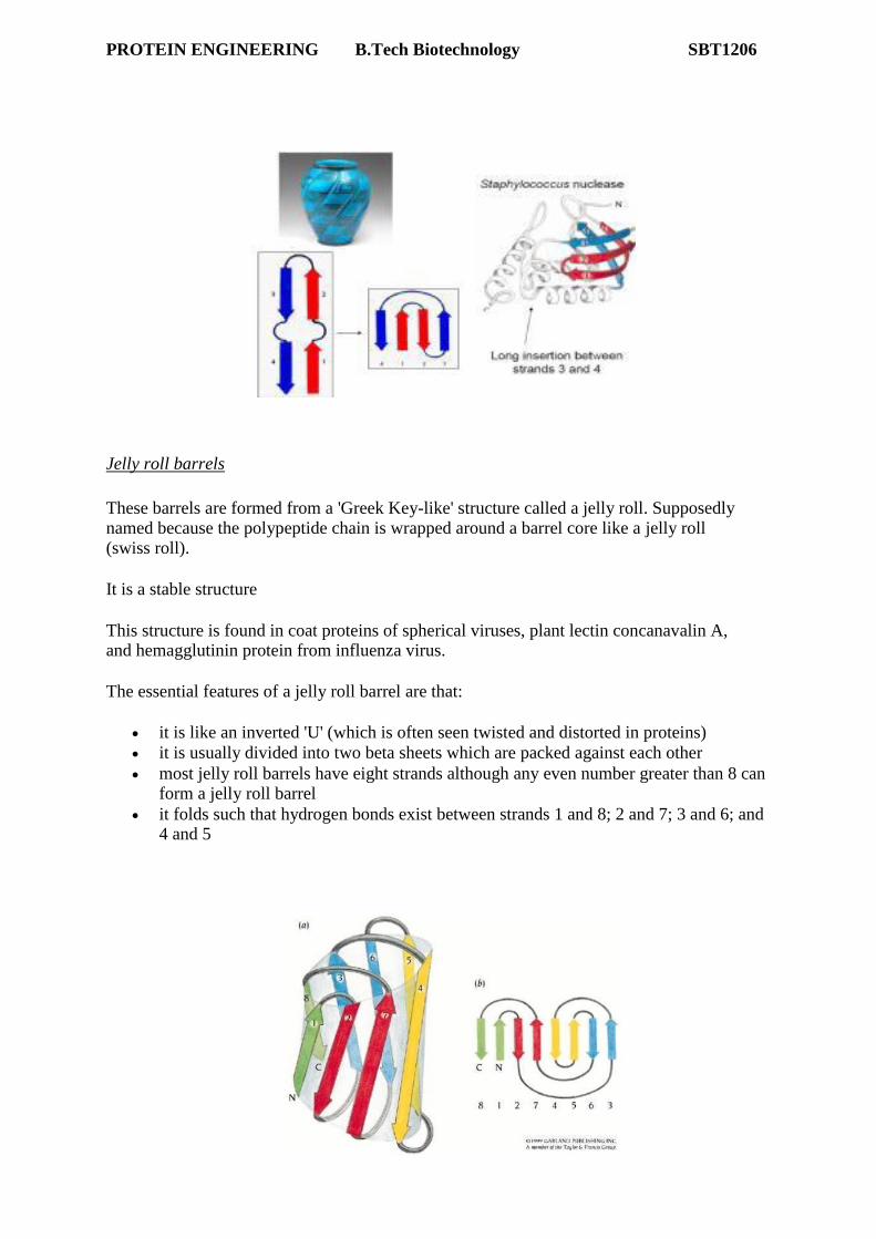
A. Ionic Bonds - Salt Bridges
Ionic bonds are formed as amino acids bearing opposite electrical charges are juxtaposed in
the hydrophobic core of proteins. Ionic bonding in the interior is rare because most charged
amino acids lie on the protein surface. Although rare, ionic bonds can be important to protein
structure because they are potent electrostatic attractions that can approach the strength of
covalent bonds. A ionic bond-salt bridge between a negatively charged O on the sidechain of
glutamic acid lies 2.8 Å from the positively charged N on the amino terminus (lysine) is
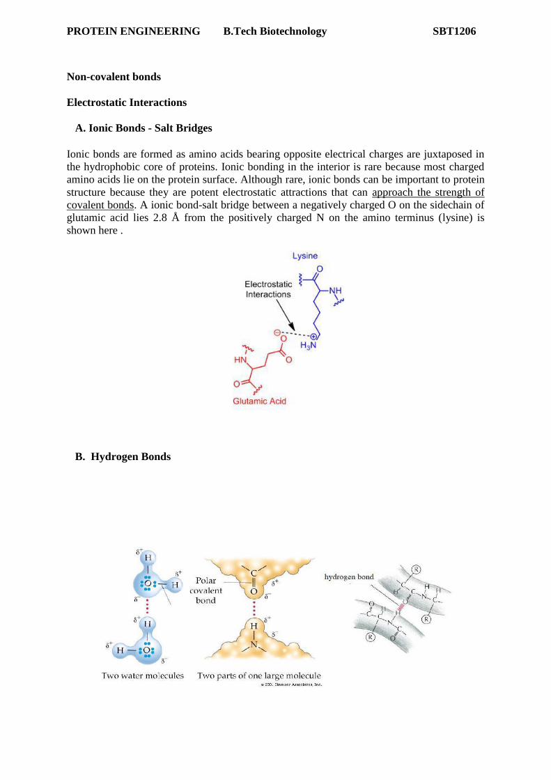
Hydrogen bonds are a particularly strong form of dipole-dipole interaction. Because atoms of
different elements differ in their tendencies to hold onto electrons -- that is, because they
have different electronegativities -- all bonds between unlike atoms are polarized, with more
electron density residing on the more electronegative atom of the bonded pair. Separation of
partial charges creates a dipole, which you can think of as a mini-magnet with a positive and
a negative end. In any system, dipoles will tend to align so that the positive end of one dipole
and the negative end of another dipole are in close proximity. This alignment is favorable.
Hydrogen bonds are dipole-dipole interactions that form between heteroatoms in which one
heteroatom (e.g. nitrogen) contains a bond to hydrogen and the other(e.g. oxygen) contains an
available lone pair of electrons. You can think of the hydrogen in a hydrogen bond as being
shared between the two heteroatoms, which is highly favorable. Hydrogen bonds have an
ideal X-H-X angle of 180°, and the shorter they are, the stronger they are. Hydrogen bonds
play an important role in the formation of secondary structure. Alpha helices are hydrogen
bonded internally along the backbone whereas beta strands are hydrogen bonded to other beta
strands. Side chains can also participate in hydrogen bonding interactions. You should be
able to list the side chains that can participate in hydrogen bonds now that you know the
structures of the side chains. Because hydrogen bonds are directional, meaning the
participating dipoles must be aligned properly for a hydrogen bond to form (another w ay of
saying it is that the hydrogen bonding angle must be larger than about 135°, with an optimum
of 180°), and because unfavorable alignment of participating dipoles is repulsive, hydrogen
bonds between side chains play key roles in determining the unique structures that different
proteins form.
Hydrophobic Bonds
Hydrophobic bonds are a major force driving proper protein folding. Burying the nonpolar
surfaces in the interior of a protein creates a situation where the water molecules can
hydrogen bond with each other without becoming excessively ordered. Thus, the energy of
the system goes down.
Therefore, an important factor governing the folding of any protein is the distribution of its
polar and nonpolar amino acids. The nonpolar (hydrophobic) side chains in a protein such as
those belonging to phenylalanine, leucine, isoleucine, valine, methionine and tryptophan tend
to cluster in the interior of the molecule (just as hydrophobic oil droplets coalesce in water to
PROTEIN ENGINEERING B.Tech Biotechnology SBT1206
form one large droplet). In contrast, polar side chains such as those belonging to arginine,
glutamine, glutamate, lysine, etc. tend to arrange themselves near the outside of the molecule,
where they can form hydrogen bonds with water and with other polar molecules. There are
some polar amino acids in protein interiors, however, and these are very important in defining
the precise shape adopted by the protein because the pairing of opposite poles is even more
significant than it is in water.
.
Van der Waals Forces
The Van der Waals force is a transient, weak electrical attraction of one atom for another.
Van der Waals attractions exist because every atom has an electron cloud that can fluctuate,
yielding a temporary electric dipole. The transient dipole in one atom can induce a
complementary dipole in another atom, provided the two atoms are quite close. These short-
lived, complementary dipoles provide a weak electrostatic attraction, the Van der Waals force.
Of course, if the two electron clouds of adjacent atoms are too close, repulsive forces come
into play because of the negatively-charged electrons. The appropriate distance required for
Van der Waals attractions differs from atom to atom, based on the size of each electron cloud,
and is referred to as the Van der Waals radius. The dots around atoms in this and other
displays represent Van der Waals radii.
Van der Waals attractions, although transient and weak, can provide an important component of protein structure because of their sheer number. Most atoms of a protein are packed
sufficiently close to others to be involved in transient Van der Waals attractions.
Van der Waals forces can play important roles in protein-protein recognition when complementary shapes are involved. This is the case in antibody-antigen recognition, where a "lock and key" fit of the two molecules yields extensive Van der Waals attractions.
PROTEIN ENGINEERING B.Tech Biotechnology SBT1206
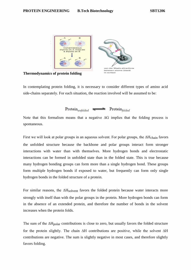
Thermodynamics of protein folding
In contemplating protein folding, it is necessary to consider different types of amino acid
side-chains separately. For each situation, the reaction involved will be assumed to be:
Note that this formalism means that a negative ∆G implies that the folding process is
spontaneous.
First we will look at polar groups in an aqueous solvent. For polar groups, the ΔHchain favors
the unfolded structure because the backbone and polar groups interact form stronger
interactions with water than with themselves. More hydrogen bonds and electrostatic
interactions can be formed in unfolded state than in the folded state. This is true because
many hydrogen bonding groups can form more than a single hydrogen bond. These groups
form multiple hydrogen bonds if exposed to water, but frequently can form only single
hydrogen bonds in the folded structure of a protein.
For similar reasons, the ΔHsolvent favors the folded protein because water interacts more
strongly with itself than with the polar groups in the protein. More hydrogen bonds can form
in the absence of an extended protein, and therefore the number of bonds in the solvent
increases when the protein folds.
The sum of the ΔHpolar contributions is close to zero, but usually favors the folded structure
for the protein slightly. The chain ∆H contributions are positive, while the solvent ∆H
contributions are negative. The sum is slightly negative in most cases, and therefore slightly
favors folding.
PROTEIN ENGINEERING B.Tech Biotechnology SBT1206
of the polar groups favors the unfolded state, because the chain is much more
disordered in the unfolded state. In contrast, the ΔSsolvent favors the folded state,
because the solvent is more disordered with the protein in the folded state. In most cases, the
sum of the ΔSpolar favors the unfolded state slightly. In other words, the ordering of the chain
during the folding process outweighs the other entropic factors.
The ΔGpolar that is obtained from the values of ΔHpolar and ΔSpolar for the polar groups
varies somewhat, but usually tends to favor the unfolded protein. In other words, the folding
of proteins comprised of polar residues is usually a nonspontaneous process.
Next, we will consider a chain constructed from non-polar groups in aqueous solvent. Once
again, the ΔHchain usually favors the unfolded state slightly. Once again, the reason is that
the backbone can interact with water in the unfolded state. However, the effect is smaller for
non-polar groups, due to the greater number of favorable van der Waals interactions in the
folded state. This is a result of the fact that non-polar atoms form better van der Waals
contacts with other non-polar groups than with water; in some cases, these effects mean that
the ΔHchain for nonpolar residues is slightly negative.
As with the polar groups, the ΔHsolvent for non-polar groups favors the folded state. In the
case of non-polar residues, ΔHsolvent favors folding more than it does for polar groups,
because water interacts much more strongly with itself than it does with non-polar groups.
The ΔSchain
PROTEIN ENGINEERING B.Tech Biotechnology SBT1206
The sum of the ΔHnon-polar favors folding somewhat. The magnitude of the ΔHnonpolar is not
very large, but is larger than the magnitude of the ∆Hpolar, which also tends to slightly favor
folding.
The ΔSchain of the non-polar groups favors the less ordered unfolded state. However, the
ΔSsolvent highly favors the folded state, due to the hydrophobic effect. During the burying of
the non-polar side chains, the solvent becomes more disordered. The ΔSsolvent is a major
driving force for protein folding which is called conformational entropy.
The ΔGnon-polar is therefore negative, due largely to the powerful contribution of the ΔSsolvent.
Adding together the terms for ΔGpolar and ΔGnon-polar gives a slightly negative overall ΔG for protein folding, and therefore, proteins generally fold spontaneously.
Raising the temperature, however, tends to greatly increase the magnitude of the TΔSchain
term, and therefore to result in unfolding of the protein.
The folded state is the sum of many interactions. Some favor folding, and some favor the
unfolded state. The qualitative discussion above did not include the magnitudes of the effects.
For real proteins, the various ∆H and ∆S values are difficult to measure accurately. However,
PROTEIN ENGINEERING B.Tech Biotechnology SBT1206
for many proteins it is possible to estimate the overall ∆G of folding. Measurements of this
value have shown that the overall ΔG for protein folding is very small: only about –10 to –50
kJoules/mol. This corresponds to a few salt bridges or hydrogen bonds.
Studies of protein folding have revealed one other important point: the hydrophobic effect is
very important, but it is relatively non-specific. Any hydrophobic group will interact with
essentially any other hydrophobic group. While the hydrophobic effect is a major driving
force for protein folding, it is the constrains imposed by the more geometrically specific
hydrogen bonding and electrostatic interactions in conjunction with the hydrophobic
interactions that largely determine the overall folded structure of the protein.
PROTEIN FOLDING MECHANISM
Protein Folding
Protein folding is a process in which a polypeptide folds into a specific, stable,
functional, three-dimensional structure. It is the process by which a protein structure assumes
its functional shape or conformation Proteins are formed from long chains of amino acids;
they exist in an array of different structures which often dictate their functions. Proteins
follow energetically favorable pathways to form stable, orderly, structures; this is known as
the proteins‘ native structure. Most proteins can only perform their various functions when
they are folded. The proteins‘ folding pathway, or mechanism, is the typical sequence of
structural changes the protein undergoes in order to reach its native structure. Protein folding
takes place in a highly crowded, complex, molecular environment within the cell, and often
requires the assistance of molecular chaperones, in order to avoid aggregation or mis folding.
Proteins are comprised of amino acids with various types of side chains, which may be
hydrophobic, hydrophilic, or electrically charged. The characteristics of these side chains
affect what shape the protein will form because they will interact differently intra molecularly
and with the surrounding environment, favoring certain conformations nd structures over
others. Scientists believe that the instructions for folding a protein are encoded in the
sequence. Researchers and scientists can easily determine the sequence of a protein, but have
not cracked the code that governs folding.
Protein Folding theory and experiment
Early scientists who studied proteomics and its structure speculated that proteins had
templates that resulted in their native conformations. This theory resulted in a search for how
proteins fold to attain their complex structure. It is now well known that under physiological
conditions, proteins normally spontaneously fold into their native conformations. As a result,
a protein's primary structure is valuable since it determines the three-dimensional structure of
a protein. Normally, most biological structures do not have the need for external templates to
help with their formation and are thus called self-assembling.
Protein Renaturation
Protein renaturation known since the 1930s. However, it was not until 1957 when
Christian Anfinsen performed an experiment on bovine pancreatic RNase A that protein
renaturation was quantified. RNase A is a single chain protein consisting of 124 residues. In
8M urea solution of 2-mercaptoethanol, the RNase A is completely unfolded and has its four
disulfide bonds cleaved through reduction. Through dialysisof urea and introducing the
solution to O2 at pH 8, the enzymatically active protein is physically incapable of being
recognized from RNase A. As a result, this experiment demonstrated that the protein
spontaneously renatured.
One criteria for the renaturation of RNase A is for its four disulfide bonds to reform.
The likelihood of one of the eight Cys residues from RNase A reforming a disulfide bond
with its native residue compared to the other seven Cys residues is 1/7. Futhermore, the next
one of remaining six Cys residues randomly forming the next disulfide bond is 1/5 and etc.
As a result, the probability of RNase A reforming four native disulfide links at random is (1/7 * 1/5 * 1/3 * 1/1 = 1/105). The result of this probability demonstrates that forming the
disulfide bonds from RNase A is not a random activity.
When RNase A is reoxidized utilizing 8M urea, allowing the disulfide bonds to
reform when the polypeptide chain is a random coil, then RNase A will only be around 1
percent enzymatically active after urea is removed. However, by using 2-mercaptoethanol,
the protein can be made fully active once again when disulfide bond interchange reactions
occur and the protein is back to its native state. The native state of the RNase A is
thermodynamically stable under physiological conditions, especially since a more stable
protein that is more stable than that of the native state requires a larger activation barrier, and
is kinetically inaccessible.By using the enzyme protein disulfide isomerase (PDI), the time it
takes for randomized RNase A is minimized to about 2 minutes. This enzyme helps facilitate
the disulfide interchange reactions. In order for PDI to be active, its two active site Cys
residues needs to be in the -SH form. Furthermore, PDI helps with random cleavage and the
reformation of the disulfide bonds of the protein as it attain thermodynamically favorable
conformations.
Post translationally Modified Proteins Might Not Renature
Proteins in a "scrambled" state go through PDI to renature, and their native state does
not utilize PDI because native proteins are in their stable conformations. However, proteins
that are posttranslationally modified need the disulfide bonds to stabilize their rather unstable
native form. One example of this is insulin, a polypeptide hormone. This 51 residue
polypeptide has two disulfide bonds that is inactivated by PDI. The following link is an image
showing insulin with its two disulfide bonds. Through observation of this phenomena,
scientists were able to find that insulin is made from proinsulin, an 84-residue single chain.
This link provides more information on the structure of proinsulin and its progression on
becoming insulin. The disulfide bonds of proinsulin need to be intact before conversion of
becoming insulin through proteolytic excision of its C chain which is an internal 33-residue
segment. However according to two findings, the C chain is not what dictates the folding of
the A and B chains, but instead holds them together to allow formation of the disulfide bonds.
For one, with the right renaturing conditions in place, scrambled insulin can become its native
form with a 30% yield. This yield can be increased if the A and B chains are cross-linked.
Secondly, through analysis of sequences of proinsulin from many species, mutations are
permitted at the C chain eight times more than if it were for A and B chains.
The Protein Folding Process Considerable evidence suggests that all of the information to describe the three dimensional
conformation of a protein is contained within the primary structure. However, for the most
part, we cannot fully interpret the information contained within the sequence. To understand
why this is true, we need to take a more careful look at proteins and how they fold. The polypeptide chain for most proteins is quite long. It therefore has many possible
conformations. If you assume that all residues could have 2 possible combinations of and
angles (real peptides can have many more than this), a 100 amino acid peptide could have
2100
(~1030
) possible conformations. If the polypeptide tested a billion conformations/second,
it would still take over 1013
years to find the correct conformation. (Note that the universe is
only ~1010
years old, and that a 100 residue polypeptide is a relatively small protein.) The
observation that proteins cannot fold by random tests of all possible conformations is referred
to as the Levinthal paradox. Folding pathways In classical transition state theory, the reaction diagram
for a spontaneous two state system is considered to have a
high-energy starting material, a lower energy product, and
an energy barrier between them. While the typical
diagram that describes the process (such as the one shown
at right) is useful, it is incomplete. The process for the conversion of S to P could actually
take many pathways; the pathway shown is merely the minimum energy route from one state
to another. The true situation is described by an energy landscape, with the minimum energy
route being the equivalent of a pass between two mountains. Thus, although the pathway
involves an energy barrier, other pathways require passing through even higher energy states.
A large part of the reason that single pathways (or small numbers of pathways) exist for chemical reactions is that most reactions involve the cleavage and reformation of covalent
bonds. The energy barrier for breaking a covalent bond is usually quite high. In protein folding, however, the interactions involved are weak. Because the thermal energy of a protein
molecule is comparable to the typical
non covalent interaction strength, an
unfolded polypeptide is present in a
large variety of rapidly changing
conformations. This realization led to
the Levinthal paradox: because the
unfolded protein should be constantly
changing its shape due to thermal
motions of the different parts of the polypeptide, it seemed unlikely that the protein would be able to find the correct state to begin transiting a fixed folding pathway.
An alternate hypothesis has been proposed, in which portions of the protein self-
organize, followed by folding into the final structure. Because the different parts of the
protein begin the folding process independently, the shape of the partially folded protein can
be very variable. In this model, the protein folds by a variety of different paths on an energy
landscape. The folding energy landscape has the general shape of a funnel. In the folding
process, as long as the overall process results in progressively lower energies, there can be a
large variety of different pathways to the final folded state.
The folding funnel shown above has a smooth surface. Actual folding funnels may be
fairly smooth, or may have irregularities in the surface that can act to trap the polypeptide chain in misfolded states. Alternatively, the folding funnel may direct the
polypeptide into a metastable state. Metastable states are local minima in the landscape; if the
energy barriers that surround the state are high enough, the metastable state may exist for a
long time – metastable states are stable for kinetic rather than thermodynamic reasons.
The difficulty in refolding many proteins in vitro suggests that the folded state of at
least some complex proteins may be in a metastable state rather than a global energy
minimum.
Folding process
The lower energies observed toward the depression in the folding funnel are thought
to be largely due to the collapse of an extended polypeptide due to the hydrophobic effect. In
addition to the hydrophobic effect, de solvation of the backbone is necessary for protein
folding, at least for portions of the backbone that will become buried. One method for
desolvation of the backbone is the formation of secondary structure. This is especially true
for helical structures, which can form tightly organized regions of hydrogen bonding while
PROTEIN ENGINEERING B.Tech Biotechnology SBT1206
excluding water from the backbone structure. A general outline for the process experienced
by a folding protein seems to look like this: A general outline for the process experienced by a folding protein seems to look like
this:
1. Some segments of a polypeptide may rapidly attain a relatively stable, organized
structure (largely due to organization of secondary structural Elements). 2. These structures provide nuclei for further folding. 3. During the folding process, the protein is proposed to form a state called a
Molten globule. This state readily rearranges to allow interactions between different parts
of the protein.
4. These nucleated, partially folded domains then coalesce into the folded protein. If
this general pathway is correct, it seems likely that at least some of the residues within the
sequence of most proteins function to guide the protein into the proper folding pathway, and
prevent the ―trapping‖ of the polypeptide in unproductive Partially folded states.
Folding inside cells
Real cells contain many proteins at a high overall protein concentration. The protein
concentration inside a cell is ~150 mg/ml. folding inside cells differs from most experiments
used to study folding in vitro:
• Proteins are synthesized on ribosomes. The entire chain is not available to fold at
once, as is the case for an experimentally unfolded protein in a test tube. • Within cells, the optimum ionic concentration, pH, and macromolecule
Concentration for each protein to fold properly cannot be controlled as tightly as in an
experimental system. • Major problems could arise if unfolded or partially folded proteins encountered one
another. Exposed hydrophobic regions might interact, and form potentially lethal insoluble
aggregates within the cell.
One mechanism for limiting problems with folding proteins inside cells volves
specialized proteins called molecular chaperones, which assist in folding proteins.
Molecular chaperones were first observed to be involved in responses to elevated temperature
(i.e. ―heat shock‖) to stabilize existing proteins and prevent protein aggregation and were
called heat-shock proteins (abbreviated as ―hsp‖). Additional research revealed that heat
shock proteins are present in all cells, and that they decrease or prevent non-specific protein
aggregation and assist in protein folding.
MOLECULAR CHAPERONES
In molecular biology, molecular chaperones are proteins that assist the covalent
folding or unfolding and the assembly or disassembly of other macromolecular structures.
Chaperones are present when the macromolecules perform their normal biological functions
and have correctly completed the processes of folding and/or assembly. The chaperones are
concerned primarily with protein folding. The first protein to be called a chaperone assists the
assembly of nucleosomes from folded histones and DNA and such assembly chaperones,
tendency to aggregate increases as proteins are denatured by stress. In this case, chaperones
do not convey any additional stericinformation required for proteins to fold. However, some
highly specific 'steric chaperones' do convey unique structural (steric) information onto
proteins, which cannot be folded spontaneously. Such proteins violate Anfinsen's dogma.
Various approaches have been applied to study the structure, dynamics and
functioning of chaperones. Bulk biochemical measurements have informed us on the protein
folding efficiency, and prevention of aggregation when chaperones are present during protein
folding. Recent advances in single-molecule analysis have brought insights into structural
heterogeneity of chaperones, folding intermediates and affinity of chaperones for
unstructured and structured protein chains.
Properties
Molecular chaperones interact with unfolded or partially folded protein subunits, e.g. nascent chains emerging from the ribosome, or extended chains being translocated across subcellular membranes.
They stabilize non-native conformation and facilitate correct folding of protein subunits.
They do not interact with native proteins, nor do they form part of the final folded structures.
Some chaperones are non-specific, and interact with a wide variety of polypeptide chains, but others are restricted to specific targets.
They often couple ATP binding/hydrolysis to the folding process.
Essential for viability, their expression is often increased by cellular stress.
Main role: They prevent inappropriate association or aggregation of exposed hydrophobic
surfaces and direct their substrates into productive folding, transport or degradation pathways.
Location and Function
Many chaperones are heat shock proteins, that is, proteins expressed in response to
elevated temperatures or other cellular stresses. The reason for this behaviour is thatprotein
folding is severely affected by heat and, therefore, some chaperones act to prevent or correct
damage caused by misfolding. Other chaperones are involved in folding newly made proteins
as they are extruded from the ribosome. Although most newly synthesized proteins can fold
in absence of chaperones, a minority strictly requires them for the same. Some chaperone systems work as foldases: they support the folding of proteins in an ATP-
dependent manner (for example, the GroEL/GroES or the DnaK/DnaJ/GrpE system). Other
chaperones work as holdases: they bind folding intermediates to prevent their aggregation,
in a translocation-competent (generally unfolded) state and guides them to the translocon.
New functions for chaperones continue to be discovered, such as assistance in protein
degradation, bacterial adhesin activity, and in responding to diseases linked to protein
aggregation (e.g. see prion) and cancer maintenance.
CHEPARONINE
Chaperonins are proteins that provide favourable conditions for the correct folding of other
proteins, thus preventing aggregation. Newly made proteins usually must fold from a linear chain of amino acids into a three-dimensional form. Chaperonins belong to a large class of
molecules that assist protein folding, called molecular chaperones. The energy to fold proteins is supplied by adenosine triphosphate GroupI Chaperonins
GroupI Chaperonins are found in bacteria as welas organelles of endosymbiotic origin: chloroplasts and mitochondria. The GroEL/GroES complex in E. coli is a Group I chaperonin and the best characterized large (~ 1 MDa) chaperonin complex.
1.GroEL is a double-ring 14mer with a greasy hydrophobic patch at its opening and
can accommodate the native folding of substrates 15-60 kDa in size. 2.GroES is a single-ring heptamer that binds to GroEL in the presence of ATP or
transition state analogues of ATP hydrolysis, such as ADP-AlF3. It's like a cover that covers GroEL (box/bottle). GroEL/GroES may not be able to undo protein aggregates, but kinetically it competes in the pathway of misfolding and aggregation, thereby preventing aggregate formation. Group II Chaperonins