Protein Kinases: Starting a Molecular Systems View of Endocytosis Prisca Liberali, Pauli R ¨ am ¨ o, and Lucas Pelkmans Institute of Molecular Systems Biology, ETH Zurich, CH-8093 Zurich, Switzerland; email: [email protected]Annu. Rev. Cell Dev. Biol. 2008. 24:501–23 First published online as a Review in Advance on July 3, 2008 The Annual Review of Cell and Developmental Biology is online at cellbio.annualreviews.org This article’s doi: 10.1146/annurev.cellbio.041008.145637 Copyright c 2008 by Annual Reviews. All rights reserved 1081-0706/08/1110-0501$20.00 Key Words membrane trafficking, phosphorylation, signal transduction, complexity, nonlinear systems, genetical physics Abstract The field of endocytosis is in strong need of formal biophysical model- ing and mathematical analysis. At the same time, endocytosis must be much better integrated into cellular physiology to understand the for- mer’s complex behavior in such a wide range of phenotypic variations. Furthermore, the concept that endocytosis provides the space-time for signal transduction can now be experimentally addressed. In this review, we discuss these principles and argue for a systematic and top-down ap- proach to study the endocytic membrane system. We provide a summary of published observations on protein kinases regulating endocytic ma- chinery components and discuss global unbiased approaches to further map out kinase regulatory networks. In particular, protein phosphoryla- tion is at the heart of controlling the physical properties of endocytosis and of integrating these physical properties into the signal transduction networks of the cell to allow a fine-tuned response to the continuously varying physiological conditions of a cell. 501 Click here for quick links to Annual Reviews content online, including: • Other articles in this volume • Top cited articles • Top downloaded articles • Our comprehensive search Further ANNUAL REVIEWS Annu. Rev. Cell Dev. Biol. 2008.24:501-523. Downloaded from www.annualreviews.org by Haifa University on 08/26/13. For personal use only.
Transcript
ANRV356-CB24-20 ARI 3 September 2008 19:11
Protein Kinases: Startinga Molecular Systems Viewof EndocytosisPrisca Liberali, Pauli Ramo, and Lucas PelkmansInstitute of Molecular Systems Biology, ETH Zurich, CH-8093 Zurich, Switzerland;email: [email protected]
Annu. Rev. Cell Dev. Biol. 2008. 24:501–23
First published online as a Review in Advance onJuly 3, 2008
The Annual Review of Cell and DevelopmentalBiology is online at cellbio.annualreviews.org
This article’s doi:10.1146/annurev.cellbio.041008.145637
membrane trafficking, phosphorylation, signal transduction,complexity, nonlinear systems, genetical physics
AbstractThe field of endocytosis is in strong need of formal biophysical model-ing and mathematical analysis. At the same time, endocytosis must bemuch better integrated into cellular physiology to understand the for-mer’s complex behavior in such a wide range of phenotypic variations.Furthermore, the concept that endocytosis provides the space-time forsignal transduction can now be experimentally addressed. In this review,we discuss these principles and argue for a systematic and top-down ap-proach to study the endocytic membrane system. We provide a summaryof published observations on protein kinases regulating endocytic ma-chinery components and discuss global unbiased approaches to furthermap out kinase regulatory networks. In particular, protein phosphoryla-tion is at the heart of controlling the physical properties of endocytosisand of integrating these physical properties into the signal transductionnetworks of the cell to allow a fine-tuned response to the continuouslyvarying physiological conditions of a cell.
The endocytic membrane system in mam-malian cells is complex. The basic steps of mem-brane trafficking—cargo recruitment, vesicleformation, vesicle transport, vesicle docking,and vesicle fusion—are a concerted seriesof events that involves many different pro-teins and lipids (Gruenberg 2001, McNiven &
Thompson 2006, Miaczynska & Zerial 2002,Soldati & Schliwa 2006). Although the gen-eral principle of endocytosis is always thesame, there is not one particular series ofmolecular events that always applies. Recentprogress in the field of endocytosis is rapidlydismissing our textbook view. Not only is thecontribution of clathrin-dependent processescompared with alternative endocytic routes de-bated in numerous instances, but also the def-inition of clathrin-mediated endocytosis findsitself on loose ground. The canonical clathrin-mediated route now appears to have differ-ent variants, which can make use of differentadaptors, different GTPases, and different traf-ficking itineraries and can bypass the canoni-cal early endosome (Lakadamyali et al. 2006,Schmid & McMahon 2007). Also, recent workon Listeria monocytogenes entry has shown thatclathrin can assemble into very large latticesthat appear to support a form of phagocyto-sis (Veiga & Cossart 2005). The more thesedifferences are studied, the more it becomesclear that only clathrin is the common factor.Thus, although the term clathrin-dependentendocytosis incorporates all these variants, itis hard to maintain the view that from afunctional perspective, these are all just oneroute. Similar controversies are programmedto arise (if they have not already arisen) forterms such as caveolae-mediated, fluid phase, ormacropinocytosis (Lajoie & Nabi 2007, Mayor& Pagano 2007).
The above-discussed impasse in the field ofendocytosis is probably exemplary for severalimpasses in molecular cell biology and shouldbe blamed on our highly reductionist, molecu-lar, and deterministic approach to these prob-lems in a time when global and unbiased com-parisons were not possible. All pathways in cellbiology will likely have to be redefined with un-biased global and quantitative methods, reveal-ing a standardized set of rules and definitions(Kirschner 2005, Mogilner et al. 2006). In onesense, we can compare this transition in biologywith that in chemistry approximately 150 yearsago. At that time, alchemy (reluctantly) gaveway to modern-day chemistry, founded on a
502 Liberali · Ramo · Pelkmans
Ann
u. R
ev. C
ell D
ev. B
iol.
2008
.24:
501-
523.
Dow
nloa
ded
from
ww
w.a
nnua
lrev
iew
s.or
gby
Hai
fa U
nive
rsity
on
08/2
6/13
. For
per
sona
l use
onl
y.
ANRV356-CB24-20 ARI 3 September 2008 19:11
standardized set of rules and definitions dis-covered by Antoine Lavoisier, who started tometiculously weigh metals, gases, liquids, andall kinds of materials and chemicals without anya priori hypothesis of a periodic table (Lavoisier1789). He might have believed in finding some-thing fundamental in the data, but there wasno scientific foundation for that belief. Thisprobably seems boring to many modern-dayscientists, but from the accurate study of a com-prehensive set of measurements, a systematicpattern was discovered.
We will thus have to go through a phase ofpainstakingly measuring, in an unbiased man-ner, as many relevant properties of cellular sys-tems as possible. Essential for this phase will bemethods to quantify large populations of sin-gle molecules and single particles; to quantifylarge populations of individual cells (eventu-ally within tissues); and to quantify functionalroles of whole genomes (of which the protein-encoding part is just a fraction), of whole pro-teomes (including the enormous complexity ofposttranslational modifications), and of the in-teractions between them. One can predict thatwhen genome-wide functional analysis of en-docytosis in mammalian cells becomes moreaccurate and quantitative and can incorpo-rate quantitative properties of all vesicles ina cell and all cells in a cell population, wemight be astonished by the number of differ-ences between the internalization of two ligandsthat both use clathrin-dependent endocytosis.Nevertheless, when this is done for a dozenof such ligands, the data might reveal certainpatterns, some of which we have no notion oftoday. We will find functional modules of cel-lular components, which can be linked to func-tional groups of physical properties that con-stitute certain design principles of a vesiclepattern (Milo et al. 2002). Moreover, we mightfind that ligands will fall into groups of pathwaysassembled from similar functional modules ofmolecular components and similar physical de-sign principles. Such information, when quan-titative, will allow us to create a set of formaland standardized rules with which to define theproperties of the endocytic membrane system
DEFINITION OF COMPLEXITY
A complex system is a system composed of interconnected partsthat as a whole displays properties not obvious from the individ-ual parts (Adami 2002, Ricard 2003). This makes every biologicalsystem with some nonlinear properties (like a simple feedbackloop) complex. The field of complex systems theory adds that asystem is complex when there are difficulties with its bottom-upformal modeling and simulation. For systems biology, this meansthat the system cannot be accurately modeled by a set of deter-ministic equations (for instance, differential equations) (Huang &Wikswo 2006). It is often argued that this is because the system’scomponents, their concentrations, and their ways of interactionare not (yet) known. But there are fundamental nondeterministicproperties (such as stochastic behavior) that, when amplified ordampened in nonlinear ways, make nonstatistical models inap-propriate. One may regard this basic property as the uncertaintyprinciple in biology. The biological uncertainty principle seemsparticularly relevant for dynamic systems that consist of manydifferent components and interactions that span several orders ofmagnitude on the space-time scale. It is likely that a crucial systemsuch as endocytosis has many built-in mechanisms to deal withthis uncertainty, but this remains to be experimentally addressed.
in human cells. This phase may be expensive,take a long time, and require a large and com-plex infrastructure. However, this concept canbe applied to smaller sets of genes, proteins, andphysical properties (Pelkmans et al. 2005). Theiterative process of thinking about the principalcomponents of the endocytic membrane systemon which to focus first to reveal the data and touse those data to think about how to expand theinitial focus will reveal sets of rules and defini-tions better and quicker and will optimize themalong the way.
WHY IS THE ENDOCYTICMEMBRANE SYSTEM INHUMAN CELLS SO COMPLEX?
The complexity of endocytosis is a specific traitof cells from multicellular organisms. There is alarge increase in complexity from Saccharomycescerevisiae to Caenorhabditis elegans and Drosophilamelanogaster to Homo sapiens ( Jekely 2007, Toret
www.annualreviews.org • Protein Kinases 503
Ann
u. R
ev. C
ell D
ev. B
iol.
2008
.24:
501-
523.
Dow
nloa
ded
from
ww
w.a
nnua
lrev
iew
s.or
gby
Hai
fa U
nive
rsity
on
08/2
6/13
. For
per
sona
l use
onl
y.
ANRV356-CB24-20 ARI 3 September 2008 19:11
STRUCTURAL VERSUS SYSTEMS BIOLOGYIN ENDOCYTOSIS
For certain aspects of endocytosis, structural biologists have vi-sualized functional modules with a remarkable degree of resolu-tion. Often, structural biology is seen as the ultimate foundationon which to attempt bottom-up modeling of biological systems.The molecular structure itself is a model, either an average ofmany possible conformations or a specific, trapped conformationthat allows the growth of a crystal. The biological uncertaintyprinciple will point out that one averaged or one specific struc-ture will not be able to account for the complexity of the system.Thus, systematic structural biology using nuclear magnetic reso-nance (NMR) or X-ray crystallography on isolated componentswill allow us to reveal basic structural rules that are generally ap-plied in endocytosis (e.g., the clathrin cage or matricity) (Fotinet al. 2004, Schmid & McMahon 2007), whereas single-particleor single-molecule methods, such as cryoelectron and optical mi-croscopy with nanometer resolution, will be necessary to revealstructural variation principles that underlie systems behavior.
& Drubin 2006). Whereas the complexity ofone or two core endocytic routes, and theircore machinery, appeared earlier in evolutionand is conserved through evolution, the partic-ular properties of endocytosis in human cellsappeared late.
In a multicellular organism, the endocyticmembrane system needs to demonstrate ex-treme plasticity (Kennedy & Ehlers 2006,Mostov et al. 2003). It needs to transcytosemassive amounts of liquid in epithelial cells ofthe renal duct, to transport vesicles over verylong distances in neurons (Rodman et al. 1990,Sudhof 2004), to relocalize specific membranecomponents for cell polarization and migration,and to shift from a sampling state to an antigen-presenting state during the maturation ofdendritic cells (Le Roy & Wrana 2005). In thesedifferent functions, the endocytic membranesystem displays very different properties. Some-times the vesicles are of a very narrow size rangeand always contain the same amount of cargo(neurons) (Voglmaier & Edwards 2007), some-times the endocytic organelles reshape intolong tubular structures that fuse with the cell
surface (maturing dendritic cells) (Kleijmeeret al. 2001), and sometimes the endosomes arelocated at the leading or ruffling edge of a cell(migrating cells) (Rappoport & Simon 2003).Also, within an individual cell, the endocyticmembrane system can display very different be-havior, depending on extrinsically and intrin-sically varying conditions. Recycling of mem-brane components is blocked during mitosiswhen the cells round up and overshoots thenormal activity when mitosis is completed andcells spread out again (Boucrot & Kirchhausen2007). Fluid-phase endocytosis is regulated asa function of the metabolic state and size ofa cell (Hennig et al. 2006), and the activityof raft-mediated endocytosis seems to dependon the adhesive state of cells (Echarri & DelPozo 2006, Pelkmans 2005). Molecular biol-ogy tends to explain differences in organellebehavior by the existence of cell-type- or cell-state-specific proteins. Indeed, there is tissue-,cell-type-, and cell-state-specific expression ofproteins that are part of the endocytic machin-ery. It is, however, unclear if the behavior ofthe endocytic system in one cell can be changedinto that of another cell by just expressing thesespecific proteins.
There is also a fundamental need for com-plexity in systems like endocytosis. Complexitymakes dynamic systems robust, permits themto evolve, allows them to oscillate or be noisy(by applying negative-feedback loops) or sta-ble (by applying positive-feedback loops), andcan contain built-in adaptation principles (e.g.bistability, criticality) that result in different be-havior of the system when a few key param-eters are changed (Kholodenko 2006, Shinaret al. 2007). The emergent properties that arisefrom such changes may appear to the molec-ular biologist as a completely different systemwith different molecular requirements. But itis the complexity itself that allows one systemconsisting of one set of components to behavedifferently under different conditions (Balazsi& Oltvai 2005, Mayo et al. 2006).
This complexity brings us to an alterna-tive principle of how plasticity of the endocyticmembrane system might be achieved. Perhaps
504 Liberali · Ramo · Pelkmans
Ann
u. R
ev. C
ell D
ev. B
iol.
2008
.24:
501-
523.
Dow
nloa
ded
from
ww
w.a
nnua
lrev
iew
s.or
gby
Hai
fa U
nive
rsity
on
08/2
6/13
. For
per
sona
l use
onl
y.
ANRV356-CB24-20 ARI 3 September 2008 19:11
it is the particular topology of certain regulatorycircuits within the endocytic membrane systemthat leads to a certain behavior (Kashtan et al.2004). If so, the endocytic membrane systemmust have all properties intrinsically built-in.We can then imagine why the endocytic mem-brane system in any cell from a multicellular or-ganism is so complex: It must have the intrinsicability to demonstrate a wide range of differentstates and behaviors. This possibility remains tobe empirically addressed, but we must be pre-pared for the prospect that in large-scale pertur-bation screens, all these different behaviors canemerge, even in a population of simple, nonpo-larizing, nondifferentiating tissue culture cells.
ENDOCYTOSIS: SPACE-TIMEFOR SIGNAL TRANSDUCTION
Another reason why the endocytic membranesystem is so complex is its essential role incell signaling. Here we discuss how the en-docytic membrane system might regulate thespace-time in which an input signal (extracellu-lar and intracellular) is transduced, processed,and translated in the cell.
A variety of sensors continuously monitorthe physiological status of the cell and the ex-tracellular environment. Well-known examplesare sensors that measure energy status, nutrientstatus, ion concentrations, levels of oxysterols,or the amount of cell stretching (Cota et al.2006, Janowski et al. 1996). Many sensors con-sist of components that are associated with orspan cellular membranes, such as integrins, ionchannels, growth hormone and cytokine recep-tors, Toll-like receptors, lipid sensors on endo-somes, pH sensors (e.g., the vacuolar ATPase),or redox-potential sensors (Chang et al. 2006,Morgan et al. 2007). By movement of thesesensors between cellular compartments, mem-brane trafficking, and endocytosis in particular,will have a major impact on the sensing capa-bility of a cell.
Endocytosis also plays important roles insignal transduction and processing (Miaczynskaet al. 2004, von Zastrow & Sorkin 2007). Re-ceptor kinases and other membrane-associated
DEFINITIONS IN SIGNAL TRANSDUCTION
The field of signal processing defines the following componentsof a signaling system: the primary signal, the sensor, the signaltransducer, the acceptor, and the effector. This applies equallywell to signal transduction in human cells. Signals are diverse andcan be a sterol, a growth hormone, protons, calcium, ADP:ATPratio, or a stretched integrin. Sensors can be (a) a cytosolic kinasethat becomes activated when the concentration of cAMP changesor (b) a hormone receptor on the cell surface. Transducers oftenare also kinases but can be GTPases or ubiquitin ligases. Theseare activated by the sensors and transfer information to a down-stream acceptor. Various transducers normally pass on a signalin complex ways. An acceptor receives the signal and activates aresponse to the signal via an effector. The specific topology of thenetwork by which such information is received, transduced, andaccepted can achieve complex signal integration and translation.Many separate signals can be integrated to create one output thatis short- or long-lasting, that oscillates, or that is binary, and asimple signal can be translated into a complex output that mayhave everlasting consequences for the cell (such as irreversiblecell differentiation).
kinases, but also signaling adaptors and sig-naling GTPases, can be localized to specificmembrane vesicles and compartments in thecell. Such molecules can be brought into closeproximity to each other by vesicle transportand membrane fusion, can be separated fromeach other by sorting and membrane fission,or can be inactivated or degraded by the endo-cytic membrane system. Not only can receptorkinases be targeted for degradation by trans-port to the lysosome, but the actual processof invaginating vesicles into the lumen of lateendosomes (multivesicular bodies), and theirregulated backfusion or degradation, providesthe cell with a mechanism to target cytosolicproteins for temporary inactivation (they areshielded from the cytosol) or degradation in-dependent of the proteasome (Hurley & Emr2006, van der Goot & Gruenberg 2006).
Endocytosis is also essentially involved insignal translation. The eventual outcome ofmany signal-processing events involves the re-localization of membrane components. For
www.annualreviews.org • Protein Kinases 505
Ann
u. R
ev. C
ell D
ev. B
iol.
2008
.24:
501-
523.
Dow
nloa
ded
from
ww
w.a
nnua
lrev
iew
s.or
gby
Hai
fa U
nive
rsity
on
08/2
6/13
. For
per
sona
l use
onl
y.
ANRV356-CB24-20 ARI 3 September 2008 19:11
example, the response might be to internalizeintegrins or stably assemble them in adhesioncomplexes on the surface, or to internalize neu-rotransmitter receptors or glucose channels orto accumulate them on the surface. Other cel-lular responses might be to migrate in one par-ticular direction, to grow in size, or to round upduring the mitotic cycle, which all need massiverelocation of the surface membrane.
Thus, the endocytic membrane system liesat the very heart of signal transduction, pro-cessing, and translation. It is then likely thatpart of the complexity of the endocytic mem-brane system has evolved to incorporate spe-cific properties that allow the system to playthis central role. This picture would predict thatthe endocytic machinery provides many pointsof interaction with protein kinases (and othermolecules) that sense and transduce cellularsignals.
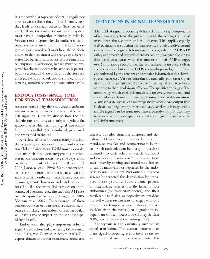
The above discussion raises another point.If the endocytic membrane system defines to asignificant extent the actual space-time in whichthe regulation of signaling networks takes place,standard reaction-diffusion diagrams will notbe appropriate for modeling signal processing.The interaction between signaling moleculeswill not be dictated by diffusion but by mem-brane dynamics, will not be able to rely on ahomogeneous concentration in the cell, andwill concern only a few active molecules. Thecollective behavior of vesicles, tubules, andorganelles does not display a random walk,nor does it display consistent active motion(Holcman & Triller 2006, Taflia & Holcman2007). Time-lapse images of cells containingfluorescent vesicles and organelles reveal thenotion that vesicles display a form of mixedbehavior (Holcman & Triller 2006, Taflia &Holcman 2007). It is not clear which formalmodels can describe this type of behavior,but perhaps certain agent-based models, suchas Brownian agents, may prove useful in thefuture.
Furthermore, at small scales, membranesprovide a quasi-two-dimensional surface(Kholodenko et al. 2000). From surfacechemistry, we know that chemical reactions on
surfaces proceed according to different princi-ples. But at larger scales, the three-dimensionalstructure of a vesicle or an organelle must betaken into account. Approximate simulations ofcomplex shapes indicate that particle geometrycan strongly influence reaction-diffusion ki-netics (Sbalzarini et al. 2005), but formal rulesstill need to be discovered. It thus seems ap-propriate to assume that signaling that dependson the endocytic membrane system will behaveaccording to physical rules that we do not yetknow but that will be fundamentally differentfrom standard reaction-diffusion kinetics. InFigure 1, we summarize these concepts.
If the endocytic membrane system consti-tutes the space-time for signal transductionreactions (at least to a certain extent), and sig-nal transduction is able to change the prop-erties of the endocytic membrane system, wedeal with the situation that the actors (sensors,signal transducers, and acceptors) influencetheir own space-time (dynamic shapes, patternsand interactions of membranes, vesicles, andorganelles), which in turn will influence theactors. In other words, the physical rules deter-mining the diffusion-reaction kinetics of signal-ing components are influenced by the compo-nents themselves (Figure 1). This introducesan aspect of complexity that is usually not con-sidered in systems biology. It is a type of feed-forward or feedback loop, but the effect is onthe space-time dimension in which the signal-ing reactions take place, which is fundamen-tally different from the kind of loops we usuallyconsider in standard signal processing diagrams(Figure 1). Given the importance of dynam-ics of complex shapes in biology, this may bea fundamental principle of processes in livingsystems.
PROTEIN KINASES REGULATINGENDOCYTIC MACHINERY
The previous three sections laid out our igno-rance of the integrated activities of membranetrafficking and signal transduction and the needto readjust our mindset for studying them. Toa certain extent, however, there is a molecular
506 Liberali · Ramo · Pelkmans
Ann
u. R
ev. C
ell D
ev. B
iol.
2008
.24:
501-
523.
Dow
nloa
ded
from
ww
w.a
nnua
lrev
iew
s.or
gby
Hai
fa U
nive
rsity
on
08/2
6/13
. For
per
sona
l use
onl
y.
ANRV356-CB24-20 ARI 3 September 2008 19:11
+ -
Brownian motion
Processive motion bybranched actin propulsion
Stop-and-go bidirectionalmotion on microtubules orF-actin filaments
Membrane fusion
Membrane fission
Flat 2-D surface(nanometer scale)
Curved surface on
complex structure
(micrometer scale)
Endosome with dynamic shape
+
-
Dkinase (P)Dkinase
Ckinase (P)Ckinase
Bkinase (P)Bkinase Arp2/3 (P)
Kinesin (P)
Syntaxin (P)
Ekinase (P)Ekinase Sortingnexin (P)
Akinase (P)Akinase Dynamin (P)
Nonlinear signaltransduction
Space-time: mixed vesicledynamics and complex surfaces
Figure 1Endocytosis defines a flexible space-time for signal transduction. This conceptual overview shows a hypothetical membrane-dependentkinase phosphorylation cascade (left) in which each kinase of the cascade is localized somewhere in the endocytic membrane system. Byphosphorylation of endocytic machinery components (a dynamin, Arp2/3, a kinesin, a syntaxin, a sorting nexin) along the way, thecascade will change (either increase or decrease) the dynamics of endocytic vesicles, fission and fusion reactions, and the shapes ofendocytic organelles. This defines the space-time for the reaction-diffusion kinetics of signal transduction, creating a mechanism bywhich the cascade can regulate its own transduction kinetics (either negatively or positively). We can also imagine the existence ofgeneric kinases that broadly influence the space-time of membrane-dependent signal transduction by regulating many endocyticmachinery components. In this scenario, signal transduction reactions occur at steady state and do not require any specific input butwill change their rate and direction when the dynamics of the endocytic membrane system are changed, resulting in specific signalingcascades.
foundation on which to build systems models ofendocytosis, and protein phosphorylation takesa central role in this process. Phosphorylationallows the actors of signaling cascades (proteinkinases) to control their space-time by regulat-ing membrane deformation, fission, trafficking,docking, and fusion (the endocytic machinery).Phosphorylation is also a tangible observationthat has been documented for many proteinsand protein kinases for several decades. There-fore, we expect that a systematic inventory ofthese reactions will provide a first molecular
foundation on which to further develop graph-ical models of signaling-endocytosis networks(Pelkmans et al. 2005). Although such mod-els do not explain systems behavior, they doprovide a useful way to navigate through thesystem’s components and to think about howsystems properties might emerge.
We collected all known direct phos-phorylation reactions of endocytic machin-ery components assigned to specific proteinkinases from the literature (SupplementalTable 1; follow the Supplemental Material
In general, a network consists of nodes and edges. Nodes arediscrete entities, genes, proteins, or metabolites. Edges are inter-actions between the nodes and can be of any type. Many biol-ogists understand an interaction as something physical, such asa (non)covalent binding between the two nodes, a biochemicalmodification of one node by another (such as phosphorylation),or a biochemical transition (metabolic networks). Increasingly,one finds networks in which statistical correlations between twonodes are displayed as an interaction. Such networks can be de-rived from transcriptome profiling, in which the profile of mRNAabundance in a series of particular conditions or in particular tis-sues is used to correlate genes. Above a certain correlation thresh-old, a connection is drawn. In functional RNAi screens, when thephenotype is described by a quantitative multivariate expressionor phenotype feature vector (statistically similar to a transcrip-tion profile), clusters of phenotypes (phenoclusters) can be made.These distances can be used as connections in a network, in whicha link between two nodes indicates that they have similar loss-of-function phenotypes.
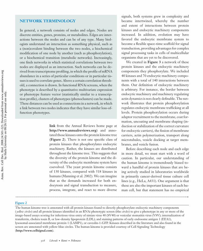
link from the Annual Reviews home page athttp://www.annualreviews.org) and anno-tated those kinases onto the protein kinome tree(Figure 2). There is not one specific class ofprotein kinases that phosphorylates endocyticmachinery. Rather, the kinases are distributedthroughout the kinome tree. This suggests thatthe diversity of the protein kinome and the di-versity of the endocytic membrane system havecoevolved. The yeast protein kinome consistsof 130 kinases, compared with 518 kinases inhumans (Manning et al. 2002). We can imaginethat as the demands increased for both en-docytosis and signal transduction to measure,process, integrate, and react to more diverse
signals, both systems grew in complexity andbecame intertwined, whereby the numberand extent of interactions between proteinkinases and endocytic machinery componentsincreased. In addition, evolution may havefavored the endocytic membrane system tobecome a flexible space-time scaffold for signaltransduction, providing advantages for complexsignal processing tasks in cells of multicellularorganisms that are yet to be discovered.
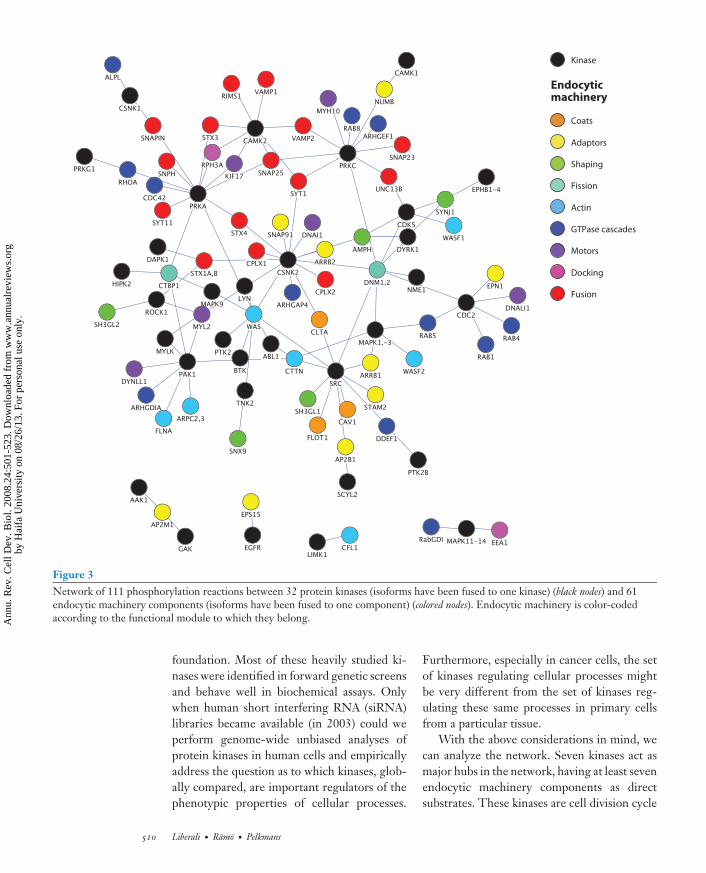
We created in Figure 3 a network of theseprotein kinases and the endocytic machinerycomponents they phosphorylate. We included40 kinases and 70 endocytic machinery compo-nents with a total of 140 interactions betweenthem. Our definition of endocytic machineryis arbitrary. For instance, the border betweenendocytic machinery and machinery regulatingactin dynamics is not clearly definable. The net-work illustrates that protein phosphorylationregulates endocytic membrane trafficking at alllevels. Protein phosphorylation occurs duringadaptor recruitment to the membrane, coat for-mation, uncoating and membrane shaping (in-duction or stabilization of the correct curvaturefor endocytic carriers), the fission of membranecarriers, actin polymerization, transport alongmicrotubules, vesicle docking at target mem-branes, and vesicle fusion.
Before describing each node and each edgein more detail, we must start with a word ofcaution. In particular, our understanding ofthe human kinome is tremendously biased to-ward a handful of protein kinases that are be-ing actively studied in laboratories worldwidein primarily cancer-derived tissue culture celllines (e.g., HeLa, A431). One argument is thatthese are also the important kinases of each hu-man cell, but that statement has no empirical
−−−−−−−−−−−−−−−−−−−−−−−−−−−−−−−−−−−−−−−−−−−−−−−−−−−−−−−−−−−−−−−−−−−−−−−−−−−−−−−−−−−−−−−−−→Figure 2The human kinome tree is annotated with all protein kinases found to directly phosphorylate endocytic machinery components( yellow circles) and all protein kinases identified in an RNAi phenotypic screen (blue circles) to give a phenotype in any or more of theimage-based assays scoring for infectious virus entry of simian virus 40 (SV40) or vesicular stomatitis virus (VSV); internalization oftransferrin, cholera toxin B, or low-density lipoprotein (LDL); and staining patterns of early endosome antigen 1 (EEA1),lysosomal-associated membrane protein 1 (LAMP1), or caveolin-1-GFP. Kinases described in the literature and also found in thescreen are annotated with yellow-blue circles. The human kinome is provided courtesy of Cell Signaling Technology(http://www.cellsignal.com).
508 Liberali · Ramo · Pelkmans
Ann
u. R
ev. C
ell D
ev. B
iol.
2008
.24:
501-
523.
Dow
nloa
ded
from
ww
w.a
nnua
lrev
iew
s.or
gby
Hai
fa U
nive
rsity
on
08/2
6/13
. For
per
sona
l use
onl
y.
ANRV356-CB24-20 ARI 3 September 2008 19:11
Screen
Published
Both
TKL
STE
TK
CK1
AGC
CAMK
CMGC
www.annualreviews.org • Protein Kinases 509
Ann
u. R
ev. C
ell D
ev. B
iol.
2008
.24:
501-
523.
Dow
nloa
ded
from
ww
w.a
nnua
lrev
iew
s.or
gby
Hai
fa U
nive
rsity
on
08/2
6/13
. For
per
sona
l use
onl
y.
ANRV356-CB24-20 ARI 3 September 2008 19:11
Coats
Adaptors
Shaping
Fission
Actin
GTPase cascades
Motors
Docking
Fusion
Kinase
Endocyticmachinery
Figure 3Network of 111 phosphorylation reactions between 32 protein kinases (isoforms have been fused to one kinase) (black nodes) and 61endocytic machinery components (isoforms have been fused to one component) (colored nodes). Endocytic machinery is color-codedaccording to the functional module to which they belong.
foundation. Most of these heavily studied ki-nases were identified in forward genetic screensand behave well in biochemical assays. Onlywhen human short interfering RNA (siRNA)libraries became available (in 2003) could weperform genome-wide unbiased analyses ofprotein kinases in human cells and empiricallyaddress the question as to which kinases, glob-ally compared, are important regulators of thephenotypic properties of cellular processes.
Furthermore, especially in cancer cells, the setof kinases regulating cellular processes mightbe very different from the set of kinases reg-ulating these same processes in primary cellsfrom a particular tissue.
With the above considerations in mind, wecan analyze the network. Seven kinases act asmajor hubs in the network, having at least sevenendocytic machinery components as directsubstrates. These kinases are cell division cycle
510 Liberali · Ramo · Pelkmans
Ann
u. R
ev. C
ell D
ev. B
iol.
2008
.24:
501-
523.
Dow
nloa
ded
from
ww
w.a
nnua
lrev
iew
s.or
gby
Hai
fa U
nive
rsity
on
08/2
6/13
. For
per
sona
l use
onl
y.
ANRV356-CB24-20 ARI 3 September 2008 19:11
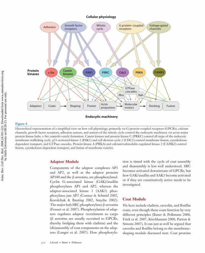
2 (CDC2), c-Src (SRC), casein kinase 1 and2 (CSNK1 and -2, respectively) [here treatedas one kinase, protein kinase C (PRKC)],protein kinase A (PRKA), p21-activated kinase1 (PAK1), and calcium/calmodulin-regulatedkinase 2B (CAMK2). CDC2 primarily phos-phorylates RabGTPases, which is believed tooccur during the mitotic cycle. SRC is a heavilystudied signaling kinase that is membraneanchored and associated with the plasmamembrane and perhaps also with intracellularorganelles. It phosphorylates componentsinvolved in early events of the endocyticmembrane trafficking cycle, namely vesicle for-mation, fission, and actin-mediated propulsion.It thus appears that SRC can act as a generalon switch for endocytic membrane trafficking,downstream from growth factor receptors andintegrins, which activate SRC. Casein kinasehas a very broad range of substrates and is usu-ally considered to be a nonspecific switch thatmodulates structural properties of many differ-ent types of proteins. It is activated by manyreceptor signals (e.g., growth factor receptors,cadherins) during mitosis. PRKC has beenstudied extensively in membrane traffickingand endocytosis; it has 10 isoforms, which wehere, for sake of simplicity, collectively treatas one kinase. PRKC regulates fission, fusion,and the RabGTPase cycle. Many signalscan activate PRKC, but most prominentlyamong these are activated G protein–coupledreceptors (GPCRs) and growth-factor re-ceptors. PRKA regulates membrane fission,microtubule transport, actin propulsion, andmembrane fusion. It is activated by cAMPdownstream of GPCRs and has an importantrole in nutrient signaling. PAK1 receives sig-nals from growth factor receptors, GPCRs, andintegrins on the cell surface and is a well-knownhub in the regulation of the actin cytoskeletonand microtubule-dependent transport. It isessential for ruffle formation, cup closure,and internalization of the membrane carriersduring macropinocytosis. In our network, itregulates actin-binding and -branching pro-teins, molecular motors, membrane fission, andRhoGTPases. The last hub is CAMK2, which
is activated by calmodulin as soon as intracellu-lar Ca2+ concentrations rise. CAMK2 regulatesprimarily membrane fusion. The effect of in-tracellular Ca2+ on membrane fusion hasbeen extensively studied for synaptic vesicles,where the arrival of an electrical impulse at thesynapse leads to immediate opening of Ca2+
channels and immediate fusion of numeroussynaptic vesicles already docked on the synapsemembrane. Ca2+-regulated fusion is not aspecific characteristic of membrane traffickingin synapses but is seen in any cell type. Thisis an example of an element of the endocyticmembrane system that is generally built-in butused more predominantly in a specific cell type(e.g., neurons). In Figure 4, we created a sim-plified hierarchical network to illustrate that, bymere consideration of these seven kinase hubs,cellular physiology is already linked in complexnetworks to the endocytic machinery modules.
MODULES OF ENDOCYTICMACHINERY COMPONENTSREGULATED BYPHOSPHORYLATION
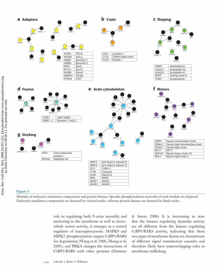
We next separated the phosphorylation net-works surrounding and interconnecting eachfunctional module of endocytic machinerycomponents for more detailed discussion(Figure 5). We do not embark here on de-tailed descriptions of the mechanics by whichthese modules operate. For that, we refer toa series of excellent reviews (Cai et al. 2007,D’Souza-Schorey & Chavrier 2006, Farsad& De Camilli 2003, Jahn & Scheller 2006,McNiven & Thompson 2006, Miaczynska &Zerial 2002, Robinson 2004, Roth 2007, Soldati& Schliwa 2006). The purpose here is to createan informational network of all known phos-phorylation reactions. In most cases, it is notyet understood how reversible phosphoryla-tion can change the mechanics of the endocyticmachinery. For the few cases in which more de-tailed insights have been obtained, we refer tothe original work and their reviews (Henderson& Conner 2007, Mace et al. 2005, Yarar et al.2007).
www.annualreviews.org • Protein Kinases 511
Ann
u. R
ev. C
ell D
ev. B
iol.
2008
.24:
501-
523.
Dow
nloa
ded
from
ww
w.a
nnua
lrev
iew
s.or
gby
Hai
fa U
nive
rsity
on
08/2
6/13
. For
per
sona
l use
onl
y.
ANRV356-CB24-20 ARI 3 September 2008 19:11
Adaptors Coats Shaping FissionActinpropulsion
Molecularmotors
Docking Fusion
GTPasecascades
RabRho
CAMK2c-Src PAK1 PRKCCaseinkinases Cdc2 PRKA
Endocytic machinery
Cellular physiology
AdhesionVoltage-gatedchannels
G protein–coupledreceptors
Mitoticcycle
Growth factorreceptors
Proteinkinases
Figure 4Hierarchical representation of a simplified view on how cell physiology, primarily via G protein–coupled receptors (GPCRs), calciumchannels, growth factor receptors, adhesion sensors, and sensors of the mitotic cycle control the endocytic machinery via seven majorprotein kinase hubs. c-Src controls vesicle formation. Casein kinases and protein kinase C (PRKC) control all steps of the endocyticmembrane trafficking cycle. p21-activated kinase 1 (PAK1) and cell division cycle 2 (CDC2) control membrane fission, cytoskeleton-dependent transport, and GTPase cascades. Protein kinase A (PRKA) and calcium/calmodulin-regulated kinase 2 (CAMK2) controlfission, cytoskeleton-dependent transport, and fusion of membrane vesicles.
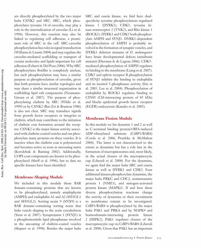
Adaptor Module
Components of the adaptor complexes AP1and AP2, as well as the adaptor proteinsAP180 and the β arrestins, are phosphorylated.Cyclin G–associated kinase (GAK)/auxillinphosphorylates AP1 and AP2, whereas theadaptor-associated kinase 1 (AAK1) phos-phorylates just AP2 (Conner & Schmid 2002,Korolchuk & Banting 2002, Smythe 2002).The major hub SRC phosphorylates β arrestins(Fessart et al. 2007). Phosphorylation of adap-tors regulates adaptor recruitment to cargo(β arrestins are usually recruited to GPCRs,thereby bridging them with clathrin) and the(dis)assembly of coat components on the adap-tors (Langer et al. 2007). How phosphoryla-
tion is timed with the cycle of coat assemblyand disassembly is less well understood. SRCbecomes activated downstream of GPCRs, buthow GAK/auxillin and AAK1 become activatedor if they are constitutively active needs to beinvestigated.
Coat Module
We here include clathrin, caveolin, and flotillincoats, even though these coats function by verydifferent principles (Bauer & Pelkmans 2006,Frick et al. 2007, Kirchhausen 2000, Parton &Simons 2007). It can just as well be argued thatcaveolin and flotillin belong to the membrane-shaping module discussed next. Coat proteins
512 Liberali · Ramo · Pelkmans
Ann
u. R
ev. C
ell D
ev. B
iol.
2008
.24:
501-
523.
Dow
nloa
ded
from
ww
w.a
nnua
lrev
iew
s.or
gby
Hai
fa U
nive
rsity
on
08/2
6/13
. For
per
sona
l use
onl
y.
ANRV356-CB24-20 ARI 3 September 2008 19:11
are directly phosphorylated by the two majorhubs CSNK2 and SRC. SRC, which phos-phorylates tyrosine 14 of caveolin, may play arole in the internalization of caveolae (Li et al.1996). However, this reaction may also belinked to regulating cell adhesion, a promi-nent role of SRC in the cell. SRC-caveolinphosphorylation has roles in signal transduction(Williams & Lisanti 2004) and may regulate thecaveolin-mediated scaffolding or transport ofcertain molecules and lipids important for celladhesion (Echarri & Del Pozo 2006). Why SRCphosphorylates flotillin is completely unclear,but such phosphorylation may have a similarpurpose as phosphorylation of caveolin, giventhat both proteins have similar topologies andmay share a similar structural organization inscaffolding lipid raft components (Neumann-Giesen et al. 2007). The purpose of phos-phorylating clathrin by SRC (Wilde et al.1999) or by CSNK2 (Bar-Zvi & Branton 1986)is also not clear. SRC may transduce signalsfrom growth factor receptors or integrins toclathrin, which may contribute to the initiationof clathrin coat formation around the recep-tor. CSNK2 is the major kinase activity associ-ated with clathrin-coated vesicles and can phos-phorylate many proteins on these vesicles. It isinactive when the clathrin coat is polymerizedand becomes active as soon as uncoating starts(Korolchuk & Banting 2002). Additionally,COPI-coat components are known to be phos-phorylated (Sheff et al. 1996), but to date nospecific kinases have been identified.
Membrane-Shaping Module
We included in this module those BARdomain–containing proteins that are knownto be phosphorylated, namely amphiphysin(AMPH) and endophilin A1 and A2 (SH3GL1and SH3GL2). Sorting nexin 9 (SNX9) is aBAR domain–containing sorting nexin thatlinks vesicle shaping to the actin cytoskeleton(Yarar et al. 2007). Synaptojanin 1 (SYNJ1) isa phosphoinositide lipid phosphatase involvedin the uncoating of clathrin-coated vesicles(Slepnev et al. 1998). Besides the major hubs
SRC and casein kinase, we find here dual-specificity tyrosine phosphorylation–regulatedkinase 1 (DYRK1), CDK5, tyrosine ki-nase nonreceptor 2 (TNK2), and Rho kinase 1(ROCK1). DYRK1 and CDK5 both phosphor-ylate AMPH and SYNJ1. DYRK1-dependentphosphorylation of AMPH is probably in-volved in the formation of synaptic vesicles, andDYRK1 deletion mutants of D. melanogasterhave brain developmental defects (minibrainmutant) (Dierssen & de Lagran 2006). CDK5-mediated phosphorylation of AMPH regulatesits binding to the membrane (Liang et al. 2007).CDK5 and ephrin receptor B phosphorylationof SYNJ1 inhibits the binding to endophilinand its inositol 5-phosphatase activity (Irie etal. 2005, Lee et al. 2004). Phosphorylation ofendophilin by ROCK1 regulates binding toCIN85 (Cbl-interacting protein of 85 kDa)and blocks epidermal growth factor receptor(EGFR) endocytosis (Kaneko et al. 2005).
Membrane Fission Module
In this module we list dynamin 1 and 2 as wellas C-terminal binding protein1/BFA-inducedADP-ribosylated substrate (CtBP1/BARS)(Corda et al. 2006, Praefcke & McMahon2004). The latter is not characterized to theextent as dynamins but has a role late in theformation of macropinosomes and, most likely,in the actual closure of the macropinocyticcup (Liberali et al. 2008). For the dynamins,we again find the major hubs SRC and caseinkinase as well as DYRK1 and CDK5. Fouradditional kinases phosphorylate dynamins, themajor hubs PRKC and CDC2, nonmetastaticprotein 1 (NME1), and mitogen-activatedprotein kinase (MAPK)1. If and how thesediverse phosphorylation reactions changethe activity of dynamins or their recruitmentto membranes remain to be investigated.CtBP1/BARS is phosphorylated by the majorhubs PAK1 and PRKA and by MAPK9 andhomeodomain-interacting protein kinase2 (HIPK2). PAK1 regulates closure of themacropinocytic cup via CtBP1/BARS (Liberaliet al. 2008). Given that PAK1 has an important
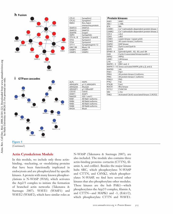
Figure 5Modules of endocytic machinery components and protein kinases. Specific phosphorylation networks of each module are depicted.Endocytic machinery components are denoted by colored nodes, whereas protein kinases are denoted by black nodes.
role in regulating both F-actin assembly andanchoring to the membrane as well as micro-tubule motor activity, it emerges as a centralregulator of macropinocytosis. MAPK9 andHIPK2 phosphorylation targets CtBP1/BARSfor degradation (Wang et al. 2006, Zhang et al.2005), and PRKA changes the interactions ofCtBP1/BARS with other proteins (Dammer
& Sewer 2008). It is interesting to notethat the kinases regulating dynamin activityare all different from the kinases regulatingCtBP1/BARS activity, indicating that thesetwo types of membrane fission are downstreamof different signal transduction cascades andtherefore likely have nonoverlapping roles inmembrane trafficking.
EGFREphrinB-EphB1, -B2, -B3, and -B4Cyclin G associated kinase/auxilin 2HIPK2
LIM kinase
LynERK1 and -2
Stress-activated MAPK p38 α, β, and Δ
Jnk
Pak1All protein kinase A isoforms
All protein kinase C isoforms
PKGFak
Fyk2Rho kinaseCVAK104
c-Src
Activated Cdc42-associated kinase-2 (ACK2)
h
i
Figure 5(Continued )
Actin Cytoskeleton Module
In this module, we include only those actin-binding, -nucleating, or -modulating proteinsthat have been functionally implicated inendocytosis and are phosphorylated by specifickinases. A protein with many known phosphor-ylations is N-WASP (WAS), which activatesthe Arp2/3 complex to initiate the formationof branched actin networks (Takenawa &Suetsugu 2007). WAVE1 (WASF1) andWAVE2 (WASF2), which have similar roles as
N-WASP (Takenawa & Suetsugu 2007), arealso included. The module also contains threeactin-binding proteins: cortactin (CTTN), fil-amin A, and cofilin1. Besides the major kinasehubs SRC, which phosphorylates N-WASPand CTTN, and CSNK2, which phosphor-ylates N-WASP, we find here several otherkinases that also phosphorylate other modules.These kinases are the hub PAK1—whichphosphorylates the Arp2/3 complex, filamin A,and CTTN—and MAPK1 and -3, (Erk1/2),which phosphorylate CTTN and WAVE1.
www.annualreviews.org • Protein Kinases 515
Ann
u. R
ev. C
ell D
ev. B
iol.
2008
.24:
501-
523.
Dow
nloa
ded
from
ww
w.a
nnua
lrev
iew
s.or
gby
Hai
fa U
nive
rsity
on
08/2
6/13
. For
per
sona
l use
onl
y.
ANRV356-CB24-20 ARI 3 September 2008 19:11
We also find here the major hub PRKA aswell as CDK5, both known to phosphorylateN-WASP. The module furthermore consists ofAbelson cytoplasmic tyrosine kinase 1 (ABL1),which regulates CTTN and N-WASP, as wellas MAPK9, the SRC family tyrosine kinaseLYN, TNK2 (activated Cdc42-associatedkinase), PTK2 (focal adhesion kinase), andBruton’s tyrosine kinase (BTK), all known tophosphorylate WASP. Finally, we include LIMkinase 1, which phosphorylates cofilin1.
Molecular-Motors Module
The following molecular motors with roles inendocytosis are known to be phosphorylatedby specific kinases: (a) three subunits of thedynein motor complex, the dynein light chain1 (DYNLL1), the dynein light intermediatechain 1 (DNALI1), and the dynein intermedi-ate chain 1 (DNAI1); (b) myosin motor 2 heavychain 10 (MYH10) and myosin light chain 2(MYL2); and (c) the kinesin KIF17. The ki-nases responsible for this are the hubs CDC2,CSNK2, PAK1, PRKA, PRKC, and CAMK2 aswell as the nonhubs myosin light chain kinase(MYLK) and ROCK1. A set of five kinases co-ordinates the activity of MYL2 and the dyneincomplex. PAK1 and CSNK2 are interesting be-cause they regulate both a component of thedynein complex and myosin 2. For many endo-cytic events, myosin and dynein motors need toact sequentially to switch from movement on F-actin to movement on microtubules. Sequentialphosphorylation of the responsible motor mayestablish this switch.
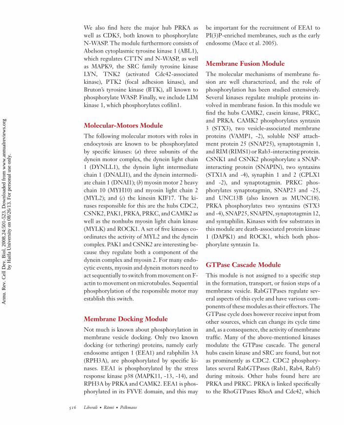
Membrane Docking Module
Not much is known about phosphorylation inmembrane vesicle docking. Only two knowndocking (or tethering) proteins, namely earlyendosome antigen 1 (EEA1) and rabphilin 3A(RPH3A), are phosphorylated by specific ki-nases. EEA1 is phosphorylated by the stressresponse kinase p38 (MAPK11, -13, -14), andRPH3A by PRKA and CAMK2. EEA1 is phos-phorylated in its FYVE domain, and this may
be important for the recruitment of EEA1 toPI(3)P-enriched membranes, such as the earlyendosome (Mace et al. 2005).
Membrane Fusion Module
The molecular mechanisms of membrane fu-sion are well characterized, and the role ofphosphorylation has been studied extensively.Several kinases regulate multiple proteins in-volved in membrane fusion. In this module wefind the hubs CAMK2, casein kinase, PRKC,and PRKA. CAMK2 phosphorylates syntaxin3 (STX3), two vesicle-associated membraneproteins (VAMP1, -2), soluble NSF attach-ment protein 25 (SNAP25), synaptotagmin 1,and RIM (RIMS1) or Rab3-interacting protein.CSNK1 and CSNK2 phosphorylate a SNAP-interacting protein (SNAPIN), two syntaxins(STX1A and -4), synaphin 1 and 2 (CPLX1and -2), and synaptotagmin. PRKC phos-phorylates synaptotagmin, SNAP23 and -25,and UNC13B (also known as MUNC18).PRKA phosphorylates two syntaxins (STX3and -4), SNAP25, SNAPIN, synaptotagmin 12,and syntaphilin. Kinases with few substrates inthis module are death-associated protein kinase1 (DAPK1) and ROCK1, which both phos-phorylate syntaxin 1a.
GTPase Cascade Module
This module is not assigned to a specific stepin the formation, transport, or fusion steps of amembrane vesicle. RabGTPases regulate sev-eral aspects of this cycle and have various com-ponents of these modules as their effectors. TheGTPase cycle does however receive input fromother sources, which can change its cycle timeand, as a consequence, the activity of membranetraffic. Many of the above-mentioned kinasesmodulate the GTPase cascade. The generalhubs casein kinase and SRC are found, but notas prominently as CDC2. CDC2 phosphory-lates several RabGTPases (Rab1, Rab4, Rab5)during mitosis. Other hubs found here arePRKA and PRKC. PRKA is linked specificallyto the RhoGTPases RhoA and Cdc42, which
516 Liberali · Ramo · Pelkmans
Ann
u. R
ev. C
ell D
ev. B
iol.
2008
.24:
501-
523.
Dow
nloa
ded
from
ww
w.a
nnua
lrev
iew
s.or
gby
Hai
fa U
nive
rsity
on
08/2
6/13
. For
per
sona
l use
onl
y.
ANRV356-CB24-20 ARI 3 September 2008 19:11
play (distant) roles in endocytosis by regulat-ing the cytoskeleton. PRKC has been specifi-cally linked to Rab8 and a Rho GDP/GTP ex-change factor (ARHGEF1). Interestingly, wealso find two major MAPKs: p38 (MAPK11,-13, -14) and Erk1/2 (MAPK1, -3). p38 is themajor transducer of stress response signaling inthe cell and regulates Rab GDP dissociation in-hibitor (GDI). Erk1/2, the major bottleneck intransducing growth factor signals to cell prolif-eration, phosphorylates Rab5.
UNBIASED GLOBALAPPROACHES TO LINKENDOCYTOSIS TO KINASEREGULATORY NETWORKSOF THE CELL
The previous section demonstrates that allmodules of the endocytic machinery are highlyinterconnected by protein kinases and that cer-tain modules are enriched in phosphorylationreactions. These are membrane fission, theGTPase cascade, membrane fusion, and actin-mediated propulsion (in particular N-WASP).
However, if an unbiased analysis of pro-tein phosphorylation of all these modules wereavailable, the picture would probably look quitedifferent. The first step toward such a globalview was taken several years ago by systemati-cally silencing each kinase of the human kinomeusing siRNA and studying how this affects avariety of properties of the endocytic mem-brane system (Pelkmans et al. 2005). This studyincluded the infectious entry of simian virus40 through caveolae/raft-mediated endocyto-sis; the infectious entry of vesicular stomatitisvirus through clathrin-mediated endocytosis;and 23 parameters of the endocytic membranesystem describing internalization patterns offluorescent transferrin, of cholera toxin subunitB, and of low-density lipoprotein and the in-tracellular distributions of early endosomes, oflate endosomes, and of caveolin-1. Because itwas the first study of its kind, and inherent toany high-throughput study, some experimentalnoise as well as false-positive and false-negativeobservations must be expected. The rapid tech-
nological development in this field, includingbetter siRNA libraries and advanced computa-tional methods for quantifying and classifyingloss-of-function phenotypes (Kittler et al. 2007,Lamprecht et al. 2007, Pepperkok & Ellenberg2006, Reimers & Carey 2006, Root et al. 2006),will allow much improved global and unbiasedanalysis of the endocytic membrane system inthe future. The importance of this approach forcreating a set of formal and standardized ruleswith which to define the properties of the endo-cytic membrane system justifies a large invest-ment by the cell biology community to makethese methods more mature, well adapted to theproblems of membrane trafficking, and moremainstream.
The results of this first study showed thatprotein kinase silencing has widespread effectson the endocytic membrane system. The 140protein kinases found to have a loss-of-functionphenotype in any of the parameters studied arealso annotated on the kinome tree in Figure 2.Of the 32 published kinases regulating endo-cytic machinery components, 26 had a loss-of-function phenotype in the screen. Importantly,several of these were actually found to regu-late endocytic machinery components after theRNAi screen was completed (see Supplemen-tal Table 1), thus independently validating sev-eral observations. It can be expected that a num-ber of the protein kinases found in a phenotypicscreen do not directly phosphorylate endocyticmachinery components but have a role moreupstream in signal transduction to the endo-cytic machinery. This hypothesis can be testedin a network in which one adds a layer of proteinkinases that are upstream of the protein kinasesdirectly phosphorylating the endocytic machin-ery (Figure 6). The information to constructsuch a network was derived from STRING(http://string.embl.de), a protein associationnetwork that quantitatively integrates predictedand known physical and functional interac-tion data from various sources. In this com-plex graph, we can now link more than halfof the protein kinases identified in the screento endocytic machinery components. There isno doubt that more extensive bioinformatics
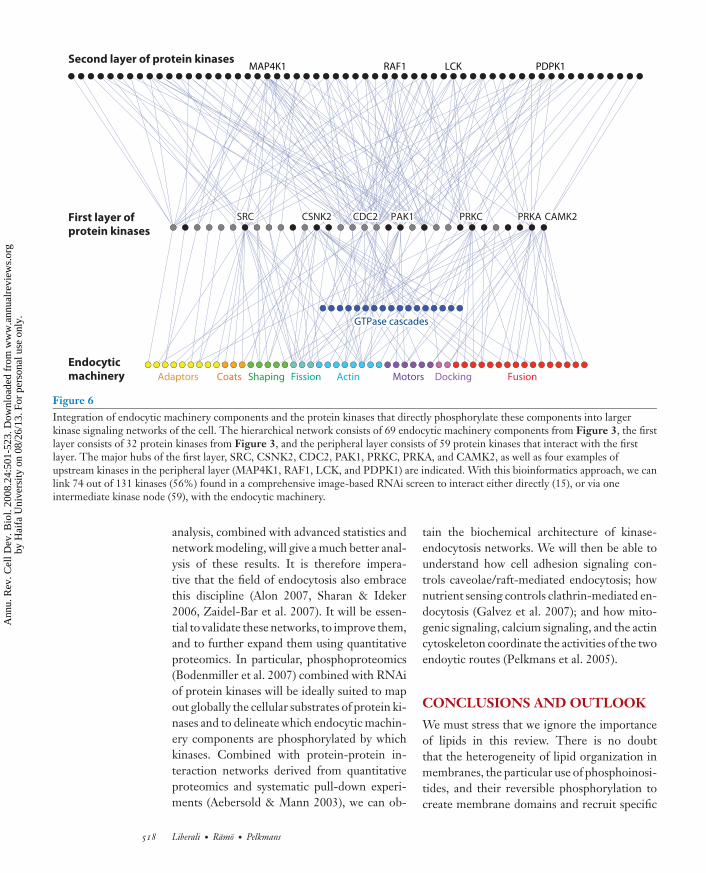
Figure 6Integration of endocytic machinery components and the protein kinases that directly phosphorylate these components into largerkinase signaling networks of the cell. The hierarchical network consists of 69 endocytic machinery components from Figure 3, the firstlayer consists of 32 protein kinases from Figure 3, and the peripheral layer consists of 59 protein kinases that interact with the firstlayer. The major hubs of the first layer, SRC, CSNK2, CDC2, PAK1, PRKC, PRKA, and CAMK2, as well as four examples ofupstream kinases in the peripheral layer (MAP4K1, RAF1, LCK, and PDPK1) are indicated. With this bioinformatics approach, we canlink 74 out of 131 kinases (56%) found in a comprehensive image-based RNAi screen to interact either directly (15), or via oneintermediate kinase node (59), with the endocytic machinery.
analysis, combined with advanced statistics andnetwork modeling, will give a much better anal-ysis of these results. It is therefore impera-tive that the field of endocytosis also embracethis discipline (Alon 2007, Sharan & Ideker2006, Zaidel-Bar et al. 2007). It will be essen-tial to validate these networks, to improve them,and to further expand them using quantitativeproteomics. In particular, phosphoproteomics(Bodenmiller et al. 2007) combined with RNAiof protein kinases will be ideally suited to mapout globally the cellular substrates of protein ki-nases and to delineate which endocytic machin-ery components are phosphorylated by whichkinases. Combined with protein-protein in-teraction networks derived from quantitativeproteomics and systematic pull-down experi-ments (Aebersold & Mann 2003), we can ob-
tain the biochemical architecture of kinase-endocytosis networks. We will then be able tounderstand how cell adhesion signaling con-trols caveolae/raft-mediated endocytosis; hownutrient sensing controls clathrin-mediated en-docytosis (Galvez et al. 2007); and how mito-genic signaling, calcium signaling, and the actincytoskeleton coordinate the activities of the twoendoytic routes (Pelkmans et al. 2005).
CONCLUSIONS AND OUTLOOK
We must stress that we ignore the importanceof lipids in this review. There is no doubtthat the heterogeneity of lipid organization inmembranes, the particular use of phosphoinosi-tides, and their reversible phosphorylation tocreate membrane domains and recruit specific
518 Liberali · Ramo · Pelkmans
Ann
u. R
ev. C
ell D
ev. B
iol.
2008
.24:
501-
523.
Dow
nloa
ded
from
ww
w.a
nnua
lrev
iew
s.or
gby
Hai
fa U
nive
rsity
on
08/2
6/13
. For
per
sona
l use
onl
y.
ANRV356-CB24-20 ARI 3 September 2008 19:11
endocytic machinery to membranes as well aslipid second messengers are crucial elementsof the endocytic membrane system (Di Paolo& De Camilli 2006). Cellular signaling regu-lates many lipid kinases and phosphatases, hy-drolases, carbohydrate transferases, flippases,pumps, and nonvesicular transferases betweencompartments, which strongly influences thebehavior of membranes (Di Paolo & De Camilli2006). This topic deserves a review of its own.
If we have the ambition to create a phys-ical explanation of the behavior of the endo-cytic membrane system, we need to fill in anenormous conceptual and informational gap,requiring both top-down and bottom-up ap-proaches. In the top-down approach, we mustattempt to model and predict the activity of en-docytic pathways and their phenotypic diversityby identifying patterns in large sets of measure-ments from individual cells and vesicles. Image-based screens of individual cells in whole cellpopulations combined with novel image analy-sis methods and advanced statistical and math-ematical analysis tools will be essential. Even-tually, this may lead to the discovery of formalrules that describe basic principles of the en-docytic membrane system, which is required toseparate nondeterministic aspects from deter-ministic aspects of the system. Only for the lat-ter will bottom-up approaches be useful. Mean-while, we must identify the principal proteins,enzymes, lipids, and metabolites responsible forthis deterministic behavior and delineate thetightly controlled interaction schemes between
them. We have already come a long way to iden-tifying these molecules, and the various-omicsdisciplines will make this list more complete.Systematic analysis of the effect of silencing,inactivating, or overexpressing these moleculeswill allow us to link these molecules to the phys-ical principles underlying phenotypic complex-ity of endocytosis. We here coin the term genet-ical physics for this approach. Genetical physicslinks genetic perturbations (e.g., by RNAi) tospecific parameters and degrees of freedom ofthe physical principles underlying the cellularsystem under scrutiny.
For accurate deterministic bottom-up mod-els, we will have to determine the abundancesand turnover of all these molecules, measureall the association and dissociation constantsbetween these molecules, and ascertain howthese variables are regulated over time. How-ever, the biological uncertainty principle (dueto small and noisy numbers of interacting com-ponents and unpredictable interaction kinetics)will prevent fully deterministic descriptions atthis level. This last aim therefore lies in the veryfar future for a complex system such as endo-cytic membrane trafficking, and we may haveto ask ourselves to what extent we should pur-sue the bottom-up approach. If we can iden-tify predictive physical principles and link themto sets of individual molecules without havinga complete biochemical model of how thesemolecules interact, they may be remarkably suf-ficient in explaining the roles of the endocyticmembrane system in cellular physiology.
SUMMARY POINTS
1. Protein kinases play an essential role in regulating the endocytic membrane system.
2. Protein kinases are the interface between the endocytic machinery and the physiologicalstatus of the cell.
3. Protein kinases allow the endocytic machinery to respond to changing demands fromthe cell and thereby ensure that the appropriate cellular phenotype is established ormaintained.
Genetical physics:a new discipline thatquantitatively linksgenetic perturbationsto degrees of freedomin a formal, physicalmodel of the biologicalsystem of interest
www.annualreviews.org • Protein Kinases 519
Ann
u. R
ev. C
ell D
ev. B
iol.
2008
.24:
501-
523.
Dow
nloa
ded
from
ww
w.a
nnua
lrev
iew
s.or
gby
Hai
fa U
nive
rsity
on
08/2
6/13
. For
per
sona
l use
onl
y.
ANRV356-CB24-20 ARI 3 September 2008 19:11
4. The endocytic membrane system in turn plays an essential role in signal transductionand provides a flexible and changeable space-time for signal transduction reactions.
5. Signal transduction and the endocytic membrane system have most likely coevolved intothe complex and integrated system that we observe in mammalian cells today.
DISCLOSURE STATEMENT
The authors are not aware of any biases that might be perceived as affecting the objectivity of thisreview.
ACKNOWLEDGMENTS
We thank all members of the laboratory for stimulating discussions. L.P. is supported by the SwissNational Science Foundation, SystemsX.ch, the European Union, and the ETH Zurich. P.L. isa long-term fellow of the Federation of European Biochemical Societies (FEBS), and P.R. is along-term fellow of the European Molecular Biology Organization (EMBO).
LITERATURE CITED
Adami C. 2002. What is complexity? BioEssays 24:1085–94Excellent overview ofthe power of modern-day quantitativeproteomics.
Aebersold R, Mann M. 2003. Mass spectrometry-based proteomics. Nature 422:198–207Alon U. 2007. Network motifs: theory and experimental approaches. Nat. Rev. Genet. 8:450–61Balazsi G, Oltvai ZN. 2005. Sensing your surroundings: how transcription-regulatory networks of the cell
Early paper identifyingone of the kinaseactivities in clathrin-coated vesicles.
Bar-Zvi D, Branton D. 1986. Clathrin-coated vesicles contain two protein kinase activities. Phos-phorylation of clathrin β-light chain by casein kinase II. J. Biol. Chem. 261:9614–21
Bauer M, Pelkmans L. 2006. A new paradigm for membrane-organizing and -shaping scaffolds. FEBS Lett.580:5559–64
Bodenmiller B, Mueller LN, Mueller M, Domon B, Aebersold R. 2007. Reproducible isolation of distinct,overlapping segments of the phosphoproteome. Nat. Methods 4:231–37
Boucrot E, Kirchhausen T. 2007. Endosomal recycling controls plasma membrane area during mitosis. Proc.Natl. Acad. Sci. USA 104:7939–44
Cai H, Reinisch K, Ferro-Novick S. 2007. Coats, tethers, Rabs, and SNAREs work together to mediate theintracellular destination of a transport vesicle. Dev. Cell 12:671–82
Chang TY, Chang CC, Ohgami N, Yamauchi Y. 2006. Cholesterol sensing, trafficking, and esterification.Annu. Rev. Cell Dev. Biol. 22:129–57
Identification of thekinase responsible foradaptor complex 2phosphorylation.
Conner SD, Schmid SL. 2002. Identification of an adaptor-associated kinase, AAK1, as a regulator ofclathrin-mediated endocytosis. J. Cell Biol. 156:921–29
Corda D, Colanzi A, Luini A. 2006. The multiple activities of CtBP/BARS proteins: the Golgi view. TrendsCell Biol. 16:167–73
Cota D, Proulx K, Smith KA, Kozma SC, Thomas G, et al. 2006. Hypothalamic mTOR signaling regulatesfood intake. Science 312:927–30
D’Souza-Schorey C, Chavrier P. 2006. ARF proteins: roles in membrane traffic and beyond. Nat. Rev. Mol.Cell Biol. 7:347–58
Excellent discussion ofthe role ofphosphoinositides inmembrane traffic.
Di Paolo G, De Camilli P. 2006. Phosphoinositides in cell regulation and membrane dynamics. Nature
443:651–57Dierssen M, de Lagran MM. 2006. DYRK1A (dual-specificity tyrosine-phosphorylated and -regulated kinase
1A): a gene with dosage effect during development and neurogenesis. Sci. World J. 6:1911–22
520 Liberali · Ramo · Pelkmans
Ann
u. R
ev. C
ell D
ev. B
iol.
2008
.24:
501-
523.
Dow
nloa
ded
from
ww
w.a
nnua
lrev
iew
s.or
gby
Hai
fa U
nive
rsity
on
08/2
6/13
. For
per
sona
l use
onl
y.
ANRV356-CB24-20 ARI 3 September 2008 19:11
Summarizes severalobservations from theDel Pozo lab thatcaveolae-mediatedendocytosis regulatescell adhesion signaling.
Echarri A, Del Pozo MA. 2006. Caveolae internalization regulates integrin-dependent signalingpathways. Cell Cycle 5:2179–82
Farsad K, De Camilli P. 2003. Mechanisms of membrane deformation. Curr. Opin. Cell Biol. 15:372–81Fessart D, Simaan M, Zimmerman B, Comeau J, Hamdan FF, et al. 2007. Src-dependent phosphorylation of
β2-adaptin dissociates the β-arrestin-AP-2 complex. J. Cell Sci. 120:1723–32Fotin A, Cheng Y, Sliz P, Grigorieff N, Harrison SC, et al. 2004. Molecular model for a complete clathrin
lattice from electron cryomicroscopy. Nature 432:573–79Frick M, Bright NA, Riento K, Bray A, Merrified C, Nichols BJ. 2007. Coassembly of flotillins induces
formation of membrane microdomains, membrane curvature, and vesicle budding. Curr. Biol. 17:1151–56
Image-based RNAiscreen on transferrinuptake, in whichautomated image-processing algorithmswere used. The authorsdemonstrate that themTOR signalingpathway regulates thenumber of transferrinreceptor molecules perendocytic vesicle.
Galvez T, Teruel MN, Heo WD, Jones JT, Kim ML, et al. 2007. siRNA screen of the human signal-ing proteome identifies the PtdIns(3,4,5)P3-mTOR signaling pathway as a primary regulator oftransferrin uptake. Genome Biol. 8:R142
Gruenberg J. 2001. The endocytic pathway: a mosaic of domains. Nat. Rev. Mol. Cell Biol. 2:721–30Henderson DM, Conner SD. 2007. A novel AAK1 splice variant functions at multiple steps of the endocytic
pathway. Mol. Biol. Cell 18:2698–706Hennig KM, Colombani J, Neufeld TP. 2006. TOR coordinates bulk and targeted endocytosis in the Drosophila
melanogaster fat body to regulate cell growth. J. Cell Biol. 173:963–74Holcman D, Triller A. 2006. Modeling synaptic dynamics driven by receptor lateral diffusion. Biophys. J.
91:2405–15Huang S, Wikswo J. 2006. Dimensions of systems biology. Rev. Physiol. Biochem. Pharmacol. 157:81–104Hurley JH, Emr SD. 2006. The ESCRT complexes: structure and mechanism of a membrane-trafficking
network. Annu. Rev. Biophys. Biomol. Struct. 35:277–98Irie F, Okuno M, Pasquale EB, Yamaguchi Y. 2005. EphrinB-EphB signalling regulates clathrin-mediated
endocytosis through tyrosine phosphorylation of synaptojanin 1. Nat. Cell Biol. 7:501–9Jahn R, Scheller RH. 2006. SNAREs—engines for membrane fusion. Nat. Rev. Mol. Cell Biol. 7:631–43Janowski BA, Willy PJ, Devi TR, Falck JR, Mangelsdorf DJ. 1996. An oxysterol signalling pathway mediated
by the nuclear receptor LXRα. Nature 383:728–31Jekely G. 2007. Origin of eukaryotic endomembranes: a critical evaluation of different model scenarios.
Adv. Exp. Med. Biol. 607:38–51Kaneko T, Maeda A, Takefuji M, Aoyama H, Nakayama M, et al. 2005. Rho mediates endocytosis of epidermal
growth factor receptor through phosphorylation of endophilin A1 by Rho-kinase. Genes Cells 10:973–87
Kashtan N, Itzkovitz S, Milo R, Alon U. 2004. Topological generalizations of network motifs. Phys. Rev. E70:031909
Kennedy MJ, Ehlers MD. 2006. Organelles and trafficking machinery for postsynaptic plasticity. Annu. Rev.Neurosci. 29:325–62
Kholodenko BN. 2006. Cell-signalling dynamics in time and space. Nat. Rev. Mol. Cell Biol. 7:165–76
These two discussionsby Kholodenko andcolleagues nicely lay outthe concept thatmembranes define aspace-time for signaltransduction that isdifferent from standardreaction-diffusionkinetics.
Kholodenko BN, Hoek JB, Westerhoff HV. 2000. Why cytoplasmic signalling proteins should berecruited to cell membranes. Trends Cell Biol. 10:173–78
Kirchhausen T. 2000. Three ways to make a vesicle. Nat. Rev. Mol. Cell Biol. 1:187–98Kirschner MW. 2005. The meaning of systems biology. Cell 121:503–4Kittler R, Pelletier L, Heninger AK, Slabicki M, Theis M, et al. 2007. Genome-scale RNAi profiling of cell
division in human tissue culture cells. Nat. Cell Biol. 9:1401–12Kleijmeer M, Ramm G, Schuurhuis D, Griffith J, Rescigno M, et al. 2001. Reorganization of multi-
vesicular bodies regulates MHC class II antigen presentation by dendritic cells. J. Cell Biol. 155:53–63
Korolchuk VI, Banting G. 2002. CK2 and GAK/auxilin2 are major protein kinases in clathrin-coated vesicles.Traffic 3:428–39
Lajoie P, Nabi IR. 2007. Regulation of raft-dependent endocytosis. J. Cell Mol. Med. 11:644–53
Indicates that thecanonical endocyticpathway must beconsidered as apopulation of vesiclesthat display specificheterogeneity in thekinetics ofinternalization andtransport of cargo.Lakadamyali M, Rust MJ, Zhuang X. 2006. Ligands for clathrin-mediated endocytosis are differentially
sorted into distinct populations of early endosomes. Cell 124:997–1009
www.annualreviews.org • Protein Kinases 521
Ann
u. R
ev. C
ell D
ev. B
iol.
2008
.24:
501-
523.
Dow
nloa
ded
from
ww
w.a
nnua
lrev
iew
s.or
gby
Hai
fa U
nive
rsity
on
08/2
6/13
. For
per
sona
l use
onl
y.
ANRV356-CB24-20 ARI 3 September 2008 19:11
Demonstrates theimportance of theopen-source mentalityin the life sciences toshare image-processingtools. CellProfiler isbased on MatLab andprovides an easy-to-usegraphical user interfaceenabling every biologistto do computationalimage processing.
Langer JD, Stoops EH, Bethune J, Wieland FT. 2007. Conformational changes of coat proteins during vesicleformation. FEBS Lett. 581:2083–88
Lavoisier A. 1789. Traite Elementaire de Chimie. Paris: CuchetLe Roy C, Wrana JL. 2005. Signaling and endocytosis: a team effort for cell migration. Dev. Cell 9:167–68Lee SY, Wenk MR, Kim Y, Nairn AC, De Camilli P. 2004. Regulation of synaptojanin 1 by cyclin-dependent
kinase 5 at synapses. Proc. Natl. Acad. Sci. USA 101:546–51Li S, Seitz R, Lisanti MP. 1996. Phosphorylation of caveolin by Src tyrosine kinases. The α-isoform of caveolin
is selectively phosphorylated by v-Src in vivo. J. Biol. Chem. 271:3863–68Liang S, Wei FY, Wu YM, Tanabe K, Abe T, et al. 2007. Major Cdk5-dependent phosphorylation sites of
amphiphysin 1 are implicated in the regulation of the membrane binding and endocytosis. J. Neurochem.102:1466–76
Liberali P, Kakkonen E, Turacchio G, Valente C, Spaar A, et al. 2008. The closure of Pak1-dependentmacropinosomes requires the phosphorylation of CtBP1/BARS. EMBO J. 27:970–81
Mace G, Miaczynska M, Zerial M, Nebreda AR. 2005. Phosphorylation of EEA1 by p38 MAP kinase regulatesμ opioid receptor endocytosis. EMBO J. 24:3235–46
Hallmark discussion ofthe phylogeny of thewhole human proteinkinome.
Manning G, Whyte DB, Martinez R, Hunter T, Sudarsanam S. 2002. The protein kinase complementof the human genome. Science 298:1912–34
Mayo AE, Setty Y, Shavit S, Zaslaver A, Alon U. 2006. Plasticity of the cis-regulatory input function of a gene.PLoS Biol. 4:e45
Mayor S, Pagano RE. 2007. Pathways of clathrin-independent endocytosis. Nat. Rev. Mol. Cell Biol. 8:603–12McNiven MA, Thompson HM. 2006. Vesicle formation at the plasma membrane and trans-Golgi network:
the same but different. Science 313:1591–94Miaczynska M, Pelkmans L, Zerial M. 2004. Not just a sink: endosomes in control of signal transduction.
Curr. Opin. Cell Biol. 16:400–6Miaczynska M, Zerial M. 2002. Mosaic organization of the endocytic pathway. Exp. Cell Res. 272:8–14Milo R, Shen-Orr S, Itzkovitz S, Kashtan N, Chklovskii D, Alon U. 2002. Network motifs: simple building
blocks of complex networks. Science 298:824–27Mogilner A, Wollman R, Marshall WF. 2006. Quantitative modeling in cell biology: What is it good for?
Dev. Cell 11:279–87Morgan MR, Humphries MJ, Bass MD. 2007. Synergistic control of cell adhesion by integrins and syndecans.
Nat. Rev. Mol. Cell Biol. 8:957–69Mostov K, Su T, ter Beest M. 2003. Polarized epithelial membrane traffic: conservation and plasticity.
Nat. Cell Biol. 5:287–93Neumann-Giesen C, Fernow I, Amaddii M, Tikkanen R. 2007. Role of EGF-induced tyrosine phosphorylation
of reggie-1/flotillin-2 in cell spreading and signaling to the actin cytoskeleton. J. Cell Sci. 120:395–406Parton RG, Simons K. 2007. The multiple faces of caveolae. Nat. Rev. Mol. Cell Biol. 8:185–94Pelkmans L. 2005. Secrets of caveolae- and lipid raft-mediated endocytosis revealed by mammalian viruses.
Biochim. Biophys. Acta 1746:295–304Pelkmans L, Fava E, Grabner H, Hannus M, Habermann B, et al. 2005. Genome-wide analysis of human
kinases in clathrin- and caveolae/raft-mediated endocytosis. Nature 436:78–86Pepperkok R, Ellenberg J. 2006. High-throughput fluorescence microscopy for systems biology. Nat. Rev.
Mol. Cell Biol. 7:690–96Praefcke GJ, McMahon HT. 2004. The dynamin superfamily: universal membrane tubulation and fission
molecules? Nat. Rev. Mol. Cell Biol. 5:133–47Rappoport JZ, Simon SM. 2003. Real-time analysis of clathrin-mediated endocytosis during cell migration.
J. Cell Sci. 116:847–55Reimers M, Carey VJ. 2006. Bioconductor: an open source framework for bioinformatics and computational
biology. Methods Enzymol. 411:119–34Ricard J. 2003. What do we mean by biological complexity? C. R. Biol. 326:133–40Robinson MS. 2004. Adaptable adaptors for coated vesicles. Trends Cell Biol. 14:167–74
522 Liberali · Ramo · Pelkmans
Ann
u. R
ev. C
ell D
ev. B
iol.
2008
.24:
501-
523.
Dow
nloa
ded
from
ww
w.a
nnua
lrev
iew
s.or
gby
Hai
fa U
nive
rsity
on
08/2
6/13
. For
per
sona
l use
onl
y.
ANRV356-CB24-20 ARI 3 September 2008 19:11
Rodman JS, Mercer RW, Stahl PD. 1990. Endocytosis and transcytosis. Curr. Opin. Cell Biol. 2:664–72Root DE, Hacohen N, Hahn WC, Lander ES, Sabatini DM. 2006. Genome-scale loss-of-function screening
with a lentiviral RNAi library. Nat. Methods 3:715–19Roth MG. 2007. Integrating actin assembly and endocytosis. Dev. Cell 13:3–4
By applying computersimulation of diffusionin complex-shapedmembranes (theendoplasmic reticulum),the authors showed thatorganelle shape hasstrong influences ondiffusion at themacroscopic scale.
Sbalzarini IF, Mezzacasa A, Helenius A, Koumoutsakos P. 2005. Effects of organelle shape on fluo-rescence recovery after photobleaching. Biophys. J. 89:1482–92
Schmid EM, McMahon HT. 2007. Integrating molecular and network biology to decode endocytosis.Nature 448:883–88
Sharan R, Ideker T. 2006. Modeling cellular machinery through biological network comparison. Nat.Biotechnol. 24:427–33
Sheff D, Lowe M, Kreis TE, Mellman I. 1996. Biochemical heterogeneity and phosphorylation of coatomersubunits. J. Biol. Chem. 271:7230–36
Shinar G, Milo R, Martinez MR, Alon U. 2007. Input output robustness in simple bacterial signaling systems.Proc. Natl. Acad. Sci. USA 104:19931–35
Slepnev VI, Ochoa GC, Butler MH, Grabs D, De Camilli P. 1998. Role of phosphorylation in regulation ofthe assembly of endocytic coat complexes. Science 281:821–24
Smythe E. 2002. Regulating the clathrin-coated vesicle cycle by AP2 subunit phosphorylation. Trends Cell Biol.12:352–54
Soldati T, Schliwa M. 2006. Powering membrane traffic in endocytosis and recycling. Nat. Rev. Mol. Cell Biol.7:897–908
Important theoreticalconsiderations of howcellularcompartmentalizationwill affect Brownianmotion.
Taflia A, Holcman D. 2007. Dwell time of a Brownian molecule in a microdomain with traps and asmall hole on the boundary. J. Chem. Phys. 126:234107
Takenawa T, Suetsugu S. 2007. The WASP-WAVE protein network: connecting the membrane to thecytoskeleton. Nat. Rev. Mol. Cell Biol. 8:37–48
Toret CP, Drubin DG. 2006. The budding yeast endocytic pathway. J. Cell Sci. 119:4585–87Van Der Goot FG, Gruenberg J. 2006. Intra-endosomal membrane traffic. Trends Cell Biol. 16:514–21Veiga E, Cossart P. 2005. Listeria hijacks the clathrin-dependent endocytic machinery to invade mammalian
cells. Nat. Cell Biol. 7:894–900Voglmaier SM, Edwards RH. 2007. Do different endocytic pathways make different synaptic vesicles? Curr.
Opin. Neurobiol. 17:374–80von Zastrow M, Sorkin A. 2007. Signaling on the endocytic pathway. Curr. Opin. Cell Biol. 19:436–45Wang SY, Iordanov M, Zhang Q. 2006. c-Jun NH2-terminal kinase promotes apoptosis by down-regulating
the transcriptional corepressor CtBP. J. Biol. Chem. 281:34810–15Wilde A, Beattie EC, Lem L, Riethof DA, Liu SH, et al. 1999. EGF receptor signaling stimulates SRC kinase
phosphorylation of clathrin, influencing clathrin redistribution and EGF uptake. Cell 96:677–87Williams TM, Lisanti MP. 2004. The Caveolin genes: from cell biology to medicine. Ann. Med. 36:584–95Yarar D, Waterman-Storer CM, Schmid SL. 2007. SNX9 couples actin assembly to phosphoinositide signals
and is required for membrane remodeling during endocytosis. Dev. Cell 13:43–56Zaidel-Bar R, Itzkovitz S, Ma’ayan A, Iyengar R, Geiger B. 2007. Functional atlas of the integrin adhesome.
Nat. Cell Biol. 9:858–67Zhang Q, Nottke A, Goodman RH. 2005. Homeodomain-interacting protein kinase-2 mediates CtBP
phosphorylation and degradation in UV-triggered apoptosis. Proc. Natl. Acad. Sci. USA 102:2802–7
Dscam-Mediated Cell Recognition Regulates NeuralCircuit FormationDaisuke Hattori, S. Sean Millard, Woj M. Wojtowicz, and S. Lawrence Zipursky � � � � � 597