46
Protein Modification, targeting and degradation
| Date post: | 17-Dec-2015 |
| Category: |
Documents |
| Upload: | kory-kennedy |
| View: | 225 times |
| Download: | 7 times |
Protein Modification, targeting and degradation

Protein modification
• Proteins undergo a variety of modifications that are critical for function. There are numerous amino acid modifications such as collagen.
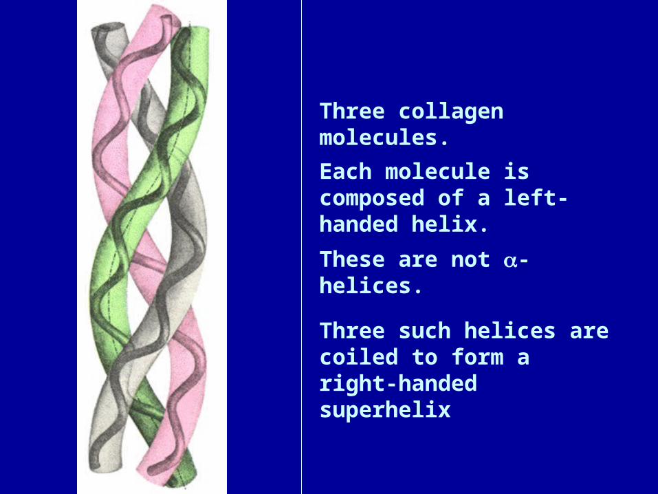
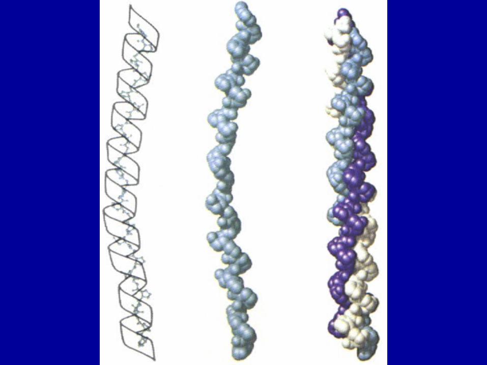
Three collagen molecules.
Each molecule is composed of a left-handed helix.
These are not -helices.
Three such helices are coiled to form a right-handed superhelix

Collagen’s unique blend of amino acids
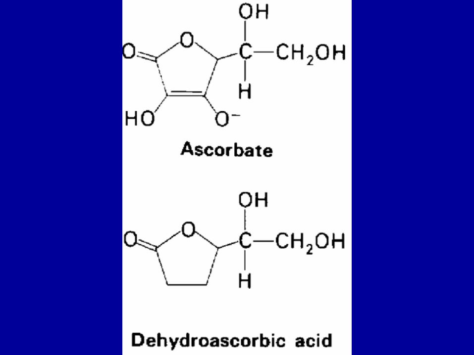
• ~ 30% of residues are Glycine • ~ 30 of residues are Proline or Hydroxyproline (HyPro) • 5-hydroxylysine (HyLys) also occurs; a site for glycosylation• Hydroxylation of Pro, Lys is a post-translational modification,
requires vitamin C as a reactant• The sequence of collagen bears long stretches of
Gly--Pro/HyPro-X repeats
RO
N
R
OH
12
34
5
RO
N
R
OH
12
34
5
3-HyPro4-HyPro
ONH
NH2
R
R
OH
5-HyLys
Structure of Collagen
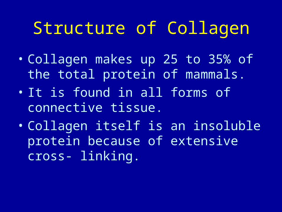
• Collagen makes up 25 to 35% of the total protein of mammals.
• It is found in all forms of connective tissue.
• Collagen itself is an insoluble protein because of extensive cross- linking.
Structure of Collagen(continued)
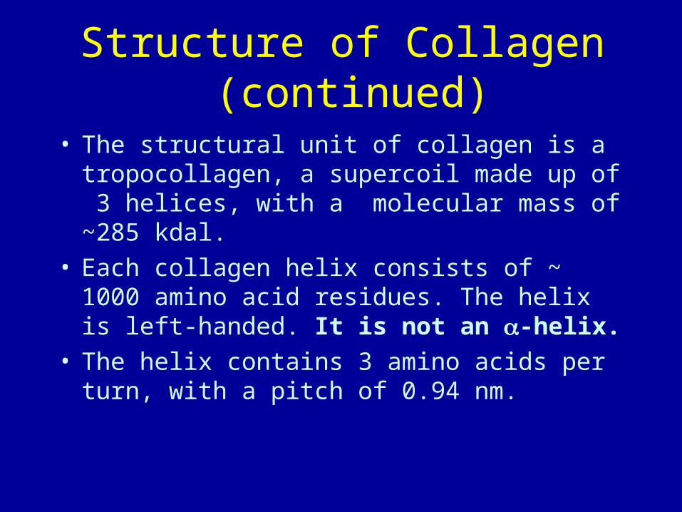
• The structural unit of collagen is a tropocollagen, a supercoil made up of 3 helices, with a molecular mass of ~285 kdal.
• Each collagen helix consists of ~ 1000 amino acid residues. The helix is left-handed. It is not an -helix.
• The helix contains 3 amino acids per turn, with a pitch of 0.94 nm.
Structure of Collagen(continued)
• Each unit of tropocollagen is about 1.5 nm wide and 300 nm long. Bundles of these 3-stranded supercoils can be seen as collagen fibres in the electron microscope.
• Mature collagen has extensive covalent crosslinkages between individual collagen molecules.

An electron micrograph of collagen from skin

Structure of Collagen(continued)
• There is no intra-helical H-bonding in collagen helices.
• Rather, H-bonding occurs between the amide N of glycine residues in the central axis and the carbonyls of other residues in the adjacent chains. Often proline and hydroxyproline are involved.
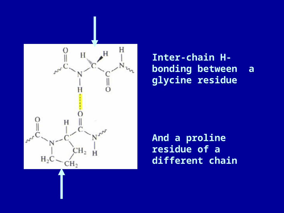
Inter-chain H-bonding between a glycine residue
And a proline residue of a different chain
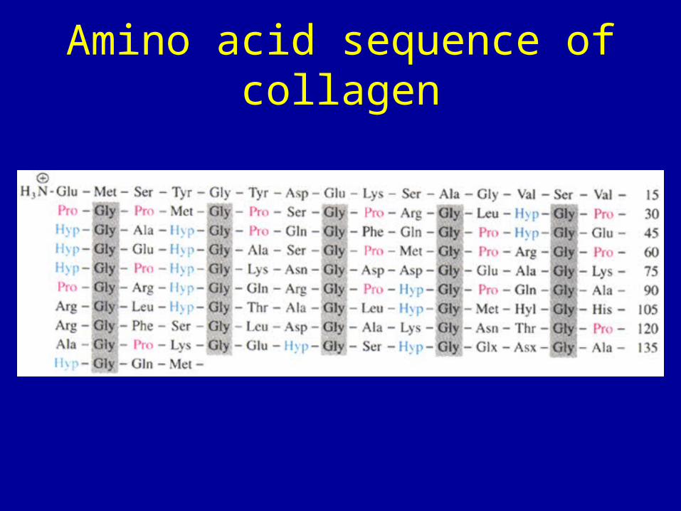
Amino acid sequence of collagen

Defective Hydroxylation Is One Of TheBiochemical Lesions in Scurvy
The importance of the hydroxylation of collagen becomes evident in scurvy. A vivid description of this disease was given by Jacques Cartier in 1536, when it afflicted his men as they were exploring the Saint Lawrence River:
Some did lose all their strength, and could not stand on their feet… Others also had all their skins spotted with spots of blood of a purple colour: then did it ascend up to their ankles, knees, thighs, shoulders, arms, and necks. Their mouths became stinking, their gums so rotten, that all the flesh did fall off, even to the roots of the teeth, which did also almost all fall out.

The means of preventing scurvy was succintly stated by James Lind, a Scottish physician, in 1753:
Experience indeed sufficiently shows that as greens or fresh vegetables, with ripe fruits, are the best remedies for it, so they prove the most effectual preservatives against it.
Lind urged the inclusion of lemon juice in the diet of sailors. His advice was adopted by the British Naby some forty years later.
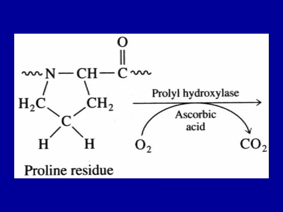

The presence of hyp residues greatly increases the potential for H-bonding between chains.
Hyp and pro make up 25% of the residues of collagen
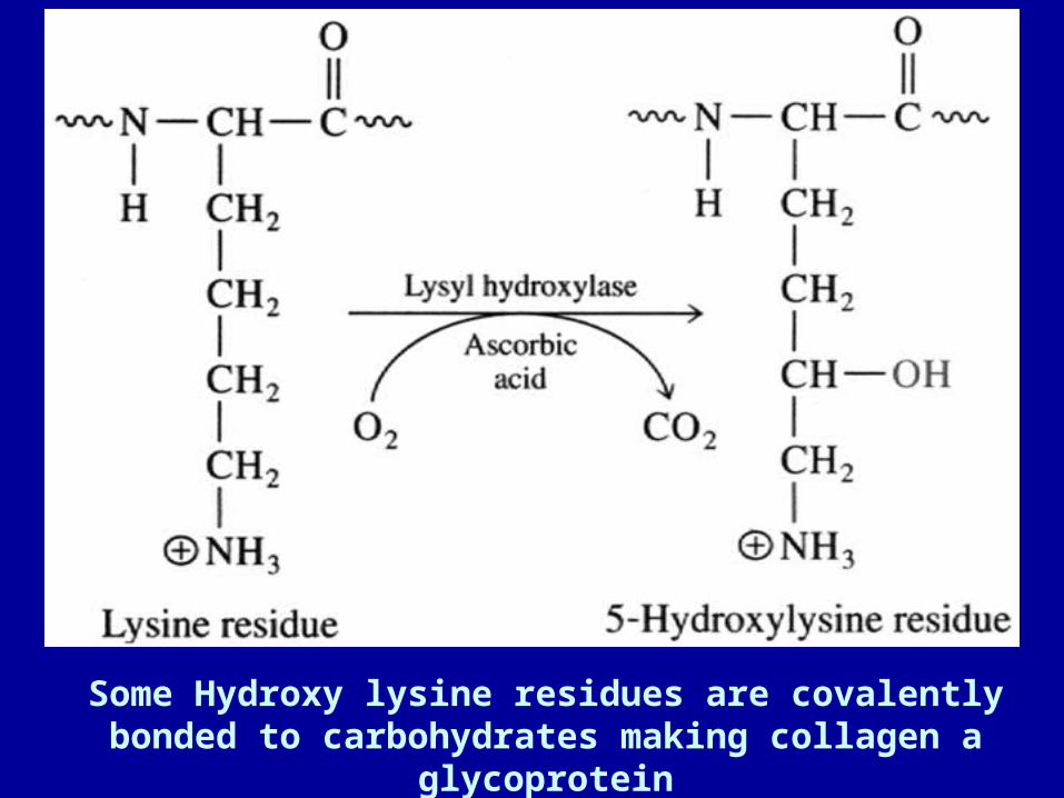
Some Hydroxy lysine residues are covalently bonded to carbohydrates making collagen a glycoprotein

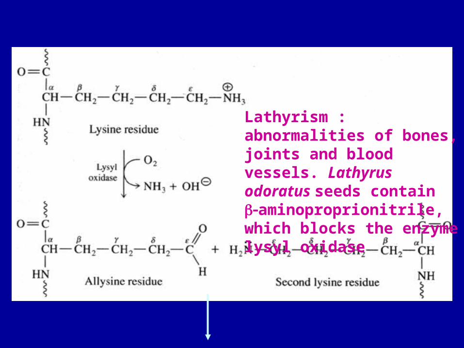
Lathyrism : abnormalities of bones, joints and blood vessels. Lathyrus odoratus seeds contain aminoproprionitrile, which blocks the enzyme lysyl oxidase

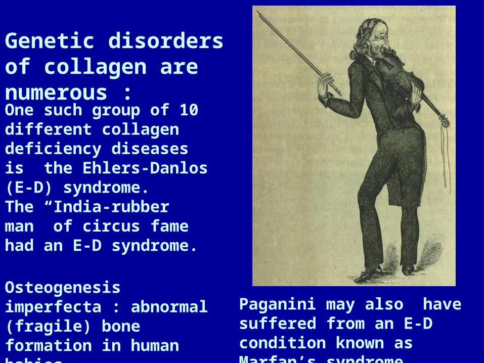
Genetic disorders of collagen are numerous :
One such group of 10 different collagen deficiency diseases is the Ehlers-Danlos (E-D) syndrome.
The “India-rubber man” of circus fame had an E-D syndrome.
Paganini may also have suffered from an E-D condition known as Marfan’s syndrome.
Osteogenesis imperfecta : abnormal (fragile) bone formation in human babies
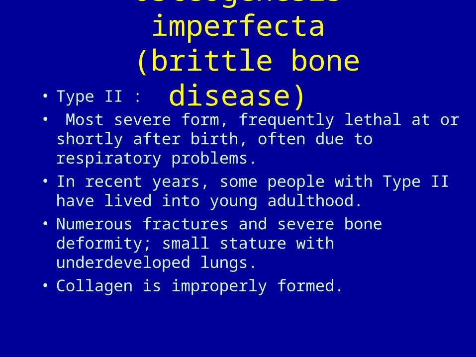
Osteogenesis imperfecta (brittle bone disease)
• Type II :• Most severe form, frequently lethal at or shortly
after birth, often due to respiratory problems. • In recent years, some people with Type II have
lived into young adulthood. • Numerous fractures and severe bone deformity;
small stature with underdeveloped lungs. • Collagen is improperly formed.
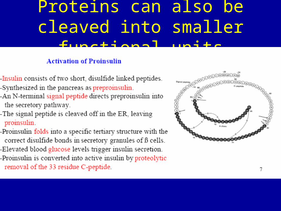
Proteins can also be cleaved into smaller functional units

How do molecules get in and out of the nucleus?
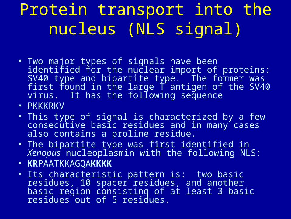
Protein transport into the nucleus (NLS signal)
• Two major types of signals have been identified for the nuclear import of proteins: SV40 type and bipartite type. The former was first found in the large T antigen of the SV40 virus. It has the following sequence
• PKKKRKV• This type of signal is characterized by a few consecutive
basic residues and in many cases also contains a proline residue.
• The bipartite type was first identified in Xenopus nucleoplasmin with the following NLS:
• KRPAATKKAGQAKKKK• Its characteristic pattern is: two basic residues, 10
spacer residues, and another basic region consisting of at least 3 basic residues out of 5 residues.
Importins and exportins• After RNA molecules (mRNA, tRNA and rRNA)
are produced in the nucleus, they must be exported to the cytoplasm for protein synthesis. In addition, proteins operating in the nucleus must be imported from the cytoplasm. The traffic through the nuclear envelope is mediated by a protein family which can be divided into exportins and importins. Binding of a molecule (a "cargo") to exportins facilitates its export to the cytoplasm. Importins facilitate import into the nucleus.
Improtins and exportins are regulated by Ran
• Like other G proteins, Ran can switch between GTP-bound and GDP-bound states. Transition from the GTP-bound to the GDP-bound state is catalyzed by a GTPase-activating protein (GAP) which induces hydrolysis of the bound GTP. The reverse transition is catalyzed by guanine nucleotide exchange factor (GEF) which induces exchange between the bound GDP and the cellular GTP.
How Ran works
• The GEF of Ran (denoted by RanGEF) is located predominantly in the nucleus while RanGAP is located almost exclusively in the cytoplasm. Therefore, in the nucleus Ran will be mainly in the GTP-bound state due to the action of RanGEF while cytoplasmic Ran will be mainly loaded with GDP. This asymmetric distribution has led to the following model for the function of exportins and importins.
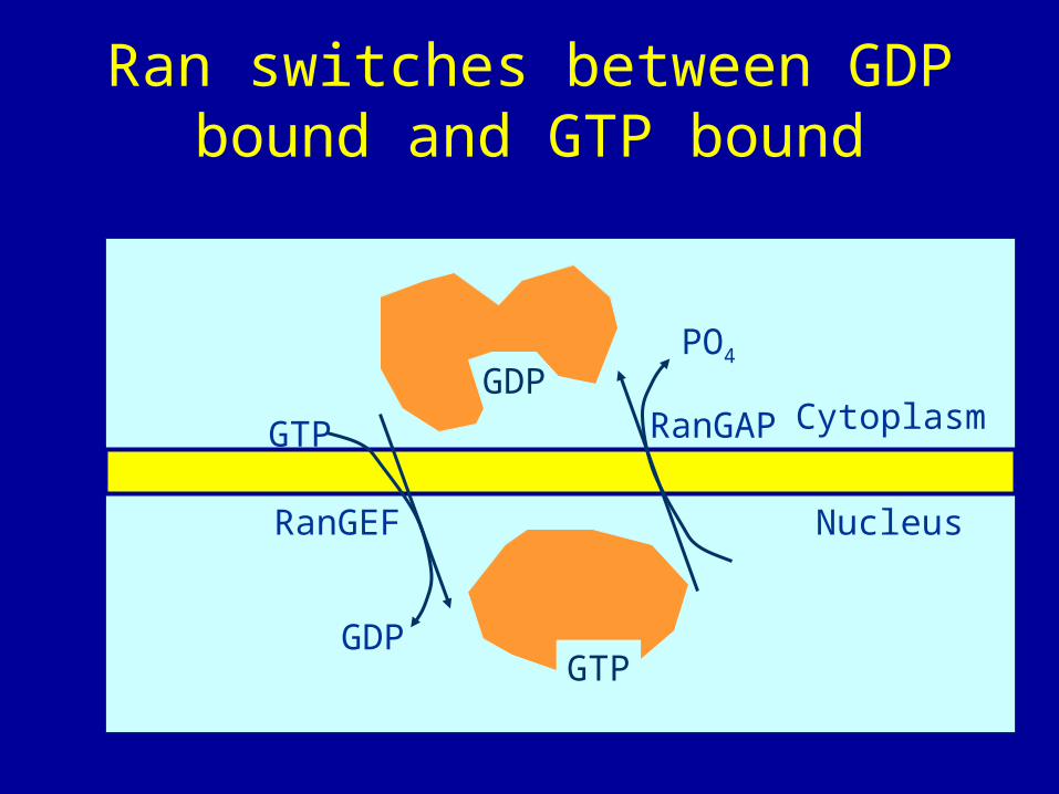
Ran switches between GDP bound and GTP bound
GTP
GDP
Nucleus
CytoplasmRanGAP
PO4
GTP
GDP
RanGEF
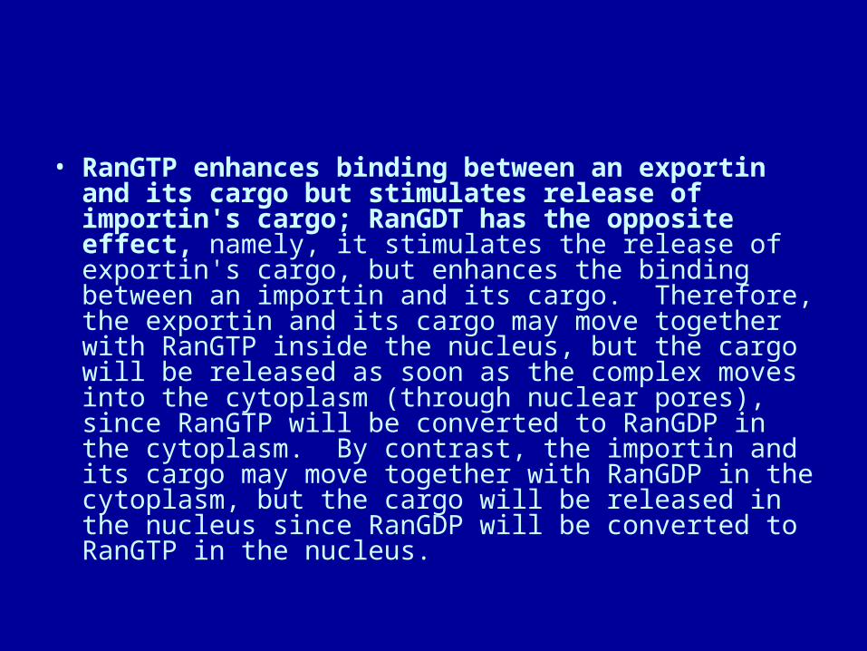
• RanGTP enhances binding between an exportin and its cargo but stimulates release of importin's cargo; RanGDT has the opposite effect, namely, it stimulates the release of exportin's cargo, but enhances the binding between an importin and its cargo. Therefore, the exportin and its cargo may move together with RanGTP inside the nucleus, but the cargo will be released as soon as the complex moves into the cytoplasm (through nuclear pores), since RanGTP will be converted to RanGDP in the cytoplasm. By contrast, the importin and its cargo may move together with RanGDP in the cytoplasm, but the cargo will be released in the nucleus since RanGDP will be converted to RanGTP in the nucleus.

Ran helps move importins and exportins and their cargo in and out of the nucleus
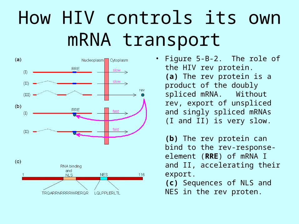
How HIV controls its own mRNA transport
• Figure 5-B-2. The role of the HIV rev protein. (a) The rev protein is a product of the doubly spliced mRNA. Without rev, export of unspliced and singly spliced mRNAs (I and II) is very slow.
(b) The rev protein can bind to the rev-response-element (RRE) of mRNA I and II, accelerating their export. (c) Sequences of NLS and NES in the rev proten.
How do proteins find their way to the endoplasmic reticulum
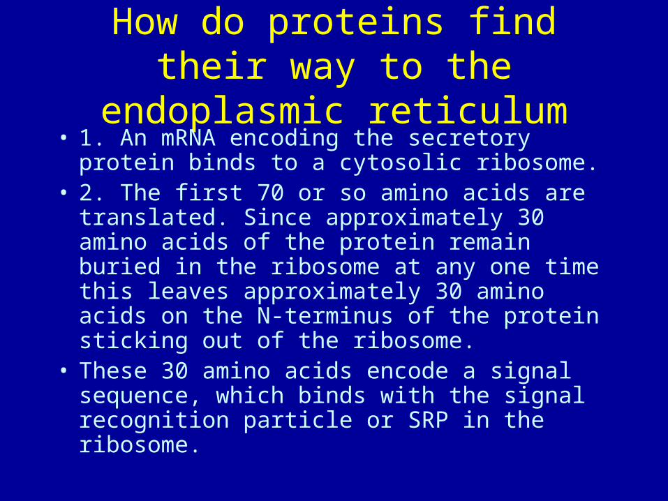
• 1. An mRNA encoding the secretory protein binds to a cytosolic ribosome.
• 2. The first 70 or so amino acids are translated. Since approximately 30 amino acids of the protein remain buried in the ribosome at any one time this leaves approximately 30 amino acids on the N-terminus of the protein sticking out of the ribosome.
• These 30 amino acids encode a signal sequence, which binds with the signal recognition particle or SRP in the ribosome.
Then
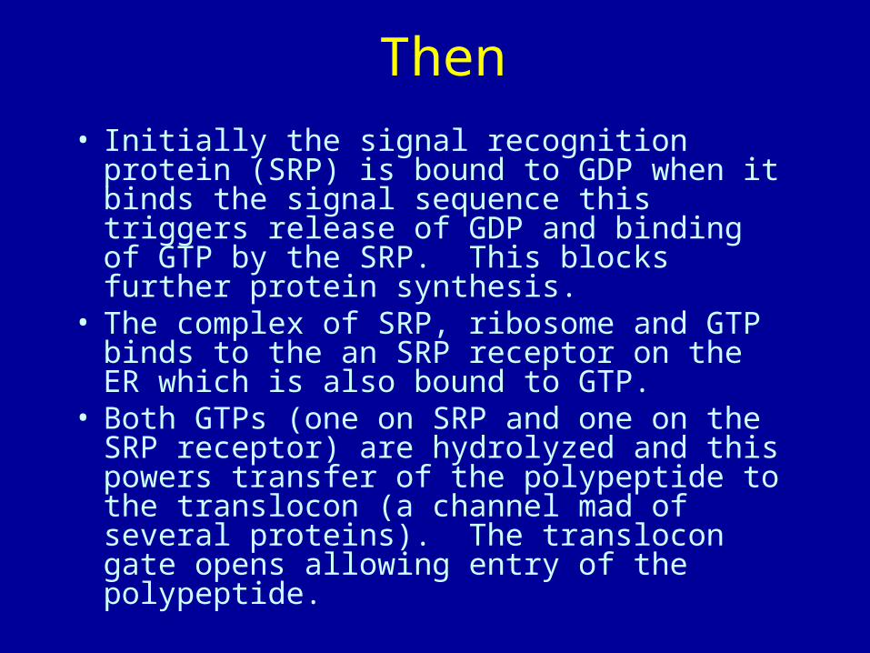
• Initially the signal recognition protein (SRP) is bound to GDP when it binds the signal sequence this triggers release of GDP and binding of GTP by the SRP. This blocks further protein synthesis.
• The complex of SRP, ribosome and GTP binds to the an SRP receptor on the ER which is also bound to GTP.
• Both GTPs (one on SRP and one on the SRP receptor) are hydrolyzed and this powers transfer of the polypeptide to the translocon (a channel mad of several proteins). The translocon gate opens allowing entry of the polypeptide.
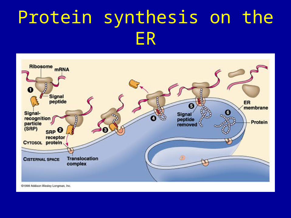
Protein synthesis on the ER
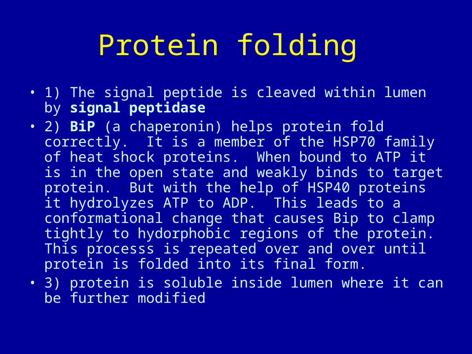
Protein folding
• 1) The signal peptide is cleaved within lumen by signal peptidase
• 2) BiP (a chaperonin) helps protein fold correctly. It is a member of the HSP70 family of heat shock proteins. When bound to ATP it is in the open state and weakly binds to target protein. But with the help of HSP40 proteins it hydrolyzes ATP to ADP. This leads to a conformational change that causes Bip to clamp tightly to hydorphobic regions of the protein. This processs is repeated over and over until protein is folded into its final form.
• 3) protein is soluble inside lumen where it can be further modified

Chaperones• Chaperones are proteins in the cell which function to
help proteins to fold correctly• molecular chaparones guide folding of proteins present in the
cytosol, lumen of RER, mitochondria, etc. • chaparones also promote the assembly of protein complexes from
subunits • chaperones prevent the aggregation of unfolded proteins
– heat shock proteins • a set of proteins induced by a brief exposure of cells to elevated
temperature (42° C) • many of molecular chaperones are heat shock proteins (hsp) • the heat shock causes many proteins to unfold or misfold and the
hsp are induced to help refold these proteins correctly – examples of heat shock proteins:
• hsp 70 - cytosol hsp 60 - cytosol BiP - in lumen of RER (BiP is an abbreviation for
• binding protein) mitochondrial hsp - (mhsp 60 and mhsp 70) – mechanism of action:– bind to exposed hydrophobic regions of proteins to achieve
proper folding ATP requirement for action
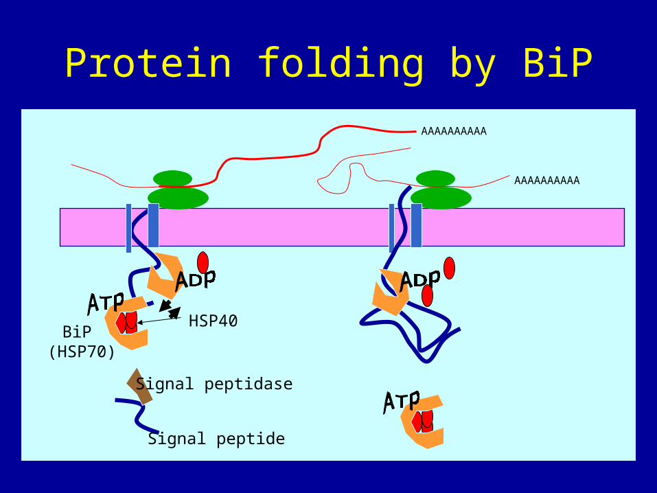
Protein folding by BiP
Signal peptidase
Signal peptide
AAAAAAAAAA
AAAAAAAAAA
HSP40BiP
(HSP70)

Membrane proteins• Complications: proteins embedded in membranes• protein contains a stop-transfer sequence which is too
hydrophobic to emerge into aqueous environment of ER lumen• stop-transfer sequence therefore gets stuck in membrane• ribosome lets go of translocon, finishes job in cytoplasm • translocon dissociates, leaves protein embedded in membrane • example = LDL receptor
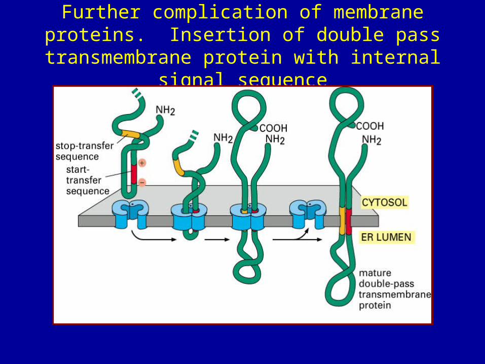
Further complication of membrane proteins. Insertion of double pass transmembrane protein
with internal signal sequence

Multiple transmembrane sequences are common
Disulfide bonds form between cysteines
• PDI protein disulfide isomerase works in the ER. In the cytosol most Cystines are in the reduced state partly because of active oxygen radical scavengers. In the ER PDI works by forming disulfide bonds with the target protein and then transferring that bond to another cystine within the target protein.
Further protein modification
Why glycosylation?– Aids in proper protein folding.
– Provides protection against proteases (e.g. lysosomal membrane proteins)
– Employed for signaling.
• Most soluble and membrane-bound proteins made in the ER are glycoproteins, in contrast to cytsolic proteins.
• Glycoprotein synthesis is a 3-part process:1. Assembly of the precursor oligosaccharide2. En-bloc transfer to the protein3. Modification of the oligosaccharide by removal of sugars
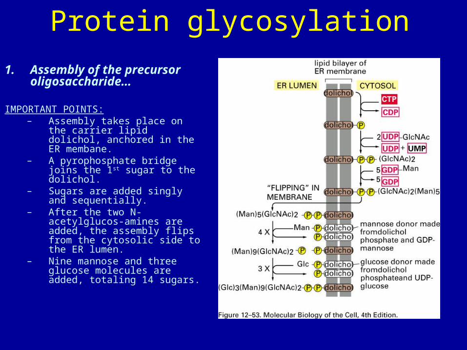
Protein glycosylation
1. Assembly of the precursor oligosaccharide…
IMPORTANT POINTS:– Assembly takes place on the
carrier lipid dolichol, anchored in the ER membane.
– A pyrophosphate bridge joins the 1st sugar to the dolichol.
– Sugars are added singly and sequentially.
– After the two N-acetylglucos-amines are added, the assembly flips from the cytosolic side to the ER lumen.
– Nine mannose and three glucose molecules are added, totaling 14 sugars.

Now in the second step, attachment
2. En-bloc transfer of the oligosaccharide to the protein
• One step transfer, catalyzed by oligosaccharyl transferase, which is bound to the membrane at the translocator.
• Covalently attached to certain asparagines in the polypeptide chain (said to be “N-linked” glycosylation).
• Attaches to NH2 side chain of Asn but only in the context:
Asn-x-Ser or Asn-x-Thr


