j ourna l homepage: www.e lsev ie r .com/ locate /ymvre
Protein phosphatase 2A activity is required for functional adherentjunctions in endothelial cells☆
Anita Kása a, István Czikora a,1, Alexander D. Verin b, Pál Gergely a, Csilla Csortos a,⁎a Department of Medical Chemistry, University of Debrecen Medical and Health Science Center, Debrecen H-4032, Egyetem tér 1., Hungaryb Vascular Biology Center, Georgia Health Science University, 1459 Laney Walker Blvd, CB-3701, Augusta, GA 30912, USA
☆ This work was supported by the Hungarian Science RFaculty of Medicine Research Fund (Bridging Fund 20KONV-2012-0025 project (co-financed by the EuropeanFund), and NIH (PO1-HL101902 and R01-HL67307).⁎ Corresponding author. Fax: +36 52412566.
E-mail address: [email protected] (C. Csortos)1 Present address: Vascular Biology Center, Georgia H
Laney Walker Blvd, CB-3714, Augusta, GA 30912, USA.
Article history:Accepted 18 May 2013Available online 28 May 2013
Reversible Ser/Thr phosphorylation of cytoskeletal and adherent junction (AJ) proteins has a critical role in theregulation of endothelial cell (EC) barrier function. We have demonstrated earlier that protein phosphatase 2A(PP2A) activity is important in EC barrier integrity. In the present work, macro- and microvascular EC were ex-amined andwe provided further evidence on the significance of PP2A in themaintenance of EC cytoskeleton andbarrier functionwith special focus on the Bα (regulatory) subunit of PP2A. Immunofluorescent staining revealedthat the inhibition of PP2A results in changes in the organization of EC cytoskeleton as microtubule dissolutionand actin re-arrangement were detected. Depletion of Bα regulatory subunit of PP2A had similar effect on thecytoskeleton structure of the cells. Furthermore, transendothelial electric resistancemeasurements demonstrat-ed significantly slower barrier recovery of Bαdepleted EC after thrombin treatment. AJ proteins, VE-cadherin andβ-catenin, were detected along with Bα in pull-down assay. Also, the inhibition of PP2A (by okadaic acid orfostriecin) or depletion of Bα caused β-catenin translocation from the membrane to the cytoplasm in parallelwith its phosphorylation on Ser552. In conclusion, our data suggest that the A/Bα/C holoenzyme form of PP2Ais essential in EC barrier integrity both in micro- and macrovascular EC.
PP2A is a ubiquitously expressed Ser/Thr protein phosphatase re-sponsible for dephosphorylation and regulation of many moleculartargets involved in the regulation of numerous cellular processes(Janssens and Goris, 2001; Zolnierowicz, 2000). PP2A holoenzymes areheterotrimers, consisting of a core dimer (PP2AD) which contains a65 kDa structural subunit (PP2A A) and a 36 kDa catalytic subunit(PP2A C). The dimer is associated with a third variable regulatory Bsubunit. Three unrelated families of B subunits, B, B′ and B″, have beenidentified, all encoded by multiple genes and with multiple splice vari-ants creating a huge diversity of these regulatory subunits and theirisoforms (Csortos et al., 1996; Hendrix et al., 1993; McCright andVirshup, 1995; Strack et al., 1999). They have a crucial role in the deter-mination of substrate specificity and in the targeting of PP2A to differentsubcellular localizations (Ferrigno et al., 1993; McCright and Virshup,1995). The methylation and phosphorylation on Tyr and Thr residuesin the C terminal region of the catalytic subunit selectively affect the
esearch Fund (CNK80709), UD12) and TÁMOP-4.2.2.A-11/1/Union and the European Social
.ealth Science University, 1459
rights reserved.
binding of the B subunit to PP2AD (Janssens et al., 2008). Distinct B fam-ilies have two separate A subunit binding domains which are conserved(Li and Virshup, 2002). These facts are in agreementwith the large num-ber of cellular functions recognized so far to be regulated by PP2A andsupport that the different subunit compositions define specific functionsfor distinct PP2A heterotrimer complexes (Silverstein et al., 2002).
ABαC is the most abundant and ubiquitous PP2A holoenzyme, andBα is the most widespread regulatory subunit of PP2A (Hendrix et al.,1993; Mayer et al., 1991), while the localization of Bβ and Bγ is limitedto neuronal tissues. In contrast to Bα, which is expressed at a constantlevel, the expression of Bβ and Bγ is developmentally controlled(Strack et al., 1998). Bα was demonstrated to have functional role inthe cytoskeleton, cytoplasm, nucleus, plasma membrane, Golgi, and inthe endoplasmic reticulum (Sontag, 2001). The ABαC trimer has severalspecific functions in the cytoskeleton as it has been shown to associatewithmicrotubules in epithelial cells, fibroblasts, and neurons (Sontag etal., 1999).
The vascular endothelium dynamically regulates the liquid andmacromolecule transport between the blood and the interstitialspace. Protein phosphorylation and dephosphorylation are principalregulating mechanisms in the endothelial barrier function becauseSer, Thr, and Tyr residues of many cytoskeletal proteins and cytoskel-eton associated proteins, as well as cell junction proteins are modu-lated by reversible phosphorylation (Csortos et al., 2007). The roleof PP2A in macrovascular endothelial barrier function has been stud-ied previously by our group. It was demonstrated that PP2A and its
87A. Kása et al. / Microvascular Research 89 (2013) 86–94
substrates are implicated in the maintenance of normal barrier func-tion and PP2A protects ECs from thrombin or nocodazole induced gapformation and barrier dysfunction (Tar et al., 2004, 2006).
Endothelial cells communicate through junctional structures such astight junctions (TJs), adherent junctions (AJs), and gap junctions (GJs)(Dejana et al., 1999;Wallez andHuber, 2008) formedby transmembraneproteins, as part of the paracellular pathway (Lum and Malik, 1994). AJs
Fig. 1. PP2A inhibition affects the organization of cytoskeleton structure. HLMVEC (A–F) mono100 nM fostriecin for 1 h (E,F), then the cells were double stained as described in Materphalloidin (B,D,F) to visualize the microtubules and microfilaments, respectively. Pictur200 μm. A and B, C and D, E and F are parallel images. Shown are representative data of th
play a dominant role in EC barrier function, and their regulation dependson the phosphorylation state of the adherent proteins (Huber andWeis,2001). AJs consist of the transmembrane VE-cadherin and its intracellu-lar components β-catenin and plakoglobin which bind α-catenin,supporting the linkage between the AJ complex and the actin cytoskele-ton (Ben-Ze'ev and Geiger, 1998). In addition, β-catenin is a key com-ponent of the Wnt-signaling pathway, it plays an important role in
layers were treated either with 0.1% DMSO (A,B), with 5 nM OA for 90 min (C,D), or withials and methods with anti-β-tubulin primary antibody (A,C,E) and with Texas Red–es were taken with an Olympus Fluoview FV1000 confocal microscope, scale bars:ree independent experiments.
88 A. Kása et al. / Microvascular Research 89 (2013) 86–94
embryonic development and tumorigenesis (Nusslein-Volhard andWieschaus, 1980). Phosphorylation of β-catenin by casein kinase I andGSK3β on Ser45 and subsequently on Ser33/37, Thr41, respectively,leads to its ubiquitination and proteosomal degradation (Aberle et al.,1997; Rubinfeld et al., 1996). AKT and PKA phosphorylate β-catenin onSer552 and Ser675 residues (Fang et al., 2007; Taurin et al., 2008) andthe phosphorylation modulates the transcriptional activity of β-cateninand promotes the proliferation of vascular smooth muscle cells, respec-tively. However, Ser/Thr protein phosphatases involved in the dephos-phorylation of the adherent junction proteins are not characterized inEC. Here we provide evidence that the ABαC holoenzyme form of PP2Ais involved in the regulation of pulmonary EC cytoskeleton organization.Furthermore, we demonstrate that Bα controls the barrier function of ECvia direct or non-direct regulation of the dephosphorylation ofβ-catenin.
Materials and methods
Proteins and reagents
Proteins and reagents were obtained as follows: Protease InhibitorCocktail Set III EMD Biosciences (San Diego, CA); TRIZOL AppliedBiosystems (Foster City, CA); M-MLV reverse transcriptase Promega(Madison,WI); Fostriecin Tocris Bioscience (Bristol, UK) anti-V5 antibodyInvitrogen (Carlsbad, CA); antibodies against PP2A B, rabbit β-catenin,β-catenin (Ser552), VE-cadherin Cell Signaling Technology, Inc. (Beverly,MA); mouse anti-β-catenin and anti-actin antibodies SIGMA (St Louis,MO); and anti-β-tubulin antibody Millipore (Billerica, MA). Alexa 488-,594-conjugated antibodies, Texas Red-phalloidin, ProLong Gold Antifademedium with DAPI Molecular Probes (Eugene, OR). Substances for cellculturing were from Invitrogen. All other chemicals were from Sigma(St Louis, MO).
Cell cultures
Human pulmonary artery endothelial cells (HPAEC) and Humanlung microvascular endothelial cells (HLMVEC) obtained from LonzaGroup Ltd. (Walkersville, MD), were propagated in culture mediumEGM-2-MV (Lonza) supplemented with 5% (v/v) fetal bovine serum(FBS; HyClone,Waltham,MA) and used at passages 3–7. Bovine pulmo-nary artery endothelial cells (BPAEC) (culture line-CCL209) fromAmer-ican Type Tissue Culture Collection (Rockville, MD) were maintained inMEMwith 10% FBS, 1% sodium pyruvate, 1% MEM non-essential aminoacids, 1% antibiotic–antimycotic mixture. Human Embryonic Kidney293T (HEK 293T) cells from European Collection of Cell Cultures(Salisbury, UK) were cultured in DMEM with 2 mM glutamine, 10%FBS and 1% antibiotic-antimycotic mixture. All cells were maintainedat 37 °C in a humidified atmosphere of 5% CO2.
Fig. 2. Subcellular localization of regulatory B subunit. Immunofluorescent staining of BPAECsualize F-actin Texas Red-phalloidin (B) was applied. Pictures were taken with an Olympus Fdouble-stained cells, panel C is the merged image of A and B. Shown are representative da
Immunofluorescent staining
Immunofluorescent stainingwas performed as described in (Csortoset al., 2008). The coverslips were observed with an Olympus FluoviewFV1000 confocal microscope using UPLSAPO 60 × 1.35 NA oil immer-sion objective or with Zeiss Axiolab microscope using 63× oil immer-sion objective. Images were processed with FV10-ASW v1.5 software.
Image analysis of stress fiber formation
Texas Red-stained EC monolayers treated with either thrombin orPP2A Bα siRNA were observed under Zeiss Axiolab microscope using63× oil immersion objective. 8 bit images were analyzed using ImageJ 1.46R as described previously (Birukova et al., 2004). Briefly, theratio to the cell area covered by stress fibers to the whole cell areawas determined. Statistical analysis of the data was done by GraphPadPrism 5.
Pull-down assay
HEK cells transfected with PP2A Bα/pcDNA3.1/V5-His constructusing 10 μg DNA: 20 μl PEI ratio were lysed with lysis buffer (10 mMTris–HCl pH 7.5; 140 mM NaCl; 1% Triton-X-100; protease inhibitorcocktail (1:200); 10 mM EDTA; 0.1% SDS), scraped and centrifuged for10 min at 8200 g. The supernatant was applied onto anti-V5-affinitygel and incubated for 4 h at 4 °C to bind recombinant Bα. Next theresin was centrifuged for 30 s at 8200 g. BPAEC were washed withPBS and scraped in lysis buffer followed by sonication and centrifuga-tion. These extracts were added to the resin to which the recombinantBα was bonded in advance, and incubated for 4 h at 4 °C, centrifugedfor 30 min at 8200 g, washed with PBS, then boiled with 2× samplebuffer.
Depletion of endogenous PP2A Bα
Cells (HLMVEC, HPAEC and BPAEC) were treated with SMARTselection-designed PP2A Bα-specific and nontargeting (#1) siRNA du-plexes (50 nM) (Thermo Fisher Scientific, Lafayette, CO). Cells weretransfected at 70% confluence using DharmaFECT1 (Thermo FisherScientific) and were utilized after 48–72 h.
RNA isolation, RT-PCR, and PP2A Bα expression construct preparation
RNA isolated from HPAEC and HLMVEC with TRIZOL was tran-scribed using M-MLV reverse transcriptase and oligo dT. Primers forPCR: PP2A Bα (partial coding sequence to check silencing) 5′CAGCACCTTCCAGAGCCA3′; 5′GGCAGATGCCCTCATGTC3′; full length PP2A Bα 5′ATGGCAGGAGCTGGAG3′; 5′ATTCACTTTGTCTTGAAATATATAC
monolayer was performed using rabbit polyclonal antibody against PP2A B (A). To vi-luoview FV1000 confocal microscope, scale bars: 200 μm. A and B are parallel images ofta of three independent experiments.
89A. Kása et al. / Microvascular Research 89 (2013) 86–94
AG3′. EcoRI was used for subcloning Bα into pcDNA3.1/V5-His fromthe initial Bα/pCR2.1-TOPO construct.
Western blot
Cells were lysed in 2× sample buffer. Proteins separated by SDS-PAGE were transferred to nitrocellulose membrane, and probed withspecific antibodies.
Fig. 3. PP2A Bα depletion affects endothelial cytoskeleton organization and barrier function.non-silencing (non-si) RNA. Panel A: Immunofluorescent staining of BPAEC transfected withwithout any further treatment were double-stained with PP2A B specific antibody (a,b) anthrombin (50 nM, 30 min) (e,f) is also shown. Stress fiber formation induced by thrombin wand methods (g). The results are presented as means n = 14, ±SEM. Significant changes aretaken with a Zeiss Axiolab microscope, scale bars: 200 μm. Panel B: HPAEC (a) and HLMVEC (Bα siRNA. 72 h later TER was measured. Arrow indicates the time point when thrombin (a:RT-PCR (Bc) using specific primer pairs; and by Western blot (Bd) of the lysate of control cespecific antibody. GAPDH and actin signals were used as inner and loading controls, respectefficiency of depletion was about the same, results shown (Bc–d) were acquired on HLMVE
Measurement of transendothelial electrical resistance
Transendothelial electrical resistance (TER) was measured in re-sponse to EC barrier disruptive agent (thrombin) using an electricalcell substrate impedance sensing system (ECIS; Applied Biophysics,Troy, NY) as previously described (Bogatcheva et al., 2007; Kolosovaet al., 2005). HPAEC and HLMVEC transfected with small interferingRNA (siRNA) specific to the regulatory subunit of PP2A were platedon gold microelectrodes. TER was measured 72 h later.
Cells were transfected with small interfering RNA (siRNA) specific for PP2A Bα or withPP2A Bα specific siRNA (a,c,e) and non-silencing siRNA (b,d,f). Transfected monolayersd with Texas Red-phalloidin (c,d). Actin staining of transfected cells challenged withas evaluated by morphometric analysis using Image J program as described in Materialsindicated by * (P b 0.05 vs control), and # (P b 0.05 vs non-si/thrombin). Pictures wereb) plated on gold microelectrodes were transfected with non-targeting or specific PP2A50 nM or b: 0.5 M) or vehicle was added to the medium. The depletion was verified bylls and lysates of cells transfected with non-si or PP2A B specific siRNA using B subunitively. RT-PCR and Western blot verification was done for all cell types investigated. TheC. Shown are representative data of three independent experiments.
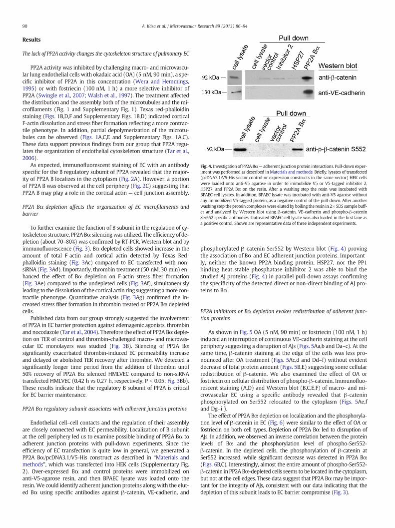
Fig. 4. Investigation of PP2A Bα— adherent junction protein interactions. Pull-down exper-iment was performed as described inMaterials andmethods. Briefly, lysates of transfected(pcDNA3.1/V5-His vector control or expression constructs in the same vector) HEK cellswere loaded onto anti-V5 agarose in order to immobilize V5 or V5-tagged inhibitor 2,HSP27, and PP2A Bα on the resin. After a washing step the resin was incubated withBPAEC cell lysates. In addition, BPAEC lysate was incubated with anti-V5 agarose withoutany immobilized V5-tagged protein, as a negative control of the pull-down. After anotherwashing step theprotein complexeswere eluted by boiling the resin in 2× SDS sample buff-er and analyzed by Western blot using β-catenin, VE-cadherin and phospho-β-cateninSer552 specific antibodies. Untreated BPAEC cell lysate was also loaded in the first lane asa positive control. Shown are representative data of three independent experiments.
90 A. Kása et al. / Microvascular Research 89 (2013) 86–94
Results
The lack of PP2A activity changes the cytoskeleton structure of pulmonary EC
PP2A activity was inhibited by challenging macro- and microvascu-lar lung endothelial cells with okadaic acid (OA) (5 nM, 90 min), a spe-cific inhibitor of PP2A in this concentration (Wera and Hemmings,1995) or with fostriecin (100 nM, 1 h) a more selective inhibitor ofPP2A (Swingle et al., 2007; Walsh et al., 1997). The treatment affectedthe distribution and the assembly both of themicrotubules and the mi-crofilaments (Fig. 1 and Supplementary Fig. 1). Texas red-phalloidinstaining (Figs. 1B,D,F and Supplementary Figs. 1B,D) indicated corticalF-actin dissolution and stress fiber formation reflecting a more contrac-tile phenotype. In addition, partial depolymerization of the microtu-bules can be observed (Figs. 1A,C,E and Supplementary Figs. 1A,C).These data support previous findings from our group that PP2A regu-lates the organization of endothelial cytoskeleton structure (Tar et al.,2006).
As expected, immunofluorescent staining of EC with an antibodyspecific for the B regulatory subunit of PP2A revealed that the major-ity of PP2A B localizes in the cytoplasm (Fig. 2A). However, a portionof PP2A B was observed at the cell periphery (Fig. 2C) suggesting thatPP2A B may play a role in the cortical actin — cell junction assembly.
PP2A Bα depletion affects the organization of EC microfilaments andbarrier
To further examine the function of B subunit in the regulation of cy-toskeleton structure, PP2ABα silencingwas utilized. The efficiency of de-pletion (about 70–80%) was confirmed by RT-PCR, Western blot and byimmunofluorescence (Fig. 3). Bα depleted cells showed increase in theamount of total F-actin and cortical actin detected by Texas Red-phalloidin staining (Fig. 3Ac) compared to EC transfected with non-siRNA (Fig. 3Ad). Importantly, thrombin treatment (50 nM, 30 min) en-hanced the effect of Bα depletion on F-actin stress fiber formation(Fig. 3Ae) compared to the undepleted cells (Fig. 3Af), simultaneouslyleading to the dissolution of the cortical actin ring suggesting amore con-tractile phenotype. Quantitative analysis (Fig. 3Ag) confirmed the in-creased stress fiber formation in thrombin treated or PP2A Bα depletedcells.
Published data from our group strongly suggested the involvementof PP2A in EC barrier protection against edemagenic agonists, thrombinand nocodazole (Tar et al., 2004). Therefore the effect of PP2A Bα deple-tion on TER of control and thrombin-challenged macro- and microvas-cular EC monolayers was studied (Fig. 3B). Silencing of PP2A Bαsignificantly exacerbated thrombin-induced EC permeability increaseand delayed or abolished TER recovery after thrombin. We detected asignificantly longer time period from the addition of thrombin until50% recovery of PP2A Bα silenced HMLVEC compared to non-siRNAtransfected HMLVEC (0.42 h vs 0.27 h, respectively, P b 0.05; Fig. 3Bb).These results indicate that the regulatory B subunit of PP2A is criticalfor EC barrier maintenance.
PP2A Bα regulatory subunit associates with adherent junction proteins
Endothelial cell–cell contacts and the regulation of their assemblyare closely connected with EC permeability. Localization of B subunitat the cell periphery led us to examine possible binding of PP2A Bα toadherent junction proteins with pull-down experiments. Since theefficiency of EC transfection is quite low in general, we generated aPP2A Bα/pcDNA3.1/V5-His construct as described in “Materials andmethods”, which was transfected into HEK cells (Supplementary Fig.2). Over-expressed Bα and control proteins were immobilized onanti-V5-agarose resin, and then BPAEC lysate was loaded onto theresin.We could identify adherent junction proteins alongwith the elut-ed Bα using specific antibodies against β-catenin, VE-cadherin, and
phosphorylated β-catenin Ser552 by Western blot (Fig. 4) provingthe association of Bα and EC adherent junction proteins. Important-ly, neither the known PP2A binding protein, HSP27, nor the PP1binding heat-stable phosphatase inhibitor 2 was able to bind thestudied AJ proteins (Fig. 4) in parallel pull-down assays confirmingthe specificity of the detected direct or non-direct binding of AJ pro-teins to Bα.
PP2A inhibitors or Bα depletion evokes redistribution of adherent junc-tion proteins
As shown in Fig. 5 OA (5 nM, 90 min) or fostriecin (100 nM, 1 h)induced an interruption of continuous VE-cadherin staining at the cellperiphery suggesting a disruption of AJs (Figs. 5Aa,b and Da–c). At thesame time, β-catenin staining at the edge of the cells was less pro-nounced after OA treatment (Figs. 5Ac,d and Dd–f) without evidentdecrease of total protein amount (Figs. 5B,E) suggesting some cellularredistribution of β-catenin. We also examined the effect of OA orfostriecin on cellular distribution of phospho-β-catenin. Immunofluo-rescent staining (A,D) and Western blot (B,C,E,F) of macro- and mi-crovascular EC using a specific antibody revealed that β-cateninphosphorylated on Ser552 relocated to the cytoplasm (Figs. 5Ae,fand Dg–i ).
The effect of PP2A Bα depletion on localization and the phosphoryla-tion level of β-catenin in EC (Fig. 6) were similar to the effect of OA orfostriecin on both cell types. Depletion of PP2A Bα led to disruption ofAJs. In addition, we observed an inverse correlation between the proteinlevels of Bα and the phosphorylation level of phospho-Ser552-β-catenin. In the depleted cells, the phosphorylation of β-catenin atSer552 increased, while significant decrease was detected in PP2A Bα(Figs. 6B,C). Interestingly, almost the entire amount of phospho-Ser552-β-catenin in PP2ABα-depleted cells seems to be located in the cytoplasm,but not at the cell edges. These data suggest that PP2A Bαmay be impor-tant for the integrity of AJs, consistent with our data indicating that thedepletion of this subunit leads to EC barrier compromise (Fig. 3).
Fig. 5. Okadaic acid and fostriecin treatment evoke redistribution of adherent junction proteins. BPAEC (A,B) and HLMVEC (D,E) monolayers were treated with vehicle (Panel A: a,c,e; Panel D: a,d,g), 5 nM okadaic acid (OA) for 90 min (Panel A: b,d.f; Panel D:b,e,h), or with 100 nM fostriecin for 1 h (Panel D: c,f,i). Afterwards the cells were fixed for immuno-fluorescent staining (A, D) or lysed for Western blot analysis (B,C) as described in Materials and methods. Antibodies against VE-cadherin, β-catenin and phospho-β-catenin Ser552were used as indicated in the immunofluorescent pictures to detect the subcellular distribution of adherent junction proteins. Pictures were taken with a Zeiss Axiolab microscope,scale bars: 200 μm. (B, E) Phosphorylation level of β-catenin on Ser552 was analyzed by Western blot using specific antibody against phospho-β-catenin Ser552. β-Catenin andactin were also detected as loading controls. (C, F) Phosphorylation level of β-catenin was normalized to total β-catenin and quantified by the densitometry of Western blots(n = 3, p b 0.05 vs control). Shown are representative data of three independent experiments.
91A. Kása et al. / Microvascular Research 89 (2013) 86–94
Also, these data suggest the involvement of PP2A in the regulationof β-catenin phosphorylation and cellular distribution, which affect AJassembly and EC permeability.
Discussion
PP2A, a major Ser/Thr phosphatase, is involved in many physiologi-cal processes in the cell. Our previous results have proved that PP2Aplays a critical role in the endothelial barrier protection and in the ar-rangement of cytoskeletal proteins (Tar et al., 2004, 2006). To better
understand the functional role of PP2A in the regulation of endothelialcytoskeleton organization we employed PP2A inhibitors, okadaic acid(OA), a well-known cell-permeable inhibitor of PP2A (Haystead et al.,1989), and fostriecin, an antitumor antibiotic produced by Streptomycespulveraceus (Walsh et al., 1997). OA and fostriecin inhibit PP2Amore ef-ficiently (Ki = 0.2 nM and 3.2 nM, respectively) compared to PP1(Ki = 2 μM and 131 μM, respectively) (Cohen et al., 1990; Walsh etal., 1997). Several earlier studies demonstrated considerable effect ofokadaic acid on the cytoskeleton of different cell types. It has beenshown that OA causes depolymerization of interphase microtubules
Fig. 6. Depletion of PP2A Bα evokes the redistribution of β-catenin and p-β-catenin coinciding with the disruption of cell–cell contacts. BPAEC were transfected with specific PP2ABα siRNA and with non-silencing RNA. (A) Immunofluorescent staining with anti-β-catenin (a,b) and anti-phospho-β-catenin Ser552 (c,d) antibodies as described in Materials andmethods. Pictures were taken with a Zeiss Axiolab microscope, scale bars: 200 μm. (B) Western blotting of cell lysates using phospho-β-catenin Ser552, PP2A B, and actin specificantibodies. (C) Densitometric quantification of data demonstrated on panel B (n = 3, p b 0.05). Shown are representative data of three independent experiments.
92 A. Kása et al. / Microvascular Research 89 (2013) 86–94
and abnormalities in the mitotic spindle in LLC-PK cells (Vandre andWills, 1992) and also promotes PP2A-mediated microtubule destabili-zation and phosphorylation of PP2A-sensitive microtubule-associatedproteins (Sontag et al., 1996). In our experiments, the inhibition ofPP2A affected the actin and tubulin organization in EC suggesting apivotal role of PP2A in the maintenance of cytoskeleton structures.However, our group previously showed that OA (5 nM) had no signifi-cant effect on BPAEC permeability (Verin et al., 1995). In addition, stain-ing of F-actin showed that OA (5 nM) treatment did not cause anydetectable change of the actin cytoskeleton in HPAEC (Tar et al.,2004). This apparent controversy can be resolved by the differing cul-turing conditions used. In both of the published works the human andbovine ECs were maintained in M199 containing 20% bovine serum,and endothelial cell growth supplement. In the present work we usedMEM, according to the recommendation of ATCC, to maintain BPAECsupplemented with only 10% of bovine serum for better comparabilityto serum starved silencing conditions. This serum concentration wasroutinely used for maintaining the very same cell type (CCL-209) inother laboratories as reported in (Drew et al., 2010; Duthu and Smith,1980; Ludwig et al., 2005; Wu et al., 2010).
PP2A is one of the most abundant phospho-Ser/Thr-specific proteinphosphatases. The large families of PP2A holoenzymes have wide sub-strate specificity; therefore PP2A is involved in many basic processes ofthe cell. Since we have previously shown that the over-expression ofthe catalytic C, and structural A subunits significantly attenuatedthrombin or nocodazole-induced barrier dysfunction and cytoskeletonrearrangement (Tar et al., 2006), to narrow the affected processes, thepresent study was rather focused on the possible regulatory role of theBα subunit of PP2A in endothelial cells. Endogenous localization and dis-tribution of the B subunits have been described in several different tis-sues and cell types, but endothelium (Janssens et al., 2008; Mayer etal., 1991). In agreement with the earlier findings in other cell types, im-munofluorescent staining of the endogenous Bα demonstrated mainlycytoplasmic localization in BPAEC. Nevertheless, B also localizes atthe cell periphery, seemingly with the cortical actin ring. Moreover,
alterationof PP2A activity by reduction (about 80%) of Bαprotein level af-fected the organization of F-actin and cortical actin; bothwere detected inan increased level in Bα depleted cells, and thrombin-induced stress fiberformation becamemore prominent in Bα silenced cells compared to con-trols transfected with non-silencing RNA. Importantly, our TER measure-ments also support that Bα plays a role in the barrier maintainingfunction of PP2A. Based on these findings, we hypothesized that PP2A ac-tivity may be required not only in the regulation of the phosphorylationlevel of cytoskeleton associated proteins, as we suggested earlier, but itmay have an important role in the intercellular junctions as well.
Adherent junctions are themost abundant communicating structuresamong endothelial cells. β-Catenin was originally identified associatingwith the cytoplasmic domains of cadherins and found to have a crucialrole in Ca2+ dependent cell adhesion (Aberle et al., 1996). Besidesbeing a structural component of AJ, β-catenin has several functions,which are regulated via phosphorylation of Ser/Thr and Tyr residues(Valenta et al., 2012). It is well known that β-catenin is a component ofthe Wnt signaling pathway (Kikuchi, 2003). It is interesting to notethat during Wnt-signaling in Drosophila the interaction of PP2A Bαwith β-catenin regulates the phosphorylation state of Ser33/37 andThr41 residues and the degradation of β-catenin (Zhang et al., 2009).The phosphorylation state of the VE-cadherin and β-catenin determinesthe stability of their complex. Phosphorylation of β-catenin at Tyr654and the Tyr860 residue in cadherin prevents their interaction. Phosphor-ylation of Ser residues in cadherin was also shown to inhibit the interac-tion. On the other hand, phosphorylation of specific Ser residues incadherin may enhance the binding of β-catenin (Bazzoni and Dejana,2004; Kemler, 1993; Lilien and Balsamo, 2005).We investigated the pos-sible interaction between the Bα regulatory subunit of PP2A and adher-ent junction proteins in BPAEC to seewhether PP2A activity is significantin the stability of the VE-cadherin–β-catenin complex. Recombinant Bαwas utilized as bait in pull-down experiments and adherent proteins,VE-cadherin and β-catenin, from EC were detected in the bound proteincomplex. To learn whether PP2A activity triggers the dephosphorylationof β-catenin and/or VE-cadherin or dephosphorylates them directly, and
93A. Kása et al. / Microvascular Research 89 (2013) 86–94
the identification of the concerned amino acid residues require furtherstudies.
Inhibition of PP2A with OA or fostriecin challenge, or the depletionof Bα resulted in a redistribution of VE-cadherin and β-catenin fromthe cell membranes (to the cytoplasm or nuclei) and the loss ofcell–cell contacts, demonstrating the critical role of PP2A activity inwell-functioning adherent junctions. Consistent with our observa-tions, OA treatment caused re-localization of VE-cadherin from themembrane to the cytoplasm in human keratinocytes (Serres et al.,2000) and thrombin induced dissociation of β-catenin and p120from the cell membrane has been described recently in human umbil-ical vein ECs (Beckers et al., 2008). It was also suggested that the cat-alytic subunit of PP2A is important in the stabilization of E-cadherin/β-catenin complex at the plasma membrane (Gotz et al., 2000). Inter-estingly, PP2A activity was shown to increase paracellular permeabil-ity of epithelial cells by dephosphorylating tight junction proteins(Nunbhakdi-Craig et al., 2002). The differing cell- and junction typesexamined could explain this apparently opposing result. It may alsosuggest that adherent and tight junctions might be differently regu-lated via the phosphorylation state of their components. It shouldalso be noted, that the authors reported no noticeable change in theF-actin structure upon OA treatment of the epithelial cells, whileour results reflected changes in the cytoskeleton arrangement afterthe inhibition of PP2A in EC.
It is known that Ser552 phosphorylation site of β-catenin is not re-lated to the degradation of the protein. Different kinasesmay phosphor-ylate β-catenin at Ser552, and it was reported that it caused thedissociation of β-catenin from cell–cell contacts and an increase in itstranscriptional activity (Fang et al., 2007; Taurin et al., 2008). We ob-served that the phosphorylation level of Ser552 in EC is low, and ap-pears mostly at the cell membrane. After OA or fostriecin treatment,or silencing of Bα the Ser552 residue of β-catenin was phosphorylated,and this phosphorylated form appeared to be present in the cytoplasm.Thesefindings indicate that Bα andmost likely PP2A aswell, have a piv-otal role in regulating the phosphorylation level of the Ser552 side chainof β-catenin. However, the role of this site-specific Ser phosphorylationof β-catenin on EC cytoskeleton/barrier/AJ assembly and the exact roleof PP2A in β-catenin dephosphorylation remains to be determined. Arecent paper published during the preparation of this manuscript alsoclaims that PP2A activity is necessary to maintain brain EC monolayerintegrity (Le Guelte et al., 2012).
Conclusions
Taken together these results further strengthen our previous find-ings that PP2A is a critical component in the maintenance of the en-dothelial cytoskeleton structure, and suggest that Bα is at least oneamong the regulatory B subunits of the PP2A holoenzyme having asignificant role in this aspect. Furthermore, association of Bα and AJproteins was detected suggesting that the activity of the ABαC holo-enzyme form of PP2A might be necessary for functional adherentjunctions in EC. However, our results do not exclude the possibilityof other PP2A holoenzyme forms containing a different B subunitbeing also involved in EC barrier maintenance through the regulationof the phosphorylation state of cytoskeleton associated and cell junc-tion proteins via opposing or parallel effects of the different PP2Aactivities.
Supplementary data to this article can be found online at http://dx.doi.org/10.1016/j.mvr.2013.05.003.
Acknowledgments
The authors are most thankful to Dr. György Vámosi (University ofDebrecen, Department of Biophysics and Cell Biology) for his help inconfocal microscopy.
References
Aberle, H., et al., 1996. Single amino acid substitutions in proteins of the armadillo genefamily abolish their binding to alpha-catenin. J. Biol. Chem. 271, 1520–1526.
Aberle, H., et al., 1997. beta-catenin is a target for the ubiquitin-proteasome pathway.EMBO J. 16, 3797–3804.
Bazzoni, G., Dejana, E., 2004. Endothelial cell-to-cell junctions: molecular organizationand role in vascular homeostasis. Physiol. Rev. 84, 869–901.
Beckers, C.M., et al., 2008. Nuclear targeting of beta-catenin and p120ctn duringthrombin-induced endothelial barrier dysfunction. Cardiovasc. Res. 79, 679–688.
Ben-Ze'ev, A., Geiger, B., 1998. Differential molecular interactions of beta-catenin andplakoglobin in adhesion, signaling and cancer. Curr. Opin. Cell Biol. 10, 629–639.
Birukova, A.A., et al., 2004. Role of Rho GTPases in thrombin-induced lung vascular en-dothelial cells barrier dysfunction. Microvasc. Res. 67, 64–77.
Bogatcheva, N.V., et al., 2007. Involvement of microtubules, p38, and Rho kinasespathway in 2-methoxyestradiol-induced lung vascular barrier dysfunction.Am. J. Physiol. Lung Cell. Mol. Physiol. 292, L487–L499.
Cohen, P., et al., 1990. Okadaic acid: a new probe for the study of cellular regulation.Trends Biochem. Sci. 15, 98–102.
Csortos, C., et al., 1996. High complexity in the expression of the B′ subunit of protein phos-phatase 2A0. Evidence for the existence of at least seven novel isoforms. J. Biol.Chem. 271, 2578–2588.
Csortos, C., et al., 2007. Regulation of vascular endothelial cell barrier function and cy-toskeleton structure by protein phosphatases of the PPP family. Am. J. Physiol. LungCell. Mol. Physiol. 293, L843–L854.
Csortos, C., et al., 2008. TIMAP is a positive regulator of pulmonary endothelial barrierfunction. Am. J. Physiol. Lung Cell. Mol. Physiol. 295, L440–L450.
Dejana, E., et al., 1999. The role of endothelial cell-to-cell junctions in vascular morpho-genesis. Thromb. Haemost. 82, 755–761.
Drew, C.P., et al., 2010. Bluetongue virus infection alters the impedance of monolayersof bovine endothelial cells as a result of cell death. Vet. Immunol. Immunopathol.136, 108–115.
Duthu, G.S., Smith, J.R., 1980. In vitro proliferation and lifespan of bovine aorta endo-thelial cells: effect of culture conditions and fibroblast growth factor. J. Cell. Phys-iol. 103, 385–392.
Fang, D., et al., 2007. Phosphorylation of beta-catenin by AKT promotes beta-catenintranscriptional activity. J. Biol. Chem. 282, 11221–11229.
Ferrigno, P., et al., 1993. Protein phosphatase 2A1 is the major enzyme in vertebratecell extracts that dephosphorylates several physiological substrates for cyclin-dependent protein kinases. Mol. Biol. Cell 4, 669–677.
Gotz, J., et al., 2000. Distinct role of protein phosphatase 2A subunit Calpha in the reg-ulation of E-cadherin and beta-catenin during development. Mech. Dev. 93, 83–93.
Haystead, T.A., et al., 1989. Effects of the tumour promoter okadaic acid on intracellularprotein phosphorylation and metabolism. Nature 337, 78–81.
Hendrix, P., et al., 1993. Analysis of subunit isoforms in protein phosphatase 2A holoen-zymes from rabbit and Xenopus. J. Biol. Chem. 268, 7330–7337.
Huber, A.H., Weis, W.I., 2001. The structure of the beta-catenin/E-cadherin complex andthe molecular basis of diverse ligand recognition by beta-catenin. Cell 105, 391–402.
Janssens, V., Goris, J., 2001. Protein phosphatase 2A: a highly regulated family of serine/threonine phosphatases implicated in cell growth and signalling. Biochem. J. 353,417–439.
Janssens, V., et al., 2008. PP2A holoenzyme assembly: in cauda venenum (the sting is inthe tail). Trends Biochem. Sci. 33, 113–121.
Kemler, R., 1993. From cadherins to catenins: cytoplasmic protein interactions and reg-ulation of cell adhesion. Trends Genet. 9, 317–321.
Kikuchi, A., 2003. Tumor formation by genetic mutations in the components of theWntsignaling pathway. Cancer Sci. 94, 225–229.
Kolosova, I.A., et al., 2005. Signaling pathways involved in adenosine triphosphate-induced endothelial cell barrier enhancement. Circ. Res. 97, 115–124.
Le Guelte, A., et al., 2012. Semaphorin 3A elevates endothelial cell permeability throughPP2A inactivation. J. Cell Sci. 125, 4137–4146.
Li, X., Virshup, D.M., 2002. Two conserved domains in regulatory B subunits mediatebinding to the A subunit of protein phosphatase 2A. Eur. J. Biochem. 269, 546–552.
Lilien, J., Balsamo, J., 2005. The regulation of cadherin-mediated adhesion by tyrosine phos-phorylation/dephosphorylation of beta-catenin. Curr. Opin. Cell Biol. 17, 459–465.
Ludwig, A., et al., 2005. Effect of statins on the proteasomal activity in mammalian en-dothelial and vascular smooth muscle cells. Biochem. Pharmacol. 70, 520–526.
Lum, H., Malik, A.B., 1994. Regulation of vascular endothelial barrier function.Am. J. Physiol. 267, L223–L241.
Mayer, R.E., et al., 1991. Structure of the 55-kDa regulatory subunit of protein phospha-tase 2A: evidence for a neuronal-specific isoform. Biochemistry 30, 3589–3597.
McCright, B., Virshup, D.M., 1995. Identification of a new family of protein phosphatase2A regulatory subunits. J. Biol. Chem. 270, 26123–26128.
Nunbhakdi-Craig, V., et al., 2002. Protein phosphatase 2A associates with and regulatesatypical PKC and the epithelial tight junction complex. J. Cell Biol. 158, 967–978.
Nusslein-Volhard, C., Wieschaus, E., 1980. Mutations affecting segment number andpolarity in Drosophila. Nature 287, 795–801.
Rubinfeld, B., et al., 1996. Binding of GSK3beta to the APC-beta–catenin complex andregulation of complex assembly. Science 272, 1023–1026.
Serres, M., et al., 2000. The disruption of adherens junctions is associated with a decreaseof E-cadherin phosphorylation by protein kinase CK2. Exp. Cell Res. 257, 255–264.
Silverstein, A.M., et al., 2002. Actions of PP2A on the MAP kinase pathway and apopto-sis are mediated by distinct regulatory subunits. Proc. Natl. Acad. Sci. U. S. A. 99,4221–4226.
Sontag, E., 2001. Protein phosphatase 2A: the Trojan Horse of cellular signaling. Cell.Signal. 13, 7–16.
94 A. Kása et al. / Microvascular Research 89 (2013) 86–94
Sontag, E., et al., 1996. Regulation of the phosphorylation state and microtubule-binding activity of Tau by protein phosphatase 2A. Neuron 17, 1201–1207.
Sontag, E., et al., 1999.Molecular interactions among protein phosphatase 2A, tau, and mi-crotubules. Implications for the regulation of tau phosphorylation and the develop-ment of tauopathies. J. Biol. Chem. 274, 25490–25498.
Strack, S., et al., 1998. Brain protein phosphatase 2A: developmental regulation and dis-tinct cellular and subcellular localization byB subunits. J. Comp.Neurol. 392, 515–527.
Strack, S., et al., 1999. Cloning and characterization of B delta, a novel regulatorysubunit of protein phosphatase 2A. FEBS Lett. 460, 462–466.
Swingle, M., et al., 2007. Small-molecule inhibitors of ser/thr protein phosphatases:specificity, use and common forms of abuse. Methods Mol. Biol. 365, 23–38.
Tar, K., et al., 2004. Phosphatase 2A is involved in endothelial cell microtubule remod-eling and barrier regulation. J. Cell. Biochem. 92, 534–546.
Tar, K., et al., 2006. Role of protein phosphatase 2A in the regulation of endothelial cellcytoskeleton structure. J. Cell. Biochem. 98, 931–953.
Taurin, S., et al., 2008. Phosphorylation of beta-catenin by PKA promotes ATP-induced prolif-eration of vascular smooth muscle cells. Am. J. Physiol. Cell Physiol. 294, C1169–C1174.
Valenta, T., et al., 2012. The many faces and functions of beta-catenin. EMBO J. 31,2714–2736.
Vandre, D.D., Wills, V.L., 1992. Inhibition of mitosis by okadaic acid: possible involve-ment of a protein phosphatase 2A in the transition from metaphase to anaphase.J. Cell Sci. 101 (Pt. 1), 79–91.
Verin, A.D., et al., 1995. Regulation of endothelial cell gap formation and barrier func-tion by myosin-associated phosphatase activities. Am. J. Physiol. 269, L99–L108.
Wallez, Y., Huber, P., 2008. Endothelial adherens and tight junctions in vascular homeosta-sis, inflammation and angiogenesis. Biochim. Biophys. Acta 1778, 794–809.
Walsh, A.H., et al., 1997. Fostriecin, an antitumor antibiotic with inhibitory activityagainst serine/threonine protein phosphatases types 1 (PP1) and 2A (PP2A), is highlyselective for PP2A. FEBS Lett. 416, 230–234.
Wera, S., Hemmings, B.A., 1995. Serine/threonine protein phosphatases. Biochem. J. 311(Pt. 1), 17–29.
Wu, X., et al., 2010. Statin post-treatment provides protection against simulated ische-mia in bovine pulmonary arterial endothelial cells. Eur. J. Pharmacol. 636, 114–120.
Zhang, W., et al., 2009. PR55 alpha, a regulatory subunit of PP2A, specificallyregulates PP2A-mediated beta-catenin dephosphorylation. J. Biol. Chem. 284,22649–22656.
Zolnierowicz, S., 2000. Type 2A protein phosphatase, the complex regulator of numer-ous signaling pathways. Biochem. Pharmacol. 60, 1225–1235.