33
Protein Structure Stryer Short Course Chapter 4
| Date post: | 31-Dec-2015 |
| Category: |
Documents |
| Upload: | catherine-young |
| View: | 249 times |
| Download: | 7 times |
Protein Structure
Stryer Short CourseChapter 4

Peptide bonds
• Amide bond• Primary structure• N- and C-terminus• Condensation and hydrolysis

Polypeptides

Drawing Peptides
• Sidechains• Stereochemistry• Ionization states
• Example: Draw the peptide AHSCVE at pH 8.
• Steps– Backbone– Stereochemistry– Sidechains– Check ionization
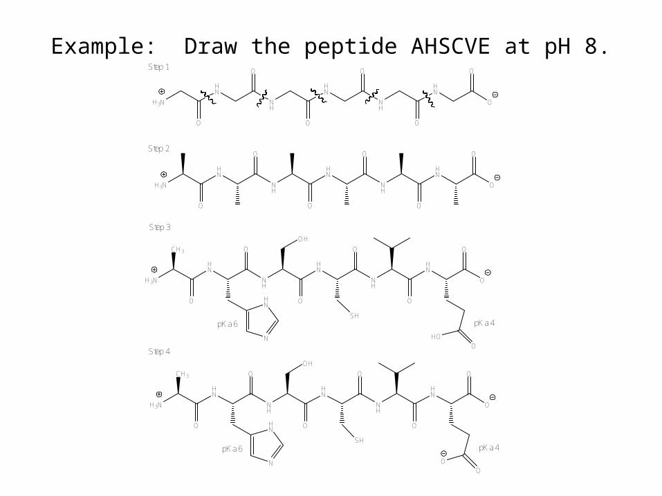
Example: Draw the peptide AHSCVE at pH 8.
H3N
HN
NH
HN
NH
HN
O
O
O
O
O
O
O
H3N
HN
NH
HN
NH
HN
O
O
O
O
O
O
O
H3N
HN
NH
HN
NH
HN
O
O
O
O
O
O
OCH3
N
HN
OH
SH
OHO
pKa 4pKa 6
Step 1
Step 2
Step 3
H3N
HN
NH
HN
NH
HN
O
O
O
O
O
O
OCH3
N
HN
OH
SH
OO
pKa 4pKa 6
Step 4
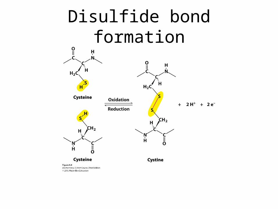
Disulfide bond formation

Primary Structure
• Protein defined by unique primary sequence• Structure defined by primary sequence• Function dictated by structure• Basis of understanding mutation


Basis of Secondary Structure
• Polarity• Rigidity• Planarity

Cis and Trans Peptide Bonds
• Double bond character• Slowly interchangeable• Trans heavily favored—steric interactions

Conformational Constraint
NOT cis/trans

Ramachandran Plots

Alpha Helix
• Right handed• Polarity• n and n + 4• Gly and Pro
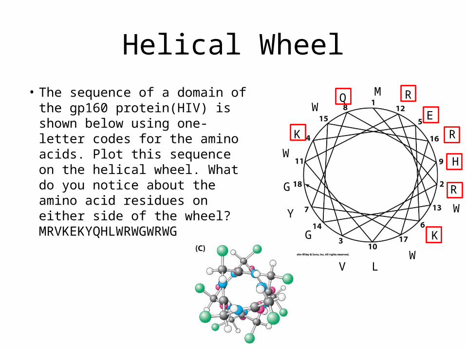
Helical Wheel• The sequence of a domain of the
gp160 protein(HIV) is shown below using one-letter codes for the amino acids. Plot this sequence on the helical wheel. What do you notice about the amino acid residues on either side of the wheel? MRVKEKYQHLWRWGWRWG
M
R
V
K
E
K
Y
Q
H
L
W
R
W
G
W
R
W
G

Beta Sheets
• Parallel• Antiparallel• mixed


Amphipathic Sheets
• Alternating sidechains can lead to amphipathic sheets

Irregular Secondary Structure
• Nonrepeating loops and turns
• Change of direction
• Turns have about 4 residues
• Internal H-bonds• Gly, Pro
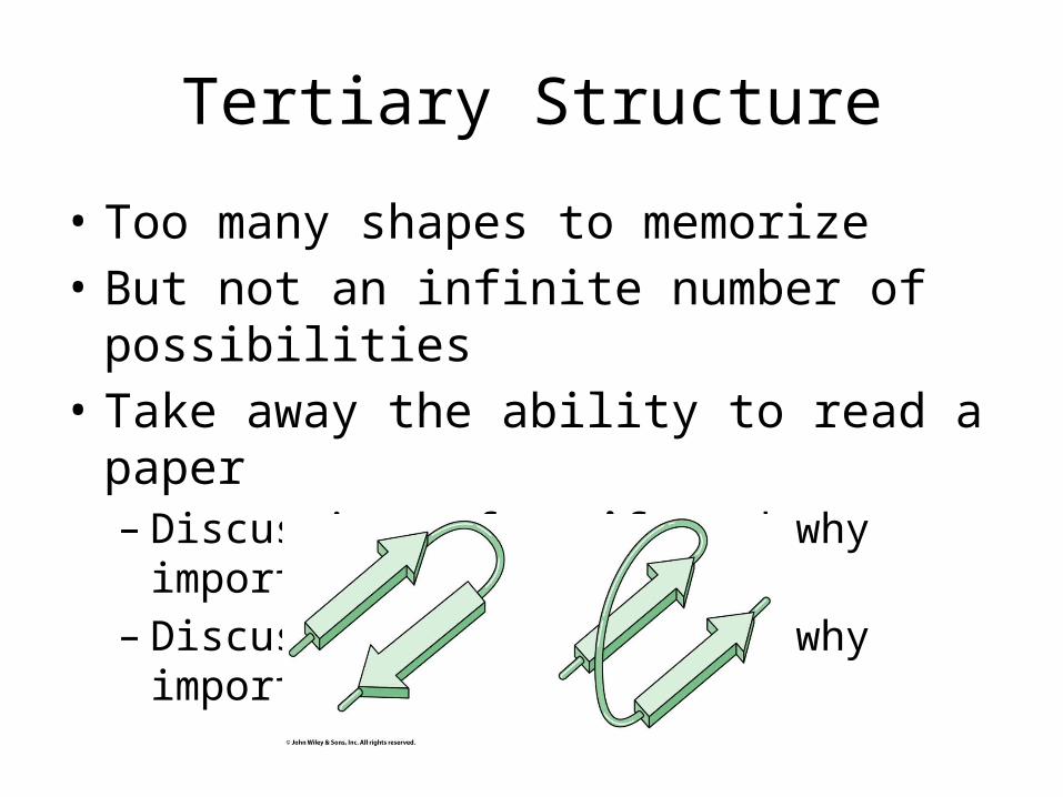
Tertiary Structure
• Too many shapes to memorize• But not an infinite number of possibilities• Take away the ability to read a paper– Discussions of motifs and why important– Discussion of domains and why important
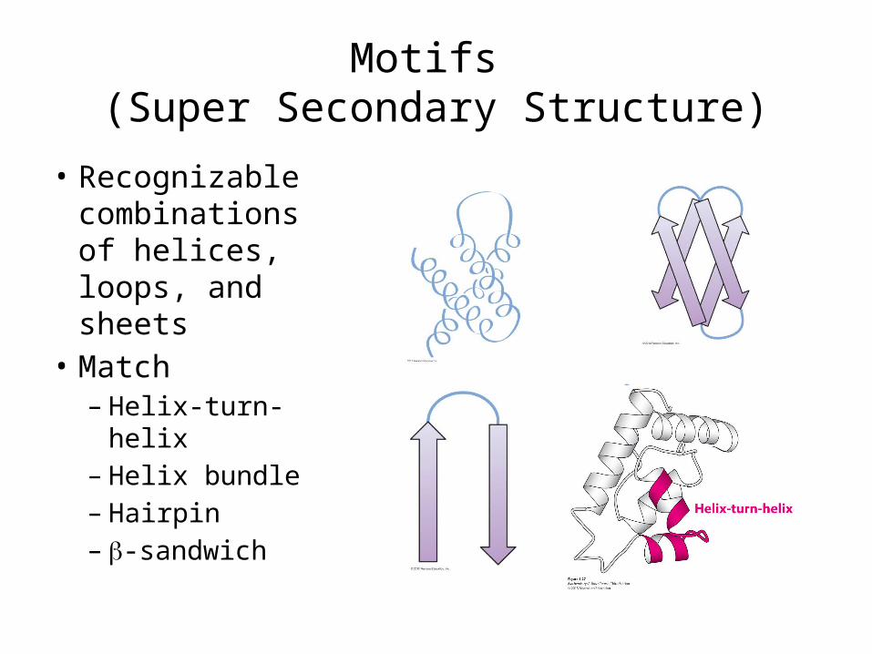
Motifs (Super Secondary Structure)
• Recognizable combinations of helices, loops, and sheets
• Match– Helix-turn-helix– Helix bundle– Hairpin– b-sandwich

Studying Motifs
• Some Motifs are highly studied
• Know the lingo– Leucine zipper– Zinc finger
• Often have recurring applications

Structural and Functional Domains• Discrete, independently
folded unit (may maintain shape when cleaved on loop)
• May have separate activities: “ATP binding domain” or “catalytic domain”
• Similar activity = similar structure across many proteins
• Binding pockets at interfaces
How many domains?

Common Domains

Quaternary Structure
• Multiple subunits: Oligomers
• Homodimer, heterotetramer
• Advantages– Economy– Stability– regulation
a2a3
a2b2
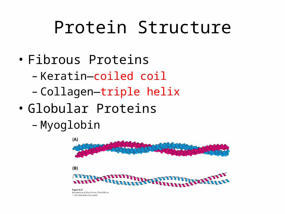
Protein Structure
• Fibrous Proteins– Keratin—coiled coil– Collagen—triple helix
• Globular Proteins– Myoglobin
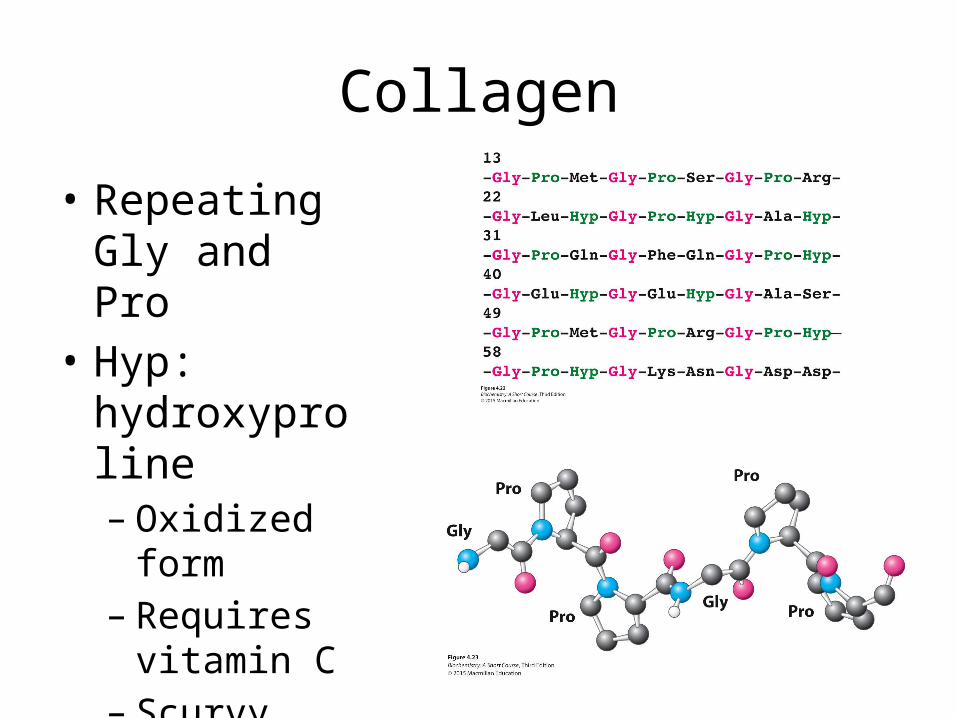
Collagen
• Repeating Gly and Pro
• Hyp: hydroxyproline– Oxidized form– Requires
vitamin C– Scurvy
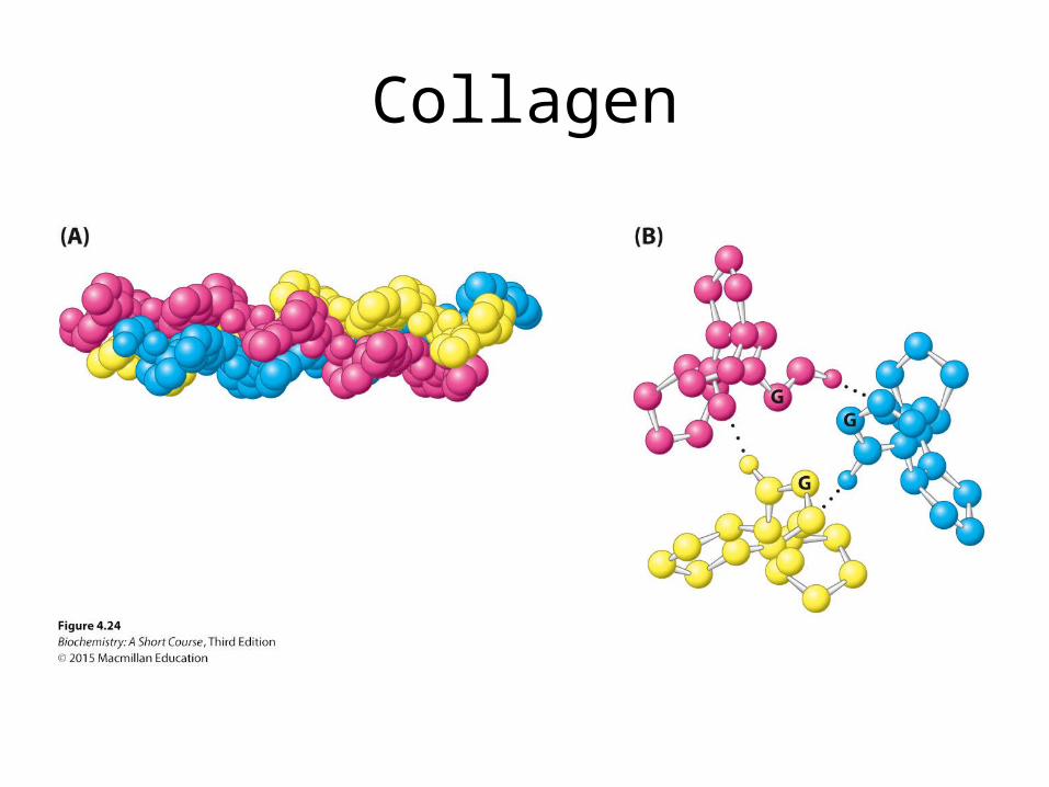
Collagen

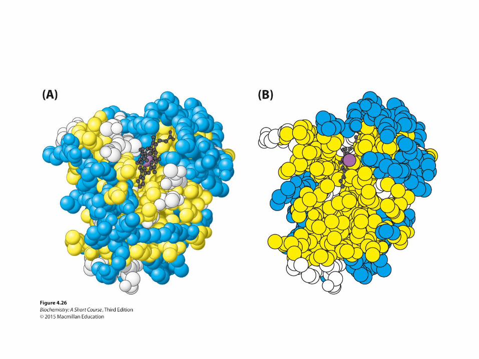
Myoglobin
• Globular Protein• Hydrophobic
effect• Helix bundle• Polar loops• Nonpolar core

Protein Folding
• Native vs denatured states• DG might be 40 kJ/mol for small protein (about 2
H-bonds)• Classic Anfinsen experiments show folding info
contained in primary sequence (in many cases)

Thermodynamics and Kinetics
• Levinthal’s paradox: not random sampling of all possible conformations
• Energy funnel• Series of irreversible
steps• Entropy traps

More than one fold
• Traditionally, one protein = one fold
• Intrinsically Unstructured Proteins (IUPs) are more common than originally thought
• Metamorphic proteins– Cytokine with equilibrium
of two structures with necessary function

Misfolding Pathology
• Amyloidoses– Alzheimer, Parkinson,
Huntington, prion
• Formation of amyloid fibers– The less stable protein
form accumulates into a nucleus, which grows to a fibril
– Aggregations cause damage—oxidation??
• Mad Cow Disease: prions as the infectious agent


