Protein Kinase C (PKC) Activity Regulates Functional Effectsof Kv�1.3 Subunit on KV1.5 ChannelsIDENTIFICATION OF A CARDIAC Kv1.5 CHANNELOSOME*□S
Received for publication, December 2, 2011, and in revised form, April 13, 2012 Published, JBC Papers in Press, April 30, 2012, DOI 10.1074/jbc.M111.328278
Miren David‡1,2, Álvaro Macías‡1,3, Cristina Moreno‡4, Ángela Prieto‡4, Ramón Martínez-Mármol§, Rubén Vicente¶,Teresa González‡5, Antonio Felipe§, Michael M. Tamkun�, and Carmen Valenzuela‡6
From the ‡Instituto de Investigaciones Biomédicas, Madrid Consejo Superior de Investigaciones Cientıficas-Universidad Autonomade Madrid, C/Arturo Duperier 4, 28029 Madrid, Spain, the §Molecular Physiology Laboratory, Departamento de Bioquímica iBiología Molecular, Institut de Biomedicina (IBUB). Universitat de Barcelona, 08028 Barcelona, Spain, the ¶Laboratory of MolecularPhysiology and Channelopathies, Department of Experimental and Health Sciences, Universitat Pompeu Fabra, Edifici PRBB, C/Dr.Aiguader 88, 08003 Barcelona, Spain, and the �Program in Molecular, Cellular, and Developmental Neuroscience, Department ofBiomedical Sciences and Department of Biochemistry and Molecular Biology, Colorado State University,Fort Collins, Colorado 80523
Background: Kv�1.3 fast inactivation conferred onto Kv1.5 is PKC-dependent.Results: PKC inhibition shifts Kv�1.3-induced inactivation curve without altering Kv1.5-Kv�1.3 interaction. A Kv1.5 channelo-some is characterized.Conclusion: Kv1.5 channelosome is composed of several PKC isoforms (�I, �II, and �), Kv�1.3 and RACK1 in HEK293 and inrat ventricular cells.Significance: This is the first evidence of a cardiac Kv1.5-Kv�1.3-RACK1-PKC macromolecular complex.
Kv1.5 channels are the primary channels contributing to theultrarapid outward potassium current (IKur). The regulatoryKv�1.3 subunit converts Kv1.5 channels from delayed rectifierswith a modest degree of slow inactivation to channels with bothfast and slow inactivation components. Previous studies haveshown that inhibition of PKC with calphostin C abolishes thefast inactivation induced by Kv�1.3. In this study, we investi-gated the mechanisms underlying this phenomenon usingelectrophysiological, biochemical, and confocal microscopyapproaches. To achieve this, we used HEK293 cells (which lackKv� subunits) transiently cotransfectedwith Kv1.5�Kv�1.3 andalso rat ventricular and atrial tissue to study native �-� subunitinteractions. Immunocytochemistry assays demonstrated thatthese channel subunits colocalize in control conditions andafter calphostin C treatment. Moreover, coimmunoprecipita-tion studies showed that Kv1.5 and Kv�1.3 remain associatedafter PKC inhibition. After knocking down all PKC isoforms bysiRNAor inhibitingPKCwith calphostinC,Kv�1.3-induced fastinactivation at�60mVwas abolished. However, depolarizationto �100 mV revealed Kv�1.3-induced inactivation, indicatingthat PKC inhibition causes a dramatic positive shift of the inac-
tivation curve. Our results demonstrate that calphostin C-me-diated abolishment of fast inactivation is not due to the dissoci-ation of Kv1.5 and Kv�1.3. Finally, immunoprecipitation andimmunocytochemistry experiments revealed an associationbetween Kv1.5, Kv�1.3, the receptor for activated C kinase(RACK1), PKC�I, PKC�II, and PKC� in HEK293 cells. A verysimilar Kv1.5 channelosome was found in rat ventricular tissuebut not in atrial tissue.
The outward potassium current IKur,7 the main currentresponsible for human atrial repolarization, is generated fol-lowing the activation of Kv1.5 channels. The slow and partialinactivation and the voltage dependence of these channelsunderlie their key role in the regulation of the atrial actionpotential duration (1, 2). This slow inactivation is modified bythe assembly of Kv1.5 subunits with� subunits (Kv�1.2, Kv�1.3,and Kv�2.1) present in the human myocardium (3, 4). TheKv�1.3 subunit provides a number of functions, including a fast,partial inactivation component, a greater degree of slow inactiva-tion, a shift of the activation curve toward more negative poten-tials, a 7-fold decrease in the sensitivity of the channel to the blockinduced by antiarrhythmic drugs and local anesthetics, and adecrease in the degree of stereoselective blockage (5–7).IKur is highly susceptible to adrenergic regulation, which is
differentiallymodulated by�- and�-stimulation (8) via proteinkinases C (PKC) and A (PKA), respectively (9–11). This phe-nomenon is very important because the expression levels of �-
* This work was supported by Ministerio de Ciencia e Innovación (MICINN)Grants SAF2010-14916, Ministerio de Educacion y Ciencia-PR2003-0056, andRed Cooperativa de Enfermedades Cardiovasculares RECAVA FIS RD06/0014/0006 (to C. V.) and BFU2011-23268 and CSD2008-00005 (to A. F.).
□S This article contains supplemental Fig. S1 and Table S1.1 Both authors contributed equally to this work.2 Recipient of an RECAVA grant.3 Recipient of a JAE predoctoral grant.4 Recipients of an FPI grant.5 Recipient of a JAE doctoral grant.6 Recipient of Grant MEC-PR2003-0056. To whom correspondence should be
and �-adrenergic receptors are altered in several cardiacpathologies as well as the release of catecholamines (12, 13). Infact, cardiac hypertrophy is associated with an up-regulation ofdifferent PKC isoforms (14–17). Similarly, �-calmodulin kinaseII (�-CaMKII) expression increases during atrial fibrillation(18). Furthermore, one of the most effective treatments foratrial fibrillation is the oral administration of �-blockers (19),which induce a pharmacological remodeling that is capable ofreversing the electrical dysfunction typically observed duringatrial fibrillation (20, 21). PKC and PKA activities are alsorequired for the Kv1.5 modulation by the auxiliary subunitsKv�1.2 and Kv�1.3 (9, 10, 22). Indeed, PKC inhibition by cal-phostin C reverses the Kv�1.3-dependent electrophysiologicaleffects (9). Calphostin C, a potent and selective inhibitor ofmultiple protein kinase C (PKC) isoforms, acts via interactionwith the regulatory diacylglycerol (DAG) binding site (23).PKCs comprise a cluster of at least 11 different isoforms that
include three subfamilies according to their sensitivities to sec-ond messengers such as Ca2� and DAG. Classical isoforms(PKC�, -�I, -�II, and -�) are activated by Ca2� and DAG,whereas novel isoforms (PKC�, -�, -�, and �) are sensitive toDAG but insensitive to Ca2�. Finally, atypical isoforms (PKC�and /) do not require Ca2� or DAG for their activation,whereas they are dependent on ceramide, arachidonic acid, andother lipids (24–26). Upon activation, most PKC isoformsundergo subcellular relocation depending on the cell type (27).This specific compartmentation is further fine-tuned by inter-actions with specific PKC adaptor proteins named receptor foractivated C kinase (RACK) (28).PKC isoforms translocate to different subcellular sites after
their activation by specific second messengers, eliciting uniquecellular effects (29, 30) that are dependent on the substrate to bephosphorylated (26, 31). The interaction of PKCs with severalsubstrates is believed to occur through a family of proteins thatare collectively named RACKs (32). These adaptor proteins donot have any intrinsic functional effects. However, they are ableto translocate bound proteins to different subcellular locationsthrough their protein-protein interaction domains (WD40)(33). Thus, they are capable of binding several enzymes con-comitantly to form an enzymatic complex that is localized closeto the substrate of the enzyme. This process allows them toregulate different cellular responses (32). RACK1 is one of theadaptor proteins that has been identified as an anchoring pro-tein for PKC.Additionally, several ion channels (KCa1.1, Cav1.2,Kir3.1, TRPC3, and NMDA receptor) have been reported to bemodulated by PKC via this scaffold protein (34–39). The bind-ing of RACK1 to PKC�II enhances the enzymatic activity of thelatter by 4–6-fold (34, 40).In the present study, we analyzed the effects of PKC inhibi-
tion on the Kv1.5�Kv�1.3 interaction. None of the isoform-selective PKC inhibitors removed the Kv�1.3-mediated fastinactivation. By silencing all of the PKC isoformswith siRNAorinhibiting PKCs with calphostin C, fast inactivation was abol-ished at membrane potentials up to �60 mV. However, atmembrane potentials more positive than �100 mV, fast inacti-vation was evident, which indicated that Kv�1.3 and Kv1.5remained assembled after PKC inhibition and that withoutPKC activity, the voltage dependence of Kv�1.3-induced inac-
tivation is dramatically shifted in the positive direction. Addi-tionally, we demonstrate that Kv1.5�Kv�1.3 channels inter-acted with RACK1, PKC�I, PKC�II, and PKC�, either directlyor through scaffold proteins, generating an emerging Kv1.5channelosome in HEK293 cells and ventricular tissue.
EXPERIMENTAL PROCEDURES
Expression Plasmids, Cell Culture, and Transient Trans-fection—HumanKv1.5 andKv�1.3 in pBKhave been extensivelycharacterized (4). Human Kv1.5 (�22 to 1,894 nucleotides) andKv�1.3 (�53 to 1,500 nucleotides) were inserted into the samepBK vector, with the Kv1.5 subunit placed 3� to the Kv�1.3subunit and preceded by an internal ribosome entrysequence, thus generating a bicistronic messenger RNA asdescribed previously (9). The gene encoding Kv�1.3 was sub-cloned between the SacII and NotI restriction sites withinthe polylinker of the pCMV-Tag5A vector, which generateda recombinant Kv�1.3-Myc protein. To generate Kv2.1-HA,the HA epitope was inserted after Gly-217 of the rat Kv2.1cDNA, placing the epitope in the extracellular S1-S2 loop(41). In some experiments, a construct with an HA tag intro-duced into the Kv1.5 S1-S2 loop was used (kindly provided byProf. D. J. Snyders).HEK293 cells were cultured in Dulbecco’s modified Eagle’s
medium supplemented with 10% fetal bovine serum, 10units/ml penicillin-streptomycin (Sigma-Aldrich), and 1%nonessential amino acids. For the electrophysiological experi-ments, cells were transfected with Kv1.5 (0.4 �g) orKv1.5�Kv�1.3 channels (0.3 �g) and a reporter plasmidexpressing CD8 (1.6 �g) using Lipofectamine 2000 (10 �l)(Invitrogen). Before experimental use, the cells were incubatedwith polystyrene microbeads precoated with an anti-CD8 anti-body (Dynabeads M450, Dynal) as described previously (5, 7,42). For the immunocytochemistry and immunoprecipitationstudies, the cells were cotransfected with 0.5 �g of Kv1.5 orKv2.1-HA and 1 �g of Kv�1.3-Myc cDNA.Inhibitors, Antibodies, and siRNA—Calphostin C, Gö6976,
Gö6983, hispidin, and PKC� pseudosubstrate inhibitor (PKC�-PI) were from Calbiochem (Merck KGaA). Donkey anti-goatantibodywas fromSantaCruz Biotechnology (Santa Cruz, CA),and goat anti-mouse and goat anti-rabbit antibodies were fromCalbiochem (1:10000); all of them were horseradish peroxi-dase-conjugated. The polyclonal rabbit antibody anti-Kv1.5(1:1000) was purchased from Alomone Labs, the monoclonalanti-Myc (1:500) and anti-�-actin (1:40000) antibodies werefrom Santa Cruz Biotechnology, anti-Kv�1 (1:500) was fromAbcam (Abcam Limited), and anti-HA (1:250) was fromNovus(Novus Biologicals). Monoclonal antibodies specific for thePKC isoforms (1:200) and anti-RACK1 (1:500) were from SantaCruz Biotechnology.To knock down all PKC isoforms, we transfected HEK293
cells with PKC-specific small interfering RNAs (siRNAs) pur-chased from Santa Cruz Biotechnology, according to the man-ufacturer’s instructions. The PKC siRNA contains five target-specific 19–25-nucleotide siRNAs that are designed to knockdown gene expression. The most efficient transfection resultswere obtained with 50 nM of the siRNA duplexes transfected72 h prior to the experiments.
Kv1.5-Kv�1.3 and PKC
JUNE 15, 2012 • VOLUME 287 • NUMBER 25 JOURNAL OF BIOLOGICAL CHEMISTRY 21417
Electrophysiological Recordings and Data Acquisition—Theintracellular pipette filling solution contained the following (inmM): 80 potassium aspartate, 42 KCl, 3 phosphocreatine, 10KH2PO4, 3 MgATP, 5 HEPES-K, and 5 EGTA-K (adjusted topH 7.25 with KOH). The bath solution contained the following(in mM): 140 NaCl, 4 KCl, 1.8 CaCl2, 1 MgCl2, 10 HEPES-Na,and 10 glucose (adjusted to pH7.40withNaOH).Currentswererecorded using the whole-cell configuration of the patch clamptechnique with a patch clamp amplifier (Axopatch-200B patchclamp amplifier; Molecular Devices) and stored on a personalcomputer (TD Systems) with a DigiData 1440A analog-to-dig-ital converter (Molecular Devices). PClamp version 10 softwarewas used for both data acquisition and analysis (MolecularDevices). Currents were recorded at room temperature (21–23 °C) at a stimulation frequency of 0.1 Hz and were sampled at4 kHz after antialias filtering at 2 kHz. The average pipetteresistance ranged between 1 and 3 megaohms (n � 70).Gigaohm seal formation was achieved by suction (2–5gigaohms, n � 70). After seal formation, the cells were liftedfrom the bath, and the membrane patch was ruptured with abrief additional suction. The capacitive transients elicited bysymmetrical 10-mV steps from �80 mV were recorded at 50kHz and filtered at 10 kHz for subsequent calculations of thecapacitive surface area, access resistance, and input impedance.Thereafter, the capacitance and series resistance compensationwere optimized, and usually 80% compensation of the effectiveaccess resistance was obtained. MicroCal Origin 7.05 (Origin-Lab Co) and the Clampfit utility of pClamp 9 were used toperform least squares fitting and for data presentation. Deacti-vation and inactivation were fitted to a biexponential processwith an equation of the form y�A1exp(�t/�1)�A2exp(�t/�2)� C, where �1 and �2 are the system time constants, A1 and A2are the amplitudes of each component of the exponential, andCis the baseline value. The voltage dependence of the activationcurves was fitted with a Boltzmann equation: y � 1/(1 �exp(�(V � Vh)/s)), where s represents the slope factor, V rep-resents the membrane potential, and Vh represents the voltageat which 50% of the channels are open.Protein Extracts, Immunoprecipitation, and Western Blot—
For total protein extraction from HEK293 cells, the cells werewashed twice in chilled phosphate-buffered saline (PBS) andcentrifuged at 3,000� g for 10min. The pellet was then lysed inice-cold lysis solution (20mMHEPES, pH 7.4, 1 mM EDTA, 255mM sucrose supplemented with Complete protease inhibitormixture tablets (Roche Diagnostics)), and homogenized byrepeated passage (10 times) through a 25-gauge (0.45� 16mm)needle.Homogenateswere further centrifuged at 10,000� g for5min to remove nuclei and organelles. Samples were separatedinto aliquots and stored at �80 °C. For immunoprecipitationassays, we isolated membrane protein from the total proteinextract by an additional centrifugation at �150,000 � g for 90min. The pellet was resuspended in 30mMHEPES (pH 7.4), andthe protein content was determined using the Bradford Bio-Rad protein assay (Bio-Rad). Ventricular (principal coronaryarteries excluded) and atrial tissues frommaleWistar rats werekindly provided by Drs. A. Cogolludo and F. Pérez-Vizcaíno(Universidad Complutense deMadrid, Spain). After dissection,cardiac tissuewas frozen in liquid nitrogen andhomogenized in
a glass potter (300�l and 3ml of the lysis buffer described abovewere used for atria and ventricles, respectively). The homoge-nate was centrifuged at 6000 � g for 10 min at 4 °C. The super-natant was collected, separated into aliquots, and stored at�80 °C until its posterior analysis.For the coimmunoprecipitation experiments, the homoge-
nates were resuspended in 150 �l of immunoprecipitationbuffer (1% Nonidet P-40, 10% glycerol, 10 mM HEPES, and 150mMNaCl supplementedwithComplete protease inhibitormix-ture tablets (pH � 7.8) (Roche Diagnostics)) and homogenizedby orbital shaking at 4 °C for 1 h. 300 �g of crude membraneproteinwas used forHEK293 cells, 500�gwas used for rat atria,and 1500 �g was used for the ventricular tissue. Proteins werethen incubated with 20 �l of immunoprecipitation buffer-pre-washed Sepharose protein A/G beads (Santa Cruz Biotechnol-ogy) for 2 h at 4 °C, and contaminant-bound Sepharose beadswere separated by centrifugation for 30 s at 5000 � g at 4 °C.The supernatant was incubated with 4 ng of polyclonal anti-Kv1.5 (Alomone Labs) or monoclonal anti-RACK1 antibody(Santa Cruz Biotechnology) for each microgram of protein,overnight at 4 °Cwith orbital shaking. Approximately 20–30�lof PBS-washed Sepharose proteinA/G beadswas then added tothe mixture followed by incubation for 2 h. Sepharose beadsbound to antibody-protein complexes were precipitated bycentrifugation (30 s at 5000 � g at 4 °C), and antibody-boundbeads were then washed twice with immunoprecipitationbuffer and centrifuged for 30 s at 5000� g at room temperature.In the case of cardiac tissue samples, coimmunoprecipitationwas performed using Pierce� Direct IP kit (Thermo Scientific)following the manufacturer’s instructions.Total protein extracts and immunoprecipitated protein sam-
ples were resuspended in 1� SDS (2% �-mercaptoethanol) andboiled at 100 °C for 5 min. The samples were then centrifugedfor 3 min at 5,000 � g at room temperature, and 25–50 �l ofprotein extract was separated by SDS-PAGE (7, 10, or 15%acrylamide/bisacrylamide) gels. The proteins, transferred toPVDF membranes, were probed with anti-Kv1.5, anti-Myc,anti-PKC, anti-Kv�1, and anti-RACK1 antibodies. Secondaryantibodies were developed by ECL-Plus Western blottingreagent (Amersham Biosciences).Immunostaining and Confocal Microscopy—For immuno-
staining, HEK293 cells were grown on gelatin-coated coverslipsin Dulbecco’s modified Eagle’s medium containing 10% fetalbovine serum. Twenty-four hours after transfection, the cellswere washed three times with PBS. For antibody-inducedpatching experiments, after 30min of incubation with blockingsolution (10% goat serum, 5% nonfat dry milk, PBS), the cellswere incubated with the S1-S2 Kv1.5 external epitope antibody(diluted 1:1000) or anti-HA (diluted 1:250) in HEPES-basedculture medium for 1 h at room temperature (43). Next, thecells were fixed with 4% paraformaldehyde in PBS for 10 minand blocked overnight (PBS � 5%w/v dry milk). The cells werewashed and permeabilized three times with PBS-CHAPS andthen incubated with anti-Myc antibody (1:500; PBS-CHAPSwith 10% goat serum). Next, the cells were washed three timeswith PBS-CHAPS and incubated with Alexa Fluor 488 anti-rabbit (1:500) and Alexa Fluor 594 anti-mouse (1:500) antibod-ies from Molecular Probes. The samples were mounted with
Kv1.5-Kv�1.3 and PKC
21418 JOURNAL OF BIOLOGICAL CHEMISTRY VOLUME 287 • NUMBER 25 • JUNE 15, 2012
Aqua Poly/Mount (Polysciences, Inc). Stained cells were visu-alized using LSM510 ZEISS (Carl Zeiss) or Leica TCS SP5 con-focal microscopes, and images were analyzed with Zeiss, Leica,and ImageJ software (44).Statistical Analysis—Data are presented as the mean � S.E.
One-way analysis of variance was used to compare more thantwo groups. Statistical significance was set at p � 0.05. Thecurve-fitting procedure used a nonlinear least squares (Gauss-Newton) algorithm; the results are displayed in a linear andsemilogarithmic format together with the difference plot.Goodness of fit was determined using the 2-square criterionand by inspection of systematic nonrandom trends in the dif-ference plot.
RESULTS
Modulation of Kv�1.3-induced Fast Inactivation Is NotDriven by a single PKC Isoform—Although PKC inhibition pre-vents Kv�1.3-induced fast inactivation of Kv1.5 channels
(9–11), the identity of the specific isoform responsible for thisphenomenon remains unknown. To that end, we first deter-mined which PKC isoforms are expressed in HEK293 cells.Supplemental Fig. S1 shows that HEK293 cells express all PKCisoforms (classical: �, �, and �; novel: �, �, and �; and atypical: �and /) with the exception of the novel PKC� isoform.Next, we performed electrophysiological experiments using
different PKC inhibitors. Fig. 1 shows representative currenttraces obtained under control conditions and after PKC inhibi-tion with different inhibitors. The characteristics of these PKCinhibitors are shown in Table 1.With the exception of calphos-tinC, PKC inhibitors (Gö6976,Gö6983, hispidin, and PKC�-PI)failed to abolish Kv�1.3-induced fast inactivation. Only afterPKC inhibition with calphostin C did the current-voltage rela-tionship (IV) exhibit the linearity observed when the Kv1.5 cur-rent is recorded in the absence of Kv�1.3. Different mecha-nisms of action of PKC inhibitors may explain these results.Table 2 shows the values obtained for the degrees of inactiva-
FIGURE 1. Modulation of Kv�1.3-induced fast inactivation is not due to a single PKC isoform. HEK293 cells were transiently transfected with Kv1.5�Kv�1.3.Representative current traces elicited by 250-ms depolarizing pulses from a holding potential of �80 mV to voltages between �80 and �60 mV in 10-mV stepsand the IV relationships in which the current magnitude measured at the end of 250-ms pulses was plotted versus the membrane potential are shown. Tailcurrents were obtained upon return to �40 mV. The specific inhibitor used is indicated above each trace family.
Kv1.5-Kv�1.3 and PKC
JUNE 15, 2012 • VOLUME 287 • NUMBER 25 JOURNAL OF BIOLOGICAL CHEMISTRY 21419
tion,Vh, and the inactivation and deactivation kinetics (�) of thecurrents generated after treating the cells with various PKCinhibitors. Remarkably, all of the tested PKC inhibitors
decreased the degree of fast inactivation and slowed the fasttime constant of the inactivation process (Table 2).siRNA-induced Down-regulation of PKC Mimics Effects of
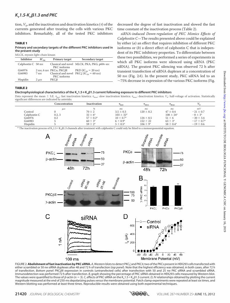
Calphostin C—The results presented above could be explainedby either (a) an effect that requires inhibition of different PKCisoforms or (b) a direct effect of calphostin C that is indepen-dent of its PKC inhibitory properties. To differentiate betweenthese two possibilities, we performed a series of experiments inwhich all PKC isoforms were silenced using siRNA (PKCsiRNA). The greatest PKC silencing was observed 72 h aftertransient transfection of siRNA duplexes at a concentration of50 nM (Fig. 2A). In the present study, PKC siRNA led to an�75% decrease in expression of the various PKC isoforms (Fig.
FIGURE 2. Abolishment of fast inactivation by PKC siRNA. A, Western blots to detect PKC� and PKC�, two of the PKCs present in HEK293 cells transfected witheither scrambled or 50 nM siRNA duplexes after 48 and 72 h of transfection (top panel). Note that the highest efficiency was obtained, in both cases, after 72 hof transfection. Bottom panel: PKC�II expression in controls (untransfected cells) after transfection with 50 and 25 nM PKC siRNA and scrambled siRNA.Immunodetection was performed 72 h after transfection. B, graph showing the percentage of PKC siRNA obtained in HEK293 cells measured by Western blot.The values were quantified to those of �-actin (n � 3). C, effects of PKC siRNA on the Kv1.5�Kv�1.3 current. D, IV relationships obtained by plotting the currentmagnitude measured at the end of 250-ms depolarizing pulses versus the membrane potential. Patch clamp experiments were repeated at least six times, andWestern blotting was performed at least three times. Reproducible results were obtained using both experimental techniques.
TABLE 1Primary and secondary targets of the different PKC inhibitors used inthe present studyMLCK, myosin light-chain kinase.
Inhibitor IC50 Primary target Secondary target
Calphostin C 50 nM Classical and novelPKC isoforms
TABLE 2Electrophysiological characteristics of the Kv1.5�Kv�1.3 current following exposure to different PKC inhibitorsData represent the mean � S.E. �fast: fast inactivation kinetics; �slow: slow inactivation kinetics; �deac: deactivation kinetics; Vh: half-voltage of activation. Statisticallysignificant differences are indicated by asterisks.
2B, supplemental Table S1). Fig. 2C shows non-inactivatingelectrophysiological recordings of the Kv1.5�Kv�1.3 channelsafter PKC siRNA treatment. Voltage-dependent potassiumcurrents were evoked by applying depolarizing pulses from aholding potential of�80mV to different voltages between�80to �60 mV in 10-mV steps. Fig. 2D shows the IV relationshipsmeasured at the end of the 250-ms pulses under these experi-mental conditions. Under control conditions, the IV relation-ship reached a plateau at around �15 mV. Binding of theinactivating particle of the Kv�1.3 subunit is a highly voltage-dependent process; atmore positive test potentials, this bindingismore likely to occur. PKC siRNA eliminated fast inactivation,and thus, the IV relationships obtained under these experimen-tal conditions resembled those obtained after activation ofKv1.5 channels in the absence of Kv�1.3 subunits (7). Hence,this treatment mimicked the effects of calphostin C on the cur-rent generated after activation of Kv1.5�Kv�1.3 channels,which ruled out a possible direct effect of calphostin C.Kv1.5 and Kv�1.3 Still Colocalize in Cell Membrane during
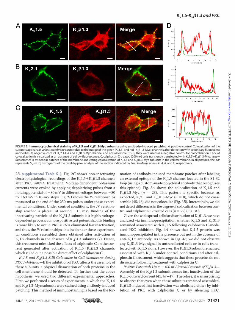
PKC Inhibition—If the inhibition of PKCaffects the assembly ofthese subunits, a physical dissociation of both proteins in thecell membrane should be detected. To further test the abovehypothesis, we used two different experimental approaches.First, we performed a series of experiments in which the Kv1.5andKv�1.3-Myc subunits were stained using antibody-inducedpatching. This method of immunostaining is based on the for-
mation of antibody-induced membrane patches after labelingan external epitope of the Kv1.5 channel located in the S1-S2loop (using a custom-made polyclonal antibody that recognizesthis epitope). Fig. 3A shows the colocalization of Kv1.5 andKv�1.3-Myc (n � 28). This pattern is specific because, asexpected, Kv2.1 and Kv�1.3-Myc (n � 8), which do not coas-semble (45, 46), did not colocalize (Fig. 3B). Interestingly, we didnot detect differences in the degree of colocalizationbetween con-trol and calphostin C-treated cells (n � 29) (Fig. 3D).Given thewidespread cellular distribution of Kv�1.3, we next
analyzed via immunoprecipitation whether Kv1.5 and Kv�1.3remained associated with Kv1.5 following calphostin C-medi-ated PKC inhibition. Fig. 4A shows that Kv1.5 protein wasimmunoprecipitated in the presence but not in the absence ofanti-Kv1.5 antibody. As shown in Fig. 4B, we did not observeany Kv�1.3-Myc signal in untransfected cells or in cells trans-fectedwith Kv1.5 alone. However, the Kv�1.3 subunit remainedassociated with Kv1.5 under control conditions and after cal-phostin C treatment, which suggests that these proteins do notdissociate following treatment with calphostin C.Positive Potentials Up to �100mV Reveal Presence of Kv�1.3—
Assembly of the Kv�1.3 subunit causes fast inactivation of theKv1.5 outward current (45, 47–49). Therefore, it was surprisingto observe that even when these subunits remained assembled,Kv�1.3-induced fast inactivation was abolished either by inhi-bition of PKC with calphostin C or by silencing PKC.
FIGURE 3. Immunocytochemical staining of Kv1.5 and Kv�1.3-Myc subunits using antibody-induced patching. A, positive control. Colocalization of thesubunits appears as yellow membrane clusters due to the merge of the green (Kv1.5) and red (Kv�1.3-Myc) channels after detection with secondary fluorescentantibodies. B, negative control. Kv2.1-HA and Kv�1.3-Myc channels do not assemble. Thus, they were used as a negative control for colocalization. Lack ofcolocalization is visualized as an absence of yellow fluorescence. C, calphostin C-treated (200 nM) cells transiently transfected with Kv1.5�Kv�1.3-Myc; yellowfluorescence is evident in patches of the membrane, indicating colocalization of Kv1.5 and Kv�1.3-Myc subunits in the cell membrane. In all pictures, the barrepresents 5 �m. D, histograms of the pixel-by-pixel analysis of the section indicated by lines in Merge panels in A, B, and C, respectively.
Kv1.5-Kv�1.3 and PKC
JUNE 15, 2012 • VOLUME 287 • NUMBER 25 JOURNAL OF BIOLOGICAL CHEMISTRY 21421
Kv1.5�Kv�1.3 current is activated at�30mVand inactivates atpotentials positive to 0 mV, exhibiting a degree of inactivationof 70 � 2% (n � 23) when measured at �60 mV (Fig. 1). AfterPKC inhibition with calphostin C- or siRNA-mediated PKCsilencing, Kv1.5�Kv�1.3 currents showed a degree of slow inac-tivation that ranged between 20 and 30%,whereas pulse steps topotentials positive to�60mV revealed the presence of the typ-ical Kv�1.3-induced fast inactivation (Fig. 5A). Fig. 5A alsoshows the mean values of the inactivation degrees obtained at�100 mV recorded in at least four experiments. These resultsfurther suggest that Kv1.5 and Kv�1.3 remain assembled afterPKC inhibition.This outcome suggests that after PKC inhibition, the inacti-
vation curve of Kv1.5-Kv�1.3 current is shifted toward morepositive membrane potentials. To test this hypothesis, a seriesof experiments was performed in which a double-pulse proto-col was applied (Fig. 5B). These results show that the inactiva-tion curve in calphostin C-treated cells was shifted towardmore positive potentials (Vh � �26.5 � 0.6 versus �8.5 � 1.9mV in the presence and in the absence of calphostin C, respec-tively; n � 10; see Fig. 5C and Table 3). Moreover, the degree ofinactivation in calphostin C-treated cells decreased from47.3 � 5.7% (n � 5) to 34.6 � 2.2% (n � 7) (p � 0.05).Effects of PIP2 and OAG on Kv�1.3-induced Fast
Inactivation—It has been proposed that PIP2 associates withthe N terminus of the Kv�1.3 subunit, and when the � subunitdissociates from PIP2, it assumes a hairpin structure that canenter the central cavity of an open Kv1.5 channel, triggeringN-type inactivation. Therefore, it has been suggested thatKv�1.3-induced fast inactivation is mediated by equilibriumbinding of the N terminus of Kv�1.3, which switches betweenbinding to phosphoinositides (PIPs) and the inner pore regionof Kv1.5 channels (50). Moreover, stimulation of �1-receptorsactivates PLC�, which is capable of cleaving PIP2 into IP3 andDAG, thus activating classical andnovel PKCs. Thus, the effectsof PIP2 andOAG (a nondegradableDAGanalog) were analyzedby adding them to the internal solution (Fig. 6). Cells dialyzedwith PIP2 exhibited a lower degree of fast inactivation just afterpatch rupture in comparison with control cells (55 � 4% versus
68 � 6%, n � 4, p � 0.05) (Fig. 6A). Also, after 8 min of dialysiswith PIP2, the degree of N-type inactivation increased signifi-cantly (from 55 � 4% to 70 � 4%, n � 4, p � 0.05) (Fig. 6A).These effects could be due to a decrease of the PIP2 concentra-tion due to its cleavage into IP3 and DAG. In fact, the degrada-tion of PIP2 would increase the ability of the N terminus ofKv�1.3 to inactivate Kv1.5 (50). In contrast, cells dialyzed withOAG exhibited a degree of N-type inactivation similar to con-trol cells, both after patch rupture and after 8 min of dialysiswithOAG (65� 3% to 67� 2%,n� 5, p 0.05) (Fig. 6B). Theseresults are in agreement with the involvement of classical andnovel PKC isoforms in the effects of calphostin C and PKCsiRNAon the fast Kv�1.3-induced inactivation. To test whetherthe PIP2 effects at different times (t � 0 or 8 min) were due tothe cleavage of PIP2 into DAG and IP3, a series of experimentsin which cells previously incubatedwith a PLC inhibitor (U73122,10 �M) and dialyzed with PIP2 was performed (Fig. 6C). Underthese experimental conditions, the degree of fast inactivation wasreduced (from 69 � 2% to 62 � 2%, n � 9, p � 0.01), the timeconstant of the fast inactivation was increased, and the contribu-tion of the fast component of inactivation was decreased.The Kv1.5 Macromolecular Complex Contains Kv1.5, Kv�1.3,
RACK1, PKC�I, PKC�II, and PKC�—Several ion channels havebeen reported to be modulated by PKC via RACK1 (34). Wehypothesized that Kv1.5, Kv�1.3, and PKC form a functionalcomplex in which PKC activity is an essential requirement forthe induction of fast and incomplete channel inactivation byKv�1.3. To test this hypothesis, immunocytochemistry experi-ments were performed (Fig. 7). Fig. 7A shows confocal imagesof cells transfected with Kv1.5-HA and Kv�1.3, in which westained for Kv1.5 and RACK1. Fig. 7B shows confocal images ofcells transfected with Kv1.5 and Kv�1.3-Myc, in which westained for Kv�1.3 and RACK1. As shown, under both experi-mental conditions, colocalizationwas consistent betweenKv1.5and RACK1, as well as between Kv�1.3 and RACK1. Given thewidespread immunolocalization patterns, we confirmed sub-unit association using immunoprecipitation experiments. Weimmunoprecipitated Kv1.5 channels and blotted for PKC�II toconfirm the presence of this enzyme in the protein complexes.As shown in Fig. 8A, PKC�II signal was absent in PKCsiRNA-transfected cells. Furthermore, blots against RACK1confirmed the interaction of Kv1.5 with this adaptor protein(Fig. 8B).Moreover, reverse coimmunoprecipitation (immuno-precipitation of RACK1) yielded similar results, revealing thepresence not only of PKC�II (data not shown) but also of theKv1.5 and Kv�1.3 subunits (Fig. 8C). Collectively, these resultsdemonstrate the presence of a functional complex or channelo-some that includes Kv1.5, Kv�1.3, RACK1, and PKC�II. Thisinteraction was demonstrated in controls and was found to beabsent in PKC-silenced cells (Fig. 8A). On the other hand,because calphostinC inhibits PKCby binding to theDAGbind-ing site, the reduced level of PKC�II in calphostin C-treatedcells is not surprising, as inhibited PKC�II may not be able tobind RACK1 in the channelosome.To determine which PKC isoforms are present in this
macromolecular complex, a series of coimmunoprecipita-tion experiments were performed in which we immunopre-cipitated Kv1.5 channels and blotted for the PKC isoforms
FIGURE 4. Immunoprecipitation of Kv1.5 with Kv�1.3-Myc. A, negative(Ab�) and positive (Ab�) control immunoprecipitations (IP) blotted againstKv1.5 channels. Supernatants (S) are also shown. WB, Western blot. B, immu-noprecipitation of Kv1.5 channels and blotting against Kv�1.3-Myc revealedthe coimmunoprecipitation of both subunits, indicating that the proteinassembly was not abolished by PKC inhibition. The arrow indicates theKv�1.3-Myc epitope. The band that appears with a molecular mass of 50 kDacorresponds to the immunoglobulin heavy chain.
Kv1.5-Kv�1.3 and PKC
21422 JOURNAL OF BIOLOGICAL CHEMISTRY VOLUME 287 • NUMBER 25 • JUNE 15, 2012
present in HEK293 cells (Fig. 9). We observed that onlyPKC�I, PKC�II, and PKC� were present in the Kv1.5channelosome.The Kv1.5 Channelosome Is Present in Rat Ventricular Myo-
cytes but Not in Atrial Myocytes—Finally, we performed exper-iments in cardiac tissue (Fig. 10). Coimmunoprecipitationexperiments were performed in which we immunoprecipi-tated Kv1.5 channels and blotted for PKC�I, PKC�II, PKC�,RACK1, and Kv�1.x. Samples from ventricular homogenatespresented a Kv1.5 channelosome composed of Kv1.5, Kv�1,RACK1, PKC�I, and PKC�II (Fig. 10A). Importantly, atrialtissue did not display this Kv1.5 protein complex (Fig. 10B)because none of the proteins studied forming the Kv1.5channelosome in ventricle coimmunoprecipitated withKv1.5 in atrial tissue.
DISCUSSION
In the present study, we have analyzed the mechanisms bywhich calphostinC-mediated PKC inhibition abolishesKv�1.3-induced fast inactivation of the Kv1.5 channel. We have dem-onstrated that the inhibition of at least classical and novel PKCisoforms is required to abolishKv�1.3-induced fast inactivation(9, 10). Furthermore, this effect was not due to Kv1.5�Kv�1.3dissociation but due to a positive shift of the inactivation curvedriven by PKC inhibition because both subunits remainedassembled as shown by immunocytochemistry, immunopre-cipitation, and electrophysiological experiments. We have also
shown that at least Kv�1.3, RACK1, PKC�I, PKC�II, and PKC�
are associated with Kv1.5 in HEK293 cells, forming a channelo-some. Finally, for the first time, we provide evidence pointing tothe existence of a native ventricular cardiac Kv1.5 channelo-some, whose composition is similar to that found in HEK293cells (with the exception of PKC�, absent in this tissue accord-ing to our Western blot analyses, data not shown).The stimulation of �1-receptors leads to activation of PLC�.
This enzyme cleaves PIP2, generating IP3 andDAG, which acti-vate most PKC isoforms either alone or with Ca2�, with theexception of atypical PKCs. The N terminus of the Kv�1.3 sub-unit associates with membrane-bound PIP2, and when it disso-ciates from PIP2, it assumes a hairpin structure that enters thecentral cavity of an open Kv1.5 channel, inducing fast inactiva-tion. Thus, Kv�1.3-induced fast inactivation is mediated by acompetitive binding between PIPs and the inner pore region ofKv1.5 channels for the N terminus of Kv�1.3 (50). The presentresults obtained for PIP2 and OAG are in agreement with pre-vious studies and indicate a fine-tuned regulation of Kv�1.3-induced fast inactivation by PKC and PIP2. Moreover, theeffects of PIP2 in cells in which the PLCwas inhibited produceda marked decrease of the Kv�1.3-induced fast inactivation. Inthe absence of PIP2 into the internal solution, the effectsobserved were qualitatively similar (data not shown), suggest-ing a role of PLC on the fast inactivation induced by this �subunit.
FIGURE 5. Voltage dependence of inactivation of Kv1.5 and Kv1.5�Kv�1.3. A, current traces obtained after depolarization from a holding potential of �80mV to �60 (red) and �100 mV (black) in HEK293 cells transfected with Kv1.5�Kv�1.3 and with scrambled siRNA (50 nM) (left), with PKC siRNA-transfected cells(middle) and in calphostin C-treated cells (right). B, original current records obtained in control and calphostin C-treated cells. C, voltage dependence of theinactivation under control and after calphostin C treatment. Note that calphostin C shifts the inactivation curve to more positive potentials.
TABLE 3Voltage-dependent inactivation parametersVh: half voltage of inactivation. s: slope of the Boltzmann equation. Data represent the mean � S.E. of n � 7 experiments. Statistically significant differences are indicatedby asterisks.
FIGURE 6. Effects of PIP2, OAG, and PLC on Kv�1.3-induced fast inactivation. A, representative current traces obtained after depolarization from a holdingpotential of �80 mV to �60 mV, just after patch rupture (t � 0 min) and after 8 min (t � 8 min), in control conditions and during PIP2 dialysis. The graph showsthe degree of inactivation in control cells and in cells dialyzed with PIP2 at t � 0 min and t � 8 min. Note that at t � 0, the degree of fast inactivation wassignificantly lower than that after 8 min of dialysis in PIP2 and control dialyzed cells. B, representative current traces obtained at �60 mV at t � 0 and t � 8 minin control conditions and during OAG dialysis. The graph shows the degree of fast inactivation in control cells and in cells dialyzed with OAG at t � 0 min andt � 8 min. The degree of fast inactivation was similar in all experimental conditions. C, representative current traces obtained at �60 mV at t � 0 (U73122, 10�M) and t � 8 min (U73122 � PIP2) after inhibition of PLC with U73122 and after PIP2 dialysis. The upper right, bottom left, and bottom right graphs show thedegree of fast inactivation, the fast inactivation kinetics, and the contribution of the fast component of inactivation to the total process (Afast/(Afast � Aslow))under both experimental conditions, respectively. *, p � 0.05; **, p � 0.01; ***, p � 0.001.
Kv1.5-Kv�1.3 and PKC
21424 JOURNAL OF BIOLOGICAL CHEMISTRY VOLUME 287 • NUMBER 25 • JUNE 15, 2012
Most PKC isoforms redistribute into different subcellularcompartments upon activation, depending on the PKC isoformand the cell type (36). A surprising finding of the present studywas that all of the PKC inhibitors tested (Gö6976, Gö6983, his-pidin, and PKC�-PI), with the exception of calphostin C, failedto abolish Kv�1.3-induced fast inactivation. This result is likelydue to the differentmechanisms of action of the PKC inhibitorsused. Indeed, calphostin C inhibits PKC by binding to its C1domain (DAG and phorbol ester binding site) and then irre-versibly inactivates the enzyme. Gö6983 and Gö6976 are com-petitive inhibitors of ATP binding to the catalytic domain ofPKC, whereas hispidin affects both PKC�I and PKC�II trans-location (23, 51–54). Our results suggest that inhibition of clas-sical and novel, but not atypical, PKC isoforms counteractsKv�1.3-induced fast inactivation and shifts theVh of the activa-tion curve to values closer to those of Kv1.5 observed in the
absence of Kv�1.3, as has been described previously (9). Thepotential dissociation of �-� caused by PKC inhibition was alsoanalyzed in the present study. Colocalization, immunoprecipi-tation, and electrophysiological experiments revealed thatthese subunits remained assembled in the plasma membranedespite PKC inhibition by calphostin C. These results are inagreement with the notion that Kv1.5 and Kv�1.3 subunitsassemble in the endoplasmic reticulum during the early stagesof their biosynthesis (55), and a 2-h incubation with calphostinC is sufficient to eliminate the typical fast inactivation inducedby Kv�1.3 (9). Moreover, previous results reported by Kwak etal. (9) showed that a tandem construction of Kv1.5 and Kv�1.3,which generates a unique protein, showed similar responses tocalphostin C (a shift inVh to more positive potentials and a lossof fast inactivation). These results indicate that mechanismsother than subunit dissociation must be involved in the abol-ishment of Kv�1.3-induced fast inactivation produced by cal-phostin C.Although we were able to rule out a role for atypical PKCs,
the involvement of a specific PKC isoform in this process hasnot yet been demonstrated. In addition, experiments in whichthemembrane potential was depolarized to�100mV indicatedthat despite PKC inhibition (calphostin C or PKC siRNA),Kv�1.3 was still capable of conferring fast inactivation and thusremained associated with Kv1.5. Furthermore, the coimmuno-precipitation experiments showed that Kv�1.3 remained asso-ciated with Kv1.5 despite PKC inhibition by calphostin C or byPKC silencing by siRNA. These data suggest that Kv1.5 andKv�1.3 subunits form a stable complex following their biosyn-thesis in the endoplasmic reticulum and that PKC inhibitionand/or activationmodulates the inactivating effect of Kv�1.3. Ithas been demonstrated that residues Arg-5 and Thr-6 of theKv�1.3 subunit are involved in PIP2 binding and thatmutations(alanine or cysteine) at these residues dramatically increase thedegree of Kv�1.3-induced fast inactivation. It has beendescribed that calphostin C does not modify the gating of theKv1.5 in the absence of � subunits (9). Therefore, it is likely thatthe effects shown in the present study involve the phosphory-lation of some residues present in the Kv�1.3 subunit. One can-didate could be threonine at position 6 (Thr-6) of Kv�1.3. Thus,we may hypothesize that PKC phosphorylation of this threo-nine or another threonine/serine at the N terminus of theKv�1.3 subunit can lead to a diminished capability to bind PIP2,thus avoiding the abrogation of fast inactivation caused by PIP2.These findings provide an explanation for the effects of PKCinhibition on the Kv�1.3-induced inactivation, assuming thatphosphorylated Kv�1.3 cannot bind PIP2. NonphosphorylatedKv�1.3 would be able to bind PIP2, and this bindingwould abol-ish N-type inactivation (50). However, further experiments arenecessary to elucidate the residue(s) involved in this effect.In the past decade, myriads of protein-protein interactions
involved in intracellular signaling have been described (56–59).Determination of the subcellular localization of signal trans-duction proteins, enzymes, substrates, and mediators hasrevealed the rapid, efficient transmission of signals either fromthe extracellularmediumor from intracellular sites as an essen-tial feature of optimal signaling (60). Despite the high degree ofhomology between PKC isoforms at both sequence and struc-
FIGURE 7. Immunocytochemical staining of Kv1.5-HA or Kv�1.3-Myc andRACK1 proteins. A, cells stained with anti-Kv1.5-HA, anti-Kv�1.3-Myc, or anti-RACK1. Colocalization appears as yellow fluorescence due to the merge of thegreen (Kv1.5-HA) and red (RACK1) channels after detection with secondaryfluorescent antibodies. Cells were cotransfected with Kv1.5-HA�Kv�1.3-Myc.RACK1 is an endogenous protein. B, cells stained with anti-Myc (green) andanti-RACK1 (red). Cells were transfected with Kv�1.3-Myc alone. In all pictures,the bar represents 5 �m.
FIGURE 8. Immunoprecipitation of Kv1.5�Kv�1.3 channel or RACK1 withPKC�II. A, PKC�II coimmunoprecipitated (IP) with Kv1.5. B, RACK1 coimmu-noprecipitated with Kv1.5 under all experimental conditions. The results arerepresentative of three independent experiments. C, coimmunoprecipitationof RACK1 and immunodetection of Kv1.5 and c-Myc-tagged Kv�1.
Kv1.5-Kv�1.3 and PKC
JUNE 15, 2012 • VOLUME 287 • NUMBER 25 JOURNAL OF BIOLOGICAL CHEMISTRY 21425
tural levels, especially within the catalytic domain, each PKCisoformmediates unique subcellular functions (29, 30) that aredependent on the target substrate (26, 31). These localizationmechanisms are especially common in plasma membrane pro-teins because incoming stimuli must be integrated and trans-mitted with a high degree of efficiency. In the present study, we
have demonstrated that Kv1.5 and Kv�1.3 form a highly stableprotein complex, which is tightly regulated by classical andnovel, but not atypical, PKC isoforms. Furthermore, our resultsshow that PKC�I, PKC�II, and RACK1 coassemble with Kv1.5and Kv�1.3. Therefore, the Kv1.5 functional channelosomecontains at least Kv1.5, Kv�1.3, RACK1, PKC�I, PKC�II, and
FIGURE 9. Immunoprecipitation of Kv1.5�Kv�1.3 channel with PKCs (n � 3). Negative (Ab�) and positive (Ab�) immunoprecipitations (IP) blotted againstthe different PKCs isoforms present in HEK293 cells. Note that only PKC�I, PKC�II, and PKC� coimmunoprecipitated with Kv1.5�Kv�1.3 channels. WB, Westernblot; S, supernatants.
FIGURE 10. Immunoprecipitation of Kv1.5 channel in cardiac tissue (n � 3). Negative (Ab�) and positive (Ab�) immunoprecipitations (IP) blotted againstthe different PKC isoforms present in HEK293 channelosome (Fig. 9). Note that a very similar channelosome has been found in ventricular (A), but not in atrial(B), tissue. WB, Western blot; S, supernatants.
Kv1.5-Kv�1.3 and PKC
21426 JOURNAL OF BIOLOGICAL CHEMISTRY VOLUME 287 • NUMBER 25 • JUNE 15, 2012
PKC�. Besides, a similar channelosome has been found in ven-tricular tissue, but not in atrial tissue, showing a parallel distri-bution to that of Kv�1.3 (47), which suggests an important roleof this � subunit in the constitution of the macromolecularcomplex.The presence of RACK1 in other ion channel complexes has
been previously reported. Indeed, RACK1 binds to the carboxylterminus of KCa1.1 channels (35). Moreover, the overexpres-sion of RACK1 with KCa1.1 channels in Xenopus oocytes shiftsthe activation curve toward more positive potentials in theabsence, but not in the presence, of ectopically expressed �channel subunits (35). Furthermore, PKC�II, which is recruitedby RACK1, down-regulates Cav1.2 activity following activation(36). Besides, the functional Kir3.1 complex contains PKA, pro-tein phosphatase 1 (PP1), PP2A/C, and RACK1. Within thiscomplex, RACK1 binds directly to G�� (37) and PKC (34).Recently, it has been described that TRPC3 regulates IP3 recep-tor (IP3R) function by mediating interaction between IP3R andthe scaffolding protein RACK1, and the importance of theOrai1-STIM1-TRPC3-RACK1-IP3R complexes in the finemodulation of intracellular Ca2� stimulated by agonists ofthese receptors has been reported (38). Thus, RACK1 is emerg-ing as an important adaptor protein that may play very impor-tant roles in themodulation of different ion channels and in theinteraction between ion pores and ancillary subunits. The vari-ety of signaling proteins linked to RACK1 (32), which includePKC�II (34, 40), PLC� (61), Src (62), and dynamin-1 (63),amongothers, could explain the diverse effects of PKAandPKCon the Kv1.5�Kv�1.3 current (8–11) and the requirement forsimultaneous inhibition of numerous PKC isoforms to abolishfast inactivation.In conclusion, we have analyzed the mechanisms by which
calphostin C abolishes Kv�1.3-induced fast inactivation. Ourexperiments demonstrate that this effect is not due to the dis-sociation of Kv1.5 and Kv�1.3 subunits and that these subunitsremain assembled following PKC inhibition. Our experimentsadditionally demonstrate that PLC is also involved in the regu-lation of the Kv�1.3-induced fast inactivation.In addition, we have characterized a Kv1.5 channelosome in
which Kv1.5, Kv�1.3, RACK1, PKC�I, PKC�II, and PKC� phys-ically and functionally interact. Importantly, we have identifieda very similar macromolecular complex in rat ventricular tis-sue. The description and functional characterization of thischannelosome open up a variety of possible mechanisms toexplain the differences between IKur recorded in ventricle andatrium in different animal species. Differential composition ofthis Kv1.5 complex could constitute a primary mechanism ofcapital importance for the modulation of cardiac excitability.
Acknowledgments—We thank U. Lyakhovych for technical assistanceand Drs. L. Sastre, A. Cogolludo, and F. Perez-Vizcaíno for valuablecomments and discussions regarding this manuscript. We thank theeditorial assistance of American Journal Experts.
REFERENCES1. Fedida, D., Wible, B., Wang, Z., Fermini, B., Faust, F., Nattel, S., and
Brown, A. M. (1993) Identity of a novel delayed rectifier current fromhuman heart with a cloned K� channel current. Circ. Res. 73, 210–216
2. Snyders, D. J., Tamkun,M.M., and Bennett, P. B. (1993) A rapidly activat-ing and slowly inactivating potassium channel cloned from human heart:Functional analysis after stable mammalian cell culture expression. J. Gen.Physiol. 101, 513–543
3. Uebele, V.N., England, S. K., Chaudhary, A., Tamkun,M.M., and Snyders,D. J. (1996) Functional differences in Kv1.5 currents expressed in mam-malian cell lines are due to the presence of endogenous Kv�2.1 subunits.J. Biol. Chem. 271, 2406–2412
4. Uebele, V. N., England, S. K., Gallagher, D. J., Snyders, D. J., Bennett, P. B.,and Tamkun, M. M. (1998) Distinct domains of the voltage-gated K�
5. González, T., Navarro-Polanco, R., Arias, C., Caballero, R., Moreno, I.,Delpón, E., Tamargo, J., Tamkun, M. M., and Valenzuela, C. (2002) As-sembly with the Kv�1.3 subunit modulates drug block of hKv1.5 channels.Mol. Pharmacol. 62, 1456–1463
6. Decher, N., Kumar, P., Gonzalez, T., Renigunta, V., and Sanguinetti, M. C.(2005) Structural basis for competition between drug binding and Kv�1.3accessory subunit-induced N-type inactivation of Kv1.5 channels. Mol.Pharmacol. 68, 995–1005
7. Arias, C., Guizy, M., David, M., Marzian, S., González, T., Decher, N., andValenzuela, C. (2007) Kv�1.3 reduces the degree of stereoselectivebupivacaine block of Kv1.5 channels. Anesthesiology 107, 641–651
8. Li, G. R., Feng, J., Wang, Z., Fermini, B., and Nattel, S. (1996) Adrenergicmodulation of ultrarapid delayed rectifier K� current in human atrialmyocytes. Circ. Res. 78, 903–915
9. Kwak, Y. G., Navarro-Polanco, R. A., Grobaski, T., Gallagher, D. J., andTamkun, M.M. (1999) Phosphorylation is required for alteration of Kv1.5K� channel function by the Kv�1.3 subunit. J. Biol. Chem. 274,25355–25361
10. Kwak, Y. G., Hu, N., Wei, J., George, A. L., Jr., Grobaski, T. D., Tamkun,M. M., and Murray, K. T. (1999) Protein kinase A phosphorylation altersKv�1.3 subunit-mediated inactivation of the Kv1.5 potassium channel.J. Biol. Chem. 274, 13928–13932
11. Murray, K. T., Fahrig, S. A., Deal, K. K., Po, S. S., Hu, N. N., Snyders, D. J.,Tamkun, M. M., and Bennett, P. B. (1994) Modulation of an inactivatinghuman cardiac K� channel by protein kinase C. Circ. Res. 75, 999–1005
12. Schlaich, M. P., Kaye, D. M., Lambert, E., Sommerville, M., Socratous, F.,and Esler, M. D. (2003) Relation between cardiac sympathetic activity andhypertensive left ventricular hypertrophy. Circulation 108, 560–565
13. Schlaich, M. P., Kaye, D. M., Lambert, E., Hastings, J., Campbell, D. J.,Lambert, G., and Esler, M. D. (2005) Angiotensin II and norepinephrinerelease: interaction and effects on the heart. J. Hypertens. 23, 1077–1082
14. Takeishi, Y., Jalili, T., Ball, N. A., and Walsh, R. A. (1999) Responses ofcardiac protein kinase C isoforms to distinct pathological stimuli are dif-ferentially regulated. Circ. Res. 85, 264–271
15. Kerkelä, R., Ilves, M., Pikkarainen, S., Tokola, H., Ronkainen, J., Vuoltee-naho, O., Leppäluoto, J., and Ruskoaho, H. (2002) Identification of PKC�
isoform-specific effects in cardiac myocytes using antisense phosphoro-thioate oligonucleotides.Mol. Pharmacol. 62, 1482–1491
16. Braz, J. C., Bueno, O. F., DeWindt, L. J., andMolkentin, J. D. (2002) PKC�
regulates the hypertrophic growth of cardiomyocytes through extracellu-lar signal-regulated kinase1/2 (ERK1/2). J. Cell Biol. 156, 905–919
17. Hahn, H. S., Marreez, Y., Odley, A., Sterbling, A., Yussman, M. G., Hilty,K. C., Bodi, I., Liggett, S. B., Schwartz, A., and Dorn, G. W. (2003) Proteinkinase C� negatively regulates systolic and diastolic function in patholog-ical hypertrophy. Circ. Res. 93, 1111–1119
18. Tessier, S., Karczewski, P., Krause, E. G., Pansard, Y., Acar, C., Lang-Laz-dunski, M., Mercadier, J. J., and Hatem, S. N. (1999) Regulation of thetransient outward K� current by Ca2�/calmodulin-dependent protein ki-nases II in human atrial myocytes. Circ. Res. 85, 810–819
19. Kühlkamp, V., Schirdewan, A., Stangl, K., Homberg, M., Ploch, M., andBeck, O. A. (2000) Use of metoprolol CR/XL to maintain sinus rhythmafter conversion from persistent atrial fibrillation: a randomized, double-blind, placebo-controlled study. J. Am. Coll. Cardiol. 36, 139–146
20. Workman, A. J., Kane, K. A., Russell, J. A., Norrie, J., and Rankin, A. C.(2003) Chronic �-adrenoceptor blockade and human atrial cell electro-physiology: evidence of pharmacological remodeling. Cardiovasc. Res. 58,
Kv1.5-Kv�1.3 and PKC
JUNE 15, 2012 • VOLUME 287 • NUMBER 25 JOURNAL OF BIOLOGICAL CHEMISTRY 21427
518–52521. Valenzuela, C. (2003) Pharmacological electrical remodeling in human
atria induced by chronic �-blockade. Cardiovasc. Res. 58, 498–50022. Williams, C. P., Hu, N., Shen, W., Mashburn, A. B., and Murray, K. T.
(2002) Modulation of the human Kv1.5 channel by protein kinase C acti-vation: role of the Kv�1.2 subunit. J Pharmacol. Exp. Ther. 302, 545–550
23. Kobayashi, E., Nakano, H., Morimoto, M., and Tamaoki, T. (1989) Cal-phostin C (UCN-1028C), a novel microbial compound, is a highly potentand specific inhibitor of protein kinase C. Biochem. Biophys. Res. Com-mun. 159, 548–553
24. Mellor, H., and Parker, P. J. (1998) The extended protein kinase C super-family. Biochem. J. 332, 281–292
25. Nishizuka, Y. (1988) Themolecular heterogeneity of protein kinase C andits implications for cellular regulation. Nature 334, 661–665
26. Carpenter, D., Jackson, T., and Hanley, M. R. (1987) Protein kinase Cs:coping with a growing family. Nature 325, 107–108
27. Dai, S., Hall, D. D., and Hell, J. W. (2009) Supramolecular assemblies andlocalized regulation of voltage-gated ion channels. Physiol. Rev. 89, 411–452
28. Souroujon, M. C., and Mochly-Rosen, D. (1998) Peptide modulators ofprotein-protein interactions in intracellular signaling.Nat. Biotechnol. 16,919–924
29. Dempsey, E. C., Newton, A. C., Mochly-Rosen, D., Fields, A. P., Reyland,M. E., Insel, P. A., andMessing, R.O. (2000) Protein kinase C isozymes andthe regulation of diverse cell responses. Am. J. Physiol. Lung Cell. Mol.Physiol. 279, L429–L438
30. Jaken, S., and Parker, P. J. (2000) Protein kinase C binding partners. Bioes-says 22, 245–254
31. Parker, P. J., Kour, G., Marais, R. M., Mitchell, F., Pears, C., Schaap, D.,Stabel, S., and Webster, C. (1989) Protein kinase C: a family affair. Mol.Cell. Endocrinol. 65, 1–11
32. Schechtman, D., and Mochly-Rosen, D. (2001) Adaptor proteins in pro-tein kinase C-mediated signal transduction. Oncogene 20, 6339–6347
33. Neer, E. J., Schmidt, C. J., Nambudripad, R., and Smith, T. F. (1994) Theancient regulatory-protein family of WD repeat proteins. Nature 371,297–300
34. Ron, D., Chen, C. H., Caldwell, J., Jamieson, L., Orr, E., andMochly-Rosen,D. (1994) Cloning of an intracellular receptor for protein kinase C: ahomolog of the � subunit of G proteins. Proc. Natl. Acad. Sci. U.S.A. 91,839–843
35. Isacson, C. K., Lu, Q., Karas, R. H., andCox, D.H. (2007) RACK1 is a BKCachannel-binding protein. Am. J. Physiol. Cell Physiol. 292, C1459–C1466
36. Dorn, G. W., 2nd, and Mochly-Rosen, D. (2002) Intracellular transportmechanisms of signal transducers. Annu. Rev. Physiol. 64, 407–429
37. Dell, E. J., Connor, J., Chen, S., Stebbins, E. G., Skiba, N. P., Mochly-Rosen,D., and Hamm, H. E. (2002) The �� subunit of heterotrimeric G proteinsinteracts with RACK1 and two other WD repeat proteins. J. Biol. Chem.277, 49888–49895
38. Woodard, G. E., López, J. J., Jardín, I., Salido, G. M., and Rosado, J. A.(2010) TRPC3 regulates agonist-stimulated Ca2� mobilization by medi-ating the interaction between type I inositol 1,4,5-trisphosphate receptor,RACK1, and Orai1. J. Biol. Chem. 285, 8045–8053
39. Yaka, R., Thornton, C., Vagts, A. J., Phamluong, K., Bonci, A., and Ron, D.(2002) NMDA receptor function is regulated by the inhibitory scaffoldingprotein, RACK1. Proc. Natl. Acad. Sci. U.S.A. 99, 5710–5715
40. Csukai, M., Chen, C. H., De Matteis, M. A., and Mochly-Rosen, D. (1997)The coatomer protein ��-COP, a selective binding protein (RACK) forprotein kinase C�. J. Biol. Chem. 272, 29200–29206
41. O’Connell, K. M., and Tamkun, M. M. (2005) Targeting of voltage-gatedpotassium channel isoforms to distinct cell surface microdomains. J. CellSci. 118, 2155–2166
42. González, T., Longobardo, M., Caballero, R., Delpón, E., Tamargo, J., andValenzuela, C. (2001) Effects of bupivacaine and a novel local anesthetic,IQB-9302, on human cardiac K� channels. J. Pharmacol. Exp. Ther. 296,573–583
43. Mays, D. J., Foose, J. M., Philipson, L. H., and Tamkun, M. M. (1995)Localization of the Kv1.5 K� channel protein in explanted cardiac tissue.
J. Clin. Invest. 96, 282–29244. Bréchet, A., Fache,M. P., Brachet, A., Ferracci, G., Baude, A., Irondelle,M.,
Pereira, S., Leterrier, C., and Dargent, B. (2008) Protein kinase CK2 con-tributes to the organization of sodium channels in axonal membranes byregulating their interactions with ankyrin G. J. Cell Biol. 183, 1101–1114
45. England, S. K., Uebele, V. N., Shear, H., Kodali, J., Bennett, P. B., andTamkun, M. M. (1995) Characterization of a voltage-gated K� channel �subunit expressed in human heart. Proc. Natl. Acad. Sci. U.S.A. 92,6309–6313
46. Rhodes, K. J., Keilbaugh, S. A., Barrezueta, N. X., Lopez, K. L., and Trim-mer, J. S. (1995) Association and colocalization of K� channel � and �subunit polypeptides in rat brain. J. Neurosci. 15, 5360–5371
47. England, S. K., Uebele, V. N., Kodali, J., Bennett, P. B., and Tamkun,M.M.(1995) A novel K� channel� subunit (hKv�1.3) is produced via alternativemRNA splicing. J. Biol. Chem. 270, 28531–28534
48. Leicher, T., Roeper, J., Weber, K., Wang, X., and Pongs, O. (1996) Struc-tural and functional characterization of human potassium channel sub-unit �1 (KCNA1B). Neuropharmacology 35, 787–795
49. Hoshi, T., Zagotta, W. N., and Aldrich, R. W. (1990) Biophysical andmolecular mechanisms of Shaker potassium channel inactivation. Science250, 533–538
50. Decher, N., Gonzalez, T., Streit, A. K., Sachse, F. B., Renigunta, V., Soom,M., Heinemann, S. H., Daut, J., and Sanguinetti, M. C. (2008) Structuraldeterminants of Kv�1.3-induced channel inactivation: a hairpin modu-lated by PIP2. EMBO J. 27, 3164–3174
51. Qatsha, K. A., Rudolph, C., Marmé, D., Schächtele, C., and May, W. S.(1993) Gö6976, a selective inhibitor of protein kinase C, is a potent antag-onist of human immunodeficiency virus 1 induction from latent/low-lev-el-producing reservoir cells in vitro. Proc. Natl. Acad. Sci. U.S.A. 90,4674–4678
52. Young, L. H., Balin, B. J., and Weis, M. T. (2005) Gö6983: a fast actingprotein kinase C inhibitor that attenuates myocardial ischemia/reperfu-sion injury. Cardiovasc. Drug Rev. 23, 255–272
53. Wadsworth, S. J., andGoldfine, H. (2002)Mobilization of protein kinase Cin macrophages induced by Listeria monocytogenes affects its internaliza-tion and escape from the phagosome. Infect. Immun. 70, 4650–4660
54. Sossin, W. S. (1997) An autonomous kinase generated during long-termfacilitation in Aplysia is related to the Ca2�-independent protein kinase CApl II. Learn Mem. 3, 389–401
55. Nagaya, N., and Papazian, D. M. (1997) Potassium channel � and � sub-units assemble in the endoplasmic reticulum. J. Biol. Chem. 272,3022–3027
56. Burack, W. R., and Shaw, A. S. (2000) Signal transduction: hanging on ascaffold. Curr. Opin. Cell Biol. 12, 211–216
57. Hubbard,M. J., and Cohen, P. (1993) On target with a newmechanism forthe regulation of protein phosphorylation. Trends Biochem. Sci. 18,172–177
58. Wang, H., Zhang, Y., Cao, L., Han, H., Wang, J., Yang, B., Nattel, S., andWang, Z. (2002) HERG K� channel, a regulator of tumor cell apoptosisand proliferation. Cancer Res. 62, 4843–4848
59. Mochly-Rosen, D. (1995) Localization of protein kinases by anchoringproteins: a theme in signal transduction. Science 268, 247–251
60. Pawson, T., and Nash, P. (2003) Assembly of cell regulatory systemsthrough protein interaction domains. Science 300, 445–452
61. Disatnik, M. H., Hernandez-Sotomayor, S. M., Jones, G., Carpenter, G.,andMochly-Rosen, D. (1994) Phospholipase C-�1 binding to intracellularreceptors for activated protein kinase C. Proc. Natl. Acad. Sci. U.S.A. 91,559–563
62. Chang, B. Y., Conroy, K. B.,Machleder, E.M., andCartwright, C. A. (1998)RACK1, a receptor for activated C kinase and a homolog of the � subunitof G proteins, inhibits activity of Src tyrosine kinases and growth of NIH3T3 cells.Mol. Cell. Biol. 18, 3245–3256
63. Rodriguez, M. M., Ron, D., Touhara, K., Chen, C. H., and Mochly-Rosen,D. (1999) RACK1, a protein kinase C anchoring protein, coordinates thebinding of activated protein kinase C and select pleckstrin homology do-mains in vitro. Biochemistry 38, 13787–13794
Kv1.5-Kv�1.3 and PKC
21428 JOURNAL OF BIOLOGICAL CHEMISTRY VOLUME 287 • NUMBER 25 • JUNE 15, 2012