5 Respiratory Diseases and Lung Transplantation, Department Internal and Specialist Medicine, University ofSiena, Viale Bracci 16, 53100 Siena, Italy
* Correspondence: [email protected] (C.L.); [email protected] (E.B.)† The authors considered that these authors should be regarded as joined First Authors.‡ Prasse A. and Bini L. have been acting as senior investigators and should be considered equally Last Authors.
Abstract: In the longtime challenge of identifying specific, easily detectable and reliable biomarkersof IPF, BALF proteomics is providing interesting new insights into its pathogenesis. To the best of ourknowledge, the present study is the first shotgun proteomic investigation of EVs isolated from BALFof IPF patients. Our main aim was to characterize the proteome of the vesicular component of BALFand to explore its individual impact on the pathogenesis of IPF. To this purpose, ultracentrifugationwas chosen as the EVs isolation technique, and their purification was assessed by TEM, 2DE andLC-MS/MS. Our 2DE data and scatter plots showed considerable differences between the proteomeof EVs and that of whole BALF and of its fluid component. Analysis of protein content and pro-tein functions evidenced that EV proteins are predominantly involved in cytoskeleton remodeling,adenosine signaling, adrenergic signaling, C-peptide signaling and lipid metabolism. Our findingsmay suggest a wider system involvement in the disease pathogenesis and support the importance ofpre-fractioning of complex samples, such as BALF, in order to let low-abundant proteins-mediatedpathways emerge.
Keywords: shotgun proteomics; EVs; IPF; BALF
1. Introduction
Bronchoalveolar lavage (BAL) is a relatively non-invasive procedure used in pul-monary medicine, consisting of washing selected lobes of the lung with saline buffersolution by fiberoptic bronchoscopy and in the recovery of the fluid. This fluid consistsmainly of cells, both resident alveolar cells and recruited inflammatory cells, their secretedproducts and proteins leaked across the endothelial–epithelial barrier. Its cell-free compo-nent, commonly referred to as “Bronchoalveolar Lavage Fluid (BALF),” is quite similarin composition to other biological fluids, especially plasma, consisting mainly of phos-pholipids, lipids, nucleic acids, peptides and proteins derived from resident cells and/orpassive/active diffusion through the alveolar–capillary barrier [1,2]. A key point to under-standing its diagnostic potential is that BALF in some measure collects so-called “epithelial
Int. J. Mol. Sci. 2021, 22, 5696. https://doi.org/10.3390/ijms22115696 https://www.mdpi.com/journal/ijms
lining fluid (ELF),” a set of soluble components responsible for the structural integrity ofairspaces, gas-exchange maintenance and immune protection in the airways and alveoli.Interestingly, the protein composition of ELF, and also BALF, is affected by external factorsand/or pathological conditions affecting the lung; therefore, it closely reflects the patho-logical status given by certain pulmonary disorders. BALF examination is, therefore, anoptimal tool of ELF assessment and of diagnosis and monitoring of pulmonary diseases [3].To this purpose, BAL testing is particularly useful for the diagnosis of “interstitial lungdiseases (ILDs),” especially those of unknown etiology such as “Idiopathic PulmonaryFibrosis (IPF)” [4]. The latter can, indeed, be defined as a chronic progressive fibroprolifera-tive disease characterized by fibroblast and myofibroblast deposition in the alveolar wallsand uninterrupted production of extracellular matrix, leading to impaired parenchymastructure and gas exchange [5,6]. As a differential diagnosis of this disorder is particularlydifficult, BAL cytological analysis must be performed in combination with other diagnosticprocedures. Consequently, many advances and improvements have been made to deeplyimprove ILDs discrimination [7]. In particular, BALF proteomics has surged ahead, nowproviding not only qualitative descriptive results but also clinically applicable and quantita-tive ones [8]. Indeed, studies of the BALF proteome have been providing new insights intopathophysiological biochemical mechanisms and suggesting novel potential biomarkers oflung diseases [3,5,9,10]. In order to fulfil this objective, many research groups have beeninvestigating the pathophysiological role of extracellular vesicles (EVs) in lung diseases.As generally known, EVs are lipid-bound vesicles secreted by cells into the extracellularspace, and they are classified into three subtypes according to size: exosomes (20–200 nm),microvesicles (200–1000 nm) and apoptotic bodies (1000–5000 nm). These structures trans-fer regulatory signals, mainly proteins, lipids, DNA, mRNAs and miRNAs, to target cellsexploiting a remarkable cell-to-cell communication [11]. Interestingly, the compositionand content of these vesicles change in the course of lung diseases, emphasizing theirvalue as a new source of biomarkers and potential therapeutic vehicles [12]. Despite greatinterest in EVs’ potential, few studies have focused on their role in IPF. In detail, Njocket al. investigated exosomes from the sputum of IPF patients, demonstrating a correlationof three specific exosomal miRNAs (miR-142-3p, miR-33a-5p, let-7d-5p) with disease sever-ity [13]. Other reports have focused on serum EVs evaluation in IPF patients; for instance,Yamada et al. proved a correlation of serum EV miR-21-5p with progression and prognosisof the disease [14]. Nonetheless, very few studies have investigated EVs in BALF from IPFsubjects and, most highlighted miRNAs in EVs: for instance, Lee et al. found miRNA-richEVs in BALF from a healthy mouse model and set up a specific isolation protocol [15], whileLiu et al. investigated the expression pattern of miRNAs in exosomes from BALF of elderlyIPF patients, identifying downregulation of miR-30a-5p in IPF subjects [16]. Furthermore,Martin-Medina et al. demonstrated that EVs from BALF of IPF patients act as carriersof signaling mediator WNT5A, contributing to disease pathogenesis [17]. Given thesepoints, this study aimed at the proteome characterization of EVs from BALF of IPF patientsby first setting up the EVs isolation protocol by ultracentrifugation, followed by qualitycontrol assessment by Transmission Electron Microscopy (TEM) and two-dimensionalelectrophoresis (2DE), in order to highlight the distinctive vesicular proteomic profile. As afurther step, proteome identification was carried out by shotgun LC-MS/MS, then followedby enrichment analysis of EVs’ exclusive proteins.
2. Results2.1. Quality Control Assessment of EVs from BALF of IPF Patients
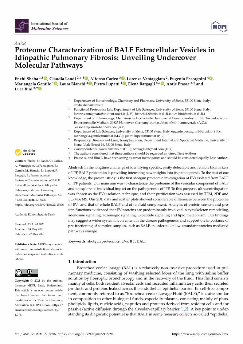
EVs were isolated from BALF of IPF patients (Table 1) by sequential ultracentrifuga-tion, and their purification was assessed by TEM (Figure 1). As illustrated in the TEMimages of EVs from BALF of IPF patients (Figure 1a), this easily reproducible isolationtechnique enabled the separation of a wide size range (40–2000 nm) of vesicles with spheri-cal morphology, sometimes assembled into small aggregates. The isolation method andthe subsequent variety of isolated EVs were further assessed by performing TEM images
Int. J. Mol. Sci. 2021, 22, 5696 3 of 16
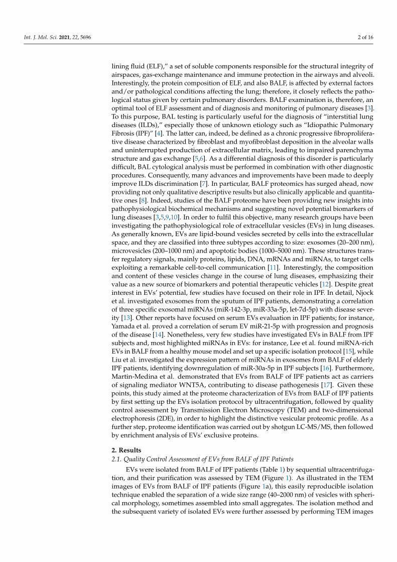
of EVs isolated from BALF of other ILD patients. In order to further confirm the correctisolation of EVs, 2DE of EVs and of the BALF supernatant was performed. The resulting2D gels showed patterns of proteins ranging from MW 200–10 kDa and from pH in iso-electric points 3.5–10. The EVs proteomic profile obtained (Figure 2a) was considerablydifferent from that of the supernatant and deviated sharply from that of the whole BALF(Figure 2b,c, respectively). Conversely, 2DE gels showed a strong similarity between thewhole BALF and the supernatant. To easily correlate and visualize 2DE data variationsbetween the three components, a heatmap was performed (Figure 2d). The whole BALFand the supernatant 2DE data sets cluster together, while EVs’ data set distantly correlatewith the supernatant and whole BALF. In addition, Figure 2d clearly displays that eachgroup shows a different enriched subgroup of spots.
Table 1. Demographic and clinical data of enrolled patients. Table reports the diagnosis, the gender,age, smoking habit and clinical information (FVC, DLCO and GAP) of enrolled patients.
Diagnosis Gender Age Smoking FVC DLCO GAP
IPF Male 74 ex 62 43 2IPF Male 63 ex 39 22 3IPF Male 69 never 50 32 3
Int. J. Mol. Sci. 2021, 22, x FOR PEER REVIEW 3 of 16
EVs were isolated from BALF of IPF patients (Table 1) by sequential ultracentrifuga-tion, and their purification was assessed by TEM (Figure 1). As illustrated in the TEM images of EVs from BALF of IPF patients (Figure 1a), this easily reproducible isolation technique enabled the separation of a wide size range (40–2000 nm) of vesicles with spher-ical morphology, sometimes assembled into small aggregates. The isolation method and the subsequent variety of isolated EVs were further assessed by performing TEM images of EVs isolated from BALF of other ILD patients. In order to further confirm the correct isolation of EVs, 2DE of EVs and of the BALF supernatant was performed. The resulting 2D gels showed patterns of proteins ranging from MW 200–10 kDa and from pH in isoelectric points 3.5–10. The EVs proteomic profile obtained (Figure 2a) was considerably different from that of the supernatant and deviated sharply from that of the whole BALF (Figure 2b,c, respec-tively). Conversely, 2DE gels showed a strong similarity between the whole BALF and the supernatant. To easily correlate and visualize 2DE data variations between the three com-ponents, a heatmap was performed (Figure 2d). The whole BALF and the supernatant 2DE data sets cluster together, while EVs’ data set distantly correlate with the supernatant and whole BALF. In addition, Figure 2d clearly displays that each group shows a different enriched subgroup of spots.
Table 1. Demographic and clinical data of enrolled patients. Table reports the diagnosis, the gen-der, age, smoking habit and clinical information (FVC, DLCO and GAP) of enrolled patients.
Diagnosis Gender Age Smoking FVC DLCO GAP IPF Male 74 ex 62 43 2 IPF Male 63 ex 39 22 3 IPF Male 69 never 50 32 3
Figure 1. TEM of EVs from BALF of IPF patients (a) and of other ILD patients (b). EVs of both IPF and other ILD BALFsamples show a variety of EVs ranging from smaller sizes (40–150 nm) to medium and larger sizes (150–2000 nm).
Int. J. Mol. Sci. 2021, 22, 5696 4 of 16
Int. J. Mol. Sci. 2021, 22, x FOR PEER REVIEW 4 of 16
Figure 1. TEM of EVs from BALF of IPF patients (a) and of other ILD patients (b). EVs of both IPF and other ILD BALF samples show a variety of EVs ranging from smaller sizes (40–150 nm) to medium and larger sizes (150–2000 nm).
Figure 2. Two-dimensional gel electropherograms of BALF EVs, BALF supernatant and whole BALF and corresponding heatmap based on 2DE data sets. (a) 2DE gel of BALF EVs, (b) 2DE gel of BALF supernatant, (c) 2DE gel of whole BALF, (d) heatmap of EVs, whole BALF and supernatant, displaying variations of an abundance of 2DE spots data ranging from low abundant (blue) to high abundant (red) state.
2.2. Shotgun Proteomics of BALF EVs and BALF Fluidic Portion The proteome of the vesicular component of BALF was obtained by the shotgun ap-
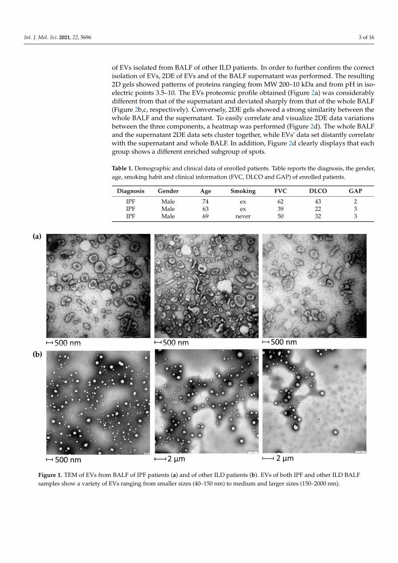
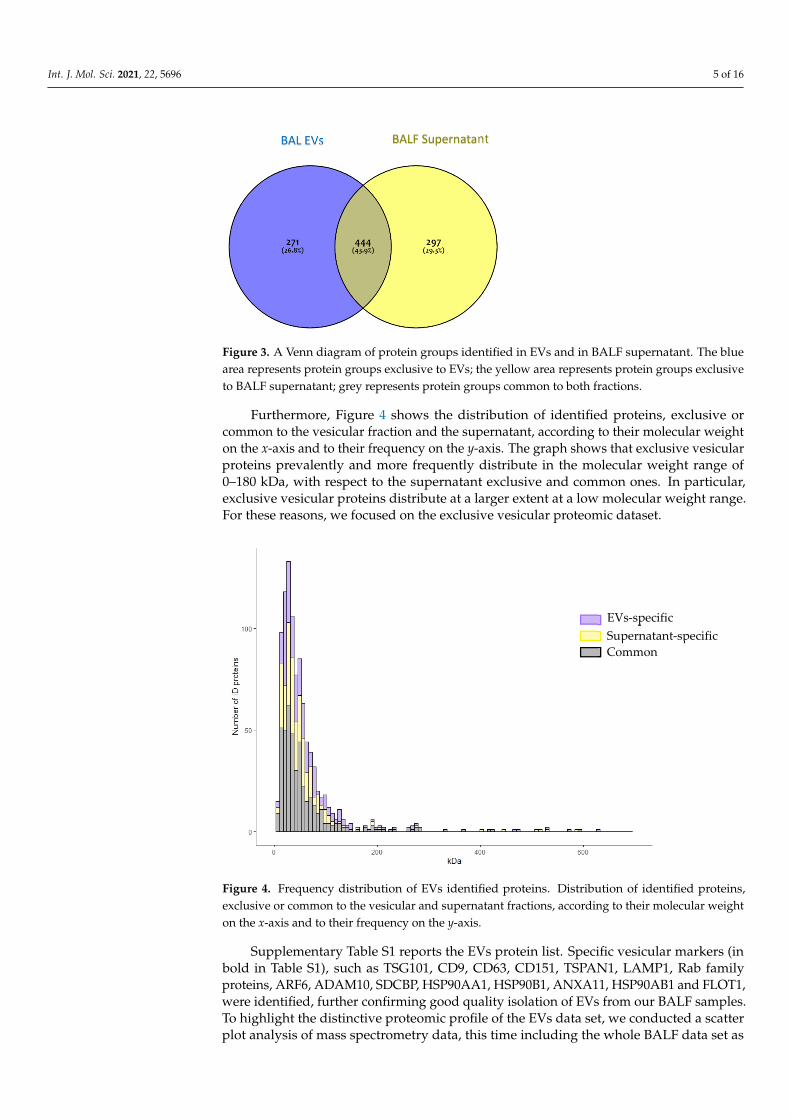
proach. The mass spectrometry proteomics data have been deposited to the Proteo-meXchange Consortium via the PRIDE [18] partner repository with the dataset identifier PXD025590. The proteome of the vesicular component of BALF was obtained by the shot-gun approach. In order to identify those proteins specific to the vesicular fraction, a Venn diagram was performed submitting the proteome lists of EVs and of the complementary supernatant, which accounts for 715 and 741 proteins, respectively (Figure 3). As the Venn diagram shows, 271 proteins (26.8%) were exclusive to EVs, 297 (29.3%) were exclusive to the BALF supernatant, and 444 (43.9%) were common to both.
Figure 2. Two-dimensional gel electropherograms of BALF EVs, BALF supernatant and whole BALF and correspondingheatmap based on 2DE data sets. (a) 2DE gel of BALF EVs, (b) 2DE gel of BALF supernatant, (c) 2DE gel of whole BALF, (d)heatmap of EVs, whole BALF and supernatant, displaying variations of an abundance of 2DE spots data ranging from lowabundant (blue) to high abundant (red) state.
2.2. Shotgun Proteomics of BALF EVs and BALF Fluidic Portion
The proteome of the vesicular component of BALF was obtained by the shotgunapproach. The mass spectrometry proteomics data have been deposited to the ProteomeX-change Consortium via the PRIDE [18] partner repository with the dataset identifierPXD025590. The proteome of the vesicular component of BALF was obtained by the shot-gun approach. In order to identify those proteins specific to the vesicular fraction, a Venndiagram was performed submitting the proteome lists of EVs and of the complementarysupernatant, which accounts for 715 and 741 proteins, respectively (Figure 3). As the Venndiagram shows, 271 proteins (26.8%) were exclusive to EVs, 297 (29.3%) were exclusive tothe BALF supernatant, and 444 (43.9%) were common to both.
Int. J. Mol. Sci. 2021, 22, 5696 5 of 16Int. J. Mol. Sci. 2021, 22, x FOR PEER REVIEW 5 of 16
Figure 3. A Venn diagram of protein groups identified in EVs and in BALF supernatant. The blue area represents protein groups exclusive to EVs; the yellow area represents protein groups exclu-sive to BALF supernatant; grey represents protein groups common to both fractions.
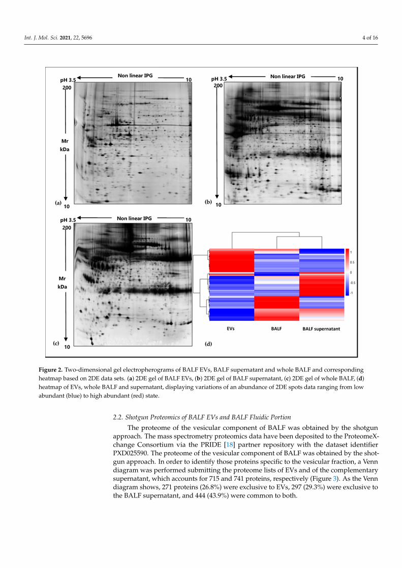
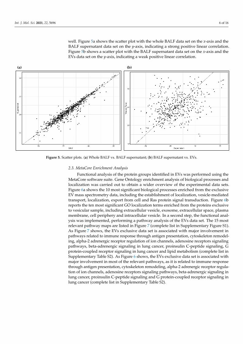
Furthermore, Figure 4 shows the distribution of identified proteins, exclusive or com-mon to the vesicular fraction and the supernatant, according to their molecular weight on the x-axis and to their frequency on the y-axis. The graph shows that exclusive vesicular proteins prevalently and more frequently distribute in the molecular weight range of 0–180 kDa, with respect to the supernatant exclusive and common ones. In particular, exclu-sive vesicular proteins distribute at a larger extent at a low molecular weight range. For these reasons, we focused on the exclusive vesicular proteomic dataset.
Figure 4. Frequency distribution of EVs identified proteins. Distribution of identified proteins, exclusive or common to the vesicular and supernatant fractions, according to their molecular weight on the x-axis and to their frequency on the y-axis.
Supplementary Table S1 reports the EVs protein list. Specific vesicular markers (in bold in Table S1), such as TSG101, CD9, CD63, CD151, TSPAN1, LAMP1, Rab family pro-teins, ARF6, ADAM10, SDCBP, HSP90AA1, HSP90B1, ANXA11, HSP90AB1 and FLOT1, were identified, further confirming good quality isolation of EVs from our BALF samples.
BALF Supernatant BAL EVs
EVs-specific Supernatant-specific Common
Figure 3. A Venn diagram of protein groups identified in EVs and in BALF supernatant. The bluearea represents protein groups exclusive to EVs; the yellow area represents protein groups exclusiveto BALF supernatant; grey represents protein groups common to both fractions.
Furthermore, Figure 4 shows the distribution of identified proteins, exclusive orcommon to the vesicular fraction and the supernatant, according to their molecular weighton the x-axis and to their frequency on the y-axis. The graph shows that exclusive vesicularproteins prevalently and more frequently distribute in the molecular weight range of0–180 kDa, with respect to the supernatant exclusive and common ones. In particular,exclusive vesicular proteins distribute at a larger extent at a low molecular weight range.For these reasons, we focused on the exclusive vesicular proteomic dataset.
Int. J. Mol. Sci. 2021, 22, x FOR PEER REVIEW 5 of 16
Figure 3. A Venn diagram of protein groups identified in EVs and in BALF supernatant. The blue area represents protein groups exclusive to EVs; the yellow area represents protein groups exclu-sive to BALF supernatant; grey represents protein groups common to both fractions.
Furthermore, Figure 4 shows the distribution of identified proteins, exclusive or com-mon to the vesicular fraction and the supernatant, according to their molecular weight on the x-axis and to their frequency on the y-axis. The graph shows that exclusive vesicular proteins prevalently and more frequently distribute in the molecular weight range of 0–180 kDa, with respect to the supernatant exclusive and common ones. In particular, exclu-sive vesicular proteins distribute at a larger extent at a low molecular weight range. For these reasons, we focused on the exclusive vesicular proteomic dataset.
Figure 4. Frequency distribution of EVs identified proteins. Distribution of identified proteins, exclusive or common to the vesicular and supernatant fractions, according to their molecular weight on the x-axis and to their frequency on the y-axis.
Supplementary Table S1 reports the EVs protein list. Specific vesicular markers (in bold in Table S1), such as TSG101, CD9, CD63, CD151, TSPAN1, LAMP1, Rab family pro-teins, ARF6, ADAM10, SDCBP, HSP90AA1, HSP90B1, ANXA11, HSP90AB1 and FLOT1, were identified, further confirming good quality isolation of EVs from our BALF samples.
BALF Supernatant BAL EVs
EVs-specific Supernatant-specific Common
Figure 4. Frequency distribution of EVs identified proteins. Distribution of identified proteins,exclusive or common to the vesicular and supernatant fractions, according to their molecular weighton the x-axis and to their frequency on the y-axis.
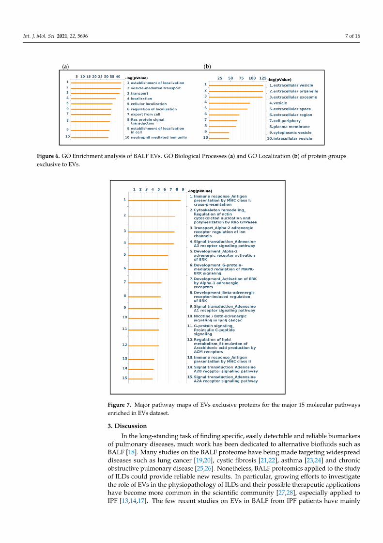
Supplementary Table S1 reports the EVs protein list. Specific vesicular markers (inbold in Table S1), such as TSG101, CD9, CD63, CD151, TSPAN1, LAMP1, Rab familyproteins, ARF6, ADAM10, SDCBP, HSP90AA1, HSP90B1, ANXA11, HSP90AB1 and FLOT1,were identified, further confirming good quality isolation of EVs from our BALF samples.To highlight the distinctive proteomic profile of the EVs data set, we conducted a scatterplot analysis of mass spectrometry data, this time including the whole BALF data set as
Int. J. Mol. Sci. 2021, 22, 5696 6 of 16
well. Figure 5a shows the scatter plot with the whole BALF data set on the x-axis and theBALF supernatant data set on the y-axis, indicating a strong positive linear correlation.Figure 5b shows a scatter plot with the BALF supernatant data set on the x-axis and theEVs data set on the y-axis, indicating a weak positive linear correlation.
Figure 5. Scatter plots
(a)Whole BALF vs BALF supernatant; (b) BALF supernatant vs EVs
(a) (b)
Figure 5. Scatter plots. (a) Whole BALF vs. BALF supernatant; (b) BALF supernatant vs. EVs.
2.3. MetaCore Enrichment Analysis
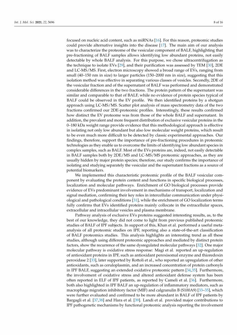
Functional analysis of the protein groups identified in EVs was performed using theMetaCore software suite. Gene Ontology enrichment analysis of biological processes andlocalization was carried out to obtain a wider overview of the experimental data sets.Figure 6a shows the 10 most significant biological processes enriched from the exclusiveEV mass spectrometry data, including the establishment of localization, vesicle-mediatedtransport, localization, export from cell and Ras protein signal transduction. Figure 6breports the ten most significant GO localization terms enriched from the proteins exclusiveto vesicular sample, including extracellular vesicle, exosome, extracellular space, plasmamembrane, cell periphery and intracellular vesicle. In a second step, the functional anal-ysis was implemented, performing a pathway analysis of the EVs data set. The 15 mostrelevant pathway maps are listed in Figure 7 (complete list in Supplementary Figure S1).As Figure 7 shows, the EVs exclusive data set is associated with major involvement inpathways related to immune response through antigen presentation, cytoskeleton remodel-ing, alpha-2 adrenergic receptor regulation of ion channels, adenosine receptors signalingpathways, beta-adrenergic signaling in lung cancer, proinsulin C-peptide signaling, Gprotein-coupled receptor signaling in lung cancer and lipid metabolism (complete list inSupplementary Table S2). As Figure 6 shows, the EVs exclusive data set is associated withmajor involvement in most of the relevant pathways, as it is related to immune responsethrough antigen presentation, cytoskeleton remodeling, alpha-2 adrenergic receptor regula-tion of ion channels, adenosine receptors signaling pathways, beta-adrenergic signaling inlung cancer, proinsulin C-peptide signaling and G protein-coupled receptor signaling inlung cancer (complete list in Supplementary Table S2).
Int. J. Mol. Sci. 2021, 22, 5696 7 of 16Int. J. Mol. Sci. 2021, 22, x FOR PEER REVIEW 7 of 16
Figure 6. GO Enrichment analysis of BALF EVs. GO Biological Processes (a) and GO Localization (b) of protein groups exclusive to EVs.
Figure 7. Major pathway maps of EVs exclusive proteins for the major 15 molecular pathways enriched in EVs dataset.
3. Discussion In the long-standing task of finding specific, easily detectable and reliable biomarkers
of pulmonary diseases, much work has been dedicated to alternative biofluids such as BALF [18]. Many studies on the BALF proteome have being made targeting widespread diseases such as lung cancer [19,20], cystic fibrosis [21,22], asthma [23,24] and chronic ob-structive pulmonary disease [25,26]. Nonetheless, BALF proteomics applied to the study of ILDs could provide reliable new results. In particular, growing efforts to investigate the role of EVs in the physiopathology of ILDs and their possible therapeutic applications have become more common in the scientific community [27,28], especially applied to IPF [13,14,17]. The few recent studies on EVs in BALF from IPF patients have mainly focused on nucleic acid content, such as miRNAs [16]. For this reason, proteomic studies could
(a) (b)
Figure 6. GO Enrichment analysis of BALF EVs. GO Biological Processes (a) and GO Localization (b) of protein groupsexclusive to EVs.
Int. J. Mol. Sci. 2021, 22, x FOR PEER REVIEW 7 of 16
Figure 6. GO Enrichment analysis of BALF EVs. GO Biological Processes (a) and GO Localization (b) of protein groups exclusive to EVs.
Figure 7. Major pathway maps of EVs exclusive proteins for the major 15 molecular pathways enriched in EVs dataset.
3. Discussion In the long-standing task of finding specific, easily detectable and reliable biomarkers
of pulmonary diseases, much work has been dedicated to alternative biofluids such as BALF [18]. Many studies on the BALF proteome have being made targeting widespread diseases such as lung cancer [19,20], cystic fibrosis [21,22], asthma [23,24] and chronic ob-structive pulmonary disease [25,26]. Nonetheless, BALF proteomics applied to the study of ILDs could provide reliable new results. In particular, growing efforts to investigate the role of EVs in the physiopathology of ILDs and their possible therapeutic applications have become more common in the scientific community [27,28], especially applied to IPF [13,14,17]. The few recent studies on EVs in BALF from IPF patients have mainly focused on nucleic acid content, such as miRNAs [16]. For this reason, proteomic studies could
(a) (b)
Figure 7. Major pathway maps of EVs exclusive proteins for the major 15 molecular pathwaysenriched in EVs dataset.
3. Discussion
In the long-standing task of finding specific, easily detectable and reliable biomarkersof pulmonary diseases, much work has been dedicated to alternative biofluids such asBALF [18]. Many studies on the BALF proteome have being made targeting widespreaddiseases such as lung cancer [19,20], cystic fibrosis [21,22], asthma [23,24] and chronicobstructive pulmonary disease [25,26]. Nonetheless, BALF proteomics applied to the studyof ILDs could provide reliable new results. In particular, growing efforts to investigatethe role of EVs in the physiopathology of ILDs and their possible therapeutic applicationshave become more common in the scientific community [27,28], especially applied toIPF [13,14,17]. The few recent studies on EVs in BALF from IPF patients have mainly
Int. J. Mol. Sci. 2021, 22, 5696 8 of 16
focused on nucleic acid content, such as miRNAs [16]. For this reason, proteomic studiescould provide alternative insights into the disease [17]. The main aim of our analysiswas to characterize the proteome of the vesicular component of BALF, highlighting thatpre-fractioning of BALF samples allows identifying low abundant proteins, not easilydetectable by whole BALF analysis. For this purpose, we chose ultracentrifugation asthe technique to isolate EVs [29], and their purification was assessed by TEM [30], 2DEand LC-MS/MS. First, electron microscopy showed a broad range of EVs, ranging fromsmall (40–150 nm in size) to larger particles (150–2000 nm in size), suggesting that thisisolation method was effective in separating various classes of vesicles. Secondly, 2DE ofthe vesicular fraction and of the supernatant of BALF was performed and demonstratedconsiderable differences in the two fractions. The protein pattern of the supernatant wassimilar and comparable to that of BALF, while no evidence of protein species typical ofBALF could be observed in the EV profile. We then identified proteins by a shotgunapproach using LC-MS/MS. Scatter plot analysis of mass spectrometry data of the twofractions confirmed our 2DE-proteomic profiles. Interestingly, these results confirmedhow distinct the EV proteome was from those of the whole BALF and supernatant. Inaddition, the prevalent and more frequent distribution of exclusive vesicular proteins in the0–180 kDa weight range provide evidence that this methodological approach is effectivein isolating not only low abundant but also low molecular weight proteins, which resultto be even much more difficult to be detected by classic experimental approaches. Ourfindings, therefore, support the importance of pre-fractioning procedures in proteomictechnologies as they enable us to overcome the limits of identifying low abundant species incomplex samples, such as BALF. Most of the EVs proteins are, indeed, not easily detectablein BALF samples both by 2DE/MS and LC-MS/MS proteomic approaches, as they areusually hidden by major protein species; therefore, our study confirms the importance ofisolating and studying separately the vesicular and the supernatant fractions as a source ofpotential biomarkers.
We implemented this characteristic proteomic profile of the BALF vesicular com-ponent by evaluating the protein content and functions in specific biological processes,localization and molecular pathways. Enrichment of GO biological processes provideevidence of EVs predominant involvement in mechanisms of transport, localization andsignal mediation, confirming their key roles in intercellular communication under physi-ological and pathological conditions [31], while the enrichment of GO localization termsfully confirms that EVs identified proteins mainly collocate in the extracellular spaces,extracellular and intracellular vesicles and plasma membrane.
Pathway analysis of exclusive EVs proteins suggested interesting results, as, to thebest of our knowledge, they did not come to light from previous published proteomicstudies of BALF of IPF subjects. In support of this, Khan et al. performed a useful meta-analysis of all proteomic studies on IPF, reporting also a state-of-the-art classificationof BALF proteomics studies. This analysis highlights an interesting trend as all thesestudies, although using different proteomic approaches and mediated by distinct proteinfactors, show the recurrence of the same dysregulated molecular pathways [32]. One majormolecular pathway is oxidative stress response: Magi et al. reported an up-regulationof antioxidant proteins in IPF, such as antioxidant peroxisomal enzyme and thioredoxinperoxidase 2 [33], later supported by Rottoli et al., who reported an upregulation of otherantioxidants, such as ceruloplasmin, and an increased concentration of protein carbonylsin IPF BALF, suggesting an extended oxidative proteomic pattern [34,35]. Furthermore,the involvement of oxidative stress and altered antioxidant defense system has beenoften reported in ELF of IPF patients, as reported by Cameli et al. [36]. Furthermore,both also highlighted in IPF BALF an up-regulation of inflammatory mediators, such asmacrophage migration inhibitory factor (MIF) and calgranulin B (S100A9) [33–35], whichwere further evaluated and confirmed to be more abundant in BALF of IPF patients byBargagli et al. [37,38] and Hara et al. [39]. Landi et al. provided major contributions toIPF pathogenetic mechanisms by functional proteomic analysis reporting the involvement
Int. J. Mol. Sci. 2021, 22, 5696 9 of 16
of several molecular pathways. Some of those are protein folding, Slit-Robo signaling,hypoxia response, blood coagulation system and complement-mediated immune responseand angiotensin system [5,9]. Furthermore, Carleo et al. confirmed and implementedprevious results suggesting the involvement of the Wnt-β-catenin transduction signaling,lung carcinogenesis pathway and a protease/antiprotease imbalance in IPF BALF patientswith acute exacerbations, in addition to the involvement of ER stress, ion homeostasis andwound healing processes [6,40]. The most suggested molecular pathway is representedby pro-fibrotic mechanisms mediated by several factors, such as osteopontin, matrix-metalloproteinases, CCL24, CXCL7 and CCL18, as reported by Foster et al. by shotgunproteomic analysis of IPF BALF [41].
Interestingly, our analysis highlighted various molecular pathways, probably medi-ated by low abundant proteins, which are hardly uncovered as hidden by the abundanceof major protein species.
Remarkably, pathways analysis of EVs proteins showed that they are prevalentlyinvolved in antigen presentation by MHC class I and II, cytoskeleton remodeling, adenosinesignaling, adrenergic signaling, G protein signaling, specific G protein C-peptide signalingand lipid metabolism. C-peptide (proinsulin), prevalently studied in diabetes, exerts itsbiological activities via a specific G-protein coupled receptor also expressed on endothelialcells and fibroblasts [42]. Its signaling involves ERK1/2, PI3K-Akt, PKC, eNOS and NF-kB, well-known factors in TGF-β signaling [43,44], the key regulator of fibrosis, therebysuggesting C-peptide involvement in fibrogenic processes [45]. Indeed, recent studies showan association of C-peptide with fibrosis progression in different pathologies [46,47]. Somestudies have reported the association of C-peptide with the transcription factor peroxisomeproliferator-activated receptor-γ (PPARγ) [48], whose modulation balances adipogenesisand fibrogenesis [49], in line with the concept of metabolic dysregulation as an additionalimpacting cause of fibrosis, especially in IPF [45,49].
Given also the identification in EVs of monoacylglycerol lipase (MAGL), a pro-inflammatory enzyme catalyzing the arachidonic acid production, pathway analysis sug-gests the involvement of lipid metabolism mediated by the stimulation of the productionof arachidonic acid. Costola-de-Souza et al. report interesting results according to whichMAGL inhibition displays anti-inflammatory and protective effects during acute lunginjury in mice [50], while a more recent study of Habib et al. demonstrated that MAGLinhibitors have a powerful impact on liver fibrosis as it delays fibrosis progression andpromotes its regression [51].
Our identification of several Rho GTPases in EVs suggests their potential action on cy-toskeleton remodeling by mediating actin filament rearrangement via ROCK [52]. Indeed,interesting studies report that ROCK signaling pathways are involved in myofibroblast dif-ferentiation and fibrogenic processes, especially pulmonary fibrosis such as IPF [40,53,54].Curiously, given the growing attention to Wnt signaling in the regulation of cellular adhe-sions and its involvement in IPF pathogenic mechanisms, Franco et al. reported that RhoGTPases’ regulation modulates cell migration and polarity via a β-catenin-independentWnt pathway [55]. Another remarkable cytoskeleton-related protein detected in EVs isprofilin, which triggers fibrogenic pathways such as PI3K-Akt and ERK 1/2 [56].
Our results indicate the involvement of another interesting pathway related to fibroge-nesis, which has recently attracted attention: the adenosine signaling pathway. Adenosineexerts its functions by binding to G-protein coupled receptors A2A and A2B, leading tofibroblast activation and collagen synthesis [57]. Indeed, several studies already report acorrelation between A2B adenosine receptor (A2BAR) activation and regulation of inflam-mation and fibrosis in IPF, specifically indicating macrophages as major mediators [58,59].Some signal transduction factors of this signaling, such as PKA, are detected in EVs. Cu-riously, a recent study demonstrated the key role of A2BAR in the modulation of theEMT process in IPF by two signaling pathways, cAMP/PKA and MAP/ERK [60]. Ourenrichment analysis suggests a direct link between PKA and CREB1 activation, inducingVEGF-A transcription, a major player in IPF onset [61].
Int. J. Mol. Sci. 2021, 22, 5696 10 of 16
We also found another molecular pathway whose relation to IPF pathogenesis isnot often considered: the alpha- and beta-adrenergic systems. Rassler B. demonstratedthat continuous stimulation of beta- and especially alpha-adrenergic signaling in ratsleads to pulmonary fibrosis. Adrenergic-stimulated histological lung fibrosis is associatedwith a remarkable increase in TGFβ1, collagen I, MMP-2 and TIMP-2 mRNA expression,suggesting a link between adrenergic stimulation, the up-regulation of ECM moleculesand the promotion of fibrotic processes [62].
Although these altered molecular pathways were detected by proteins directly identi-fied in BAL samples, our results provide evidence of a potential wider systemic involve-ment. In other words, altered vesicular protein-mediated processes may be regulatedin situ by cellular protein release and by systemic circulating molecules, which may bereleased in the lung environment by plasma exudation into epithelial lining fluid. Likewise,vesicular proteins may be of systemic origin as well as pulmonary origin. Altered sys-temic metabolic pathways could therefore use EVs as a communication system to induce aspecific pro-fibrotic response in the lung environment, leading to IPF.
4. Materials and Methods4.1. Population
Male IPF patients, mean age 69 ± 5 years, 2 ex-smokers and 1 never smoker, wereenrolled in the study. The patients were diagnosed according to ATS/ERS guidelines at theMedizinische Hoschschule Pneumology Clinic (Hannover, Germany). Demographic dataand smoking habits of the three patients used for this sample characterization were reportedin Table 1 together with other clinical data such as FVC, DLCO and GAP percentagesaccording to ATS/ERS guidelines. The diagnosis of IPF was formulated in the context of themultidisciplinary discussion. After informed consent of the patients, BAL was performedfor diagnostic purposes in order to exclude other ILDs. Samples were provided by theresearch group directed by Prof. Antje Prasse at Fraunhofer ITEM (Hannover, Germany).
4.2. EVs Isolation from BALF
Human IPF BALF samples were centrifuged at 800× g for 5 min at 4 ◦C, as a routineprocedure to separate BALF and cell components [63,64]. In particular, BALF samplesfrom IPF patients were prepared for the analysis, specifically using 15 mL per each asstarting volume. First, BALF samples were centrifuged at 12,000× g for 45 min at 4 ◦C. Thepellet was discarded, and the supernatant was collected in ultracentrifuge tubes. Then,supernatants were centrifuged at 110,000× g for 2 h at 4 ◦C (Beckman Coulter Optima XE,Type 70 Ti Fixed-Angle Titanium Rotor, Beckman Coulter Life Sciences, Brea, CA, USA). Atthis step, the supernatant was collected in a new tube and stored on ice, as this fractionwas the complementary portion of BALF whole fluid without EVs (BALF supernatant).Conversely, the pellet was resuspended in PBS and filtered into a new ultracentrifuge tubethrough a 0.22 µm filter and centrifuged at 110,000× g for 70 min at 4 ◦C. Following this,the supernatant was discarded, and the pellet was resuspended in PBS and centrifugedagain at 110,000× g for 70 min at 4 ◦C; at this point, the pellet containing BALF EVs wastransferred into a new Eppendorf [29]. The concentration of BALF EVs was detected byNanoDrop (NanoDrop ND-1000 spectrophotometer).
4.3. TEM
As the first checkpoint of isolation of BALF EVs, TEM was performed by Dr. EugenioPaccagnini and Dr. Mariangela Gentile, members of the research group directed by Prof.Pietro Lupetti of the Life Sciences Department at the University of Siena. In detail, about3 µL of EV fraction was loaded on a 300 mesh formvar coated copper grid for 2 min. Afterblotting the excess, the grid was negatively stained with 2% aqueous ammonium molybdatefor 30 s and analyzed using a Thermo Fisher Scientific Tecnai G2 Spirit transmission electronmicroscope operating at 120 kV equipped with an EMSIS Veleta 2048 × 2048 CCD camera.
Int. J. Mol. Sci. 2021, 22, 5696 11 of 16
4.4. Samples Preparation for 2DE Analysis
Dialysis of the BALF EV and supernatant was performed against four changes ofdistilled water at 4 ◦C for 12 h to eliminate salts. Samples were lyophilized and dis-solved in lysis buffer (8 M urea, 4% w/v 3-[(3-cholamidopropyl) dimethylammonia]-1-propanesulfonate hydrate (CHAPS), 40 mM Tris base, 1% w/v dithioerythritol (DTE) andtrace amounts of bromophenol blue).
Before adding bromophenol blue, the protein concentration of the BALF supernatantwas determined by Bradford assay [65] in order to load 60 µg of protein per gel, while EVs’total protein content was used.
4.5. 2D-Electrophoresis
2DE was carried out using the Immobiline polyacrylamide system on a preformedimmobilized nonlinear pH gradient from pH 3 to 10, 18 cm in length (Cytiva, formerlyGE Healthcare, Uppsala, Sweden). The 2D run was performed using Ettan™ IPGphor™system (Cytiva, formerly GE Healthcare, Uppsala, Sweden) at 16 ◦C, applying the followingelectrical conditions: 200 V for 8 h, from 200 to 3500 V in 2 h, 3500 V for 2 h, from 3500 to5000 V in 2 h, 5000 V for 3 h, from 5000 to 8000 V in 1 h, 8000 V for 3 h, from 8000 to 10,000 Vin 1 h, 10,000 V, for a total of 90,000 VhT (total Volts per hour). Gel strips were rehydratedwith lysis buffer and traces of bromophenol blue overnight at room temperature; then,0.2% carrier ampholyte was added to samples, and the run was performed by cup loading,with the cup placed at the cathodic end of the strips. After the first dimensional run, stripswere equilibrated in 6 M urea, 2% w/v SDS, 2% w/v DTE, 30% v/v glycerol and 0.5 MTris–HCl pH 6.8 for 12 min and for a further 5 min in 6 M urea, 2% w/v SDS, 2.5% w/viodoacetamide, 30% v/v glycerol, 0.5 M Tris–HCl pH 6.8 and a trace of bromophenol blue.Then, the second dimension was performed on 9–16% SDS polyacrylamide linear gradientgels (18 × 20 cm × 1.5 mm) at 40 mA/gel constant current and 9 ◦C until the dye reachedthe bottom of the gel. Gels were finally stained with ammoniacal silver nitrate. Gels werethen digitalized using the Image Scanner III laser densitometer supplied with the LabScan6.0 software (GE Healthcare), image analysis was performed using Melanie Classic 9.0software and normalization of the 2DE data was performed. The heatmap was performedon normalized spot volumes (%V) using RStudio Desktop 1.1.463 (Integrated Developmentfor RStudio, Inc., Boston, MA, USA, https://www.rstudio.com (accessed on 1 April 2021).
4.6. MS-Preparative SDS-PAGE
For shotgun proteomic analysis, BALF whole fluid of the patients was also prepared,and BALF whole fluid and BALF supernatant underwent cold acetone precipitation (1:4)overnight at −20 ◦C. They were then centrifuged at 4542× g for 10 min at 4 ◦C. The pelletwas resuspended in acetone and centrifuged again at 15,000× g for 10 min at 4 ◦C. At thisstep, the three components of each sample (whole BALF, BALF supernatant and EVs) weresolubilized in a denaturating solution composed of 8 M urea and 4% w/v CHAPS, andtheir protein concentration was determined by Bradford assay [65].
MS-preparative SDS-PAGE was carried out using pre-cast 12% polyacrylamide gels(Criterion™ XT Bis-Tris Protein Gel, Bio-Rad, Hercules, CA, USA) in a Criterion™ VerticalElectrophoresis Cell (Bio-Rad, Hercules, CA, USA) with the following voltage conditions:60 V for stacking gel and 120 V for separating gel. The amount of protein loaded was50 µg for BALF and BALF supernatant and the total protein content for EVs. Sampleswere centrifuged, and the XT sample buffer and XT reducing agent (Bio-Rad, Hercules,CA, USA) were added to samples, which were then held at 95 ◦C for 5 min. After that, theproteins were alkylated by adding 40% acrylamide at a final concentration of 2%, and thesamples were loaded in the gel. After the run, the gel was incubated in 50% v/v methanoland 10% v/v glacial acetic acid fixing solution for 1 h under gentle agitation. It was thenstained in Coomassie Blue solution composed of 0.1% w/v Coomassie Brilliant Blue R-250,50% v/v methanol and 10% v/v glacial acetic acid for 20 min under gentle agitation.
Protein bands were cut out and minced into 3 1 mm pieces. The pieces were destainedtwice in 50% v/v acetonitrile (ACN)/20 mM Ammonium Bicarbonate (ABC) at 37 ◦C undershaking (Thermomixer, Eppendorf AG, Hamburg, Germany) at 700 rpm for 30 min. The gelpieces were then dehydrated in 100% ACN at room temperature for 10 min, and the solventwas removed in a vacuum centrifuge (Speedvac, Thermo Fischer ScientificTM, Waltham,MA, USA) for 30 min. Then, a solution of 10 ng/µL trypsin in 10% v/v ACN/20 mMABC solution for protein digestion was added to the gel pieces, which were rehydratedon ice for 60 min; then, covered with 10% v/v ACN/20 mM ABC solution and digestedovernight at 37 ◦C under shaking at 350 rpm. Digestion was then stopped by adding 50%v/v ACN/5% v/v trifluoroacetic acid (TFA) solution. The gel pieces were incubated at24 ◦C under shaking at 700 rpm for 30 min. The supernatant containing peptide extractswas collected into a new vial and dried in a vacuum centrifuge for 30 min. A 50% v/vACN/0.5% v/v TFA solution was added, and the mixture was incubated under the sameprevious conditions. The supernatant was pooled with the previous one and dried againin the vacuum centrifuge for 30 min. Then, 100% ACN was added to gel pieces, and theywere incubated at 24 ◦C under shaking at 700 rpm for 20 min. Finally, the supernatant wascollected and dried in a vacuum centrifuge for 3 h.
4.8. LC-MS/MS Analysis and Protein Identification
Peptides of mono-dimensional gel digestion were analyzed by LC-MS/MS. Driedsamples were dissolved in 2% v/v ACN/0.1% v/v TFA solution and incubated at 24 ◦Cunder shaking at 350 rpm for 30 min, then centrifuged at 20,000× g for 30 min at roomtemperature and the supernatants were transferred to an LC sample vial. An appropriateamount of each sample was injected into a Dionex Ultimate 3000 high-performance LCsystem (Thermo Fisher Scientific, Waltham, MA, USA). Peptides were loaded on a C18trap column (2 cm long, 75 µm i.d., Acclaim PepMap, Dionex) at 6 µL/min and washedwith 0.1% v/v TFA loading buffer. After 5 min, the trap column was switched in line withthe C18 nanoflow separation column (50 cm long, 75 µm i.d., Acclaim PepMap, Dionex)and the peptides were eluted with a linear gradient of elution buffers A (0.1% v/v formicacid) and B (80% v/v acetonitrile, 0.1% v/v formic acid) at 250 nl/min. The LC system wasconnected to the nanoESI source of an LTQ Orbitrap Lumos Mass Spectrometer (ThermoFisher Scientific, USA). The Orbitrap mass analyzer recorded the survey scans selecting themost intense precursor ions of charge state ≥ 2 for collision-induced fragmentation with anormalized collision energy of 38%. Fragments were scanned out in the Orbitrap mass ana-lyzer in centroid mode, and the raw data were processed with MaxQuant software (Version1.6.50, https://maxquant.net/maxquant/ (accessed on 20 June 2020). For peptide identifi-cation, MS/MS spectra were searched against human entries in the UniProtKB/Swiss-Protdatabase and were considered to be identified for false discovery rates (FDR) on proteinand peptide level ≤ 0.01. Oxidation of methionine residues, N-terminal acetylation, deami-dation of asparagine and glutamine residues and propionamidation of cysteine residueswere selected as variable modifications. A maximum of two missed cleavages was accepted.A minimum ratio count of one unique or razor peptide was required for quantification. Theprotein groups identified were processed for statistical purposes using Perseus software(Version 1.5.2.6, https://maxquant.net/perseus/ (accessed on 20 June 2020). Shotgunexperiments were performed in collaboration with Dr. Alfonso Carleo, a member of theProf. Antje Prasse research group at Fraunhofer ITEM (Hannover, Germany).
4.9. Enrichment Analysis
Enrichment analysis was performed by submitting the gene names of identified pro-teins to the MetaCore 6.8 network building tool (http://portal.genego.com (accessed on 1April 2021), Clarivate Analytics, Philadelphia, PA, USA). Specifically, we first performed en-richment analysis by GeneGo ontology biological processes, localization and then pathwaymaps analysis. The software establishes a hierarchical list of pathway maps, prioritized
according to their statistical significance (p ≤ 0.001), and each is equivalent to a canonicalmap that has multiple sequential steps of interactions, defining a well-established signalingmechanism. Each step is also well-defined, experimentally validated and accepted in theresearch field.
5. Conclusions
In conclusion, EVs’ isolation and separation from its complementary fraction turnedout to be extremely useful as they shed light on unexpected and infrequently IPF-associatedmolecular pathways, which might be hardly identifiable by classic experimental ap-proaches. Interestingly, the protein content of BALF EVs regulates distinct molecularpathways, suggesting a particular new disease scenario in which systemic and metabolicdysregulation may be a cause and/or a consequence of IPF development. Vesicular pro-teins may indeed potentially cooperate with proteins in the complementary fractions to thepathogenesis and maintenance of the disease through pro-fibrotic and pro-inflammatorysignals. These findings support the much-needed improvement in proteomic analysis infractioning complex samples, allowing low abundant proteins to emerge and to unveilparticular signaling pathways. This might represent a valuable starting point for furtherstudy in a larger cohort of patients where IPF samples could be compared with other ILDsand control samples in order to provide new insights into the pathophysiology of thedisease and biomarkers discovery.
Supplementary Materials: The following are available online at https://www.mdpi.com/article/10.3390/ijms22115696/s1, Figure S1: Pathway maps enrichment of EVs proteins from IPF BALF.Complete list of the 50 most statistically significant pathway maps of vesicular proteins from IPFBALF; Table S1: MS complete protein lists of vesicular proteins, reported by gene name and proteinname, identified by LC-MS/MS. Vesicular markers are highlighted in bold; Table S2: Pathway mapsof proteins exclusive to EVs. Excel report of the 50 most significantly enriched pathway maps ofproteins exclusive to EVs: maps, p-value, FDR, number of proteins associated, network objects.
Author Contributions: Conceptualization, C.L., A.P. and L.B. (Luca Bini); Formal analysis, E.S., A.C.and L.V.; Methodology, E.S., A.C., E.P., M.G. and L.B. (Laura Bianchi); Writing—original draft, E.S.and C.L.; Writing—review & editing, P.L., E.B., A.P. and L.B. (Luca Bini). All authors have read andagreed to the published version of the manuscript.
Funding: This research project is funded by Tuscany Region for HIDE IPF project.
Institutional Review Board Statement: The study was conducted according to the guidelines of theDeclaration of Helsinki, and approved by the Institutional Review Board (or Ethics Committee) ofUniversity of Siena (protocol code 17431, 15/06/2020).
Informed Consent Statement: Informed consent was obtained from all subjects involved in thestudy.
Data Availability Statement: Requests for further information about resources, reagents and dataavailability should be directed to the corresponding author.
Acknowledgments: The authors thank Helen Ampt for revising the English.
Conflicts of Interest: The authors declare no conflict of interest.
References1. Reynolds, H.Y. Use of Bronchoalveolar Lavage in Humans—Past Necessity and Future Imperative. Lung 2000, 178, 271–293.
[CrossRef]2. Govender, P.; Dunn, M.J.; Donnelly, S.C. Proteomics and the lung: Analysis of bronchoalveolar lavage fluid. Proteom. Clin. Appl.
2009, 3, 1044–1051. [CrossRef]3. Nguyen, E.V.; Gharib, S.A.; Schnapp, L.M.; Goodlett, D.R. Shotgun MS proteomic analysis of bronchoalveolar lavage fluid in
normal subjects. Proteom. Clin. Appl. 2014, 8, 737–747. [CrossRef]
4. Travis, W.D.; Costabel, U.; Hansell, D.M.; King, T.E.; Lynch, D.A.; Nicholson, A.G.; Ryerson, C.J.; Ryu, J.H.; Selman, M.; Wells,A.U.; et al. An Official American Thoracic Society/European Respiratory Society Statement: Update of the InternationalMultidisciplinary Classification of the Idiopathic Interstitial Pneumonias. Am. J. Respir. Crit. Care Med. 2013, 188, 733–748.[CrossRef]
5. Landi, C.; Bargagli, E.; Carleo, A.; Bianchi, L.; Gagliardi, A.; Prasse, A.; Perari, M.G.; Refini, R.M.; Bini, L.; Rottoli, P. A systembiology study of BALF from patients affected by idiopathic pulmonary fibrosis (IPF) and healthy controls. Proteom. Clin. Appl.2014, 8, 932–950. [CrossRef] [PubMed]
7. Efared, B.; Ebang-Atsame, G.; Rabiou, S.; Diarra, A.S.; Tahiri, L.; Hammas, N.; Smahi, M.; Amara, B.; Benjelloun, M.C.; Serraj,M.; et al. The diagnostic value of the bronchoalveolar lavage in interstitial lung diseases. J. Negat. Results Biomed. 2017, 16, 4.[CrossRef] [PubMed]
8. Rottoli, P.; Bargagli, E.; Landi, C.; Magi, B. Proteomic analysis in interstitial lung diseases: A review. Curr. Opin. Pulm. Med. 2009,15, 470–478. [CrossRef]
9. Landi, C.; Bargagli, E.; Bianchi, L.; Gagliardi, A.; Carleo, A.; Bennett, D.; Perari, M.G.; Armini, A.; Prasse, A.; Rottoli, P.; et al.Towards a functional proteomics approach to the comprehension of idiopathic pulmonary fibrosis, sarcoidosis, systemic sclerosisand pulmonary Langerhans cell histiocytosis. J. Proteom. 2013, 83, 60–75. [CrossRef]
10. D’Alessandro, M.; Bergantini, L.; Refini, R.M.; Cameli, P.; Perillo, F.; Landi, C.; Icorne, F.; Perrone, A.; Sestini, P.; Bonella, F.; et al.Adiponectin and leptin levels in idiopathic pulmonary fibrosis: A new method for BAL and serum assessment. Immunobiology2020, 225, 151997. [CrossRef]
11. Doyle, L.M.; Wang, M.Z. Overview of Extracellular Vesicles, Their Origin, Composition, Purpose, and Methods for ExosomeIsolation and Analysis. Cells 2019, 8, 727. [CrossRef]
12. Rollet-Cohen, V.; Bourderioux, M.; Lipecka, J.; Chhuon, C.; Jung, V.A.; Mesbahi, M.; Nguyen-Khoa, T.; Guérin-Pfyffer, S.; Schmitt,A.; Edelman, A.; et al. Comparative proteomics of respiratory exosomes in cystic fibrosis, primary ciliary dyskinesia and asthma.J. Proteom. 2018, 185, 1–7. [CrossRef]
14. Yamada, M. The Roles of MicroRNAs and Extracellular Vesicles in the Pathogeneses of Idiopathic Pulmonary Fibrosis and AcuteRespiratory Distress Syndrome. Tohoku J. Exp. Med. 2020, 251, 313–326. [CrossRef]
15. Lee, H.; Groot, M.; Pinilla-Vera, M.; Fredenburgh, L.E.; Jin, Y. Identification of miRNA-rich vesicles in bronchoalveolar lavagefluid: Insights into the function and heterogeneity of extracellular vesicles. J. Control. Release 2019, 294, 43–52. [CrossRef]
16. Liu, B.; Jiang, T.; Hu, X.; Liu, Z.; Zhao, L.; Liu, H.; Liu, Z.; Ma, L. Downregulation of microRNA-30a in bronchoalveolar lavagefluid from idiopathic pulmonary fibrosis patients. Mol. Med. Rep. 2018, 18, 5799–5806. [CrossRef] [PubMed]
17. Martin-Medina, A.; Lehmann, M.; Burgy, O.; Hermann, S.; Baarsma, H.A.; Wagner, D.E.; De Santis, M.M.; Ciolek, F.; Hofer, T.P.;Frankenberger, M.; et al. Increased Extracellular Vesicles Mediate WNT5A Signaling in Idiopathic Pulmonary Fibrosis. Am. J.Respir. Crit. Care Med. 2018, 198, 1527–1538. [CrossRef] [PubMed]
18. Wheelock, C.E.; Goss, V.M.; Balgoma, D.; Nicholas, B.; Brandsma, J.; Skipp, P.J.; Snowden, S.; Burg, D.; D’Amico, A.; Horvath, I.;et al. Application of ’omics technologies to biomarker discovery in inflammatory lung diseases. Eur. Respir. J. 2013, 42, 802–825.[CrossRef] [PubMed]
19. Sim, S.Y.; Choi, Y.R.; Lee, J.H.; Lim, J.M.; Lee, S.H.; Kim, K.P.; Kim, J.Y.; Kim, M. In-Depth Proteomic Analysis of HumanBronchoalveolar Lavage Fluid toward the Biomarker Discovery for Lung Cancers. Proteom. Clin. Appl. 2019, 13, e1900028.[CrossRef]
20. Matthiesen, R. MS-Based Biomarker Discovery in Bronchoalveolar Lavage Fluid for Lung Cancer. Proteom. Clin. Appl. 2020, 14,e1900077. [CrossRef]
21. Liessi, N.; Pedemonte, N.; Armirotti, A.; Braccia, C. Proteomics and Metabolomics for Cystic Fibrosis Research. Int. J. Mol. Sci.2020, 21, 5439. [CrossRef]
22. Braccia, C.; Tomati, V.; Caci, E.; Pedemonte, N.; Armirotti, A. SWATH label-free proteomics for cystic fibrosis research. J. Cyst.Fibros. 2019, 18, 501–506. [CrossRef] [PubMed]
23. Landi, C.; Cameli, P.; Vantaggiato, L.; Bergantini, L.; D’Alessandro, M.; Perruzza, M.; Carleo, A.; Shaba, E.; Di Giuseppe, F.;Angelucci, S.; et al. Ceruloplasmin and oxidative stress in severe eosinophilic asthma patients treated with Mepolizumab andBenralizumab. Biochim. et Biophys. Acta (BBA) Proteins Proteom. 2021, 1869, 140563. [CrossRef] [PubMed]
24. Xu, P.; Wang, L.; Chen, D.; Feng, M.; Lu, Y.; Chen, R.; Qiu, C.; Li, J. The application of proteomics in the diagnosis and treatmentof bronchial asthma. Ann. Transl. Med. 2020, 8, 132. [CrossRef]
25. Moon, J.-Y.; Filho, F.S.L.; Shahangian, K.; Takiguchi, H.; Sin, D.D. Blood and sputum protein biomarkers for chronic obstructivepulmonary disease (COPD). Expert Rev. Proteom. 2018, 15, 923–935. [CrossRef] [PubMed]
26. Liu, Y.; Liu, H.; Li, C.; Ma, C.; Ge, W. Proteome Profiling of Lung Tissues in Chronic Obstructive Pulmonary Disease (COPD):Platelet and Macrophage Dysfunction Contribute to the Pathogenesis of COPD. Int. J. Chronic Obstr. Pulm. Dis. 2020, 15, 973–980.[CrossRef] [PubMed]
27. Neri, T.; Tavanti, L.; De Magistris, S.; Lombardi, S.; Romei, C.; Falaschi, F.; Paggiaro, P.; Celi, A. Endothelial Cell-DerivedExtracellular Vesicles as Potential Biomarkers in Chronic Interstitial Lung Diseases. Ann. Clin. Lab. Sci. 2019, 49, 608–610.[PubMed]
28. McVey, M.J.; Maishan, M.; Blokland, K.E.C.; Bartlett, N.; Kuebler, W.M. Extracellular vesicles in lung health, disease, and therapy.Am. J. Physiol.-Lung Cell. Mol. Physiol. 2019, 316, L977–L989. [CrossRef] [PubMed]
29. Théry, C.; Amigorena, S.; Raposo, G.; Clayton, A. Isolation and Characterization of Exosomes from Cell Culture Supernatants andBiological Fluids. Curr. Protoc. Cell Biol. 2006, 30, 3–22. [CrossRef] [PubMed]
30. Cizmar, P.; Yuana, Y. Detection and Characterization of Extracellular Vesicles by Transmission and Cryo-Transmission Elec-tronMicroscopy. In Extracellular Vesicles; Humana Press: New York, NY, USA, 2017; Volume 1660, pp. 221–232. [CrossRef]
31. Kugeratski, F.G.; Kalluri, R. Exosomes as mediators of immune regulation and immunotherapy in cancer. FEBS J. 2021, 288, 10–35.[CrossRef]
32. Khan, T.; Dasgupta, S.; Ghosh, N.; Chaudhury, K. Proteomics in idiopathic pulmonary fibrosis: The quest for biomarkers. Mol.Omics 2021, 17, 43–58. [CrossRef]
33. Magi, B.; Bini, L.; Perari, M.G.; Fossi, A.; Sanchez, J.-C.; Hochstrasser, D.; Paesano, S.; Raggiaschi, R.; Santucci, A.; Pallini,V.; et al. Bronchoalveolar lavage fluid protein composition in patients with sarcoidosis and idiopathic pulmonary fibrosis: Atwo-dimensional electrophoretic study. Electrophoresis 2002, 23, 3434–3444. [CrossRef]
34. Rottoli, P.; Magi, B.; Cianti, R.; Bargagli, E.; Vagaggini, C.; Nikiforakis, N.; Pallini, V.; Bini, L. Carbonylated proteins inbronchoalveolar lavage of patients with sarcoidosis, pulmonary fibrosis associated with systemic sclerosis and idiopathicpulmonary fibrosis. Proteomics 2005, 5, 2612–2618. [CrossRef] [PubMed]
35. Rottoli, P.; Magi, B.; Perari, M.G.; Liberatori, S.; Nikiforakis, N.; Bargagli, E.; Cianti, R.; Bini, L.; Pallini, V. Cytokine profile andproteome analysis in bronchoalveolar lavage of patients with sarcoidosis, pulmonary fibrosis associated with systemic sclerosisand idiopathic pulmonary fibrosis. Proteomics 2005, 5, 1423–1430. [CrossRef] [PubMed]
37. Bargagli, E.; Olivieri, C.; Nikiforakis, N.; Cintorino, M.; Magi, B.; Perari, M.G.; Vagaggini, C.; Spina, D.; Prasse, A.; Rottoli,P. Analysis of macrophage migration inhibitory factor (MIF) in patients with idiopathic pulmonary fibrosis. Respir. Physiol.Neurobiol. 2009, 167, 261–267. [CrossRef]
38. Bargagli, E.; Olivieri, C.; Prasse, A.; Bianchi, N.; Magi, B.; Cianti, R.; Bini, L.; Rottoli, P. Calgranulin B (S100A9) Levels inBronchoalveolar Lavage Fluid of Patients with Interstitial Lung Diseases. Inflammation 2008, 31, 351–354. [CrossRef]
39. Hara, A.; Sakamoto, N.; Ishimatsu, Y.; Kakugawa, T.; Nakashima, S.; Hara, S.; Adachi, M.; Fujita, H.; Mukae, H.; Kohno, S. S100A9in BALF is a candidate biomarker of idiopathic pulmonary fibrosis. Respir. Med. 2012, 106, 571–580. [CrossRef]
40. Carleo, A.; Bargagli, E.; Landi, C.; Bennett, D.; Bianchi, L.; Gagliardi, A.; Carnemolla, C.; Perari, M.G.; Cillis, G.; Armini, A.; et al.Comparative proteomic analysis of bronchoalveolar lavage of familial and sporadic cases of idiopathic pulmonary fibrosis. J.Breath Res. 2016, 10, 026007. [CrossRef]
41. Foster, M.W.; Morrison, L.D.; Todd, J.L.; Snyder, L.; Thompson, J.W.; Soderblom, E.J.; Plonk, K.; Weinhold, K.J.; Townsend, R.;Minnich, A.; et al. Quantitative Proteomics of Bronchoalveolar Lavage Fluid in Idiopathic Pulmonary Fibrosis. J. Proteome Res.2015, 14, 1238–1249. [CrossRef]
42. Yaribeygi, H.; Maleki, M.; Sathyapalan, T.; Sahebkar, A. The effect of C-peptide on diabetic nephropathy: A review of molecularmechanisms. Life Sci. 2019, 237, 116950. [CrossRef]
43. Venugopal, S.K.; Mowery, M.L.; Jialal, I. C Peptide; StatPearls Publishing: Treasure Island, FL, USA, 2020.44. Kitamura, T.; Kimura, K.; Jung, B.D.; Makondo, K.; Okamoto, S.; Cañas, X.; Sakane, N.; Yoshida, T.; Saito, M. Proinsulin C-peptide
rapidly stimulates mitogen-activated protein kinases in Swiss 3T3 fibroblasts: Requirement of protein kinase C, phosphoinositide3-kinase and pertussis toxin-sensitive G-protein. Biochem. J. 2001, 355, 123–129. [CrossRef] [PubMed]
46. Wang, N.; Wang, Y.; Zhang, W.; Chen, Y.; Chen, X.; Wang, C.; Li, Q.; Chen, C.; Jiang, B.; Lu, Y. C-peptide is associated withNAFLD inflammatory and fibrotic progression in type 2 diabetes. Diabetes/Metab. Res. Rev. 2020, 36, e3210. [CrossRef] [PubMed]
47. Li, Y.; Zhong, Y.; Gong, W.; Gao, X.; Qi, H.; Liu, K.; Qi, J. C-peptide prevents SMAD3 binding to alpha promoters to inhibitcollagen type IV synthesis. J. Mol. Endocrinol. 2018, 61, 47–56. [CrossRef] [PubMed]
48. Chima, R.S.; Lamontagne, T.; Piraino, G.; Hake, P.W.; Denenberg, A.; Zingarelli, B. C-peptide, a novel inhibitor of lunginflammation following hemorrhagic shock. Am. J. Physiol. Cell. Mol. Physiol. 2011, 300, L730–L739. [CrossRef] [PubMed]
49. Bargagli, E.; Refini, R.M.; D’Alessandro, M.; Bergantini, L.; Cameli, P.; Vantaggiato, L.; Bini, L.; Landi, C. Metabolic Dysregulationin Idiopathic Pulmonary Fibrosis. Int. J. Mol. Sci. 2020, 21, 5663. [CrossRef]
50. Costola-De-Souza, C.; Ribeiro, A.; Ferraz-De-Paula, V.; Calefi, A.S.; Aloia, T.P.A.; Junior, J.A.G.; De Almeida, V.I.; Pinheiro, M.L.;Palermo-Neto, J. Monoacylglycerol Lipase (MAGL) Inhibition Attenuates Acute Lung Injury in Mice. PLoS ONE 2013, 8, e77706.[CrossRef]
51. Habib, A.; Chokr, D.; Wan, J.; Hegde, P.; Mabire, M.; Siebert, M.; Ribeiro-Parenti, L.; Le Gall, M.; Lettéron, P.; Pilard, N.; et al.Inhibition of monoacylglycerol lipase, an anti-inflammatory and antifibrogenic strategy in the liver. Gut 2019, 68, 522–532.[CrossRef] [PubMed]
60. Giacomelli, C.; Daniele, S.; Romei, C.; Tavanti, L.; Neri, T.; Piano, I.; Celi, A.; Martini, C.; Trincavelli, M.L. The A2B AdenosineReceptor Modulates the Epithelial– Mesenchymal Transition through the Balance of cAMP/PKA and MAPK/ERK PathwayActivation in Human Epithelial Lung Cells. Front. Pharmacol. 2018, 9, 54. [CrossRef]
61. Landi, C.; Carleo, A.; Vantaggiato, L.; Bergantini, L.; d’Alessandro, M.; Cameli, P.; Sebastiani, G.; Dotta, F.; Bargagli, E. CommonMolecular Pathways Targeted by Nintedanib in Cancer and IPF: A Bioinformatic Study. Pulm. Pharmacol. Ther. 2020, 64, 101941.[CrossRef]
62. Rassler, B. Role of α- and β-adrenergic Mechanisms in the Pathogenesis of Pulmonary Injuries Characterized by Edema,Inflammation and Fibrosis. Cardiovasc. Hematol. Disord. Targets 2014, 13, 197–207. [CrossRef]
63. Prasse, A.; Binder, H.; Schupp, J.; Kayser, G.; Bargagli, E.; Jaeger, B.; Hess, M.; Rittinghausen, S.; Vuga, L.; Lynn, H.; et al. BAL CellGene Expression Is Indicative of Outcome and Airway Basal Cell Involvement in Idiopathic Pulmonary Fibrosis. Am. J. Respir.Crit. Care Med. 2019, 199, 622–630. [CrossRef] [PubMed]
64. Huppertz, C.; Jäger, B.; Wieczorek, G.; Engelhard, P.; Oliver, S.J.; Bauernfeind, F.-G.; Littlewood-Evans, A.; Welte, T.; Hornung, V.;Prasse, A. The NLRP3 inflammasome pathway is activated in sarcoidosis and involved in granuloma formation. Eur. Respir. J.2020, 55, 1900119. [CrossRef] [PubMed]
65. Harlow, E.; Lane, D. Bradford Assay. Cold Spring Harb. Protoc. 2006, 2006. [CrossRef] [PubMed]