+ All Categories
Home > Documents > Proteomic Dissection of Nanotopography-Sensitive ... 2018... · Paolo Milani [email protected]...

Proteomic Dissection of Nanotopography-Sensitive ... 2018... · Paolo Milani [email protected]...

Date post: 16-Feb-2019
Category:
Upload: dinhnhu
View: 218 times
Download: 0 times
Share this document with a friend
32
ORIGINAL RESEARCH published: 04 January 2018 doi: 10.3389/fncel.2017.00417 Frontiers in Cellular Neuroscience | www.frontiersin.org 1 January 2018 | Volume 11 | Article 417 Edited by: Francesco Moccia, University of Pavia, Italy Reviewed by: Erik B. Malarkey, Vertex Pharmaceuticals, United States Ola Hermanson, Karolinska Institute (KI), Sweden *Correspondence: Elisa Maffioli elisa.maffi[email protected] Carsten Schulte [email protected] Paolo Milani [email protected] Gabriella Tedeschi [email protected] These authors have contributed equally to this work. Shared the last authorship. Received: 15 September 2017 Accepted: 12 December 2017 Published: 04 January 2018 Citation: Maffioli E, Schulte C, Nonnis S, Grassi Scalvini F, Piazzoni C, Lenardi C, Negri A, Milani P and Tedeschi G (2018) Proteomic Dissection of Nanotopography-Sensitive Mechanotransductive Signaling Hubs that Foster Neuronal Differentiation in PC12 Cells. Front. Cell. Neurosci. 11:417. doi: 10.3389/fncel.2017.00417 Proteomic Dissection of Nanotopography-Sensitive Mechanotransductive Signaling Hubs that Foster Neuronal Differentiation in PC12 Cells Elisa Maffioli 1 * †‡ , Carsten Schulte 2, 3 * †‡ , Simona Nonnis 1, 3 , Francesca Grassi Scalvini 1, 3 , Claudio Piazzoni 2 , Cristina Lenardi 2 , Armando Negri 1, 3 , Paolo Milani 2 * and Gabriella Tedeschi 1, 3 * 1 Department of Veterinary Medicine, Università degli Studi di Milano, Milan, Italy, 2 Centre for Nanostructured Materials and Interfaces, Università degli Studi di Milano, Milan, Italy, 3 Fondazione Filarete, Milan, Italy Neuronal cells are competent in precisely sensing nanotopographical features of their microenvironment. The perceived microenvironmental information will be “interpreted” by mechanotransductive processes and impacts on neuronal functioning and differentiation. Attempts to influence neuronal differentiation by engineering substrates that mimic appropriate extracellular matrix (ECM) topographies are hampered by the fact that profound details of mechanosensing/-transduction complexity remain elusive. Introducing omics methods into these biomaterial approaches has the potential to provide a deeper insight into the molecular processes and signaling cascades underlying mechanosensing/-transduction but their exigence in cellular material is often opposed by technical limitations of major substrate top-down fabrication methods. Supersonic cluster beam deposition (SCBD) allows instead the bottom-up fabrication of nanostructured substrates over large areas characterized by a quantitatively controllable ECM-like nanoroughness that has been recently shown to foster neuron differentiation and maturation. Exploiting this capacity of SCBD, we challenged mechanosensing/-transduction and differentiative behavior of neuron-like PC12 cells with diverse nanotopographies and/or changes of their biomechanical status, and analyzed their phosphoproteomic profiles in these settings. Versatile proteins that can be associated to significant processes along the mechanotransductive signal sequence, i.e., cell/cell interaction, glycocalyx and ECM, membrane/f-actin linkage and integrin activation, cell/substrate interaction, integrin adhesion complex, actomyosin organization/cellular mechanics, nuclear organization, and transcriptional regulation, were affected. The phosphoproteomic data suggested furthermore an involvement of ILK, mTOR, Wnt, and calcium signaling in these nanotopography- and/or cell mechanics-related processes. Altogether, potential nanotopography-sensitive mechanotransductive signaling hubs participating in neuronal differentiation were dissected. Keywords: mechanotransduction, integrin signaling, quantitative shot gun proteomics, biophysics, biomaterial, neuronal differentiation, cell adhesion
Transcript

ORIGINAL RESEARCHpublished: 04 January 2018

doi: 10.3389/fncel.2017.00417

Frontiers in Cellular Neuroscience | www.frontiersin.org 1 January 2018 | Volume 11 | Article 417

Edited by:

Francesco Moccia,

University of Pavia, Italy

Reviewed by:

Erik B. Malarkey,

Vertex Pharmaceuticals, United States

Ola Hermanson,

Karolinska Institute (KI), Sweden

*Correspondence:

Elisa Maffioli

[email protected]

Carsten Schulte

[email protected]

Paolo Milani

[email protected]

Gabriella Tedeschi

[email protected]

†These authors have contributed

equally to this work.‡Shared the last authorship.

Received: 15 September 2017

Accepted: 12 December 2017

Published: 04 January 2018

Citation:

Maffioli E, Schulte C, Nonnis S,

Grassi Scalvini F, Piazzoni C,

Lenardi C, Negri A, Milani P and

Tedeschi G (2018)

Proteomic Dissection of

Nanotopography-Sensitive

Mechanotransductive Signaling Hubs

that Foster Neuronal Differentiation in

PC12 Cells.

Front. Cell. Neurosci. 11:417.

doi: 10.3389/fncel.2017.00417

Proteomic Dissection ofNanotopography-SensitiveMechanotransductive Signaling Hubsthat Foster Neuronal Differentiationin PC12 CellsElisa Maffioli 1*†‡, Carsten Schulte 2, 3*†‡, Simona Nonnis 1, 3, Francesca Grassi Scalvini 1, 3,

Claudio Piazzoni 2, Cristina Lenardi 2, Armando Negri 1, 3, Paolo Milani 2*‡ and

Gabriella Tedeschi 1, 3*‡

1Department of Veterinary Medicine, Università degli Studi di Milano, Milan, Italy, 2Centre for Nanostructured Materials and

Interfaces, Università degli Studi di Milano, Milan, Italy, 3 Fondazione Filarete, Milan, Italy

Neuronal cells are competent in precisely sensing nanotopographical features of their

microenvironment. The perceived microenvironmental information will be “interpreted”

by mechanotransductive processes and impacts on neuronal functioning and

differentiation. Attempts to influence neuronal differentiation by engineering substrates

that mimic appropriate extracellular matrix (ECM) topographies are hampered by

the fact that profound details of mechanosensing/-transduction complexity remain

elusive. Introducing omics methods into these biomaterial approaches has the

potential to provide a deeper insight into the molecular processes and signaling

cascades underlying mechanosensing/-transduction but their exigence in cellular

material is often opposed by technical limitations of major substrate top-down

fabrication methods. Supersonic cluster beam deposition (SCBD) allows instead the

bottom-up fabrication of nanostructured substrates over large areas characterized by

a quantitatively controllable ECM-like nanoroughness that has been recently shown

to foster neuron differentiation and maturation. Exploiting this capacity of SCBD, we

challenged mechanosensing/-transduction and differentiative behavior of neuron-like

PC12 cells with diverse nanotopographies and/or changes of their biomechanical

status, and analyzed their phosphoproteomic profiles in these settings. Versatile

proteins that can be associated to significant processes along the mechanotransductive

signal sequence, i.e., cell/cell interaction, glycocalyx and ECM, membrane/f-actin

linkage and integrin activation, cell/substrate interaction, integrin adhesion complex,

actomyosin organization/cellular mechanics, nuclear organization, and transcriptional

regulation, were affected. The phosphoproteomic data suggested furthermore an

involvement of ILK, mTOR, Wnt, and calcium signaling in these nanotopography- and/or

cell mechanics-related processes. Altogether, potential nanotopography-sensitive

mechanotransductive signaling hubs participating in neuronal differentiation were

dissected.

Keywords: mechanotransduction, integrin signaling, quantitative shot gun proteomics, biophysics, biomaterial,

neuronal differentiation, cell adhesion

Maffioli et al. Nanotopography-Sensitive Mechanotransductive Signaling in Neurons

INTRODUCTION

Cells can sense nanotopographical cues deriving from theextracellular matrix (ECM), predominantly through integrinadhesion complexes (IAC), and the microenvironmentalinformation is converted into alterations of the cytoskeletalorganization and mechanics. Mechanosensitive signalingcascades and nuclear rearrangements eventually translate thesemodulations into cellular responses; this entire sequenceis defined as mechanotransduction. Mechanosensingof the substrate nanotopography and the subsequentmechanotransduction strongly impact many physiologicalcellular behaviors, in particular in the context of celldifferentiation (Geiger et al., 2009; Wang et al., 2009; Chenet al., 2014; Dalby et al., 2014; Jansen et al., 2015), but they mightalso influence pathophysiological processes, such as metastaticcell migration (Park et al., 2016).

Many aspects regarding the cellular capacity of sensingnanoscale topographical cues and how the information isintegrated into complex mechanotransductive signals are stilllargely unknown (Chen et al., 2014; Dalby et al., 2014).Artificial nanostructured surfaces produced by diverse top-down microfabrication techniques (typical of the semiconductorindustry) have been useful tools that helped to understandprincipal surface topography-related parameters controllingmechanotransductive processes (Chen et al., 2014; Dalby et al.,2014) which are hard to access in vivo. This approachhas two major limitations. First of all, starting from simplesurface motifs it is extremely difficult to reconstruct themorphological complexity of the ECM. Secondly, achieving anin-depth comprehension of the mechanotransductive processesand signaling requires systematic high-throughput and omicapproaches (Cranford et al., 2013; Groen et al., 2016).Admittedly, many micro-/nanofabrication techniques havetechnical limitations hindering a feasible and cost-effective scale-up necessary to provide sufficiently large surface areas with adefined nanotopography (Chen et al., 2014). However, the yieldof a reasonable amount of cellular material is mandatory for theimplementation of omics approaches.

In this framework, we use the bottom-up nanofabricationmethod supersonic cluster beam deposition (SCBD) toquantitatively address the influence of nanoscale surfacetopography on mechanotransduction. SCBD permits theengineering of nanostructured surfaces with a reproduciblenanoscale roughness parameter by assembling transitionmetal oxide clusters (Schulte et al., 2017), thereby realizingtopographies that mimic ECM nano-features (Gasiorowski et al.,2013). SCBD can be applied efficiently to produce biocompatiblesubstrates (made by titania or zirconia clusters) on large

Abbreviations: AFM, Atomic force microscopy; ECM, Extracellular matrix; FA,

Focal adhesion(s); FBS, Fetal bovine serum; FC, Focal complex(es); HS, Horse

serum; IAC, Integrin adhesion complex(es); IPA, Ingenuity pathway analysis; min,

Minutes; mM, Milli molar; NGF, Nerve growth factor; ns-Zr, Nanostructured

zirconia; PCA, Principal component analysis; PLL, Poly-L-lysine; RMS, Root

mean square; SAC, Stretch-activated channels; SCBD, Supersonic cluster beam

deposition; SD, Standard deviation; TrkA, Tropomyosin receptor kinase A; TRPC,

Transient receptor potential cation channels.

macroscopic areas rendering it compatible with profoundbiological analyses, such as proteomic studies (Schulte et al.,2017).

Recently, using PC12 cells as a broadly accepted modelsystem for neuronal differentiation, we demonstrated thatappropriate biophysical stimuli; provided by the cellularinteraction with nanotopographical cues of titania or zirconiasurfaces produced by SCBD, promote neuronal differentiationprocesses (Tamplenizza et al., 2013; Schulte et al., 2016a). Thispotential of the cluster-assembled surfaces was also confirmedin hippocampal neurons (Schulte et al., 2016b). In bothcellular models we applied label-free shotgun proteomicsas an essential technique to examine the impact of thenanotopography on cellular differentiation processes sincethis quantitative approach can achieve simultaneously:(a) the identification of thousands of proteins isolatedfrom a cellular model and (b) the quantification of eachprotein/phosphosite. It is therefore well suited to studydifferences in global protein expression between differentsamples, providing substantial information to delineate andprofoundly understand cell signaling pathways and modulationsof the cellular program (Toffolo et al., 2014; Zanotti et al.,2016), in particular also in the context of integrin-mediatedmechanotransduction (Humphries et al., 2015; Robertson et al.,2015).

Neuronal differentiation is a specifically interesting biologicalprocess in this mechanobiological context. It is accompaniedby drastic morphological and cytoskeletal rearrangementsthroughout the realization of neurites, dendrites and axons,strongly controlled by point contact-mediated neuron/ECMinteraction (Myers et al., 2011; Flynn, 2013; Kerstein et al.,2015). In fact, in PC12 cells the extent of nanotopography-triggered differentiation (even in the absence of a biochemicalstimulus) was comparable to the canonical differentiationmediated by NGF-induced TrkA activation. In each case,either NGF- or nanotopography-induced, the outcome was theoutgrowth of neurites and a differentiated cell. Our previousstudy furthermore revealed that in the latter condition, complexmechanotransductive events were at the basis of cellularprocesses that lead to the onset of neuritogenesis and neuronaldifferentiation. However, at the proteome level we only comparedthe nanostructured surface with a roughness parameter Rq of15 nm root mean square (RMS) (which induced the strongestneuritogenesis, called ns-Zr15 hereafter) against a flat zirconiasurface (which even after NGF stimulus impeded neuronaldifferentiation, named flat-Zr herafter). The cells on ns-Zr15were found to have small IAC (predominantly focal contact(FC) size), few to none stress fibers and a low cell rigidity,contrary to the large IAC (focal adhesion (FA) size), stressfibers and an increased cellular rigidity on flat-Zr (Schulte et al.,2016a).

Besides these two conditions (ns-Zr15 and flat-Zr), theproteomic analyses in this study comprise instead PC12 inmore versatile experimental conditions including a surfacenanotopography with higher roughness, the biochemically NGF-induced canonical neuronal differentiation and manipulationsthat affect the biomechanical status of the PC12 cell. The

Frontiers in Cellular Neuroscience | www.frontiersin.org 2 January 2018 | Volume 11 | Article 417

Maffioli et al. Nanotopography-Sensitive Mechanotransductive Signaling in Neurons

characteristics of these additional experimental conditionsevaluated in this extended proteomic analyses are (summarizedin Figure 1):

(1) The surface with an increased roughness Rq of 25nm RMS (ns-Zr25) has asperities that display subtledifferences in diameter and dimension compared tons-Zr15. This roughness induced neuritogenesis to alower extent with respect to ns-Zr15 (Schulte et al.,2016a).

(2 and 3) As canonical reference, representing the broadlystudied biochemically triggered neuritogenesis andneuronal differentiation, PC12 cells on PLL-coatedglass, in the absence (PLL) or presence of NGF (NGF),were introduced into the analysis. The cells exhibitedlarge IAC (FA size), stress fibers and an intermediatecell rigidity without the NGF stimulus. The IAC size(to FC), stress fibers frequency and cellular rigiditydecreased upon NGF-induced differentiation (Schulteet al., 2016a).

Moreover, the role of cellular biomechanics in thismechanotransductive sequence was approached byadding two conditions affecting the cellular tension:

(4) Cells grown on ns-Zr15 in hypoosmotic mediumto increase the cellular tension by osmotic swellingwhich counteracts the lower rigidity of the cells onns-Zr15 and inhibits the nanotopography-triggeredneuritogenesis (Schulte et al., 2016a).

(5) Cells on PLL exposed to a short hyperosmoticshock (decrease in cellular tension), a treatment thatmorphologically triggered the outgrowth of neurites(Figure S1).

A correlation of these alterations in cellular and topographicalcharacteristics to changes in the PC12 neuronal proteomeallowed us to obtain a deeper understanding of cellularnanotopography sensing and mechanotransductive signalintegration. We were able to define potentially relevant surfacenanotopography-sensitive mechanotransductive proteins andsignaling networks/hubs that might play a key role in the signal

FIGURE 1 | Representations of the cell morphology in the different conditions and a summary of the results presented in a previous publication (Schulte et al., 2016a).

The figure summarizes the results of our previous publication (Schulte et al., 2016a) which provided the basis for the selection of the experimental conditions of the

extended phosphoproteomic analyses of this work. In the upper row representations of the surface nanotopographies are displayed which were also used in this work

(PLL-coated glass, flat-Zr, ns-Zr15, and ns-Zr25). Underneath example photos demonstrate the morphology of PC12 cells in the indicated conditions. In the table the

impact of these different conditions on examined cellular parameter are recapitulated (FA, focal adhesions; FC, focal complexes; IAC, integrin adhesion complex; n.a.,

not analyzed).

Frontiers in Cellular Neuroscience | www.frontiersin.org 3 January 2018 | Volume 11 | Article 417

Maffioli et al. Nanotopography-Sensitive Mechanotransductive Signaling in Neurons

integration driving, in this case, neuronal differentiation. Forthe sake of clarity (considering the many diverse conditions),in the main text we will talk more generally about signalingpathways and cellular processes affected or modulated inthe different experimental settings, highlighting only a fewparticularly interesting proteins dissected from the global picturethat introduce new aspects. Further examples of proteins andinformation on their to-date reported functions relevant in thiscontext will be provided in the corresponding tables we referto. The precise analysis of biomolecular events triggered bycell/nanotopography interaction, combined with the technicalcapacities of SCBD, constitutes the necessary foundationfor efficient near-future exploitation of SCBD for versatilebio-applications.

EXPERIMENTAL PROCEDURES

Substrate PreparationsAs a basis for all substrates standard microscope glass slides withthe dimensions of 76× 26mm (surface area∼20 cm2) were used.

On this carrier, we produced the cluster-assemblednanostructured films by supersonic cluster beam deposition(SCBD) of zirconia clusters obtained through a pulsedmicroplasma cluster source. Specific details on this bottom-up nanofabrication approach can be found in Wegner et al.(2006) and Schulte et al. (2017). These cluster-assembledzirconia surfaces are given the abbreviation ns-Zr throughoutthe manuscript. The number after Zr indicates the roughnessparameter Rq. Two batches were fabricated for this workwith roughness parameters of 15 nm RMS (ns-Zr15) and 25nm RMS (ns-Zr25). The roughness and the morphologicalparameters have been systematically characterized by atomicforce microscopy (AFM) (Podestà et al., 2015; Borghi et al.,2016). The capacity of SCBD to reliably cover large macroscopicareas with nanostructured films of a predefined roughnessallowed us to perform the experiments on microscope glassslides with the dimensions of 76 × 26mm (∼20 cm2 surfacearea). This rendered possible the yield of sufficient cellularmaterial from the different experimental situations to obtainthe analyzed information, e.g., also data on the phosphorylationstatus of the proteins.

The flat zirconia surfaces (flat-Zr) with a roughness of ∼0.4nm RMS were obtained with electron beam evaporation. For thecanonical reference, the microscope glass slides were coated withpoly-L-lysine (PLL) (Sigma-Aldrich, St. Louis, USA, Missouri)for 30min at room temperature (RT), after cleaning with 70%ethanol and washing twice with PBS. This coating was donedirectly before plating the cells.

All substrates were sterilized with UV light for 10min beforeseeding the cells.

Cell Culture and Preparation of the Cellsfor the ExperimentsPC12 cells (PC-12 Adh ATCC Catalog no. CRL-1721.1TM) wereroutinely kept in culture in RPMI-1640 medium (Sigma-Aldrich)

which was supplemented with 10% horse serum (HS, Sigma-Aldrich), 5% fetal bovine serum (FBS, Sigma-Aldrich), 2 mM L-glutamine, 10mMHEPES, 100 units/ml penicillin, 1 mMpyruvicacid, and 100 µg/ml streptomycin. The culture conditionswere 37◦C and 5% CO2 (98% air-humified). Subculturingwas performed every 2–3 days by detaching the cells with 1mM EDTA in HBSS or a trypsin solution (Sigma-Aldrich),centrifugation at 1,000× g (5 min) and resuspension in theculture medium.

For the experiments the PC12 cells were detached with 1 mMEDTA in HBSS and centrifuged at 1,000× g (5 min), washed withlow serum medium (RPMI-1640 with all the supplements, butonly 1% HS and without FBS), and centrifuged again at 1,000× g(5 min). Before plating the cells on the different substrateconditions, the cells were counted with an improved Neubauerchamber and then seeded with the concentration of ∼4,000cells/cm2 (after resuspension in RPMI low serum medium) ontothe microscope slides that were placed into non-treated 4 welldishes with the dimensions 127.8× 85.5 mm (Thermo Fisher).

For the NGF condition, the NGF stimulus (human NGF-β,Sigma-Aldrich) was added to the medium right after plating thecells making a final concentration of 50 ng/ml. For the ns-Zr15hypo condition, the cells were re-suspended in RPMI low serummedium diluted 7.5/2.5 with deionised water (supplements werekept at the aforementioned concentrations) and pre-incubatedin the hypoosmotic medium for 15 min before plating the cellseventually into the well, always in the hypoosmotic medium. Forthe PLL hyper condition, after the adhesion of the cells (1 h afterplating) a hyperosmotic shock was applied to the cells (150 mMsucrose final concentration in the RPMI low serum medium) for15min, and washed once with RPMI low serum medium. Thecells were left in RPMI low serum medium for the rest of theexperiment.

The cells were left in the incubator for 24 h in all conditions.After washing twice with PBS, the cellular material wasyielded for the proteomic analysis by scratching the cells fromthe microscope slides with cell scrapers (TPP, Trasadingen,Switzerland) in the presence of icecold PBS supplemented withprotease (Roche, Basel, Switzerland) and phosphatase inhibitors(phosphatase inhibitor cocktail (Cell Signaling Technology),calyculine A (serine/threonine phosphatase inhibitor) 10 nM(Cell Signaling Technology), microcystin-LR 10 nM (Enzo LifeSciences).

For the inhibition experiments with SKF96365 (Sigma-Aldrich) and GsMTx4 (Alomone Labs, Israel), the resuspendedcells were preincubated with the inhibitors (SKF96365 15µM;GsMTx4 10µM) in RPMI low serum medium (supplementedwith 50 ng/ml NGF in the PLL +NGF condition) for 15min in suspension before plating. The inhibitor treatmentwas maintained for 1 h, and then the medium was discardedand exchanged with new RPMI low serum medium (plus50 ng/ml NGF in the PLL +NGF condition). For therapamycin inhibition (Sigma-Aldrich), the cells were treatedwith the indicated rapamycin concentrations for the wholeduration of the experiment. After 24 h, respectively 48 h forthe rapamycin experiments, the morphology of the PC12 cellswas recorded with an inverted Axiovert 40 CFL microscope

Frontiers in Cellular Neuroscience | www.frontiersin.org 4 January 2018 | Volume 11 | Article 417

Maffioli et al. Nanotopography-Sensitive Mechanotransductive Signaling in Neurons

(Zeiss, Oberkochen, Germany) equipped with LD A-Plan 20x/0.3Ph1 or CP-ACHROMAT 10x/0.25 Ph1 objectives (both Zeiss)and the analysis was performed with ImageJ (NIH, NewYork, USA). Cells with neurites >10µm were counted asdifferentiated and only neurites with a length >10µm wereconsidered for neurite length quantification. If cells havemultiple neurites only the longest two were taken into thequantification, and in case of neurite branching the longestbranch was measured. The neurite morphology was comparablebetween the canonical biochemically (NGF-)induced and thenanotopography-triggered neuritogenesis with 1.82 ± 0.42neurites per cell for the first and 1.66± 0.21 for the latter (in total160 differentiated cells for each condition were quantified from 8independent experiments) (Figure S2). In both cases the medianwas 2 neurites per cell and the vast majority of cells bore 1 or 2neurites (together 82%, respectively 90%).

All the inhibition experiments were performed on coverslipswith a diameter of 13 mm. The substrate preparation itself wasthe same as in the precedent section.

Shotgun Mass Spectrometry and LabelFree QuantificationAfter reduction and derivatisation, the proteins were digestedwith trypsin sequence grade (Roche) for 16 h at 37◦C usinga trypsin:protein ratio of 1:20. LC-ESI-MS/MS analysis wasperformed on a Dionex UltiMate 3000 HPLC System with aPicoFrit ProteoPrep C18 column (200 mm, internal diameterof 75 µm) (New Objective, USA). Gradient: 1% ACN in 0.1%formic acid for 10 min, 1–4% ACN in 0.1% formic acid for 6min, 4–30% ACN in 0.1% formic acid for 147 min and 30–50%ACN in 0.1% formic for 3 min at a flow rate of 0.3 µl/min.The eluate was electrosprayed into an LTQ Orbitrap Velos(Thermo Fisher Scientific, Bremen, Germany) through a Proxeonnanoelectrospray ion source (Thermo Fisher Scientific). TheLTQ-Orbitrap was operated in positive mode in data-dependentacquisition mode to automatically alternate between a full scan(350–2,000m/z) in the Orbitrap (at resolution 60000, AGC target1000000) and subsequent CIDMS/MS in the linear ion trap of the20most intense peaks from full scan (normalized collision energyof 35%, 10 ms activation). Isolation window: 3 Da, unassignedcharge states: rejected, charge state 1: rejected, charge states 2+,3+, 4+: not rejected; dynamic exclusion enabled (60 s, exclusionlist size: 200). Four technical replicate analyses of each samplewere performed. Data acquisition was controlled by Xcalibur 2.0and Tune 2.4 software (Thermo Fisher Scientific). Mass spectrawere analyzed using MaxQuant software (version 1.3.0.5). Theinitial maximum allowed mass deviation was set to 6 ppm formonoisotopic precursor ions and 0.5 Da for MS/MS peaks.Enzyme specificity was set to trypsin, defined as C-terminal toarginine and lysine excluding proline, and a maximum of twomissed cleavages were allowed. Carbamidomethylcysteine was setas a fixed modification, while N-terminal acetylation, methionineoxidation and Ser/Thr/Tyr phosphorylation were set as variablemodifications. The spectra were searched by the Andromedasearch engine against the rat Uniprot sequence database (release29.05.2013). Protein identification required at least one unique or

razor peptide per protein group. Quantification inMaxQuant wasperformed using the built in XIC-based label free quantification(LFQ) algorithm using fast LFQ. The required false positive rate(FDR) was set to 1% at the peptide, 1% at the protein and 1%at the site-modification level, and the minimum required peptidelength was set to 6 amino acids.

The mass spectrometry proteomics data have been depositedto the ProteomeXchange Consortium via the PRIDE (Vizcaínoet al., 2016) partner repository with the dataset identifierPXD007644.

Statistical and Bioinformatics AnalysesStatistical analyses were performed using the Perseus software(version 1.4.0.6, www.biochem.mpg.de/mann/tools/). Onlyproteins/phosphopeptides present and quantified in at least3 out of 4 technical repeats were considered as positivelyidentified in a sample and used for statistical analyses. AnAnova test (Permutation based FDR 0.05) was carried outto identify proteins/phosphopeptides differentially expressedamong the different conditions. To tackle specific biologicalissues we then compared subsets of three proteomic datarelated to specific conditions, namely: [ns-Zr15, NGF, PLL],[ns-Zr15, NGF, flat-Zr], [ns-Zr15, NGF, ns-Zr25]. Therefore,proteins/phosphopeptides were considered differentiallyexpressed if they were present only in one condition or showed aPost-hoc Bonferroni test p < 0.0167.

Regarding the proteomic data of ns-Zr15 hypo and PLL hyperwhich refer to peculiar cell conditions, the following comparisonswere performed: [ns-Zr15, ns-Zr15 hypo], [PLL hyper and PLL],and [ns-Zr15 hypo, PLL hyper]. Proteins/phosphopeptides wereconsidered differentially expressed if they were present only inone condition or showed a significant Welch t-test difference(cut-off at 5% permutation based FDR).

Bioinformatic analyses were carried out by DAVID software(release 6.7) (Huang et al., 2009), Panther software (Mi et al.,2017), ClueGO application of Cytoskape software (release 3.2.0)(http://www.cytoscape.org/), and Ingenuity Pathway Analysis(IPA R©) (QIAGEN Redwood City, www.qiagen.com/ingenuity)to cluster enriched annotation groups of Molecular Function,Biological Processes, Pathways, and Networks within the set ofidentified proteins/phosphopeptides. The compared data sets areindicated in the relative figures. Functional grouping was basedon a Fisher Exact test p ≤ 0.05 and at least two counts.

RESULTS AND DISCUSSION

Similarities and Differences betweenBiochemically and MechanotransductivelyPromoted Neuronal Differentiation at theProtein LevelFocus on ns-Zr15, NGF, PLL, flat-ZrThe versatile conditions included in the extended proteomicapproach presented here are summarized in the introduction andin Figure 1. Altogether, they address different aspects of surfacenanotopography of the substrate and/or biomechanics of the cell,

Frontiers in Cellular Neuroscience | www.frontiersin.org 5 January 2018 | Volume 11 | Article 417

Maffioli et al. Nanotopography-Sensitive Mechanotransductive Signaling in Neurons

integrating also information on the phosphorylation status of theproteins.

To dissect similarities and differences between thebiochemically and mechanotransductively promoted neuronaldifferentiation at the proteome level we compared the data setsof ns-Zr15 (neuritogenesis-triggering cluster-assembled zirconiasurface), PLL and NGF [canonical condition on PLL-coatedglass, in the presence (NGF) or absence (PLL) of NGF]. TheVenn diagram (Figure S3A), the work flow (Figure S3B), theVolcano plots (Figure S3C) and the corresponding lists ofdifferently expressed proteins (Tables S1–S6) are reported in theindicated Supplementary Information.

The proteomic analysis of NGFvsPLL (Table S1) comparedto ns-Zr15vsPLL (Table S3) highlights the common outcome ofneuronal differentiation, independent of whether initiatedcanonically by NGF stimulation (NGF) or instead bymechanotransductive processes (ns-Zr15). 11 out of 35proteins found to be significantly altered in NGFvsPLL aredifferentially expressed in the same manner also in ns-Zr15vsPLL(the common proteins are marked in gray in Table S3). Several

of these proteins indeed have prominent and versatile knownroles in the regulation of neuronal functioning and neurogenicprocesses [such as e.g., Htra1 (Launay et al., 2008; Tennstaedtet al., 2012); Vps35 (Wang et al., 2012; Tang et al., 2015); Fasn(Knobloch et al., 2013); Pdia3/ERp57 (Castillo et al., 2015;Bargsted et al., 2016); C3 (Stevens et al., 2007); RPL19 (Zhouet al., 2010); details in Table 1: Similarities].

The comparison of ns-Zr15vsPLL (Table S3) with ns-Zr15vsflat-Zr (Schulte et al., 2016a) shows that the impact of ns-Zr15 on the protein expression profile is very similar (24 proteinsaltered in the same manner; marked X in Table S3) with respectto the two flat surfaces (flat-Zr and PLL).

Pointing instead more specifically toward the differencesbetween ns-Zr15 and NGF (ns-Zr15vsNGF, Table S2), thecomparison reveals that 19 proteins (37%) are involved incell proliferation and differentiation, 11 (22%) are receptors orplayers in signal transduction processes and 4 (8%) are relatedto Ca2+ signaling. Moreover, the ClueGo analysis highlights thatthese proteins are mainly involved in neurofilament formationand assembly (e.g., vimentin, an intermediate filament protein

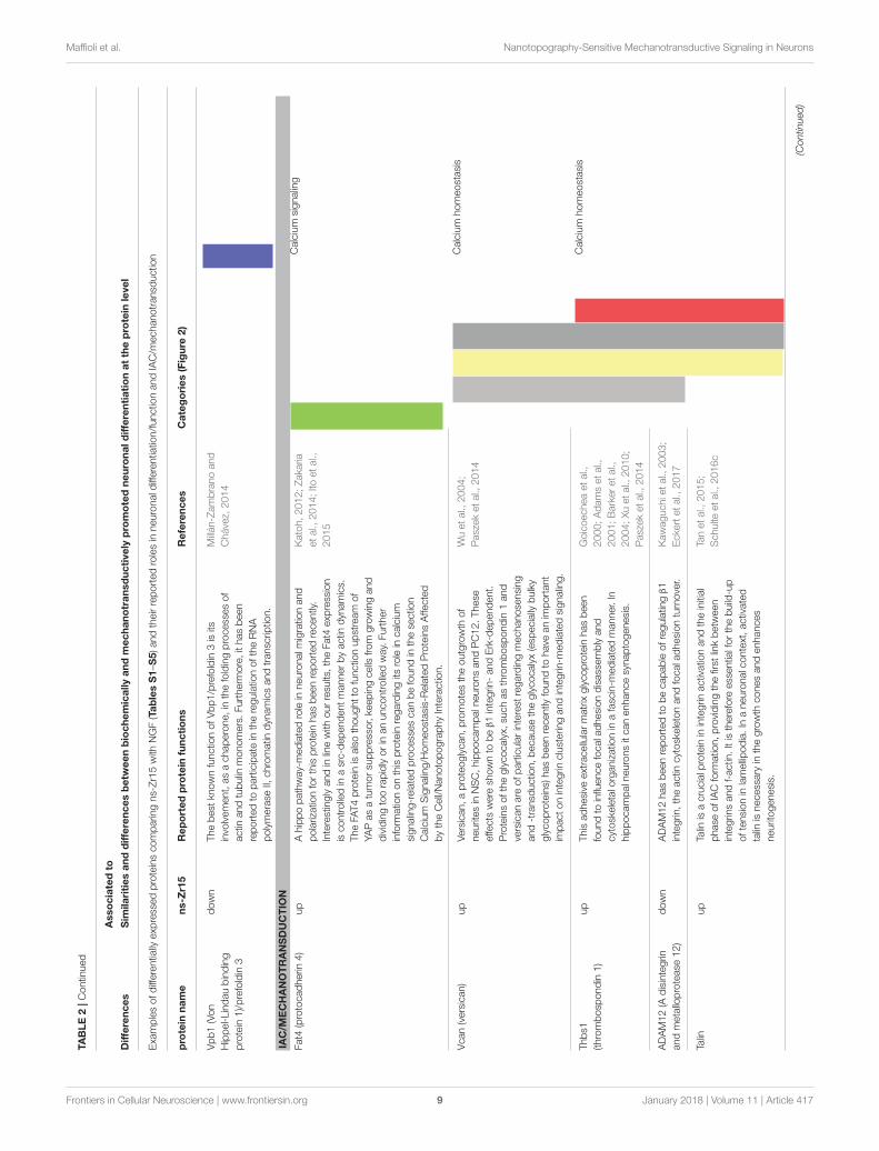
TABLE 1 | Similarities between biochemically and mechanotransductively promoted neuronal differentiation at the protein level by comparing the conditions ns-Zr15,

NGF, and PLL.

Associated to

Similarities Similarities and differences between biochemically and

mechanotransductively promoted neuronal differentiation at the protein level

Examples of proteins differentially expressed in the same manner in NGFvsPLL (Table S1) and ns-Zr15vsPLL (Table S3) and their reported roles in a neuronal context

Protein name NGF and

ns-Zr15

Reported protein functions References Category

Htra1 (high temperature

responsive antigen 1)

up Htra1 is a microtubule-associated serine protease that

has been found to be crucial for neuronal protein

quality control (e.g., tau), survival and maturation.

Launay et al., 2008; Tennstaedt

et al., 2012

Neuronal protein quality

control

Vps35 (vacuolar protein

sorting-associated protein 35)

up Vps35 belongs to the retromer complex and

contributes therefore essentially to endosomal

trafficking. It promotes e.g., neuronal morphogenesis in

hippocampal neurons by contributing to the retrograde

trafficking of BACE1. In addition, mitochondria were

found fused and dysfunctional in Vps35-deficient

dopaminergic neurons, causing the loss of these

neurons.

Wang et al., 2012; Tang et al.,

2015

Vesicle trafficking

Fasn (fatty acid synthase) up Fasn is essential in the lipogenesis control of neural

stem cells and its deletion impairs adult neurogenesis.

Knobloch et al., 2013 Lipogenesis

Pdia3 (protein

disulfide-isomerase 3)/ERp57

up This disulfide isomerase belongs to the endoplasmic

reticulum proteostasis network with a neuroprotective

function against misfolded prions. It is involved in

axonal regeneration after peripheral nerve injury.

Castillo et al., 2015; Bargsted

et al., 2016

Proteostasis network

C3 (complement component 3) down C3 is downregulated in the adult central nervous

system and known to participate in synapse

elimination.

Stevens et al., 2007 Complement system

RPL19 (60S ribosomal protein

L19)

up RPL19 was recommended as a very stable

differentiation reference independent of the

differentiation stimulus.

Zhou et al., 2010

Further information on a selection of proteins with versatile known roles in the regulation of neuronal functioning and neurogenic processes found to be differentially expressed in the

same manner in NGF and ns-Zr15, compared to PLL.

Frontiers in Cellular Neuroscience | www.frontiersin.org 6 January 2018 | Volume 11 | Article 417

Maffioli et al. Nanotopography-Sensitive Mechanotransductive Signaling in Neurons

needed during initiation of neuritogenesis; Boyne et al., 1996) andsome in protection against oxidative damage (Figure S3D).

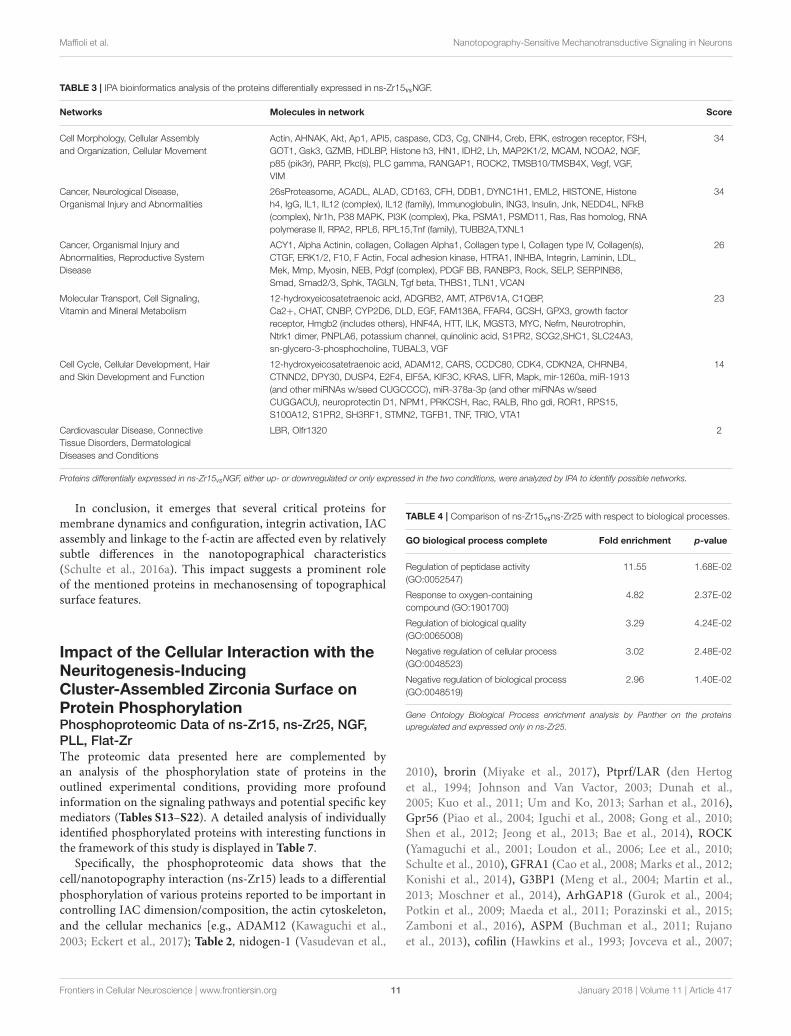
For some of these proteins [including also proteins appearingonly in ns-Zr15vsPLL (Table S3) and not in NGFvsPLL(Table S1), or expressed exclusively in ns-Zr15 (Table S5)]crucial and versatile functions in (post-)transcriptional andepigenetic regulation have been observed [e.g., DDB1 (Canget al., 2006), Nedd4 (Drinjakovic et al., 2010; Wiszniak et al.,2013; Hsia et al., 2014); Dpy30 (Jiang et al., 2011); Nsun2 (Blancoet al., 2014; Hussain and Bashir, 2015), HMGB2 (Abrahamet al., 2013), hnRNP A1 (Han et al., 2005; Li et al., 2007),Vbp1/prefoldin 3 (Millán-Zambrano and Chávez, 2014); detailsin Table 2: Differences–Neuronal differentiation/function].Moreover, various proteins are of particular interest regardinga potential connection of mechanotransductive signaling andneuronal differentiation processes in the nanotopography-induced setting [e.g., Fat4 (Katoh, 2012; Zakaria et al., 2014;Ito et al., 2015), Versican (Wu et al., 2004; Paszek et al., 2014),Thrombospondin (Goicoechea et al., 2000; Adams et al., 2001;Barker et al., 2004; Xu et al., 2010; Paszek et al., 2014), ADAM12(Kawaguchi et al., 2003; Eckert et al., 2017), Talin (Tan et al., 2015;Schulte et al., 2016c), NCoa2 (Voegel et al., 1996; Wyszynskiet al., 2002; Spangler and Hoogenraad, 2007; Essmann et al.,2008; Asperti et al., 2009, 2010; Selak et al., 2009; Dasgupta et al.,2014; Geiger et al., 2014; Heisler et al., 2014), Ran(bp3/GAP)(Yudin and Fainzilber, 2009); marked in gray in Tables S2, S5;details in Table 2: Differences–IAC/Mechanotransduction]. Thisinformation is additionally validated by the IPA bioinformaticsanalysis of the proteins differentially expressed in ns-Zr15vsNGF(Table S2). This evaluation detected relevant protein networksmodulated by the surface nanotopography related to: cellmorphology, cellular assembly and organization, cellularmovement, molecular transport, cell signaling, vitamin andmineral metabolism, cancer and invasion (Table 3).

Influence of the Surface NanotopographyRoughness on the Protein ExpressionFocus on ns-Zr15, ns-Zr25, NGF, PLLAs shown in Figure 1, the higher surface roughness Rq 25nm RMS (ns-Zr25) also induced neuritogenesis in PC12 butto a lower degree (Schulte et al., 2016a). The rationale of thisphenomenon is not completely clear yet. Therefore, in additionto the mentioned ns-Zr15, we have included ns-Zr25 into thisproteomic approach (Tables S7–S12). The Venn diagram andwork flow for the comparison of NGF, ns-Zr15, ns-Zr25 areshown in Figures S4A,B, respectively.

Several proteins (7 out of 26; marked in light gray in Table S8)found to be upregulated in ns-Zr25vsNGF (Table S8) are alsoupregulated in the comparison ns-Zr15vsNGF (Table S2) or ns-Zr15vsPLL (Table S3), suggesting that the biological processestriggered by the cell/nanostructure interaction are partiallysimilar, even if the roughness parameter is increasing.

The proteins upregulated only in ns-Zr25vsNGF(Tables S8, S12), and not in the other conditions, are e.g.,stress-induced proteins (such as CASC5, GPX3, A1M, andHSP90) and proteins involved in transport and trafficking. The

data further demonstrates that the interaction of PC12 cells withhigher roughness is accompanied by an increase of proteinsrelated to formation/degradation of atherosclerosis plaques(APOB, SERPIND1), secretion, anti-inflammation activityand stress response (HMOX1, LOC681468, TXN, HMGN1,Cybasc3) while others are directly involved in gene expressioncontrol. Accordingly, the enrichment analysis of GO biologicalprocesses carried out by Panther on the proteins upregulated oronly expressed in ns-Zr25 (Table 4) shows that the roughnessincrease triggers the expression of proteins involved in responseto oxygen-containing compounds.

The proteome analysis of cells grown on ns-Zr25 also displaysan increased expression of proteins involved in regulation ofcell proliferation, differentiation and apoptosis, adhesion andtrafficking, as well as intercellular signaling pathways. Someof these proteins indicate a strong neuronal differentiation-promotive effect also for this substrate [e.g., syntaxin 4 (Kennedyet al., 2010; Mohanasundaram and Shanmugam, 2010), clathrin(Heuser and Reese, 1973; Cosker and Segal, 2014), HMGN1(Deng et al., 2013; Nagao et al., 2014), and SCN1B (Namaduraiet al., 2015), details in Table 5], consistent with our resultsin primary hippocampal neurons where ns-Zr25 had the mostsignificant effect on neuron differentiation and maturation(Schulte et al., 2016b). However, the induction of many stress-related proteins suggests that the substrate situation is becomingsuboptimal for PC12 cells leading to the altered expression ofproteins that are essential for the regulation of neuronal survival[e.g., CREM (Mantamadiotis et al., 2002; Wu et al., 2012), NPM1(Qing et al., 2008; Pfister and D’Mello, 2015); details in Table 5].

Compared to ns-Zr15, in ns-Zr25 (Table S7) there wasdecreased protein expression of tumor suppressors involvedin apoptosis (PARK7, GZMB, SRSF1, FAT4) and cytoskeletalproteins that play essential roles in the integrin signaling.The IPA confirms the latter observation by identifying ILK(integrin-linked kinase) signaling as the only canonical pathwaysignificantly decreased on ns-Zr25 (Z score-1, proteins CDH1,FN1, ACTN4, TMSB10/TMSB4X) (Table 6). Intriguingly, thispathway has been reported to be pivotal in the regulation ofIAC architecture/composition and to be sensitive to integrinligand density of the substrate (Elad et al., 2013). In the contextof mechanosensing, lysophosphatidylcholine acyltransferase(Lpcat2b) expression only in the ns-Zr25 condition is intriguing(Table S12). This protein converts lysophosphatidylcholinein phosphatidylcholine; a process essential in the regulationof membrane dynamics (i.e., curvature/bending, tension),recruitment of F-BAR proteins and membrane/f-actin linkage(Anitei and Hoflack, 2012). Also regarding cell/cell contact,IAC and actomyosin organization some changes are noteworthy[such as Rab14 (Linford et al., 2012), clathrin (Ezratty et al.,2009), nischarin (Alahari et al., 2000, 2004; Zhang and Abdel-Rahman, 2006; Ding et al., 2008, 2015; Pouwels et al., 2012),ArhGEF1/P115-RhoGEF (Hart et al., 1998; Kozasa et al., 1998;Dubash et al., 2007); details in Table 5]. In addition, SF3B2 and 5(upregulated in the cells on ns-Zr15, Table S11), are componentsof the spliceosomal U2 small nuclear ribonucleoprotein particlethat has an important role in neuronal transcriptional regulation(Jia et al., 2012) (details in Table 5).

Frontiers in Cellular Neuroscience | www.frontiersin.org 7 January 2018 | Volume 11 | Article 417

Maffioli et al. Nanotopography-Sensitive Mechanotransductive Signaling in Neurons

TABLE2|Differencesbetw

eenbiochemically

andmechanotransd

uctivelypromotedneuronald

ifferentiatio

nattheprotein

levelb

ycomparin

gtheconditionsns-Zr15,NGF,andPLL.

Associatedto

Differences

Sim

ilaritiesanddifferencesbetw

eenbiochemicallyandmechanotransductively

promotedneuronaldifferentiationattheprotein

level

Exa

mplesofdifferentially

exp

ressedproteinscomparin

gns-Zr15with

NGF(TablesS1–S

5)andtheirreportedrolesinneuronald

ifferentiatio

n/functio

nandIAC/m

echanotransd

uctio

n

Protein

name

ns-Z

r15

Reportedprotein

functions

References

Categories(Figure

2)

NEURONALDIFFERENTIATIO

N/FUNCTIO

N

Cell/cellinteractio

n

GlycocalyxandECM

Membrane/f-actin

linka

geandintegrin

activatio

n

Cell/su

bstrate

interactio

n,IAC

Actomyo

sinorganizatio

n/C

ellularmechanics

Nuclearorganizatio

nandtransc

riptio

n

Otherfunctio

ns

DDB1(DNA

damage-binding

protein

1)

up

DDB1andNedd4are

both

invo

lvedin

ubiquitinligatio

n

andare

essentialfortheregulatio

nofneuronalcellsu

rvival,

axo

nbranchingandthemaintenanceofstem

cell

propertiesofneuralcrest

cells.Nedd4hasmany

substrateswith

importantfunctio

nin

neurons(e.g.ion

channels,receptors).ThemRNAexp

ressionofNedd4itself

isregulateddownstream

ofthePTEN/m

TORC1pathway.

Cangetal.,

2006

Ubiquitinligatio

n

Nedd4(neuronal

precursorcellexp

ressed

anddevelopmentally

downregulated

protein

4)

down

Drin

jako

vicetal.,

2010;

Wiszn

iaketal.,

2013;

Hsiaetal.,

2014

Ubiquitinligatio

n

Dpy3

0(histone

methyltrasferase

complexregulatory

subunit)

up

Dpy3

0,with

itsrolein

histonemethylatio

n,hasbeenfound

tocontrolembryonicstem

cellfate

specificatio

n,in

particulartoward

theneurallineage.

Jiangetal.,

2011

Nsu

n2(NOP2/sun

domain

family,member

2)

up

Mutatio

nsinthiscytosine-5

RNAmethyltransferase

can

leadto

microcephaly(Dubowitz

syndrome)andother

neurodevelopmentald

isorders.Nsu

n2canbefoundin

neuronald

endritesanddueto

itsmRNAmethylatio

n

activity

itisinvo

lvedintheepigenetic

controlo

f

postsynaptic

functio

nsandplasticity.

Blancoetal.,

2014;

HussainandBash

ir,

2015

HMGB2(highmobility

groupB2)

down

HMGB2belongsto

thefamily

ofchromatin

structural

proteinsandisim

portantin

thecontrolo

fneuralstem

cell

proliferatio

nandtheregulatio

nofneurogenesisand

neuronald

ifferentiatio

n.

Abraham

etal.,

2013

hnRNPA1

(heterogeneousnuclear

ribonucleoprotein

A1)

down

hnRNPA1iskn

ownto

beinvo

lvedinalternativepre-m

RNA

splicing,in

theneuronalsettingasasilencingfactor.

Hanetal.,

2005;Li

etal.,

2007

(Continued)

Frontiers in Cellular Neuroscience | www.frontiersin.org 8 January 2018 | Volume 11 | Article 417

Maffioli et al. Nanotopography-Sensitive Mechanotransductive Signaling in Neurons

TABLE2|Contin

ued

Associatedto

Differences

Sim

ilaritiesanddifferencesbetw

eenbiochemicallyandmechanotransductively

promotedneuronaldifferentiationattheprotein

level

Exa

mplesofdifferentially

exp

ressedproteinscomparin

gns-Zr15with

NGF(TablesS1–S

5)andtheirreportedrolesinneuronald

ifferentiatio

n/functio

nandIAC/m

echanotransd

uctio

n

protein

name

ns-Z

r15

Reportedprotein

functions

References

Categories(Figure

2)

Vpb1(Von

Hippel-Lindaubinding

protein

1)/prefoldin3

down

Thebest

knownfunctio

nofVbp1/prefoldin3isits

invo

lvement,asachaperone,in

thefoldingprocessesof

actin

andtubulin

monomers.Furtherm

ore,ithasbeen

reportedto

participate

intheregulatio

noftheRNA

polymerase

II,chromatin

dyn

amicsandtransc

riptio

n.

Millán-Zambranoand

Chávez,

2014

IAC/M

ECHANOTRANSDUCTIO

N

Fat4

(protocadherin

4)

up

Ahippopathway-mediatedrolein

neuronalm

igratio

nand

polarizatio

nforthisprotein

hasbeenreportedrecently.

Interestinglyandin

linewith

ourresu

lts,theFat4

exp

ression

iscontrolledin

asrc-dependentmannerbyactin

dyn

amics.

TheFA

T4protein

isalsothoughtto

functio

nupstream

of

YAPasatumorsu

ppressor,ke

epingcells

from

growingand

dividingtoorapidlyorin

anuncontrolledway.Further

inform

atio

nonthisprotein

regardingits

roleincalcium

signaling-relatedprocessescanbefoundinthese

ctio

n

Calcium

Signaling/H

omeostasis-RelatedProteinsAffected

bytheCell/NanotopographyInteractio

n.

Katoh,2012;Zaka

ria

etal.,

2014;Itoetal.,

2015

Calcium

signaling

Vcan(versican)

up

Versican,aproteoglycan,promotestheoutgrowth

of

neuritesin

NSC,hippocampaln

euronsandPC12.These

effects

were

shownto

be

β1integrin

-andErk-dependent.

Proteinsoftheglycocalyx,

suchasthrombosp

ondin1and

versicanare

ofparticularinterest

regardingmechanose

nsing

and-transd

uctio

n,because

theglycocalyx(esp

ecially

bulky

glycoproteins)

hasbeenrecently

foundto

have

anim

portant

impactonintegrin

clusterin

gandintegrin

-mediatedsignaling.

Wuetal.,

2004;

Paszeketal.,

2014

Calcium

homeostasis

Thbs1

(thrombosp

ondin1)

up

Thisadhesive

extracellularmatrixglycoprotein

hasbeen

foundto

influencefocaladhesiondisassemblyand

cytoskeletalo

rganizatio

nin

afasc

in-m

ediatedmanner.In

hippocampaln

euronsitcanenhancesynaptogenesis.

Goicoecheaetal.,

2000;Adamsetal.,

2001;Barkeretal.,

2004;Xuetal.,

2010;

Paszeketal.,

2014

Calcium

homeostasis

ADAM12(A

disintegrin

andmetalloprotease

12)

down

ADAM12hasbeenreportedto

becapableofregulatin

gβ1

integrin

,theactin

cytoskeletonandfocaladhesionturnover.

Kawaguchietal.,

2003;

Ecke

rtetal.,

2017

Talin

up

Talin

isacrucialp

rotein

inintegrin

activatio

nandtheinitial

phase

ofIACform

atio

n,providingthefirst

linkbetw

een

integrin

sandf-actin

.Itistherefore

essentialforthebuild-up

oftensionin

lamellipodia.In

aneuronalcontext,activated

talin

isnecessary

inthegrowth

conesandenhances

neuritogenesis.

Tanetal.,

2015;

Schulte

etal.,

2016c

(Continued)

Frontiers in Cellular Neuroscience | www.frontiersin.org 9 January 2018 | Volume 11 | Article 417

Maffioli et al. Nanotopography-Sensitive Mechanotransductive Signaling in Neurons

TABLE2|Contin

ued

Associatedto

Differences

Sim

ilaritiesanddifferencesbetw

eenbiochemicallyandmechanotransductively

promotedneuronaldifferentiationattheprotein

level

Exa

mplesofdifferentially

exp

ressedproteinscomparin

gns-Zr15with

NGF(TablesS1–S

5)andtheirreportedrolesinneuronald

ifferentiatio

n/functio

nandIAC/m

echanotransd

uctio

n

Protein

name

ns-Z

r15

Reportedprotein

functions

References

Categories(Figure

2)

NCoa2(nuclearreceptor

coactivator

2)/GRIP1/TIF-2

up

NCoA2represe

nts

ontheonehandatransc

riptio

nal

coregulatorforvario

usnuclearreceptors

(e.g.,androgen,

estrogen,glucocorticoid,steroid

receptor)andfacilitates

theaccess

forDNAtransc

riptio

nbyhistoneacetylatio

n.

Ontheotherhandithasbeenreportedto

beim

portantfor

essentialn

euronalfunctio

nsanddendrito-or

synaptogenesis,

inparticularbycoordinatin

gneuronaland

synaptic

cargoandreceptor(kainite,AMPA)trafficking.It

cancooperate

with

othercrucialp

roteinsin

these

processes,

suchasSNAP25,14-3-3

orliprin

-α.Thelatter

protein

isfurtherm

ore

knownto

beinvo

lvedin

the

distributio

nandregulatio

nofβ1integrin

activatio

nby

stabilizingitin

itsinactiveconform

atio

natthecell

membrane.

Voegeletal.,

1996;

Wyszynskietal.,

2002;

Spanglerand

Hoogenraad,2007;

Essmannetal.,

2008;

Asp

ertietal.,

2009,

2010;Selaketal.,

2009;Dasg

upta

etal.,

2014;Geigeretal.,

2014;Heisleretal.,

2014

Vesicletrafficking

Ran(bp3)

RanGAP1

up

down

TheRanGTPase

(andits

regulators)have

versatilerolesin

neuronalcells,su

chasnucleocytoplasm

ictransp

ort,

trafficking,axo

nretrogradesignalingandcytoskeletal

dyn

amics.

Yudin

andFainzilber,

2009

Trafficking

Furtherinformationonaselectionofproteinswithprominentreportedrolesintheregulationofneuronalfunctioningandneurogenicprocessesand/orrelevanceformechanotransductiondifferentially

expressedcomparingns-Zr15

andNGF.

Frontiers in Cellular Neuroscience | www.frontiersin.org 10 January 2018 | Volume 11 | Article 417

Maffioli et al. Nanotopography-Sensitive Mechanotransductive Signaling in Neurons

TABLE 3 | IPA bioinformatics analysis of the proteins differentially expressed in ns-Zr15vsNGF.

Networks Molecules in network Score

Cell Morphology, Cellular Assembly

and Organization, Cellular Movement

Actin, AHNAK, Akt, Ap1, API5, caspase, CD3, Cg, CNIH4, Creb, ERK, estrogen receptor, FSH,

GOT1, Gsk3, GZMB, HDLBP, Histone h3, HN1, IDH2, Lh, MAP2K1/2, MCAM, NCOA2, NGF,

p85 (pik3r), PARP, Pkc(s), PLC gamma, RANGAP1, ROCK2, TMSB10/TMSB4X, Vegf, VGF,

VIM

34

Cancer, Neurological Disease,

Organismal Injury and Abnormalities

26sProteasome, ACADL, ALAD, CD163, CFH, DDB1, DYNC1H1, EML2, HISTONE, Histone

h4, IgG, IL1, IL12 (complex), IL12 (family), Immunoglobulin, ING3, Insulin, Jnk, NEDD4L, NFkB

(complex), Nr1h, P38 MAPK, PI3K (complex), Pka, PSMA1, PSMD11, Ras, Ras homolog, RNA

polymerase II, RPA2, RPL6, RPL15,Tnf (family), TUBB2A,TXNL1

34

Cancer, Organismal Injury and

Abnormalities, Reproductive System

Disease

ACY1, Alpha Actinin, collagen, Collagen Alpha1, Collagen type I, Collagen type IV, Collagen(s),

CTGF, ERK1/2, F10, F Actin, Focal adhesion kinase, HTRA1, INHBA, Integrin, Laminin, LDL,

Mek, Mmp, Myosin, NEB, Pdgf (complex), PDGF BB, RANBP3, Rock, SELP, SERPINB8,

Smad, Smad2/3, Sphk, TAGLN, Tgf beta, THBS1, TLN1, VCAN

26

Molecular Transport, Cell Signaling,

Vitamin and Mineral Metabolism

12-hydroxyeicosatetraenoic acid, ADGRB2, AMT, ATP6V1A, C1QBP,

Ca2+, CHAT, CNBP, CYP2D6, DLD, EGF, FAM136A, FFAR4, GCSH, GPX3, growth factor

receptor, Hmgb2 (includes others), HNF4A, HTT, ILK, MGST3, MYC, Nefm, Neurotrophin,

Ntrk1 dimer, PNPLA6, potassium channel, quinolinic acid, S1PR2, SCG2,SHC1, SLC24A3,

sn-glycero-3-phosphocholine, TUBAL3, VGF

23

Cell Cycle, Cellular Development, Hair

and Skin Development and Function

12-hydroxyeicosatetraenoic acid, ADAM12, CARS, CCDC80, CDK4, CDKN2A, CHRNB4,

CTNND2, DPY30, DUSP4, E2F4, EIF5A, KIF3C, KRAS, LIFR, Mapk, mir-1260a, miR-1913

(and other miRNAs w/seed CUGCCCC), miR-378a-3p (and other miRNAs w/seed

CUGGACU), neuroprotectin D1, NPM1, PRKCSH, Rac, RALB, Rho gdi, ROR1, RPS15,

S100A12, S1PR2, SH3RF1, STMN2, TGFB1, TNF, TRIO, VTA1

14

Cardiovascular Disease, Connective

Tissue Disorders, Dermatological

Diseases and Conditions

LBR, Olfr1320 2

Proteins differentially expressed in ns-Zr15vsNGF, either up- or downregulated or only expressed in the two conditions, were analyzed by IPA to identify possible networks.

In conclusion, it emerges that several critical proteins formembrane dynamics and configuration, integrin activation, IACassembly and linkage to the f-actin are affected even by relativelysubtle differences in the nanotopographical characteristics(Schulte et al., 2016a). This impact suggests a prominent roleof the mentioned proteins in mechanosensing of topographicalsurface features.

Impact of the Cellular Interaction with theNeuritogenesis-InducingCluster-Assembled Zirconia Surface onProtein PhosphorylationPhosphoproteomic Data of ns-Zr15, ns-Zr25, NGF,

PLL, Flat-ZrThe proteomic data presented here are complemented byan analysis of the phosphorylation state of proteins in theoutlined experimental conditions, providing more profoundinformation on the signaling pathways and potential specific keymediators (Tables S13–S22). A detailed analysis of individuallyidentified phosphorylated proteins with interesting functions inthe framework of this study is displayed in Table 7.

Specifically, the phosphoproteomic data shows that the

cell/nanotopography interaction (ns-Zr15) leads to a differential

phosphorylation of various proteins reported to be important incontrolling IAC dimension/composition, the actin cytoskeleton,

and the cellular mechanics [e.g., ADAM12 (Kawaguchi et al.,

2003; Eckert et al., 2017); Table 2, nidogen-1 (Vasudevan et al.,

TABLE 4 | Comparison of ns-Zr15vsns-Zr25 with respect to biological processes.

GO biological process complete Fold enrichment p-value

Regulation of peptidase activity

(GO:0052547)

11.55 1.68E-02

Response to oxygen-containing

compound (GO:1901700)

4.82 2.37E-02

Regulation of biological quality

(GO:0065008)

3.29 4.24E-02

Negative regulation of cellular process

(GO:0048523)

3.02 2.48E-02

Negative regulation of biological process

(GO:0048519)

2.96 1.40E-02

Gene Ontology Biological Process enrichment analysis by Panther on the proteins

upregulated and expressed only in ns-Zr25.

2010), brorin (Miyake et al., 2017), Ptprf/LAR (den Hertog

et al., 1994; Johnson and Van Vactor, 2003; Dunah et al.,2005; Kuo et al., 2011; Um and Ko, 2013; Sarhan et al., 2016),Gpr56 (Piao et al., 2004; Iguchi et al., 2008; Gong et al., 2010;Shen et al., 2012; Jeong et al., 2013; Bae et al., 2014), ROCK

(Yamaguchi et al., 2001; Loudon et al., 2006; Lee et al., 2010;Schulte et al., 2010), GFRA1 (Cao et al., 2008; Marks et al., 2012;Konishi et al., 2014), G3BP1 (Meng et al., 2004; Martin et al.,

2013; Moschner et al., 2014), ArhGAP18 (Gurok et al., 2004;Potkin et al., 2009; Maeda et al., 2011; Porazinski et al., 2015;Zamboni et al., 2016), ASPM (Buchman et al., 2011; Rujano

et al., 2013), cofilin (Hawkins et al., 1993; Jovceva et al., 2007;

Frontiers in Cellular Neuroscience | www.frontiersin.org 11 January 2018 | Volume 11 | Article 417

Maffioli et al. Nanotopography-Sensitive Mechanotransductive Signaling in Neurons

TABLE5|Comparisonofns-Zr25vsns-Zr15regardingtheinfluenceofthesu

rfacenanotopographyroughness

ontheprotein

exp

ression.

Associatedto

Influenceofthesurfacenanotopographyroughnessontheprotein

expression

Protein

name

ns-Z

r15

Reportedprotein

functions

References

Categories(Figure

2)

Cell/cellinteractio

n

GlycocalyxandECM

Membrane/f-actin

linka

geandintegrin

activatio

n

Cell/su

bstrate

interactio

n,IAC

Actomyo

sinorganizatio

n/C

ellularmechanics

Nuclearorganizatio

nandtransc

riptio

n

Otherfunctio

ns

Rab14

up

Rab14isinvo

lvedin

endocyticrecyclingandregulates

therewith

ADAM-dependentN-C

adherin

shedding

affectin

gcell-celladherensjunctio

ns,

cellpolarizatio

nand

migratio

n.

Linford

etal.,

2012

Vesicletrafficking

CLT

C(clathrin

heavy

chain

1)

down

Clathrin

islong-knownto

playacrucialrolein

neuronal

signalingbymediatin

gsynaptic

vesicleendocytosis,

receptorretrievalandretrogradetransp

ortregulatin

g

neuronalfunctio

ns(e.g.,neuronsu

rvival,axo

ngrowth

and

guidance).Apartfrom

thisrolein

neuronalsignalingby

mediatin

gendocytosisofsynaptic

receptors,clathrin

controlsalsofocaladhesiondim

ensionsin

migratin

gcells

byregulatin

gtheendocytosisofinactiveintegrin

s.

Heuse

randReese

,

1973;Ezrattyetal.,

2009;Coskerand

Segal,2014

Vesicletrafficking

Nischarin

up

Nischarin

isassociatedwith

versatileandcomplex

processes.

Ithasbeenreportedto

negativelyregulate

cell

migratio

nbutto

fosterneuriteoutgrowth,alsoin

PC12

cells.Itbindsnon-ligatedintegrin

s(e.g.,

α5β1),actin

gasa

sortofintegrin

inhibito

r,andmodulatestheLIM

K/cofilin

pathwayandPAK/R

ac1activatio

naffectin

gtherebythe

actin

dyn

amics.

Inaddition,in

differentiatedPC12

nischarin

behavesasI1-imidazo

linereceptorandactivates

protein

kinase

C(PKC),extracellularsignal-regulated

kinase

(ERK)andc-junN-term

inalkinase

(JNK)promotin

g

theneuronald

ifferentiatio

nandsu

rvival.

Alaharietal.,

2000,

2004;Zhangand

Abdel-Rahman,2006;

Dingetal.,

2008,2015;

Pouwelsetal.,

2012

I1-imidazo

linereceptor

ArhGEF1/P115-

RhoGEF

up

ArhGEF1interacts

with

Gα13andregulatesRhoGTPase

activity

andcontrolsfocaladhesionandstress

fiber

form

atio

nin

resp

onse

tofib

ronectin

.

Hartetal.,

1998;

Kozasa

etal.,

1998;

Dubash

etal.,

2007

HMGN1(highmobility

groupnucleoso

me

bindingdomain

1)

down

HGMN1,anucleoso

me-bindingprotein,isim

portantfor

thestructuralo

rganizatio

nofthechromatin

andfine-tunes

transc

riptio

nprofilesalongtheneuronallineage.Theloss

ofthisprotein

lowers

theamountofneuralp

rogenito

rcells

inthebrain,whereasincrease

dexp

ressionfosters

astrogenesisinsteadofneurogenesis.

Dengetal.,

2013;

Nagaoetal.,

2014

(Continued)

Frontiers in Cellular Neuroscience | www.frontiersin.org 12 January 2018 | Volume 11 | Article 417

Maffioli et al. Nanotopography-Sensitive Mechanotransductive Signaling in Neurons

TABLE5|Contin

ued

Associatedto

Influenceofthesurfacenanotopographyroughnessontheprotein

expression

Protein

name

ns-Z

r15

Reportedprotein

functions

References

Categories(Figure

2)

SF3B2and5(splicing

factor3Bsu

bunit2

and5)

up

SF3B2and5belongto

theU2sn

RNPsp

liceoso

me

complexandistherefore

invo

lvedin

thesp

licing

pre-m

RNA.Dysfunctio

nofthiscomplexleadsto

ageneral

perturbatio

nofalternativesp

licingcausing

neurodegeneratio

n.

Jiaetal.,

2012

CREM

(cAMP-resp

onsive

elementmodulator)

CREM

isamemberofthecAMP-resp

onsive

element

bindingprotein

family

andim

portantin

thetransc

riptio

nal

regulatio

n.Itisupregulatedasresp

onse

tonervetissu

e

injury

andrepair,probablyasapro-apoptotic

factor.

Mantamadiotis

etal.,

2002;Wuetal.,

2012

Apoptosis

NPM1(nucleolar

phosp

hoproteinB23,

numatrin)/nucleophosm

in/

B23

down

NPM1isanucleolarprotein

with

versatilefunctio

n,

esp

ecially

alsoin

aneuronalcontext

where

itisinvo

lvedin

thecontrolo

fgrowth,differentiatio

nandapoptosis.

Exp

ressedin

levelshigherthanthephysiologicalo

nes,

it

cancause

neuronald

eath.

Qingetal.,

2008;

PfisterandD’M

ello,

2015

Apoptosis

Stx4(syn

taxin4)

down

Syn

taxin4isanessentialp

ostsynaptic

t-SNAREprotein

invo

lvedin

exo

cytosisandsynaptic

plasticity.

Kennedyetal.,

2010;

Mohanasu

ndaram

and

Shanmugam,2010

Vesicletrafficking

SCN1B(sodium

voltage-gatedchannel

beta

subunit1)

down

SCN1Bbelongsto

theNaVchannelfamily

whichis

importantin

theinitiatio

nofactio

npotentials.Thebeta

subunits

are

associatedto

thepore-form

ingalphasu

bunits

andfine-tunethechannels’exc

itability.

Namaduraietal.,

2015

Channel

Selectedproteinswithreportedrolesinneuronalsurvival,functioning,differentiationand/ormechanotransductionthatshoweddifferentialexpressioninthens-Zr15vsns-Zr25comparison.

Frontiers in Cellular Neuroscience | www.frontiersin.org 13 January 2018 | Volume 11 | Article 417

Maffioli et al. Nanotopography-Sensitive Mechanotransductive Signaling in Neurons

TABLE 6 | IPA bioinformatics comparison of ns-Zr15vsns-Zr25.

Ingenuity canonical pathways p-Value z-score Molecules

Epithelial Adherens Junction Signaling 5,37E-03 NaN CDH1, ACTN4, TUBB

Germ Cell-Sertoli Cell Junction Signaling 8,71E-03 NaN CDH1, ACTN4, TUBB

Sertoli Cell-Sertoli Cell Junction Signaling 1,02E-02 NaN CDH1, ACTN4, TUBB

GIalpha12/13 Signaling 3,98E-02 NaN CDH1, ARHGEF1

ILK Signaling 1,32E-03 −1 CDH1, FN1, ACTN4, TMSB10/TMSB4X

Actin Cytoskeleton Signaling 2,34E-03 0 FN1, ARHGEF1, ACTN4, TMSB10/TMSB4X

Heme Degradation 3,09E-04 NaN HMOX1, BLVRB

IL-10 Signaling 1,29E-02 NaN HMOX1, BLVRB

Unfolded protein response 7,41E-03 NaN P4HB, EIF2AK3

Acute Phase Response Signaling 7,59E-04 NaN PLG, HMOX1, FN1, SERPIND1

Coagulation System 3,24E-03 NaN PLG, SERPIND1

Insulin Receptor Signaling 4,90E-02 NaN PTPN1, STX4

Proteins expressed in a differential manner in the comparion ns-Zr15vsns-Zr25 were analyzed by IPA to identify canonical pathways affected by the different nanotopography

characteristics.

Flynn et al., 2012), septin-2 (Spiliotis and Nelson, 2006; Joo

et al., 2007); details in Table 7]. Furthermore, several proteinsessential in epigenetic and (post-)transcriptional regulation of

gene expression are modulated at the phosphorylation level [e.g.,

KMT2D (Dhar et al., 2012), RtcB (Kosmaczewski et al., 2015),E2F4 (Persengiev et al., 1999); details in Table 7]. Regarding thislatter aspect, it is noteworthy that lipin-1 phosphorylation isaffected by the interaction with ns-Zr15. This phosphatidic acidphosphatase is important in lipid synthesis and SREBP-mediatedtranscriptional regulation (e.g., Fasn expression, see Table 1).Its phosphorylation is regulated by mTOR which thereby alsocontrols its intracellular localisation and the lamin A-dependentnuclear organization (Peterson et al., 2011; Eaton et al., 2013).

The alterations (regarding expression and phosphorylationlevels) extend in a consistent manner our previous results(Schulte et al., 2016a), accentuating additionally the impact ofthe cell/nanotopography interaction on mechanotransductiveprocesses and defining more precisely nanotopography-sensitivesignaling hubs (Figure 2).

To find relevant patterns and specific differences in signalingprocesses related to the diverse conditions (PLL, NGF, ns-Zr15,ns-Zr25, and flat-Zr) a principal component analysis (PCA)was carried out on the corresponding phosphoproteomes. Theanalysis, applied to all the peptides found phosphorylated inthese 5 conditions, reveals at a glance that the phosphoproteomesof NGF and ns-Zr15 cluster together (confirming again thecommon outcome of a differentiated cell). Flat-Zr and PLLinstead are at the opposite ends of the plot (Figure 3A),suggesting that the cells on these two substrates behave verydifferently as far as protein phosphorylation concerns, inagreement with the other data reported so far.

If the same analysis is carried out focusing only on thesequence phospho-motifs present in the phosphoproteome data,a similar plot can be obtained (Figure 3B), but in this case amore evident separation can be observed between NGF andns-Zr15, indicating that the kinases and phosphatases involvedare, at least in part, different. Figure 3C reports in green allthe substrate motifs that are more relevant in the PCA analysis,

accounting for the 16% of all the phospho-motifs present in thephosphopeptides.

The enrichment analysis of these phospho-motifs, carried outby Panther and David, shows that there is a highly significantenrichment (p ≤ 0.05) of few signaling pathways in the cellson ns-Zr15 (Table 8). The angiogenesis and VEGF signalingpathways are in line with the processes mentioned throughoutthis work as they comprise many players also involved in focaladhesion, MAPK and Ca2+ signaling. In addition, the resultssuggest that the differences between PC12 cells grown on ns-Zr15(compared to the NGF condition) could be partially ascribed toa modulation within the Wnt pathway (Table 8). An indicatoris e.g., the downregulation of E-cadherin in the cells on ns-Zr15, considering the known crosstalk between (E-)cadherin celladhesion and canonical Wnt signaling by release of β-catenin(Heuberger and Birchmeier, 2010). Interestingly, Wnt expressionin PC12 cells leads to an upregulation of E-cadherin and a flatepithelial-like cell morphology associated with unresponsivenessto NGF-induced neuritogenesis (Bradley et al., 1993). Inepithelial cells E-cadherin-mediated cell/cell adhesions areessential in mechanically connecting the intercellular actomyosinmachineries to regulate tissue organization (Lecuit and Yap,2015). Moreover, it has been demonstrated in human embryonicstem cells that the surface nanotopography has an impact onE-cadherin expression level (Chen et al., 2012). The potentialimpact of the substrate nanotopography on Wnt signaling ina neuronal setting is therefore an interesting issue for furtherinvestigations.

In the comparison ns-Zr15vsns-Zr25 (Tables S20–S22), apartfrom various already mentioned proteins, lamin A appearedas differentially phosphorylated. This protein is particularlyinteresting in the context of mechanotransduction representingone of the intermediate filaments that forms the interior of thenuclear envelope. It was found to be involved in the regulationof nuclear architecture/biophysics, chromatin organization, andtranscription regulation at the end of mechanotransductivesignaling cascades that influence differentiative processes (Swiftet al., 2013). Its differential expression during adult neurogenesis

Frontiers in Cellular Neuroscience | www.frontiersin.org 14 January 2018 | Volume 11 | Article 417

Maffioli et al. Nanotopography-Sensitive Mechanotransductive Signaling in Neurons

TABLE7|Comparisonoftheconditionsns-Zr15,ns-Zr25,NGF,PLLandflat-Zrto

analyse

theim

pactofthecellularinteractio

nwith

theneuritogenesis-inducingcluster-assembledzirconiasu

rfaceonprotein

phosp

horylatio

n.

Associatedto

Impactofthecellularinteractionwiththeneuritogenesis-inducingcluster-assembledzirconia

surfaceonprotein

phosphorylation

Protein

name

Reportedprotein

functions

References

Categories(Figure

2)

Cell/cellinteractio

n

GlycocalyxandECM

Membrane/f-actin

linkageandintegrinactivatio

n

Cell/su

bstrate

interactio

n,IAC

Actomyo

sinorganizatio

n/C

ellularmechanics

Nuclearorganizatio

nandtransc

riptio

n

Otherfunctio

ns

Nidogen-1

Thebase

mentmembraneproteinnidogen-1

isknownto

beim

portantin

theregulatio

nofhippocampalsynaptic

plasticity

andnetw

ork

exc

itability.

Vasu

devanetal.,

2010

Brorin

Brorinhasbeenreportedto

favo

rneurogenesisandto

inhibitastrogenesis

contributin

gto

axo

nguidancein

thezebrafishforebrain.

Miyakeetal.,

2017

Galectin

-8Galectin

-8isase

cretedECM

proteinandmatricellularmodulatorofcell

adhesionthatisboundbyintegrinswhichregulatescelladhesionand

survival,promotin

gorinhibiting,dependentonwhetheritisprese

ntin

a

solubleorim

mobilizedmanner.

Hadarietal.,

2000;Zicketal.,

2004

Ptprf(receptor-type

tyrosine-protein

phosp

hatase

F)/LAR

(leukocytecommon

antig

en-related)

ThePtprf/LARreceptor,aneuronaladhesionmoleculeessentia

lin

synapse

maturatio

n,isparticularlyinterestingwith

resp

ectto

IAC,

mechanotransd

uctio

nandcalcium

signaling.Theprese

nceofthis

receptorinfocaladhesions(FA)iscontrolledin

anegativemannerby

myo

sinII-generatedforceandhasbeensh

ownto

have

thecapacity

to

regulate

FAs(in

mouse

embryonicfib

roblasts).Itinteracts

inaCaMKII

(Ca2+/calm

odulin-dependentproteinkinase

II)-regulatedwaywith

liprin-α

1.Theliprin-α

1/LARinteractio

ndeterm

inesLARdistributio

nandtherefore

synapse

morphogenesis.

Moreover,Ptprf/LARcanbefound,to

aminor

extent,tyrosine-phosp

horylatedintheadultbrain.Thefunctio

nofthis

phosp

horylatio

nisyetunknownbutcould

beim

portantforthebindingof

SH2/SH3domain-containingadaptorproteins.

denHertogetal.,

1994;Johnso

nand

VanVactor,2003;Dunahetal.,

2005;

Kuoetal.,

2011;Um

andKo,2013;

Sarhanetal.,

2016

Gpr56(G

protein-coupled

receptor56)

Malfunctio

nsofGpr56cancause

theneurodevelopmentald

isease

polymicrogyria.In

thebrainitispredominantly

exp

ressedin

neuronal

progenito

rcells

(NPC)in

regionsofpostnatalneurogenesiswhere

itis

invo

lvedin

thecontrolo

fbrainconvo

lutio

n/patterningin

thecerebral

cortexinanintegrin

α3β1-dependentmanner.Moreover,Gpr56operates

togetherwith

Gα13in

theRho-m

ediatedregulatio

nofNPC

adhesion/m

igratio

n.G

α13again

isessentia

linintegrinsignaling.

Piaoetal.,

2004;Iguchietal.,

2008;

Gongetal.,

2010;Shenetal.,

2012;

Jeongetal.,

2013;Baeetal.,

2014

ROCK

(Rho-associated,

coiled-coil-containing

proteinkinase

)

ROCK/R

hoAactivity

hasacomplexroleinneuritogenesis.

Alth

oughonthe

onehanditisknownto

beinhibito

ryforneuritogenesis,

andin

particular

fortheinitialn

eurite

form

atio

n,ontheotherhandsp

atia

llyrestricted

ROCK/R

hoAactivity

isalsoessentia

ltosu

ppress

lamellipodialp

rotrusions,

therebyconso

lidatin

gneurites/axo

nsbymaintainingthegrowth

cone

polarity.ROCKandits

RhoAbindingactivity

istig

htly

regulatedby

phosp

horylatio

ndownstream

ofsrcandcontributesto

themodulatio

nof

focaladhesionturnover.

Yamaguchietal.,

2001;Loudonetal.,

2006;Leeetal.,

2010;Schulte

etal.,

2010

Frontiers in Cellular Neuroscience | www.frontiersin.org 15 January 2018 | Volume 11 | Article 417

Maffioli et al. Nanotopography-Sensitive Mechanotransductive Signaling in Neurons

TABLE7|Contin

ued

Associatedto

Impactofthecellularinteractionwiththeneuritogenesis-inducingcluster-assembledzirconia

surfaceonprotein

phosphorylation

Protein

name

Reportedprotein

functions

References

Categories(Figure

2)

GFRA1(GDNFfamily

receptorα-1)

GFRA1wasfoundto

form

acomplexwith

β1integrin,togetherwith

Ret

andNCAM-140,andto

playanim

portantroleinthedifferentia

tionof

neuronsintheolfactory

system

andthesu

rvivalo

fglutamatergiccortical

neurons.

Caoetal.,

2008;Marksetal.,

2012;

Konishietal.,

2014

G3BP1(Ras

GTPase

-activatin

g

protein-binding

protein1)

G3BP1hasbeenreportedto

have

anim

pactonneuronalsproutin

gby

promotin

gtheform

atio

noftaumRNAribonucleoprotein

granulesandcan

befoundassociatedwith

α5β1integrin-containingcomplexe

s.Moreover,

G3BP1deficiencyim

pairsthesynaptic

plasticity

andcalcium

homeostasis

inhippocampalneurons.

Mengetal.,

2004;Martinetal.,

2013;

Mosc

hneretal.,

2014

Calcium

homeostasis

ArhGAP18(Rho

GTPase

activatin

g

protein18)

ArhGAP18hasbeensh

ownto

beinvo

lved,asnegativeregulator,in

the

controlo

fRhoAactivity

andstress

fiberform

atio

nbyincreasingthe

GTPase

activity

ofRhoandstabilizingtheRhoA-G

DPinactiveform

.The

protein,interactin

gwith

RhoA,hasbeendesc

ribedrecently

asYAP

effectorintheactomyo

sin-dependentregulatio

noftissu

etension.A

specificroleofthisproteinin

neuronshasnotbeenreportedso

far,butits

exp

ressionleveld

ecrease

sinneurosp

heresduringdifferentia

tionandit

appearedasageneassociatedwith

schizophreniain

asc

reeningfor

singlenucleotid

epolymorphisms.

ForanotherArhGAPfamily

member,

ArhGAP15,acontributio

nin

theneurogenesisofhippocampalneurons

hasbeensh

ownvery

recently.

Guroketal.,

2004;Potkin

etal.,

2009;

Maedaetal.,

2011;Porazinskietal.,

2015;Zambonietal.,

2016

Wntsignaling

ASPM

(Abnorm

al

spindle-like

microcephaly-

associated

protein)

ASPM

isknownto

contribute

totheregulatio

nofneuronald

ifferentia

tion

processesbyactomyo

sin-dependentactio

ns.

Astheproteinname

indicates,

thisproteinisinvo

lvedin

thecontrolo

fbrainsize

andmutatio

ns

ofthisproteincanberesp

onsiblefordevelopingtheneurald

isorder

microcephaly.ASPM

isapositiveregulatorforWntsignalingandits

exp

ressionisessentia

lforaccurate

neurogenesis.

Furtherm

ore,recently

it

hasbeenfoundthatthedroso

phila

orthologofthisproteininteracts

with

andregulatesmyo

sinIIlocalizatio

n,therebycontrollingneuroepith

elium

morphogenesisbymechanobiologicalevents.

Buchmanetal.,

2011;Rujanoetal.,

2013

Cofilin/destrin/ADF

(actin

depolymerising

factor)

Cofilin

isessentia

lforactin

cytoskeletalo

rganizatio

nbyregulatin

gthe

severingoff-actin

andtheturnoverrate

ofactin

andtherewith

,in

the

neuronalcontext,theactin

retrogradeflow

inneurite

growth

conesofthe

developingbrain.Phosp

horylatio

nnegativelyregulatesits

actin

binding

andtherebycontrolsthef-actin

homeostasis.

Hawkinsetal.,

1993;Jovceva

etal.,

2007;Flynnetal.,

2012

Septin

-2Septin

-2modulatesactomyo

sincontractility

bybindingmyo

sinIIand

recruitingregulatory

proteins.

Septin

phosp

horylatio

ncontrolsthe

assemblyofse

ptin

sinto

highlyorderedpolymers.Interestingly,se

ptin

-2

hasbeenfoundto

bephosp

horylatedin

post-m

itotic

neurons.

Spiliotis

andNelson,2006;Jooetal.,

2007

Frontiers in Cellular Neuroscience | www.frontiersin.org 16 January 2018 | Volume 11 | Article 417

Maffioli et al. Nanotopography-Sensitive Mechanotransductive Signaling in Neurons

TABLE7|Contin

ued

Associatedto

Impactofthecellularinteractionwiththeneuritogenesis-inducingcluster-assembledzirconia

surfaceonprotein

phosphorylation

Protein

name

Reportedprotein

functions

References

Categories(Figure

2)

KMT2D(histone-lysine

N-m

ethyltransferase

2D)/MLL4

(mixed-lineage

leukemia4)

ThisproteinisamammalianhistoneH3lysine4(H3K4)

mono-m

ethyltransferase

essentia

lindifferentia

tion-specificgene

activatio

n.Ithasbeensh

ownto

participate

intheregulatio

nofneuronal

differentia

tion,facilitatin

gtheactivatio

nofdifferentia

tion-specificgenes

(e.g.,nestin).

Dharetal.,

2012

RTCB(RNA2′ ,3′ -cyclic

phosp

hate

and5′ -OH

ligase

)

ThisRtcBRNAligase

participatesin

tRNAligatio

nanditisinvo

lvedinthe

regulatio

nofneuronalg

rowth

andaxo

nregeneratio

n.

Kosm

aczewskietal.,

2015

E2F4

Thistransc

riptio

nfactorhasbeensh

ownto

playarolein

neuronal

differentia

tionandneuritogenesis.

Persengievetal.,

1999

Rab23(Ras-related

protein23)

Rab23participatesto

endocyticvesicletraffickingandisinvo

lvedinthe

regulatio

nofso

nichedgehogsignalingin

neuraltubepatterning.

Eggensc

hwileretal.,

2001;Evans

etal.,

2003

Vesicletrafficking

Selectedproteinsfoundtobedifferentiallyphosphorylatedthathave

essentialknownfunctionswithrespecttoprocessesrelevantformechanotransductive

signalingand/orneuronalfunctioninganddifferentiation.

proposes a potential role in it; however, to date details remainstill unclear (Takamori et al., 2007). Further proteins found tobe phosphorylated on ns-Zr25 are Galectin-8 (Hadari et al.,2000; Zick et al., 2004), and Rab23 (Eggenschwiler et al., 2001;Evans et al., 2003) which have essential reported functions incell adhesion/survival and neuronal development, respectively(Table 7).

Altogether, many proteins that are important in neurogenicand/or mechanotransductive processes are differentiallyexpressed and/or phosphorylated upon cellular interactionwith the cluster-assembled zirconia surface that promotesneuritogenesis. Combining the analysis of our proteomicdata with information available on these proteins and theirfunctions, suggests a dynamic and complex modulation of anentire signaling network by the cell/nanotopography interactionthat is in control of cellular behavior and fate, i.e., in this caseneuronal differentiation. We were able to dissect potentialnanotopography-sensitive key elements regulated within amechanotransductive sequence, identifying many proteins thatcan be assigned to these principal categories: cell/cell adhesion,ECM and glycocalyx, cell/substrate interaction and IAC, integrinactivation and membrane/f-actin linkage, integrin adhesioncomplexes, actomyosin organization/cellular mechanics andnuclear organization and transcriptional regulation (Figure 2).

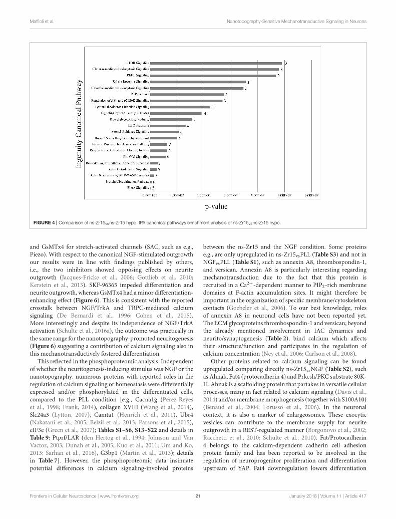
Alterations in Cellular Processes andSignaling by the Modulation of CellularTensionFocus on Phosphoproteomic Data of ns-Zr15,

ns-Zr15 hypo, PLL hyperIn our previous paper we identified the alteration of thecellular nanomechanical properties as critical for thesignal integration within the nanotopography-dependentmechanotransductive sequence that fostered neuronaldifferentiation. The interaction with the nanostructured surfacedictated the IAC nanoarchitecture/dynamics and cytoskeletalorganization in a manner that consequentially resulted in a softermembrane/cytoskeletal layer of the neuronal cell. Compensatingthis effect by a hypoosmotic gradient (causing cell swellingand an increase of cell tension) counteracted gradually thenanostructure-induced neuritogenesis on the morphologicallevel (Schulte et al., 2016a).

The mechanotransduction dependency of thenanotopography-promoted differentiation was broadly validatedby a proteomic comparison (Tables S23–S24) of PC12 cellsinteracting with the ns-Zr15 in the isoosmotic standardmedium (ns-Zr15) or instead in the presence of hypoosmoticmedium (ns-Zr15 hypo). Many proteins found to be expressedonly in ns-Zr15 (Table S24) or to be downregulated in ns-Zr15hypo (Table S23) can be classified as proteins involved inRhoGTPase-controlled cytoskeletal organization according tothe IPA canonical pathways enrichment analysis (Figure 4).Consistent with the hypoosmotic manipulation of the membranetension, clathrin- and caveolar-mediated endocytosis appearedamong the five most affected pathways in this evaluation(Figure 4). These pathways are regulated by and respond to

Frontiers in Cellular Neuroscience | www.frontiersin.org 17 January 2018 | Volume 11 | Article 417

Maffioli et al. Nanotopography-Sensitive Mechanotransductive Signaling in Neurons

FIG

URE2|Schematic

represe

ntatio

nofthepotentialrelatio

nandcrosstalkamongdifferentsignalingpathways

modulatedbytheneuron/nanotopographyinteractio

n.Onthelefttheillustratio

narrangesproteins

disse

ctedfrom

thewhole(phosp

ho)proteomicdata

set[i.e.,proteinswith

alteredexp

ressionand/orphosp

horylatio

nlevelintheindicatedconditions,

includingalsoso

meproteinsfrom

Schulte

etal.(2016a)]into

severalcategorie

sandse

tsthem

into

a(potential)relatio

nto

eachotherwith

resp

ectto

theirreportedcellbiologicalfunctio

nandcontext.Themain

categorie

sare

visu

alizedasfollows;

cell/cellinteractio

n(green

box),glycocalyxandECM

(boxwith

graypatternedfilling),cellsu

bstrate

interactio

nandIAC(graybox,

grayovalreprese

nts

theIAC),membrane/f-actin

linka

geandintegrin

activatio

n(yellow

box),actomyo

sin

organizatio

n/cellularmechanics(redbox),nuclearorganizatio

n,andtransc

riptio

nalregulatio

n(bluecirc

le,represe

ntin

gthenucleus).Furtherinform

atio

nonthereportedsp

ecificfunctio

nsoftheindividualp

roteins,

justifyingtheircategorisatio

n,canbefoundin

thecorresp

ondingtablesthroughoutthemanusc

ript.Moreover,ontherig

htase

lectio

nofproteinsislistedwith

associatio

nto

theirfunctio

nsin

neuronald

ifferentiatio

n

processes,

particularly

inneuritegrowth

cones.

Frontiers in Cellular Neuroscience | www.frontiersin.org 18 January 2018 | Volume 11 | Article 417

Maffioli et al. Nanotopography-Sensitive Mechanotransductive Signaling in Neurons

FIGURE 3 | Principal component analysis (PCA) on the phosphoproteome of PC12 cells grown in different experimental conditions. (A) PCA analysis of the

phosphopeptides of PC12 cells in the experimental conditions PLL, NGF, ns-Zr15, ns-Zr25, and flat-Zr. (B) PCA analysis of the sequence phospho-motifs present in

the phosphoproteome data of PLL, NGF, ns-Zr15, ns-Zr25, and flat-Zr. (C) Visual representation of the PCA analysis of the sequence phospho-motifs. All the

substrate motifs that are more relevant in the PCA analysis are marked in green. (D) PCA analysis of the phosphopeptides of PC12 cells in the experimental conditions

PLL, NGF, ns-Zr15, PLL hyper, and ns-Zr15 hypo.

modulations of the membrane tension and are crucially involvedin cell volume and shape control (Raucher and Sheetz, 1999;Sinha et al., 2011; Gauthier et al., 2012).

Intriguingly, mTOR signaling emerged as the strongestmodulated pathway in this analysis (Figure 4). mTOR signalingrepresents a highly conserved pathway known to be anintegrative master regulator of many cellular processes/pathwaysat the interface of intracellular and extracellular signals (Laplanteand Sabatini, 2009), also in regard to neurogenic events

(Garza-Lombó and Gonsebatt, 2016). Inhibition of mTOR(C1)with rapamycin had different effects on flat and nanostructuredsubstrates. On PLL there was an increase of neurite outgrowthupon rapamycin inhibition both in the absence, or presence,of NGF. In the latter, the effect was additive to the NGF-induced increase, reproducing data reported by others (Parkeret al., 2000). On ns-Zr15 instead no significant impact, neitherpromotive nor inhibitory, with respect to neurite outgrowthwas observable (Figure 5). It can be speculated that the boosted

Frontiers in Cellular Neuroscience | www.frontiersin.org 19 January 2018 | Volume 11 | Article 417

Maffioli et al. Nanotopography-Sensitive Mechanotransductive Signaling in Neurons

TABLE 8 | Enrichment analysis of the kinases substrate motifs that are more

relevant in the PCA analysis of phospho-sites differently expressed in

ns-Zr15vsNGF.

Fold enrichment p-value

PANTHER PATHWAYS

Heterotrimeric G-protein signaling

pathway-rod outer segment

phototransduction

>100 1.47E-02

VEGF signaling pathway 78.4 4.40E-02

Parkinson disease 64.84 1.72E-03

CCKR signaling map 54.74 8.16E-05

Angiogenesis 45.05 5.07E-03

Wnt signaling pathway 22.45 3.96E-02

DAVID PATHWAYS

Wnt signaling pathway 27.58865 1.93E-04

VEGF signaling pathway 48.625 1.198-03

GABAergic synapse 33.53448 2.503-03

Gap junction 33.15341 2.56-03

GnRH signaling pathway 31.71196 2.795-03

Inflammatory mediator regulation of

TRP channels

25.36957 4.333-03

Thyroid hormone signaling pathway 25.36957 4.333-03

Tight junction 20.54577 6.542-03

Oxytocin signaling pathway 18.23438 8.247-03

The ns-Zr15vsNGF analysis of the phospho-motifs was carried out by Panther and David

(p ≤ 0.05).

neuritogenesis on PLL is due to an induction of mTORC2activation triggered by the rapamycin-mediated inhibition ofmTORC1 as a negative feedback between the two mTORCs isknown (Xie and Proud, 2013). Moreover, mTORC2 has beenshown to be involved in the regulation of actin dynamicsand morphology of neurons in a Rac/PAK-dependent signalingpathway that controls cofilin phosphorylation (Huang et al.,2013; Thomanetz et al., 2013). Very recently, it has beendemonstrated in DRG neurons that topographical features canpotentiate mTORC2 guiding neurite outgrowth (Thomson et al.,2017). On ns-Zr15 the mTORC2 might already be inducedby the cell/nanotopography interaction and thus rapamycintreatment does not further affect neurite outgrowth. The alteredcofilin phosphorylation (Table 7) is in line with this (Huanget al., 2013). The varying impact of mTOR(C1) inhibition byrapamycin depending on whether the cells interact with a flat or ananotopographical surface makes mTOR signaling an interestingand promising target for further studies in this context but goesbeyond the scope of this work.

Moreover, prominent markers for (developing) neurons andneurite outgrowth [such as BASP1, MAP1B, or β-tubulin(TUBB5)] are strongly downregulated in the hypoosmoticcondition. The same is true for many proteins involved in theactin polymerisationmachinery and the cytoskeletal organization[(e.g., Capg, Arpc1b, 3 and 5, Capzb, fascin) which are crucialfor the realization of neuritogenesis (da Silva and Dotti, 2002;Fletcher and Mullins, 2010); (Tables S23, S24)]. The alterationsin the protein expression profile are largely mirror-inverted to

those seen in the comparison ns-Zr15vsflat-Zr (Schulte et al.,2016a). 37 proteins have an opposite expression level in these twocomparisons (marked X in Table S23), whereas only 5 proteinsare altered in the same way (marked x in Table S23).

On the other hand, a hyperosmotic shock applied to cells onPLL-coated glass (resulting in a decrease of membrane tension)led morphologically to the outgrowth of neurites (Figure 1,Figure S1). The proteomic data (Table S25) disclosed that theneuritogenesis was accompanied by a modification of the proteinprofile similar to those found in ns-Zr15vsflat-Zr (Schulte et al.,2016a). 39 proteins had the same alteration of the expressionlevel (marked in x in Table S25). However, proteins knownto be involved in IAC (abundant e.g., in the ns-Zr15vsflat-Zrcomparison; Schulte et al., 2016a) are basically missing here. Inaddition, 16 proteins also showed an opposite expression levelmodification (marked X in Table S25).

The PCA analysis carried out on the phosphopeptidesdifferentially expressed in these conditions (ns-Zr15, ns-Zr15hypo, NGF, PLL hyper, PLL) indicates that either hypoosmoticswelling on ns-Zr15, as well as the hyperosmotic shock onPLL, moves the profiles partially closer to the NGF condition.This emphasizes again that ns-Zr15 hypo basically lost itsnanotopography-specific features, whereas PLL hyper has gainedat least some characteristics of the NGF condition (Figure 3D).

Overall, these proteomic data further reinforce that themodulation of the cellular nanomechanical properties is a keyintegrating signal causally linked to the change of the cellularprogram and the differentiation processes that we discussedin our previous work (Schulte et al., 2016a), with a potentialinvolvement of mTOR signaling constituents.

Calcium Signaling/Homeostasis-RelatedProteins Affected by theCell/Nanotopography InteractionFocus on Proteins Involved in Calcium Signaling and

HomeostasisAlterations of the integrin/ECM interaction (e.g., in growthcone filopodia; Kuhn et al., 1998; Gomez et al., 2001;Gui et al., 2006) and cellular biomechanics can modulateanother important mechanotransduction-susceptible pathway;that is calcium signaling regulated by Ca2+ influx passingmechanosensitive membrane channels (Vogel and Sheetz, 2006;Sukharev and Sachs, 2012). In turn, it has long been known thatintegrin/ligand binding is affected by divalent cations (also Ca2+)(Zhang and Chen, 2012). Local changes in calcium concentrationinfluence integrin adhesion dynamics in growth cones and axonguidance in a calpain/talin-dependent manner (Kerstein et al.,2017), mediated e.g., by Piezo1/Fam38A (McHugh et al., 2010).However, despite this acknowledged role of calcium signalsin neuronal differentiative processes, the exact spatiotemporalregulation and impact of calcium signaling is rather complex andintricate with many details still elusive (Gomez and Zheng, 2006;Leclerc et al., 2012; Kerstein et al., 2013; Toth et al., 2016).

To study the involvement of mechanosensitive channel typesin our experimental context, we used the inhibitors SKF-96365 for transient receptor potential cation channels (TRPC)

Frontiers in Cellular Neuroscience | www.frontiersin.org 20 January 2018 | Volume 11 | Article 417

Maffioli et al. Nanotopography-Sensitive Mechanotransductive Signaling in Neurons

FIGURE 4 | Comparison of ns-Zr15vsns-Zr15 hypo. IPA canonical pathways enrichment analysis of ns-Zr15vsns-Zr15 hypo.

and GsMTx4 for stretch-activated channels (SAC, such as e.g.,Piezo). With respect to the canonical NGF-stimulated outgrowthour results were in line with findings published by others,i.e., the two inhibitors showed opposing effects on neuriteoutgrowth (Jacques-Fricke et al., 2006; Gottlieb et al., 2010;Kerstein et al., 2013). SKF-96365 impeded differentiation andneurite outgrowth, whereas GsMTx4 had aminor differentiation-enhancing effect (Figure 6). This is consistent with the reportedcrosstalk between NGF/TrkA and TRPC-mediated calciumsignaling (De Bernardi et al., 1996; Cohen et al., 2015).More interestingly and despite its independence of NGF/TrkAactivation (Schulte et al., 2016a), the outcome was practically inthe same range for the nanotopography-promoted neuritogenesis(Figure 6) suggesting a contribution of calcium signaling also inthis mechanotransductively fostered differentiation.

This reflected in the phosphoproteomic analysis. Independentof whether the neuritogenesis-inducing stimulus was NGF or thenanotopography, numerous proteins with reported roles in theregulation of calcium signaling or homeostasis were differentiallyexpressed and/or phosphorylated in the differentiated cells,compared to the PLL condition [e.g., Cacna1g (Perez-Reyeset al., 1998; Frank, 2014), collagen XVIII (Wang et al., 2014),Slc24a3 (Lytton, 2007), Camta1 (Henrich et al., 2011), Ubr4(Nakatani et al., 2005; Belzil et al., 2013; Parsons et al., 2015),eIF3e (Green et al., 2007); Tables S1–S6, S13–S22 and details inTable 9; Ptprf/LAR (den Hertog et al., 1994; Johnson and VanVactor, 2003; Dunah et al., 2005; Kuo et al., 2011; Um and Ko,2013; Sarhan et al., 2016), G3bp1 (Martin et al., 2013); detailsin Table 7]. However, the phosphoproteomic data insinuatepotential differences in calcium signaling-involved proteins

between the ns-Zr15 and the NGF condition. Some proteinse.g., are only upregulated in ns-Zr15vsPLL (Table S3) and not inNGFvsPLL (Table S1), such as annexin A8, thrombospondin-1,and versican. Annexin A8 is particularly interesting regardingmechanotransduction due to the fact that this protein isrecruited in a Ca2+-dependent manner to PIP2-rich membranedomains at F-actin accumulation sites. It might therefore beimportant in the organization of specific membrane/cytoskeletoncontacts (Goebeler et al., 2006). To our best knowledge, rolesof annexin A8 in neuronal cells have not been reported yet.The ECM glycoproteins thrombospondin-1 and versican; beyondthe already mentioned involvement in IAC dynamics andneurito/synaptogenesis (Table 2), bind calcium which affectstheir structure/function and participates in the regulation ofcalcium concentration (Ney et al., 2006; Carlson et al., 2008).

Other proteins related to calcium signaling can be foundupregulated comparing directly ns-Zr15vsNGF (Table S2), suchas Ahnak, Fat4 (protocadherin 4) and Prkcsh/PKC substrate 80K-H. Ahnak is a scaffolding protein that partakes in versatile cellularprocesses, many in fact related to calcium signaling (Davis et al.,2014) and/ormembranemorphogenesis (together with S100A10)(Benaud et al., 2004; Lorusso et al., 2006). In the neuronalcontext, it is also a marker of enlargeosomes. These exocyticvesicles can contribute to the membrane supply for neuriteoutgrowth in a REST-regulated manner (Borgonovo et al., 2002;Racchetti et al., 2010; Schulte et al., 2010). Fat/Protocadherin4 belongs to the calcium-dependent cadherin cell adhesionprotein family and has been reported to be involved in theregulation of neuroprogenitor proliferation and differentiationupstream of YAP. Fat4 downregulation lowers differentiation

Frontiers in Cellular Neuroscience | www.frontiersin.org 21 January 2018 | Volume 11 | Article 417

Maffioli et al. Nanotopography-Sensitive Mechanotransductive Signaling in Neurons

FIGURE 5 | Effect of rapamycin inhibition on neurite outgrowth of PC12 cells on PLL and ns-Zr15. The phase contrast images and the graph display the reaction of

PC12 cells on PLL (± NGF) or on ns-Zr15 after rapamycin treatment at two different concentrations: 0.1 and 1µM. The graph summarizes the global statistics of two

independent experiments showing the change of the differentiation rate and neurite length compared to the PLL –NGF Ø inhibition, with in total 367–943 cells and

216–661 neurites quantified for the differentiation rate (black bars), respectively neurite length (white bars). The bars show the average (mean ± s.d.) of the two

experiments.

of neuroprogenitors into neurons in the cerebellum (Cappelloet al., 2013). Prkcsh/PKC substrate 80K-H, which colocaliseswith IP3R1, modulates IP3-induced calcium release and mighttherefore have a role in synaptic plasticity (Kawaai et al., 2009).

In comparison to ns-Zr25, in addition to already mentionedones (here or in the tables, such as e.g., Fat4, Cacna1g,G3BP1), the S100 calcium-binding protein A10 (Benaud et al.,

2004; Rescher and Gerke, 2008; Jung et al., 2010) differentiallyphosphorylated in the cells on ns-Zr15 (details in Table 9).

In summary, Ca2+ signaling is important for both, NGF-and nanotopography-triggered neuritogenesis, but the proteomicdata suggest that the cell/nanotopography interaction mightinfluence some specific proteins prominently involved in calciumhomeostasis and/or signaling.

Frontiers in Cellular Neuroscience | www.frontiersin.org 22 January 2018 | Volume 11 | Article 417

Maffioli et al. Nanotopography-Sensitive Mechanotransductive Signaling in Neurons

FIGURE 6 | Impact of treatment with drugs affecting different types of calcium channels (SKF-96365 and GsMTx4). The phase contrast and the graph show the effect

of the drugs SKF-96365 (15µM) and GsMTx4 (10µM), affecting transient receptor potential cation channels (TRPC), respectively stretch-activated channels (SAC), on

PC12 differentiation grown in the PLL +NGF and ns-Zr15 condition. The graph represents the global statistics of two independent experiments with the change of

differentiation rate (left graph) and neurite length (right graph) in comparison to the corresponding condition Ø inhibition (white bars: PLL + NGF, gray bars: ns-Zr15).

The bars represent the average (mean ± s.d.) of the two experiments (comprising in total 434–650 cells and 108–387 neurites quantified).

CONCLUSIONS

In recent years the relevance of microenvironmental and cellularmechanobiological aspects with respect to differentiationprocesses have become evident (Geiger et al., 2009;Wang et al., 2009; Dalby et al., 2014; Jansen et al., 2015),particularly in the neuronal context (Franze et al., 2013).However, many details regarding the mechanosensing of themicroenvironment, the signal transmission, and integrationwithin the mechanotransductive sequence remain elusive(Dalby et al., 2014; Humphries et al., 2015; Jansen et al., 2015).In previous publications we have introduced how ECM-likenanotopographical features of cluster-assembled titania and

zirconia sufaces, produced by SCBD, can foster important eventsin neuronal differentiation (such as e.g., neuritogenesis; Schulteet al., 2016a,b, synaptogenesis and network maturation; Schulteet al., 2016b) through mechanotransductive pathways.

The presently adopted quantitative proteomic approach(associated to a systematic characterization) challenges PC12cells with diverse experimental situations that address the impactof substrate nanotopography and/or cellular biomechanics onneuronal cell fate. The analyzed conditions comprised thecanonical PC12 cell differentiation setting with a biochemicalstimulus (PLL ±NGF; i.e., PLL and NGF), three differentzirconia surface topographies with distinct nanoscale roughnessparameters (flat-Zr, ns-Zr15, ns-Zr25) and treatments that affect

Frontiers in Cellular Neuroscience | www.frontiersin.org 23 January 2018 | Volume 11 | Article 417

Maffioli et al. Nanotopography-Sensitive Mechanotransductive Signaling in Neurons

TABLE 9 | Comparison among all conditions with focus on proteins involved in calcium signaling and homeostasis.

Associated to

Calcium signaling/homeostasis-related proteins affected by the cell/nanotopography interaction

Protein name Reported protein functions References Category

Cacna1g (voltage-dependent, T-type, α

1G subunit calcium channel)

Cacna1g is a member of the CaV family, a protein

family which is essential for the postsynaptic

homeostasis of synaptic plasticity and thus the

proper functioning of synapses.

Perez-Reyes et al., 1998;

Frank, 2014

Channel

Collagen XVIII The drosophila homologue of collagen

XVIII/endostatin have been found to be involved in

the homeostatic presynaptic plasticity by interacting

with CaV2.1 calcium channels and regulating

calcium influx.

Wang et al., 2014 Extracellular matrix

Slc24a3 (solute carrier family 24,

sodium/potassium/calcium exchanger,

member 3)/NCKX3

(Na(+)/K(+)/Ca(2+)-exchange protein 3)

The potassium-dependent sodium/calcium

exchanger slc24a3/NCKX3 exchanges, as the

name implies, in a potassium-dependent manner

sodium or calcium primarily in neurons.

Lytton, 2007 Channel

CAMTA1 (Calmodulin-binding transcription

activator 1)

CAMTA1, which is deleted in neuroblastoma,

induces neurites and expression of neuronal

differentiation markers.

Henrich et al., 2011 Transcription factor

Ubr4 (E3 ubiquitin-protein ligase)/p600 Ubr4 is involved in membrane morphogenesis and

integrin signaling. It is required for neuronal survival

in a calcium/calmodulin-dependent mechanism.

The protein has versatile functions in the CNS,

calcium signaling and cytoskeletal organization. The

phosphorylation level of p600 at cyclin-dependent

consensus site varies during cell cycle. The function

of this phosphorylation, in particular in a neuronal

context, is unknown, though.

Nakatani et al., 2005; Belzil

et al., 2013; Parsons et al.,

2015

Neuronal survival, membrane

remodeling, integrin signaling,

cytoskeletal organization

eIF3e (eukaryotic initiation factor 3 subunit) This tumor suppressor has been shown to

contribute to the trafficking of CaV calcium channels

and therefore the regulation regulation of calcium

influx and intracellular calcium levels by these type

of channels.

Green et al., 2007 Transcription, trafficking

S100A10 (S100 calcium-binding protein

A10)

This protein is, as the only member of the S100

family, not capable of Ca2+ binding. It is

nevertheless known to be implicated in calcium

signaling, e.g., by regulating the trafficking of ion

channels (also TRP). It participates also to the

transport of neurotransmitter (receptors).

Furthermore, together with AHNAK, it is involved in

governing f-actin and cell membrane organization.

Benaud et al., 2004;

Rescher and Gerke, 2008;

Jung et al., 2010

Trafficking, membrane

remodeling, cytoskeletal

organization

Selected proteins differentially expressed after cell/nanotopography interaction that have reported functions in calcium signaling/homeostasis.

the tensional state of the cell (ns-Zr15 hypo, PLL hyper).This approach enabled us to acquire an extensive molecularimage of the processes and pathways that are sensitive tochanges in the microenvironmental nanotopography and/orcellular nanomechanical properties, and to identify potential keyelements therein. The data thus provide various starting pointsand indications on how the nanotopographical sensitivity isachieved and integrated into signaling pathways.

We found various common, but also distinctive features in theprotein expression and phosphorylation profiles comparing thecanonical biochemically (NGF-)induced neuronal differentiation

and the one triggered by the cell/nanotopography interaction.In this neuron-like PC12 cell model the mechanotransductivestimulus provided by an appropriate nanotopography isalone sufficient to achieve the necessary change in thecellular program that implements the neuronal differentiation.There are indications in the phosphoproteomic data that thenanotopographical stimulus is even more effective. A robustengagement of proteins involved in cell morphology, cellularassembly and organization, and cellular movement (see IPA,Table 3) was observed and many proteins with acknowledgedroles related to IAC/mechanotransduction were altered. In

Frontiers in Cellular Neuroscience | www.frontiersin.org 24 January 2018 | Volume 11 | Article 417

Maffioli et al. Nanotopography-Sensitive Mechanotransductive Signaling in Neurons

addition, versatile proteins with tasks in neuronal functioningand differentiation have been identified to be modulated in thenanotopography setting; as well as several proteins essentiallyinvolved in epigenetic and (post-)transcriptional regulationduring neuronal differentiation (Tables 1, 2, 5, 7). This is inline with the hypothesis of a potential epigenetic regulation ofcell reprogramming and differentiation dependent on cellularmechanics and microenvironmental cues (such as in this casesurface nanotopography) (Downing et al., 2013; Crowder et al.,2016).

We have seen on the morphological level that only specificroughness parameters of the cluster-assembled zirconia surfaces(for PC12 cells ns-Zr15) provide appropriate biophysical cuesto gain full neuritogenesis. An increased nanotopographyroughness (ns-Zr25) instead leads to an only partial effect onneuritogenesis (Schulte et al., 2016a). In line with this, theproteomic analysis demonstrated that there is a partial overlapin the molecular alterations between ns-Zr15 and ns-Zr25,compared to NGF. Yet, the comparison revealed and specifiedalso some decisive differences regarding proteins important forintegrin activation and cytoskeletal organization. Notably, theIPA emphasized the importance of IAC in the mechanosensingto interpret the distinct natures of the topographies as ILK(integrin-linked kinase) signaling is the only decreased pathway(Table 6). This is striking because ILK signaling is essentiallyinvolved in determining the molecular architecture of IAC andits signaling is sensitive to variations in ligand spacing/density.The proteomic data suggests furthermore that the increasein roughness starts to cause cellular stress in this PC12 cellmodel.

In summary, many of the proteins found to be altered atthe expression and/or phosphorylation level (Tables 2, 5, 7)can be associated with the following categories which all havehigh relevance with respect to mechanotransductive signaling:cell/cell adhesion, glycocalyx and ECM, integrin activationand membrane/f-actin linkage, cell/substrate interactionand IAC, actomyosin organization/cellular mechanics,and nuclear organization and transcriptional regulation. Byintegrating the dissected alterations into a potential context,a complex nanotopography-sensitive network with broadcrosstalk opportunities crystallizes, capable of regulating thecell/microenvironment interface and consequentially cellularcytoskeletal mechanics and signaling, here in control of neuronaldifferentiation processes (Figure 2).

This comprehensive proteomic analysis insinuates that otherpathways with strong correlation to mechanotransduction, suchas Wnt (Table 8), mTOR (Figures 4, 5) and Ca2+ signaling(Table 9, Figure 6), might also be affected by, and involved in,the nanotopography-triggered cellular processes. However, moreprofound future studies are required regarding these pathways.

Altogether, this proteomic-based analysis definednanotopography-sensitive signaling hubs and key elementspotentially important in the promotion of neuronaldifferentiation by nanotopographical cues. It deliveredseveral interesting starting points to evaluate in morespecific studies, and in a wider context with respect to therole of certain proteins in mechanotransductive signaling

that regulates neuron development and maturation.In the framework of biomaterials that are based onnanoscale surface features, an in-depth understanding ofthe impact of cell/nanotopography interaction on cellularprocesses and fate is the indispensable prerequisite. Animproved insight might help to harness and effectivelycontrol the potential of these biomaterials in biomedicalapplications. Vice versa, information obtained by advancedbiomaterial approaches could provide conclusions for abetter comprehension of the difficult to access in vivomode of operation of microenvironmental and cellularmechanobiological processes, e.g., regarding epigeneticregulation.

AUTHOR CONTRIBUTIONS

The project was mostly conceived by CS and GT. EM, CS,and GT wrote the principal part of the manuscript andrealized the figures. The proteomic approach and relateddata analyses were done by EM, SN, FG, AN, CS, and GT.CS performed the cell biological inhibition experiments andcorresponding quantifications. CP executed the fabrication ofthe nanostructured surfaces by SCBD and flat-Zr by electronbeam evaporation. CL and PM participated in the projectconception/creation and the realization of the manuscript,contributing moreover reagents, materials, analysis tools andconsiderations regarding the nanotechnological aspects of thework.

ACKNOWLEDGMENTS

PM, GT, and CS acknowledge support from the European Unionproject “FutureNanoNeeds” grant “Framework to respond toregulatory needs of future nanomaterials and markets” (FP7-NMP-2013-LARGE-7). We are grateful for the contribution ofAndrea Notarnicola to the experiments leading to Figure S1,Figure 6. We thank Justin Mayer for the proofreading of themanuscript.

SUPPLEMENTARY MATERIAL

The Supplementary Material for this article can be foundonline at: https://www.frontiersin.org/articles/10.3389/fncel.2017.00417/full#supplementary-material

Figure S1 | Induction of neurite outgrowth in PC12 cells by hyperosmotic shock

on PLL. The phase contrast images in the panel illustrate the response of PC12

cells on PLL after applying a hyperosmotic shock (15min, 150mM sucrose), with

the conditions PLL ± NGF as references. The graph represents the global statistic

of two independent experiments (in total 343–398 cells and 226–251 neurites

quantified). The bars show the average (mean) change compared to the PLL

–NGF condition (white bars: differentiation rate; black bars: neurite length) and are

flanked by the standard deviation (s.d.).

Figure S2 | Comparison of the neurite morphology of PC12 cells in the between

NGF and ns-Zr15 condition. The graph shows on the left the average number

neurites per differentiated PC12 cell grown on PLL and stimulated with NGF (white

bar) or interacting with ns-Zr15 (gray bar). The bars are flanked by the standard

deviation (s.d.). On the right, the bars demonstrate the frequency of the different

categories indicating the number of neurites per differentiated cells in the two

Frontiers in Cellular Neuroscience | www.frontiersin.org 25 January 2018 | Volume 11 | Article 417

Maffioli et al. Nanotopography-Sensitive Mechanotransductive Signaling in Neurons

conditions. Representative examples of the cell morphology in these conditions

can be found in the phase contrast images of Figures 1, 5, 6. In total 160 cells

from 8 independent experiments were quantified.

Figure S3 | Comparison of PLL, NGF and ns-Zr15. (A) Venn diagram; (B) work

flow; (C) Vulcano plots; (D) ClueGo analysis of proteins upregulated or expressed

only in cells grown on ns-Zr15vsNGF. Functional grouping was based on p ≤ 0.05

and at least two counts.

Figure S4 | Comparison of NGF, ns-Zr15, ns-Zr25. (A) Venn diagram; (B) work

flow

Table S1 | Proteins differently expressed comparing NGFvsPLL. List of the

proteins annotated with Perseus software. Categorical annotation was supplied in

form of Gene Ontology (GO) biological process (BP), molecular function (MF),

cellular component (CC), keywords as well as in participation in a KEGG pathway,

Reactome and molecular complexes (CORUM).

Table S2 | Proteins differently expressed comparing NGFvsns-Zr15. List of the

proteins annotated with Perseus software. Categorical annotation was supplied

in form of Gene Ontology (GO) biological process (BP), molecular function

(MF), cellular component (CC), keywords as well as in participation in a KEGG

pathway, Reactome and molecular complexes (CORUM). Proteins that are of

particular interest regarding a potential connection of IAC/mechanotransductive

signaling and neuronal differentiation processes in the nanotopography-induced

setting are marked in gray.

Table S3 | Proteins differently expressed comparing ns-Zr15vsPLL. List of the

proteins annotated with Perseus software. Categorical annotation was supplied in

form of Gene Ontology (GO) biological process (BP), molecular function (MF),

cellular component (CC), keywords as well as in participation in a KEGG pathway,

Reactome and molecular complexes (CORUM). X = proteins differentially

expressed in the same manner in ns-Zr15vsflat-Zr and ns-Zr15vsPLL. In gray,

proteins differentially expressed in the same manner in NGFvsPLL and

ns-Zr15vsPLL.

Table S4 | Proteins expressed only in NGF (with respect to the comparison PLL,

NGF, ns-Zr15). List of the proteins annotated with Perseus software. Categorical

annotation was supplied in form of Gene Ontology (GO) biological process (BP),

molecular function (MF), cellular component (CC), keywords as well as in

participation in a KEGG pathway and molecular complexes (CORUM).

Table S5 | Proteins expressed only in ns-Zr15. List of the proteins annotated with

Perseus software. Categorical annotation was supplied in form of Gene Ontology

(GO) biological process (BP), molecular function (MF), cellular component (CC),

keywords as well as in participation in a KEGG pathway and molecular complexes

(CORUM). Proteins that are of particular interest regarding a potential connection

of IAC/mechanotransductive signaling and neuronal differentiation processes in

the nanotopography-induced setting are marked in

gray.

Table S6 | Proteins expressed only in PLL. List of the proteins annotated with

Perseus software. Categorical annotation was supplied in form of Gene Ontology

(GO) biological process (BP), molecular function (MF), cellular component (CC),

keywords as well as in participation in a KEGG pathway and molecular complexes

(CORUM).

Table S7 | Proteins differently expressed comparing ns-Zr25vsns-Zr15. List of the

proteins annotated with Perseus software. Categorical annotation was supplied in

form of Gene Ontology (GO) biological process (BP), molecular function (MF),

cellular component (CC), keywords as well as in participation in a KEGG pathway

and molecular complexes (CORUM).

Table S8 | Proteins differently expressed comparing ns-Zr25vsNGF. List of the

proteins annotated with Perseus software. Categorical annotation was supplied in

form of Gene Ontology (GO) biological process (BP), molecular function (MF),

cellular component (CC), keywords as well as in participation in a KEGG pathway,

Reactome and molecular complexes (CORUM). Proteins found to be upregulated

in ns-Zr25vsNGF and also up-regulated in the comparison ns-Zr15vsNGF or

ns-Zr15vsPLL are marked in gray.

Table S9 | Proteins differently expressed comparing ns-Zr15vsNGF. List of the

proteins annotated with Perseus software. Categorical annotation was supplied in

form of Gene Ontology (GO) biological process (BP), molecular function (MF),

cellular component (CC), keywords as well as in participation in a KEGG pathway

and molecular complexes (CORUM).

Table S10 | Proteins expressed only in NGF (with respect to the comparison NGF,

ns-Zr15, ns-Zr25). List of the proteins annotated with Perseus software.

Categorical annotation was supplied in form of Gene Ontology (GO) biological

process (BP), molecular function (MF), cellular component (CC), keywords as well

as in participation in a KEGG pathway and molecular complexes (CORUM).

Table S11 | Proteins expressed only in ns-Zr15 (with respect to the comparison

NGF, ns-Zr15, ns-Zr25). List of the proteins annotated with Perseus software.

Categorical annotation was supplied in form of Gene Ontology (GO) biological

process (BP), molecular function (MF), cellular component (CC), keywords as well

as in participation in a KEGG pathway and molecular complexes (CORUM).

Table S12 | Proteins expressed only in ns-Zr25. List of the proteins annotated

with Perseus software. Categorical annotation was supplied in form of Gene

Ontology (GO) biological process (BP), molecular function (MF), cellular

component (CC), keywords as well as in participation in a KEGG pathway and

molecular complexes (CORUM).

Table S13 | Proteins differently phosphorylated in NGFvsPLL. The table lists all

phosphorylated proteins annotated with Perseus software. Annotated terms

include GOBP, GOMF, GOCC, Keywords, KEGG, CORUM, together with the

enriched sequence motifs of phosphopeptides data set.

Table S14 | Proteins differently phosphorylated in ns-Zr15vsPLL. The table lists all

phosphorylated proteins annotated with Perseus software. Annotated terms

include GOBP, GOMF, GOCC, Keywords, KEGG, CORUM, together with the

enriched sequence motifs of phosphopeptides data set.

Table S15 | Proteins phosphorylated only in NGF. The table lists all

phosphorylated proteins annotated with Perseus software. Annotated terms

include GOBP, GOMF, GOCC, Keywords, KEGG, CORUM, together with the

enriched sequence motifs of phosphopeptides data set.

Table S16 | Proteins phosphorylated only in ns-Zr15. The table lists all

phosphorylated proteins annotated with Perseus software. Annotated terms

include GOBP, GOMF, GOCC, Keywords, KEGG, CORUM, together with the

enriched sequence motifs of phosphopeptides data set.

Table S17 | Proteins differently phosphorylated in ns-Zr15vsflat-Zr. The table lists

all phosphorylated proteins annotated with Perseus software. Annotated terms

include GOBP, GOMF, GOCC, Keywords, KEGG, CORUM, together with the

enriched sequence motifs of phosphopeptides data set.

Table S18 | Proteins exclusively phosphorylated in ns-Zr15 (with respect to the

comparison PLL, NGF, ns-Zr15, flat-Zr). The table lists all phosphorylated proteins

annotated with Perseus software. Annotated terms include GOBP, GOMF, GOCC,

Keywords, KEGG, CORUM, together with the enriched sequence motifs of

phosphopeptides data set.

Table S19 | Proteins exclusively phosphorylated in flat-Zr. The table lists all

phosphorylated proteins annotated with Perseus software. Annotated terms

include GOBP, GOMF, GOCC, Keywords, KEGG, CORUM, together with the

enriched sequence motifs of phosphopeptides data set.

Table S20 | Proteins differently phosphorylated in ns-Zr15vsns-Zr25. The table

lists all phosphorylated proteins annotated with Perseus software. Annotated

terms include GOBP, GOMF, GOCC, Keywords, KEGG, CORUM, together with

the enriched sequence motifs of phosphopeptides data set.

Table S21 | Proteins differently phosphorylated in ns-Zr25vsNGF. The table lists all

phosphorylated proteins annotated with Perseus software. Annotated terms

include GOBP, GOMF, GOCC, Keywords, KEGG, CORUM, together with the

enriched sequence motifs of phosphopeptides data set.

Table S22 | Proteins exclusively phosphorylated in ns-Zr15 (with respect to the

comparison ns-Zr15, ns-Zr25). The table lists all phosphorylated proteins

annotated with Perseus software. Annotated terms include GOBP, GOMF, GOCC,

Keywords, KEGG, CORUM, together with the enriched sequence motifs of

phosphopeptides data set.

Frontiers in Cellular Neuroscience | www.frontiersin.org 26 January 2018 | Volume 11 | Article 417

Maffioli et al. Nanotopography-Sensitive Mechanotransductive Signaling in Neurons

Table S23 | Proteins differently expressed comparing ns-Zr15 hypovsns-Zr15. List

of the proteins annotated with Perseus software. Categorical annotation was

supplied in form of Gene Ontology (GO) biological process (BP), molecular

function (MF), cellular component (CC), keywords as well as in participation in a

KEGG pathway and molecular complexes (CORUM). X = proteins with an

opposed alteration of the expression level comparing ns-Zr15vsflat-Zr with

ns-Zr15 hypovsns-Zr15; Y = proteins altered in the same way in ns-Zr15vsflat-Zr

and ns-Zr15 hypovsns-Zr15.

Table S24 | Proteins expressed only in ns-Zr15 (with respect to the comparison

ns-Zr15, ns-Zr15 hypo). List of the proteins annotated with Perseus software.

Categorical annotation was supplied in form of Gene Ontology (GO) biological

process (BP), molecular function (MF), cellular component (CC), keywords as well

as in participation in a KEGG pathway and molecular complexes (CORUM).

Table S25 | Proteins differently expressed comparing PLL hypervsPLL. List of the

proteins annotated with Perseus software. Categorical annotation was supplied in

form of Gene Ontology (GO) biological process (BP), molecular function (MF),

cellular component (CC), keywords as well as in participation in a KEGG pathway

and molecular complexes (CORUM). x = proteins with a similar alteration of the

expression level comparing ns-Zr15vsflat-Zr and PLL hypervsPLL; X = proteins

altered in an opposite manner in ns-Zr15vsflat-Zr and PLL hypervsPLL.

REFERENCES

Abraham, A. B., Bronstein, R., Reddy, A. S., Maletic-Savatic, M., Aguirre, A., and

Tsirka, S. E. (2013). Aberrant neural stem cell proliferation and increased adult

neurogenesis in mice lacking chromatin protein HMGB2. PloS ONE 8:e84838.

doi: 10.1371/journal.pone.0084838

Adams, J. C., Kureishy, N., and Taylor, A. L. (2001). A role for syndecan-1

in coupling fascin spike formation by thrombospondin-1. J. Cell Biol. 152,

1169–1182. doi: 10.1083/jcb.152.6.1169

Alahari, S. K., Lee, J. W., and Juliano, R. L. (2000). Nischarin, a novel protein that

interacts with the integrin alpha5 subunit and inhibits cell migration. J. Cell

Biol. 151, 1141–1154. doi: 10.1083/jcb.151.6.1141

Alahari, S. K., Reddig, P. J., and Juliano, R. L. (2004). The integrin-binding protein

Nischarin regulates cell migration by inhibiting PAK. EMBO J. 23, 2777–2788.

doi: 10.1038/sj.emboj.7600291

Anitei, M., and Hoflack, B. (2012). Bridging membrane and cytoskeleton

dynamics in the secretory and endocytic pathways. Nat. Cell Biol. 14, 11–19.

doi: 10.1038/ncb2409

Asperti, C., Astro, V., Totaro, A., Paris, S., and de Curtis, I. (2009). Liprin-alpha1

promotes cell spreading on the extracellular matrix by affecting the distribution

of activated integrins. J. Cell Sci. 122(Pt 18), 3225–3232. doi: 10.1242/jcs.

054155

Asperti, C., Pettinato, E., and de Curtis, I. (2010). Liprin-alpha1 affects the

distribution of low-affinity beta1 integrins and stabilizes their permanence

at the cell surface. Exp. Cell Res. 316, 915–926. doi: 10.1016/j.yexcr.2010.

01.017

Bae, B. I., Tietjen, I., Atabay, K. D., Evrony, G. D., Johnson, M. B., Asare, E., et al.

(2014). Evolutionarily dynamic alternative splicing of GPR56 regulates regional

cerebral cortical patterning. Science 343, 764–768. doi: 10.1126/science.1244392

Bargsted, L., Hetz, C., and Matus, S. (2016). ERp57 in neurodegeneration and

regeneration. Neural Regen. Res. 11, 232–233. doi: 10.4103/1673-5374.177722

Barker, T. H., Pallero, M. A., MacEwen, M. W., Tilden, S. G., Woods,

A., Murphy-Ullrich, J. E., et al. (2004). Thrombospondin-1-induced focal

adhesion disassembly in fibroblasts requires Thy-1 surface expression,

lipid raft integrity, and Src activation. J. Biol. Chem. 279, 23510–23516.

doi: 10.1074/jbc.M402169200

Belzil, C., Neumayer, G., Vassilev, A. P., Yap, K. L., Konishi, H., Rivest, S.,

et al. (2013). A Ca2+-dependent mechanism of neuronal survival mediated

by the microtubule-associated protein p600. J. Biol. Chem. 288, 24452–24464.

doi: 10.1074/jbc.M113.483107

Benaud, C., Gentil, B. J., Assard, N., Court, M., Garin, J., Delphin, C.,

et al. (2004). AHNAK interaction with the annexin 2/S100A10 complex

regulates cell membrane cytoarchitecture. J. Cell Biol. 164, 133–144.

doi: 10.1083/jcb.200307098

Blanco, S., Dietmann, S., Flores, J. V., Hussain, S., Kutter, C., Humphreys,

P., et al. (2014). Aberrant methylation of tRNAs links cellular

stress to neuro-developmental disorders. EMBO J. 33, 2020–2039.

doi: 10.15252/embj.201489282

Borghi, F., Sogne, E., Lenardi, C., Podestà, A., Merlini, M., Ducati, C., et al.

(2016). Cluster-assembled cubic zirconia films with tunable and stable

nanoscale morphology against thermal annealing. J. Appl. Phys. 120:055302.

doi: 10.1063/1.4960441

Borgonovo, B., Cocucci, E., Racchetti, G., Podini, P., Bachi, A., and Meldolesi, J.

(2002). Regulated exocytosis: a novel, widely expressed system.Nat. Cell Biol. 4,

955–962. doi: 10.1038/ncb888

Boyne, L. J., Fischer, I., and Shea, T. B. (1996). Role of vimentin in early stages

of neuritogenesis in cultured hippocampal neurons. Int. J. Dev. Neurosci. 14,

739–748. doi: 10.1016/S0736-5748(96)00053-6

Bradley, R. S., Cowin, P., and Brown, A. M. (1993). Expression of Wnt-1 in PC12

cells results inmodulation of plakoglobin and E-cadherin and increased cellular

adhesion. J. Cell Biol. 123(6 Pt 2), 1857–65. doi: 10.1083/jcb.123.6.1857

Buchman, J. J., Durak, O., and Tsai, L. H. (2011). ASPM regulates Wnt

signaling pathway activity in the developing brain. Genes Dev. 25, 1909–1914.

doi: 10.1101/gad.16830211

Cang, Y., Zhang, J., Nicholas, S. A., Bastien, J., Li, B., Zhou, P., et al. (2006).

Deletion of DDB1 in mouse brain and lens leads to p53-dependent elimination

of proliferating cells. Cell 127, 929–940. doi: 10.1016/j.cell.2006.09.045

Cao, J. P., Yu, J. K., Li, C., Sun, Y., Yuan, H. H., Wang, H. J., et al. (2008). Integrin

beta1 is involved in the signaling of glial cell line-derived neurotrophic factor.

J. Comp. Neurol. 509, 203–210. doi: 10.1002/cne.21739

Cappello, S., Gray, M. J., Badouel, C., Lange, S., Einsiedler, M., Srour, M., et al.

(2013). Mutations in genes encoding the cadherin receptor-ligand pair DCHS1

and FAT4 disrupt cerebral cortical development. Nat. Genet. 45, 1300–1308.

doi: 10.1038/ng.2765

Carlson, C. B., Lawler, J., andMosher, D. F. (2008). Structures of thrombospondins.

Cell Mol. Life Sci. 65, 672–686. doi: 10.1007/s00018-007-7484-1

Castillo, V., Oñate, M., Woehlbier, U., Rozas, P., Andreu, C., Medinas,

D., et al. (2015). Functional role of the Disulfide Isomerase ERp57 in

Axonal Regeneration. PLOS ONE. 10:e0136620. doi: 10.1371/journal.pone.

0136620

Chen, W., Shao, Y., Li, X., Zhao, G., and Fu, J. (2014). Nanotopographical surfaces

for stem cell fate control: engineering mechanobiology from the bottom. Nano

Today 9, 759–784. doi: 10.1016/j.nantod.2014.12.002

Chen, W., Villa-Diaz, L. G., Sun, Y., Weng, S., Kim, J. K., Lam, R. H. W., et al.

(2012). Nanotopography influences adhesion, spreading, and self-renewal of

human embryonic stem cells. ACS Nano 6, 4094–4103. doi: 10.1021/nn3004923

Cohen, M. R., Johnson, W. M., Pilat, J. M., Kiselar, J., DeFrancesco-Lisowitz,

A., Zigmond, R. E., et al. (2015). Nerve growth factor regulates transient

receptor potential vanilloid 2 via extracellular signal-regulated kinase signaling

to enhance neurite outgrowth in developing neurons. Mol. Cell Biol. 35,

4238–4252. doi: 10.1128/MCB.00549-15

Cosker, K. E., and Segal, R. A. (2014). Neuronal Signaling through Endocytosis.

Cold Spring Harb Perspect Biol. 6:a020669. doi: 10.1101/cshperspect.a020669

Cranford, S. W., de Boer, J., van Blitterswijk, C., and Buehler, M. J. (2013).

Materiomics: an -omics approach to biomaterials research. Adv. Mater.

Deerfield Beach Fla. 25, 802–824. doi: 10.1002/adma.201202553

Crowder, S.W., Leonardo, V.,Whittaker, T., Papathanasiou, P., and Stevens, M.M.

(2016). Material cues as potent regulators of epigenetics and stem cell function.

Cell Stem Cell 18, 39–52. doi: 10.1016/j.stem.2015.12.012

Dalby, M. J., Gadegaard, N., and Oreffo, R. O. C. (2014). Harnessing

nanotopography and integrin-matrix interactions to influence stem cell fate.

Nat. Mater. 13, 558–569. doi: 10.1038/nmat3980

Dasgupta, S., Lonard, D. M., and O’Malley, B. W. (2014). Nuclear receptor

coactivators: master regulators of human health and disease. Annu. Rev. Med.

65, 279–292. doi: 10.1146/annurev-med-051812-145316

da Silva, J. S., and Dotti, C. G. (2002). Breaking the neuronal sphere: regulation

of the actin cytoskeleton in neuritogenesis. Nat. Rev. Neurosci. 3, 694–704.

doi: 10.1038/nrn918

Davis, T. A., Loos, B., and Engelbrecht, A.-M. (2014). AHNAK: the giant jack of all

trades. Cell Signal. 26, 2683–2693. doi: 10.1016/j.cellsig.2014.08.017

Frontiers in Cellular Neuroscience | www.frontiersin.org 27 January 2018 | Volume 11 | Article 417

Maffioli et al. Nanotopography-Sensitive Mechanotransductive Signaling in Neurons

De Bernardi, M. A., Rabins, S. J., Colangelo, A. M., Brooker, G., and Mocchetti,

I. (1996). TrkA mediates the nerve growth factor-induced intracellular

calcium accumulation. J. Biol. Chem. 271, 6092–6098. doi: 10.1074/jbc.271.1

1.6092

Deng, T., Zhu, Z. I., Zhang, S., Leng, F., Cherukuri, S., Hansen, L., et al. (2013).

HMGN1 modulates Nucleosome occupancy and DNase I Hypersensitivity

at the CpG island promoters of Embryonic stem cells. Mol. Cell Biol. 33,

3377–3389. doi: 10.1128/MCB.00435-13

den Hertog, J., Tracy, S., and Hunter, T. (1994). Phosphorylation of receptor

protein-tyrosine phosphatase alpha on Tyr789, a binding site for the SH3-SH2-

SH3 adaptor protein GRB-2 in vivo. EMBO J. 13, 3020–3032.

Dhar, S. S., Lee, S. H., Kan, P. Y., Voigt, P., Ma, L., Shi, X., et al. (2012). Trans-

tail regulation of MLL4-catalyzed H3K4 methylation by H4R3 symmetric

dimethylation is mediated by a tandem PHD of MLL4. Genes Dev. 26,

2749–2762. doi: 10.1101/gad.203356.112

Ding, Y., Li, Y., Lu, L., Zhang, R., Zeng, L., Wang, L., et al. (2015). Inhibition of

Nischarin expression promotes Neurite outgrowth through regulation of PAK

activity. PLOS ONE 10:e0144948. doi: 10.1371/journal.pone.0144948

Ding, Y., Milosavljevic, T., and Alahari, S. K. (2008). Nischarin inhibits LIM

kinase to regulate cofilin phosphorylation and cell invasion. Mol. Cell Biol. 28,

3742–3756. doi: 10.1128/MCB.01832-07

Downing, T. L., Soto, J., Morez, C., Houssin, T., Fritz, A., Yuan, F., et al. (2013).

Biophysical regulation of epigenetic state and cell reprogramming. Nat. Mater.

12, 1154–1162. doi: 10.1038/nmat3777

Drinjakovic, J., Jung, H., Campbell, D. S., Strochlic, L., Dwivedy, A., and Holt, C.

E. (2010). E3 ligase Nedd4 promotes axon branching by downregulating PTEN.

Neuron. 65, 341–357. doi: 10.1016/j.neuron.2010.01.017

Dubash, A. D., Wennerberg, K., García-Mata, R., Menold, M. M., Arthur, W. T.,

and Burridge, K. (2007). A novel role for Lsc/p115 RhoGEF and LARG in

regulating RhoA activity downstream of adhesion to fibronectin. J. Cell Sci.

120(Pt 22), 3989–98. doi: 10.1242/jcs.003806

Dunah, A. W., Hueske, E., Wyszynski, M., Hoogenraad, C. C., Jaworski, J.,

Pak, D. T., et al. (2005). LAR receptor protein tyrosine phosphatases in

the development and maintenance of excitatory synapses. Nat. Neurosci. 8,

458–467. doi: 10.1038/nn1416

Eaton, J. M., Mullins, G. R., Brindley, D. N., and Harris, T. E. (2013).

Phosphorylation of lipin 1 and charge on the phosphatidic acid head group

control its phosphatidic acid phosphatase activity and membrane association.

J. Biol. Chem. 288, 9933–9945. doi: 10.1074/jbc.M112.441493

Eckert, M. A., Santiago-Medina, M., Lwin, T. M., Kim, J., Courtneidge, S. A., and

Yang, J. (2017). ADAM12 induction by TWIST1 promotes tumor invasion and

metastasis via regulation of invadopodia and focal adhesions. J. Cell Sci. 130,

2036-2048. doi: 10.1242/jcs.198200

Eggenschwiler, J. T., Espinoza, E., and Anderson, K. V. (2001). Rab23 is an essential

negative regulator of themouse Sonic hedgehog signalling pathway.Nature 412,

194–198. doi: 10.1038/35084089

Elad, N., Volberg, T., Patla, I., Hirschfeld-Warneken, V., Grashoff, C., Spatz, J. P.,

et al. (2013). The role of integrin-linked kinase in the molecular architecture

of focal adhesions. J. Cell Sci. 126(Pt 18), 4099–4107. doi: 10.1242/jcs.

120295

Essmann, C. L., Martinez, E., Geiger, J. C., Zimmer,M., Traut, M. H., Stein, V., et al.

(2008). Serine phosphorylation of ephrinB2 regulates trafficking of synaptic

AMPA receptors. Nat. Neurosci. 11, 1035–1043. doi: 10.1038/nn.2171

Evans, T. M., Ferguson, C., Wainwright, B. J., Parton, R. G., and Wicking,

C. (2003). Rab23, a negative regulator of hedgehog signaling, localizes

to the plasma membrane and the endocytic pathway. Traffic 4, 869–884.

doi: 10.1046/j.1600-0854.2003.00141.x

Ezratty, E. J., Bertaux, C., Marcantonio, E. E., and Gundersen, G. G. (2009).

Clathrin mediates integrin endocytosis for focal adhesion disassembly in

migrating cells. J. Cell Biol. 187, 733–747. doi: 10.1083/jcb.200904054

Fletcher, D. A., and Mullins, R. D. (2010). Cell mechanics and the cytoskeleton.

Nature 463, 485–492. doi: 10.1038/nature08908

Flynn, K. C. (2013). The cytoskeleton and neurite initiation. Bioarchitecture 3,

86–109. doi: 10.4161/bioa.26259

Flynn, K. C., Hellal, F., Neukirchen, D., Jacob, S., Tahirovic, S., Dupraz,

S., et al. (2012). ADF/cofilin-mediated actin retrograde flow directs

neurite formation in the developing brain. Neuron 76, 1091–1107.

doi: 10.1016/j.neuron.2012.09.038

Frank, C. A. (2014). How voltage-gated calcium channels gate forms

of homeostatic synaptic plasticity. Front. Cell Neurosci. 8:40.

doi: 10.3389/fncel.2014.00040

Franze, K., Janmey, P. A., and Guck, J. (2013). Mechanics in neuronal

development and repair. Annu. Rev. Biomed. Eng. 15, 227–251.

doi: 10.1146/annurev-bioeng-071811-150045

Garza-Lombó, C., and Gonsebatt, M. E. (2016). Mammalian target of rapamycin:

its role in early neural development and in adult and aged brain function. Front.

Cell Neurosci. 10:157. doi: 10.3389/fncel.2016.00157

Gasiorowski, J. Z., Murphy, C. J., and Nealey, P. F. (2013). Biophysical cues and cell

behavior: the big impact of little things. Annu. Rev. Biomed. Eng. 15:155–176.

doi: 10.1146/annurev-bioeng-071811-150021

Gauthier, N. C., Masters, T. A., and Sheetz, M. P. (2012). Mechanical feedback

between membrane tension and dynamics. Trends Cell Biol. 22, 527–535.

doi: 10.1016/j.tcb.2012.07.005

Geiger, B., Spatz, J. P., and Bershadsky, A. D. (2009). Environmental

sensing through focal adhesions. Nat. Rev. Mol. Cell Biol. 10, 21–33.

doi: 10.1038/nrm2593

Geiger, J. C., Lipka, J., Segura, I., Hoyer, S., Schlager, M. A., Wulf, P. S., et al.

(2014). The GRIP1/14-3-3 pathway coordinates cargo trafficking and dendrite

development. Dev. Cell 28, 381–393. doi: 10.1016/j.devcel.2014.01.018

Goebeler, V., Ruhe, D., Gerke, V., and Rescher, U. (2006). Annexin A8

displays unique phospholipid and F-actin binding properties. FEBS Lett. 580,

2430–2434. doi: 10.1016/j.febslet.2006.03.076

Goicoechea, S., Orr, A. W., Pallero, M. A., Eggleton, P., and Murphy-Ullrich,

J. E. (2000). Thrombospondin mediates focal adhesion disassembly through

interactions with cell surface calreticulin. J. Biol. Chem. 275, 36358–36368.

doi: 10.1074/jbc.M005951200

Gomez, T. M., Robles, E., Poo, M., and Spitzer, N. C. (2001). Filopodial calcium

transients promote substrate-dependent growth cone turning. Science 291,

1983–1987. doi: 10.1126/science.1056490

Gomez, T. M., and Zheng, J. Q. (2006). The molecular basis for calcium-dependent

axon pathfinding. Nat. Rev. Neurosci. 7, 115–125. doi: 10.1038/nrn1844

Gong, H., Shen, B., Flevaris, P., Chow, C., Lam, S. C. T., Voyno-Yasenetskaya,

T. A., et al. (2010). G protein subunit Galpha13 binds to integrin

alphaIIbbeta3 and mediates integrin ‘outside-in’ signaling. Science 327,

340–343. doi: 10.1126/science.1174779

Gottlieb, P. A., Barone, T., Sachs, F., and Plunkett, R. (2010). Neurite outgrowth

from PC12 cells is enhanced by an inhibitor of mechanical channels. Neurosci.

Lett. 481, 115–119. doi: 10.1016/j.neulet.2010.06.066

Green, E. M., Barrett, C. F., Bultynck, G., Shamah, S. M., and Dolmetsch,

R. E. (2007). The Tumor Suppressor eIF3e mediates calcium-dependent

internalization of the L-Type calcium channel CaV1.2. Neuron. 55, 615–632.

doi: 10.1016/j.neuron.2007.07.024

Groen, N., Guvendiren, M., Rabitz, H., Welsh, W. J., Kohn, J., and de

Boer, J. (2016). Stepping into the omics era: opportunities and challenges

for biomaterials science and engineering. Acta Biomater. 34, 133–142.

doi: 10.1016/j.actbio.2016.02.015

Gui, P., Wu, X., Ling, S., Stotz, S. C., Winkfein, R. J., Wilson, E., et al.

(2006). Integrin receptor activation triggers converging regulation of Cav1.2

calcium channels by c-Src and protein kinase A pathways. J. Biol. Chem. 281,

14015–14025. doi: 10.1074/jbc.M600433200

Gurok, U., Steinhoff, C., Lipkowitz, B., Ropers, H. H., Scharff, C.,

and Nuber, U. A. (2004). Gene expression changes in the course

of neural progenitor cell differentiation. J. Neurosci. 24, 5982–6002.

doi: 10.1523/JNEUROSCI.0809-04.2004

Hadari, Y. R., Arbel-Goren, R., Levy, Y., Amsterdam, A., Alon, R., Zakut, R.,

et al. (2000). Galectin-8 binding to integrins inhibits cell adhesion and induces

apoptosis. J. Cell Sci. 113(Pt 13), 2385–2397.

Han, K., Yeo, G., An, P., Burge, C. B., and Grabowski, P. J. (2005). A combinatorial

code for splicing silencing: UAGG and GGGG motifs. PLoS Biol. 3:e158.

doi: 10.1371/journal.pbio.0030158

Hart, M. J., Jiang, X., Kozasa, T., Roscoe, W., Singer, W. D., Gilman,

A. G., et al. (1998). Direct stimulation of the guanine nucleotide

exchange activity of p115 RhoGEF by Galpha13. Science. 280, 2112–2114.

doi: 10.1126/science.280.5372.2112

Hawkins, M., Pope, B., Maciver, S. K., and Weeds, A. G. (1993). Human

actin depolymerizing factor mediates a pH-sensitive destruction

Frontiers in Cellular Neuroscience | www.frontiersin.org 28 January 2018 | Volume 11 | Article 417

Maffioli et al. Nanotopography-Sensitive Mechanotransductive Signaling in Neurons

of actin filaments. Biochemistry 32, 9985–9993. doi: 10.1021/bi000

89a014

Heisler, F. F., Lee, H. K., Gromova, K. V., Pechmann, Y., Schurek, B., Ruschkies, L.,

et al. (2014). GRIP1 interlinks N-cadherin and AMPA receptors at vesicles to

promote combined cargo transport into dendrites. Proc. Natl. Acad. Sci. U.S.A.

111, 5030–5035. doi: 10.1073/pnas.1304301111

Henrich, K. O., Bauer, T., Schulte, J., Ehemann, V., Deubzer, H., Gogolin, S.,

et al. (2011). CAMTA1, a 1p36 tumor suppressor candidate, inhibits growth

and activates differentiation programs in neuroblastoma cells. Cancer Res. 71,

3142–3151. doi: 10.1158/0008-5472.CAN-10-3014

Heuberger, J., and Birchmeier, W. (2010). Interplay of Cadherin-Mediated cell

Adhesion and Canonical Wnt signaling. Cold Spring Harb Perspect Biol.

2:a002915. doi: 10.1101/cshperspect.a002915

Heuser, J. E., and Reese, T. S. (1973). Evidence for recycling of synaptic vesicle

membrane during transmitter release at the frog neuromuscular junction. J.

Cell Biol. 57, 315–344. doi: 10.1083/jcb.57.2.315

Hsia, H. E., Kumar, R., Luca, R., Takeda, M., Courchet, J., Nakashima, J.,

et al. (2014). Ubiquitin E3 ligase Nedd4-1 acts as a downstream target of

PI3K/PTEN-mTORC1 signaling to promote neurite growth. Proc. Natl. Acad.

Sci. U.S.A. 111, 13205–13210. doi: 10.1073/pnas.1400737111

Huang, da. W., Sherman, B. T., and Lempicki, R. A. (2009). Bioinformatics

enrichment tools: paths toward the comprehensive functional analysis of large

gene lists. Nucleic Acids Res. 37, 1–13. doi: 10.1093/nar/gkn923

Huang, W., Zhu, P. J., Zhang, S., Zhou, H., Stoica, L., Galiano, M., et al. (2013).

mTORC2 controls actin polymerization required for consolidation of long-

term memory. Nat. Neurosci. 16, 441–448. doi: 10.1038/nn.3351

Humphries, J. D., Paul, N. R., Humphries, M. J., and Morgan, M. R. (2015).

Emerging properties of adhesion complexes: what are they and what do they

do? Trends Cell Biol. 25, 388–397. doi: 10.1016/j.tcb.2015.02.008

Hussain, S., and Bashir, Z. I. (2015). The epitranscriptome in modulating

spatiotemporal RNA translation in neuronal post-synaptic function. Front. Cell

Neurosci. 9:420. doi: 10.3389/fncel.2015.00420

Iguchi, T., Sakata, K., Yoshizaki, K., Tago, K., Mizuno, N., and Itoh, H. (2008).

Orphan G protein-coupled receptor GPR56 regulates neural progenitor cell

migration via a G alpha 12/13 and Rho pathway. J. Biol. Chem. 283,

14469–14478. doi: 10.1074/jbc.M708919200

Ito, T., Taniguchi, H., Fukagai, K., Okamuro, S., and Kobayashi, A. (2015).

Inhibitory mechanism of FAT4 gene expression in response to actin

dynamics during Src-induced carcinogenesis. PloS ONE 10:e0118336.

doi: 10.1371/journal.pone.0118336

Jacques-Fricke, B. T., Seow, Y., Gottlieb, P. A., Sachs, F., and Gomez, T. M. (2006).

Ca2+ Influx through Mechanosensitive channels inhibits neurite outgrowth

in opposition to other influx pathways and release from intracellular stores. J.

Neurosci. 26, 5656–5664. doi: 10.1523/JNEUROSCI.0675-06.2006

Jansen, K. A., Donato, D. M., Balcioglu, H. E., Schmidt, T., Danen, E. H. J., and

Koenderink, G. H. (2015). A guide to mechanobiology: where biology and

physics meet. Biochim. Biophys. Acta Mol. Cell Res. 1853(11 Pt B), 3043–3052.

doi: 10.1016/j.bbamcr.2015.05.007

Jeong, S. J., Luo, R., Singer, K., Giera, S., Kreidberg, J., Kiyozumi, D., et al. (2013).

GPR56 functions together with α3β1 integrin in regulating cerebral cortical

development. PloS ONE 8:e68781. doi: 10.1371/journal.pone.0068781

Jia, Y., Mu, J. C., and Ackerman, S. L. (2012). Mutation of a U2 snRNA gene

causes global disruption of alternative splicing and neurodegeneration. Cell

148, 296–308. doi: 10.1016/j.cell.2011.11.057

Jiang, H., Shukla, A., Wang, X., Chen, W., Bernstein, B. E., and Roeder,

R. G. (2011). Role for Dpy-30 in ES cell-fate specification by regulation

of H3K4 methylation within bivalent domains. Cell 144, 513–525.

doi: 10.1016/j.cell.2011.01.020

Johnson, K. G., and Van Vactor, D. (2003). Receptor protein tyrosine

phosphatases in nervous system development. Physiol. Rev. 83, 1–24.

doi: 10.1152/physrev.00016.2002

Joo, E., Surka, M. C., and Trimble, W. S. (2007). Mammalian SEPT2 Is required

for scaffolding nonmuscle Myosin, I. I., and its Kinases. Dev. Cell 13, 677–690.

doi: 10.1016/j.devcel.2007.09.001

Jovceva, E., Larsen, M. R., Waterfield, M. D., Baum, B., and Timms, J. F.

(2007). Dynamic cofilin phosphorylation in the control of lamellipodial

actin homeostasis. J. Cell Sci. 120(Pt 11), 1888–1897. doi: 10.1242/jcs.

004366

Jung, M. J., Murzik, U., Wehder, L., Hemmerich, P., and Melle, C. (2010).

Regulation of cellular actin architecture by S100A10. Exp. Cell Res. 316,

1234–1240. doi: 10.1016/j.yexcr.2010.01.022

Katoh, M. (2012). Function and cancer genomics of FAT family genes (review). Int.

J. Oncol. 41, 1913–1918. doi: 10.3892/ijo.2012.1669

Kawaai, K., Hisatsune, C., Kuroda, Y., Mizutani, A., Tashiro, T., and Mikoshiba,

K. (2009). 80K-H interacts with inositol 1,4,5-trisphosphate (IP3) receptors

and regulates IP3-induced calcium release activity. J. Biol. Chem. 284, 372–380.

doi: 10.1074/jbc.M805828200

Kawaguchi, N., Sundberg, C., Kveiborg, M., Moghadaszadeh, B., Asmar, M.,

Dietrich, N., et al. (2003). ADAM12 induces actin cytoskeleton and extracellular

matrix reorganization during early adipocyte differentiation by regulating beta1

integrin function. J. Cell Sci. 116(Pt 19), 3893–904. doi: 10.1242/jcs.00699

Kennedy,M. J., Davison, I. G., Robinson, C. G., and Ehlers, M. D. (2010). Syntaxin-

4 defines a domain for activity-dependent exocytosis in dendritic spines. Cell

141, 524–535. doi: 10.1016/j.cell.2010.02.042

Kerstein, P. C., Jacques-Fricke, B. T., Rengifo, J., Mogen, B. J., Williams, J. C.,

Gottlieb, P. A., et al. (2013). Mechanosensitive TRPC1 channels promote

calpain proteolysis of talin to regulate spinal axon outgrowth. J. Neurosci. 33,

273–285. doi: 10.1523/JNEUROSCI.2142-12.2013

Kerstein, P. C., Nichol, R. H., and Gomez, T. M. (2015). Mechanochemical

regulation of growth cone motility. Front. Cell Neurosci. 9:244.

doi: 10.3389/fncel.2015.00244

Kerstein, P. C., Patel, K. M., and Gomez, T. M. (2017). Calpain-Mediated

Proteolysis of Talin and FAK regulates Adhesion Dynamics necessary for Axon

guidance. J. Neurosci. 37, 1568–1580. doi: 10.1523/JNEUROSCI.2769-16.2016

Knobloch, M., Braun, S. M. G., Zurkirchen, L., von Schoultz, C., Zamboni,

N., Araúzo-Bravo, M. J., et al. (2013). Metabolic control of adult neural

stem cell activity by Fasn-dependent lipogenesis. Nature 493, 226–230.

doi: 10.1038/nature11689

Konishi, Y., Yang, L.-B., He, P., Lindholm, K., Lu, B., Li, R., et al. (2014). Deficiency

of GDNF Receptor GFRα1 in Alzheimer’s neurons results in neuronal death. J.

Neurosci. 34, 13127–13138. doi: 10.1523/JNEUROSCI.2582-13.2014

Kosmaczewski, S. G., Han, S. M., Han, B., Irving Meyer, B., Baig, H. S., Athar, W.,

et al. (2015). RNA ligation in neurons by RtcB inhibits axon regeneration. Proc.

Natl. Acad. Sci. U.S.A. 112, 8451–8456. doi: 10.1073/pnas.1502948112

Kozasa, T., Jiang, X., Hart,M. J., Sternweis, P.M., Singer,W.D., Gilman, A. G., et al.

(1998). p115 RhoGEF, a GTPase activating protein for Galpha12 and Galpha13.

Science. 280, 2109–2111. doi: 10.1126/science.280.5372.2109

Kuhn, T. B., Williams, C. V., Dou, P., and Kater, S. B. (1998). Laminin directs

growth cone navigation via two temporally and functionally distinct calcium

signals. J. Neurosci. 18, 184–194.

Kuo, J. C., Han, X., Hsiao, C.-T., Yates, J. R., andWaterman, C. M. (2011). Analysis

of the myosin-II-responsive focal adhesion proteome reveals a role for β-Pix in

negative regulation of focal adhesion maturation. Nat. Cell Biol. 13, 383–393.

doi: 10.1038/ncb2216

Laplante, M., and Sabatini, D. M. (2009). mTOR signaling at a glance. J. Cell Sci.

122, 3589–3594. doi: 10.1242/jcs.051011

Launay, S., Maubert, E., Lebeurrier, N., Tennstaedt, A., Campioni, M., Docagne, F.,

et al. (2008). HtrA1-dependent proteolysis of TGF-beta controls both neuronal

maturation and developmental survival. Cell Death Differ. 15, 1408–1416.

doi: 10.1038/cdd.2008.82

Leclerc, C., Néant, I., and Moreau, M. (2012). The calcium: an early signal that

initiates the formation of the nervous system during embryogenesis. Front.Mol.

Neurosci. 5:3. doi: 10.3389/fnmol.2012.00064

Lecuit, T., and Yap, A. S. (2015). E-cadherin junctions as active

mechanical integrators in tissue dynamics. Nat. Cell. Biol. 17, 533–539.

doi: 10.1038/ncb3136

Lee, H. H., Tien, S. C., Jou, T. S., Chang, Y. C., Jhong, J. G., and Chang, Z. F.

(2010). Src-dependent phosphorylation of ROCK participates in regulation of

focal adhesion dynamics. J. Cell Sci. 123(Pt 19), 3368–3377. doi: 10.1242/jcs.

071555

Li, Q., Lee, J. A., and Black, D. L. (2007). Neuronal regulation of alternative

pre-mRNA splicing. Nat. Rev. Neurosci. 8, 819–831. doi: 10.1038/nrn2237

Linford, A., Yoshimura, S., Nunes Bastos, R., Langemeyer, L., Gerondopoulos,

A., Rigden, D. J., et al. (2012). Rab14 and its exchange factor FAM116 link

endocytic recycling and adherens junction stability in migrating cells. Dev. Cell

22, 952–966. doi: 10.1016/j.devcel.2012.04.010

Frontiers in Cellular Neuroscience | www.frontiersin.org 29 January 2018 | Volume 11 | Article 417

Maffioli et al. Nanotopography-Sensitive Mechanotransductive Signaling in Neurons

Lorusso, A., Covino, C., Priori, G., Bachi, A., Meldolesi, J., and Chieregatti,

E. (2006). Annexin2 coating the surface of enlargeosomes is needed for

their regulated exocytosis. EMBO J. 25, 5443–5456. doi: 10.1038/sj.emboj.

7601419

Loudon, R. P., Silver, L. D., Yee, H. F., and Gallo, G. (2006). RhoA-

kinase and myosin II are required for the maintenance of growth cone

polarity and guidance by nerve growth factor. J. Neurobiol. 66, 847–867.

doi: 10.1002/neu.20258

Lytton, J. (2007). Na+/Ca2+ exchangers: three mammalian gene families control

Ca2+ transport. Biochem. J. 406, 365–382. doi: 10.1042/BJ20070619

Maeda, M., Hasegawa, H., Hyodo, T., Ito, S., Asano, E., Yuang, H., et al.

(2011). ARHGAP18, a GTPase-activating protein for RhoA, controls

cell shape, spreading, and motility. Mol. Biol. Cell 22, 3840–3852.

doi: 10.1091/mbc.E11-04-0364

Mantamadiotis, T., Lemberger, T., Bleckmann, S. C., Kern, H., Kretz, O.,

Villalba, A. M., et al. (2002). Disruption of CREB function in brain leads to

neurodegeneration. Nat. Genet. 31, 47–54. doi: 10.1038/ng882

Marks, C., Belluscio, L., and Ibáñez, C. F. (2012). Critical role of GFRα1 in

the development and function of the main olfactory system. J. Neurosci. 32,

17306–17320. doi: 10.1523/JNEUROSCI.1522-12.2012

Martin, S., Zekri, L., Metz, A., Maurice, T., Chebli, K., Vignes, M., et al. (2013).

Deficiency of G3BP1, the stress granules assembly factor, results in abnormal

synaptic plasticity and calcium homeostasis in neurons. J. Neurochem. 125,

175–184. doi: 10.1111/jnc.12189

McHugh, B. J., Buttery, R., Lad, Y., Banks, S., Haslett, C., and Sethi, T. (2010).

Integrin activation by Fam38A uses a novel mechanism of R-Ras targeting

to the endoplasmic reticulum. J. Cell Sci. 123(Pt 1), 51–61. doi: 10.1242/jcs.0

56424

Meng, X., Krishnan, J., She, Y., Ens, W., Standing, K., and Wilkins, J. A. (2004).

Association of rasGAPSH3 binding protein 1, G3BP1, and rasGap120 with

integrin containing complexes induced by an adhesion blocking antibody. J.

Proteome Res. 3, 506–516. doi: 10.1021/pr0340983

Mi, H., Huang, X., Muruganujan, A., Tang, H., Mills, C., Kang, D., et al. (2017).

PANTHER version 11: expanded annotation data from Gene Ontology and

Reactome pathways, and data analysis tool enhancements. Nucleic Acids Res.

45, D183–D189. doi: 10.1093/nar/gkw1138

Millán-Zambrano, G., and Chávez, S. (2014). Nuclear functions of prefoldin.Open

Biol. 4:140085. doi: 10.1098/rsob.140085

Miyake, A., Mekata, Y., Fujibayashi, H., Nakanishi, K., Konishi, M., and

Itoh, N. (2017). Brorin is required for neurogenesis, gliogenesis, and

commissural axon guidance in the zebrafish forebrain. PLoS ONE 12:e0176036.

doi: 10.1371/journal.pone.0176036

Mohanasundaram, P., and Shanmugam, M. M. (2010). Role of syntaxin 4 in

activity-dependent exocytosis and synaptic plasticity in hippocampal neurons.

Sci. Signal. 3:jc7. doi: 10.1126/scisignal.3144jc7

Moschner, K., Sündermann, F., Meyer, H., da Graca, A. P., Appel, N.,

Paululat, A., et al. (2014). RNA protein granules modulate tau isoform

expression and induce neuronal sprouting. J. Biol. Chem. 289, 16814–16825.

doi: 10.1074/jbc.M113.541425

Myers, J. P., Santiago-Medina, M., and Gomez, T. M. (2011). Regulation of axonal

outgrowth and pathfinding by integrin-ECM interactions. Dev. Neurobiol. 71,

901–923. doi: 10.1002/dneu.20931

Nagao, M., Lanjakornsiripan, D., Itoh, Y., Kishi, Y., Ogata, T., and Gotoh, Y.

(2014). High mobility group nucleosome-binding family proteins promote

astrocyte differentiation of neural precursor cells. Stem Cells 32, 2983–2997.

doi: 10.1002/stem.1787

Nakatani, Y., Konishi, H., Vassilev, A., Kurooka, H., Ishiguro, K., Sawada, J., et al.

(2005). p600, a unique protein required for membrane morphogenesis

and cell survival. Proc. Natl. Acad. Sci. U.S.A. 102, 15093–15098.

doi: 10.1073/pnas.0507458102

Namadurai, S., Yereddi, N. R., Cusdin, F. S., Huang, C. L. H., Chirgadze, D. Y.,

and Jackson, A. P. (2015). A new look at sodium channel β subunits. Open Biol.

5:140192. doi: 10.1098/rsob.140192

Ney, A., Booms, P., Epple, G., Mörgelin, M., Guo, G., Kettelgerdes, G., et al. (2006).

Calcium-dependent self-association of the C-type lectin domain of versican.

Int. J. Biochem. Cell Biol. 38, 23–29. doi: 10.1016/j.biocel.2005.07.007

Park, J., Kim, D. H., Kim, H. N., Wang, C. J., Kwak, M. K., Hur, E.,

et al. (2016). Directed migration of cancer cells guided by the graded

texture of the underlying matrix. Nat. Mater. 15, 792–801. doi: 10.1038/nma

t4586

Parker, E. M., Monopoli, A., Ongini, E., Lozza, G., and Babij, C. M. (2000).

Rapamycin, but not FK506 and GPI-1046, increases neurite outgrowth in PC12

cells by inhibiting cell cycle progression. Neuropharmacology 39, 1913–1919.

doi: 10.1016/S0028-3908(00)00028-9

Parsons, K., Nakatani, Y., and Nguyen, M. D. (2015). p600/UBR4

in the central nervous system. Cell Mol. Life Sci. 72, 1149–1160.

doi: 10.1007/s00018-014-1788-8

Paszek, M. J., DuFort, C. C., Rossier, O., Bainer, R., Mouw, J. K., Godula, K., et al.

(2014). The cancer glycocalyx mechanically primes integrin-mediated growth

and survival. Nature 511, 319–325. doi: 10.1038/nature13535

Perez-Reyes, E., Cribbs, L. L., Daud, A., Lacerda, A. E., Barclay, J., Williamson, M.

P., et al. (1998). Molecular characterization of a neuronal low-voltage-activated

T-type calcium channel. Nature 391, 896–900. doi: 10.1038/36110

Persengiev, S. P., Kondova, I. I., and Kilpatrick, D. L. (1999). E2F4 actively

promotes the initiation and maintenance of nerve growth factor-induced

cell differentiation. Mol. Cell Biol. 19, 6048–6056. doi: 10.1128/MCB.

19.9.6048

Peterson, T. R., Sengupta, S. S., Harris, T. E., Carmack, A. E., Kang, S. A., Balderas,

E., et al. (2011). mTOR complex 1 regulates lipin 1 localization to control the

SREBP pathway. Cell 146, 408–420. doi: 10.1016/j.cell.2011.06.034

Pfister, J. A., and D’Mello, S. R. (2015). Insights into the regulation of

neuronal viability by nucleophosmin/B23. Exp. Biol. Med. 240, 774–786.

doi: 10.1177/1535370215579168

Piao, X., Hill, R. S., Bodell, A., Chang, B. S., Basel-Vanagaite, L., Straussberg, R.,

et al. (2004). G protein-coupled receptor-dependent development of human

frontal cortex. Science 303, 2033–2036. doi: 10.1126/science.1092780

Podestà, A., Borghi, F., Indrieri, M., Bovio, S., Piazzoni, C., and Milani, P.

(2015). Nanomanufacturing of titania interfaces with controlled structural and

functional properties by supersonic cluster beam deposition. J. Appl. Phys.

118:234309. doi: 10.1063/1.4937549

Porazinski, S., Wang, H., Asaoka, Y., Behrndt, M., Miyamoto, T., Morita, H., et al.

(2015). YAP is essential for tissue tension to ensure vertebrate 3D body shape.

Nature 521, 217–221. doi: 10.1038/nature14215

Potkin, S. G., Turner, J. A., Fallon, J. A., Lakatos, A., Keator, D. B., Guffanti,

G., et al. (2009). Gene discovery through imaging genetics: identification of

two novel genes associated with schizophrenia. Mol. Psychiatry 14, 416–428.

doi: 10.1038/mp.2008.127

Pouwels, J., Nevo, J., Pellinen, T., Ylänne, J., and Ivaska, J. (2012). Negative

regulators of integrin activity. J. Cell Sci. 125(Pt 14), 3271–3280.

doi: 10.1242/jcs.093641

Qing, Y., Yingmao, G., Lujun, B., and Shaoling, L. (2008). Role of Npm1 in

proliferation, apoptosis and differentiation of neural stem cells. J. Neurol. Sci.

266, 131–137. doi: 10.1016/j.jns.2007.09.029

Racchetti, G., Lorusso, A., Schulte, C., Gavello, D., Carabelli, V., D’Alessandro, R.,

et al. (2010). Rapid neurite outgrowth in neurosecretory cells and neurons is

sustained by the exocytosis of a cytoplasmic organelle, the enlargeosome. J. Cell

Sci. 123(Pt 2), 165–170. doi: 10.1242/jcs.059634

Raucher, D., and Sheetz, M. P. (1999). Membrane expansion increases endocytosis

rate during mitosis. J. Cell Biol. 144, 497–506. doi: 10.1083/jcb.144.3.497

Rescher, U., and Gerke, V. (2008). S100A10/p11: family, friends and functions.

Pflugers Arch. 455, 575–582. doi: 10.1007/s00424-007-0313-4

Robertson, J., Jacquemet, G., Byron, A., Jones, M. C., Warwood, S., Selley,

J. N., et al. (2015). Defining the phospho-adhesome through the

phosphoproteomic analysis of integrin signalling. Nat. Commun. 6:6265.

doi: 10.1038/ncomms7265

Rujano, M. A., Sanchez-Pulido, L., Pennetier, C., le Dez, G., and Basto, R. (2013).

The microcephaly protein Asp regulates neuroepithelium morphogenesis by

controlling the spatial distribution of myosin II. Nat. Cell Biol. 15, 1294–1306.

doi: 10.1038/ncb2858

Sarhan, A. R., Patel, T. R., Cowell, A. R., Tomlinson, M. G., Hellberg, C., Heath,

J. K., et al. (2016). LAR protein tyrosine phosphatase regulates focal adhesions

through CDK1. J. Cell Sci. 129, 2962–2971. doi: 10.1242/jcs.191379

Schulte, C., Podestà, A., Lenardi, C., Tedeschi, G., and Milani, P.

(2017). Quantitative control of protein and cell interaction with

nanostructured surfaces by cluster assembling. Acc. Chem. Res. 50, 231–239.

doi: 10.1021/acs.accounts.6b00433

Frontiers in Cellular Neuroscience | www.frontiersin.org 30 January 2018 | Volume 11 | Article 417

Maffioli et al. Nanotopography-Sensitive Mechanotransductive Signaling in Neurons

Schulte, C., Racchetti, G., D’Alessandro, R., and Meldolesi, J. (2010).

A new form of neurite outgrowth sustained by the exocytosis of

enlargeosomes expressed under the control of REST. Traffic 11, 1304–1314.

doi: 10.1111/j.1600-0854.2010.01095.x

Schulte, C., Rodighiero, S., Cappelluti, M. A., Puricelli, L., Maffioli, E.,

Borghi, F., et al. (2016a). Conversion of nanoscale topographical

information of cluster-assembled zirconia surfaces into mechanotransductive

events promotes neuronal differentiation. J. Nanobiotechnol. 14:18.

doi: 10.1186/s12951-016-0171-3

Schulte, C., Ripamonti, M., Maffioli, E., Cappelluti, M. A., Nonnis, S.,

Puricelli, L., et al. (2016b). Scale invariant disordered nanotopography

promotes hippocampal neuron development and maturation with

involvement of mechanotransductive pathways. Front. Cell Neurosci. 10:267.

doi: 10.3389/fncel.2016.00267

Schulte, C., Ferraris, G. M. S., Oldani, A., Galluzzi, M., Podestà, A., Puricelli, L.,

et al. (2016c). Lamellipodial tension, not integrin/ligand binding, is the crucial

factor to realise integrin activation and cell migration. Eur. J. Cell Biol. 95, 1–14.

doi: 10.1016/j.ejcb.2015.10.002

Selak, S., Paternain, A. V., Aller,M. I., Aller, I. M., Picó, E., Rivera, R., et al. (2009). A

role for SNAP25 in internalization of kainate receptors and synaptic plasticity.

Neuron. 63, 357–371. doi: 10.1016/j.neuron.2009.07.017

Shen, B., Delaney, M. K., and Du, X. (2012). Inside-out, outside-in,

and inside-outside-in: G protein signaling in integrin-mediated cell

adhesion, spreading, and retraction. Curr. Opin. Cell Biol. 24, 600–606.

doi: 10.1016/j.ceb.2012.08.011

Sinha, B., Köster, D., Ruez, R., Gonnord, P., Bastiani, M., Abankwa, D., et al. (2011).

Cells respond to mechanical stress by rapid disassembly of caveolae. Cell 144,

402–413. doi: 10.1016/j.cell.2010.12.031

Spangler, S. A., and Hoogenraad, C. C. (2007). Liprin-alpha proteins: scaffold

molecules for synapse maturation. Biochem. Soc. Trans. 35(Pt 5), 1278–1282.

doi: 10.1042/BST0351278

Spiliotis, E. T., and Nelson, W. J. (2006). Here come the septins: novel polymers

that coordinate intracellular functions and organization. J. Cell Sci. 119, 4–10.

doi: 10.1242/jcs.02746

Stevens, B., Allen, N. J., Vazquez, L. E., Howell, G. R., Christopherson, K. S., Nouri,

N., et al. (2007). The classical complement cascade mediates CNS synapse

elimination. Cell 131, 1164–1178. doi: 10.1016/j.cell.2007.10.036

Sukharev, S., and Sachs, F. (2012). Molecular force transduction by ion

channels: diversity and unifying principles. J. Cell Sci. 125(Pt 13), 3075–3083.

doi: 10.1242/jcs.092353

Swift, J., Ivanovska, I. L., Buxboim, A., Harada, T., Dingal, P. C., Pinter, J., et al.

(2013). Nuclear lamin-A scales with tissue stiffness and enhances matrix-

directed differentiation. Science 341:1240104. doi: 10.1126/science.1240104

Takamori, Y., Tamura, Y., Kataoka, Y., Cui, Y., Seo, S., Kanazawa, T., et al. (2007).

Differential expression of nuclear lamin, the major component of nuclear

lamina, during neurogenesis in two germinal regions of adult rat brain. Eur.

J. Neurosci. 25, 1653–1662. doi: 10.1111/j.1460-9568.2007.05450.x

Tamplenizza, M., Lenardi, C., Maffioli, E., Nonnis, S., Negri, A., Forti, S.,

et al. (2013). Nitric oxide synthase mediates PC12 differentiation induced

by the surface topography of nanostructured TiO2. J. Nanobiotechnol. 11:35.

doi: 10.1186/1477-3155-11-35

Tan, C. L., Kwok, J. C., Heller, J. P., Zhao, R., Eva, R., and Fawcett, J. W. (2015).

Full length talin stimulates integrin activation and axon regeneration.Mol. Cell

Neurosci. 68:1–8. doi: 10.1016/j.mcn.2015.03.011

Tang, F.-L., Liu, W., Hu, J.-X., Erion, J. R., Ye, J., Mei, L., et al. (2015).

VPS35 deficiency or mutation causes dopaminergic neuronal loss by

impairing mitochondrial fusion and function. Cell Rep. 12, 1631–1643.

doi: 10.1016/j.celrep.2015.08.001

Tennstaedt, A., Pöpsel, S., Truebestein, L., Hauske, P., Brockmann, A., Schmidt,

N., et al. (2012). Human high temperature requirement serine protease A1

(HTRA1) degrades tau protein aggregates. J. Biol. Chem. 287, 20931–20941.

doi: 10.1074/jbc.M111.316232

Thomanetz, V., Angliker, N., Cloëtta, D., Lustenberger, R. M., Schweighauser, M.,

Oliveri, F., et al. (2013). Ablation of the mTORC2 component rictor in brain

or Purkinje cells affects size and neuron morphology. J. Cell Biol. 201, 293–308.

doi: 10.1083/jcb.201205030

Thomson, S. E., Charalambous, C., Smith, C. A., Tsimbouri, P. M., Déjardin, T.,

Kingham, P. J., et al. (2017).Microtopographical cues promote peripheral nerve

regeneration via transient mTORC2 activation. Acta Biomater. 60, 220–231.

doi: 10.1016/j.actbio.2017.07.031

Toffolo, E., Rusconi, F., Paganini, L., Tortorici, M., Pilotto, S., Heise, C.,

et al. (2014). Phosphorylation of neuronal Lysine-Specific Demethylase

1LSD1/KDM1A impairs transcriptional repression by regulating interaction

with CoREST and histone deacetylases HDAC1/2. J. Neurochem. 128, 603–616.

doi: 10.1111/jnc.12457

Toth, A. B., Shum, A. K., and Prakriya, M. (2016). Regulation of neurogenesis

by calcium signaling. Cell Calcium 59, 124–34. doi: 10.1016/j.ceca.2016.

02.011

Um, J. W., and Ko, J. (2013). LAR-RPTPs: synaptic adhesion molecules

that shape synapse development. Trends Cell Biol. 23, 465–475.

doi: 10.1016/j.tcb.2013.07.004

Vasudevan, A., Ho,M. S. P.,Weiergräber, M., Nischt, R., Schneider, T., Lie, A., et al.

(2010). Basement membrane protein nidogen-1 shapes hippocampal synaptic

plasticity and excitability. Hippocampus 20, 608–620. doi: 10.1002/hipo.

20660

Vizcaíno, J. A., Csordas, A., del-Toro, N., Dianes, J. A., Griss, J., Lavidas, I., et al.

(2016). update of the PRIDE database and its related tools. Nucleic Acids Res.

44, D447–D456. doi: 10.1093/nar/gkw880

Voegel, J. J., Heine, M. J., Zechel, C., Chambon, P., and Gronemeyer,

H. (1996). TIF2, a 160 kDa transcriptional mediator for the ligand-

dependent activation function AF-2 of nuclear receptors. EMBO J. 15,

3667–3675.

Vogel, V., and Sheetz, M. (2006). Local force and geometry sensing regulate cell

functions. Nat. Rev. Mol. Cell Biol. 7, 265–275. doi: 10.1038/nrm1890

Wang, C. L., Tang, F. L., Peng, Y., Shen, C. Y., Mei, L., and Xiong, W. C. (2012).

VPS35 regulates developing mouse hippocampal neuronal morphogenesis

by promoting retrograde trafficking of BACE1. Biol. Open 1, 1248–1257.

doi: 10.1242/bio.20122451

Wang, N., Tytell, J. D., and Ingber, D. E. (2009). Mechanotransduction at a

distance: mechanically coupling the extracellular matrix with the nucleus. Nat.

Rev. Mol. Cell Biol. 10, 75–82. doi: 10.1038/nrm2594

Wang, T., Hauswirth, A. G., Tong, A., Dickman, D., and Davis, G. W. (2014).

Endostatin is a Trans-Synaptic signal for Homeostatic Synaptic Plasticity.

Neuron 83, 616–629. doi: 10.1016/j.neuron.2014.07.003

Wegner, K., Piseri, P., Tafreshi, H. V., and Milani, P. (2006). Cluster beam

deposition: a tool for nanoscale science and technology. J. Phys. Appl. Phys.

39:R439. doi: 10.1088/0022-3727/39/22/R02

Wiszniak, S., Kabbara, S., Lumb, R., Scherer, M., Secker, G., Harvey, N., et al.

(2013). The ubiquitin ligase Nedd4 regulates craniofacial development by

promoting cranial neural crest cell survival and stem-cell like properties. Dev.

Biol. 383, 186–200. doi: 10.1016/j.ydbio.2013.09.024

Wu, X., Jin, W., Liu, X., Fu, H., Gong, P., Xu, J., et al. (2012). Cyclic

AMP response element modulator-1 (CREM-1) involves in Neuronal

Apoptosis after Traumatic brain injury. J. Mol. Neurosci. 47, 357–367.

doi: 10.1007/s12031-012-9761-1

Wu, Y., Sheng, W., Chen, L., Dong, H., Lee, V., Lu, F., et al. (2004). Versican

V1 isoform induces neuronal differentiation and promotes neurite outgrowth.

Mol. Biol. Cell 15, 2093–2104. doi: 10.1091/mbc.E03-09-0667

Wyszynski, M., Kim, E., Dunah, A. W., Passafaro, M., Valtschanoff, J.

G., Serra-Pagès, C., et al. (2002). Interaction between GRIP and liprin-

alpha/SYD2 is required for AMPA receptor targeting. Neuron 34, 39–52.

doi: 10.1016/S0896-6273(02)00640-2

Xie, J., and Proud, C. G. (2013). Crosstalk between mTOR complexes. Nat. Cell

Biol. 15, 1263–1265. doi: 10.1038/ncb2877

Xu, J., Xiao, N., and Xia, J. (2010). Thrombospondin 1 accelerates synaptogenesis

in hippocampal neurons through neuroligin 1. Nat. Neurosci. 13, 22–24.

doi: 10.1038/nn.2459

Yamaguchi, Y., Katoh, H., Yasui, H., Mori, K., and Negishi, M. (2001). RhoA

inhibits the nerve growth factor-induced Rac1 activation through Rho-

associated kinase-dependent pathway. J. Biol. Chem. 276, 18977–18983.

doi: 10.1074/jbc.M100254200

Yudin, D., and Fainzilber, M. (2009). Ran on tracks – cytoplasmic roles for a

nuclear regulator. J. Cell Sci. 122, 587–593. doi: 10.1242/jcs.015289

Zakaria, S., Mao, Y., Kuta, A., de Sousa, C. F., Gaufo, G. O., McNeill, H., et al.

(2014). Regulation of neuronal migration by Dchs1-Fat4 planar cell polarity.

Curr. Biol. 24, 1620–1627. doi: 10.1016/j.cub.2014.05.067

Frontiers in Cellular Neuroscience | www.frontiersin.org 31 January 2018 | Volume 11 | Article 417

Maffioli et al. Nanotopography-Sensitive Mechanotransductive Signaling in Neurons

Zamboni, V., Armentano, M., Sarò, G., Ciraolo, E., Ghigo, A., Germena, G.,

et al. (2016). Disruption of ArhGAP15 results in hyperactive Rac1, affects

the architecture and function of hippocampal inhibitory neurons and causes

cognitive deficits. Sci. Rep. 6:34877. doi: 10.1038/srep34877

Zanotti, L., Angioni, R., Calì, B., Soldani, C., Ploia, C., Moalli, F., et al. (2016).

Mouse mesenchymal stem cells inhibit high endothelial cell activation and

lymphocyte homing to lymph nodes by releasing TIMP-1. Leukemia 30,

1143–1154. doi: 10.1038/leu.2016.33

Zhang, J., and Abdel-Rahman, A. A. (2006). Nischarin as a functional imidazoline

(I1) receptor. FEBS Lett. 580, 3070–3074. doi: 10.1016/j.febslet.2006.04.058

Zhang, K., and Chen, J. (2012). The regulation of integrin function by divalent

cations. Cell Adhes. Migr. 6, 20–29. doi: 10.4161/cam.18702

Zhou, L., Lim, Q.-E., Wan, G., and Too, H.-P. (2010). Normalization with

genes encoding ribosomal proteins but not GAPDH provides an accurate

quantification of gene expressions in neuronal differentiation of PC12 cells.

BMC Genomics. 11:75. doi: 10.1186/1471-2164-11-75

Zick, Y., Eisenstein, M., Goren, R. A., Hadari, Y. R., Levy, Y., and Ronen,

D. (2004). Role of galectin-8 as a modulator of cell adhesion and

cell growth. Glycoconj. J. 19, 517–526. doi: 10.1023/B:GLYC.0000014081.

55445.af

Conflict of Interest Statement: The authors declare that the research was

conducted in the absence of any commercial or financial relationships that could

be construed as a potential conflict of interest.

Copyright © 2018 Maffioli, Schulte, Nonnis, Grassi Scalvini, Piazzoni, Lenardi,

Negri, Milani and Tedeschi. This is an open-access article distributed under the

terms of the Creative Commons Attribution License (CC BY). The use, distribution or

reproduction in other forums is permitted, provided the original author(s) or licensor

are credited and that the original publication in this journal is cited, in accordance

with accepted academic practice. No use, distribution or reproduction is permitted

which does not comply with these terms.

Frontiers in Cellular Neuroscience | www.frontiersin.org 32 January 2018 | Volume 11 | Article 417


Recommended