104
The International Federation of Head and Neck Oncologic Societies Current Concepts in Head and Neck Surgery and Oncology 2017 www.ifhnos.net
| Date post: | 26-Feb-2018 |
| Category: |
Documents |
| Upload: | duongnguyet |
| View: | 214 times |
| Download: | 0 times |

The International Federation of Head and Neck Oncologic Societies
Current Concepts in Head and Neck Surgery and Oncology 2017
www.ifhnos.net
The International Federation of Head and Neck Oncologic Societies
Current Concepts in Head and Neck Surgery and Oncology 2017
Paranasal Sinuses and Skull Base: Open approaches
Dr.Patrick Gullane
2017
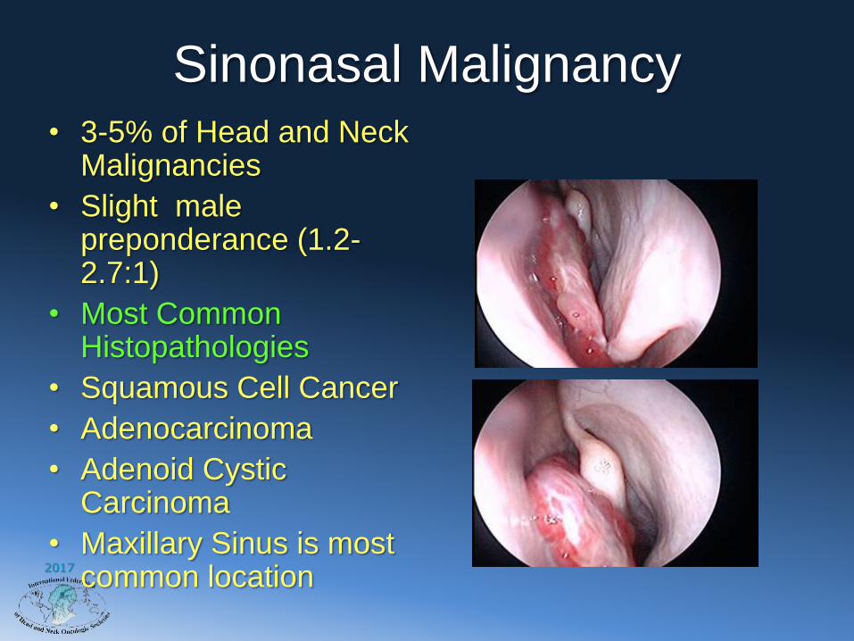
Sinonasal Malignancy• 3-5% of Head and Neck
Malignancies
• Slight male preponderance (1.2-2.7:1)
• Most Common Histopathologies
• Squamous Cell Cancer
• Adenocarcinoma
• Adenoid Cystic Carcinoma
• Maxillary Sinus is most common location

2017
Incidence by Sinus
• Maxillary sinus: 70-80 %
• Ethmoid sinus: 10-20%
• Frontal sinus:<5%
• Sphenoid sinus:<5%
• Nasal cavity: 20-30%

2017
Are we making ProgressThe Good News:
We are Doing Better!
Gil Z, et al. Improvement in survival during the past 4 decades among patients with anterior skull base cancer. Head Neck. 2012;34(9):1212-7.

2017
What is the right treatment?
• Surgery
– Endoscopic
– Open Surgery (Maxillectomy, Craniofacial Resection)
• Radiotherapy
• Chemotherapy
• Molecular targeted agents

2017
Clinical Practice Guidelines

2017
The Problem-Histology
Patel et al. Craniofacial Surgery for Malignant Skull Base Tumours, Cancer 2003;98:1179-87.
2017
Right Tool for the Right JobImpact of Skull Base Surgery
2017
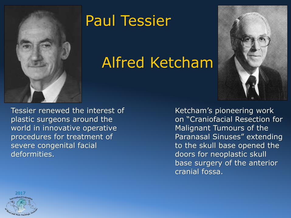
Paul Tessier
Alfred Ketcham
Tessier renewed the interest of plastic surgeons around the world in innovative operative procedures for treatment of severe congenital facial deformities.
Ketcham’s pioneering work on “Craniofacial Resection for Malignant Tumours of the Paranasal Sinuses” extending to the skull base opened the doors for neoplastic skull base surgery of the anterior cranial fossa.

2017
The Surgical Gold Standard
Ketcham AS. A combined intracranial facial approach to the paranasal sinuses. Am J Surg. 1963;106:698-703

2017
Evolution of Skull Base Surgery
Early pioneers
Open skull base
Radiosurgery
Endoscopic skull base
Multiport
Carl Snyderman
2017
Entr
y in
SB
S
Enth
usi
asm
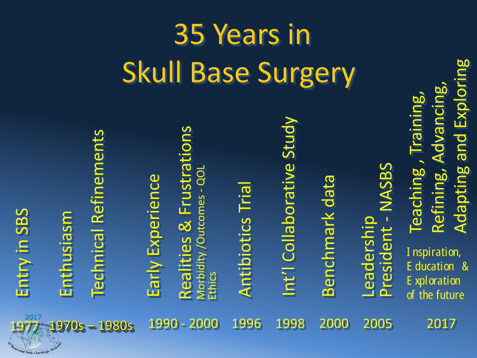
35 Years in Skull Base Surgery
Tech
nic
al R
efin
emen
ts
1977 1990 - 2000 19961970s – 1980s
Earl
y Ex
per
ien
ce
Rea
litie
s &
Fru
stra
tio
ns
Mo
rbid
ity
/Ou
tco
mes
-Q
OL
Eth
ics
2017
Teac
hin
g , T
rain
ing,
Ref
inin
g, A
dva
nci
ng,
Ad
apti
ng
and
Exp
lori
ng
2005
Lead
ersh
ipP
resi
den
t -
NA
SBS
1998
An
tib
ioti
cs T
rial
Int’
l Co
llab
ora
tive
Stu
dy
2000B
ench
mar
k d
ata
Inspiration, Education &Exploration of the future

2017
1960’s 1970’s 1980’s 1990’s 2017
Intr
od
uctio
n
Skep
ticis
m
Enth
usia
sm
Ag
gre
ssiv
en
ess
Techniq
ues
Experi
ence
Techniq
ues –
Com
plic
ations
Realit
ies
Outc
om
es –
QO
L
Cost
effectiven
ess
Eth
ics
0
10
20
30
40
50
60
60's 70's 80's 90's 00's
Survival %
Improved Survival1960 – 2017
“The great thing in the world is not so much where we stand,as in what direction we are moving.”
Oliver Wendell Holmes
Evolution in the Approaches in Skull Base Surgery:
Has Effected Outcome with-

2017
Definition of a Skull Base Tumour
“A tumour than transcends the skull base necessitating a combined intra
and extracranial approach for its ablation.”

2017
Skull Base Classification 1989
Region I Anterior
Region II Anterolateral
Region III Lateral/
Posterolateral
Irish, Gullane, Gentili, Dolan 1988

2017
Region I
• Tumours Involving the Anterior Cranial fossa
• Cancers arising– Sinuses– Orbit– Bone of skull
base– Skin– Intracranial
origin

2017
Region I -Selection of Approach
Open approach vs. Endoscopic
Depended on 3 Factors– Tumour type
– Extent of neoplasm
– Need for pre/post op radiation

2017
TreatmentOften Requires Combined Therapy
• Surgery
• Radiation
– IMRT
– Proton beam
–Neutron beam
• Chemotherapy

2017
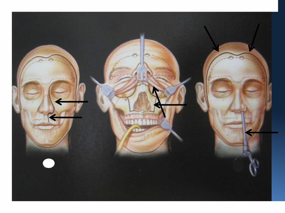
Conventional Surgical Approaches: Craniotomy + one below
• Degloving
± maxillotomy
± maxillectomy
± le Fort I
• Lateral rhinotomy ± medial maxillectomy
• Weber Ferguson Incision
± maxillectomy ± orbit
± maxillotomy
• Supra maxillectomy ± skin, orbit
• Extended maxillectomy ± skin, orbit
• Subcranial approach
• Trans-orbital approach

2017
Realised Reconstruction was VitalLesson Learned
• Closure withoutspecific reconstruction
• Local flaps
• Pedicled flaps
• Free flaps
Pitfalls of no reconstruction.

2017
How Reconstruction Impacted the Outcome of Skull Base
Surgery.Toronto Experience
• 1988 – Dr.Alf Ketcham
• “Born 20 years too soon”
Development

2017
Goals of Cranial Base Reconstruction
• Provision of secure dural seal – Earlymajor complication using endoscopic approach
• Dead space obliteration• Suspension and support of neural
structures• Provision of bone and soft tissue cover• Maintenance of function• Achievement of optimal cosmesis

2017
Reconstructive Options Region I-Limited Defect
Pericranial flap
+ Skin Graft70%
Fasciocutaneous flapForearmLateral ArmAnterolateral Thigh

2017
Analysis of Best ApproachWithin Each Region
When to use Endoscopic or
Open Resection?
Controversial and
Revolutionary

2017
Open approach
Endoscopic
KassamSnyderman
Beginning of the RevolutionShah, Draft, Stamm, Stamberger, Wilson
Planning the Next Attack on Skull Base Cancer2006-Open or Endoscopic?Revolution
vs

2017
Conventional Approaches
• Conventional –
• Lateral Rhinotomy + Craniotomy
• Recognised that Improved Reconstruction reduced complication rate and changed the outcome.
2017

2017
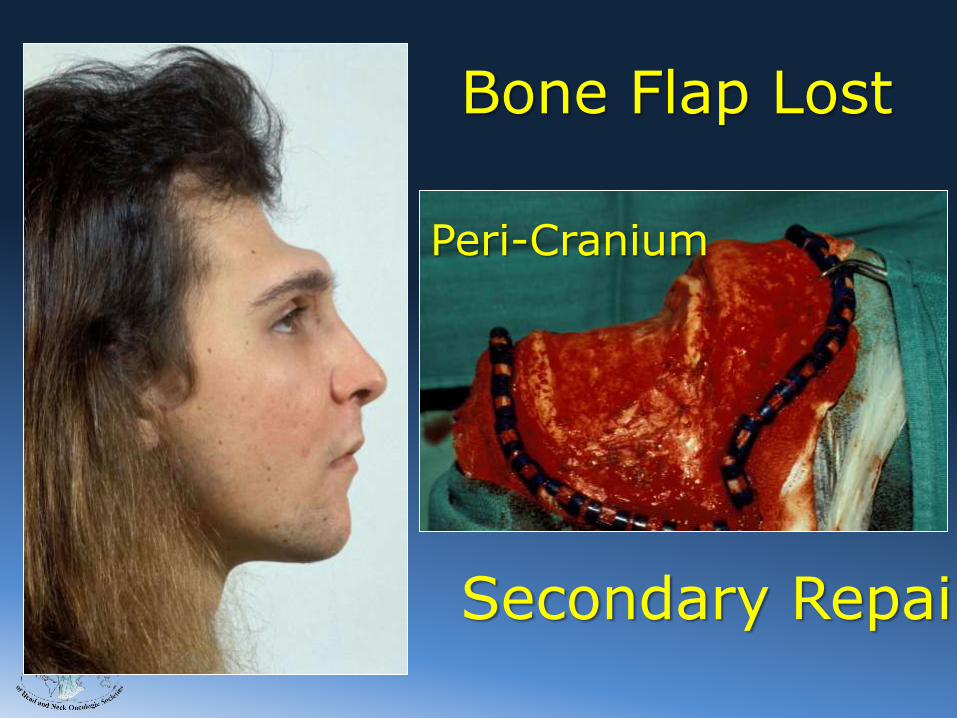
1989-18 year old: Poor Results SCC Right Ethmoid Sinus

2017
Pneumocephalus
2017
Bone Flap Lost
Secondary Repair
Peri-Cranium

2017
7 years post op
Secondary Reconstruction
Hydroxyapatite

2017
LateralRhinotomy
CraniotomyPericranialFlap
Proptosis-Chondrosarcoma

2017
10 yrs Postop
Rectus Abdominis Muscle
Inferior Epigastric Artery

2017
Approach to Extensive Tumours
Tumours that involve brain, orbit and skin.
“Will always need an open approach in my opinion”

2017
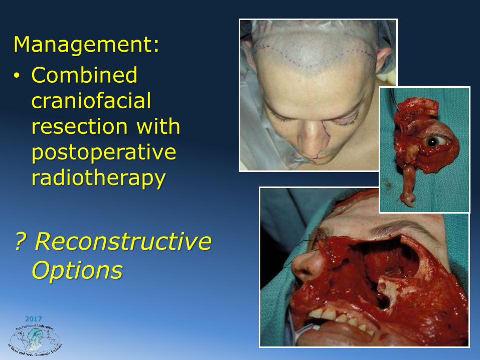
Selection of Approach-Eye invaded
• 29 year old University Professor of music
• 3 month history of diplopia
• Diagnosis - left ethmoid retro orbital squamous cell carcinoma
Open most appropriate

2017
Investigations:• CT Scan• MRI Scan
• The imaging revealed invasion of the anterior skull base, orbit and maxillary sinus.
Options in treatment?
2017
Management:
• Combined craniofacial resection with postoperative radiotherapy
? Reconstructive Options

2017

2017
3 weeks post-op 6 months post-op and 66 Gy radiation
Orbital defect – How would you manage it?

2017
9 years post-op 15 years post-op
2017
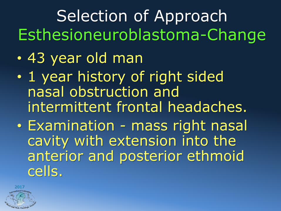
Selection of ApproachEsthesioneuroblastoma-Change
• 43 year old man
• 1 year history of right sided nasal obstruction and intermittent frontal headaches.
• Examination - mass right nasal cavity with extension into the anterior and posterior ethmoid cells.

2017
Investigations:–CT Scan
–MRI Scan
–Biopsy -Esthesioneuroblastoma
What approach wouldyou use to-day?
Endoscopic approach

2017
Management – 1996?
Endoscopic – 2017
• Combined craniofacial resection with postoperative radiotherapy.
• Craniofacial resection with postop irradiation and chemotherapy.
• Endoscopic transnasal excision with postoperative radiotherapy.
• Irradiation alone.5 yrs post-op

2017
Intracranial En-Plaque Recurrence at 7 yrs
Maybe endoscopic resection with post-operative radiotherapy may reduce the possibility of
en-plaque recurrence? Reduce risk of dural seeding
Irish JC, Dasgupta R, Freeman J, Gullane PJ, Gentili F, Brown D, Neligan P, O’Sullivan B. Outcome and Analysis of the Surgical Management of
Esthesioneuroblastoma. Jour of Otol. 26(1): 1-7, 1997

2017
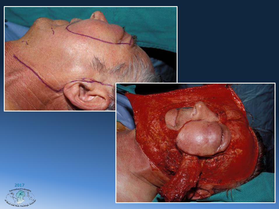
Squamous Cell
Carcinoma left
maxilla/skull base invasion
2017

2017
Combined Cranofacial Resection
Rectus Abdominis Flap

2017
8 years post Combined Therapy
Minimal access surgery not an option.
2017
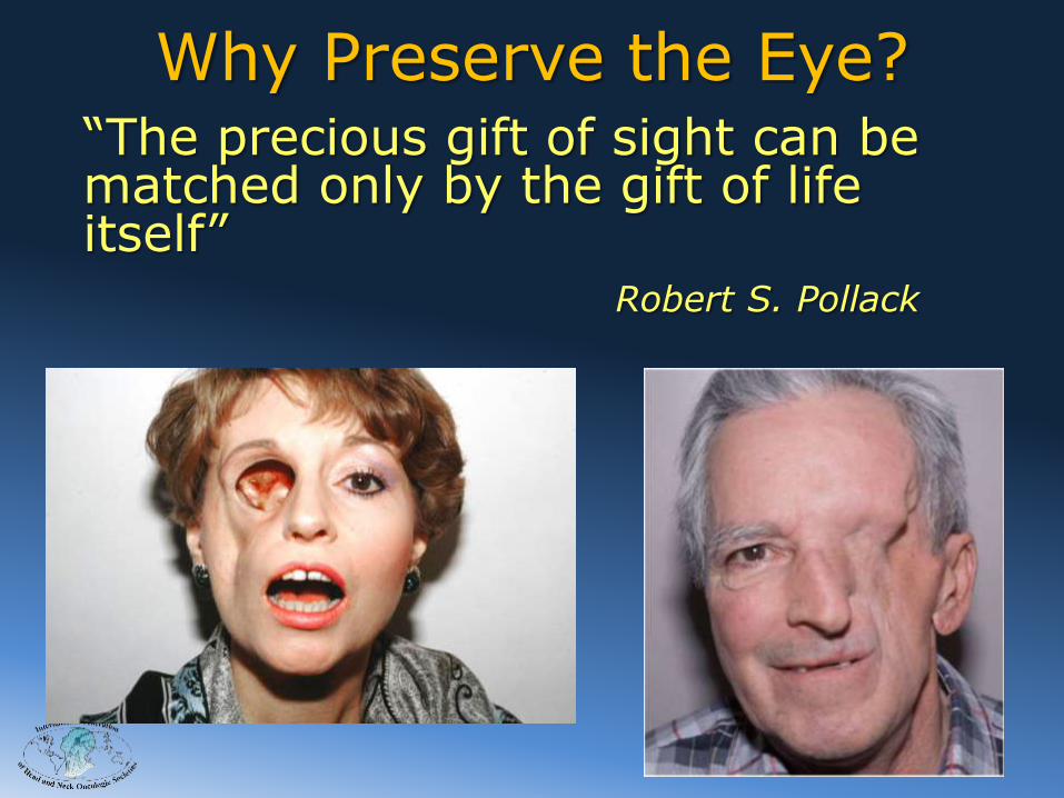
Why Preserve the Eye?“The precious gift of sight can be matched only by the gift of life itself”
Robert S. Pollack

2017
What about Quality of Life?

2017
How Reconstruction Impacted the Outcome of Skull Base Surgery.
Toronto Experience
• 1988 – Ketcham
• “Born 20 years too soon”
Development

2017 Flap Selection in Cranial Base Reconstruction.Plastic & Reconstructive Surgery. 98(7):1159-1166, December 1996.
Neligan, P. C. M.B., F.R.C.S.(C); Mulholland, S. M.D.; Irish, J. M.D., F.R.C.S.(C); Gullane, P. J. M.B., F.R.C.S.(C); Boyd, J. B. M.D., F.R.C.S.(C); Gentili, F. M.D., F.R.C.S.(C); Brown, D. M.D., F.R.C.S.(C); Freeman, J. M.D.,
F.R.C.S.(C)
Impact of Cranial Base Reconstruction

2017
Impact of Cranial Base ReconstructionUncomplicated Wound Healing

2017
Impact of Cranial Base Reconstruction

2017
Impact of Cranial Base Reconstruction

2017
Impact of Cranial Base ReconstructionIncidence of Abscess
2017
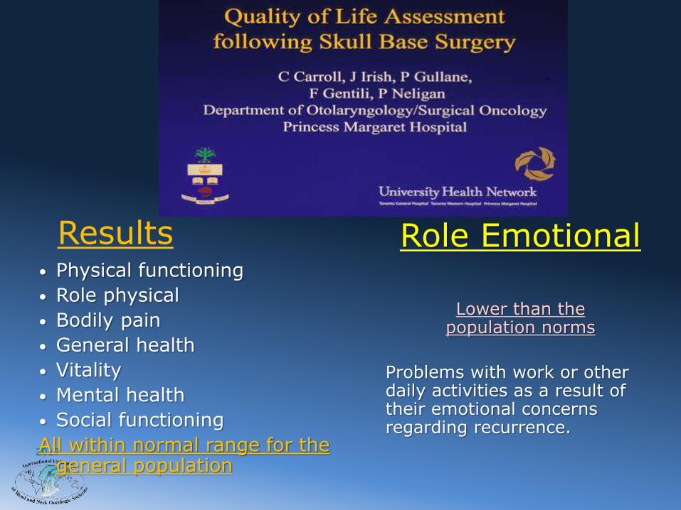
Results• Physical functioning
• Role physical
• Bodily pain
• General health
• Vitality
• Mental health
• Social functioning
All within normal range for the general population
Role Emotional
Lower than thepopulation norms
Problems with work or other daily activities as a result of their emotional concerns regarding recurrence.

2017
VSOpen Endoscopic
We Are at a Crossroads

2017
The Trade-off
• Open Approaches
– Better visualization
– En bloc resection
– Dealing with vascular injuries
– Access for more extensive surgery
• Endoscopic Approaches
– Better visualization
– Less brain retraction
– Lower complication rates?
– Better quality of life
– Shorter hospital stay
✔
✖
✔
✔
✔
✔
✔
✔✖
✔

2017
Entr
y in
SB
S
Enth
usi
asm
35 Years in Skull Base Surgery
Tech
nic
al R
efin
emen
ts
1977 1990 - 20001970s – 1980s
Earl
y Ex
per
ien
ce
Rea
litie
s &
Fru
stra
tio
ns
Mo
rbid
ity
/Ou
tco
mes
-Q
OL
Eth
ics
• Complications - 25-40%
• Infection was a problem
• Reported outcomes data were not satisfactory
• Case selection was a problem
• Quality of life of the patient was not studied

2017
Mortality/MorbidityFrom Skull Base Surgery
Complications +40%Overall
Mortality< 10%
Minor +15% Complications
Major +25%Complications
Collective review from literature in 1990

2017
Complications of Craniofacial Surgery
• Infection• Wound sepsis• Osteomyelitis• Meningitis
Avoidance
• Smaller bone flaps• Subcranial approach• Isolation of bone flapwith galeal-pericranialflap
• “Wrapping technique”

2017
Entr
y in
SB
S
Enth
usi
asm
35 Years in Skull Base Surgery
Tech
nic
al R
efin
emen
ts
1977 1990 - 2000 19961970s – 1980s
Earl
y Ex
per
ien
ce
Rea
litie
s &
Fru
stra
tio
ns
Mo
rbid
ity
/Ou
tco
mes
-Q
OL
Eth
ics
An
tib
ioti
cs T
rial
• Prophylactic antibiotics were given at random
• Choice of antibiotics was random
• The length of time for antibiotics was variable
• A definitive study was necessary

2017
Study of Prophylactic Antibiotics for
Cranio Facial Surgery• Audit of all infectious complications till 1996
• Review of culture reports
• Preliminary trial with three sets of Antibiotic regimen
• Optimal choice of Antibiotic combination
• Ceftazidime, Vancomycin
and MetronidazolRate of Infectious complications is down to less than 2%, since 1996, and all are minor.
Sal Caruana and Dennis Kraus ( Triological Thesis)

2017
Entr
y in
SB
S
Enth
usi
asm
35 Years in Skull Base Surgery
Tech
nic
al R
efin
emen
ts
1977 1990 - 2000 19961970s – 1980s
Earl
y Ex
per
ien
ce
Rea
litie
s &
Fru
stra
tio
ns
Mo
rbid
ity
/Ou
tco
mes
-Q
OL
Eth
ics
1998
An
tib
ioti
cs T
rial
Int’
l Co
llab
ora
tive
Stu
dy
2000B
ench
mar
k d
ata • No reliable
data were available.
• Benchmark data were necessary to move forward

2017
Results of Skull Base Surgery for Malignant Tumours 1960-2000
5 year survival
Ketchum 1963– 1960’s – 1970’s 50%
Cheeseman– 1970’s – 1980’s 50%
Shah 1992– 1980’s – 1990’s 63%
Irish, Gullane, Gentili 1994– 1983 – 1992
54%
2017
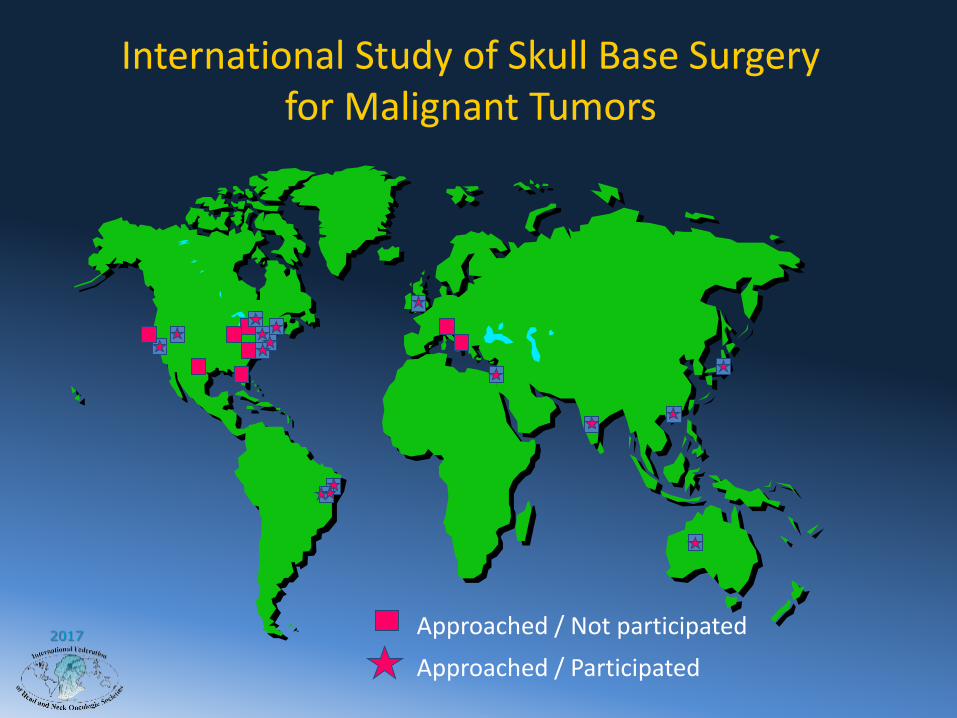
International Study of Skull Base Surgeryfor Malignant Tumors
Approached / Not participated
Approached / Participated
2017
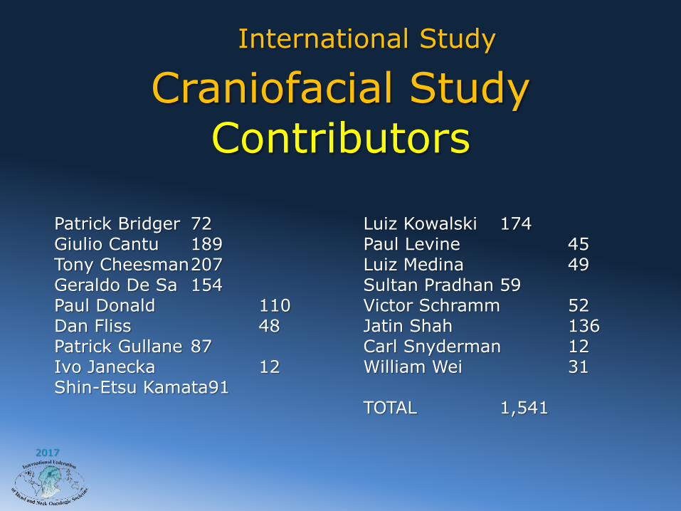
Craniofacial Study Contributors
International Study
Patrick Bridger 72Giulio Cantu 189Tony Cheesman207Geraldo De Sa 154Paul Donald 110Dan Fliss 48Patrick Gullane 87Ivo Janecka 12Shin-Etsu Kamata91
Luiz Kowalski 174Paul Levine 45Luiz Medina 49Sultan Pradhan 59Victor Schramm 52Jatin Shah 136Carl Snyderman 12William Wei 31
TOTAL 1,541

2017
• 1541 patients treated between 1956-2000 were accrued
• Exclusions:No pathologic information = 26 (2%)No Follow-up data = 88 (6%)Benign tumors = 120 (8%)
• 1307 patients eligible for analysis
International Collaborative Study Group
Craniofacial Surgery for Malignant Skull Base Tumors

2017
0.0
0.1
0.2
0.3
0.4
0.5
0.6
0.7
0.8
0.9
1.0
Pro
port
ion S
urv
ivin
g
0 12 24 36 48 60 72 84 96 108 120
Follow up Interval (Months)
5-year recurrence free survival 53%
ICSG for CFS Survival

2017
0.0
0.1
0.2
0.3
0.4
0.5
0.6
0.7
0.8
0.9
1.0
Pro
po
rtio
n S
urv
ivin
g
0 12 24 36 48 60 72 84 96 108 120
Follow-up Interval (Months)
Group I: ENB, Skin, 79% Low grade sarcoma
Group 2: Hi grade sarcoma, SCC, 57% Salivary, Other malignancies
Group 3: Mucosal melanoma, 30% Undifferentiated/anaplastic
ICSG for CFS
DSS: Impact of Histology
p<.0001

2017
ICSG for CFS
DSS: Impact of Intracranial Extension
0.0
0.1
0.2
0.3
0.4
0.5
0.6
0.7
0.8
0.9
1.0
Pro
po
rtio
n S
urv
ivin
g
0 12 24 36 48 60 72 84 96 108 120
Follow-up Interval (Months)
None 66%
Bone 59%
Dura 54%
Brain 29%
p<.0001

2017
ICSG for CFS
Margins of Surgical Resection
Close
17%
Negative
63%
Positive
15%
*Data not available in 185 patients (14%)

2017
0.0
0.1
0.2
0.3
0.4
0.5
0.6
0.7
0.8
0.9
1.0
Pro
po
rtio
n S
urv
ivin
g
0 12 24 36 48 60 72 84 96 108 120
Follow-up Interval (Months)
Negative 74%
Positive 38%
ICSG for CFS DSS: Impact of Surgical Margin Status

2017
ICSG for CFS Prognostic Predictors of Disease-
Specific Survival
Prognostic covariate RFS DSS OS
Uni Multi Uni Multi Uni Multi
Age NS - NS - NS -
Gender NS - NS - NS -
Medical Comorbidity NS - NS - SIG SIG
Anatomic Location SIG NS SIG NS SIG NS
Histology SIG SIG SIG SIG SIG SIG
Orbital Involvement SIG NS SIG NS SIG NS
Intracranial
Involvement
SIG SIG SIG SIG SIG SIG
Surgical Margins SIG SIG SIG SIG SIG SIG

2017
Predictors of Survival
Independent predictors of overall, disease-specific and recurrence-free survival on multivariate analysis were:
1. Status of surgical margins
2. Histology
3. Extracranial extent
Head and Neck, Vol 27, # 6, Pgs: 445-451, June 2005.

2017
Complications
Head and Neck, Vol 27, #6, Pgs: 445-451, June 2005

2017
Factors Associated with Complications

2017
Benefits from the International Collaborative Study
• Benchmark outcomes data are now available from across the world
• Factors impacting upon outcomes are identified by multivariate analysis
• Case selection is made possible for better outcomes
• Outcomes from future interventions will have to be compared with these data.

2017
Entr
y in
SB
S
Enth
usi
asm
35 Years in Skull Base Surgery
Tech
nic
al R
efin
emen
ts
1977 1990 - 2000 19961970s – 1980s
Earl
y Ex
per
ien
ce
Rea
litie
s &
Fru
stra
tio
ns
Mo
rbid
ity
/Ou
tco
mes
-Q
OL
Eth
ics
Wisdom
2005
Lead
ersh
ipP
resi
den
t -
NA
SBS
1998
An
tib
ioti
cs T
rial
Int’
l Co
llab
ora
tive
Stu
dy
2000B
ench
mar
k d
ata

2017
Case selectionCost effectiveness
EconomicsEthics
Biology of the tumorQuality of life
Issues / Wisdom
Progress in Skull Base Surgery

2017
Disadvantages of External Skull Base Surgery
• External incisions
• Need for a craniotomy
• Morbidity and complications
• Cost (Length of Hospitalization)
• Esthetic and functional outcomes
• Quality of life

2017
How to reduce the Morbidityof External Skull Base Surgery

2017
Advantages of EndonasalSkull Base Surgery
• Avoids external incisions
• No esthetic sequela
• Avoids Craniotomy
• Reduced blood loss
• Total tumor resection possible
• Dural repair possible

2017
1,500 + Patients
Potential Candidates for Endonasal skull base surgeryInternational Collaborative Study Group for Craniofacial Surgery
• Paranasal Sinus Tumors• 11% T1 or T2 – 21% No orbital or cranial involvement
• Esthesioneuroblastoma• 22% Kadish A – 40% no orbital or cranial involvement
• Low Grade Sarcoma• 10% T2 – 35%
• High Grade Sarcoma•25% T2 – 23%
2017
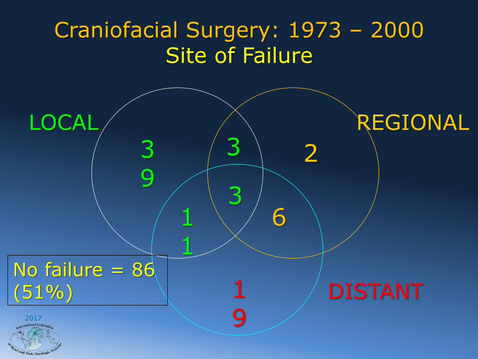
Craniofacial Surgery: 1973 – 2000Site of Failure
LOCAL REGIONAL
DISTANT
39
2
19
3
361
1No failure = 86 (51%)

2017
Local Recurrence is the most common cause of treatment
failure
• Case Selection•Favorable histology• Monobloc resection• Secure negative margins• Post operative R.T.
Outcome Prevention

2017
-- Issues of Concern --
Endonasal Endoscopic Surgery for
Malignant Tumors Involving the Skull Base
• Safety• Operative• Oncologic
• Expertise• Learning Curve
• Case Selection• Anatomic Extent• Histology• Biology

2017
Entr
y in
SB
S
Enth
usi
asm
35 Years in Skull Base Surgery
Tech
nic
al R
efin
emen
ts
1977 1990 - 2000 19961970s – 1980s
Earl
y Ex
per
ien
ce
Rea
litie
s &
Fru
stra
tio
ns
Mo
rbid
ity
/Ou
tco
mes
-Q
OL
Eth
ics
2017
Teac
hin
g , T
rain
ing,
Ref
inin
g, A
dva
nci
ng,
Ad
apti
ng
and
Exp
lori
ng
2005
Lead
ersh
ipP
resi
den
t -
NA
SBS
1998
An
tib
ioti
cs T
rial
Int’
l Co
llab
ora
tive
Stu
dy
2000B
ench
mar
k d
ata
Inspiration, Education &Exploration of the future

2017
Case Selection (Endonasal resection) Select HistologyWide resection (Dura,Brain?,Orbit,Bone)Secure marginsDural repair / graftReconstruction with free flapsTime tested techniquesOutcomes data available
Wisdom

2017
• CFR is a safe and effective treatment option for malignant skull base tumors.
• Histology of the primary tumor, its intracranial extent and status of surgical margins are independent predictors of RFS, OS and DSS.
• Future role of Endoscopic Approaches still in development and we must be diligent to monitor progress using this approach.
ICSG for CFSConclusions

2017
ENDOSCOPIC APPROACH
OPEN APPROACH
NASAL CAVITY TUMORS ETHMOID TUMORSSKULL BASE EROSION
MAXILLARY SINUS TUMORSORBITAL INVASION PALATE INVASIONNASAL BONE INVASIONSKIN/SOFT TISSUE
DURAL DISEASEBRAIN INVASION
Choosing the Right Approach

2017
ENDOSCOPIC APPROACH OPEN APPROACH
NASAL CAVITY TUMORS ETHMOID TUMORSSKULL BASE EROSION
MAXILLARY SINUS TUMORSORBITAL INVASION PALATE INVASIONNASAL BONE INVASIONSKIN/SOFT TISSUE
DURAL DISEASEBRAIN INVASION
Choosing the Right Approach
courtesy John de Almeida

2017
OPEN CRANIOTOMY
ENDOSCOPIC

2017
Analysis of Best ApproachWithin Each Region
When to use Endoscopic or
Open Resection?
Controversial and
Revolutionary

2017
“The future just ain’twhat it used to be”
Mark Twain
CRANIOTOMY

2017
• “It has become appallingly obviousthat our technology has exceededour humanity.”
Albert Einstein

2017
Am J Rhinol 22, 308-316, 2008

2017
Histology

2017
Follow Up & Recurrence

2017
Future of Skull Base Oncology
“The best way to predict the
future is to invent it.”Alan Kay
2017
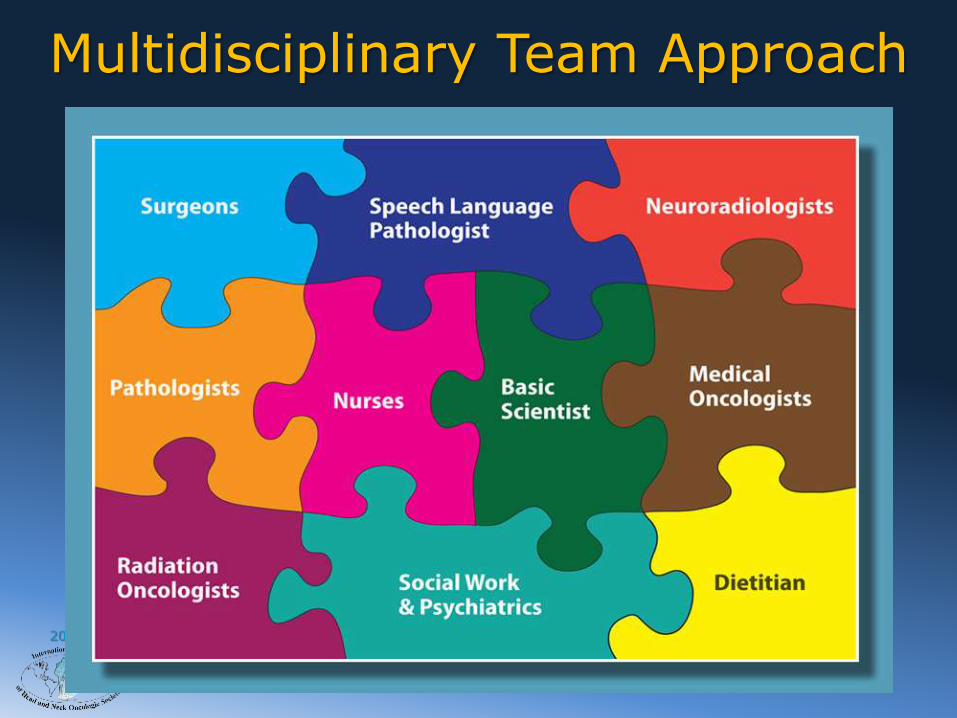
Multidisciplinary Team Approach

2017
Our Future-Read the Book –A brief history of To-Morrow


