Page 1
FINAL
9-17-2014
Provisional Peer-Reviewed Toxicity Values for
Ethyl Acrylate (CASRN 140-88-5)
Superfund Health Risk Technical Support Center
National Center for Environmental Assessment
Office of Research and Development
U.S. Environmental Protection Agency
Cincinnati, OH 45268
Page 2
i
AUTHORS, CONTRIBUTORS, AND REVIEWERS
CHEMICAL MANAGERS
Jason C. Lambert, PhD, DABT
National Center for Environmental Assessment, Cincinnati, OH
Carrie Fleming, PhD
National Center for Environmental Assessment, Cincinnati, OH
DRAFT DOCUMENT PREPARED BY
SRC, Inc.
7502 Round Pond Road
North Syracuse, NY 13212
PRIMARY INTERNAL REVIEWERS
Sanju Diwan, PhD
National Center for Environmental Assessment, Washington, DC
Anuradha Mudipalli, MSc, PhD
National Center for Environmental Assessment, Research Triangle Park, NC
This document was externally peer reviewed under contract to
Eastern Research Group, Inc.
110 Hartwell Avenue
Lexington, MA 02421-3136
Questions regarding the contents of this document may be directed to the U.S. EPA Office of
Research and Development’s National Center for Environmental Assessment, Superfund Health
Risk Technical Support Center (513-569-7300).
Page 3
ii
TABLE OF CONTENTS
BACKGROUND ............................................................................................................................ 1
DISCLAIMERS .............................................................................................................................. 1
QUESTIONS REGARDING PPRTVs ........................................................................................... 1
INTRODUCTION .......................................................................................................................... 2
REVIEW OF PERTINENT DATA ................................................................................................ 5
HUMAN STUDIES .................................................................................................................... 5
Oral Exposure ......................................................................................................................... 5
Inhalation Exposure ................................................................................................................ 5
ANIMAL STUDIES ................................................................................................................... 6
Oral Exposure ......................................................................................................................... 6
Inhalation Exposure .............................................................................................................. 17
OTHER STUDIES .................................................................................................................... 24
Toxicokinetics ....................................................................................................................... 24
Acute or Short-term Studies .................................................................................................. 26
Other Routes ......................................................................................................................... 29
Mechanistic Studies .............................................................................................................. 29
Genotoxicity .......................................................................................................................... 31
DERIVATION OF PROVISIONAL SUBCHRONIC AND CHRONIC RfD VALUES FOR
ETHYL ACRYLATE ................................................................................................................... 34
SUBCHRONIC p-RfD.............................................................................................................. 34
CHRONIC p-RfD ..................................................................................................................... 38
DERIVATION OF PROVISIONAL SUBCHRONIC AND CHRONIC INHALATION RfC
VALUES FOR ETHYL ACRYLATE .......................................................................................... 40
SUBCHRONIC AND CHRONIC p-RfC ................................................................................. 43
PROVISIONAL CARCINOGENICITY ASSESSMENT FOR ETHYL ACRYLATE .............. 45
WEIGHT-OF-EVIDENCE (WOE) DESCRIPTOR ................................................................. 45
MODE-OF-ACTION DISCUSSION ....................................................................................... 46
Key Events ............................................................................................................................ 46
Strength, Consistency, and Specificity of Association ......................................................... 47
Dose-response Concordance ................................................................................................. 47
Temporal Relationships ........................................................................................................ 47
Biological Plausibility and Coherence .................................................................................. 49
Conclusion ............................................................................................................................ 50
QUANTITATIVE ESTIMATES OF CARCINOGENIC RISK .............................................. 50
Oral Exposure ....................................................................................................................... 50
Inhalation Exposure .............................................................................................................. 50
APPENDIX A. DERIVATION OF A SCREENING SUBCHRONIC ORAL VALUE FOR
ETHYL ACRYLATE (CASRN 140-88-5) .................................................................................. 51
APPENDIX B. DETAILS OF BENCHMARK DOSE MODELING FOR SCREENING
SUBCHRONIC p-RfD.................................................................................................................. 54
APPENDIX C. DETAILS OF BENCHMARK DOSE MODELING FOR CHRONIC p-RfD . 60
APENDIX D. REFRENCES ....................................................................................................... 80
Page 4
iii
COMMONLY USED ABBREVIATIONS AND ACRONYMS
α2u-g alpha 2u-globulin
ACGIH American Conference of Governmental
Industrial Hygienists
AIC Akaike’s information criterion
ALD approximate lethal dosage
ALT alanine aminotransferase
AST aspartate aminotransferase
atm atmosphere
ATSDR Agency for Toxic Substances and
Disease Registry
BMD benchmark dose
BMDL benchmark dose lower confidence limit
BMDS Benchmark Dose Software
BMR benchmark response
BUN blood urea nitrogen
BW body weight
CA chromosomal aberration
CAS Chemical Abstracts Service
CASRN Chemical Abstracts Service Registry
Number
CBI covalent binding index
CHO Chinese hamster ovary (cell line cells)
CL confidence limit
CNS central nervous system
CPN chronic progressive nephropathy
CYP450 cytochrome P450
DAF dosimetric adjustment factor
DEN diethylnitrosamine
DMSO dimethylsulfoxide
DNA deoxyribonucleic acid
EPA Environmental Protection Agency
FDA Food and Drug Administration
FEV1 forced expiratory volume of 1 second
GD gestation day
GDH glutamate dehydrogenase
GGT γ-glutamyl transferase
GSH glutathione
GST glutathione-S-transferase
Hb/g-A animal blood-gas partition coefficient
Hb/g-H human blood-gas partition coefficient
HEC human equivalent concentration
HED human equivalent dose
i.p. intraperitoneal
IRIS Integrated Risk Information System
IVF in vitro fertilization
LC50 median lethal concentration
LD50 median lethal dose
LOAEL lowest-observed-adverse-effect level
MN micronuclei
MNPCE micronucleated polychromatic
erythrocyte
MOA mode-of-action
MTD maximum tolerated dose
NAG N-acetyl-β-D-glucosaminidase
NCEA National Center for Environmental
Assessment
NCI National Cancer Institute
NOAEL no-observed-adverse-effect level
NTP National Toxicology Program
NZW New Zealand White (rabbit breed)
OCT ornithine carbamoyl transferase
ORD Office of Research and Development
PBPK physiologically based pharmacokinetic
PCNA proliferating cell nuclear antigen
PND postnatal day
POD point of departure
POD[ADJ] duration-adjusted POD
QSAR quantitative structure-activity
relationship
RBC red blood cell
RDS replicative DNA synthesis
RfC inhalation reference concentration
RfD oral reference dose
RGDR regional gas dose ratio
RNA ribonucleic acid
SAR structure activity relationship
SCE sister chromatid exchange
SD standard deviation
SDH sorbitol dehydrogenase
SE standard error
SGOT glutamic oxaloacetic transaminase, also
known as AST
SGPT glutamic pyruvic transaminase, also
known as ALT
SSD systemic scleroderma
TCA trichloroacetic acid
TCE trichloroethylene
TWA time-weighted average
UF uncertainty factor
UFA interspecies uncertainty factor
UFH intraspecies uncertainty factor
UFS subchronic-to-chronic uncertainty factor
UFD database uncertainty factor
U.S. United States of America
WBC white blood cell
Page 5
FINAL
9-17-2014
1 Ethyl acrylate
PROVISIONAL PEER-REVIEWED TOXICITY VALUES FOR
ETHYL ACRYLATE (CASRN 140-88-5)
BACKGROUND
A Provisional Peer-Reviewed Toxicity Value (PPRTV) is defined as a toxicity value
derived for use in the Superfund Program. PPRTVs are derived after a review of the relevant
scientific literature using established Agency guidance on human health toxicity value
derivations. All PPRTV assessments receive internal review by a standing panel of National
Center for Environment Assessment (NCEA) scientists and an independent external peer review
by three scientific experts.
The purpose of this document is to provide support for the hazard and dose-response
assessment pertaining to chronic and subchronic exposures to substances of concern, to present
the major conclusions reached in the hazard identification and derivation of the PPRTVs, and to
characterize the overall confidence in these conclusions and toxicity values. It is not intended to
be a comprehensive treatise on the chemical or toxicological nature of this substance.
The PPRTV review process provides needed toxicity values in a quick turnaround
timeframe while maintaining scientific quality. PPRTV assessments are updated approximately
on a 5-year cycle for new data or methodologies that might impact the toxicity values or
characterization of potential for adverse human health effects and are revised as appropriate. It is
important to utilize the PPRTV database (http://hhpprtv.ornl.gov) to obtain the current
information available. When a final Integrated Risk Information System (IRIS) assessment is
made publicly available on the Internet (http://www.epa.gov/iris), the respective PPRTVs are
removed from the database.
DISCLAIMERS
The PPRTV document provides toxicity values and information about the adverse effects
of the chemical and the evidence on which the value is based, including the strengths and
limitations of the data. All users are advised to review the information provided in this
document to ensure that the PPRTV used is appropriate for the types of exposures and
circumstances at the site in question and the risk management decision that would be supported
by the risk assessment.
Other U.S. Environmental Protection Agency (EPA) programs or external parties who
may choose to use PPRTVs are advised that Superfund resources will not generally be used to
respond to challenges, if any, of PPRTVs used in a context outside of the Superfund program.
QUESTIONS REGARDING PPRTVs
Questions regarding the contents and appropriate use of this PPRTV assessment should
be directed to the EPA Office of Research and Development’s National Center for
Environmental Assessment, Superfund Health Risk Technical Support Center (513-569-7300).
Page 6
FINAL
9-17-2014
2 Ethyl acrylate
INTRODUCTION
Ethyl acrylate (2-propenoic acid, ethyl ester, C5H8O2) is a colorless liquid with a
penetrating acrid odor. See Figure 1 for chemical structure of ethyl acrylate. It is soluble in
ethanol, ether, and chloroform and is slightly soluble in water (NTP, 1986). Ethyl acrylate is
used to produce polymers and copolymers for latex paints, textiles, paper coatings and fabric
finishes, and has been used as a fragrance since the 1950s. It also occurs naturally in pineapples
and raspberries and has been approved by the U.S. Food and Drug Administration as a flavoring
agent (NTP, 1986). A table of physicochemical properties is provided below (see Table 1).
Figure 1. Chemical Structure of Ethyl Acrylate
Table 1. Physicochemical Properties Table (Ethyl Acrylate)a
Property (unit) Value
Boiling point (°C) 99.8 at 760 mm Hg
Melting point (°C) -71.2
Density (g/cm3) 0.9234
Vapor pressure (mm Hg at 20°C) 29
Solubility in water (mg/ mL at 20°C) 10−50
Molecular weight (g/mol) 100.12
Flash point (°C) 9
Octanol/water partition coefficient (Log P) 3.5
aNTP (1998).
A summary of available toxicity values for ethyl acrylate (CASRN 140-88-5) from
U.S. EPA and other agencies/organizations is provided in Table 2.
Page 7
FINAL
9-17-2014
3 Ethyl acrylate
Table 2. Summary of Available Toxicity Values for Ethyl Acrylate (CASRN 140-88-5)
Source/Parametera
Value
(Applicability) Notes Reference Date Accessed
Noncancer
ACGIH 8-hr TLV-TWA:
5 ppm (20 mg/m3)
15-min
TLV-STEL:
15 ppm
TLVs based on upper respiratory
tract, gastrointestinal, eye, and skin
irritation; central nervous system
impairment; and skin sensitization.
ACGIH (2013) NA
ATSDR NV NA ATSDR (2013) NA
Cal/EPA NV NA Cal/EPA (2014a)b 9-10-2014b
NIOSH IDLH: 300 ppm IDLH is based on toxicity data in
humans (Nemec and Bauer, 1978)
and animals (Oberly and Tansy,
1985; de Ceaurriz et al., 1981;
Pozzani et al., 1949; Treon et al.,
1949).
NIOSH (1995) NA
OSHA PEL: 25 ppm
(100 mg/m3)
PEL is for occupational exposure to
ethyl acrylate, with skin irritation as
a potential concern.
OSHA (2011;
2006)
NA
IRIS NV NA U.S. EPA 9-10-2014
Drinking water NV NA U.S. EPA (2012a) NA
HEAST NV NA U.S. EPA (2011a) NA
CARA HEEP NV The CARA list includes a HEEP for
ethyl acrylate but no RfD or RfC
values.
U.S. EPA (1994a;
1987)
NA
WHO NV NA WHO 9-10-2014
Cancer
ACGIH WOE: A4 (“Not
Classifiable as a
Human
Carcinogen”)
NA ACGIH (2013) NA
IRIS NV NA U.S. EPA 9-10-2014
Drinking water NV NA U.S. EPA (2012a) NA
HEAST OSF: 4.8 × 10⁻²
(mg/kg-d)-1
IUR: 1.4 × 10⁻⁶
µg/L
WOE: B2
(“Probable Human
Carcinogen”)
Cites HEEP (U.S. EPA, 1987) as the
source of these values. The OSF
was based on an increased incidence
of squamous cell
papillomas/carcinomas of the
forestomach in male rats (NTP,
1986).
U.S. EPA (2011a) NA
Page 8
FINAL
9-17-2014
4 Ethyl acrylate
Table 2. Summary of Available Toxicity Values for Ethyl Acrylate (CASRN 140-88-5)
Source/Parametera
Value
(Applicability) Notes Reference Date Accessed
IARC WOE: Group 2B
(“Possibly
Carcinogenic to
Humans”)
Based on sufficient evidence of
carcinogenicity in experimental
animals
IARC (1999;
1986)
NA
NIOSH REL: “Ca”
(“Potential
Occupational
Carcinogen”;
exposure should be
limited to the
lowest feasible
concentration)
NA NIOSH (2010) NA
NTP NV NTP (1986) concluded that ethyl
acrylate was carcinogenic to the
forestomach of rats and mice in their
studies, but the chemical was
delisted during development of the
11th Report on Carcinogens (NTP,
2005) and remains delisted in the
12th Report on Carcinogens (NTP,
2011).
NTP (2011;
2005)
NA
Cal/EPA “Known to the
State [of
California] to
Cause Cancer”
NA Cal/EPA (2014b;
2011)b
9-10-2014b
aSources: American Conference of Governmental Industrial Hygienists (ACGIH); Agency for Toxic Substances and
Disease Registry (ATSDR); California Environmental Protection Agency (Cal/EPA); National Institute for
Occupational Safety and Health (NIOSH); Occupational Safety and Health Administration (OSHA); Chemical
Assessments and Related Activities (CARA); Health and Environmental Effects Profile (HEEP); World Health
Organization (WHO); Integrated Risk Information System (IRIS); Health Effects Assessment Summary Tables
(HEAST); International Agency for Research on Cancer (IARC); National Toxicology Program (NTP). bThe Cal/EPA Office of Environmental Health Hazard Assessment (OEHHA) Toxicity Criteria Database
(http://oehha.ca.gov/tcdb/index.asp) was also reviewed and found to contain no information on ethyl acrylate.
IDLH = immediately dangerous to life or health; IUR = inhalation unit risk; NA = not applicable; NSRL = no
significant risk level; NV = not available; OSF = oral slope factor; PEL = permissible exposure level;
REL = recommended exposure level; STEL = short-term exposure limit; TLV = threshold limit value; TWA = time
weighted average.
Page 9
FINAL
9-17-2014
5 Ethyl acrylate
Literature searches were conducted on sources published from 1900 through August 2014
for studies relevant to the derivation of provisional toxicity values for ethyl acrylate (CASRN
140-88-5). The following databases were searched by chemical name, synonyms, or CASRN:
ACGIH, ANEUPL, ATSDR, BIOSIS, Cal EPA, CCRIS, CDAT, ChemIDplus, CIS, CRISP,
DART, EMIC, EPIDEM, ETICBACK, FEDRIP, GENE-TOX, HAPAB, HERO, HMTC, HSDB,
IARC, INCHEM IPCS, IPA, ITER, IUCLID, LactMed, NIOSH, NTIS, NTP, OSHA, OPP/RED,
PESTAB, PPBIB, PPRTV, PubMed (toxicology subset), RISKLINE, RTECS, TOXLINE, TRI,
U.S. EPA IRIS, U.S. EPA HEAST, U.S. EPA HEEP, U.S. EPA OW, and U.S. EPA
TSCATS/TSCATS2. The following databases were searched for toxicity values or exposure
limits: ACGIH, ATSDR, Cal/EPA, U.S. EPA IRIS, U.S. EPA HEAST, U.S. EPA HEEP,
U.S. EPA OW, U.S. EPA TSCATS/TSCATS2, NIOSH, NTP, OSHA, and RTECS.
REVIEW OF PERTINENT DATA
The phrase “statistical significance,” used throughout the document, indicates a p-value
of <0.05 unless otherwise noted.
HUMAN STUDIES
Oral Exposure
Human studies on oral exposure to ethyl acrylate were not located in the literature.
Inhalation Exposure
Occupational epidemiology studies of 13,863 white male workers from two U.S. plants
producing acrylic sheet were reported by Walker et al. (1991). In the Bristol, Pennsylvania
plant, two cohorts were evaluated (1) the Early Bristol cohort consisting of 3,934 individuals
employed between January 1, 1933, and December 31, 1945 (of which, approximately
74% employees were hired between 1941 and 1945), and (2) the Later Bristol cohort of
6,548 individuals hired between January 1, 1946, and December 31, 1986. In the Knoxville,
Tennessee plant, the cohort consisted of 3,381 workers employed between January 1, 1943, and
December 31, 1982. All groups were followed from the first day of employment or
January 1, 1933, whichever came later. Assessment of exposure to ethyl acrylate and/or methyl
methacrylate was based on job history and a job-specific exposure scale. The total exposure for
each job held by each worker was estimated by multiplying the exposure intensity by the interval
in days from the start to the end of employment in the job divided by 365.25. Mortality rates
(from death certificates) were tabulated, and standardized mortality rates were calculated to
assess whether occupational exposures were associated with increased incidences of colon and
rectal cancers. In the Early Bristol cohort, an excess of mortality due to colon cancer was
observed. Colon cancer-associated mortality appeared at least 20 years after the equivalent of
3 years of employment in jobs producing the highest exposure to ethyl acrylate and/or methyl
methacrylate vapor and volatile byproducts of polymerization. Cancer of the rectum was also
significantly increased in this cohort. However, assessment of the Later Bristol and Knoxville
cohorts did not show excess mortality from either colon or rectal cancer. Quantitative levels of
exposures to ethyl acrylate, methyl methacrylate, and byproducts of polymerization were not
available in any cohort. No adjustment was made for confounding variables such as age,
smoking, and alcohol consumption. No information was available on whether exposures
associated with job categories were different between the Early and Later Bristol cohorts. The
Page 10
FINAL
9-17-2014
6 Ethyl acrylate
study authors concluded that the excess of colon and rectal cancers in the Early Bristol study was
unlikely to be associated with acrylate exposure.
Rohm and Haas Co (1987) reported a statistically increased incidence of respiratory
cancers in the Knoxville plant workers (cohort described above) as compared with nonfactory
workers. However, there was no relationship to length of employment or to job categories with
the highest exposures. As noted for Walker et al. (1991), exposure was not quantified, and no
adjustment was made for confounding variables. This study is not suitable for quantitative risk
assessment.
A prospective cohort study on the effects of occupational exposure to chemicals
(including ethyl acrylate) involved in the production of acrylic acid, acrylic acid esters and
acrylate was conducted in 1992−1999 (Tucek et al., 2002). Exposure to the chemicals was
determined by personal passive dosimetry. Workers (60 exposed and 60 controls) were assessed
annually for general health (interview), a general medical examination, clinical chemistry
(aminotransferases, ALT, AST, GMT, alkaline phosphatase, glucose, total protein, uric acid,
triacylglycerols, cholesterol [total, HDL and LDL], urea, creatinine, and bilirubin), urinalysis
(pH, protein, glucose, acetone, urobilinogen, sediment), hematology (automated blood count),
serum immunity (immunoglobulins G, A, M, E; complements C3 and C4; lysozyme;
orosomucoid; transferring; prealbumin; ceruloplasmin, alpha-1-fetoprotein; alpha-1-antitrypsin;
alpha-2-macroglobulin; albumin; haptoglobin; hemopexin; C-reactive protein; rheumatoid factor;
antistreptolysin-O and circulating immunocomplexes), selected tumor markers
(carcinoembryonic antigen, neuron specific enolase, thymidine kinase), and spirometry.
Exposures were generally found to be low (below maximum allowable concentrations values or
suggested limits for each chemical). No differences were noted over the 8-year duration of the
study between control and exposed groups that could be attributed to acrylate exposure.
ANIMAL STUDIES
Oral Exposure
Subchronic-duration Studies
Bernacki et al. (1987a)
In an unpublished industry study, ethyl acrylate (>99% purity) was administered in the
drinking water of male and female F344 rats (40/group for males and 20/group for females) at
concentrations of 0 (water control); 200, 1,000, 2,000, or 4,000 ppm, 7 days/week, for 13 weeks
(Bernacki et al., 1987a). Based on the study authors’ calculations, compound intake averaged 0,
17, 70, 135, and 249 mg/kg-day, for males and 0, 20, 87, 161, and 293 mg/kg-day, for females.
Interim sacrifices consisted of 10 males/group after Study Weeks 1 and 2, and 10 rats/sex/group
after Study Week 4. Samples of drinking water were analyzed for ethyl acrylate concentrations
and did not differ significantly from target concentrations. Animals were observed twice daily
for mortality and morbidity during the week and once daily on weekends and holidays. Livers
and kidneys were weighed at the 4- and 13-week sacrifices. At all necropsy intervals, the entire
stomach was removed, weighed, dissected free of other tissues, and opened along the greater
curvature, weighed, and fixed for staining and analysis. The following tissues from all rats were
preserved similarly: liver, kidneys, heart, adrenals, thyroid/parathyroid, spleen, gonads,
esophagus (only at 13 weeks), and gross lesions. Histopathology was performed on
hematoxylin- and eosin-stained sections of both the forestomach and glandular stomach and on
gross lesions from all dose groups.
Page 11
FINAL
9-17-2014
7 Ethyl acrylate
No deaths or clinical signs of toxicity were reported (Bernacki et al., 1987a). Male body
weights were significantly decreased in all treatment groups (4, 9, 15, and 17% less than controls
from low through high doses, respectively; p < 0.05), whereas there were no changes in female
body weight throughout the study. Food consumption was decreased in males from all treated
groups throughout the study and in females receiving ≥87 mg/kg-day. A dose-dependent
decrease in water consumption (~20−40% less than controls, p < 0.05) was observed in both
sexes. The study authors considered the effects on male body weights to be secondary to
decreased drinking water and unrelated to treatment. However, it is not clear whether the
treatment-related changes in water consumption resulted from unpalatability, or whether they
may have been related to irritation of the stomach, as described below. Dose- and time-related
changes in both absolute and relative stomach weights were noted at all necropsy intervals.
After Week 1, absolute and relative stomach weights were increased in the high-dose male
group. Following Week 2, relative stomach weights—but not absolute stomach weights—were
increased at concentrations ≥70-mg/kg-day male group. After Week 4, increases in relative
stomach weights were observed in females at ≥87 mg/kg-day and males at ≥135 mg/kg-day,
while increases in absolute stomach weight occurred at the high dose in both sexes. At terminal
sacrifice, increased relative stomach weights were observed in males at ≥70 mg/kg-day and
females at ≥161 mg/kg-day, while absolute stomach weights were elevated in females at
≥161 mg/kg-day and males only at 249 mg/kg-day. No changes in stomach weight were
observed in either sex at any sacrifice in the low dose group. Changes in liver and kidney
weights, noted at 4 and 13 weeks, were small in magnitude and lacked a dose-response
relationship; the study authors considered these findings to be secondary to body-weight changes
and not toxicologically significant.
Gross pathology was observed only in the forestomach after 1, 2, and 4 weeks of
treatment (Bernacki et al., 1987a). After Weeks 1 and 2, findings consisted of focal/multifocal
discolorations in a small number of rats in the two highest dose groups. After Week 4,
“prominence” and/or thickening of the limiting ridge of the forestomach was noted at
≥87 mg/kg-day in females (2/10, 2/10, and 7/10 in the 87-, 161-, and 293-mg/kg-day groups,
respectively) and ≥135 mg/kg-day in males (3/10 and 5/10 in the 135- and 249-mg/kg-day
groups, respectively). No gross pathology was observed in controls or at the lowest dose at any
interim sacrifice interval. At terminal sacrifice, no gross pathology was observed in any
treatment group. However, histopathological analysis showed a diffuse hyperplasia of the
squamous epithelium of the forestomach at all time intervals, generally in a dose-related manner,
at exposure concentrations ≥1,000 ppm in both sexes (70 mg/kg-day in males and 87 mg/kg-day
in females), with no apparent sex difference at 4 or 13 weeks. Severity of the hyperplasia ranged
from minimal to moderate at the highest dose and was minimal at 1,000 ppm. Hyperplasia was
characterized as basal cell hyperplasia with an increase in number and size of basophilic cells,
arranged in a disorganized fashion. Hyperkeratosis of the forestomach occurred at ≥2,000 ppm
(135 mg/kg-day in males and 161 mg/kg-day in females) at all time intervals, generally in
conjunction with hyperplasia. The study authors reported that gross thickening of the
forestomach and/or limiting ridge generally corresponded to diffuse hyperplasia and/or
hyperkeratosis histologically. Histopathology findings in the forestomach at terminal sacrifice
are reported in Table 3. No significant gross pathology or histopathology in the glandular
stomach was observed at any concentration. Based on increased stomach weight and
histopathology in the forestomach of both males and females, as well as decreased body weight
in males, the NOAEL was 200 ppm (17 mg/kg-day in males), and the LOAEL was 1,000 ppm
(70 mg/kg-day in males).
Page 12
FINAL
9-17-2014
8 Ethyl acrylate
Table 3. Incidences of Forestomach Lesions in F344/N Rats Treated
with Ethyl Acrylate in Drinking Water for 13 Weeks
Parameter Control
17 mg/kg-d
(200 ppm)
70 mg/kg-d
(1,000 ppm)
135 mg/kg-d
(2,000 ppm)
249 mg/kg-d
(4,000 ppm)
Males
Hyperplasia, diffuse
Minimal 0/10a 0/10 8/10b 2/10 3/10
Mild 0/10 0/10 0/10 8/10b 6/10b
Moderate 0/10 0/10 0/10 0/10 1/10
Total number affected 0/10 0/10 8/10b 10/10b 10/10b
Hyperkeratosis 0/10 0/10 0/10 10/10b 10/10b
Females
Parameter Control
20 mg/kg-d
(200 ppm)
87 mg/kg-d
(1,000 ppm)
161 mg/kg-d
(2,000 ppm)
293 mg/kg-d
(4,000 ppm)
Hyperplasia, diffuse
Minimal 1/10 0/10 6/10b 9/10b 2/10
Mild 0/10 0/10 0/10 1/10 5/10b
Moderate 0/10 0/10 0/10 0/10 3/10
Total number affected 1/10 0/10 6/10b 10/10b 10/10b
Hyperkeratosis 0/10 0/10 1/10 4/10b 10/10b
aNumber affected/number examined. bSignificantly different from control at p < 0.05 based on Fisher’s exact test performed for this review.
Source: Bernacki et al. (1987a).
Bernacki et al. (1987b)
A second study using gavage dosing was also performed (Bernacki et al., 1987b). Ethyl
acrylate (>99% purity) was administered via gavage to male F344 rats (20/group) at
concentrations of 0, 0.4%, 2%, or 4% in corn oil, resulting in doses of 0, 20, 100, or 200 mg/kg,
respectively, for 5 days/week, for 13 weeks. Doses adjusted to a continuous exposure were 0,
14, 71, and 143 mg/kg-day). An additional 10 rats were treated with 200-mg/kg ethyl acrylate
for the first 4 weeks of the study and then were placed in a recovery group (corn oil only) for the
remaining 9 weeks of the study. An interim sacrifice of 10 males/group occurred after Week 4.
Animals were observed twice daily on treatment days and once daily on weekends and holidays
for mortality and morbidity. Body weights and food consumption were recorded once weekly.
Livers and kidneys were weighed at the 4- and 13-week sacrifices. At the same time, the entire
stomach was removed, weighed, dissected free of other tissues and opened along the greater
curvature, weighed, and fixed for staining and analysis. The following tissues from all rats were
preserved similarly: liver, kidneys, heart, adrenals, thyroid/parathyroid, spleen, gonads, and gross
lesions. Histopathology was performed on hematoxylin- and eosin-stained sections of both the
forestomach and glandular stomach and on gross lesions from all dose groups.
Page 13
FINAL
9-17-2014
9 Ethyl acrylate
No deaths or clinical signs of toxicity were reported (Bernacki et al., 1987b). Body
weights were significantly decreased at the end of the study in the 71-mg/kg-day,
143-mg/kg-day, and recovery groups (2.5, 7.3, and 2.5% less than controls, respectively;
p < 0.05). There were no treatment-related changes in food consumption during the study. Dose-
and time-related changes in both absolute and relative stomach weights were noted at the 4- and
13-week necropsy intervals. After Week 4, absolute and relative stomach weights were
increased in the 71-mg/kg-day (28 and 24%, respectively) and the 143-mg/kg-day (41 and 44%,
respectively) groups. At terminal sacrifice, increased absolute and relative stomach weights
were observed in the 14-mg/kg-day (7 and 9%, respectively), the 71-mg/kg-day (26 and 30%,
respectively), and the 143-mg/kg-day (50 and 63% respectively) groups. No changes in stomach
weight were observed at either sacrifice in the recovery group. Changes in liver and kidney
weights were small in magnitude and lacked a dose-response relationship; the study authors did
not consider these findings to be toxicologically significant.
Gross pathology was observed only in the forestomach after 4 and 13 weeks of treatment
in the 71- and 143-mg/kg-day groups (Bernacki et al., 1987b). After Week 4, thickening of the
forestomach (1/10 rats) and raised or discolored foci (4/10 rats) were observed in the
143-mg/kg-day group. Also, prominence of the limiting ridge was observed in the 71-mg/kg-day
group (6/10 rats) and the 143-mg/kg-day groups (10/10 rats). No gross pathology was observed
in controls or the 14-mg/kg-day group at 4 weeks. At terminal sacrifice, changes in the
forestomachs of the 143-mg/kg-day group included thickening (1/10), irregular surface (1/10),
raised plaques (5/10), nodules (2/10), enlarged stomach (2/10), and prominence of the limiting
ridge (9/10). The only change noted in the 71-mg/kg-day group was prominence of the limiting
ridge (1/10), and no changes were observed in the control, 14-mg/kg-day, or recovery groups.
Changes in the small intestine were observed in all groups and consisted of white thickened
walls with prominent Peyer’s patches and fluid content; these changes were considered to be
related to repeated dosing with corn oil and were not due to ethyl acrylate.
Histopathological changes in the forestomachs of treated rats were generally varied in a
dose-related manner; no changes were noted in the recovery group. Diffuse hyperplasia of the
squamous epithelium of the forestomach was observed at 14, 71, and 143 mg/kg-day at all time
intervals, generally in a dose-related manner. Severity of the hyperplasia ranged from minimal at
14 mg/kg-day to mild at 71 mg/kg-day and moderate at 143 mg/kg-day. Hyperplasia was
characterized as basal cell hyperplasia and generally occurred at a comparable severity in
conjunction with diffuse hyperkeratosis at 71 and 143 mg/kg-day. Other changes noted were
submucosal inflammation at 71 and 143 mg/kg-day, focal submucosal edema at 71 and
143 mg/kg-day, and focal papillomatous hyperplasia at 143 mg/kg-day. The study authors
reported that gross thickening of the forestomach and/or limiting ridge generally corresponded to
diffuse hyperplasia and/or hyperkeratosis histologically. Histopathology findings at terminal
sacrifice are reported in Table 4. No significant compound-related gross pathology or
histopathology in the glandular stomach was observed. Based on hyperplasia in the forestomach,
the LOAEL was 14 mg/kg-day, and no NOAEL was available.
Page 14
FINAL
9-17-2014
10 Ethyl acrylate
Table 4. Incidences of Forestomach Lesions in male F344/N Rats Treated
with Ethyl Acrylate by Gavage for 13 Weeks
Parameter Control
20 mg/kg
(14 mg/kg-d)a
100 mg/kg
(71 mg/kg-d)a
200 mg/kg
(143 mg/kg-d)a Recoveryb
Hyperplasia, diffuse
Minimal 0/10c 4/10 1/10 0/10 0/10
Mild 0/10 1/10 9/10d 2/10 0/10
Moderate 0/10 0/10 0/10 8/10d 0/10
Total number affected 0/10 5/10d 10/10d 10/10d 0/10
Hyperkeratosis, diffuse 0/10 0/10 10/10d 10/10d 0/10
Hyperplasia, papillomatous, focal
Marked 0/10 0/10 0/10 4/10 0/10
Severe 0/10 0/10 0/10 5/10d 0/10
Total number affected 0/10 0/10 0/10 9/10d 0/10
Hyperkeratosis, focal 0/10 0/10 0/10 9/10d 0/10
Submucosal inflammation 0/10 0/10 0/10 9/10d 0/10
Submucosal edema, focal 0/10 0/10 1/10 9/10d 0/10
aAdministered dose (duration-adjusted dose; adjusted to continuous exposure as follows:
DOSEADJ = DOSE × exposure d/7 d). bReceived 200-mg/kg ethyl acrylate for the first 4 wk, then corn oil for the remaining 9 wk of exposure. cNumber affected/number examined. dSignificantly different from control at p < 0.05 based on Fisher’s exact test performed for this review.
Source: Bernacki et al. (1987b).
NTP (1986)
Three, 13-week studies were conducted by NTP (1986) to evaluate the subchronic
toxicity of ethyl acrylate by gavage exposure: one in F344 rats and two in B6C3F1 mice. In the
rat study, ethyl acrylate (≥99% purity) in corn oil was administered via gavage
(10 rats/sex/group) at doses of 0 (vehicle control), 7, 14, 28, 55, or 110 mg/kg-day, for
5 days/week, for 13 weeks. Doses adjusted to a continuous exposure were 5, 10, 20, 39, and
79 mg/kg-day. Animals were checked for mortality and signs of morbidity twice daily. Each
animal was given a clinical examination weekly, including palpation for tissue masses.
Body-weight data were collected weekly. Animals surviving to the end of the 91-day study were
euthanized. Gross necropsies were performed on all animals, including those that died or were
sacrificed in extremis during the study. Histopathology was performed only in the control and
high-dose groups for the following organs: gross lesions, skin, mandibular and mesenteric lymph
nodes, mammary gland, salivary gland, thigh muscle, bone marrow, thymus gland, trachea, lungs
and bronchi, heart, thyroid, parathyroid, esophagus, stomach (forestomach and glandular), small
intestine, cecum, colon, liver, pancreas, spleen, kidneys, urinary bladder, testes or ovaries,
prostate or uterus, brain, and pituitary gland.
Page 15
FINAL
9-17-2014
11 Ethyl acrylate
No mortality or clinical signs of toxicity occurred during the study, and mean body
weights of dosed animals were comparable to controls. The only observed gross findings were
erythema in the duodenum of 1/10 males at the high dose of 79 mg/kg-day and “prominent”
blood vessels in the cardiac region of the stomach in 2/10 males at 79 mg/kg-day.
Treatment-related histopathology was not observed in the high-dose group as compared with
controls. The NOAEL of this rat study was considered to be 79 mg/kg-day, the highest dose
tested, and a LOAEL could not be identified.
In the first mouse study, ethyl acrylate (≥99% purity) was administered via gavage in
corn oil (10/sex/group) at doses of 0 (vehicle control), 1.5, 3, 6, 12, or 25 mg/kg-day, for
5 days/week, for 13 weeks (NTP, 1986). The second study was conducted at higher doses (0, 12,
25, 50, or 100 mg/kg-day) because no treatment-related effects were observed in the first study.
Duration adjusted doses were 0, 1, 2, 4, 9, and 18 mg/kg-day for the first experiment and 0, 9,
18, 36, and 71 mg/kg-day for the second experiment. Experimental protocols for these studies
were the same as for the rat study. In the first mouse study, 2/10 females and 1/10 males given
18 mg/kg-day and 1/10 female given 4 mg/kg-day died. The male mouse was accidentally
killed, and the causes of death of the female mice could not be determined. In the second study,
no treatment-related mortality was observed. The mortality in the first study was, therefore,
considered to be incidental to treatment. Mean body weights were comparable between dosed
and control animals in both studies. No treatment-related gross or microscopic histopathology in
the high-dose group, relative to controls, was observed. Combining the findings in both studies,
the NOAEL was identified as 71 mg/kg-day, the highest dose tested, and a LOAEL could not be
determined.
Ghanayem et al. (1991c)
As part of a series of stop-recovery studies designed to elucidate mechanisms of
pathogenesis in the rat forestomach, Ghanayem et al. (1991c) administered ethyl acrylate
(>99% purity) via gavage in corn oil vehicle to male F344 rats treated with 0- (vehicle control),
100-, or 200-mg/kg-day ethyl acrylate, for 5 days/week, for 13 weeks (0, 71, and 143 mg/kg-day,
duration adjusted). Representative samples of rats from each dose group (10−11/group) were
euthanized at 24 hours, 8 weeks, and 19 months following the last dose. Only the forestomach,
glandular stomach, and liver were examined grossly and histopathologically. At the first
sacrifice, no gross or microscopic changes were observed in the glandular stomach or liver, but
dose-related effects were observed in the forestomach. In the 71-mg/kg-day group, a thickening
of the forestomach, accompanied by moderate mucosal hyperplasia, was found in all treated
animals (10/10) as compared with 0/10 in the vehicle control. In the 143-mg/kg-day group,
randomly distributed focal and multifocal lesions with hyperplastic proliferations of the mucosa
were observed in all treated animals (11/11). Following an 8-week recovery period, there was a
significant decline in the incidence and severity of forestomach mucosal hyperplasia in both dose
groups, with most animals showing grossly and histologically normal mucosa. However, equal
to minimal hyperplasia was still observed in a small number of rats. Following 19 months of
recovery, the forestomachs of rats in both dosed groups were grossly normal with the exception
of an occasional, more opaque forestomach in the high-dose animals. Approximately one-third
of animals treated with 143 mg/kg-day had minimal focal or multifocal areas of residual
hyperplasia in the mucosa; these findings were occasionally accompanied by localized mild
submucosal inflammation. The LOAEL for this study was 71 mg/kg-day, based on forestomach
histopathology, and a NOAEL could not be determined.
Page 16
FINAL
9-17-2014
12 Ethyl acrylate
Chronic-duration Studies
NTP (1986)
Groups of F344 rats (50/sex/dose group) were administered ethyl acrylate (≥99% purity)
by gavage in corn oil at daily doses of 0 (vehicle control), 100, or 200 mg/kg-day, for
5 days/week, for 103 weeks (NTP, 1986). Duration adjusted doses were 0, 71, and
143 mg/kg-day. All animals were observed twice daily for mortality and morbidity. Body
weights were recorded once per week for the first 12 weeks and monthly thereafter. Moribund
animals and those surviving to the end of the study were sacrificed with carbon dioxide and
necropsied. Examinations for grossly visible lesions were performed on major tissues or organs.
Tissues were preserved in 10% neutral buffered formalin, embedded in paraffin, sectioned, and
stained with hematoxylin and eosin. The following tissues were examined microscopically in all
groups: tissue masses, gross lesions, abnormal lymph nodes, blood smears, mandibular or
mesenteric lymph nodes, mammary gland, salivary gland, bone marrow, femur, thymus, trachea,
lungs and bronchi, heart, thyroid, parathyroid, esophagus, stomach (forestomach and glandular
stomach), small intestine, colon, liver, pancreas, spleen, kidneys, adrenals, urinary bladder,
prostate and testes or ovaries and uterus, brain, pituitary, eyes, ears, nasal cavity, larynx, sciatic
nerve, rectum, thigh muscle, and skin.
No significant differences in survival were observed between groups of the same sex
(NTP, 1986). Two low-dose males, one high-dose male, and one high-dose female were
accidentally killed. Clinical signs of toxicity and body weights were similar between dosed
groups and controls. The only reported nonneoplastic lesions occurred in the forestomach of
both males and females and were dose related (see Table 5). These lesions included
inflammation, epithelial hyperplasia, and hyperkeratosis. Squamous epithelial hyperplasia of the
forestomach was characterized by increased basophilia and mitotic activity of the basal
epithelium and an overall increase in the number of epithelial cells. Hyperkeratosis usually
accompanied the hyperplasia. Increased cellularity of the squamous epithelium often resulted in
a grossly wrinkled appearance of the mucosa. At times, the mucosa was disorganized to the
extent that masses of keratin, cellular debris, food particles, and hair were trapped in epithelial
invaginations within the wall of the forestomach. Foreign material (hair) was sometimes found
in the submucosa adjacent to these masses and was often accompanied by an inflammatory
reaction. Based on forestomach lesions, the LOAEL was 71 mg/kg-day, and a NOAEL could
not be identified.
Neoplasms were only observed in the forestomach (NTP, 1986). These findings are
presented in Table 5. Statistically significant positive trends were observed in the incidences of
male rats with squamous cell papillomas and squamous cell carcinomas (p < 0.01); the
incidences in the dosed groups were significantly higher than those in the vehicle controls. In
females, squamous cell papillomas occurred with a significantly positive trend, and the incidence
in the high-dose group was significantly higher relative to controls. A small increase
(2/50 animals) in the incidence of squamous cell carcinomas was observed in the high-dose
females as compared with controls (0/50 animals); the difference was not statistically significant.
Other tumor findings in other target organs were considered by the study authors to be typical of
aging rats and unrelated to ethyl acrylate exposure.
Page 17
FINAL
9-17-2014
13 Ethyl acrylate
Table 5. Incidences of Forestomach Nonneoplastic and Neoplastic Lesions
in F344/N Rats Treated with Ethyl Acrylate by Gavage for 103 Weeks
Parameter Control
100 mg/kg
(71 mg/kg-d)e
200 mg/kg
(143 mg/kg-d)e
Males
Nonneoplastic lesions
Hyperkeratosis 0/50a 37/50b 46/50b
Epithelial hyperplasia 1/50 41/50b 46/50b
Acute and/or chronic inflammation 1/50 8/50b 28/50b
Neoplastic lesions
Squamous cell papilloma 1/50c 15/50d 29/50d
Squamous cell carcinoma 0/50c 5/50d 12/50d
Squamous cell papilloma or carcinoma 1/50c 18/50d 36/50d
Females
Nonneoplastic lesions
Hyperkeratosis 0/50 24/50b 46/50b
Epithelial hyperplasia 0/50 34/50b 49/50b
Acute and/or chronic inflammation 1/50 3/50 20/50b
Neoplastic lesions
Squamous cell papilloma 1/50c 6/50 9/50d
Squamous cell carcinoma 0/50 0/50 2/50
Squamous cell papilloma or carcinoma 1/50c 6/50 11/50d
aNumber affected/number examined. bSignificantly different from control at p < 0.05 based on Fisher’s exact test performed for this review. cStatistically significant trend at p < 0.01 as reported by researchers. dSignificantly different from control at p < 0.05 based on Fisher’s exact test as reported by researchers. eAdministered dose (duration-adjusted dose; adjusted to continuous exposure as follows: DOSEADJ = DOSE ×
exposure d/7d).
Source: NTP (1986).
In the same laboratory, groups of B6C3F1 mice (50/sex/dose group) were administered
ethyl acrylate (≥99% purity) by gavage in corn oil at daily doses of 0 (vehicle control), 100, or
200 mg/kg-day for 5 days/week for 103 weeks (NTP, 1986). Duration adjusted doses were 0, 71,
and 143 mg/kg-day. Dosing regimen, experimental protocol, and statistical analysis were the
same as those for the chronic-duration rat study, except that in mice, the gall bladder was
examined histopathologically in addition to other target tissues.
No significant differences in survival were observed between any groups of the same sex
(NTP, 1986). Three vehicle control, one low-dose, and eight high-dose males, and three vehicle
control and three high-dose females were accidentally killed. Mean body weights of males were
comparable between treated and control groups. In females, mean body weights of low-dose
animals, but not high-dose animals, were decreased relative to controls. The incidences of
Page 18
FINAL
9-17-2014
14 Ethyl acrylate
nonneoplastic lesions in the forestomach were dose related in both male and female mice (see
Table 6). These lesions included hyperkeratosis, ulceration, inflammation, and epithelial
hyperplasia. Epithelial hyperplasia of the forestomach was manifested by increased cellular
basophilia, elongation, and proliferation of basilar cells with increased mitotic activity, and
increased thickness of the squamous epithelium without folding of the underlying musculature.
Mild epithelial downgrowth was present in some cases. Epithelial hyperplasia was usually
associated with variable degrees of hyperkeratosis. These findings were less frequent in mice
than in rats. Pyogenic (producing pus) infection of female genital organs occurred in mice late in
the study (after Week 86) but was not compound related (11/50, 12/50, and 11/50 in the control,
low-, and high-dose groups, respectively). Although an etiologic agent for these findings was
not identified for this study, identical lesions observed in later studies in the same laboratory
were attributed to a bacterial infection (Klebsiella oxytoca). Based on forestomach lesions, the
LOAEL for this study was 71 mg/kg-day, the lowest dose tested, and a NOAEL could not be
identified.
Treatment-related neoplasms occurred only in the mouse forestomach (NTP, 1986).
These findings are presented in Table 6. Statistically significant positive trends occurred in the
incidences of male mice with squamous cell papillomas, squamous cell carcinomas, or combined
papillomas or carcinomas. The incidences of these tumors were statistically significantly
elevated in the high-dose group, and marginally so in the low-dose combined group (p = 0.03 by
Fisher’s exact test, but p = 0.06 in life table and incidental tumor tests), relative to vehicle
controls. In females, the combined incidences of squamous cell papillomas and carcinomas
showed a significantly positive trend, and the incidence at the high dose, but not the low dose,
was significantly increased as compared to controls. Other tumors in other target organs were
considered by the researchers to be typical of aging mice and unrelated to ethyl acrylate
treatment.
Ghanayem et al. (1994)
In a stop-recovery design study, Ghanayem et al. (1994) evaluated the effects of
chronic-duration gavage dosing with ethyl acrylate (99% purity). Male F344 rats were treated
with a gavage dose of 0 (vehicle control) or 200 mg/kg-day (5 days/week) for 6 or 12 months
(duration adjusted doses were 0 and 143 mg/kg-day), and groups of 5 rats were sacrificed at
various intervals following termination of exposure (immediately, and 2 and 15 months
postdosing for the 6-month treatment group; immediately, and 2 and 9 months postdosing for the
12-month group) for evaluation of forestomach and liver histopathology. Cell proliferation
(S-phase nuclei during replicative DNA synthesis) was assessed in all groups receiving ethyl
acrylate or corn oil vehicle for up to 12 months and after 2- or 9-month recovery periods, using
BrDU incorporation via subcutaneous implantation of osmotic minipumps. No other endpoints
were evaluated. A sustained increase in forestomach histopathology occurred with treatment,
with the severity of lesions increasing with exposure duration. Animals treated for 6 months and
given 2 or 15 months of recovery showed a time-dependent regression of cell proliferation and
hyperplasia and did not develop forestomach neoplasms (Ghanayem et al., 1994). In contrast,
although significant decreases in the forestomach hyperplasia/cell proliferation were observed in
rats treated for 12 months and given 2 months of recovery (relative to those examined
immediately after 12 months of treatment), two of five of these animals developed squamous cell
papillomas. Animals treated for 12 months and given 9 months of recovery exhibited squamous
cell carcinomas (3/13) and papillomas (1/13) with a combined incidence of 4/13. In animals
treated for 12 months, a marked increase in cell proliferation in forestomach squamous and basal
Page 19
FINAL
9-17-2014
15 Ethyl acrylate
epithelium cells was observed in the animals; the study authors considered morphological
evidence of increased hyperplasia as indicative of increased epithelial cell proliferation. No
lesions, increased cell proliferation, or tumors were observed in the liver. Based on severe
forestomach histopathology at the end of exposure, the LOAEL was 143 mg/kg-day, and a
NOAEL could not be identified.
Table 6. Incidences of Forestomach Nonneoplastic and Neoplastic Lesions
in B6C3F1 Mice Treated with Ethyl Acrylate by Gavage for 103 Weeks
Parameter Control
100 mg/kg
(71 mg/kg-d)e
200 mg/kg
(143 mg/kg-d)e
Males
Nonneoplastic lesions
Hyperkeratosis 0/48a 19/47b 28/50b
Epithelial hyperplasia 0/48 17/47b 26/50b
Acute and/or chronic inflammation 0/48 3/47 8/50b
Ulceration 2/48 1/47 5/50
Neoplastic lesions
Squamous cell papilloma 0/48c 4/47 9/50d
Squamous cell carcinoma 0/48c 2/47 5/50d
Papilloma or carcinoma 0/48c 5/47d 12/50d
Females
Nonneoplastic lesions
Hyperkeratosis 2/50 14/49b 32/48b
Epithelial hyperplasia 3/50 12/49b 30/48b
Acute and/or chronic inflammation 1/50 4/49 12/48b
Ulceration 0/50 1/49 6/48b
Neoplastic lesions
Squamous cell papilloma 1/50 4/49 5/48
Squamous cell carcinoma 0/50 1/49 2/48
Papilloma or carcinoma 1/50c 5/49 7/48d
aNumber affected/number examined. bSignificantly different from control at p < 0.05 based on Fisher’s exact test performed for this review. cStatistically significant trend at p < 0.05 as reported by researchers. dSignificantly different from control at p < 0.05 based on Fisher’s exact test as reported by researchers. eAdministered dose (duration-adjusted dose; adjusted to continuous exposure as follows: DOSEADJ = DOSE ×
exposure d/7d).
Source: NTP (1986).
Borzelleca et al. (1964)
Wistar rats (25/sex/group) were administered ethyl acrylate (purity not reported) in
drinking water for 104 weeks (Borzelleca et al., 1964). Exposures in the low- and mid-dose
Page 20
FINAL
9-17-2014
16 Ethyl acrylate
groups were to concentrations of 6 and 60 ppm for the first 4 months, and then 7 and 70 ppm for
the remaining 20 months; the high exposure group was maintained at 2,000 ppm throughout the
study, and the control group received untreated water. Doses of 0, 0.5, 5, and 120 mg/kg-day
(males) and 0, 0.7, 7, and 180 mg/kg-day (females) are estimated using body weights and fluid
consumption rates reported in the study. Because fluid consumption was reported as an average
over the duration of the study while body weights were reported for a number of unevenly
spaced time points (1, 3, 6, 13, 26, 52, 78, and 104 weeks), average body weight over the course
of the study was calculated as a time-weighted mean of the given time points. There was no
4-month-time point, so the 1−13-week-time points were assumed to receive 6 or 60 ppm, and the
26−104-week-time points were assumed to receive 7- or 70-ppm ethyl acrylate. Drinking water
bottles were structurally modified to reduce ethyl acrylate volatilization, and tests showed
essentially no loss of ethyl acrylate from the drinking water bottles. For the study, stock
solutions of the monomers were prepared in tightly stoppered carboys once a week, and the
drinking water bottles were filled twice a week, with water remaining in the bottles at refilling
being discarded. Animals were individually caged and weighed weekly. Drinking water
consumption was determined over a 3-day period at the end of Study Weeks 1 and 4, monthly
through 6 months, and on even months thereafter. Food consumption was measured over 3-day
periods at the same time intervals. Hematologic end points (hematocrit, hemoglobin, total and
differential white cell counts) were determined from 5 rats/sex/group at 3-month intervals.
Semi-quantitative tests for the urinary concentrations of reducing substances and protein were
performed on urine pooled from 5 rats/sex/group at 3-month intervals. At sacrifice, relative
organ weights were calculated for heart, spleen, kidney, liver, and testes. Histopathology was
conducted on animals surviving to the end of the study and those dying during the study (if not
autolyzed) in controls and in the mid- and high-dose groups. The following tissues were
examined grossly: heart, lung, liver, kidney, urinary bladder, spleen, gastroenteric (organs not
defined), skeletal muscle, bone marrow, skin, brain, thyroid, adrenal, pancreas, pituitary, and
gonads. Histopathology was not conducted on the low-dose groups.
No treatment-related mortality was observed relative to controls (Borzelleca et al., 1964).
Female body weights from the 180-mg/kg-day exposure group were significantly decreased
throughout the study (15% less than controls at termination, p < 0.05). Male body weights were
only significantly reduced during the first year of the study and in the highest exposure group
(120 mg/kg-day) and were within 10% of control weights during this time. Significantly
decreased drinking water consumption (20−25% less than controls) was observed throughout the
study at the high dose in both males and females. Overall food consumption was significantly
decreased only in high-dose females (12% less than controls, p < 0.05). All hematological values
were within normal ranges in all groups throughout the study. Similarly, urinary concentrations
of protein and reducing substances showed no dose-related trends. No effects of treatment were
observed at any dose level for relative organ weights as compared with those of controls.
Histopathologic findings showed no abnormalities or lesions, including neoplasms, in any dosed
group other than those occurring in aging rats of this strain. The LOAEL for this study was
180 mg/kg-day for body-weight decrements of ≥10% in females; the NOAEL was 7 mg/kg-day.
Purebred beagle dogs (2/sex/group) were administered ethyl acrylate (purity not reported)
dissolved in corn oil and administered in gelatin capsules (Borzelleca et al., 1964). The doses
were reported as dietary equivalents of 0, 10, 100, and 1,000 ppm feed (estimated to be
equivalent to daily doses of 0, 0.20, 2.0, and 23 mg/kg-day, based on average measured body
weight and default food consumption), for 7 days/week, for 104 weeks. All animals in the
Page 21
FINAL
9-17-2014
17 Ethyl acrylate
high-dose group vomited following the first administration of ethyl acrylate capsules. When
doses were reduced to 500 ppm (11 mg/kg-day), 2/4 animals vomited. Dosing was discontinued
for the remainder of the first week and restarted at a dietary equivalent of 300 ppm
(6.8 mg/kg-day), which was retained by all animals. Following a step-wise increase of the dose
to 1,000 ppm (23 mg/kg-day) over the first 16 weeks, the high dose was retained by the animals
and administered at this concentration for the remainder of the study. Average daily dose at the
high dose was 22 mg/kg-day after adjusting for the first 16 weeks (and assuming a steady
increase from weeks 1 to 16). Animals were individually caged and weighed weekly. Food
consumption was measured daily. Hematologic endpoints (hematocrit, hemoglobin, total and
differential white cell counts) were measured in all dogs prior to initiation of treatment, at 2, 4,
and 13 weeks, and at 3-month intervals thereafter. Pooled urine concentrations (2/sex/group) of
reducing substances and protein were assessed at the same time intervals as hematologic
endpoints. At sacrifice, relative organ weights were calculated for heart, spleen, kidney, liver,
and testes. Histopathology was conducted on all animals for the following tissues: heart, lung,
liver, kidney, urinary bladder, spleen, gastroenteric (organs not specified), skeletal muscle, bone
marrow, skin, brain, thyroid, adrenal, pancreas, pituitary, and gonads.
With the exception of body weights (for which means at several time points were
provided), no individual or summary data were given. No mortality occurred in any group
(Borzelleca et al., 1964). Except for the initial emetic effects, no clinical signs of toxicity were
observed. Slightly lower body weights occurred in high-dose dogs (up to 10% lower than
controls; statistical analysis not reported), which were associated with a slight decrease in food
consumption. Hematologic and urinary findings were within normal ranges. Relative organ
weights in treated groups did not differ significantly from controls. Histopathologic evaluation
did not show any treatment-related nonneoplastic or neoplastic effects. The NOAEL for this
study was 22 mg/kg-day; a LOAEL could not be identified.
Reproductive/Developmental Studies
Pietrowicz et al. (1980)
One gavage developmental study was reported in a secondary review (Pietrowicz et al.,
1980), as cited in European Centre for Ecotoxicology and Toxicology of Chemicals (ECETOC,
1994); the original study was published in a Polish journal and was not translated for this review.
According to the review, pregnant Wistar rats (number not reported) were given daily gavage
doses of 0, 25, 50, 100, 200, or 400 mg/kg-day of ethyl acrylate (purity and vehicle not specified)
on Gestation Days (GDs) 7−16. Dams showed a decrease in body-weight gain and in placental
weight. Fetal effects consisted of delayed ossification, shortened ribs, and skull anomalies;
however, the review indicated that the effects were not dose related. No other information was
provided in the review, including the dose levels at which the reported effects occurred. The
review authors stated that flaws in the study design precluded comprehensive evaluation of the
results. There was not enough information to identify effect levels for this study.
Inhalation Exposure
Miller et al. (1985)
Miller et al. (1985; Dow Chemical Co, 1983) conducted chronic-duration inhalation
studies of ethyl acrylate in rats and mice. F344 rats (115/sex/exposure group and 92/sex for each
of two control groups) were exposed to vaporized ethyl acrylate (>99.5% purity) at target
concentrations of 0, 25, 75, or 225 ppm (0, 102, 307, or 921 mg/m3) for 6 hours/day, for
5 days/week, for 27 months. Subgroups (10−20/sex/dose group) were sacrificed following 3, 6,
Page 22
FINAL
9-17-2014
18 Ethyl acrylate
12, and 18 months of exposure. The highest exposure was discontinued after 6 months due to
significantly reduced body-weight gain, and animals were held without further exposure for
another 21 months. At the time of discontinuation of the highest exposure, another study was
initiated using an exposure concentration of 5 ppm (21 mg/m3) (90/sex/treated group and
80/sex/control group) and the same exposure regimen as the first study. Subgroups of animals
were sacrificed at 6, 12, and 18 months following commencement of treatment (10−20/sex/dose
group), and final sacrifice was at 24 months. Animals were observed daily for mortality and
clinical signs of toxicity. In the first study, body weights were recorded prior to initiation of
exposure, weekly for the first 3 months and biweekly for Months 4−6. In the 21-mg/m3 study,
body weights were recorded prior to initiation of exposure and monthly thereafter. At the
6-month interim sacrifice, hematology (total erythrocyte counts, hemoglobin, total and
differential leukocyte counts) and clinical chemistry (alkaline phosphatase, serum glutamic
pyruvic transaminase [alanine aminotransferase], blood urea nitrogen, glucose, cholesterol,
fasting protein, triglycerides, total protein, albumin, and globulins) were analyzed. Evaluated
end points for urinalysis were urobilinogen, bilirubin, glucose, ketones, blood, pH, protein, and
specific gravity. At the 6-month interim sacrifice, liver, kidney, and brain were removed and
weighed.
Pathology and histopathology were conducted at 3 and 6 months (Miller et al., 1985;
Dow Chemical Co, 1983). The following tissues were examined grossly: liver, heart, pancreas,
spleen, brain, pituitary, vertebrae (bone and bone marrow) with spinal cord, sciatic nerve,
adrenals, kidney, stomach, small intestine, cecum, large intestine, rectum, mediastinal and
mesenteric lymph nodes, urinary bladder, testes, epididymides, seminal vesicle, coagulating
gland, prostate, ovaries, oviduct, uterus, cervix, lung, skeletal muscle, salivary gland, mediastinal
tissue, aorta, esophagus, thyroid, parathyroid, trachea, skin (including subcutaneous tissue and
mammary tissue when present), eyes, tongue, nasal turbinates, head, lacrimal glands, larynx,
Zymbal gland, mesenteric tissue, and any other grossly recognized lesions. Animals that died
during the study or were sacrificed in extremis were also necropsied. All tissues listed above
were examined microscopically in the 0- and 307-mg/m3 groups with the exception of male
mammary tissue and the rectum. Histopathology of animals in the 102- and 921-mg/m3 groups
was more limited but included evaluation of liver, kidneys, lungs, nasal turbinates, testis, brain,
heart, spleen, pancreas, adrenals, pituitary, thyroid/parathyroid, mediastinal and mesenteric
lymph nodes, and all grossly recognized lesions suggestive of tumor formation. In the 5-ppm
study, histological examination was limited to the target tissues (nasal turbinates). Nasal cavities
were processed and examined at four cross-sectional levels. Nonneoplastic lesions in the
olfactory tract were graded, based on severity and extent of distribution within the naval cavity.
Exposure-related mortality did not occur in any dosed rat group relative to controls
throughout the studies (Miller et al., 1985; Dow Chemical Co, 1983). No clinical signs of
toxicity were observed at 21, 102, or 307 mg/m3. At 921 mg/m3, rats appeared to be irritated and
aggressive at the start of the daily 6-hour dosing period and lethargic at the end. Body-weight
gains in males and females were lower than controls throughout the chronic-duration study at
307 and 921 mg/m3 (data presented graphically). Based on visual inspection of the graphs,
body-weight gains were in the range of 10−20% less than controls. Slight decreases were
observed at 21 and 102 mg/m3, but these were of a lesser magnitude (<10% of control values).
Because body-weight data were only presented graphically as body-weight gain and no
quantitative measure of the absolute body weights was available, the significance of the
decreases in body-weight gain is unclear. No effects on hematology, clinical chemistry, or
Page 23
FINAL
9-17-2014
19 Ethyl acrylate
urinalysis were noted. Absolute organ weights (organs were not specified) were statistically
decreased only in the 921-mg/m3 group (data not shown by study author), which the study
authors attributed to the significant decrease in body-weight gain. At the 3- and 6-month interim
sacrifices, histopathology was only observed in the olfactory tract in animals exposed to
concentrations ≥102 mg/m3 as compared with controls (incidence not given). Primary findings
were reported as degeneration, necrosis, and hyperplasia of the olfactory epithelium,
accompanied by an increase in glandular elements that were mostly ductal rather than secretory.
At terminal sacrifice, the only pathological and histopathological findings attributed to
ethyl acrylate were in the nasal cavity of the rats (Miller et al., 1985; Dow Chemical Co, 1983).
Treatment-related changes were present at exposure concentrations ≥102 mg/m3 and increased in
severity and extent of distribution with increasing concentrations. No qualitative or quantitative
differences were observed between the sexes. Nonneoplastic histopathology is reported in
Table 7. At 102 mg/m3, nonneoplastic lesions were generally confined to the more anterior
regions of the olfactory epithelium in the dorsal meatus and consisted of (1) a decrease in the
number of mature neurons with compensatory hyperplasia and (2) stratification of the basal and
reserve cells, accompanied by changes in glandular elements. In some animals, focal loss of
olfactory epithelium was replaced by ciliated respiratory epithelium (“respiratory metaplasia”),
generally occurring around the luminal openings of glandular elements. At 307 mg/m3,
histopathology was generally similar but was more extensive and included the ethmoid recess
area in addition to the nasal cavity proper. In addition to basal cell hyperplasia, virtually all rats
had areas of respiratory metaplasia, increased glandular elements, and focal mineralization of the
olfactory epithelium. Affected areas in the ethmoid recess were limited to the dorsal and medial
portions of the nasal cavity. Other nonneoplastic lesions in other organs and tissues were
considered by the authors to be age related and not attributed to ethyl acrylate treatment. The
authors noted that there were no appreciable changes in the extent and severity of lesions as the
study progressed. No histopathological changes occurred in the nasal cavities at 21 mg/m3.
Based on histopathology in the olfactory tract, the NOAEL and LOAEL values for rats were 21
and 102 mg/m3, respectively.
No treatment-related neoplasms occurred in rats at any concentration.
Page 24
FINAL
9-17-2014
20 Ethyl acrylate
Table 7. Nonneoplastic Histopathological Changes in the Olfactory Epithelium of F344 Rats Exposed
to Ethyl Acrylate Vapors for up to 27 Months
Observation
Exposure Concentrations in ppm (mg/m3)a
Males Females
Control Ab
(air)
Control Bb
(air)
Control Cc
(air)
5
(21)
25
(102)
75
(307)
Control Ab
(air)
Control Bb
(air)
Control Cc
(air)
5
(21)
25
(102)
75
(307)
Basal cell hyperplasia
Slight 2d 0 0 0 68 1 0 0 0 0 55 4
Moderate 0 0 0 0 9 99 0 0 0 0 16 96
Increased intraepithelial glands
Slight 0 0 0 0 42 1 0 0 0 0 12 0
Moderate 0 0 0 0 7 97 0 2 0 0 17 100
Respiratory metaplasia
Slight 0 2 4 2 13 12 0 3 0 0 4 56
Moderate 2 2 0 0 3 83 0 0 0 0 2 24
Diffuse atrophy 2 2 0 0 5 0 0 1 0 0 0 0
Multifocal
mineralization
0 0 0 0 1 87 0 0 0 0 8 87
aResults for the 225-ppm group are not shown because exposure of this group was stopped at 6 mo. bThese two control groups were run concurrently with the 25- and 75-ppm groups. cThis additional control group was run concurrently with the 5-ppm group (started 6 mo after the other groups). dNumbers are cumulative percentages of animals with observed effects over the course of the study.
Source: Miller et al. (1985) and Dow Chemical Co (1983).
Page 25
FINAL
9-17-2014
21 Ethyl acrylate
B6C3F1 mice (105/sex/exposure group and 84/sex in each of two control groups) were
exposed at the same concentrations, using the same exposure regimen and statistical
methodology as those for rats (Miller et al., 1985; Dow Chemical Co, 1983). This includes the
running of a second study with mice exposed to 21 mg/m3. However, interim sacrifices
(10−20/sex/group) were only conducted at 6, 12, and 18 months; clinical chemistry end points
evaluated were limited to alkaline phosphatase, serum glutamic pyruvic transaminase, blood urea
nitrogen, and glucose; urinalysis was not conducted; and the gall bladder was added as a target
organ for gross pathology and histopathology. No treatment-related mortality occurred. As with
rats, body-weight gains were significantly reduced relative to controls throughout the study at
307 mg/m3. At 102 mg/m3, a slight depression of body-weight gain occurred in both sexes,
particularly during the latter part of the chronic-duration study. No hematologic, clinical
chemistry, or significant organ-weight changes were reported at any dose level.
At the 6-month interim sacrifice, the only treatment-related histopathology occurred in
the olfactory tract of mice at exposure concentrations ≥102 mg/m3 (Miller et al., 1985; Dow
Chemical Co, 1983, 1978). These findings were concentration related and were similar
quantitatively and qualitatively in both sexes. The extent and severity of the histopathology
increased with increasing exposure concentration. The primary effects were (1) degeneration,
necrosis, and inflammation in the nasal turbinates and metaplasia of the olfactory epithelium,
characterized as moderate in severity, at 921 mg/m3; (2) degeneration, necrosis, and
inflammation in the nasal turbinates, but no metaplasia, characterized as slight in severity, at
307 mg/m3; and (3) focal degeneration and inflammation of the olfactory epithelium,
characterized as very slight, at 102 mg/m3. In each exposure group, all animals (5/5 in the 102-
and 307-mg/m3 groups, and 10/10 in the 921-mg/m3 group) were affected. No histopathology
was observed in the 21-mg/m3 dose group or in the control group.
Nonneoplastic lesions observed at terminal sacrifice of mice are reported in Table 8. The
most notable change at exposures ≥102 mg/m3 was respiratory metaplasia, generally occurring in
5−25% of the olfactory epithelium, accompanied by the proliferation of ductal glandular
elements in the submucosa beneath the altered epithelium (Miller et al., 1985; Dow Chemical
Co, 1983). These glandular elements were generally dilated and frequently contained purulent
exudate. A diffuse, mild inflammatory infiltrate was associated with submucosal effects in many
animals. Lesions consisted of replacement of neuroepithelium with accompanying submucosal
glandular proliferation in the nasal cavity and ethmoid recess. At 307 mg/m3, lesions were
similar but more extensive; at least 25−50% of the olfactory epithelium was replaced with
ciliated respiratory epithelium, accompanied by hyperplasia in the underlying submucosal
glands. Approximately 28−47% of mice in the control groups had identical morphological
changes occurring in a much more limited distribution (affecting ≤5% of the olfactory mucosa),
suggesting that these types of changes also occur spontaneously. No other gross or
morphological changes occurred in any other tissue or organ. The study authors noted that the
nature and extent of observed olfactory lesions were not dependent on exposure duration and did
not increase appreciably throughout the course of the study. Exposure to 21 mg/m3 of ethyl
acrylate did not induce pathological or histopathological changes in the olfactory epithelium.
Based on histopathology in the olfactory tract, the NOAEL and LOAEL values for mice were 21
and 102 mg/m3, respectively.
Page 26
FINAL
9-17-2014
22 Ethyl acrylate
Table 8. Nonneoplastic Histopathological Changes in the Olfactory Epithelium of B6C3F1 Mice Exposed
to Ethyl Acrylate Vapors for up to 27 Months
Observation
Exposure concentrations in ppm (mg/m3)a
Males Females
Control Ab
(air)
Control Bb
(air)
Control Cc
(air)
5
(21)
25
(102)
75
(307)
Control Ab
(air)
Control Bb
(air)
Control Cc
(air)
5
(21)
25
(102)
75
(307)
Hyperplasia of submucosal glands
Very slight (focal, 0−5%)d 42e 26 8 7 4 1 28 39 20 24 3 0
Slight (5−25%) 0 2 0 0 48 1 0 2 0 0 81 0
Moderate (25−50%) 0 0 0 0 41 34 0 0 0 0 3 83
Severe (>50%) 0 0 0 0 0 61 0 0 0 0 0 14
Respiratory metaplasia of olfactory epithelium
Very slight (focal, 0−5%) 47 30 6 2 0 1 28 39 14 15 3 0
Slight (5−25%) 0 3 0 0 56 1 0 2 0 2 81 0
Moderate (25−50%) 0 2 0 0 41 36 0 0 2 0 3 83
Severe (>50%) 0 0 0 0 0 61 0 0 0 0 0 14
aResults for the 225-ppm group are not shown because exposure of this group was stopped at 6 mo. bThese two control groups were run concurrently with the 25- and 75-ppm groups. cThis additional control group was run concurrently with the 5-ppm group (started 6 mo after the other groups). dValues in parentheses indicate the relative portion of olfactory epithelium with alteration. eNumbers are cumulative percentages of animals with observed effects over the course of the study.
a. Source: (Miller et al. (1985); Dow Chemical Co (1983)).
Page 27
FINAL
9-17-2014
23 Ethyl acrylate
No treatment-related neoplasms occurred in mice at any concentration.
Reproductive/Developmental Studies
Murray et al. (1981)
Pregnant Sprague-Dawley rats (33 bred rats and 29−32 pregnancies per group) were
exposed to ethyl acrylate (99.7% purity) for 6 hours/day on GDs 6−15 at inhalation
concentrations of 0 (filtered air), 50, or 150 ppm (0, 205, or 614 mg/m3) (Murray et al., 1981).
Food and water were provided ad libitum but not during exposures. Maternal body weights were
recorded on GDs 6, 8, 10, 12, 16, 18, and 21. Food and water consumption were recorded at
3-day intervals beginning on GD 6. The uterus was removed and weighed prior to sacrifice of
dams on GD 21. Maternal livers were weighed. The number of corpora lutea and the number
and position of dead, live, and resorbed fetuses were recorded. Uteri with no visible
implantation sites were stained with ammonium sulfide (10%) to detect very early resorptions.
After being weighed, measured (crown to rump length), and sexed, all fetuses were examined for
external malformations. One-third of the fetuses per litter were examined by dissection for
soft-tissue alterations. All of the fetuses from each litter were placed in 95% ethanol, stained
with alizarin red S, and examined for skeletal anomalies.
No mortality occurred, and no signs of maternal clinical toxicity were evident (Murray et
al., 1981). At 614 mg/m3, maternal body weights on GDs 8, 10, 12, 16, and 18 were
significantly (p < 0.05) decreased relative to concurrent controls. Body-weight gain was also
significantly (p < 0.05) decreased on GDs 6−7 (loss of 16 g), and 12−15 (6 g lower weight gain
than controls). Following termination of exposure on GD 15, dams in both treated groups gained
significantly more weight than controls; at sacrifice, the total weight gain from GDs 6 to 20 was
still significantly decreased at 614 mg/m3. At 614 mg/m3, food consumption was significantly
reduced during GDs 6−14, which may account for the decreased weight gain seen at this dose.
However, drinking water consumption (measured for 3-day intervals) was statistically elevated
during GDs 6−20. At 205 mg/m3, drinking water consumption was significantly increased on
GDs 12−14, and food consumption was comparable to controls. No treatment-related changes in
maternal liver weights occurred. No significant effects were observed on incidence of
pregnancy, mean litter size, number of resorptions, fetal sex ratio, or fetal crown-to-rump
lengths. Mean fetal body weight was similar to controls in the 205-mg/m3 group but was
significantly increased in the 614-mg/m3 group; this finding was not considered by the study
authors to be toxicologically significant. At 614 mg/m3, three fetuses from 3 different litters (out
of a total of 29 litters) had hypoplastic tails; one of these fetuses also had missing vertebrae,
another had a small anal opening and missing vertebrae, and the third had missing vertebrae and
centra, a small anal opening, a short trunk, ectopic ovaries, and fused ribs. Compared to
concurrent controls (32 litters), these findings were not statistically significant; however,
statistical analyses are often of little use in the analysis of rare-event malformations. Very
similar malformations occurred in fetuses from three different 614 mg/m3 litters and no control
litters, and one of these malformations (hypoplastic tail) was also above historical control levels
for the lab (~1% occurrence out of 800 litters compared to ~10% in the current study). A
significant decrease in the number of fetuses with delayed ossification was observed at
205 mg/m3 (cervical centra) and 614 mg/m3 (cervical centra, sternebrae). These findings were
considered to be normal variations and not toxicologically significant. Based on reduced
maternal body-weight gain during gestation, the maternal NOAEL and LOAEL values were 205
and 614 mg/m3, respectively. Based on fetal malformations, the NOAEL for developmental
toxicity is also 205 mg/m3, and the LOAEL is 614 mg/m3.
Page 28
FINAL
9-17-2014
24 Ethyl acrylate
Saillenfait et al. (1999)
Pregnant Sprague-Dawley rats (20 bred rats and 17−19 pregnancies per group) were
exposed to ethyl acrylate (>99% purity) for 6 hours/day on GDs 6−20 at airborne concentrations
of 0 (filtered air), 25, 50, 100, or 200 ppm (0, 102, 205, 409, or 820 mg/m3) (Saillenfait et al.,
1999). Food and water were provided ad libitum but not during exposures. Maternal body
weights were recorded on GDs 0, 6, 13, and 21. Food consumption was measured for the
intervals GDs 6−13 and 13−21. Following euthanasia of dams on GD 21, the uterus was
removed and weighed. The numbers of implantation sites, resorptions, and dead and live fetuses
were recorded. Uteri with no visible implantation sites were stained with ammonium sulfide
(10%) to detect very early resorptions. Live fetuses were weighed, sexed, and examined for
external anomalies, including those of the oral cavity. Half of the live fetuses from each litter
were preserved in Bouin’s solution and examined for internal soft-tissue changes. The other half
were fixed in 70% ethanol, eviscerated, and examined for skeletal abnormalities following
staining with alizarin red S. The litter was used as the basis for analysis of fetal variables.
No mortality was observed during the study (Saillenfait et al., 1999). Clinical signs of
toxicity during treatment were not reported. In the 821-mg/m3 group, maternal body-weight gain
throughout the exposure and absolute weight gain (corrected for uterine weight) were
significantly reduced (25−50% lower weight gain than controls at various intervals, and net loss
of 17 g absolute weight; p < 0.05 for both). No data on food consumption were available because
of a technical failure. No reproductive or developmental effects were observed for any measured
end point with the exception of fetal body weights in both sexes (7−8% lower than controls;
p < 0.01) in the 820-mg/m3 group. Single occurrences of visceral malformations were observed
in all groups, including controls. The incidences of external, visceral, and skeletal variations
were scattered among groups, with no evidence of a relationship with treatment. Based on these
findings, the maternal NOAEL and LOAEL values were 409 and 820 mg/m3, respectively, based
on reduced body-weight gain. The developmental NOAEL and LOAEL values are also 409 and
820 mg/m3, respectively, based on reduced fetal body weight.
OTHER STUDIES
Toxicokinetics
Absorption of ethyl acrylate from the gastrointestinal tract and respiratory tract is
extensive and rapid. Ghanayem et al. (1987) reported that >90% of radiolabeled ethyl acrylate at
single doses of 100, 200, or 400 mg/kg, administered by gavage in corn oil vehicle to F344 rats
was absorbed within 4 hours, with negligible amounts of radioactivity being detected in the
stomach contents at 24 hours postdosing. Tissue distribution analysis at 4 hours following
dosing demonstrated that the highest concentrations of radioactivity were found in the
forestomach, glandular stomach, intestine, liver, and kidneys. Fractionation of the forestomach
and liver showed that in the forestomach, the highest amount of radioactivity was associated with
the protein fraction, whereas in the liver, the highest percentage of radioactivity was associated
with the lipid fraction. At 24 hours postdosing, the majority of radioactivity had been cleared,
although some radioactivity was still associated with the protein fraction in the forestomach. No
binding to nucleic acids could be detected (limit of detection = 1 alkylation per 104 nucleotides).
The study authors concluded that ethyl acrylate is rapidly metabolized to C1 and C2 fractions,
which freely enter the normal synthetic pathways of the cell, and that direct alkylation does not
occur.
Page 29
FINAL
9-17-2014
25 Ethyl acrylate
During a 2-hour “nose-only” inhalation exposure to 225-ppm ethyl acrylate vapor,
absorption in rats reached an apparent plateau within 10−20 minutes and subsequently remained
relatively constant; approximately 60% of the administered dose was absorbed in the upper
respiratory tract by the end of the study (Stott and McKenna, 1984).
Metabolism of ethyl acrylate occurs via two basic pathways: hydrolysis and conjugation
(de Bethizy et al., 1987; NTP, 1986). Following in vitro or in vivo dosing, the ester bond is
rapidly hydrolyzed by carboxylesterase enzymes, generating ethanol and acrylic acid (Ghanayem
et al., 1986). Acrylic acid is subsequently metabolized oxidatively to acetyl coenzyme A via the
propionic acid pathway.
Following inhalation exposure, ethyl acrylate is rapidly hydrolyzed in the upper
respiratory tract, with the highest Vmax being observed in the olfactory epithelium (3-fold greater
than in the respiratory epithelium). The hydrolytic half-life of ethyl acrylate ranged from
0.06 seconds for olfactory epithelium to 0.23 seconds for respiratory epithelium (Frederick et al.,
1994). Stott and McKenna (1984) estimated that approximately 50% of ethyl acrylate passing
through the upper respiratory tract will be hydrolyzed in situ by carboxylesterases.
Conjugation of the ethenyl group (CH2−CH−) with the sulfhydryl group of GSH and
subsequent urinary excretion of mercapturic acid derivatives is another pathway of ethyl acrylate
metabolism. The reaction with GSH can occur either enzymatically via GSH transferases or
directly through a Michael addition reaction (ECETOC, 1994). Ethyl acrylate binds with
glutathione in vitro and has been shown to decrease tissue nonprotein sulfhydryl following in
vivo inhalation exposure (Silver and Murphy, 1981).
In vitro studies of ethyl acrylate in a range of tissues have shown that metabolism of ethyl
acrylate occurs in the forestomach, glandular stomach, stomach contents, and blood of male and
female F344 rats (NTP, 1986, Appendix M). Metabolism followed first-order kinetics and
occurred most rapidly in blood, with no significant sex differences. Estimates of half-lives were
14 or 12 (male, female) minutes in blood, 53 and 73 (male, female) minutes in stomach contents,
67 and 66 (male, female) minutes in glandular stomach tissue, and 76 and 96 (male, female)
minutes in forestomach tissue. Following in vivo gavage administration of doses up to
200 mg/kg-day (NTP, 1986, Appendix M), any systemically absorbed ethyl acrylate was shown
to rapidly hydrolyze in the blood and/or liver and not to circulate throughout the body.
Consequently, the only tissue receiving significant exposure to ethyl acrylate was the
forestomach.
Following single gavage dosing of rats with radiolabeled ethyl acrylate at 2, 20, or
200 mg/kg, the dosed radioactivity was rapidly excreted, with 60 and 75% of administered dose
eliminated at 8 and 24 hours, respectively (de Bethizy et al., 1987). The primary excretory
metabolite was CO2, accounting for 52−61% of administered radiolabel. Urinary and fecal
excretion accounted for 8−28 and 2−6% of dosed radioactivity, respectively. High performance
liquid chromatography analysis of urine showed the presence of 3-hydroxyproprionic acid and
two metabolites derived from glutathione conjugation, N-acetyl-S(carboxymethyl)cysteine and
N-acetyl-S(carboxymethyl)cysteine ethyl ester. The excretion of the N-acetyl cysteine
derivatives, expressed as a percentage of the administered dose, decreased in a dose-dependent
manner, which was attributed by the study authors to glutathione depletion. Derivatives of
glutathione conjugates of ethyl acrylate and acrylic acid in the urine of rats following gavage
Page 30
FINAL
9-17-2014
26 Ethyl acrylate
dosing were also observed by Ghanayem et al. (1991c). No evidence was found in either study
for the presence of epoxidation products, 2,3-epoxypropionic acid, N-acetyl-S(2-carboxy-
2-hydroxyethyl)cysteine and its ethyl ether, in either in vivo or in vitro studies. The study
authors concluded that epoxidation of the ethenyl group of ethyl acrylate during metabolic
transformation is unlikely.
Silver and Murphy (1981) examined the effect of the carboxylesterase inhibitor
triorthotolyl phosphate (TOTP) on metabolism and nonprotein sulfhydryl (NPSH) depletion in
selected tissues following a 4-hour inhalation exposure of ethyl acrylate (98.5% purity) by male
Holtzman rats. Plasma and lung, liver, and kidney tissue homogenates were analyzed in vitro
following termination of exposure to assay tissue-specific carboxylesterase metabolism.
Esterase activity was highest in the liver, followed by the lung (which showed one-fourth of the
activity of the liver) and the kidney. Plasma hydrolytic activity was low. No significant changes
occurred in NPSH tissue homogenates following inhalation exposure. Pretreatment of rats with
various doses of TOTP prior to sacrifice resulted in a significant inhibition in esterase activity in
all tissue homogenates examined; inhibition increased with increasing TOTP dose. TOTP
pretreatment also significantly reduced NPSH levels in liver, lung, kidney, and plasma, as
compared with corn oil vehicle, with the largest effects occurring in the lung. The study authors
suggest that hydrolysis of ethyl acrylate following inhalation exposure may be a more significant
detoxification pathway than glutathione conjugation in the examined tissues.
Acute or Short-term Studies
Sprague-Dawley rats (15/sex/dose group) were given ethyl acrylate (purity not specified)
dissolved in corn oil (2%) by gavage at a daily dose of 200 mg/kg-day for 28 days (FDRL,
1986). Five animals/sex/group were sacrificed at exposure termination; the remaining animals
were maintained untreated for either 3 or 6 additional weeks (5/sex/dose group at each time
period) to evaluate the potential reversibility of effects. Animals were observed daily for
mortality and clinical signs of toxicity. Body weight, weight gain, and food consumption were
determined weekly. All animals were given a complete gross necropsy. The forestomach and
glandular stomach were weighed and examined for gross abnormalities and histopathology.
Mortality was not observed. The only statistically significant clinical observation was an
increase in salivation in males (29% in treated groups versus 0% in controls) on Study Day 26.
Although food consumption was significantly decreased in both sexes during the study, body
weight and body-weight gain were unchanged. Both sexes showed a statistically significant
increase in absolute and relative weight of the forestomach; there was an increase in the absolute
but not relative glandular stomach weight in females only. In pathological examination, no gross
nodules were observed. However, the forestomach in treated animals was characterized by a
diffuse thickening of the mucosa in all animals of both sexes at study termination, which was
still present in 2/5 males sacrificed after the 3-week recovery period and in 3/4 males sacrificed
at 6 weeks following treatment termination. Microscopic examination at the end of the dosing
period showed statistically significant increases in the incidences of mild-to-moderate multifocal
papillomatous and nodular hyperplasia and mild-to-moderate diffuse epithelial hyperplasia,
hypertrophy of the lamina propria, and hyperkeratosis. Histopathology was still evident in
animals after 3 or 6 weeks of recovery, but the severity was decreased. No changes were
observed in the glandular stomach.
Page 31
FINAL
9-17-2014
27 Ethyl acrylate
Frederick et al. (1990) dosed male F344 rats with ethyl acrylate (≥99% purity) either by
daily gavage or in the drinking water for 2 weeks. Gavage (14 rats/treatment group) doses were
0, 2, 10, 20, 50, 100, and 200 mg/kg-day, and drinking water (20 rats/treatment group)
concentrations were 0, 200, 1,000, 2,000, and 4,000 ppm (corresponding to 0, 23, 99, 197, and
369 mg/kg-day, respectively, as calculated by study authors). In animals dosed by gavage,
irritation and lesions of the forestomach increased in incidence and severity over the
20-200 mg/kg-day-dose range. At corresponding drinking water dose levels, a much lower
incidence of forestomach irritation and less severe lesions were observed. No lesions were
observed in the glandular stomach via either dose route.
In 14-day range-finding studies conducted to identify appropriate dose levels for 13-week
and 2-year studies (NTP, 1986), no toxic effects were observed at gavage doses in corn oil
vehicle up to 200 mg/kg-day and in drinking water at concentrations up to 0.45 and 0.22% in rats
and mice, respectively. Therefore, additional studies were conducted at higher doses. F344 rats
and B6C3F1 mice (5/sex/group) received ethyl acrylate (≥99% purity) at doses of 0, 100, 200,
400, 600, or 800 mg/kg in corn oil vehicle by gavage for 14 consecutive days. A thickened
stomach wall and abdominal adhesions were observed in 1/5 male rats and 3/5 females at
100 mg/kg-day, 4/5 males and 4/5 females at 200 mg/kg-day, 4/5 males and 5/5 females at
600 mg/kg-day, and all animals of both sexes at 400 and 800 mg/kg-day. Histologically,
ulcer-like and nonulcerative inflammation of the forestomach was observed at 400 mg/kg-day
(5/5 males and 4/5 females). No inflammatory reactions or lesions were observed at lower
doses.
In mice, the forestomach was grossly thickened in all males at doses ≥200 mg/kg-day, in
l/5 males at 100 mg/kg-day, in all females at ≥400 mg/kg-day, and in 1/5 females at
200 mg/kg-day (NTP, 1986). Histologically, ulcerative inflammation in the forestomach was
found in 4/4 males and 5/5 females at 600 mg/kg-day (800 mg/kg-day group not examined) and
in 1/5 males at 400 mg/kg-day. No lesions of toxicological significance were found in mice of
either sex at doses <200 mg/kg-day.
A series of short-term studies were conducted to investigate the relationship between
dose, route, histopathology of the forestomach and glandular stomach, and recovery following
cessation of dosing (Ghanayem et al., 1986, 1985a, b). In the first study, F344 male rats
administered a single dose of ethyl acrylate (≥99% purity) in corn oil via gavage at 0 (15 rats),
100 (8 rats), 200 (8 rats), or 400 mg/kg (8 rats) (dose volume standardized to 5 mL/g) (group
sizes estimated based on the number of animals reported in data tables) developed mucosal and
submucosal edema and vacuolization of the tunica muscularis in the forestomach and mild
submucosal edema of the glandular stomach (Ghanayem et al., 1985b). Equivalent subcutaneous
or intraperitoneal dosing did not produce any toxicologically significant gastric lesions. Gavage
administration of rats with two or four consecutive oral daily doses at 200 mg/kg-day caused
mucosal edema associated with vesicle formation, mucosal hyperplasia, submucosal edema and
inflammation, vacuolization of the tunica muscularis, and mucosal erosions or ulcers in the
forestomach. Submucosal edema, inflammation, and mucosal erosions or ulcers were also
reported in the glandular stomach following repeated gavage dosing. The study authors
suggested that gastric lesions may result from localized hemodynamic changes following oral
bolus dosing and were characteristic of a classical, immediate inflammatory response to an
injurious agent at the site of administration.
Page 32
FINAL
9-17-2014
28 Ethyl acrylate
In a second study, ethyl acrylate (99% purity), at a single gavage dose of 2 mmol/kg
(400 mg/kg) to 8 male F344 rats (group sizes estimated based on the number of animals reported
in data tables), caused intercellular and intracellular mucosal edema and submucosal edema in
the forestomach (Ghanayem et al., 1985a). An estimated 15 rats (estimated from data tables)
were administered corn oil via gavage as a control. In the glandular stomach, a low incidence of
mucosal congestion and a high incidence of submucosal edema were observed. A maximum
response was reached 8 hours following ethyl acrylate administration. The forestomach edema
increased markedly when the dose was doubled to 4 mmol/kg (800 mg/kg). Decreasing the
volume of the corn oil vehicle at a 2-mmol/kg (400 mg/kg) dose increased the incidence of
gastric edema, suggesting a dose-concentration effect. This finding was more pronounced in the
forestomach than the glandular stomach. Administration of ethyl acrylate in a water mixture
(water-Emulphor) significantly increased the incidence of gastric edema, suggesting that lipid
solubility of the vehicle affected stomach absorption and, consequently, gastric toxicity by
altering the delivery of dose to the tissue. Corn oil is not as well absorbed as water by stomach
tissue, and thus, the administered dose tends to remain in stomach contents rather than enter
stomach tissue. Comparison of ethyl-acrylate-induced findings with those for equimolar
concentrations of the methyl or ethyl esters of methacrylic acid (methyl and ethyl methacrylates,
respectively) and for structural-saturated analogs of methyl or ethyl acrylates (methyl and ethyl
propionate, respectively), tested under identical conditions, did not cause gastric toxicity in either
the forestomach or glandular stomach. The study authors concluded that structural requirements
for acrylate esters to cause gastric toxicity include a complete ester linkage, an unsubstituted
double-bond between Carbons 1 and 2 of the acrylic moiety, and no substitution at Carbon 2.
In the third study, F344 male rats were given 14 daily gavage doses of 0 (24 rats),
100 (28 rats), or 200 mg/kg (28 rats) ethyl acrylate (≥99% purity) in corn oil (Ghanayem et al.,
1986). The 100-mg/kg-day group was sacrificed at 1, 7, or 14 days following the last dose; the
200-mg/kg-day group was sacrificed at 1, 14, or 28 days following the last dose. A significant
decrease in body-weight gain occurred during the 2 weeks of treatment in the high-dose group;
however, body weights after the last of the daily doses were comparable to those in the low-dose
and vehicle controls. No histopathology was observed in the glandular stomach after 14 days of
treatment. This finding differs from those in the acute studies, in which microscopic changes
were observed in the glandular stomach after 1−4 days of gavage dosing and suggests glandular
stomach adaptation to ethyl acrylate administration with repeated dosing for more than 4 days.
However, forestomach lesions were still present and were most pronounced in the high-dose
group. At 100 mg/kg-day, forestomachs were slightly thickened, whereas rats treated with
200 mg/kg-day exhibited severe papillomatous thickening and entrapment of hair shafts and feed
particles in the forestomach mucosa. Histopathological lesions included marked generalized
hyperkeratosis and moderate-to-marked generalized hyperplasia of the stratified squamous cell
mucosal layer. Additional lesions were observed at 200 mg/kg-day, including multiple mucosal
ulcers, accompanied by purulent inflammation in adjacent mucosal epithelium, and severe
submucosal inflammation in the vicinity of mucosal ulcers. The submucosal inflammation was
characterized by infiltration of neutrophils and leucocytes and onset of fibrosis. All intact
mucosal surfaces showed hyperplasia and hyperkeratosis.
At sacrifice (14 days following cessation of dosing), complete recovery had occurred in
the forestomach of rats treated with 100 mg/kg-day, whereas the forestomach of rats treated with
200 mg/kg-day still exhibited numerous lesions (Ghanayem et al., 1986). After 4 weeks of
recovery, mucosal hyperplasia was still observed in the high-dose group. Further, two novel
Page 33
FINAL
9-17-2014
29 Ethyl acrylate
lesions, submucosal fibrosis and foreign body reaction, increased during the recovery period;
these appear to have resulted from entrapment of hair and/or feeding particles in the lesions
during the course of healing.
Other Routes
No skin tumors were observed in transgenic Tg.AC mice given dermal applications of
ethyl acrylate 3 times/week for 20 weeks (Tennant et al., 1996). This transgenic mouse strain
was created by the germline insertion of a mutated v-Ha Ras under the regulation of a fetal
zeta-globin promoter. Animals of this genotype exhibit characteristics of genetically initiated
skin and, thus, have a much shorter latency period to skin tumor formation following topical
application of a chemical agent than normal mouse strains. Significant skin irritation occurred at
the site of dermal contact.
Mechanistic Studies
Several mechanistic studies have been conducted to assess the mode of action of
ethyl-acrylate-induced lesions and tumors in the forestomach following gavage, and to a lesser
extent, of lesions in the olfactory tract following inhalation exposure. These studies have
focused on changes in nonprotein sulfhydryls (glutathione substrate) and carboxylesterases
(enzymes) involved in the metabolism and detoxification of ethyl acrylate and cell proliferation.
NPSH Reduction/Depletion
Following a single gavage dose to F344 rats of either 100- or 200-mg/kg ethyl acrylate,
concentrations of NPSH were significantly reduced in both the forestomach and glandular
stomach (NTP, 1986, Appendix M). However, the time-course and extent of depletion of NPSH
varied significantly between the two target organs. At 30 and 120 minutes following dosing with
100 mg/kg, NPSH levels were approximately 30 and 16% of controls, respectively, in the
forestomach. At 200 mg/kg, NPSH levels were 17 and 14% of control values, at 30 and
120 minutes, respectively, after dosing. In the glandular stomach, NPSH was reduced to
approximately 50% of controls at 30 minutes after dosing with 100 mg/kg, and no further
decreases were observed at the 120-minute time point. At 200 mg/kg, NPSH was approximately
40% of controls at 30 minutes and did not significantly decrease further at 120 minutes. Thus,
the magnitude of NPSH depletion in the glandular stomach was less than that in the forestomach
and did not show significant time-dependent changes following dosing. The study authors
attribute the differences in NPSH depletion between the forestomach and stomach as being due
to either the unique sensitivity of this organ to ethyl acrylate and/or the high concentration
received by the forestomach following a gavage dose, as compared with the glandular stomach.
Frederick et al. (1990) measured the incidence and severity of forestomach and glandular
stomach lesions in male F344 rats dosed by gavage or drinking water for 2 weeks at a range of
doses (2−200 mg/kg-day) and determined the total NPSH content of the forestomach and
glandular stomach and the NPSH concentrations in the liver 2−24 hours after the last gavage or
drinking water dose. At a gavage dose of 200 mg/kg-day, total NPSH in the forestomach was
rapidly depleted, reaching 11% of the initial value at 6 hours postdosing. The incidence and
severity of forestomach histopathology increased at doses ≥20 mg/kg-day, with
moderate-to-severe hyperplasia, accompanied by hyperkeratosis, submucosal inflammation, and
ulceration/erosion occurring at 200 mg/kg-day. At 24 hours postdosing, NPSH content in the
200-mg/kg-day group was 4 times that of control animals (vehicle only), reflecting
excess-induced NPSH synthesis or “compensatory overshoot.” In contrast, equivalent drinking
Page 34
FINAL
9-17-2014
30 Ethyl acrylate
water doses did not induce significant NPSH depletion; tissue concentrations were only slightly
elevated, and lesions were of minimal-to-mild severity. In the glandular stomach, NPSH was
slightly depleted at 6 hours postdosing by gavage at 200 mg/kg-day and increased less than
75% relative to control values at 24 hours postdosing. Drinking water exposures at
approximately the same dose resulted in only a slight elevation of NPSH at this time period. No
significant changes were observed in the liver at 2 weeks following dosing by either route.
These observations suggest that bolus dosing of ethyl acrylate induced severe depletion of
critical cellular thiols in the forestomach, with consequent toxic effects, but not in the glandular
stomach or liver. Small changes in forestomach NPSH following drinking water exposures at a
similar dose were considered to demonstrate adaptation and detoxification without inducing
comparable forestomach toxicity at the same daily body burden.
Depletion of forestomach NPSH to 11−17% of control values, observed in the Frederick
et al. (1990) and NTP (1986), Appendix M) studies, has been associated with severe cytotoxicity
in other tissues in other studies (Clayson et al., 1990; Frederick et al., 1990).
Ghanayem et al. (1991a) examined the effects of treatment with sulfhydryl-depleting and
sulfhydryl-containing agents on forestomach edema induced by ethyl acrylate gavage
administration in F344 rats. Depletion of indigenous sulfhydryls by fasting or pretreatment with
diethylmaleate reduced the extent and magnitude of ethyl acrylate-induced forestomach edema.
In contrast, pretreatment of rats with sulfhydryl-containing chemicals such as cysteine or
cysteamine potentiated forestomach edema. The study authors suggest that modulation of
indigenous sulfhydryls plays a significant role in ethyl-acrylate-induced forestomach toxicity
(Ghanayem et al., 1991a; de Bethizy et al., 1987)).
In studies by de Bethizy et al. (1987), Sprague-Dawley rats were administered single
gavage doses of ethyl acrylate of 0 (corn oil vehicle), 2, 20, 100, or 200 mg/kg. At the highest
dose, a significant increase in forestomach weight, accompanied by gross evidence of edema,
was observed. Similar changes did not occur in the glandular stomach. An essentially linear
depletion of NPSH content of the forestomach and glandular stomach was noted at 1 hour
following dosing at the two lowest levels. However, at the two highest doses (100 and
200 mg/kg), the NPSH content did not change with dose, suggesting that the reactive thiols had
been depleted. No dose-dependent changes in NPSH content were observed in the liver or the
blood. Treatment with the carboxylesterase inhibitor, tri-o-cresyl phosphate (TOCP), 18 hours
prior to ethyl acrylate dosing increased forestomach weight but did not significantly alter NPSH
depletion in either the forestomach or the glandular stomach. However, pretreatment with TOCP
did induce significant NPSH depletion in the liver. These findings suggest that the primary route
of detoxification in the forestomach is via conjugation with glutathione, which is depleted at
higher doses, resulting in in situ toxicity. In extragastric sites such as the liver, which is remote
from the forestomach, ethyl acrylate hydrolysis by carboxylesterases is more extensive and rapid
than glutathione conjugation. Inhalation exposure experiments by Silver and Murphy (1981)
described above under Toxicokinetics, also suggest that hydrolysis of ethyl acrylate
predominates in the lung, liver, kidney, and blood; nasal turbinates were not examined in this
study.
Page 35
FINAL
9-17-2014
31 Ethyl acrylate
Increased Cell Proliferation
Ghanayem et al. (1991b) investigated the effects of gavage administration of 0, 50, 100,
or 200 mg/kg-day of ethyl acrylate on cell proliferation in the forestomach and liver, using
implanted osmotic minipumps containing bromodeoxyuridine (BrDU). In the forestomach, there
was a dose- and time-dependent increase in epithelial cell proliferation, which correlated well
with histopathological evaluation, primarily hyperplasia, at the higher doses. No increase in cell
proliferation or histopathology was observed in the liver.
Gillette and Frederick (1993) assessed forestomach and glandular stomach cell
proliferation in the F344 rat following single or multiple gavage doses of ethyl acrylate in corn
oil vehicle. At a single gavage dose of 200 mg/kg, the number of S-phase nuclei decreased
relative to concurrent control values immediately following gavage dosing, with the minimum
level being achieved at 6 hours postdosing. However, by 20 hours postdosing, the number of
S-phase nuclei had increased significantly above control values and remained elevated for at
least 48 hours following administration of the single dose. This experiment was repeated with
single doses of 0, 2, 10, 20, 50, 100, or 200 mg/kg. A significant dose-related increase in
S-phase nuclei occurred at doses ≥20 mg/kg. In a subsequent repeated dose study, rats gavaged
daily for 2 weeks at ethyl acrylate doses of 0, 10, 50, or 200 mg/kg-day showed prolonged
elevation of S-phase nuclei only in the 200-mg/kg dose group during the 24 hours after the last
dose. Increases in S-phase nuclei were not observed in lower dose groups, suggesting localized
metabolic adaptation with repeated dosing at ≤50 mg/kg-day. In the glandular stomach, a
transient increase in the S-phase response was observed only following a single 200-mg/kg dose
relative to controls; in the repeated-dose study, a marginally significant increase in S-phase
nuclei was observed only at 200 mg/kg-day at 18 hours postdosing. This increase was reported
to be declining at 24 hours postdosing, the last time point measured in the study.
These cell proliferation findings were compared with the results of the study by Frederick
et al. (1990), which measured NPSH depletion in the forestomach and glandular stomach using
the same experimental protocol. The comparison suggested that cell proliferation and NPSH
depletion are correlated and overlap temporally, with NPSH reduction preceding S-phase
induction in both target organs and being larger in magnitude in the forestomach than the
glandular stomach. This pattern of tissue changes has been reported for other forestomach
carcinogens such as butyl hydroxyanisole (BHA) (Clayson et al., 1990).
Increased cell proliferation was also reported in rats during a 12-month treatment of ethyl
acrylate at a dose (200 mg/kg-day) that also induced significant forestomach hyperplasia during
the exposure period and led to the development of forestomach tumors during a subsequent
recovery period of 2−9 months (Ghanayem et al., 1994). The study authors concluded that the
time of sustained enhancement of cell proliferation plays a critical role in ethyl-acrylate-induced
forestomach carcinogenicity, indirectly resulting in additional genetic damage and contributing
to tumor development.
Genotoxicity
Ethyl acrylate has been extensively tested for mutagenicity and genotoxicity in both in
vitro and in vivo test systems. In Salmonella typhimurium bacterial assays from a number of
different laboratories, ethyl acrylate was negative in strains TA98, TA100, TA1535, TA1537,
Page 36
FINAL
9-17-2014
32 Ethyl acrylate
and TA1538, with and without metabolic activation, in both the standard plate incorporation and
liquid preincubation assays (Zeiger et al., 1992; Waegemaekers and Bensink, 1984; Rohm and
Haas Co, 1980).
A modified S. typhimurium assay was conducted to assess the mutagenicity of ethyl
acrylate and other similar compounds (Emmert et al., 2006). To compensate for lack of
cytochrome P450 2E1 (CYP2E1) in conventional bacterial metabolizing systems,
methyltranferase-deficient S. typhimurium strain YG7108 was transfected with plasmid
pin3ERb5. This plasmid contains DNA encoding for a complete electron transport chain,
composed of P450 reductase, cytochrome b5, and CYP2E1. Under the conditions of this assay,
ethyl acrylate was negative.
In the Chinese hamster ovary (CHO) HGPRT mutation assay, ethyl acrylate was
consistently negative in the absence of metabolic activation and was not tested in the presence of
metabolic activation (Moore et al., 1988). Based on concerns that the CHO/HGPRT forward
mutation assay does not identify compounds that induce base pair deletions (because the target
gene is located on the X chromosome), an alternate in vitro study using the AS52/XPRT assay
was performed (Newton et al., 1996). The target XPRT gene is stably integrated into an
autosomal location and may have a higher sensitivity for detecting base-pair deletions. Ethyl
acrylate was negative under the conditions of this test. In general, the XPRT test system showed
good concordance with findings in the CHO/HGPRT assay.
In contrast, ethyl acrylate induced mutant colonies in the mouse lymphoma TK+/-
mutation assay with L5178Y cells with and without metabolic activation (Dearfield et al., 1991;
Moore et al., 1989, 1988). The majority of mutant colonies were reported to be small in size and
occurred at cytotoxic concentrations with low cell survival rates, indicating that observed
mutagenicity was the result of clastogenic activity (Dearfield et al., 1991; Moore et al., 1989,
1988). In clastogenic studies in vitro, ethyl acrylate induced chromosomal aberrations in the
absence of S9 mix in L5178Y cells (Moore et al., 1988) and in Chinese hamster lung (CHL) and
CHO cell lines with activation (Loveday et al., 1990; Ishidate et al., 1981). In isolated mouse
splenocytes in vitro, ethyl acrylate had no effect on chromosomal aberrations or sister chromatid
exchange (SCE) in cells tested during G0 (resting phase) of the cell cycle, even at highly toxic
concentrations (Kligerman et al., 1991). However, the chemical did induce chromosomal
aberrations—but not SCE—when tested at near-toxic concentrations on splenocytes in late
G1/early S phase of the cycle. Ethyl acrylate produced a weak increase in SCE in CHO cells
when tested with activation but not without (Loveday et al., 1990).
Ciaccio et al. (1998) investigated the mechanisms of ethyl-acrylate-induced cytotoxicity
and mutation frequency in the in vitro mouse lymphoma assay by measuring (1) NPSH levels;
(2) mitochondrial rhodamine 123 uptake; (3) DNA elution slope (single strand breakage) and
Y intercept of fitted curves (cytotoxicity and double-strand breakage) in an alkaline elution
assay; (4) cell death; and (5) pulsed-field gel electrophoretic resolution of high-molecular weight
DNA. Ethyl acrylate reduced NPSH in both a time- and concentration-dependent manner, and
marked reductions in mitochondrial rhodamine 123 uptake were observed following 4 hours of
exposure at concentrations ≥10 µg/mL. No inductions of single-strand breaks occurred in the
elution assay, and only highly cytotoxic concentrations (40−50 µg/mL) of ethyl acrylate
(producing 80−87% reduction in cell numbers) caused increases in the elution slope and parallel
drops (Y intercept) in the elution curve. At these cytotoxic concentrations, evidence for cell
Page 37
FINAL
9-17-2014
33 Ethyl acrylate
death, cell necrosis, and DNA double-strand breaks occurred; these findings were considered to
be secondary to severe cellular injury. The study authors concluded that ethyl-acrylate-induced
mutagenic responses in the mouse lymphoma assay correlated best with cellular cytotoxicity
mediated by NPSH depletion and mitochondrial membrane impairment (Ciaccio et al., 1998).
In a transformation assay using a cloned BALB/c-3T3 cell line, ethyl acrylate treatment
induced severe cytotoxicity, but transformation rates were not significantly different from
spontaneous rates for this cell line (Matthews et al., 1993).
Yang and Duerksen-Hughes (1998) investigated whether changes in p53 cell protein
levels in the mouse fibroblast cell line NCTC 929 following chemical exposure could serve as an
indicator for identifying potential DNA damage. The tumor suppressor gene p53 expresses a
p53 binding protein that can act as a positive or negative modulator of transcription and also
plays a key role in cell cycle control and prevention of uncontrolled cell proliferation. Ethyl
acrylate increased cellular p53 levels only at plate concentrations that also induced cytotoxicity.
The authors concluded that these findings are consistent with those of other studies
demonstrating that cells respond to DNA damage induced by severe cytotoxicity by increasing
their p53 protein levels, which prevents replication of damaged DNA during compensatory cell
replication (Yang and Duerksen-Hughes, 1998).
In vivo studies are predominantly negative, with four of five mouse micronucleus tests
showing that ethyl acrylate is inactive in the bone marrow (Ashby et al., 1989; Przybojewska et
al., 1984). In the study by Przybojewska et al. (1984), statistically significant increases in the
incidence of micronucleated polychromatic erythrocytes were observed in the bone marrow of
Balb/c mice following two intraperitoneal doses (24 hours apart) and sacrifice at 6 hours
following administration of the second dose. Doses ranged from 225 to 1,800 mg/kg (the highest
dose approximating the LD50). However, significant reductions in the ratio of polychromatic to
normochromatic erythrocytes were observed at all dose levels, indicating that ethyl acrylate was
cytotoxic in the bone marrow. Thus, positive findings occurred only at concentrations that
induced severe cellular injury. These positive data could not be reproduced in four micronucleus
tests in two species of mice (Balb/c and C57BL6) even when using test conditions that were
identical to those of (Ashby et al., 1989; Przybojewska et al. (1984)). The reasons for this
discrepancy are not known. However, cytotoxicity was noted to be statistically and biologically
significant, as measured by the ratio of polychromatic to normochromatic erythrocytes (Ashby et
al., 1989) in all five micronucleus studies. No significant increases in the incidence of
chromosomal aberrations or SCEs were seen in splenocytes isolated from C57BL/6 mice at
24 hours following intraperitoneal injection with ethyl acrylate at doses ranging from 125 to
1,000 mg/kg (Kligerman et al., 1991). A small elevation in micronucleus formation in
binucleated splenocytes was observed in one animal (out of five) in the highest dose group;
however, the overall mean increase was less than 2-fold as compared with background and was
not considered to be biologically significant (Kligerman et al., 1991).
Ethyl acrylate was negative in a sex-linked recessive lethal test with Drosophilia
melanogaster following either oral or intraperitoneal dosing (Valencia et al., 1985).
No DNA adducts were observed in the forestomach (limit of detection = 1 alkylation/104
nucleotides) of rats treated by gavage with doses up to 400 mg/kg (Ghanayem et al., 1987). In a
study published in Japanese and summarized in a secondary review (Morimoto et al., 1991), as
Page 38
FINAL
9-17-2014
34 Ethyl acrylate
cited in ECETOC (1994), no DNA damage was induced, as detected by alkaline elution, in the
forestomach squamous epithelium of rats administered a single gavage dose of 0.1−4%
(5−200 mg/kg) in corn oil vehicle.
In a dermal genotoxicity study, homozygous transgenic female Tg.AC mice were given
topical applications of ethyl acrylate at doses of 60, 300, or 600 µmol per mouse at a rate of
3 times/week for 20 weeks (Tice et al., 1997). Peripheral blood leukocytes were assessed for
DNA damage (single-strand breaks, alkali labile sites, DNA cross linking) at Weeks 4, 8, 12, 16,
and 20. Polychromatic and normochromatic erythrocytes in peripheral blood were evaluated for
the presence of micronuclei at Week 20. No blood effects were observed except at cytotoxic
doses, which induced cell proliferation in keratinocytes. Skin irritation was observed at the site
of ethyl acrylate administration.
DERIVATION OF PROVISIONAL SUBCHRONIC AND CHRONIC RfD
VALUES FOR ETHYL ACRYLATE
Numerous studies have demonstrated that ethyl acrylate produces forestomach lesions
(primarily irritation and hyperplasia) by oral exposure in rodents. This includes studies of
acute/short-term (Frederick et al., 1990; FDRL, 1986; Ghanayem et al., 1986; NTP, 1986;
Ghanayem et al., 1985a, b), subchronic (Ghanayem et al., 1991c; Bernacki et al., 1987a), and
chronic (Ghanayem et al., 1994; NTP, 1986) exposure durations, by drinking water and oral
gavage. Although some studies have not found forestomach lesions (most notably, the NTP
subchronic-duration gavage studies in both rats and mice), the database support for effects in this
compartment is overwhelming. There is no evidence of effects on any other tissue or organ in
any of the available studies. Table 9 presents a summary of the subchronic and chronic
noncancer data that are available.
SUBCHRONIC p-RfD
The available database for subchronic-duration oral route studies includes gavage and
drinking water exposure in rats and mice (see Table 9). Five 13-week studies (three in F344 rats
and two in B6C3F1 mice) employed gavage dosing as the route of compound administration
(Ghanayem et al., 1991c; Bernacki et al., 1987b; NTP, 1986). The subchronic drinking water
study in rats (Bernacki et al., 1987a) is identified as a potentially suitable study for quantitative
derivation of a provisional subchronic oral reference value, however based upon current standard
operating procedure, unpublished principal or influential studies must be peer-reviewed before
they can be considered for reference value derivation. Since the Bernacki et al. (1987a) study is
an unpublished submission, it is not known if the information has been peer-reviewed. As such,
while a subchronic provisional oral reference value cannot be derived here, a “screening-level”
evaluation of subchronic oral ethyl acrylate toxicity is provided in Appendix A.
Page 39
FINAL
9-17-2014
35 Ethyl acrylate
Table 9. Summary of Subchronic and Chronic Oral Noncancer Dose-response Data for Ethyl Acrylate
Species and
Study Type
(n/sex/group) Exposure
NOAEL
(mg/kg-d)
LOAEL
(mg/kg-d)
Duration-
adjusteda
NOAEL
(mg/kg-d)
Duration-
adjusteda
LOAEL
(mg/kg-d)
Responses at the
LOAEL Comments Reference
Subchronic-duration Studies
F344 rats
(10/sex/dose)
Gavage
0, 7, 14, 28, 55, or
110 mg/kg-d;
5 d/wk for 13 wk
110 N/A 79 N/A No effects on survival
or body weight; no
gross or
histopathologic
changes.
Histopathology (including
forestomach) was performed
only in the control and
high-dose groups.
NTP (1986)
F344 rats
(10−11 males/
group)
Gavage
0, 100, or
200 mg/kg-d,
5 d/wk for 13 wk
N/A 100 N/A 71 Thickening of
forestomach wall
accompanied by
mucosal hyperplasia in
all treated animals.
Only forestomach, glandular
stomach, and liver were
examined.
Ghanayem et al.
(1991c)
F344 rats
(40 males,
20 females/
group)
Drinking water
0, 17, 70, 135, and
249 mg/kg-d (M),
and
0, 20, 87, 161, and
293 (F)
7 d/wk for 13 wk
17 70 17 70 Increased relative
stomach weights,
forestomach gross
pathology, and
hyperplasia
Bernacki et al.
(1987a)
Male F344 rats
(20/dose)
Gavage
0, 20, 100, 200
mg/kg-d, 5 d/wk for
13 wk
N/A 20 N/A 14 Forestomach
hyperplasia
Bernacki et al.
(1987b)
B6C3F1 mice
(10/sex/dose)
Gavage
0, 1.5, 3, 6, 12, or
25 mg/kg-d; 5 d/wk
for 13 wk
25 N/A 18 N/A No effects on survival
or body weight; no
gross or
histopathologic
changes.
Histopathology (including
forestomach) was performed
only in the control and
high-dose groups.
NTP (1986)
Page 40
FINAL
9-17-2014
36 Ethyl acrylate
Table 9. Summary of Subchronic and Chronic Oral Noncancer Dose-response Data for Ethyl Acrylate
Species and
Study Type
(n/sex/group) Exposure
NOAEL
(mg/kg-d)
LOAEL
(mg/kg-d)
Duration-
adjusteda
NOAEL
(mg/kg-d)
Duration-
adjusteda
LOAEL
(mg/kg-d)
Responses at the
LOAEL Comments Reference
B6C3F1 mice
(10/sex/dose)
Gavage
0, 12, 25, 50, or
100 mg/kg-d;
5 d/wk for 13 wks
100 N/A 71 N/A No effects on survival
or body weight; no
gross or
histopathologic
changes.
Second study conducted at
higher doses because no
effects were observed in the
first.
NTP (1986)
Chronic-duration studies
F344 rats
(21−28 males/
exposure
duration)
Gavage
0 or 200 mg/kg-day,
5 d/wk for 6 or
12 mo
N/A 200 N/A 143 Severe forestomach
histopathology at
sacrifice at the end of
exposure
Stop-recovery studies Ghanayem et al.
(1994)
F344 rats
(50/sex/dose)
Gavage
0, 100, or
200 mg/kg-d;
5 d/wk for 103 wk
N/A 100 N/A 71 Increased incidence
and severity of
forestomach gross
pathology, hyperplasia,
and associated lesions.
NTP (1986)
Wistar rats
(25/sex/dose)
Drinking water
0, 0.5, 5, or
120 mg/kg-d (M),
and
0, 0.7, 7, or
180 mg/kg-d (F)
daily for 2 yr
M: 120
F: 7
M: N/A
F: 180
M: 120
F: 7
M: N/A
F: 180
Decreased body weight
in females. No
treatment-related
changes in survival,
organ weights, selected
hematology and
urinary parameters,
gross pathology, or
histopathology.
Body-weight changes
accompanied reduced water
intake and food consumption.
It is unclear whether
forestomach was examined.
Borzelleca et al.
(1964)
Page 41
FINAL
9-17-2014
37 Ethyl acrylate
Table 9. Summary of Subchronic and Chronic Oral Noncancer Dose-response Data for Ethyl Acrylate
Species and
Study Type
(n/sex/group) Exposure
NOAEL
(mg/kg-d)
LOAEL
(mg/kg-d)
Duration-
adjusteda
NOAEL
(mg/kg-d)
Duration-
adjusteda
LOAEL
(mg/kg-d)
Responses at the
LOAEL Comments Reference
B6C3F1 mice
(50/sex/dose)
Gavage
0, 100, or
200 mg/kg-d;
5 d/wk for 103 wk
N/A 100 N/A 71 Increased incidence
and severity of
forestomach gross
pathology, hyperplasia,
and associated lesions.
NTP (1986)
Beagle dogs
(2/sex/dose)
Gelatin capsule
0, 0.20, 2.0, and
23 mg/kg-d for
104 wk
23 N/A 22b N/A No treatment-related
changes in survival,
organ weights, selected
hematology and
urinary parameters, or
histopathology.
Slightly lower body
weights in high-dose
dogs was associated
with decreased food
consumption.
Individual and summary data
were not given, with the
exception of body weight data
(means presented only).
Borzelleca et al.
(1964)
aAdjusted to continuous exposure as follows: NOAELADJ = NOAEL × exposure d ÷ 7 d. bAdjusted for step-up dosing procedure used in high dose. See section “ANIMAL STUDIES, Oral Exposure, Chronic-duration Studies” for further details.
N/A = Not applicable.
Page 42
FINAL
9-17-2014
38 Ethyl acrylate
CHRONIC p-RfD
Chronic-duration oral toxicity studies conducted with ethyl acrylate are shown in Table 9.
NTP (1986) performed chronic-duration gavage studies in rats and mice, both of which showed
forestomach lesions at a LOAEL of 71 mg/kg-day with no NOAEL identified. Ghanayem et al.
(1994) performed a 12-month gavage study in rats that showed forestomach lesions at
143 mg/kg-day, the only dose tested. Borzelleca et al. (1964) conducted chronic-duration studies
of ethyl acrylate in rats by drinking water and dogs by gelatin capsules (ethyl acrylate dissolved
in corn oil, similar to gavage administration) that found only decreased body weight in female
rats (LOAEL = 180 mg/kg-day, NOAEL = 7 mg/kg-day) and no effects in dogs
(NOAEL = 22 mg/kg-day). However, organs examined for histopathology in the Borzelleca et
al. (1964) studies included “gastroenteric” tissues without further elaboration, and it is not
known to what extent individual organs of the gastrointestinal tract (e.g., the forestomach of the
rats) were evaluated.
The lowest LOAELs in the chronic-duration studies were identical values of
71 mg/kg-day in the NTP (1986) rat and mouse studies. No NOAEL was identified for either
study. The incidences of forestomach hyperplasia, hyperkeratosis, and inflammation in the NTP
(1986) rat study (see Table 5) and hyperplasia and hyperkeratosis in the NTP (1986) mouse
study (see Table 6) were subjected to BMD modeling using the unadjusted (5 days/week) doses.
Appendix B and Table 10 provide a summary of the modeling results. The BMDL10 estimated
for forestomach hyperkeratosis in male rats (2.3 mg/kg-day) was lower than that for the other
endpoints, as shown in Table 10, and was selected as the POD for derivation of the chronic
p-RfD.
Because the BMD modeling was done using the unadjusted (5 days/week) doses, the
resulting BMDL10 is an unadjusted dose; it was adjusted to equivalent continuous exposure as
follows:
BMDL10 ADJ = BMDL10 × 5 days ÷ 7 days
= 2.3 mg/kg-day × 5 days ÷ 7 days
= 1.6 mg/kg-day
The chronic p-RfD of 5 × 10⁻³ mg/kg-day for ethyl acrylate was then derived as follows:
Chronic p-RfD = BMDL10 ADJ ÷ UFC
= 1.6 mg/kg-day ÷ 300
= 5 × 10⁻³ mg/kg-day
The UFC of 300 is composed of the following UFs (see Table 11):
Page 43
FINAL
9-17-2014
39 Ethyl acrylate
Table 10. Summary of BMDs and BMDLs for Forestomach Endpoints
in Rats and Mice Exposed Chronically to Ethyl Acrylatea
Endpoint Species Best-Fitting Model
BMD10
(mg/kg-d)
BMDL10
(mg/kg-d)
Forestomach
hyperkeratosis
Male rat Log logistic 19.99 2.26
Forestomach hyperplasia Male rat Log probit 12.16 9.21
Forestomach inflammation Male rat Probit 80.41 63.99
Forestomach
hyperkeratosis
Female rat Multistage
(2-degree)
38.07 15.31
Forestomach hyperplasia Female rat Multistage
(1-degree)
7.63 6.13
Forestomach
Hyperkeratosis
Male mouse Log logistic 16.93 12.06
Forestomach hyperplasia Male mouse Log logistic 20.09 14.30
Forestomach
Hyperkeratosis
Female mouse Probit 57.08 45.79
Forestomach hyperplasia Female mouse Logistic 64.35 51.71
aSource of data: NTP (1986).
Table 11. UFs for the Chronic p-RfD for Ethyl Acrylate
UF Value Justification
UFA 10 For the POD based on increased incidence of forestomach lesions in rats (NTP, 1986), an UFA of
10 has been applied to account for uncertainty in characterizing the toxicodynamic and
toxicokinetic differences between rats and humans following oral ethyl acrylate exposure.
Calculation of a human equivalent dose (HED), through application of a dosimetric adjustment
factor (DAF) as outlined in EPA’s Recommended Use of Body Weight3/4 as the Default Method in
Derivation of the Oral Reference Dose (U.S. EPA, 2011b), could not be applied because the
critical effect(s) are at the portal-of-entry.
UFD 3 A factor of 3 is applied to account for database deficiencies. The toxicological database for oral
ethyl acrylate includes high-quality subchronic- and chronic-duration bioassays in two species.
One oral developmental study is available, however it lacked information about the dose level at
which effects occurred and therefore is not suitable for use in derivation of toxicity values. The
database also lacks two-generation reproductive toxicity studies. Nevertheless, the developmental
studies with inhalation exposure (Saillenfait et al., 1999; Murray et al., 1981) suggested that
developmental toxicity is not more sensitive than systemic toxicity.
UFH 10 A factor of 10 is applied for extrapolation to a potentially susceptible human subpopulation
because data for evaluating susceptible human response are insufficient.
UFL 1 An UFL of 1 has been applied for LOAEL-to-NOAEL extrapolation because the POD is a BMDL.
UFS 1 An UFS of 1 has been applied because a chronic-duration study was selected as the principal study.
UFC 300
Page 44
FINAL
9-17-2014
40 Ethyl acrylate
The confidence in the chronic p-RfD for ethyl acrylate is medium as explained in
Table 12 below.
Table 12. Confidence Descriptors for the Chronic p-RfD
Confidence Categories Designationa Discussion
Confidence in study M Confidence in the principal study is medium. The rat study (NTP, 1986)
was conducted according to standard test guidelines and used a suitable
number of animals and appropriate statistical methodology. However,
only two doses were tested, and a NOAEL was not identified.
Confidence in database M Confidence in the database is medium. The results of the rat study are
supported by the mouse study, as well as the Ghanayem et al. (1994)
gavage study, and numerous other acute/short-term and subchronic-
duration studies, although the subchronic-duration gavage studies by
NTP (1986)did not find forestomach lesions in rats or mice. No
adequate developmental or multigeneration reproduction studies were
located for ethyl acrylate by oral exposure, although inhalation studies of
developmental toxicity in rats found mild fetotoxicity at maternally toxic
doses.
Confidence in Chronic
p-RfDb
M The overall confidence in the Chronic p-RfD is medium.
aL = low, M = medium, H = high. bThe overall confidence cannot be greater than lowest entry in table.
DERIVATION OF PROVISIONAL SUBCHRONIC AND CHRONIC
INHALATION RfC VALUES FOR ETHYL ACRYLATE
The toxicological database for inhaled ethyl acrylate includes a chronic-duration
24−27-month study in rats and mice with information from interim evaluations at 3, 6, 12, and
18 months (Miller et al., 1985) and two developmental toxicity studies (Saillenfait et al., 1999;
Murray et al., 1981). In the chronic-duration study, nasal histopathology was observed in both
rats and mice at the same exposure level (102 mg/m3) after 6 and 27 months. The severity and
extent of the histopathology increased with increasing exposure concentration, but there were no
appreciable changes in the extent and severity of lesions as the study progressed. Both the rat
and mouse studies identified a NOAEL of 21 mg/m3 (5 ppm) based on nasal histopathology.
The developmental studies showed maternal and fetal effects at 614 mg/m3. A summary of the
relevant inhalation data is presented in Table 13.
To provide a basis for comparing the effect levels in the available studies, each was
converted to a human equivalent concentration (HEC), adjusting for intermittent dosing and
using the dosimetric adjustment appropriate to the observed effect (U.S. EPA, 1994b), as
follows:
NOAELHEC = animal NOAEL × hours ÷ 24 × days ÷ 7 × dosimetric adjustment
Page 45
FINAL
9-17-2014
41 Ethyl acrylate
Table 13. Summary of Inhalation Noncancer Dose-response Data for Ethyl Acrylate
Species and
Study Type
(n/sex/group) Exposure
NOAELa
(mg/m3)
LOAELa
(mg/m3) Responses at the LOAEL Reference
F344 Rats
(92/ sex/dose)
(115/sex/dose)
(115/sex/dose)
(80/sex/dose)
(90/sex/dose)
0 mg/m3, 6 hr/d, 5 d/wk for
27 mo
102 or 307 mg/m3 for 27 mo
921 mg/m3 for 6 mo
0 mg/m3 for 24 mo
21 mg/m3 for 24 mo
NOAEL = 21
NOAELHEC = 0.76
LOAEL = 102
LOAELHEC = 3.7
Dose-dependent histopathological lesions
of the olfactory tract, including respiratory
metaplasia.
Miller et al. (1985)
B6C3F1 Mice
(84/ sex/dose)
(105/sex/dose)
(105/sex/dose)
(80/sex/dose)
(90/sex/dose)
0 mg/m3, 6 hr/d, 5 d/wk for
27 mo
102 or 307 mg/m3 for 27 mo
921 mg/m3 for 6 mo
0 mg/m3 for 24 mo
21 mg/m3 for 24 mo
NOAEL = 21
Respiratory effects
NOAELHEC = 0.77
LOAEL = 102
Respiratory effects LOAELHEC = 3.8
Dose-dependent histopathological lesions
of the olfactory tract, including respiratory
metaplasia.
Miller et al. (1985)
Developmental toxicity studies
Sprague-
Dawley Rats
(33 females/
group)
0, 205, or 614 mg/m3, 6 hr/d
on GDs 6−15
Maternal and
developmental
NOAEL = 205
NOAELHEC = 51
Maternal and
developmental
LOAEL = 614
LOAELHEC = 154
Significantly decreased maternal
body-weight gain. Fetal malformations
including hypoplastic tail.
Murray et al. (1981)
Page 46
FINAL
9-17-2014
42 Ethyl acrylate
Table 13. Summary of Inhalation Noncancer Dose-response Data for Ethyl Acrylate
Species and
Study Type
(n/sex/group) Exposure
NOAELa
(mg/m3)
LOAELa
(mg/m3) Responses at the LOAEL Reference
Sprague-
Dawley rats
(20 females/
group)
0, 102, 205, 409, or
820 mg/m3, 6 hr/d on
GDs 6−20
Maternal and
developmental
NOAEL = 409
NOAELHEC = 102
Maternal and
developmental LOAEL = 820
LOAELHEC = 205
Significantly decreased maternal
body-weight gain. Mean fetal
body-weight reductions of 7−8%.
Saillenfait et al. (1999)
aHEC calculated as follows: NOAELHEC = NOAEL × exposure hr ÷ 24 hr × exposure d/7 d × dosimetric adjustment. For nonrespiratory effects, the dosimetric
adjustment is the ratio of the animal:human blood:gas partition coefficients; in the absence of experimental values, a default value of 1 is used (U.S. EPA, 1994b). For
nasal lesions, the regional gas dose ratio for extrathoracic effects (RGDRET) is used (U.S. EPA, 1994b). The RGDRET for rats in the chronic-duration toxicity study is
0.204; for mice, it is 0.206. These values were calculated using the equations and default body weights in U.S. EPA (1994b).
N/A = Not applicable.
Page 47
FINAL
9-17-2014
43 Ethyl acrylate
For ethyl acrylate, nasal lesions were among the most sensitive effects in rats and mice of
both sexes in the chronic-duration studies. Therefore, dosimetric adjustments for the effect
levels associated with these studies were performed using the Regional Gas Dose Ratio for
extrathoracic effects (RGDRET) (using Equation 4-18 and default values from U.S. EPA, 1994b),
as follows:
RGDRET = (VE ÷ SAET)animal
(VE ÷ SAET)human
where:
VE = minute volume (L/minute)
= 0.211 L/ minute for F344 rats (based on default body weight of 304 g in a
chronic-duration study, males and females combined), 0.043 L/ minute for
B6C3F1 mice (based on default body weight of 36.3 g in a
chronic-duration study, males and females combined) and 13.8 L/ minute
for humans
SAET = surface area of the extrathoracic region (cm2)
= 15 cm2 for rats, 3 cm2 for mice, 200 cm2 for humans
Systemic effects, primarily effects on body weight, were also observed in some of the
ethyl acrylate studies. For these extrarespiratory end points, the dosimetric adjustments were
made using the ratio of the animal:human blood:gas partition coefficients (U.S. EPA, 1994b).
Blood:gas partition coefficients for ethyl acrylate were not located in the available literature;
thus, the default ratio of 1.0 was used, as specified in the guidance (U.S. EPA, 1994b).
SUBCHRONIC AND CHRONIC p-RfC
Table 13 includes the HECs calculated for inhalation data on ethyl acrylate. The lowest
LOAELHEC values (~3.7 mg/m3) were those associated with nasal lesions in rats and mice in the
Miller et al. (1985) chronic-duration study, and the corresponding lower NOAELHEC is
0.76 mg/m3. Because the critical effect occurred at the portal of entry, and there were no
appreciable changes in the extent and severity of lesions over time as the study progressed, the
same toxicity value was estimated for both subchronic and chronic p-RfCs. The nasal
histopathology data in rats and mice as presented by Miller et al. (1985) are not suitable for
BMD modeling because they are presented only as cumulative percentages based on unspecified
numbers of animals sacrificed and dying at various subchronic and chronic time points during
the study. Therefore, the NOAELHEC of 0.76 mg/m3 from the rat study was selected as the POD
for derivation of both the subchronic and chronic p-RfC.
The provisional subchronic and chronic p-RfCs of 8 × 10⁻³mg/m3 for ethyl acrylate,
based on the NOAELHEC of 0.76 mg/m3 for olfactory tract histopathology in rats exposed for
6−27 months (Miller et al., 1985), is derived as follows:
Subchronic and Chronic p-RfCs = NOAELHEC ÷ UFC
= 0.76 mg/m3 ÷ 100
= 8 × 10⁻³ mg/m3
Page 48
FINAL
9-17-2014
44 Ethyl acrylate
The UFC of 100 is composed of the following UFs (see Table 14):
Table 14. UFs for the Subchronic and Chronic p-RfCs for Ethyl Acrylate
UF Value Justification
UFA 3 A factor of 3 is applied for animal-to-human extrapolation because derivation of a HEC from the
animal data partially adjusts for interspecies sensitivity (U.S. EPA, 1994b).
UFD 3 A factor of 3 is applied to account for database deficiencies. The toxicological database for
inhaled ethyl acrylate includes comprehensive chronic-duration bioassays in two species (Miller et
al., 1985). Two inhalation developmental toxicity studies are also available, although both were
conducted in rats (Saillenfait et al., 1999; Murray et al., 1981). The database lacks a
multigeneration reproductive toxicity study and a developmental study in a second species; thus, a
factor of 3 was applied for database inadequacies.
UFH 10 A factor of 10 is applied for extrapolation to a potentially susceptible human subpopulation
because data for evaluating susceptible human response are insufficient.
UFL 1 An UFL of 1 has been applied for LOAEL-to-NOAEL extrapolation because the POD is a
NOAEL.
UFS 1 A factor of 1 is applied because the irritant effects of ethyl acrylate are considered correlated with
exposure concentration rather than duration of exposure.
UFC 300
The confidence in the subchronic and chronic p-RfCs for ethyl acrylate is medium as
explained in Table 15 below.
Table 15. Confidence Descriptors for the Subchronic and Chronic p-RfCs
Confidence Categories Designationa Discussion
Confidence in study M Confidence in the principal study is medium. The Miller et al. (1985)
study used an appropriate number of animals from two species and a
wide range of inhalation exposure levels and performed a
comprehensive evaluation of endpoints. Although NOAEL and LOAEL
values were identified, the data were not reported in a manner suitable
for BMD modeling.
Confidence in database M Confidence in the database is medium. Although two inhalation
developmental toxicity studies have been conducted in rats, a
multigeneration reproductive toxicity study and a developmental study
in a second species are lacking.
Confidence in Chronic
p-RfDb
M The overall confidence in the Subchronic and Chronic p-RfCs is
medium.
aL = low, M = medium, H = high. bThe overall confidence cannot be greater than lowest entry in table.
Page 49
FINAL
9-17-2014
45 Ethyl acrylate
PROVISIONAL CARCINOGENICITY ASSESSMENT
FOR ETHYL ACRYLATE
WEIGHT-OF-EVIDENCE (WOE) DESCRIPTOR
Under the 2005 Guidelines for Carcinogen Risk Assessment (U.S. EPA, 2005), the
available evidence for ethyl acrylate supports the “Suggestive Evidence of Carcinogenic
Potential” descriptor via oral exposure at doses that induce severe irritation and cell
proliferation. With respect to the inhalation route of exposure, the available evidence for ethyl
acrylate supports the “Not Likely to be Carcinogenic to Humans”. This WOE descriptor is based
on the following information:
1. Ethyl acrylate causes forestomach tumors (but no other tumors) in both sexes of two
species (in rats and mice) after gavage administration;
2. Stop-recovery studies in rats have demonstrated that ethyl acrylate only causes
forestomach tumors with gavage exposure >6 months to doses that cause irritation and
regenerative hyperplasia;
3. No tumors were observed in a chronic-duration drinking water study in rats that included
a dose shown to be tumorigenic by administration in corn oil by gavage;
4. No tumors were observed in two species and sexes of animals exposed to ethyl acrylate
over a lifetime via inhalation;
5. Ethyl acrylate caused forestomach tumors in rats and mice by a nongenotoxic mode of
action that involves irritation of the mucosa, inflammation, and regenerative hyperplasia
leading to neoplasia. It is not genotoxic except at exposures that also cause cytotoxicity;
and
6. There is no information about ethyl acrylate carcinogenicity at low dose levels relevant to
potential human exposures.
This conclusion is consistent with that of the NTP (2011) 12th Report on Carcinogens.
Although NTP (1986) initially identified ethyl acrylate as a carcinogen based on forestomach
tumors in rats and mice in gavage studies, NTP (2005) later delisted ethyl acrylate from the
11th Report on Carcinogens because, “forestomach tumors induced in animal studies were seen
only when the chemical was administered by gavage at high concentrations of ethyl acrylate, that
induced marked local irritation and cellular proliferation, and because significant chronic human
exposure to high concentrations of ethyl acrylate monomer is unlikely.”
There are no human data on the carcinogenic effects of ethyl acrylate. In 2-year gavage
studies with rats and mice, increased incidences of squamous cell papillomas, squamous cell
carcinomas, and/or combined papillomas and carcinomas of the forestomach were observed in
rats and mice at doses of 71 and 143 mg/kg-day, with higher tumor incidences being reported in
rats (NTP, 1986). No tumors in any other target organ or tissue were observed in these gavage
studies. There are extensive mechanistic data from numerous studies, as well as a series of
well-conducted stop exposure-recovery experiments, indicating that the mode of carcinogenic
action in the forestomach is via severe cytotoxicity resulting in sustained reparative cell
proliferation. The cytotoxicity manifests as significant (both in terms of severity and extent of
distribution) forestomach hyperplasia and other associated lesions caused by continuous
high-dose gavage dosing for at least half the lifetime of the rodent (Ghanayem et al., 1994,
1993).
Page 50
FINAL
9-17-2014
46 Ethyl acrylate
No tumors were observed in a 2-year drinking water study with rats at approximate doses
up to 180 mg/kg-day (Borzelleca et al., 1964) or in a 2-year capsule bioassay with dogs at doses
up to 22 mg/kg-day (Borzelleca et al., 1964), although group sizes (2 dogs/sex/dose) were very
small in the dog study. Inhalation exposure to ethyl acrylate in chronic-duration bioassays did
not induce tumors at any site in rats or mice of both sexes exposed to vaporized concentrations
up to 307 mg/m3 (Miller et al., 1985).
Numerous genetic toxicology studies demonstrate that ethyl acrylate is not genotoxic
except at exposure levels that are also cytotoxic. In S. typhimurium mutagenicity assays, ethyl
acrylate tested negative in numerous studies and tester strains, with and without metabolic
activation (Emmert et al., 2006; Zeiger et al., 1992; Waegemaekers and Bensink, 1984; Rohm
and Haas Co, 1980). Positive results in mutagenicity assays in L5178Y cells were shown to be
due to clastogenic effects associated with cytotoxicity (Ciaccio et al., 1998; Dearfield et al.,
1991; Moore et al., 1989, 1988). Ciaccio et al. (1998) presented data that the effects resulted
from NPSH depletion and mitochondrial membrane impairment and not by direct interaction
with DNA. No DNA adducts were observed in the rat forestomach in an in vivo study at gavage
doses up to 400 mg/kg (Ghanayem et al., 1987). DNA damage was not induced in a second in
vivo study from another laboratory at gavage doses ranging from 5 to 200 mg/kg (Morimoto et
al., 1991), as cited in (ECETOC, 1994).
MODE-OF-ACTION DISCUSSION
The U.S. EPA (2005) Guidelines for Carcinogen Risk Assessment defines mode of action
as a sequence of key events and processes, starting with the interaction of an agent with a cell,
proceeding through operational and anatomical changes and resulting in cancer formation.
Toxicokinetic processes leading to the formation or distribution of the active agent (i.e., parent
material or metabolite) to the target tissue are not part of the mode of action. Examples of
possible modes of carcinogenic action for a given chemical include mutagenic, mitogenic,
antiapoptotic (inhibition of programmed cell death), cytotoxic with reparative cell proliferation,
and immunologic suppression.
Key Events
The available data support the following key events in the mode of action for ethyl
acrylate-induced forestomach tumors in rodents: (1) marked depletion of glutathione and
consequent impairment of the major detoxification pathway in the forestomach after gavage
administration; (2) cytotoxicity in the form of cellular injury, necrosis, and death; and
(3) significantly increased reparative cell replication. Cell proliferation is believed to increase
tumor formation through one or more of the following mechanisms (Butterworth et al., 1995;
Barrett, 1993):
Increased number of spontaneous initiations occurring during replication
Inhibition of apoptosis of initiated cells
Promotion of clonal expansion of initiated cells
Increased rate of neoplastic progression
Selective growth advantage of initiated cells
Reduced time available for DNA repair mechanisms
Page 51
FINAL
9-17-2014
47 Ethyl acrylate
Strength, Consistency, and Specificity of Association
An abundance of information supports the association between ethyl acrylate and the key
events outlined above. Dose-related depletion of glutathione in the rat forestomach was
demonstrated after gavage administration in several studies (Frederick et al., 1990; de Bethizy et
al., 1987; NTP, 1986). While glutathione depletion was also observed in the glandular stomach
after gavage dosing, the magnitude of effect was much less in the glandular stomach compared
with the forestomach, either due to more concentrated exposure in the forestomach or greater
sensitivity to ethyl acrylate (Frederick et al., 1990; de Bethizy et al., 1987; NTP, 1986). One
study also demonstrated that glutathione depletion did not occur after exposure via drinking
water at doses shown to deplete glutathione after gavage (Frederick et al., 1990). Increases in
cell proliferation in the forestomach were shown in two studies of ethyl acrylate administered via
gavage (Gillette and Frederick, 1993; Ghanayem et al., 1991c). In contrast, little effect on cell
proliferation was observed in the glandular stomach (Gillette and Frederick, 1993) or liver
(Ghanayem et al., 1991c). Finally, the association between gavage administration of ethyl
acrylate and both injury and hyperplasia in the forestomach has been demonstrated in both rats
and mice in numerous studies (Ghanayem et al., 1994, 1991c; Frederick et al., 1990; FDRL,
1986; Ghanayem et al., 1986; NTP, 1986).
Dose-response Concordance
Forestomach tumors have been observed only at doses where proposed precursor effects
occur and are clearly evident. NPSH depletion occurs at doses ≥2 mg/kg-day but reaches
maximum depletion (to about 10% of control values 1 hour after dosing) at 100 mg/kg-day
(Frederick et al., 1990; de Bethizy et al., 1987; NTP, 1986). Forestomach hyperplasia has been
observed at doses from 20 to 200 mg/kg-day, but the effect was seen in nearly all animals treated
at ≥100 mg/kg-day. Statistically significant increases in the incidence of forestomach tumors
(papillomas and carcinomas) were observed after 2 years of gavage dosing at ≥100 mg/kg-day
(NTP, 1986) and after 12 months at 200 mg/kg-day followed by 9 months of recovery time
(Ghanayem et al., 1994). Table 16 shows the dose-response concordance between tumors and
other lesions in the forestomach.
Temporal Relationships
A series of stop-recovery studies have demonstrated that gavage administration of ethyl
acrylate at 200 mg/kg-day to rats results in marked forestomach hyperplasia after as little as two
weeks of exposure (Ghanayem et al., 1986). Table 17 outlines the temporal relationships among
forestomach hyperplasia, hyperkeratosis, and papilloma and carcinoma formation in several
stop-recovery studies (Ghanayem et al., 1994, 1991c) and the chronic-duration rat study (NTP,
1986). These studies indicate that ethyl acrylate-induced effects on the forestomach regress with
time after exposures as long as 6 months (Ghanayem et al., 1994). In contrast, with exposure for
12 months or more, papilloma and carcinoma formation is evident, even when the severity of
hyperplasia has improved (Ghanayem et al., 1994; NTP, 1986). These data are consistent with
the hypothesis that duration of exposure to ethyl acrylate has to be sufficiently long to allow for
spontaneous mutation and/or clonal expansion of initiated cells.
Page 52
FINAL
9-17-2014
48 Ethyl acrylate
Table 16. Dose-response Concordance of Key Forestomach Effects
in Male Rats Treated via Gavage
Reference Exposure Duration Endpoint
Dose (mg/kg-d)a
0 20−28 50−55 100−110 200
NTP (1986) 2 yr Carcinoma 0/48 - - 2/47 5/50
Papilloma 0/48 - - 4/47 9/50
Hyperkeratosis 0/50 - - 37/50 46/50
Hyperplasia 1/50 - - 41/50 46/50
Inflammation 1/50 - - 8/50 28/50
Ghanayem et al.
(1991c)
13 wk, no recovery Hyperplasia 0/10 - - 10/10 11/11
13 wk, 8 wk recovery Hyperplasia 0/10 - - 1/10 6/10
13 wk, 19 mo recovery Hyperplasia 2/35 - - 2/26 9/29
NTP (1986) 13 wk Hyperplasia 0/10 0/10 0/10 0/10 -
Frederick et al. (1990) 14 d Hyperplasia 0/10 3/10 7/10 10/10 10/10
Hyperkeratosis 0/10 3/10 8/10 10/10 10/10
Ghanayem et al.
(1986)
14 d, no recovery Hyperplasia 0/24 - - 12/12 12/12
Hyperkeratosis 0/24 - - 12/12 12/12
14 d, 7 d recovery Hyperplasia 0/24 - - 6/8 -
Hyperkeratosis 0/24 - - 6/8 -
14 d, 14 d recovery Hyperplasia 0/24 - - 0/8 8/8
Hyperkeratosis 0/24 - - 0/8 8/8
14 d, 28 d recovery Hyperplasia 0/24 - - - 8/8
Hyperkeratosis 0/24 - - - 0/8
aDoses in this table are the administered dose and are not duration-adjusted.
“-” indicates that there is no data for this endpoint at this dose/duration combination.
Page 53
FINAL
9-17-2014
49 Ethyl acrylate
Table 17. Temporal Relationships Among Forestomach Effects in Male Rats
Exposed via Gavage to 200-mg/kg-day Ethyl Acrylate for Various
Exposure Durations and Untreated Recovery Periods
Recovery
Time
Exposure Duration
4 Wka 13 Wkb 26 Wkc 52 Wkc 103 Wkd
None Hyperplasia (6/6)e
Hyperkeratosis (6/6)
Severe
hyperplasia
(11/11)
Moderate
hyperplasia
(5/5)
Marked hyperplasia
(5/5)
Hyperplasia (46/50)
Hyperkeratosis (37/50)
Inflammation (28/50)
Papillomas (9/50)
Carcinomas (5/50)
6−8 wk Hyperplasia (4/4)
Hyperkeratosis (4/4)
Mild
hyperplasia
(6/10)
Hyperplasia
(0/5)
Minimal hyperplasia
(5/5)
Papillomas (2/5)
NE
36 wk NE NE NE Minimal hyperplasia
(10/13)
Papillomas (1/13)
Carcinomas (3/13)
NE
60 wk NE NE Minimal
hyperplasia
(1/18)
NE NE
76 wk NE Mild
hyperplasia
(9/29)
NE NE NE
aFDRL (1986). bGhanayem et al. (1994). cGhanayem et al. (1994). dNTP (1986). eObserved effect (incidence).
NE = not evaluated.
Biological Plausibility and Coherence
A cytotoxicity and regenerative hyperplasia mode of action for ethyl acrylate-induced
forestomach tumors is plausible and consistent with the available data based on the following
lines of evidence:
1. ethyl acrylate has been shown to increase cell proliferation in the rat forestomach—
but not the liver—at tumorigenic doses administered by gavage (Gillette and
Frederick, 1993; Ghanayem et al., 1991c);
2. nonneoplastic changes observed after ethyl acrylate exposure are largely limited to
point-of-contact lesions (both in the forestomach after oral exposure and in the nasal
passages after inhalation exposure) reflecting irritation, inflammation, and
regenerative hyperplasia;
3. withdrawal of exposure results in regression of these lesions when the duration of
exposure is less than 12 months;
Page 54
FINAL
9-17-2014
50 Ethyl acrylate
4. no tumors were observed in a drinking water study (Borzelleca et al., 1964) at a dose
that was tumorigenic by gavage, possibly due to the fact that the dose to the
forestomach was not as concentrated in the drinking water study.
Conclusion
The database of genotoxicity, mechanistic, and stop-recovery studies for ethyl acrylate
supports a mode of action for forestomach tumors induced via depletion of forestomach
glutathione, induction of cytotoxicity and regenerative hyperplasia, and increases in spontaneous
nonspecific mutation and/or clonal expansion of initiated cells. Long-term exposure studies
indicate that prolonged exposure for at least 12 months at doses causing marked irritation and
hyperplasia is a prerequisite for tumor formation, and that exposure at doses that are not
associated with these effects are unlikely to result in tumors.
QUANTITATIVE ESTIMATES OF CARCINOGENIC RISK
Oral Exposure
The forestomach tumors observed in rats and mice (Ghanayem et al., 1994; NTP, 1986)
were observed only at high doses of ethyl acrylate administered by gavage for >6 months.
Exposure to similar daily doses via drinking water for 2 years did not result in tumor formation
(Borzelleca et al., 1964). Prolonged human exposure to high concentrations of ethyl acrylate,
such as those used in the gavage studies (Ghanayem et al., 1994; NTP, 1986) is unlikely.
According to the NTP (2011) 12th Report on Carcinogens, the irritant properties of ethyl acrylate
would make chronic human exposure to high concentrations of ethyl acrylate via the oral route
of exposure unlikely. Supporting this assertion, there was a dose-related decrease in water
consumption in both of the rat drinking water studies reviewed in this document (Bernacki et al.,
1987a; Borzelleca et al., 1964). The tumor incidence data from the high concentration gavage
studies are not considered suitable for quantitative estimation of cancer risk for ethyl acrylate at
the low doses likely to be encountered by humans. The lack of sufficient information about the
potential carcinogenic activity of ethyl acrylate at lower doses that do not induce local irritation
precludes derivation of a quantitative estimate of cancer risk for ethyl acrylate by oral exposure.
Inhalation Exposure
Derivation of a quantitative estimate of cancer risk for ethyl acrylate by inhalation
exposure is precluded by the lack of data.
Page 55
FINAL
9-17-2014
51 Ethyl acrylate
APPENDIX A. DERIVATION OF A SCREENING SUBCHRONIC ORAL
VALUE FOR ETHYL ACRYLATE (CASRN 140-88-5)
For reasons noted in the derivation section of this document, it is inappropriate to derive a
subchronic oral p-RfD for ethyl acrylate based on the Bernacki et al. (1987a) 13-week drinking
water rat study. Specifically, as an unpublished, presumably non-peer-reviewed submission, any
useful data provided in such a reference is currently deemed inappropriate for the derivation of
provisional toxicity values. However, the qualitative and quantitative information in the
Bernacki et al. (1987a) rat study may be used to support derivation of an oral screening value for
ethyl acrylate (CASRN 140-88-5) that may be of use to risk assessors. In such cases, the
Superfund Health Risk Technical Support Center summarizes available information in an
Appendix. Information contained in an appendix receives the same level of internal and external
scientific peer review as the PPRTV documents to ensure their appropriateness within the
limitations detailed in the document. Users of screening values in an appendix to a PPRTV
assessment should understand that there is considerably more uncertainty associated with the
derivation of an appendix screening value than for a value presented in the body of the
assessment. Questions or concerns about the appropriate use of screening values should be
directed to the Superfund Health Risk Technical Support Center.
SCREENING SUBCHRONIC ORAL p-RfD
The available database for subchronic-duration studies includes gavage and drinking
water exposure in rats and mice (see Table 9). Five 13-week studies (three in F344 rats and two
in B6C3F1 mice) employed gavage dosing as the route of compound administration (Ghanayem
et al., 1991c; Bernacki et al., 1987b; NTP, 1986). In one rat study and two mouse studies
conducted by NTP (1986), no effects in any organs were observed. Duration-adjusted NOAELs
of 79 mg/kg-day for the rat study and 71 mg/kg-day for the two mouse studies combined were
identified; no corresponding LOAELs were available from the data. In the rat study by
Ghanayem et al. (1991c), dose-dependent gross and microscopic pathology of the forestomach
were observed at 71 and 143 mg/kg-day, using the same dosing regimen as the NTP (1986)
studies. The LOAEL was 71 mg/kg-day and a NOAEL could not be identified. In the rat study
by Bernacki et al. (1987b), dose-dependent gross and microscopic pathology of the forestomach
and increased stomach weights were observed at 14, 71, and 143 mg/kg-day. The LOAEL was
14 mg/kg-day based on forestomach hyperplasia, and a NOAEL could not be identified. A sixth
study, in which ethyl acrylate was administered in drinking water daily for 13 weeks to male and
female F344 rats (Bernacki et al., 1987a), showed (1) dose- and time-related increases in
absolute and relative stomach weights throughout the study; (2) gross pathology of the
forestomach at interim sacrifices (Weeks 1, 2, and 4) but not at terminal sacrifice; and
(3) dose-dependent histopathology ranging from minimal hyperplasia at lower doses to mild-to-
moderate hyperplasia, accompanied by hyperkeratosis, at higher doses (Bernacki et al., 1987a).
No effects were observed in the glandular stomach. The LOAEL for this drinking water study
was 70 mg/kg-day, with a corresponding NOAEL of 17 mg/kg-day. The severity and extent of
distribution of lesions in this study were significantly less than those seen in the gavage studies
(Ghanayem et al., 1991c; Bernacki et al., 1987b). It is not readily apparent why the
subchronic-duration NTP gavage studies did not produce forestomach lesions, given that such
lesions were observed in both the shorter and longer duration NTP studies and also in the other
subchronic-duration studies at identical doses (even by drinking water exposure).
Page 56
FINAL
9-17-2014
52 Ethyl acrylate
Across all of the available subchronic duration studies, the lowest LOAEL was
14 mg/kg-day for forestomach effects in the Bernacki et al. (1987b) gavage study, and a NOAEL
was not identified. However, this potential point of departure (POD) was not selected due to an
apparent enhanced sensitivity for forestomach injury; as illustrated in Table 9, all other available
subchronic duration studies, including those in the same species of rat as used in the Bernacki et
al. (1987b) gavage study, resulted in LOAELs that were consistently 5-fold higher, or, were
NOAELs in other studies, species, or exposure regimens (e.g., drinking water). For example, in
the Bernacki et al. (1987a) drinking water study, a LOAEL of 71 mg/kg-day and a NOAEL of
17 mg/kg-day for forestomach effects were identified. Although this suggests that the Bernacki
et al. (1987b) forestomach effects following gavage were more sensitive than the forestomach
effects from the Bernacki et al. (1987a) drinking water study, it may also reflect the different
means of dosing in the two studies. Therefore, it was considered appropriate to perform
benchmark dose modeling of the Bernacki et al. (1987a) drinking water data to identify the POD
even though the Bernacki et al. (1987b) gavage data indicated greater sensitivity. This decision
is supported by a 2-week study (Frederick et al., 1990) using both drinking water and gavage
dosing regimens that also showed greater severity of forestomach lesions with gavage dosing.
Bernacki et al. (1987a) reported incidences by severity score (minimal, mild, and moderate) as
well as total numbers affected; the total numbers of animals affected with hyperplasia at each
dose were used in the modeling. Appendix B provides a summary of the modeling results. The
BMDL10 estimated for forestomach hyperplasia in male rats was 17 mg/kg-day, which is
identical to the NOAEL of 17 mg/kg-day identified for males in this study. The BMDL10
estimated for forestomach hyperplasia in female rats was 31 mg/kg-day. Because the data were
from a drinking water study with continuous exposure, these BMDL10 values apply to continuous
exposure directly without any further duration adjustment.
The BMDL10 calculated for the data in male rats (17 mg/kg-day) was selected as the POD
for the derivation of the subchronic p-RfD. The screening subchronic oral p-RfD for ethyl
acrylate was derived as follows:
Screening Subchronic Oral p-RfD = BMDL10 ÷ UFC
= 17 mg/kg-day ÷ 300
= 6 × 10⁻² mg/kg-day
The composite uncertainty factor (UFC) of 300 is composed of the following UFs (see
Table A-1):
Page 57
FINAL
9-17-2014
53 Ethyl acrylate
Table A-1. UFs for the Screening Subchronic Oral p-RfD for Ethyl Acrylate
UF Value Justification
UFA 10 For the POD based on increased incidence of forestomach hyperplasia in rats (Bernacki et al., 1987a),
an UFA of 10 has been applied to account for uncertainty in characterizing the toxicodynamic and
toxicokinetic differences between rats and humans following oral ethyl acrylate exposure. Calculation
of a human equivalent dose (HED), through application of a dosimetric adjustment factor (DAF) as
outlined in EPA’s Recommended Use of Body Weight3/4 as the Default Method in Derivation of the
Oral Reference Dose (U.S. EPA, 2011b), could not be applied because the critical effect is at the
portal-of-entry.
UFD 3 A factor of 3 is applied to account for database deficiencies. The toxicological database for oral ethyl
acrylate includes high-quality subchronic- and chronic-duration bioassays in two species. One oral
developmental study is available, however it lacked information about the dose level at which effects
occurred and therefore is not suitable for use in derivation of toxicity values. The database also lacks
two-generation reproductive toxicity studies. Nevertheless, the developmental studies with inhalation
exposure (Saillenfait et al., 1999; Murray et al., 1981) suggested that developmental toxicity is not
more sensitive than systemic toxicity.
UFH 10 A factor of 10 is applied for extrapolation to a potentially susceptible human subpopulation because
data for evaluating susceptible human response are insufficient.
UFL 1 An UFL of 1 has been applied for LOAEL-to-NOAEL extrapolation because the POD is a BMDL.
UFS 1 An UFS of 1 has been applied because a subchronic-duration study was selected as the principal study.
UFC 300
The confidence in the screening subchronic p-RfD for ethyl acrylate is low as explained
in Table A-2 below.
Table A-2. Confidence Descriptors for the Screening Subchronic Oral p-RfD
Confidence
Categories Designationa Discussion
Confidence in
study
L Confidence in the principal study is low. The Bernacki et al. (1987a) drinking
water study used a wide range of doses and an acceptable number of animals and
conducted a comprehensive evaluation of appropriate end points, but was not
published in the peer-reviewed literature.
Confidence in
database
M Confidence in the database is medium. The results of the drinking water study are
supported by the Ghanayem et al. (1991c) subchronic-duration gavage study and
numerous other acute/short-term and chronic-duration studies. However,
subchronic-duration gavage studies by NTP (1986) did not find forestomach
lesions in rats or mice. No adequate developmental or multigeneration
reproduction studies were located for ethyl acrylate by oral exposure, although
inhalation studies of developmental toxicity in rats found only mild fetotoxicity at
maternally toxic doses.
Confidence in
chronic p-RfDb
L The overall confidence in the screening subchronic oral p-RfD is low.
aL = low, M = medium, H = high. bThe overall confidence cannot be greater than lowest entry in table.
Page 58
FINAL
9-17-2014
54 Ethyl acrylate
APPENDIX B. DETAILS OF BENCHMARK DOSE MODELING
FOR SCREENING SUBCHRONIC p-RfD
Model-Fitting Procedure for Quantal Noncancer Data
The model-fitting procedure for dichotomous noncancer data is as follows. All available
dichotomous models in the EPA BMDS (version 2.1) are fit to the incidence data using the extra
risk option. The multistage model is run for all polynomial degrees up to n - 1 (where n is the
number of dose groups including control). Adequate model fit is judged by three criteria:
goodness-of-fit p-value (p > 0.1), visual inspection of the dose-response curve, and scaled
residual at the data point (except the control) closest to the predefined benchmark response
(BMR). Among all the models providing adequate fit to the data, the lowest BMDL is selected
as the point of departure when the difference between the BMDLs estimated from these models
is more three-fold (unless it is an outlier); otherwise, the BMDL from the model with the lowest
Akaike Information Criterion (AIC) is chosen. In accordance with U.S. EPA (2012b) guidance,
benchmark doses (BMDs) and lower bounds on the BMD (BMDLs) associated with a BMR of
10% extra risk are calculated for all models.
Model-Fitting Results for Forestomach Hyperplasia in Male and Female Rats (Bernacki et
al., 1987a)
The procedure outlined above was applied to the data (see Table 3) for forestomach
hyperplasia in male and female rats exposed subchronically to ethyl acrylate via drinking water
for 13 weeks (Bernacki et al., 1987a). All models provided adequate fit to both data sets when
assessed by the overall χ2 goodness of fit p-value (see Tables B-1 and B-2). However, inspection
of the scaled residuals showed that fit of the 1-degree multistage/quantal linear model was poor
in the low-dose region close to the BMR for both data sets (scaled residuals of
approximately -2.0 for both). Therefore, these models were dropped from further consideration.
For both data sets, the BMDL10s from the remaining models differed by less than 3-fold, so the
models with the lowest AICs were selected. For male rats, the gamma and log-logistic models
had identical AICs; therefore, an average of the BMDL10 from the gamma model and the
BMDL10 from the log-logistic model is used for forestomach hyperplasia in male rats. The
BMD10 and BMDL10 based on the gamma model were 42 and 16 mg/kg-day, respectively; the
BMD10 and BMDL10 from the log-logistic model were 57 and 18 mg/kg-day, respectively. The
average of the BMDL10 values is 17 mg/kg-day. Fit of the gamma model to the data is shown in
Figure B-1. Fit of the log-logistic model to the data is shown in Figure B-2. For female rats, the
log-logistic model had the lowest AIC and was selected as the best fitting. The BMD10 and
BMDL10 for forestomach hyperplasia in female rats were 76 and 31 mg/kg-day, respectively.
Model fit is shown in Figure B-3. Because the data were from a drinking water study with
continuous exposure, these BMD10 and BMDL10 values apply to continuous exposure directly
without any further adjustment.
Page 59
FINAL
9-17-2014
Table B-1. Model Predictions for Forestomach Hyperplasia in Male Rats Treated
with Ethyl Acrylate in the Drinking Water for 13 Weeks
Model
Degrees of
Freedom χ2
χ2
Goodness-of-Fit
p-Valuea AIC
BMD10
(mg/kg-d)
BMDL10
(mg/kg-d)
Gammab 4 0.00 1.00 12.01 41.86 15.81
Logistic 3 0.00 1.00 14.01 60.72 21.38
Log-Logisticc 4 0.00 1.00 12.01 57.36 17.67
Log-Probitc 3 0.00 1.00 14.01 52.66 16.87
Multistage (degree = 1)d 4 4.95 0.29 20.46 5.36 3.48
Multistage (degree = 2)d 4 1.00 0.91 13.88 18.82 9.18
Multistage (degree = 3)d 4 0.23 0.99 12.46 28.54 11.91
Multistage (degree = 4)d 4 0.06 1.00 12.12 35.48 12.90
Probit 3 0.00 1.00 14.01 52.78 19.37
Weibullb 3 0.00 1.00 14.01 51.08 15.09
Quantal-Linear 4 4.95 0.29 20.46 5.36 3.48
aValues <0.10 fail to meet conventional goodness-of-fit criteria. bPower restricted to ≥1. cSlope restricted to ≥1. dBetas restricted to ≥0.
AIC = Akaike Information Criterion; BMD = maximum likelihood estimate of the dose/concentration associated
with the selected benchmark response; BMDL = 95% lower confidence limit on the BMD.
55 Ethyl acrylate
Page 60
FINAL
9-17-2014
Table B-2. Model Predictions for Forestomach Hyperplasia in Female Rats Treated
with Ethyl Acrylate in the Drinking Water for 13 Weeks
Model
Degrees of
Freedom χ2
χ2 Goodness-of-Fit
p-Valuea AIC
BMD10
(mg/kg-d)
BMDL10
(mg/kg-d)
Gammab 3 1.06 0.79 25.42 60.16 24.86
Logistic 3 2.75 0.43 26.94 34.19 19.84
Log-Logisticc 3 1.05 0.79 25.40 75.65 30.94
Log-Probitc 2 1.05 0.59 27.40 73.64 27.93
Multistage (degree = 1)d 3 6.03 0.11 33.89 8.40 5.53
Multistage (degree = 2)d 3 2.05 0.56 27.19 28.56 14.18
Multistage (degree = 3)d 3 1.21 0.75 25.69 42.86 16.84
Multistage (degree = 4)d 3 1.08 0.78 25.45 51.45 16.06
Probit 3 2.76 0.43 27.10 29.40 17.90
Weibullb 2 1.05 0.59 27.40 67.53 22.87
Quantal-Linear 3 6.03 0.11 33.89 8.40 5.53
aValues <0.10 fail to meet conventional goodness-of-fit criteria. bPower restricted to ≥1. cSlope restricted to ≥1. dBetas restricted to ≥0.
AIC = Akaike Information Criterion; BMD = maximum likelihood estimate of the dose/concentration associated
with the selected benchmark response; BMDL = 95% lower confidence limit on the BMD.
56 Ethyl acrylate
Page 61
FINAL
9-17-2014
BMDs and BMDLs indicated are associated with an extra risk of 10% and are in units
of mg/kg-day (as continuous exposure)
Figure B-1. Fit of Gamma Model to Data on Forestomach Hyperplasia in Male Rats
Treated with Ethyl Acrylate in the Drinking Water for 13 Weeks
57 Ethyl acrylate
Page 62
FINAL
9-17-2014
58 Ethyl acrylate
BMDs and BMDLs indicated are associated with an extra risk of 10% and are in units
of mg/kg-day (as continuous exposure)
Figure B-2. Fit of Log-logistic Model to Data on Forestomach Hyperplasia in Male
Rats Treated with Ethyl Acrylate in the Drinking Water for 13 Weeks
Page 63
FINAL
9-17-2014
BMDs and BMDLs indicated are associated with an extra risk of 10% and are in units of
mg/kg-day (as continuous exposure)
Figure B-3. Fit of Log-logistic Model to Data on Forestomach Hyperplasia in
Female Rats Treated with Ethyl Acrylate in the Drinking Water for 13 Weeks
59 Ethyl acrylate
Page 64
FINAL
9-17-2014
60 Ethyl acrylate
APPENDIX C. DETAILS OF BENCHMARK DOSE MODELING
FOR CHRONIC p-RfD
Model-Fitting Procedure for Quantal Noncancer Data
The model-fitting procedure for dichotomous noncancer data is as follows. All available
dichotomous models in the EPA BMDS (version 2.1) are fit to the incidence data using the extra
risk option. The multistage model is run for all polynomial degrees up to n - 1 (where n is the
number of dose groups including control). Adequate model fit is judged by three criteria:
goodness-of-fit p-value (p > 0.1), visual inspection of the dose-response curve, and scaled
residual at the data point (except the control) closest to the predefined benchmark response
(BMR). Among all the models providing adequate fit to the data, the lowest BMDL is selected
as the point of departure when the difference between the BMDLs estimated from these models
is more three-fold (unless it is an outlier); otherwise, the BMDL from the model with the lowest
Akaike Information Criterion (AIC) is chosen. In accordance with U.S. EPA (2012b) guidance,
benchmark doses (BMDs) and lower bounds on the BMD (BMDLs) associated with a BMR of
10% extra risk are calculated for all models.
Model-Fitting Results for Forestomach Hyperkeratosis in Male Rats (NTP, 1986)
Applying the procedure outlined above to the data (see Table 5) for forestomach
hyperkeratosis in male rats exposed chronically to ethyl acrylate via gavage for 103 weeks (NTP,
1986), all but the logistic and probit models provided adequate fit to the data (see Table C-1).
The BMDL10s from models providing adequate fit differed by more than 3-fold, so the model
with the lowest BMDL (log-logistic) was selected. The BMD10 and BMDL10 for forestomach
hyperkeratosis in male rats were 20 and 2.3 mg/kg-day, respectively. Figure C-1 shows the fit of
the log-logistic model to the data.
Page 65
FINAL
9-17-2014
Table C-1. Model Predictions for the Incidence of Forestomach Hyperkeratosis
in Male Rats Treated with Ethyl Acrylate by Gavage, for 5 Days/Week, for 103 Weeks
Model
Degrees of
Freedom χ2
χ2
Goodness-of-Fit
p-Valuea AIC
BMD10
(mg/kg-d)
BMDL10
(mg/kg-d)
Gammab 2 0.06 0.97 87.24 8.06 6.52
Logistic 1 13.82 0.00 102.92 32.48 24.05
Log-Logisticc 1 0.00 1.00 89.18 19.99 2.26
Log-Probitc 1 0.00 1.00 89.18 17.35 11.03
Multistage (degree = 1)d 2 0.06 0.97 87.24 8.06 6.52
Multistage (degree = 2)d 2 0.06 0.97 87.24 8.06 6.52
Probit 1 14.35 0.00 104.87 30.36 23.18
Weibullb 2 0.06 0.97 87.24 8.06 6.52
Quantal-Linear 2 0.06 0.97 87.24 8.06 6.52
aValues <0.10 fail to meet conventional goodness-of-fit criteria. bPower restricted to ≥1. cSlope restricted to ≥1. dBetas restricted to ≥0.
AIC = Akaike Information Criterion; BMD = maximum likelihood estimate of the dose/concentration associated
with the selected benchmark response; BMDL = 95% lower confidence limit on the BMD.
61 Ethyl acrylate
Page 66
FINAL
9-17-2014
BMDs and BMDLs indicated are associated with an extra risk of 10% and are in units of
mg/kg-day (5 days/week)
Figure C-1. Fit of Log-logistic Model to Data on Forestomach Hyperkeratosis in
Male Rats Treated with Ethyl Acrylate by Gavage, for 5 Days/Week, for 103 Weeks
62 Ethyl acrylate
Page 67
FINAL
9-17-2014
Model-Fitting Results for Forestomach Hyperplasia in Male Rats (NTP, 1986)
Applying the procedure outlined above to the data (see Table 5) for forestomach
hyperplasia in male rats exposed chronically to ethyl acrylate via gavage for 103 weeks (NTP,
1986), all but the logistic, log-logistic, and probit models provided adequate fit to the data (see
Table C-2). The BMDL10s from models providing adequate fit differed by less than 3-fold, so
the model with the lowest AIC (log-probit) was selected. The BMD10 and BMDL10 for
forestomach hyperplasia in male rats were 12 and 9.2 mg/kg-day, respectively. Figure C-2
shows the fit of the log-probit model to the data.
Table C-2. Model Predictions for the Incidence of Forestomach Hyperplasia
in Male Rats Treated with Ethyl Acrylate by Gavage, for 5 Days/Week, for 103 Weeks
Model
Degrees of
Freedom χ2
χ2
Goodness-of-Fit
p-Valuea AIC
BMD10
(mg/kg-d)
BMDL10
(mg/kg-d)
Gammab 1 1.39 0.24 90.14 7.18 5.77
Logistic 1 17.99 0.00 102.80 26.38 19.65
Log-Logisticc 0 0.00 NA 90.82 6.37 1.38
Log-Probitc 1 0.37 0.54 89.18 12.16 9.21
Multistage (degree = 1)d 1 1.39 0.24 90.14 7.18 5.77
Multistage (degree = 2)d 1 1.39 0.24 90.14 7.18 5.77
Probit 1 18.81 0.00 106.25 24.96 19.46
Weibullb 1 1.39 0.24 90.14 7.18 5.77
Quantal-Linear 1 1.39 0.24 90.14 7.18 5.77
aValues <0.10 fail to meet conventional goodness-of-fit criteria. bPower restricted to ≥1. cSlope restricted to ≥1. dBetas restricted to ≥0.
AIC = Akaike Information Criterion; BMD = maximum likelihood estimate of the dose/concentration associated
with the selected benchmark response; BMDL = 95% lower confidence limit on the BMD.
63 Ethyl acrylate
Page 68
FINAL
9-17-2014
BMDs and BMDLs indicated are associated with an extra risk of 10% and are in units
of mg/kg-day (5 days/week)
Figure C-2. Fit of Log-Probit Model to Data on Forestomach Hyperplasia in Male
Rats Treated with Ethyl Acrylate by Gavage, for 5 Days/Week, for 103 Weeks
64 Ethyl acrylate
Page 69
FINAL
9-17-2014
Model-Fitting Results for Forestomach Inflammation in Male Rats (NTP, 1986)
Applying the procedure outlined above to the data (see Table 5) for forestomach
inflammation in male rats exposed chronically to ethyl acrylate via gavage for 103 weeks (NTP,
1986), only the logistic, 2-degree multistage, and probit models provided adequate fit to the data
(see Table C-3). The BMDL10s from models providing adequate fit differed by less than 3-fold,
so the model with the lowest AIC (probit) was selected. The BMD10 and BMDL10 for
forestomach inflammation in male rats were 80 and 64 mg/kg-day, respectively. Figure C-3
shows the fit of the probit model to the data.
Table C-3. Model Predictions for the Incidence of Forestomach Inflammation in Male
Rats Treated with Ethyl Acrylate by Gavage, for 5 Days/Week, for 103 Weeks
Model
Degrees of
Freedom χ2
χ2 Goodness
of Fit
p-Valuea AIC
BMD10
(mg/kg-d)
BMDL10
(mg/kg-d)
Gammab 0 0.00 NA 128.36 86.85 53.13
Logistic 1 0.07 0.80 126.43 87.10 69.49
Log-Logisticc 0 0.00 NA 128.36 86.87 55.73
Log-Probitc 0 0.00 NA 128.36 88.34 59.17
Multistage (degree = 1)d 1 5.16 0.02 131.89 34.75 26.35
Multistage (degree = 2)d 1 0.34 0.56 126.72 74.54 44.55
Probit 1 0.02 0.89 126.38 80.41 63.99
Weibullb 0 0.00 NA 128.36 85.21 51.17
Quantal-Linear 1 5.16 0.02 131.89 34.76 26.35
aValues <0.10 fail to meet conventional goodness-of-fit criteria. bPower restricted to ≥1. cSlope restricted to ≥1. dBetas restricted to ≥0.
AIC = Akaike Information Criterion; BMD = maximum likelihood estimate of the dose/concentration associated
with the selected benchmark response; BMDL = 95% lower confidence limit on the BMD.
65 Ethyl acrylate
Page 70
FINAL
9-17-2014
BMDs and BMDLs indicated are associated with an extra risk of 10% and are in units
of mg/kg-day (5 days/week)
Figure C-3. Fit of Probit Model to Data on Forestomach Inflammation in Male Rats
Treated with Ethyl Acrylate by Gavage, for 5 Days/Week, for 103 Weeks
66 Ethyl acrylate
Page 71
FINAL
9-17-2014
Model-Fitting Results for Forestomach Hyperkeratosis in Female Rats (NTP, 1986)
Applying the procedure outlined above to the data (see Table 5) for forestomach
hyperkeratosis in female rats exposed chronically to ethyl acrylate via gavage for 103 weeks
(NTP, 1986), all but the logistic, 1-degree multistage, and quantal linear models provided
adequate fit to the data (see Table C-4). The BMDL10s from models providing adequate fit
differed by less than 3-fold, so the model with the lowest AIC was examined. The gamma,
log-logistic, log-probit, 2-degree multistage, and Weibull models all had the same AIC; the
2-degree multistage model, which resulted in the lowest BMDL10 among these, was selected.
The BMD10 and BMDL10 for forestomach hyperkeratosis in female rats were 38 and
15 mg/kg-day, respectively. Figure C-4 shows the fit of the 2-degree multistage model to the
data.
Table C-4. Model Predictions for the Incidence of Forestomach Hyperkeratosis in Female
Rats Treated with Ethyl Acrylate by Gavage, for 5 Days/Week, for 103 Weeks
Model
Degrees of
Freedom χ2
χ2
Goodness-of-Fit
p-Valuea AIC
BMD10
(mg/kg-d)
BMDL10
(mg/kg-d)
Gammab 1 0.00 1.00 101.11 47.68 23.50
Logistic 1 2.95 0.09 105.36 49.38 37.32
Log-Logisticc 1 0.00 1.00 101.11 55.89 35.39
Log-Probitc 1 0.00 1.00 101.11 55.63 35.30
Multistage (degree = 1)d 2 5.34 0.07 104.87 11.59 9.37
Multistage (degree = 2)d 1 0.00 1.00 101.11 38.07 15.31
Probit 1 2.55 0.11 104.66 46.78 35.03
Weibullb 1 0.00 1.00 101.11 39.20 20.05
Quantal-Linear 2 5.34 0.07 104.87 11.59 9.37
aValues <0.10 fail to meet conventional goodness-of-fit criteria. bPower restricted to ≥1. cSlope restricted to ≥1. dBetas restricted to ≥0.
AIC = Akaike Information Criterion; BMD = maximum likelihood estimate of the dose/concentration associated
with the selected benchmark response; BMDL = 95% lower confidence limit on the BMD.
67 Ethyl acrylate
Page 72
FINAL
9-17-2014
BMDs and BMDLs indicated are associated with an extra risk of 10% and are in units
of mg/kg-day (5 days/week)
Figure C-4. Fit of 2-Degree Multistage Model to Data on Forestomach
Hyperkeratosis in Female Rats Treated with Ethyl Acrylate by Gavage, for
5 Days/Week, for 103 Weeks
68 Ethyl acrylate
Page 73
FINAL
9-17-2014
Model-Fitting Results for Forestomach Hyperplasia in Female Rats (NTP, 1986)
Applying the procedure outlined above to the data (see Table 5) for forestomach
hyperplasia in female rats exposed chronically to ethyl acrylate via gavage for 103 weeks (NTP,
1986), all but the logistic and probit models provided adequate fit to the data (see Table C-5).
The BMDL10s from models providing adequate fit differed by more than 3-fold, so the models
with the lowest BMDL10 (1-degree multistage, and quantal-linear) were selected. The BMD10
and BMDL10 for forestomach hyperplasia in female rats were 7.6 and 6.1 mg/kg-day,
respectively. Figure C-5 shows the fit of the 1-degree multistage model to the data and
Figure C-6 shows the fit of the quantal-linear model to the data.
Table C-5. Model Predictions for the Incidence of Forestomach Hyperplasia in Female
Rats Treated with Ethyl Acrylate by Gavage, for 5 Days/Week, for 103 Weeks
Model
Degrees of
Freedom χ2
χ2 Goodness
of Fit
p-Valuea AIC
BMD10
(mg/kg-d)
BMDL10
(mg/kg-d)
Gammab 1 0.00 1.00 76.49 36.20 10.00
Logistic 1 3.62 0.06 79.68 44.10 30.33
Log-Logisticc 1 0.00 1.00 76.49 52.11 27.16
Log-Probitc 1 0.00 1.00 76.49 46.56 23.37
Multistage (degree = 1)d 2 2.83 0.24 77.79 7.63 6.13
Multistage (degree = 2)d 1 0.00 0.99 76.49 21.23 7.86
Probit 1 5.28 0.02 80.99 36.97 26.34
Weibullb 1 0.00 1.00 76.49 26.24 9.09
Quantal-Linear 2 2.83 0.24 77.79 7.63 6.13
aValues <0.10 fail to meet conventional goodness-of-fit criteria. bPower restricted to ≥1. cSlope restricted to ≥1. dBetas restricted to ≥0.
AIC = Akaike Information Criterion; BMD = maximum likelihood estimate of the dose/concentration associated
with the selected benchmark response; BMDL = 95% lower confidence limit on the BMD.
69 Ethyl acrylate
Page 74
FINAL
9-17-2014
BMDs and BMDLs indicated are associated with an extra risk of 10% and are in units
of mg/kg-day (5 days/week)
Figure C-5. Fit of 1-Degree Multistage Model to Data on Forestomach Hyperplasia
in Female Rats Treated with Ethyl Acrylate by Gavage, for 5 Days/Week, for
103 Weeks
70 Ethyl acrylate
Page 75
FINAL
9-17-2014
BMDs and BMDLs indicated are associated with an extra risk of 10% and are in units
of mg/kg-day (5 days/week)
Figure C-6. Fit of Quantal-Linear Model to Data on Forestomach Hyperplasia in
Female Rats Treated with Ethyl Acrylate by Gavage, for 5 Days/Week, for
103 Weeks
71 Ethyl acrylate
Page 76
FINAL
9-17-2014
Model-Fitting Results for Forestomach Hyperkeratosis in Male Mice (NTP, 1986)
Applying the procedure outlined above to the data (see Table 6) for forestomach
hyperkeratosis in male mice exposed chronically to ethyl acrylate via gavage for 103 weeks
(NTP, 1986), all but the logistic and probit models provided adequate fit to the data (see
Table C-6). The BMDL10s from models providing adequate fit differed by less than 3-fold, so
the model with the lowest AIC (log-logistic) was selected. The BMD10 and BMDL10 for
forestomach hyperkeratosis in male mice were 17 and 12 mg/kg-day, respectively. Figure C-7
shows the fit of the log-logistic model to the data.
Table C-6. Model Predictions for the Incidence of Forestomach Hyperkeratosis in Male
Mice Treated with Ethyl Acrylate by Gavage, for 5 Days/Week, for 103 Weeks
Model
Degrees of
Freedom χ2
χ2 Goodness-of-Fit
p-Valuea AIC
BMD10
(mg/kg-d)
BMDL10
(mg/kg-d)
Gammab 2 0.59 0.75 134.60 23.46 18.51
Logistic 1 9.48 0.00 148.39 61.30 49.33
Log-Logisticc 2 0.02 0.99 134.04 16.93 12.06
Log-Probitc 2 1.39 0.50 135.37 41.34 33.43
Multistage (degree = 1)d 2 0.59 0.75 134.60 23.46 18.51
Multistage (degree = 2)d 2 0.59 0.75 134.60 23.46 18.51
Probit 1 8.69 0.00 146.87 58.15 46.95
Weibullb 2 0.59 0.75 134.60 23.46 18.51
Quantal-Linear 2 0.59 0.75 134.60 23.46 18.51
aValues <0.10 fail to meet conventional goodness-of-fit criteria. bPower restricted to ≥1. cSlope restricted to ≥1. dBetas restricted to ≥0.
AIC = Akaike Information Criterion; BMD = maximum likelihood estimate of the dose/concentration associated
with the selected benchmark response; BMDL = 95% lower confidence limit on the BMD.
72 Ethyl acrylate
Page 77
FINAL
9-17-2014
BMDs and BMDLs indicated are associated with an extra risk of 10% and are in units
of mg/kg-day (5 days/week)
Figure C-7. Fit of Log-logistic Model to Data on Forestomach Hyperkeratosis in
Male Mice Treated with Ethyl Acrylate by Gavage, for 5 Days/Week, for 103 Weeks
73 Ethyl acrylate
Page 78
FINAL
9-17-2014
Model-Fitting Results for Forestomach Hyperplasia in Male Mice (NTP, 1986)
Applying the procedure outlined above to the data (see Table 6) for forestomach
hyperplasia in male mice exposed chronically to ethyl acrylate via gavage for 103 weeks (NTP,
1986), all but the logistic and probit models provided adequate fit to the data (see Table C-7).
The BMDL10s from models providing adequate fit differed by less than 3-fold, so the model with
the lowest AIC (log-logistic) was selected. The BMD10 and BMDL10 for forestomach
hyperplasia in male mice were 20 and 14 mg/kg-day, respectively. Figure C-8 shows the fit of
the log-logistic model to the data.
Table C-7. Model Predictions for the Incidence of Forestomach Hyperplasia in Male Mice
Treated with Ethyl Acrylate by Gavage, for 5 Days/Week, for 103 Weeks
Model
Degrees of
Freedom χ2
χ2 Goodness
of Fit
p-Valuea AIC
BMD10
(mg/kg-d)
BMDL10
(mg/kg-d)
Gammab 2 0.41 0.82 133.15 26.60 20.80
Logistic 1 8.29 0.00 145.62 67.29 54.26
Log-Logisticc 2 0.01 0.99 132.76 20.09 14.30
Log-Probitc 2 1.28 0.53 134.00 46.09 37.25
Multistage (degree = 1)d 2 0.41 0.82 133.15 26.60 20.80
Multistage (degree = 2)d 2 0.41 0.82 133.15 26.60 20.80
Probit 1 7.50 0.01 144.18 63.60 51.38
Weibullb 2 0.41 0.82 133.15 26.60 20.80
Quantal-Linear 2 0.41 0.82 133.15 26.60 20.80
aValues <0.10 fail to meet conventional goodness-of-fit criteria. bPower restricted to ≥1. cSlope restricted to ≥1. dBetas restricted to ≥0.
AIC = Akaike Information Criterion; BMD = maximum likelihood estimate of the dose/concentration associated
with the selected benchmark response; BMDL = 95% lower confidence limit on the BMD.
74 Ethyl acrylate
Page 79
FINAL
9-17-2014
BMDs and BMDLs indicated are associated with an extra risk of 10% and are in units
of mg/kg-day (5 days/week)
Figure C-8. Fit of Log-logistic Model to Data on Forestomach Hyperplasia in Male
Mice Treated with Ethyl Acrylate by Gavage, for 5 Days/Week, for 103 Weeks
75 Ethyl acrylate
Page 80
FINAL
9-17-2014
Model-Fitting Results for Forestomach Hyperkeratosis in Female Mice (NTP, 1986)
Applying the procedure outlined above to the data (see Table 6) for forestomach
hyperkeratosis in female mice exposed chronically to ethyl acrylate via gavage (NTP, 1986),
adequate fit to the data was provided only by the logistic, probit, and 1-degree multistage/quantal
linear models (see Table C-8). The BMDL10s from models providing adequate fit differed by
less than 3-fold, so the model with the lowest AIC (probit) was selected. The BMD10 and
BMDL10 for forestomach hyperkeratosis in female mice were 57 and 46 mg/kg-day, respectively.
Figure C-9 shows the fit of the probit model to the data.
Table C-8. Model Predictions for the Incidence of Forestomach Hyperkeratosis in Female
Mice Treated with Ethyl Acrylate by Gavage, for 5 Days/Week, for 103 Weeks
Model
Degrees of
Freedom χ2
χ2
Goodness-of-Fit
p-Valuea AIC
BMD10
(mg/kg-d)
BMDL10
(mg/kg-d)
Gammab 0 0.00 NA 142.53 60.92 24.58
Logistic 1 0.42 0.51 140.97 61.87 49.26
Log-Logisticc 0 0.00 NA 142.53 63.05 31.20
Log-Probitc 0 0.00 NA 142.53 66.13 38.09
Multistage (degree = 1)d 1 2.72 0.10 143.34 24.32 18.83
Multistage (degree = 2)d 0 0.00 NA 142.53 55.15 23.57
Probit 1 0.12 0.72 140.66 57.08 45.79
Weibullb 0 0.00 NA 142.53 57.06 24.38
Quantal-Linear 1 2.72 0.10 143.34 24.32 18.83
aValues <0.10 fail to meet conventional goodness-of-fit criteria. bPower restricted to ≥1. cSlope restricted to ≥1. dBetas restricted to ≥0.
AIC = Akaike Information Criterion; BMD = maximum likelihood estimate of the dose/concentration associated
with the selected benchmark response; BMDL = 95% lower confidence limit on the BMD.
76 Ethyl acrylate
Page 81
FINAL
9-17-2014
BMDs and BMDLs indicated are associated with an extra risk of 10% and are in units
of mg/kg-day (5 days/week)
Figure C-9. Fit of Probit Model to Data on Forestomach Hyperkeratosis in Female
Mice Treated with Ethyl Acrylate by Gavage, for 5 Days/Week, for 103 Weeks
77 Ethyl acrylate
Page 82
FINAL
9-17-2014
Model-Fitting Results for Forestomach Hyperplasia in Female Mice (NTP, 1986)
Applying the procedure outlined above to the data (see Table 6) for forestomach
hyperplasia in female mice exposed chronically to ethyl acrylate via gavage (NTP, 1986),
adequate fit to the data was provided only by the logistic, probit, and 2-degree multistage models
(see Table C-9). The BMDL10s from models providing adequate fit differed by less than 3-fold,
so the model with the lowest AIC (logistic) was selected. The BMD10 and BMDL10 for
forestomach hyperplasia in female mice were 64 and 52 mg/kg-day, respectively. Figure C-10
shows the fit of the logistic model to the data.
Table C-9. Model Predictions for the Incidence of Forestomach Hyperplasia in Female
Mice Treated with Ethyl Acrylate by Gavage, for 5 Days/Week, for 103 Weeks
Model
Degrees of
Freedom χ2
χ2
Goodness-of-Fit
p-Valuea AIC
BMD10
(mg/kg-d)
BMDL10
(mg/kg-d)
Gammab 0 0.00 NA 146.76 73.22 34.55
Logistic 1 0.00 0.99 144.76 64.35 51.71
Log-Logisticc 0 0.00 NA 146.76 73.97 39.60
Log-Probitc 0 0.00 NA 146.76 76.53 44.78
Multistage (degree = 1)d 1 3.40 0.07 148.30 28.72 21.77
Multistage (degree = 2)d 1 0.01 0.91 144.77 68.04 30.73
Probit 1 0.08 0.78 144.84 58.98 47.85
Weibullb 0 0.00 NA 146.76 70.21 33.59
Quantal-Linear 1 3.40 0.07 148.30 28.72 21.77
aValues <0.10 fail to meet conventional goodness-of-fit criteria. bPower restricted to ≥1. cSlope restricted to ≥1. dBetas restricted to ≥0.
AIC = Akaike Information Criterion; BMD = maximum likelihood estimate of the dose/concentration associated
with the selected benchmark response; BMDL = 95% lower confidence limit on the BMD.
78 Ethyl acrylate
Page 83
FINAL
9-17-2014
BMDs and BMDLs indicated are associated with an extra risk of 10% and are in units
of mg/kg-day (5 days/week)
Figure C-10. Fit of Logistic Model to Data on Forestomach Hyperplasia in Female
Mice Treated with Ethyl Acrylate by Gavage, for 5 Days/Week for 103 Weeks
79 Ethyl acrylate
Page 84
FINAL
9-17-2014
80 Ethyl acrylate
APENDIX D. REFRENCES
ACGIH (American Conference of Governmental Industrial Hygienists). (2013). 2013 TLVs and
BEIs. Based on documentation of the threshold limit values for chemical substances and
physical agents and biological exposure indices. Cincinnati, OH.
Ashby, J; Richardson, CR; Tinwell, H. (1989). Inactivity of ethyl acrylate in the mouse bone
marrow micronucleus assay. Mutagenesis 4: 283-285.
ATSDR (Agency for Toxic Substances and Disease Registry). (2013). Minimal risk levels
(MRLs) for hazardous substances. Atlanta, GA: Agency for Toxic Substances and
Disease Registry (ATSDR). Retrieved from http://www.atsdr.cdc.gov/mrls/index.asp
Barrett, JC. (1993). Mechanisms of multistep carcinogenesis and carcinogen risk assessment.
Environ Health Perspect 100: 9-20.
Bernacki, HJ; Frantz, JD; Hazelton, GA. (1987a). Ethyl acrylate: Three month drinking water
study in rats with cover letter dated 110687. (report #86r-152; OTS0518117). Spring
House, PA: Rohm and Haas Company.
Bernacki, HJ; Frantz, JD; Hazelton, GA. (1987b). Ethyl acrylate: three month oral (gavage)
study in rats. (Report No. 86R-153, OTS0518117). Spring House, PA: Rohm & Haas
Company.
Borzelleca, JF; Larson, PS; Hennigar, GR, Jr; Huf, EG; Crawford, EM; Smith, RB, Jr. (1964).
Studies on the chronic oral toxicity of monomeric ethyl acrylate and methyl methacrylate.
Toxicol Appl Pharmacol 6: 29-36. http://dx.doi.org/10.1016/0041-008X(64)90019-5
Butterworth, BE; Conolly, RB; Morgan, KT. (1995). A strategy for establishing mode of action
of chemical carcinogens as a guide for approaches to risk assessments [Review]. Cancer
Lett 93: 129-146. http://dx.doi.org/10.1016/0304-3835(95)03794-W
Cal/EPA (California Environmental Protection Agency). (2011). Hot spots unit risk and cancer
potency values. Appendix A. Sacramento, CA: Office of Environmental Health Hazard
Assessment. http://www.oehha.ca.gov/air/hot_spots/tsd052909.html
Cal/EPA (California Environmental Protection Agency). (2014a). All OEHHA acute, 8-hour and
chronic reference exposure levels (chRELs) as of June 2014. Sacramento, CA: Office of
Health Hazard Assessment. http://www.oehha.ca.gov/air/allrels.html
Cal/EPA (California Environmental Protection Agency). (2014b). Chemicals known to the state
to cause cancer or reproductive toxicity June 6, 2014. (Proposition 65 list). Sacramento,
CA: California Environmental Protection Agency, Office of Environmental Health
Hazard Assessment. http://oehha.ca.gov/prop65/prop65_list/files/P65single060614.pdf
Cal/EPA (California Environmental Protection Agency). (2014c). OEHHA toxicity criteria
database. Sacramento, CA: Office of Environmental Health Hazard Assessment.
http://www.oehha.ca.gov/tcdb/
Ciaccio, PJ; Gicquel, E; O'Neill, PJ; Scribner, HE; Vandenberghe, YL. (1998). Investigation of
the positive response of ethyl acrylate in the mouse lymphoma genotoxicity assay.
Toxicol Sci 46: 324-332. http://dx.doi.org/10.1006/toxs.1998.2537
Clayson, DB; Iverson, F; Nera, EA; Lok, E. (1990). The significance of induced forestomach
tumors. Annu Rev Pharmacol Toxicol 30: 441-463.
de Bethizy, JD; Udinsky, J. R.; Scribner, HE; Frederick, CB. (1987). The disposition and
metabolism of acrylic acid and ethyl acrylate in male Sprague-Dawley rats. Toxicol Sci
8: 549-561.
Page 85
FINAL
9-17-2014
81 Ethyl acrylate
de Ceaurriz, JC; Micillino, JC; Bonnet, P; Guenier, JP. (1981). Sensory irritation caused by
various industrial airborne chemicals. Toxicol Lett 9: 137-143.
http://dx.doi.org/10.1016/0378-4274(81)90030-8
Dearfield, KL; Harrington-Brock, K; Doerr, CL; Rabinowitz, JR; Moore, MM. (1991).
Genotoxicity in mouse lymphoma cells of chemicals capable of Michael addition.
Mutagenesis 6: 519-525.
Dow Chemical Co (Dow Chemical Company). (1978). Ethyl acrylate: summary of inhalation
studies in rats and mice with attachments, cover sheets and letter dated 092178. Midland,
MI: Dow Chemical. http://internal-pdf://Dow Chemical Company 1979-
3555649536/Dow Chemical Company 1979.pdf
Dow Chemical Co (Dow Chemical Company). (1983). Ethyl acrylate: 5 ppm chronic vapor
inhalation study in rats and mice with attachments. (Submitted under TSCA Section 8D;
NTIS No. OTS0520697). Washington, DC: U.S. Environmental Protection Agency.
ECETOC (European Centre for Ecotoxicology and Toxicology of Chemicals). (1994). Joint
assessment of commodity chemicals no. 28: Ethyl acrylate CAS No. 140-88-5. Brussels,
Belgium: European Centre for Ecotoxicology and Toxicology of Chemicals (ECETOC).
Emmert, B; Bünger, J; Keuch, K; Müller, M; Emmert, S; Hallier, E; Westphal, GA. (2006).
Mutagenicity of cytochrome P450 2E1 substrates in the Ames test with the metabolic
competent S. typhimurium strain YG7108pin3ERb5. Toxicology 228: 66-76.
http://dx.doi.org/10.1016/j.tox.2006.08.013
FDRL (Food and Drug Research Laboratories Inc.). (1986). Initial submission: 28-day oral
toxicity study of 2 methoxy-2-(1-oxo-2-propenyl)aminoacetic acid methyl ester &
ethylacrylate in rats with cover letter dated 011092. (Submitted under TSCA Section 8E;
NTIS No. OTS0533909). Washington, DC: U.S. Environmental Protection Agency.
Frederick, CB; Hazelton, GA; Frantz, JD. (1990). The histopathological and biochemical
response of the stomach of male F344/N rats following two weeks of oral dosing with
ethyl acrylate. Toxicol Pathol 18: 247-256.
http://dx.doi.org/10.1177/019262339001800203
Frederick, CB; Udinsky, JR; Finch, L. (1994). The regional hydrolysis of ethyl acrylate to acrylic
acid in the rat nasal cavity. Toxicol Lett 70: 49-56. http://dx.doi.org/10.1016/0378-
4274(94)90143-0
Ghanayem, BI; Burka, LT; Matthews, HB. (1987). Ethyl acrylate distribution, macromolecular
binding, excretion, and metabolism in male Fisher 344 rats. Fundam Appl Toxicol 9:
389-397. http://dx.doi.org/10.1016/0272-0590(87)90021-2
Ghanayem, BI; Maronpot, RR; Matthews, HB. (1985a). Ethyl acrylate-induced gastric toxicity II
Structure-toxicity relationships and mechanism. Toxicol Appl Pharmacol 80: 336-344.
Ghanayem, BI; Maronpot, RR; Matthews, HB. (1985b). Ethyl acrylate-induced gastric toxicity.
1. Effect of single and repetitive dosing. Toxicol Appl Pharmacol 80: 323-335.
http://dx.doi.org/10.1016/0041-008X(85)90090-0
Ghanayem, BI; Maronpot, RR; Matthews, HB. (1986). Ethyl acrylate-induced gastric toxicity:
III. Development and recovery of lesions. Toxicol Appl Pharmacol 83: 576-583.
http://dx.doi.org/10.1016/0041-008X(86)90240-1
Ghanayem, BI; Maronpot, RR; Matthews, HB. (1991a). Effects of sulfhydryl modulation on
ethyl acrylate-induced forestomach toxicity. Toxicol Lett 55: 215-221.
http://dx.doi.org/10.1016/0378-4274(91)90136-T
Page 86
FINAL
9-17-2014
82 Ethyl acrylate
Ghanayem, BI; Maronpot, RR; Matthews, HB. (1991b). Role of chemically induced cell
proliferation in ethyl acrylate-induced forestomach carcinogenesis. Prog Clin Biol Res
369: 337-346.
Ghanayem, BI; Matthews, HB; Maronpot, RR. (1991c). Sustainability of forestomach
hyperplasia in rats treated with ethyl acrylate for 13 weeks and regression after cessation
of dosing. Toxicol Pathol 19: 273-279. http://dx.doi.org/10.1177/019262339101900310
Ghanayem, BI; Sanchez, IM; Maronpot, RR; Elwell, MR; Matthews, HB. (1993). Relationship
between the time of sustained ethyl acrylate forestomach cell proliferation and
carcinogenicity. Environ Health Perspect 101: 277-280.
Ghanayem, BI; Sanchez, IM; Matthews, HB; Elwell, MR. (1994). Demonstration of a temporal
relationship between ethyl acrylate-induced forestomach cell proliferation and
carcinogenicity. Toxicol Pathol 22: 497-509.
http://dx.doi.org/10.1177/019262339402200504
Gillette, DM; Frederick, CB. (1993). Quantitation of an epithelial s-phase response in the rat
forestomach and glandular stomach following gavage dosing with ethyl acrylate. Toxicol
Appl Pharmacol 122: 244-257. http://dx.doi.org/10.1006/taap.1993.1193
IARC (International Agency for Research on Cancer). (1986). Ethyl acrylate. In Some chemicals
used in plastics and elastomers IARC monographs on the evaluation of carcinogenic risks
to humans, Vol 39. Lyon, France: WHO.
http://monographs.iarc.fr/ENG/Monographs/vol39/volume39.pdf
IARC (International Agency for Research on Cancer). (1999). Re-evaluation of some organic
chemicals, hydrazine, and hydrogen peroxide. Lyon, France.
http://monographs.iarc.fr/ENG/Monographs/vol71/mono71.pdf
Ishidate, M, Jr; Sofuni, T; Yoshikawa, K. (1981). Chromosomal aberration tests in vitro as a
primary screening tool for environmental mutagens and/or carcinogens. In N Inui; T
Kuroki; MA Yamada; C Heidelberger (Eds.), Mutation, promotion and transformation in
vitro (pp. 95-108). Tokyo, Japan: Japan Scientific Societies Press.
Kligerman, AD; Atwater, AL; Bryant, MF; Erexson, GL; Kwanyuen, P; Dearfield, KL. (1991).
Cytogenetic studies of ethyl acrylate using C57BL/6 mice. Mutagenesis 6: 137-141.
Loveday, KS; Anderson, BE; Resnick, MA; Zeiger, E; Holden, HE. (1990). Chromosome
aberration and sister chromatid exchange tests in Chinese hamster ovary cells in vitro. V:
Results with 46 chemicals. Environ Mol Mutagen 16: 272-303.
http://dx.doi.org/10.1002/em.2850160409
Matthews, EJ; Spalding, JW; Tennant, RW. (1993). Transformation of BALB/c-3T3 cells: V.
Transformation responses of 168 chemicals compared with mutagenicity in Salmonella
and carcinogenicity in rodent bioassays. Environ Health Perspect 101: 347-482.
Miller, RR; Young, JT; Kociba, RJ; Keyes, DG; Bodner, KM; Calhoun, LL; Ayres, JA. (1985).
Chronic toxicity and oncogenicity bioassay of inhaled ethyl acrylate in Fischer 344 rats
and B6C3F1 mice. Drug Chem Toxicol 8: 1-42.
http://dx.doi.org/10.3109/01480548509011632
Moore, MM; Amtower, A; Doerr, CL; Brock, KH; Dearfield, KL. (1988). Genotoxicity of
acrylic acid, methyl acrylate, ethyl acrylate, methyl methacrylate, and ethyl methacrylate
in L5178Y mouse lymphoma cells. Environ Mol Mutagen 11: 49-63.
http://dx.doi.org/10.1002/em.2850110107
Moore, MM; Harringtonbrock, K; Doerr, CL; Dearfield, KL. (1989). Differential mutant
quantitation at the mouse lymphoma tk and CHO hgprt loci. Mutagenesis 4: 394-403.
Page 87
FINAL
9-17-2014
83 Ethyl acrylate
Morimoto, K; Tsuji, K; Iio, T; Miyata, N; Uchida, A, : Osawa, R.; Kitsutaka, H; Takahashi, A.
(1991). DNA damage in forestomach epithelium from male F344 rats following oral
administration of tert-butylquinone, one of the forestomach metabolites of 3 BHA.
Carcinogenesis 12: 703-708.
Murray, JS; Miller, RR; Deacon, MM; Hanley, TR, Jr; Hayes, WC; Rao, KS; John, JA. (1981).
Teratological evaluation of inhaled ethyl acrylate in rats. Toxicol Appl Pharmacol 60:
106-111. http://dx.doi.org/10.1016/0041-008X(81)90140-X
Nemec, JW; Bauer, W, Jr. (1978). Acrylic acid and deviations. In Encyclopedia of chemical
toxicology (3rd ed.). New York, NY: John Wiley & Sons.
Newton, RK; Gunther, WC; Homiski, ML; Guzzie, PJ. (1996). An evaluation of 7 compounds in
the AS52/XPRT assay as an alternative test system for detecting mammalian cell
mutagens and some clastogens. Environ Mutagen51.
NIOSH (National Institute for Occupational Safety and Health). (1995). Ethyl acrylate.
Documentation for immediately dangerous to life or health concentrations (IDLHs).
Atlanta, GA: Centers for Disease Control and Prevention.
http://www.cdc.gov/niosh/idlh/140885.html.
NIOSH (National Institute for Occupational Safety and Health). (2010). NIOSH pocket guide to
chemical hazards. Index of chemical abstracts service registry numbers (CAS No.).
Atlanta, GA: Center for Disease Control and Prevention, U.S. Department of Health,
Education and Welfare. http://www.cdc.gov/niosh/npg/npgdcas.html
NTP (National Toxicology Program). (1986). Carcinogenesis studies of ethyl acrylate (CAS No.
140 88 5) in F344/N rats and B6C3F1 mice (gavage studies). (NTP technical report series
No. 259). Research Triangle Park, NC: U.S. Department of Health and Human Services,
Public Health Service. http://ntp.niehs.nih.gov/ntp/htdocs/LT_rpts/tr259.pdf.
NTP (National Toxicology Program). (1998). Final report on carcinogens, background document
for ethyl acrylate. December 2-3, 1998 meeting of the NTP Board of Scientific
Counselors report on carcinogens subcommittee. Research Triangle Park, NC: U.S.
Department of Health and Human Services, Public Health Service.
http://ntp.niehs.nih.gov/ntp/newhomeroc/other_background/EthylAcryl_NoApps_508.pdf
.
NTP (National Toxicology Program). (2005). Report on carcinogens (11th edition): Appendix B.
Research Triangle Park, NC.
NTP (National Toxicology Program). (2011). Report on carcinogens: Twelfth edition (12th ed.).
Research Triangle Park, NC: U.S. Department of Health and Human Services, Public
Health Service, National Institutes of Health.
http://ntp.niehs.nih.gov/ntp/roc/twelfth/roc12.pdf
Oberly, R; Tansy, MF. (1985). LC50 values for rats acutely exposed to vapors of acrylic and
methacrylic acid esters. J Toxicol Environ Health 16: 811-822.
http://dx.doi.org/10.1080/15287398509530790
OSHA (Occupational Safety & Health Administration). (2006). Table Z-1 limits for air
contaminants. Occupational safety and health standards, subpart Z, toxic and hazardous
substances. (OSHA standard 1910.1000). Washington, DC: U.S. Department of Labor.
http://www.osha.gov/pls/oshaweb/owadisp.show_document?p_table=STANDARDS&p_
id=9992
Page 88
FINAL
9-17-2014
84 Ethyl acrylate
OSHA (Occupational Safety & Health Administration). (2011). Air contaminants: occupational
safety and health standards for shipyard employment, subpart Z, toxic and hazardous
substances. (OSHA Standard 1915.1000). Washington, DC: U.S. Department of Labor.
http://www.osha.gov/pls/oshaweb/owadisp.show_document?p_table=STANDARDS&p_
id=10286
Pietrowicz, D; Owecka, A; Baranski, B. (1980). Disturbances in rats embryonal development
due to ethyl acrylate. Zwierzeta Lab 17: 67-72.
Pozzani, UC; Weil, CS; Carpenter, CP. (1949). Subacute vapor toxicity and range-finding data
for ethyl acrylate. J Ind Hyg Toxicol 31: 311-316.
Przybojewska, B; Dziubaltowska, E; Kowalski, Z. (1984). Genotoxic effects of ethyl acrylate
and methyl acrylate in the mouse evaluated by the micronucleus test. Mutat Res 135:
189-191. http://dx.doi.org/10.1016/0165-1218(84)90120-4
Rohm and Haas Co (Rohm and Haas Company). (1980). Documents concerning additional
information of the review of acrylate and methacrylate epidemiology studies with cover
letter dated 022687. (Submitted the U.S. EPA under TSCA Section 4; NTIS No.
OTS0518103). Washington, DC: U.S. Environmental Protection Agency.
Rohm and Haas Co (Rohm and Haas Company). (1987). Mortality study of Knoxville plant
employees (19431982) with cover letter dated 012787. (Submitted to the U.S. EPA under
TSCA section 4, NTIS No. OTS0520820). Washington, DC: U.S. Environmental
Protection Agency.
Saillenfait, AM; Bonnet, P; Gallissot, F; Protois, JC; Peltier, A; Fabries, JF. (1999). Relative
developmental toxicities of acrylates in rats following inhalation exposure. Toxicol Sci
48: 240-254.
Silver, EH; Murphy, SD. (1981). Potentiation of acrylate ester toxicity by prior treatment with
the carboxylesterase inhibitor triorthotolyl phosphate (TOTP). Toxicol Appl Pharmacol
57: 208-219.
Stott, WT; McKenna, MJ. (1984). The comparative absorption and excretion of chemical vapors
by the upper, lower, and intact respiratory tract of rats. Fundam Appl Toxicol 4: 594-602.
http://dx.doi.org/10.1016/0272-0590(84)90049-6
Tennant, RW; Spalding, J; French, JE. (1996). Evaluation of transgenic mouse bioassays for
identifying carcinogens and noncarcinogens. Mutat Res 365: 119-127.
Tice, RR; Nylander-French, LA; French, JE. (1997). Absence of systemic in vivo genotoxicity
after dermal exposure to ethyl acrylate and tripropylene glycol diacrylate in Tg.AC (v-
Ha-ras) mice. Environ Mol Mutagen 29: 240-249. http://dx.doi.org/10.1002/(SICI)1098-
2280(1997)29:3<240::AID-EM3>3.0.CO;2-G
Treon, JF; Sigmon, H; Wright, H; Kitzmiller, KV. (1949). The toxicity of methyl and ethyl
acrylate. J Ind Hyg Toxicol 31: 317-326.
Tucek, M; Tenglerov; aacute; Koll; rov; Kvasnickov; Maxa, K; Mohyluk, I; Svandov; Topolcan,
O; Vlas; K, Z; Cikrt, M. (2002). Effect of acrylate chemistry on human health. Int Arch
Occup Environ Health 75: 67-72. http://dx.doi.org/10.1007/s00420-002-0381-x
U.S. EPA (U.S. Environmental Protection Agency). (1987). Health and environmental effects
profile (HEEP) for ethyl acrylate. Cincinnati, OH: U.S. Environmental Protection
Agency, Environmental Criteria and Assessment Office. http://internal-pdf://EPA 1987
heep-1727435008/EPA 1987 heep.pdf
Page 89
FINAL
9-17-2014
85 Ethyl acrylate
U.S. EPA (U.S. Environmental Protection Agency). (1994a). Chemical assessments and related
activities (CARA) [EPA Report]. (600/R-94/904; OHEA-I-127). Washington, DC: U.S.
Environmental Protection Agency, Office of Health and Environmental Assessment.
http://nepis.epa.gov/Exe/ZyPURL.cgi?Dockey=60001G8L.txt
U.S. EPA (U.S. Environmental Protection Agency). (1994b). Methods for derivation of
inhalation reference concentrations and application of inhalation dosimetry. (EPA/600/8-
90/066F). Research Triangle Park, NC: U.S. Environmental Protection Agency,
Environmental Criteria and Assessment Office.
http://cfpub.epa.gov/ncea/cfm/recordisplay.cfm?deid=71993
U.S. EPA (U.S. Environmental Protection Agency). (2005). Supplemental guidance for assessing
susceptibility from early-life exposure to carcinogens. (EPA/630/R-03/003F).
Washington, DC: U.S. Environmental Protection Agency, Risk Assessment Forum.
http://www.epa.gov/raf/publications/pdfs/childrens_supplement_final.pdf
U.S. EPA (U.S. Environmental Protection Agency). (2011a). Health effects assessment summary
tables (HEAST). Washington, DC: U.S. Environmental Protection Agency, Office of
Emergency and Remedial Response. http://epa-heast.ornl.gov/
U.S. EPA (U.S. Environmental Protection Agency). (2011b). Recommended use of body weight
3/4 as the default method in derivation of the oral reference dose. (EPA/100/R11/0001).
Washington, DC: U.S. Environmental Protection Agency, Risk Assessment Forum.
http://www.epa.gov/raf/publications/interspecies-extrapolation.htm
U.S. EPA (U.S. Environmental Protection Agency). (2012a). 2012 Edition of the drinking water
standards and health advisories. (EPA/822/S-12/001). Washington, DC: Office of Water.
http://water.epa.gov/action/advisories/drinking/upload/dwstandards2012.pdf
U.S. EPA (U.S. Environmental Protection Agency). (2012b). Benchmark dose technical
guidance. (EPA/100/R-12/001). Washington, DC: Risk Assessment Forum.
http://www.epa.gov/raf/publications/pdfs/benchmark_dose_guidance.pdf
U.S. EPA (U.S. Environmental Protection Agency). (2012c). EPA's Integrated Risk Information
System Program. Progress report and report to Congress. Washington, DC: U.S.
Environmental Protection Agency, IRIS. Retrieved from
http://www.epa.gov/iris/index.html
Valencia, R; Mason, JM; Woodruff, RC; Zimmering, S. (1985). Chemical mutagenesis testing in
Drosophila. III. Results of 48 coded compounds tested for the National Toxicology
Program. Environ Mutagen 7: 325-348. http://dx.doi.org/10.1002/em.2860070309
Waegemaekers, THJ, M; Bensink, MPM. (1984). Non-mutagenicity of 27 aliphatic acrylate
esters in the Salmonella-microsome test. Mutat Res 137: 95-102.
http://dx.doi.org/10.1016/0165-1218(84)90097-1
Walker, AM; Cohen, AJ; Loughlin, JE; Rothman, KJ; DeFonso, LR. (1991). Mortality from
cancer of the colon or rectum among workers exposed to ethyl acrylate and methyl
methacrylate. Scand J Work Environ Health 17: 7-19.
WHO (World Health Organization). (2012). Online catalog for the Environmental Health
Criteria Series. Geneva, Switzerland: World Health Organization (WHO).
http://www.who.int/ipcs/publications/ehc/en/
Yang, J; Duerksen-Hughes, P. (1998). A new approach to identifying genotoxic carcinogens: p53
induction as an indicator of genotoxic damage. Carcinogenesis 19: 1117-1125.
Zeiger, E; Anderson, B; Haworth, S; Lawlor, T; Mortelmans, K. (1992). Salmonella
mutagenicity tests: V Results from the testing of 311 chemicals. Environ Mol Mutagen
19: 2-141. http://dx.doi.org/10.1002/em.2850190603


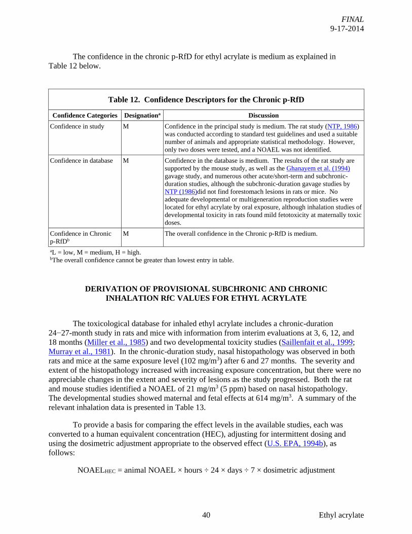


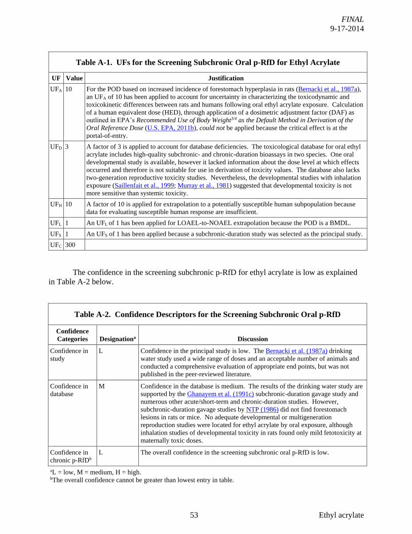


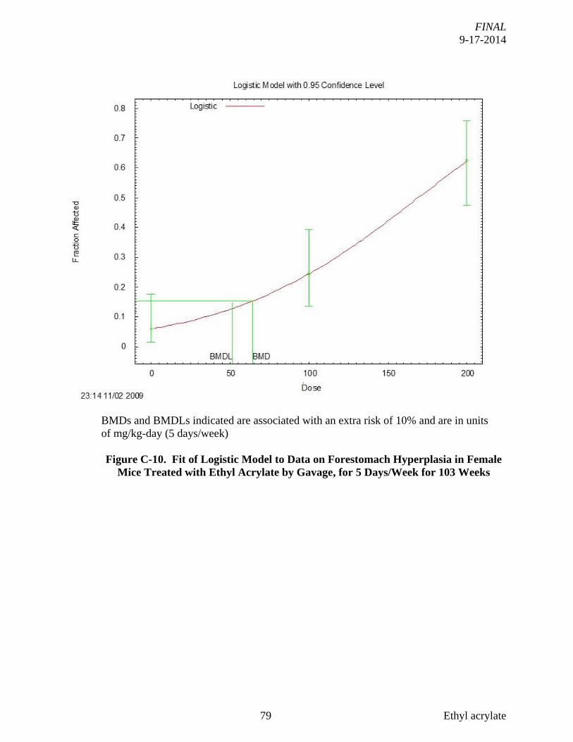


![MODIFICATION OF POLY(LACTIC ACID) BY BLENDING WITH …jestec.taylors.edu.my/Vol 12 issue 10 October 2017/12_10_16.pdfpoly(ethyl acrylate) [2] and styrene acrylic copolymer [3]. Thus,](https://static.documents.pub/doc/80x56/5e8fa2c6063d5c0c15493722/modification-of-polylactic-acid-by-blending-with-12-issue-10-october-2017121016pdf.jpg)


