Pseudomonas syringae HrpP Is a Type III Secretion Substrate SpecificitySwitch Domain Protein That Is Translocated into Plant Cells but
Functions Atypically for a Substrate-Switching Protein�
Joanne E. Morello and Alan Collmer*Department of Plant Pathology and Plant-Microbe Biology, Cornell University, Ithaca, New York 14853
Received 14 November 2008/Accepted 23 February 2009
Pseudomonas syringae delivers virulence effector proteins into plant cells via an Hrp1 type III secretionsystem (T3SS). P. syringae pv. tomato DC3000 HrpP has a C-terminal, putative T3SS substrate specificityswitch domain, like Yersinia YscP. A �hrpP DC3000 mutant could not cause disease in tomato or elicit ahypersensitive response (HR) in tobacco, but the HR could be restored by expression of HrpP in trans. ThoughHrpP is a relatively divergent protein in the T3SS of different P. syringae pathovars, hrpP from P. syringae pv.syringae 61 and P. syringae pv. phaseolicola 1448A restored HR elicitation and pathogenicity to DC3000 �hrpP.HrpP was translocated into Nicotiana benthamiana cells via the DC3000 T3SS when expressed from its nativepromoter, but it was not secreted in culture. N- and C-terminal truncations of HrpP were tested for their abilityto be translocated and to restore HR elicitation activity to the �hrpP mutant. No N-terminal truncationcompletely abolished translocation, implying that HrpP has an atypical T3SS translocation signal. Deletingmore than 20 amino acids from the C terminus abolished the ability to restore HR elicitation. HrpP fused togreen fluorescent protein was no longer translocated but could restore HR elicitation activity to the �hrpPmutant, suggesting that translocation is not essential for the function of HrpP. No T3SS substrates weredetectably secreted by DC3000 �hrpP except the pilin subunit HrpA, which unexpectedly was secreted poorly.HrpP may function somewhat differently than YscP because the P. syringae T3SS pilus likely varies in lengthdue to differing plant cell walls.
Many proteobacterial pathogens use a type III secretionsystem (T3SS) as their primary mechanism to overcome andinfect eukaryotic hosts. T3SSs are complex macromolecularmachines that span both the bacterial cell envelope and hostcell barriers to deliver proteins, commonly termed effectors,from the bacterial cytoplasm into the host cytoplasm (13, 19).After delivery into the host, effector proteins manipulate hostcell function and suppress host defenses, allowing bacterialproliferation and disease development (6, 20). Bacteria thatrely on T3SS to cause disease include plant pathogens such asPseudomonas syringae, Ralstonia solanacearum, Erwinia andXanthomonas species and animal pathogens in the genera Yer-sinia, Salmonella, Shigella, Escherichia, and Pseudomonas.While the repertoire of effectors delivered by a given T3SS isunique, the T3SS machinery is more universal (13). T3SS in-cludes a core set of eight conserved proteins. These proteins,which are also conserved in bacterial flagellar biogenesis ma-chines, make up the multiringed base structure, or basal body,that spans the bacterial membranes and cell wall. T3SS ma-chines are also comprised of less-conserved and unique pro-teins that vary between systems. These include regulatory pro-teins that orchestrate construction of the machine and theextracellular components that function to translocate effectorsacross host barriers.
The extracellular portion of the T3SS is comprised of the
pilus or needle appendage (in plant or animal pathogens, re-spectively), which acts as a conduit for effector delivery, andthe translocon complex, which creates the pore in the host cellmembrane. These substructures vary between different T3SSs;presumably these external structures have adapted to allowdifferent bacteria to infect different types of host cells. ForYersinia enterocolitica to infect macrophage cells, the T3SSneedle must be a particular length (�58 nm) to bridge thelipopolysaccharides extending from the bacterial outer mem-brane and reach the host cell membrane (35). Several otheranimal pathogens have T3SS needles of a defined length (48).Enteropathogenic Escherichia coli also has an additional ex-tension beyond the needle called the EspA filament that func-tions to span the mucous layer found outside enterocyte cells(13). In plant pathogens, however, the extracellular gap be-tween a bacterium and a plant cell includes a thick plant cellwall that is variable in width between plant species. Conse-quently, plant pathogenic Pseudomonas syringae has a pilusthat can measure over 1 �m in vitro (25).
Another major difference between the T3SS machineries ofanimal and plant pathogens is their translocon complexes. Inanimal pathogens, these are typically comprised of three es-sential proteins, but there is growing evidence that plant patho-gen translocons employ diverse, functionally redundant com-ponents (28). There is growing interest in understanding theregulatory players that orchestrate the construction of diversemachinery. It is hypothesized that the assembly of the T3SSmust involve several tightly regulated steps that allow secretionof the required components, followed by that of effectors uponcompletion. Of particular interest here is the control of pilus/needle subunit secretion, which is necessary when the pilus/
* Corresponding author. Mailing address: Department of Plant Pa-thology and Plant-Microbe Biology, Cornell University, Ithaca, NY14853. Phone: (607) 255-7843. Fax: (607) 255-4471. E-mail: [email protected].
needle is being constructed but would presumably competewith translocon and effector secretion after the T3SS is com-plete.
We study the model plant pathogen P. syringae pv. tomato(Pto) DC3000, the causal agent of bacterial speck of tomatoand Arabidopsis thaliana (8). DC3000 has a T3SS that deliversca. 28 effectors and is essential for pathogenesis (11, 12, 30,43). The P. syringae T3SS is encoded by hrp and hrc genes(hypersensitive response and pathogenicity/conserved), whichare located in a pathogenicity island on the chromosome (4).hrc genes encode the conserved core components present inevery T3SS. hrp genes encode T3SS components that are di-vergent or unique to P. syringae and enterobacterial plantpathogens, which also possess Hrp1 class T3SS (13). In con-trast, plant pathogenic Ralstonia and Xanthomonas spp. haveHrp2 class T3SS, as indicated by several different Hrp proteinsand distinct regulatory systems.
To better understand the T3SS machinery, we previouslyconducted a survey of the hrp genes of P. syringae pv. syringae(Psy) 61 to complete the inventory of all those encoding pro-teins capable of traveling the T3SS into plant cells when ex-pressed from a constitutive promoter (39). We hypothesizedthat these proteins might aid in pilus or translocon construc-tion or regulate the construction process. HrpP was one pro-tein found to be a T3SS substrate and important for secretionand translocation of the model effector AvrPto. Importantly,HrpP is related to a well-studied protein from Yersinia entero-colitica, YscP, which is a T3SS-secreted protein and a regulatorresponsible for switching the T3SS from secreting needle sub-units to secreting effector proteins (15, 38, 47). It has also beenshown that secretion of YscP into the culture medium is notessential for the switch function and that there may be two typeIII secretion signals embedded in YscP (2).
The phenotype of a yscP mutant is unregulated secretion ofthe needle subunit, no secretion of effectors, and production ofneedles of indeterminate length. The switching phenotype re-quires a domain at the C terminus of YscP called the type IIIsecretion substrate specificity switch (T3S4) domain, which is aconserved feature unifying its homologs (1). YscP has beenproposed to act as a molecular ruler because the length of theYscP protein is directly correlated with the length of the Yscneedle (26). According to this model, when the needle hasreached its proper length, YscP signals to the T3SS machineryto stop secreting needle subunits and begin secreting effectorproteins. However, other functional models have been hypoth-esized for homologs of YscP. A recent study of the Salmonellaenterica serovar Typhimurium YscP homolog InvJ showed thatan invJ mutant lacked an inner rod. When the inner rod pro-tein PrgJ was overexpressed, the length of the needle de-creased relative to that of the wild type, leading the researchersto conclude that InvJ controls the inner rod, which in turncontrols needle length (33). Recent evidence in Yersinia haslent more support to this model. YscP was found to negativelycontrol secretion of YscI, the inner rod protein (51). Also,certain YscI mutations affected needle assembly but not effec-tor secretion, implying that YscI may be a key player in sub-strate switching. Little is known about HrpB, the inner rodhomolog in P. syringae (22), other than that the protein can betranslocated into plant cells and is essential for T3SS function(39).
Other models for length control/substrate switching havebeen proposed, such as the “C-ring cup model” in flagella,which was based on the observation that certain mutations inproteins that make up the inner membrane C ring of the basalbody lead to shorter hooks (the flagellar equivalent of theneedle), thus suggesting that C-ring capacity controls hooklength (32). A more recent, flagellar “molecular-clock” modelsuggests that because overexpression of hook subunits leadsto longer hooks and hook polymerization-defective mutantsmake shorter hooks, hook polymerization initiates a count-down, and the timing, in cooperation with the YscP homologFliK, determines final hook length (34).
HrpP is considered a member of the YscP/FliK family duemostly to the presence of a T3S4 domain at its C terminus.HrpP is also proline rich (10.6%), which is considered a char-acteristic of the family. The most striking feature of HrpP is itssmall size; the protein is 189 amino acids, compared with YscPfrom Y. enterocolitica, which is 453 amino acids and 8.4%proline. We were intrigued by how HrpP functions in P. syrin-gae to regulate a pilus that can measure several hundrednanometers in length. Also, unlike animal pathogen needlesand flagellar hooks, the pilus of P. syringae is predicted to beindeterminate in length, based on the fact that plant cell wallsvary in width between species (40).
We hypothesized that HrpP would be a main player in reg-ulating pilus construction in P. syringae by allowing the systemto make the transition between secretion of pilus subunits andsecretion of translocon or effector proteins, though perhaps bya novel mechanism. In this study, we more precisely define therole of HrpP in the P. syringae T3SS. We show that HrpP is aT3SS substrate in DC3000, is translocated into plant cells atlevels equivalent to those of effectors, and is essential for thefunction of the T3SS. Though it is highly translocated andvariable, we found that HrpP from different P. syringae patho-vars could complement the DC3000 hrpP mutant. Analysis oftruncations of HrpP and an impassible HrpP-green fluorescentprotein (GFP) fusion suggests that it has structural similaritiesto YscP, but surprisingly, HrpP was found to be required forfull secretion of the pilus subunit HrpA as well as for translo-cation of HrpB.
MATERIALS AND METHODS
Bacterial strains, plasmids, and culture conditions. Bacterial strains and plas-mids used in this study are listed in Table 1. Pto strains were grown at 28°C inKing’s B (KB) medium or Hrp-inducing minimal medium (HMM) (24, 27), andEscherichia coli strains were grown at 37°C in Luria-Bertani medium or Terrificbroth for plasmid isolation. Antibiotics were used at the following concentrationswhen required: kanamycin, 50 �g/ml; rifampin, 10 �g/ml; tetracycline, 20 �g/ml;and gentamicin (Gm), 10 �g/ml.
DNA and protein manipulation techniques. Plasmids were isolated from E.coli using the Qiagen spin miniprep kit (Qiagen). Genomic DNA was isolatedusing the Wizard genomic DNA purification kit (Promega). DNA enzyme reac-tion cleanups were conducted using the Clean-up & Concentrator kit (ZymoResearch). hrpP genes, truncated hrpP genes, and hrpB from Pto DC3000, Psy 61,and P. syringae pv. phaseolicola (Pph) 1448A were cloned using PCR primersthat allow directional TOPO cloning into the pENTR/SD/D-TOPO or pENTR/D/D-TOPO vectors (Invitrogen) (Table 2). The gfp gene was fused to hrpP bySOEing PCR as previously described (21). All pENTR clones were sequenceconfirmed. Through an LR recombination reaction using the Gateway system(Invitrogen), hrpP, hrpB, and hrpP derivatives were cloned into pBS46,pCPP5295, or pCPP5371. Each recombination reaction mixture was transformedinto chemically competent E. coli TOP10. The resulting clones were conjugatedinto P. syringae with the aid of E. coli HB101 harboring the helper plasmid
pRK2013. DNA sequencing was conducted at the Cornell University Biotech-nology Resource Center using an Applied BioSystems 3730�l DNA analyzer.Sequences were analyzed using the Vector NTI software package (Invitrogen).To confirm production of cloned HrpP and its derivatives, Pseudomonas strainscarrying expression plasmids were grown overnight in 5 ml KB broth or HMM,centrifuged, and resuspended in 500 �l water. Cells were boiled, and proteinswere separated by sodium dodecyl sulfate-polyacrylamide gel electrophoresis(SDS-PAGE) and subjected to immunoblot analysis as previously described (49)using antihemagglutinin (anti-HA) (Roche), anti-Cya (Santa Cruz Biotechnol-
ogy), or anti-GFP (BD Biosciences) antibodies, followed by secondary antibodyconjugated to alkaline phosphatase.
Construction of hrpP mutant in DC3000. A nonpolar, unmarked deletion inthe hrpP gene was constructed using methods described previously (28). Onekilobase of either flank of the hrpP gene from DC3000 was amplified usingprimers P2218/2219 and P2220/2221 (Table 2), ligated together through anintroduced KpnI site and cloned into pENTR/D/D-TOPO to create pCPP5602.pCPP5602 was digested with KpnI, and a Gmr-FLP recombination target (FRT)cassette (amplified from pCPP5209) was introduced at this site, creating
TABLE 1. Bacterial strains and plasmids used in this study
Strain or plasmid Relevant characteristic(s) Source
E. coli strainsDH5� F� supE44 �(lacZYA-argF)U169 (�80dlacZ�M15) hsdR17(rK
pCPP5603. pCPP5603 was LR recombined with pCPP5301, creating pCPP5604.pCPP5604 was conjugated into DC3000, and bacteria were subcultured in KBbroth to allow for chromosomal-marker exchange. After recombination into thechromosome, the Gmr-FRT cassette was evicted by introduction of pCPP5264.The hrpP deletion mutation was confirmed by PCR and sequencing.
Cya reporter assay for translocation. Cells from 2-day-old KB plates of P.syringae carrying HrpP-Cya or HrpB-Cya hybrids were resuspended in 5 mMmorpholinoethanesulfonic acid (pH 5.5) to an optical density at 600 nm (OD600)of 0.3 and infiltrated with a blunt syringe into the leaves of 3-week-old Nicotianabenthamiana plants. Plants were kept at 24°C. Seven hours postinoculation, leafdiscs (0.32 cm2) were collected, pulverized in liquid nitrogen, and resuspended in300 �l of 0.1 M HCl. The samples were frozen at �20°C until assayed. Sampleswere assayed for cyclic AMP (cAMP) with the direct cAMP enzyme immuno-assay kit according to the manufacturer’s instructions (Assay Designs). Proteinlevels in each sample were determined through the Bradford method (7), andcAMP levels were reported as picomoles of cAMP per microgram of totalprotein.
Plant assays. For hypersensitive-response (HR) assays, Pseudomonas strainsfrom 2-day-old KB plates were resuspended in 10 mM MgCl2 to an OD600 of 0.5and infiltrated into Nicotiana tabacum cv. Xanthi, using a blunt syringe. Plantswere maintained at 24°C in the laboratory with supplemental lighting, and scoredfor visual collapse after 48 h. For tomato virulence assays, strains from 2-day-oldKB plates were resuspended in 10 mM MgCl2. Solanum lycopersicum cv. Money-maker plants were vacuum infiltrated with a suspension of 5 � 104 CFU/ml, 10mM MgCl2, and 0.02% (vol/vol) Silwet. Bacterial concentrations of the inoculumwere verified by plate count. Plants were maintained at 21°C, 60% humidity, and16-h days. At 3, 4, and 6 days postinfiltration, symptoms were visually assessed,and leaf discs were taken from three representative leaves per plant and shakenin 10 mM MgCl2 and 100 mM sucrose for 3 h at 12°C. Bacterial suspensions wereplated onto selective media, and colonies were counted to assess bacterialgrowth.
In vitro secretion assays. Pseudomonas strains were grown overnight in 8 mlKB broth at 30°C, pelleted by centrifugation and resuspended in 1 ml HMMsupplemented with 0.2% fructose. Bacteria were directly added to 100 ml HMMto an OD600 of 0.15 and grown with shaking at 22°C to an OD600 of 0.3. Cultureswere centrifuged at 5,200 � g for 15 min, and the bacterial pellet was collectedand resuspended in protein sample buffer for the “cell pellet” fraction. The top40 ml of supernatant was spun at 20,800 � g for 40 min. The top 25 ml ofsupernatant was then added to 5 ml trichloroacetic acid, shaken, and incubatedat 4°C overnight to precipitate protein. Protein was sedimented by centrifugationat 20,800 � g for 40 min, and the pellet was resuspended in protein sample bufferfor the “supernatant” fraction. Cell pellet and supernatant fractions were sepa-rated by SDS-PAGE and subjected to immunoblot analysis as previously de-scribed (49) using anti-AvrPto, anti-HrpW1, anti-HrpA, anti-HrpZ1, anti-HopA1, anti-HA (Roche), or anti-Cya (Santa Cruz) antibodies, followed by
secondary antibody conjugated to alkaline phosphatase. For two-dimensionalelectrophoresis (2DE), lawns of DC3000 and mutant derivatives were grownovernight at 30°C on KB plates with the appropriate antibiotics. Bacteria wereharvested from the plates by resuspension in 5 ml HMM followed by briefvortexing. Bacteria were directly added to 100 ml HMM (supplemented with0.2% fructose and 0.2% mannitol) to an OD600 of 0.3, and grown with shakingat 22°C to an OD600 of 0.5. Supernatant proteins from 500 ml HMM cultureswere collected as described above but were resuspended in 7 M urea, 2 Mthiourea, and 4% CHAPS {3-[(3-cholamidopropyl)-dimethylammonio]-1-pro-panesulfonate} buffer. Protein was mixed with 1% ampholyte (Bio-Rad) and 50mM dithiothreitol and allowed to passively rehydrate 17-cm, pH 3 to 10, non-linear IPG strips (Bio-Rad). The strips were subjected to isoelectric focusing,followed by second-dimension separation of proteins by SDS-PAGE (12% acryl-amide). Acrylamide gels were stained with Sypro ruby protein stain (Bio-Rad)and visualized using UV light. Gels were visually compared to Pto DC3000 2DEgel maps found at http://www.leelab.org/resources/syringae/index.html. 2DE gelmaps were prepared from proteins that were collected from supernatants ofHMM cultures in a manner similar to that described above.
RESULTS
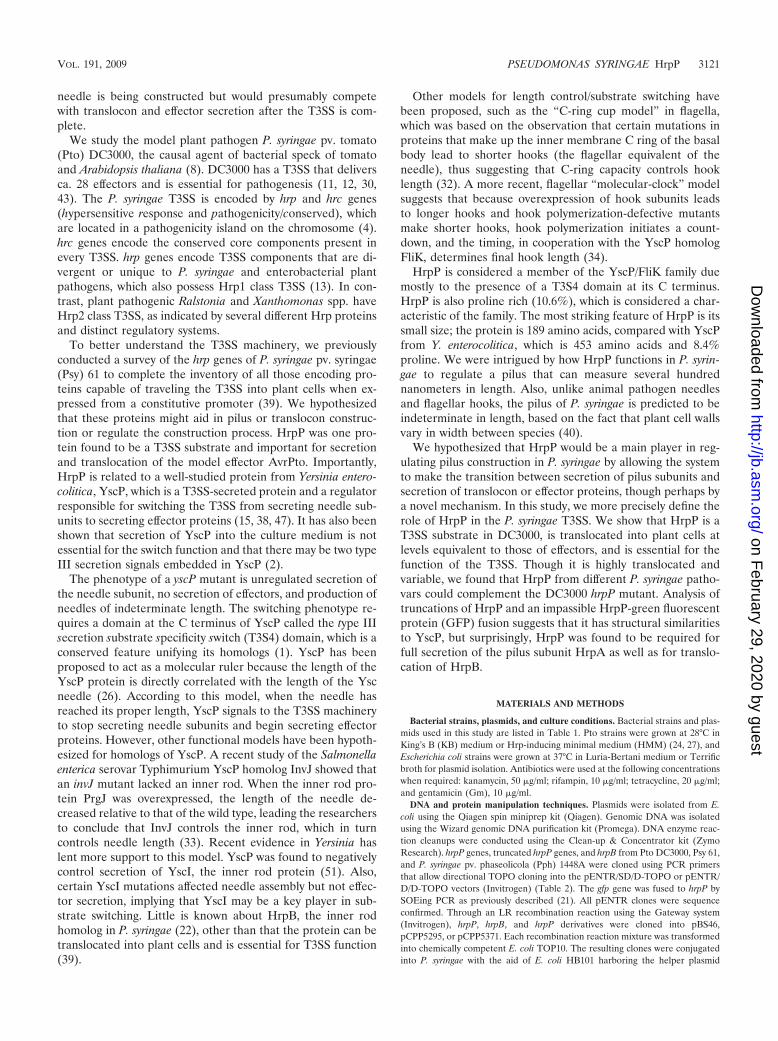
HrpP is essential for DC3000 to cause disease in tomato andelicit an HR in tobacco. To assess the function of HrpP in PtoDC3000, we generated the mutant CUCPB5453 (DC3000�hrpP), a DC3000 derivative with an unmarked, nonpolar de-letion of the entire hrpP open reading frame. The genomicregion was sequenced to verify its integrity, as hrpP is the firstgene in an essential T3SS operon. To first determine if hrpP isrequired for virulence, DC3000 �hrpP was vacuum infiltratedinto Solanum lycopersicum (tomato) cv. Moneymaker, a hostfor DC3000. DC3000 �hrpP grew equally as poorly as the typeIII mutant DC3000 �hrcC (CUCPB5112) (Fig. 1A) and pro-duced no visible symptoms (Fig. 1B).
To test if this mutant could be complemented by reintroduc-tion of the hrpP gene, pCPP5614 (expressing HrpP-HA) wasintroduced into DC3000 �hrpP. The strains were then testedfor their ability to elicit the HR in tobacco, which is dependenton effector translocation. DC3000 �hrpP(pCPP5614) was in-filtrated into N. tabacum cv. Xanthi along with DC3000,DC3000 �hrcC, and DC3000 �hrpP. DC3000 �hrpP was un-
TABLE 2. DNA oliogonucleotides used in this study
Designation Primer name Sequence
P2218 DCKpn1hrpO for TAATGGTACCCGGTGCGGTCATCGTCACP2219 DCTOPOhrpO rev CACCCGATCACCGCGCTCTACAP2220 DchrcQa for GGCGAGCGTCAAGCGCAATTP2221 DCKpn1hrcQ-a rev TAATGGTACCATCATGCATGAAGACCTTP2203 DCTOPOhrpP for CACCATGACCGCACCGATCAAP2204 DChrpP rev TGCTGAAAGGTCTTCATGP2365 DChrpPhrpbox for CACCATCGCCAGGCGCAGGTGGAAP2380 1448ATOPOhrpP for CACCATGACCGCACCGATCAAAP2381 1448AhrpP rev TGCTGCAAGATCTTCATGCAGGP2417 DCTOPOdel2-20 for CACCATGGTCGAGCGGCCAGCCACAP2419 DChrpPdel170-189 rev GTCGCGGCATGCGCCCGTP2420 DChrpPdel2-40 for CACCATGGGTGGCCTGGGCCGTGGCP2421 DChrpPdel448-end rev CAACTGCACGCTCCAGCGGTTP2427 DChrpPdelta120-end rev GTCAGGCTGATCCGGCAGGP2425 DChrpPdel2-67 for CACCATGAGTGCCGACGGCATGTTCTTTTP2426 DChrpPdel2-92 for CACCATGATAGCCTTTCCGGTAP2448 GfphrpP for GCATGAAGACCTTTCAGCAGTGAGCAAGGGCGAGGAGP2449 Gfp rev CTTGTACAGCTCGTCCATGCCGAP2446 HrpPgfp rev GCTCCTCGCCCTTGCTCACTGCTGAAAGGTCTTCATGP2239 TOPODChrpBfor CACCGTGACCATTTCCCAACTCP2363 DChrpBrev CTGCAGGTTGGTCAACTTGT
able to elicit an HR in tobacco (Fig. 1C), which is in agreementwith previous findings in Psy 61 (39). HR was restored byintroduction of pCPP5614 expressing HrpP-HA, indicating themutant is defective only in hrpP. In both compatible and in-compatible interactions with plants, DC3000 �hrpP performs
like a T3SS-negative strain, indicating that hrpP is an essentialcomponent of the P. syringae T3SS.
HrpP from multiple P. syringae pathovars can complementDC3000 �hrpP for HR and virulence. HrpP is the fifth mostvariable protein encoded by the hrp/hrc cluster when compar-ing the three representative P. syringae pathovars, Pto DC3000,Psy B728a, and Pph 1448A. The more variable components arethe pilus subunit HrpA, the harpin/translocator HrpZ1, HrpF,and HrcQB (18, 28, 39). These proteins have all been shown tobe extracellular, except HrcQB, which has a hypervariable Nterminus and is related to SpaO, a type III secreted protein ofSalmonella enterica (16, 29). The amino acid identity betweenthe HrpP proteins of DC3000 and B728a is 68.4%, that be-tween the HrpP proteins of DC3000 and 1448A is 65.8%, andthat between the HrpP proteins of B728a and 1448A is 72.1%.The finding that HrpP is relatively variable prompted us toexplore whether HrpP has evolved in certain pathovars forspecific host plants or whether it interacts with another morevariable part of the type III machinery. It was previously foundthat the hrp/hrc cluster from Psy 61 can complement a DC3000hrp/hrc cluster mutant for tomato virulence, but not Arabidop-sis virulence (18), indicating that some T3SS machinery com-ponents may be better adapted for different host species.
hrpP was cloned from Pph 1448A and Psy 61, which wehave used in past studies, and is 94.2% identical to hrpPfrom Psy B728a. Plasmids expressing either HrpPPsy61
(pCPP5700) or HrpPPph1448A (pCPP5701) from an nptIIpromoter were introduced into DC3000 �hrpP and tested tosee if they could complement the mutant as well as hrpPfrom DC3000(pCPP5614). Strains were infiltrated into N.tabacum as described above and scored for HR at 48 h.DC3000 �hrpP expressing HrpPPsy61 and HrpPPph1448A consis-tently elicited as strong an HR as did the mutant expressingHrpPPtoDC3000 (Fig. 2A). To test how these strains performedin a compatible interaction, Moneymaker tomato plants werevacuum infiltrated as described above. Both hrpP alleles com-plemented as well as hrpPPtoDC3000 for disease symptoms (Fig.2B). These results indicate that HrpP from different P. syringaepathovars can function in a DC3000 background, and thereforecan properly interact with the DC3000 T3SS and any potentialhost factors.
HrpP is translocated into plant cells at levels comparable tothose of type III effectors when expressed from its hrp pro-moter but is not secreted in culture. Previously, we found thatHrpP from P. syringae pv. syringae 61 was translocated into N.benthamiana cells in a type III-dependent manner when ex-pressed from a vector tac promoter (39). We wanted to verifythat HrpP from Pto DC3000 is also a type III substrate, butmore importantly, that it is translocated into plant cells undermore natural levels of production. In past studies, we havesuccessfully used the Cya reporter system to ascertain if aprotein is a type III substrate or effector by fusing the proteinof interest to Cya, a protein from Bordetella pertussis thatconverts ATP to cAMP in the presence of calmodulin, which ispresent only in eukaryotic cells (42, 46). hrpP from DC3000was cloned, with the genomic region immediately upstreamcontaining its hrp box promoter, in plasmid pCPP5697.pCPP5697 was transformed into both wild-type DC3000 andthe DC3000 �hrcC mutant CUCPB5112, used as a T3SS-neg-ative control. Production of HrpP-Cya in HMM was verified by
FIG. 1. A DC3000 �hrpP mutant is avirulent in tomato and fails toelicit an HR in N. tabacum. (A) Wild-type DC3000, DC3000 �hrpP(CUCPB5453), and DC3000 �hrcC (CUCPB5112 T3SS�) were vac-uum infiltrated into Solanum lycopersicum cv. Moneymaker plants at5 � 104 CFU/ml. Levels of inoculum were confirmed by dilution platecounts. At 3, 4, and 6 days postinfiltration (dpi), bacteria were recov-ered from leaf discs by shaking into 10 mM MgCl2 and 100 mMsucrose, and cell numbers were assessed by plate counts and calculatedas CFU/cm2. (B) One representative leaf from tomato plants infectedwith each strain at 6 dpi was photographed. (C) DC3000 �hrpP(CUCPB5453) was infiltrated into N. tabacum at 5 � 108 CFU/ml,along with wild-type DC3000, DC3000 �hrcC (CUCPB5112 T3SS�),and DC3000 �hrpP carrying either an empty vector (pBS60) or avector expressing HrpP-HA driven by the nptII promoter (pCPP5614).Leaves were scored for visual collapse at 48 h postinfection. Onerepresentative leaf is shown.
Western blotting (data not shown). To compare HrpP to otherknown type III substrates, DC3000 expressing the harpin/trans-locator HrpW1-Cya and the effectors AvrPto-Cya and HopA1-Cya was included in the experiment. HrpW1 is hypothesized tobe a component of the T3SS translocon complex and has lowlevels of translocation relative to those of true effectors suchas AvrPto and HopA1 (11, 28). All proteins were expressedfrom their native hrp promoters. Strains were infiltrated intoN. benthamiana leaves, sampled at 7 h postinfiltration, andassayed for cAMP. The high cAMP levels caused byDC3000(pCPP5697) indicate that HrpP-Cya is translocatedinto plant cells at levels similar to those of the true effectorsAvrPto and HopA1 when expressed from its hrp promoter(Fig. 3A), indicating that HrpP may enter the plant cytoplasmduring infection.
Due to its high level of translocation, we wanted to assess thelevel of HrpP secretion in culture. HrpP C-terminally taggedwith HA peptide was expressed from pCPP5614 from a con-stitutive nptII promoter in DC3000 and DC3000 �hrcQRSTU(CUCPB5113), a T3SS-negative strain. The nptII promoterwas used to express HrpP in this assay to maximize the chanceof detecting in vitro secretion. Strains were grown in HMM to
induce type III secretion and were separated into cell pelletand supernatant fractions. Proteins were separated by SDS-PAGE and subjected to Western analysis. Regardless of thehigh levels of HrpP-HA accumulation in the cell pellet, noHrpP-HA was ever detected in supernatant fractions (Fig. 3B).HopA1 was also not detected in these same supernatant frac-tions. With a few exceptions, including AvrPto, most P. syringaetype III effectors are not detectably secreted into HMM. Onthe other hand, HrpW1 was highly secreted into HMM thoughit is poorly translocated into plant cells, which is a noted char-
FIG. 2. HrpP from different P. syringae pathovars can complementDC3000 �hrpP for HR in tobacco and disease in tomato. (A) DC3000�hrpP (CUCPB5453) carrying a plasmid expressing from the nptIIpromoter HrpP-HA from either pathovar tomato (PtoDC3000),pathovar syringae (Psy61), or pathovar phaseolicola (Pph1448A)(pCPP5614, pCPP5700, and pCPP5701, respectively) was infiltratedinto N. tabacum at 5 � 108 CFU/ml along with wild-type DC3000,DC3000 �hrpP, and DC3000 �hrcQRSTU (CUCPB5113). Leaves werescored for visual collapse at 48 h postinfiltration (hpi). One represen-tative leaf is shown. (B) The same strains were vacuum infiltrated intoSolanum lycopersicum cv. Moneymaker plants at 5 � 104 CFU/ml.Plants were visually scored for bacterial speck disease at 6 dpi. Tworepresentative leaves for each strain are shown.
FIG. 3. HrpP-Cya is translocated into N. benthamiana cells byDC3000 at levels comparable to those of type III effectors when ex-pressed from its native hrp promoter, but it is not secreted in culture.(A) pCPP5297 (HrpP-Cya), pCPP5716 (HopA1-Cya), pCPP5312(AvrPto-Cya), and pCPP5898 (HrpW1-Cya) were introduced intowild-type DC3000 or the T3SS� derivative DC3000 �hrcQRSTU(CUCPB5113). Strains were infiltrated into N. benthamiana at 3 � 108
CFU/ml. Leaf discs were excised at 7 h postinfiltration and assayed forcAMP (using the Assay Designs direct cyclic AMP kit). Total proteinwas measured by the Bradford method. Values are the means andstandard deviation from triplicate samples. The experiment was re-peated three times with similar results. (B) DC3000 and DC3000�hrcQRSTU (CUCPB5113) carrying pCPP5614 (expressing HrpP-HAfrom the nptII promoter) were grown in HMM from an OD600 of 0.15to 0.3. Cell pellet and supernatant fractions were separated by centrif-ugation. Proteins were separated by SDS-PAGE, transferred to thepolyvinylidene difluoride (PVDF) membrane, and subjected to immu-noblot analysis using the indicated antibodies (indicated by �) andsecondary antibodies conjugated to alkaline phosphatase. CP, cell pel-let; SN, supernatant.
acteristic of translocators in P. syringae (Fig. 3A and B) (28).The lack of HrpP secretion in culture but high translocation inplanta, similar to a true effector, may indicate a biological rolefor HrpP entering the plant cell during infection.
HrpP has an atypical T3SS translocation signal and re-quires the putative T3S4 domain for function. To further in-vestigate the function of HrpP translocation, N-terminallytruncated versions of HrpP were constructed and tested fortheir ability to be translocated as well as to complementDC3000 �hrpP for HR elicitation. The N terminus of T3SSsubstrates typically contains the signal necessary for secretionthrough the pathway, and N-terminal truncations eliminatetype III secretability (36, 45). Amino acids 2 to 20, 2 to 40, 2 to67, and 2 to 92 were deleted from HrpP, and the derivatives(HrpP�2-20, HrpP�2-40, HrpP�2-67, and HrpP�2-92, respec-tively) were tested for their ability to be translocated whenfused to Cya and expressed from an hrp promoter. All trun-cated genes produced proteins of the proper size (Fig. 4A andB). In all cases, there was significant cAMP accumulation,indicating some translocation of the truncated protein (Fig. 5).Even when devoid of its N terminus, HrpP retains the ability totravel the T3SS pathway, indicating that it has an atypicaltranslocation signal and possibly a unique way of interactingwith the T3SS machinery.
To assess the functionality of these proteins, HrpP deriva-tives were expressed from an nptII promoter and tested fortheir ability to restore to the DC3000 �hrpP mutant HR elic-
itation in tobacco leaves and AvrPto secretion in culture. Wechose these assays because HR elicitation is a general test foreffector translocation and AvrPto secretion is a specific test forpathway switching to a representative, native effector. HrpP�2-20–HA and HrpP�2-40–HA can complement DC3000 �hrpPfor HR elicitation and AvrPto secretion, indicating that thevery N terminus of HrpP may not be essential for function(Fig. 5).
To assess the importance of the C terminus of HrpP, C-terminal truncations were also constructed and tested forcomplementation of DC3000 �hrpP and their own transloca-tion. The C terminus of HrpP is the location of the T3S4domain, a signature of the YscP/FliK family of proteins towhich HrpP belongs (1). This large region is believed to be theprimary functional domain in these proteins and, in HrpP, ispredicted to span amino acids 101 to 184 (1). We deletedamino acids 170 to 188, 150 to 188, and 120 to 188 from HrpP.All truncated genes produced proteins of the proper size withthe exception of HrpP�120-188–HA, which was not detectable(Fig. 4A). When fused to Cya, all C-terminal truncations weretranslocated at levels equivalent to those of HrpP-Cya, butonly HrpP�170-188–HA could complement DC3000 �hrpP forHR elicitation and AvrPto secretion (Fig. 5). This indicatesthat the deletion of only 20 to 40 amino acids from the C
FIG. 4. Production of truncated versions of HrpP in DC3000�hrpP. (A) DC3000 �hrpP carrying plasmids expressing (from thenptII promoter) full-length and truncated versions of HrpP-HA wasgrown overnight in KB broth to an OD600 of 1.0. Cells were collectedby centrifugation and lysed, and cellular proteins were separated bySDS-PAGE, transferred to the PVDF membrane, and subjected toimmunoblot analysis using an anti-HA antibody (�HA) and secondaryantibody conjugated to alkaline phosphatase. (B) Boxed portion of theWestern blot from panel A (lanes 2 and 3) overexposed to show theproduction of HrpP�2-20–HA and HrpP�2-40–HA.
FIG. 5. N- and C-terminal truncations of HrpP reveal an atypicalT3SS translocation signal and a C terminus that is important forfunction in promoting DC3000 secretion of AvrPto-Cya and elicitationof the HR in tobacco leaves. For the immunoblot data at the top of thefigure, DC3000 �hrpP (CUCPB5453) carrying plasmids expressingfull-length and truncated versions of HrpP (as described in the legendto Fig. 4) was grown in HMM from an OD600 of 0.3 to 0.5. Cell pelletsand supernatant fractions were separated by centrifugation. Superna-tant proteins were separated by SDS-PAGE, transferred to the PVDFmembrane, and subjected to immunoblot analysis using an anti-AvrPtoantibody and secondary antibody conjugated to alkaline phosphatase.For the HR tests in the middle of the figure, the same DC3000 �hrpPderivatives were infiltrated into N. tabacum at 5 � 108 CFU/ml. Leaveswere scored for visual collapse at 48 hpi. �, HR; �, no HR. For theHrp-Cya translocation tests at the bottom of the figure, full-length andtruncated versions of hrpP were fused to Cya, in pCPP5371, whichprovides expression from the AvrPto promoter, and introduced intoDC3000 or DC3000 �hrcQRSTU as indicated. Strains were infiltratedinto N. benthamiana at 3 � 108 CFU/ml. Leaf discs were taken at 7 hpiand assayed for cAMP (Assay Designs direct cyclic AMP kit). Totalprotein was measured by the Bradford method. Values are the meansand standard deviation from triplicate samples. The experiment wasrepeated three times with similar results.
terminus hinders the function of HrpP in HR elicitation andpotentially disrupts substrate switching and the T3S4 domain.As expected, the C terminus has no role in the translocation ofHrpP.
An HrpP-GFP hybrid protein cannot be translocated butcan still restore HR elicitation and AvrPto secretion to theDC3000 hrpP mutant. Because no N-terminal truncation ofHrpP eliminated its translocation into plants, we were not ableto assess the importance of HrpP translocation for its function.We therefore took the approach of fusing GFP, a protein thatfolds rapidly in the cytoplasm and cannot travel the T3SS (3),to the C terminus of HrpP. We tested the translocation of thishybrid protein following the construction of an HrpP-GFP-Cyatranslational fusion in pCPP5909. We found that DC3000 ex-pressing HrpP-GFP-Cya gave very little cAMP signal com-pared to HrpP-Cya, indicating that this protein was blockedfrom translocation (Fig. 6A). Production of this protein wasconfirmed by immunoblotting (Fig. 6B). To test its functionality,we introduced HrpP-GFP-Cya or HrpP-GFP-HA (pCPP5911)into DC3000 �hrpP and assayed for HR elicitation in compar-ison to that of HrpP-Cya or HrpP-HA (pCPP5653), all ex-pressed from hrp promoters. All HrpP derivatives comple-mented the hrpP mutant for HR elicitation equally (Fig. 6C).They also complemented equally well for speck disease intomato (data not shown). Furthermore, HrpP-HA and HrpP-GFP-HA restored to the DC3000 �hrpP mutant the ability tosecrete AvrPto in culture (Fig. 6D), and repeated experimentsrevealed no consistent difference in their ability to restoresecretion. Thus, although HrpP-GFP is not translocated, it stillretains enough functionality to allow P. syringae to elicit an HRand secrete AvrPto. We cannot however rule out the possibil-ity, though it is not evident by immunoblot analysis (Fig. 6B),that GFP could be cleaved off in the bacterial cytoplasm, thusliberating HrpP to perform its function and be delivered intothe plant cytoplasm.
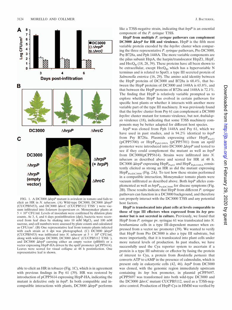
The hrpP mutant secretes only small amounts of the pilussubunit HrpA. Though we found hrpP to be required for HRand disease in planta, and for AvrPto secretion in culture, itwas important to ask if the DC3000 hrpP mutant could secreteany T3SS substrates. Due to HrpP’s similarities to YscP fromYersinia, which is required for secretion of effectors but not theneedle subunit YscF or the inner rod YscI, we hypothesizedthat DC3000 �hrpP would robustly or excessively secrete thepilus subunit HrpA and/or the inner rod protein HrpB.DC3000, DC3000 �hrpP, and DC3000 �hrcQRSTU (T3SS�)were cultured in HMM to induce type III secretion. Cell pelletand supernatant fractions were then collected as describedabove and separated by SDS-PAGE, and subjected to Westernanalysis using antibodies to HrpA, the harpin/translocatorsHrpW1 and HrpZ1, and the effector AvrPto. To assess HrpBsecretion, an HrpB-Cya fusion was expressed from pCPPP5688,and cell fractions were probed with an anti-Cya antibody. Allproteins were secreted by DC3000 in a type III-dependentmanner but not by DC3000 �hrpP (Fig. 7A), with the exceptionof HrpB-Cya, which was not secreted by either strain. HrpA,however, was detectable at low levels in the supernatant ofDC3000 �hrpP, indicating that hrpP may not be completelyessential for the secretion of HrpA. Contrary to what wasexpected though, levels of HrpA secretion in DC3000 �hrpPwere never equal to or greater than those in wild-type DC3000.
FIG. 6. An HrpP-GFP fusion cannot be translocated but can stillrestore to the DC3000 �hrpP mutant the ability to elicit HR in tobaccoand to secrete AvrPto in culture. (A) DC3000 and DC3000 �hrcQRSTU (CUCPB5113) expressing HrpP-GFP-Cya (pCPP5909) orHrpP-Cya (pCPP5697) from the vector-provided hrpP promoter wereinfiltrated into N. benthamiana at 3 � 108 CFU/ml. Leaf discs wereexcised at 7 hpi and assayed for cAMP (Assay Designs direct cyclicAMP kit). Total protein was measured by the Bradford method. Val-ues are the means and standard deviation from triplicate samples. Theexperiment was repeated three times with similar results. (B) DC3000�hrpP expressing HrpP-GFP-Cya or HrpP-GFP-HA (pCPP5911) weregrown overnight in HMM to an OD600 of 1.0. Cells were collected bycentrifugation and lysed, and cellular proteins were separated by SDS-PAGE, transferred to the PVDF membrane, and subjected to immu-noblot analysis using an anti-GFP antibody and secondary antibodiesconjugated to alkaline phosphatase. (C) DC3000, DC3000 �hrcQRSTU (CUCPB5113), and DC3000 �hrpP (CUCPB5453) andDC3000 �hrpP expressing HrpP-GFP-Cya (pCPP5909), HrpP-Cya(pCPP5697), HrpP-GFP-HA (pCPP5911), or HrpP-HA (pCPP5653)were infiltrated into N. tabacum at 5 � 108 CFU/ml. Leaves werescored for visual collapse at 48 hpi. One representative leaf is shown.(D) Immunoblot analysis, performed as described in the legend forFig. 5, indicates that AvrPto is secreted in culture by DC3000 �hrpPexpressing HrpP-HA and HrpP-GFP-HA.
Though HrpB-Cya was not secreted by wild-type DC3000, itwas found in a previous study to be weakly translocated intoplant cells (39). It was necessary to ask, then, if HrpB-Cyacould be translocated by DC3000 �hrpP. In support of previ-ous findings, expression of HrpB-Cya from pCPP5688 led totype III-dependent cAMP accumulation with DC3000 infec-tion, but not with DC3000 �hrpP, indicating that HrpB is alsonot translocated by the hrpP mutant (Fig. 7B). ConsideringDC3000 �hrpP poorly secretes the pilus subunit and fails tosecrete translocator proteins, it is likely that the translocationprocess in planta is not efficient in this mutant.
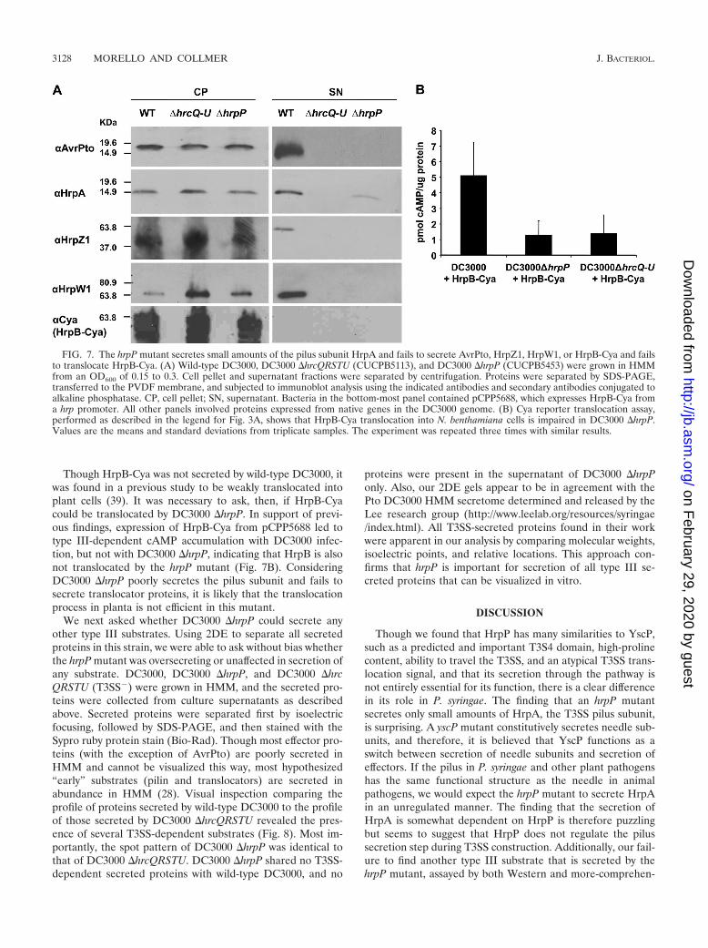
We next asked whether DC3000 �hrpP could secrete anyother type III substrates. Using 2DE to separate all secretedproteins in this strain, we were able to ask without bias whetherthe hrpP mutant was oversecreting or unaffected in secretion ofany substrate. DC3000, DC3000 �hrpP, and DC3000 �hrcQRSTU (T3SS�) were grown in HMM, and the secreted pro-teins were collected from culture supernatants as describedabove. Secreted proteins were separated first by isoelectricfocusing, followed by SDS-PAGE, and then stained with theSypro ruby protein stain (Bio-Rad). Though most effector pro-teins (with the exception of AvrPto) are poorly secreted inHMM and cannot be visualized this way, most hypothesized“early” substrates (pilin and translocators) are secreted inabundance in HMM (28). Visual inspection comparing theprofile of proteins secreted by wild-type DC3000 to the profileof those secreted by DC3000 �hrcQRSTU revealed the pres-ence of several T3SS-dependent substrates (Fig. 8). Most im-portantly, the spot pattern of DC3000 �hrpP was identical tothat of DC3000 �hrcQRSTU. DC3000 �hrpP shared no T3SS-dependent secreted proteins with wild-type DC3000, and no
proteins were present in the supernatant of DC3000 �hrpPonly. Also, our 2DE gels appear to be in agreement with thePto DC3000 HMM secretome determined and released by theLee research group (http://www.leelab.org/resources/syringae/index.html). All T3SS-secreted proteins found in their workwere apparent in our analysis by comparing molecular weights,isoelectric points, and relative locations. This approach con-firms that hrpP is important for secretion of all type III se-creted proteins that can be visualized in vitro.
DISCUSSION
Though we found that HrpP has many similarities to YscP,such as a predicted and important T3S4 domain, high-prolinecontent, ability to travel the T3SS, and an atypical T3SS trans-location signal, and that its secretion through the pathway isnot entirely essential for its function, there is a clear differencein its role in P. syringae. The finding that an hrpP mutantsecretes only small amounts of HrpA, the T3SS pilus subunit,is surprising. A yscP mutant constitutively secretes needle sub-units, and therefore, it is believed that YscP functions as aswitch between secretion of needle subunits and secretion ofeffectors. If the pilus in P. syringae and other plant pathogenshas the same functional structure as the needle in animalpathogens, we would expect the hrpP mutant to secrete HrpAin an unregulated manner. The finding that the secretion ofHrpA is somewhat dependent on HrpP is therefore puzzlingbut seems to suggest that HrpP does not regulate the pilussecretion step during T3SS construction. Additionally, our fail-ure to find another type III substrate that is secreted by thehrpP mutant, assayed by both Western and more-comprehen-
FIG. 7. The hrpP mutant secretes small amounts of the pilus subunit HrpA and fails to secrete AvrPto, HrpZ1, HrpW1, or HrpB-Cya and failsto translocate HrpB-Cya. (A) Wild-type DC3000, DC3000 �hrcQRSTU (CUCPB5113), and DC3000 �hrpP (CUCPB5453) were grown in HMMfrom an OD600 of 0.15 to 0.3. Cell pellet and supernatant fractions were separated by centrifugation. Proteins were separated by SDS-PAGE,transferred to the PVDF membrane, and subjected to immunoblot analysis using the indicated antibodies and secondary antibodies conjugated toalkaline phosphatase. CP, cell pellet; SN, supernatant. Bacteria in the bottom-most panel contained pCPP5688, which expresses HrpB-Cya froma hrp promoter. All other panels involved proteins expressed from native genes in the DC3000 genome. (B) Cya reporter translocation assay,performed as described in the legend for Fig. 3A, shows that HrpB-Cya translocation into N. benthamiana cells is impaired in DC3000 �hrpP.Values are the means and standard deviations from triplicate samples. The experiment was repeated three times with similar results.
sive 2D analysis, makes it difficult to speculate about the func-tion of this protein. If it does function as a switch, but in adifferent step of the construction process, we would have ex-pected to have found a substrate whose secretion did not
require HrpP. HrpB, the inner rod protein, whose homologYscI is secreted by the yscP mutant and has recently beenimplicated in the switching process, was not secreted by thehrpP mutant, though it was also not secreted by wild-typeDC3000. This observation also suggests a fundamental differ-ence between P. syringae and previously studied systems.
It is possible that HrpP, regardless of its similarities to YscP,does not function as a switch at all and has evolved a differentbut obviously necessary function in P. syringae. Further workwill be needed to generate functional hypotheses for HrpP.However, there remains the fact that HrpP has intriguing sim-ilarities to other T3S4-containing family members. Besides thepresence of the T3S4 domain, which truncation mutants re-vealed was almost entirely necessary for function, HrpP, likeYscP, may have multiple secretion signals and enter the T3SSpathway in a nonconventional way. Also, YscP is known tointeract with the inner membrane component YscU, whichcontains a conserved autocleavage motif important for sub-strate switching (15, 44). The YscU homolog in P. syringae,HrcU, also has this conserved motif (data not shown).
Based on the Cya reporter assay and expression from its hrppromoter, HrpP is translocated into N. benthamiana at veryhigh levels. HrpP is also not secreted in culture, like mosteffector proteins, and therefore might be classified as a “late”substrate, supporting the idea that there could be biologicalrelevance to its translocation into plants. However it is para-doxical that HrpP could be translocated into plant cells at thesame time as effectors and yet be essential for an early stage ofT3SS machinery construction. We were able to introduce HrpPfrom different pathovars into DC3000 �hrpP and show thatthey can function in HR elicitation and disease development.This suggests that HrpP does not have a specific function in agiven host species.
We also created a translocation-deficient version of HrpP byfusing it to GFP, and showed this can still function in HRelicitation. This indicates that translocation of HrpP may notbe essential for complete function. Because elicitation of theHR is a simple way to determine whether type III translocationis occurring and is a very sensitive assay, it is possible thatHrpP-GFP was not fully functional and that a subtle defect intype III secretion did exist that we did not detect. Though wecannot eliminate this possibility, we did observe that HrpP-GFP-HA was able to restore AvrPto secretion to the DC3000�hrpP mutant. In this regard, it is noteworthy that a secretion-deficient mutant of YscP was still able to secrete effectors,though it was not able to control needle length, and therefore,was still a partially functioning protein (1). Also, given thatpast work predicted two secretion signals in YscP, the reasonfor and the importance of secretion of this protein are stillenigmatic. The high level of HrpP translocation could also bean artifact of plasmid-borne expression, though this does notoccur in our system with translocator proteins, which are be-lieved to function at the plant plasma membrane and aretranslocated at only low levels when expressed from hrp pro-moters (28).
Recently, in the plant pathogen Xanthomonas campestris pv.vesicatoria, the HrpP homolog HpaC was found to be impor-tant for the secretion of effector and translocon proteins butnot that of the pilus subunit HrpE or the protein HrpB2, whichwas in fact oversecreted by an hpaC mutant (31). Although
FIG. 8. The DC3000 �hrpP mutant secretes no other T3SS substrate.The wild-type DC3000, DC3000 �hrpP (CUCPB5453), and DC3000�hrcQRSTU (CUCPB5113) strains were grown in HMM as described forFig. 7. Supernatant proteins were separated first by isoelectric focusing ona pH-3 to -10 nonlinear gradient, followed by SDS-PAGE. Gels werestained with Sypro ruby protein stain (Bio-Rad) and visualized with UVlight. One representative gel for each strain is shown. T3SS substratessecreted by DC3000 are indicated by the white circles.
HrpB2 is not in the Pfam family that contains YscI of Yersiniaspp. and HrpB of P. syringae and other plant pathogens with anHrp1 T3SS, for several reasons, HrpB2 is a good candidate forthe inner rod of Xanthomonas and other plant pathogens withan Hrp2 T3SS: (i) HrpB2 is a secreted protein (41); (ii) HrpB2is essential for T3SS function (41); (iii) the size of HrpB2 (13.6kDa) is comparable to that of YscI (12.6 kDa) and HrpB (13.0kDa); (iv) the location of hrpB2 appears syntenic with that ofyscI in Yersinia (yscI, yscJ, yscK, yscL). That is, the P. syringaehomologs of these ysc genes occur in the same arrangement(hrpB, hrcJ, hrpD, hrpE), and in the corresponding region in X.campestris pv. vesicatoria (hrpB2, hrcJ, hrpB4, hrpB5), hrcJ andhrpB5 are homologs of yscJ and yscL, respectively (9, 23).
Given these observations, apparent differences in the func-tion of HrpP and HpaC are striking. In contrast to HrpP,HpaC is not secreted (10). Whereas an hpaC mutant stronglysecretes HrpB2 (31), an hrpP mutant neither secretes nortranslocates HrpB. Also, an hrpP mutant is strongly reduced insecretion of the pilus protein (31), whereas an hpaC mutant isunaffected. On the other hand, HrpP and HpaC share thecharacteristic of being much smaller than YscP, InvJ, FliK, andother T3S4 domain proteins associated with the production offixed-length T3SS/flagellar appendages. The smaller size of theHrpP and HpaC proteins, contrasted with their associationwith having appendages of potentially much longer and inde-terminate lengths, argues against these proteins functioning asmolecular rulers. It remains unknown how pilus length andsubstrate switching from pilin to effector are controlled in plantpathogens, but obviously, HrpP functions differently from itsanimal pathogen homologs and differently from XanthomonasHpaC. The Hrp1 T3SS and Hrp2 T3SS may have indepen-dently evolved the capacity to penetrate plant cell walls, andthe surprising differences between the phenotypes of hrpP andhpaC mutants may reflect independent adaptations of the re-spective T3S4 domain proteins to the problem of regulatingT3SS traffic when pathway construction is contingent uponvariable host barriers.
ACKNOWLEDGMENTS
We thank Bryan Swingle for the construction of pBS60 and pBS46and Brian Kvitko for the construction of pCPP5898 and pCPP5296.We also thank Cynthia Damasceno and Jocelyn Rose for technicalhelp with 2DE and David J. Schneider for helpful discussions regard-ing inner rod proteins.
This work was supported by NSF grant MCB-0544066.
REFERENCES
1. Agrain, C., I. Callebaut, L. Journet, I. Sorg, C. Paroz, L. J. Mota, and G. R.Cornelis. 2005. Characterization of a type III secretion substrate specificityswitch (T3S4) domain in YscP from Yersinia enterocolitica. Mol. Microbiol.56:54–67.
2. Agrain, C., I. Sorg, C. Paroz, and G. R. Cornelis. 2005. Secretion of YscPfrom Yersinia enterocolitica is essential to control the length of the injecti-some needle but not to change the type III secretion substrate specificity.Mol. Microbiol. 57:1415–1427.
3. Akeda, Y., and J. E. Galan. 2005. Chaperone release and unfolding ofsubstrates in type III secretion. Nature 437:911–915.
4. Alfano, J. R., A. O. Charkowski, W.-L. Deng, J. L. Badel, T. Petnicki-Ocwieja, K. van Dijk, and A. Collmer. 2000. The Pseudomonas syringae Hrppathogenicity island has a tripartite mosaic structure composed of a clusterof type III secretion genes bounded by exchangeable effector and conservedeffector loci that contribute to parasitic fitness and pathogenicity in plants.Proc. Natl. Acad. Sci. USA 97:4856–4861.
5. Badel, J. L., R. Shimizu, H.-S. Oh, and A. Collmer. 2006. A Pseudomonassyringae pv. tomato avrE1/hopM1 mutant is severely reduced in growth andlesion formation in tomato. Mol. Plant-Microbe Interact. 19:99–111.
6. Block, A., G. Li, Z. Q. Fu, and J. R. Alfano. 2008. Phytopathogen type IIIeffector weaponry and their plant targets. Curr. Opin. Plant Biol. 11:396–403.
7. Bradford, M. M. 1976. A rapid and sensitive method for the quantitation ofmicrogram quantities of protein utilizing the principle of protein-dye-bind-ing. Anal. Biochem. 72:248–254.
8. Buell, C. R., V. Joardar, M. Lindeberg, J. Selengut, I. T. Paulsen, M. L.Gwinn, R. J. Dodson, R. T. Deboy, A. S. Durkin, J. F. Kolonay, R. Madupu,S. Daugherty, L. Brinkac, M. J. Beanan, D. H. Haft, W. C. Nelson, T.Davidsen, J. Liu, Q. Yuan, H. Khouri, N. Fedorova, B. Tran, D. Russell, K.Berry, T. Utterback, S. E. Vanaken, T. V. Feldblyum, M. D’Ascenzo, W.-L.Deng, A. R. Ramos, J. R. Alfano, S. Cartinhour, A. K. Chatterjee, T. P.Delaney, S. G. Lazarowitz, G. B. Martin, D. J. Schneider, X. Tang, C. L.Bender, O. White, C. M. Fraser, and A. Collmer. 2003. The complete se-quence of the Arabidopsis and tomato pathogen Pseudomonas syringae pv.tomato DC3000. Proc. Natl. Acad. Sci. USA 100:10181–10186.
9. Buttner, D., and U. Bonas. 2002. Getting across-bacterial type III effectorproteins on their way to the plant cell. EMBO J. 21:5313–5322.
10. Buttner, D., C. Lorenz, E. Weber, and U. Bonas. 2006. Targeting of twoeffector protein classes to the type III secretion system by a HpaC- andHpaB-dependent protein complex from Xanthomonas campestris pv. vesica-toria. Mol. Microbiol. 59:513–527.
11. Chang, J. H., J. M. Urbach, T. F. Law, L. W. Arnold, A. Hu, S. Gombar, S. R.Grant, F. M. Ausubel, and J. L. Dangl. 2005. A high-throughput, near-saturating screen for type III effector genes from Pseudomonas syringae.Proc. Natl. Acad. Sci. USA 102:2549–2554.
12. Collmer, A., J. L. Badel, A. O. Charkowski, W.-L. Deng, D. E. Fouts, A. R.Ramos, A. H. Rehm, D. M. Anderson, O. Schneewind, K. van Dijk, and J. R.Alfano. 2000. Pseudomonas syringae Hrp type III secretion system and effec-tor proteins. Proc. Natl. Acad. Sci. USA 97:8770–8777.
13. Cornelis, G. R. 2006. The type III secretion injectisome. Nat. Rev. Microbiol.4:811–825.
14. Cuppels, D. A. 1986. Generation and characterization of Tn5 insertion mu-tations in Pseudomonas syringae pv. tomato. Appl. Environ. Microbiol. 51:323–327.
15. Edqvist, P. J., J. Olsson, M. Lavander, L. Sundberg, A. Forsberg, H. Wolf-Watz, and S. A. Lloyd. 2003. YscP and YscU regulate substrate specificity ofthe Yersinia type III secretion system. J. Bacteriol. 185:2259–2266.
16. Fadouloglou, V. E., A. P. Tampakaki, N. M. Glykos, M. N. Bastaki, J. M.Hadden, S. E. Phillips, N. J. Panopoulos, and M. Kokkinidis. 2004. Structureof HrcQB-C, a conserved component of the bacterial type III secretionsystems. Proc. Natl. Acad. Sci. USA 101:70–75.
17. Figurski, D., and D. R. Helinski. 1979. Replication of an origin-containingderivative of plasmid RK2 dependent on a plasmid function provided intrans. Proc. Natl. Acad. Sci. USA 76:1648–1652.
18. Fouts, D. E., J. L. Badel, A. R. Ramos, R. A. Rapp, and A. Collmer. 2003. APseudomonas syringae pv. tomato DC3000 Hrp (type III secretion) deletionmutant expressing the Hrp system of bean pathogen P. syringae pv. syringae61 retains normal host specificity for tomato. Mol. Plant-Microbe Interact.16:43–52.
19. Galan, J. E., and H. Wolf-Watz. 2006. Protein delivery into eukaryotic cellsby type III secretion machines. Nature 444:567–573.
20. Gohre, V., and S. Robatzek. 2008. Breaking the barriers: microbial effectormolecules subvert plant immunity. Annu. Rev. Phytopathol. 46:189–215.
21. Horton, R. M., H. D. Hunt, S. N. Ho, J. K. Pullen, and L. R. Pease. 1989.Engineering hybrid genes without the use of restriction enzymes: gene splic-ing by overlap extension. Gene 77:61–68.
22. Huang, H.-C., R.-W. Lin, C.-J. Chang, A. Collmer, and W.-L. Deng. 1995.The complete hrp gene cluster of Pseudomonas syringae pv. syringae 61includes two blocks of genes required for harpinPss secretion that are ar-ranged colinearly with Yersinia ysc homologs. Mol. Plant-Microbe Interact.8:733–746.
23. Hueck, C. J. 1998. Type III protein secretion systems in bacterial pathogensof animals and plants. Microbiol. Mol. Biol. Rev. 62:379–433.
24. Huynh, T. V., D. Dahlbeck, and B. J. Staskawicz. 1989. Bacterial blight ofsoybean: regulation of a pathogen gene determining host cultivar specificity.Science 245:1374–1377.
25. Jin, Q., and S. Y. He. 2001. Role of the Hrp pilus in type III protein secretionin Pseudomonas syringae. Science 294:2556–2558.
26. Journet, L., C. Agrain, P. Broz, and G. R. Cornelis. 2003. The needle lengthof bacterial injectisomes is determined by a molecular ruler. Science 302:1757–1760.
27. King, E. O., M. K. Ward, and D. E. Raney. 1954. Two simple media for thedemonstration of pyocyanin and fluorescin. J. Lab. Clin. Med. 44:301–307.
28. Kvitko, B. H., A. R. Ramos, J. E. Morello, H.-S. Oh, and A. Collmer. 2007.Identification of harpins in Pseudomonas syringae pv. tomato DC3000, whichare functionally similar to HrpK1 in promoting translocation of type IIIsecretion system effectors. J. Bacteriol. 189:8059–8072.
29. Li, J., H. Ochman, E. A. Groisman, E. F. Boyd, F. Solomon, K. Nelson, andR. K. Selander. 1995. Relationship between evolutionary rate and cellularlocation among the Inv/Spa invasion proteins of Salmonella enterica. Proc.Natl. Acad. Sci. USA 92:7252–7256.
30. Lindeberg, M., S. Cartinhour, C. R. Myers, L. M. Schechter, D. J. Schneider,
and A. Collmer. 2006. Closing the circle on the discovery of genes encodingHrp regulon members and type III secretion system effectors in the genomesof three model Pseudomonas syringae strains. Mol. Plant-Microbe Interact.19:1151–1158.
31. Lorenz, C., S. Schulz, T. Wolsch, O. Rossier, U. Bonas, and D. Buttner. 2008.HpaC controls substrate specificity of the Xanthomonas type III secretionsystem. PLoS Pathog. 4:e1000094.
32. Makishima, S., K. Komoriya, S. Yamaguchi, and S. I. Aizawa. 2001. Lengthof the flagellar hook and the capacity of the type III export apparatus.Science 291:2411–2413.
33. Marlovits, T. C., T. Kubori, M. Lara-Tejero, D. Thomas, V. M. Unger, andJ. E. Galan. 2006. Assembly of the inner rod determines needle length in thetype III secretion injectisome. Nature 441:637–640.
34. Moriya, N., T. Minamino, K. T. Hughes, R. M. Macnab, and K. Namba.2006. The type III flagellar export specificity switch is dependent on FliKruler and a molecular clock. J. Mol. Biol. 359:466–477.
35. Mota, L. J., L. Journet, I. Sorg, C. Agrain, and G. R. Cornelis. 2005.Bacterial injectisomes: needle length does matter. Science 307:1278.
36. Mudgett, M. B., O. Chesnokova, D. Dahlbeck, E. T. Clark, O. Rossier, U.Bonas, and B. J. Staskawicz. 2000. Molecular signals required for type IIIsecretion and translocation of the Xanthomonas campestris AvrBs2 proteinto pepper plants. Proc. Natl. Acad. Sci. USA 97:13324–13329.
37. Oh, H.-S., B. H. Kvitko, J. E. Morello, and A. Collmer. 2007. Pseudomonassyringae lytic transglycosylases coregulated with the type III secretion systemcontribute to the translocation of effector proteins into plant cells. J. Bacte-riol. 189:8277–8289.
38. Payne, P. L., and S. C. Straley. 1999. YscP of Yersinia pestis is a secretedcomponent of the Yop secretion system. J. Bacteriol. 181:2852–2862.
39. Ramos, A. R., J. E. Morello, S. Ravindran, W.-L. Deng, H.-C. Huang, and A.Collmer. 2007. Identification of Pseudomonas syringae pv. syringae 61 typeIII secretion system Hrp proteins that can travel the type III pathway andcontribute to the translocation of effector proteins into plant cells. J. Bacte-riol. 189:5773–5778.
40. Rose, J. 2003. The plant cell wall. Blackwell Press, Ltd., Oxford, UnitedKingdom.
41. Rossier, O., G. Van den Ackerveken, and U. Bonas. 2000. HrpB2 and HrpFfrom Xanthomonas are type III-secreted proteins and essential for pathoge-nicity and recognition by the host plant. Mol. Microbiol. 38:828–838.
42. Schechter, L. M., K. A. Roberts, Y. Jamir, J. R. Alfano, and A. Collmer. 2004.Pseudomonas syringae type III secretion system targeting signals and noveleffectors studied with a Cya translocation reporter. J. Bacteriol. 186:543–555.
43. Schechter, L. M., M. Vencato, K. L. Jordan, S. E. Schneider, D. J. Schneider,and A. Collmer. 2006. Multiple approaches to a complete inventory ofPseudomonas syringae pv. tomato DC3000 type III secretion system effectorproteins. Mol. Plant-Microbe Interact. 19:1180–1192.
44. Sorg, I., S. Wagner, M. Amstutz, S. A. Muller, P. Broz, Y. Lussi, A. Engel,and G. R. Cornelis. 2007. YscU recognizes translocators as export substratesof the Yersinia injectisome. EMBO J. 26:3015–3024.
45. Sory, M.-P., A. Boland, I. Lambermont, and G. R. Cornelis. 1995. Identifi-cation of the YopE and YopH domains required for secretion and internal-ization into the cytosol of macrophages, using the cyaA gene fusionapproach. Proc. Natl. Acad. Sci. USA 92:11998–12002.
46. Sory, M.-P., and G. R. Cornelis. 1994. Translocation of a hybrid YopE-adenylate cyclase from Yersinia enterocolitica into HeLa cells. Mol. Micro-biol. 14:583–594.
47. Stainier, I., S. Bleves, C. Josenhans, L. Karmani, C. Kerbourch, I. Lamber-mont, S. Totemeyer, A. Boyd, and G. R. Cornelis. 2000. YscP, a Yersiniaprotein required for Yop secretion that is surface exposed, and released inlow Ca2�. Mol. Microbiol. 37:1005–1018.
48. Tampakaki, A. P., V. E. Fadouloglou, A. D. Gazi, N. J. Panopoulos, and M.Kokkinidis. 2004. Conserved features of type III secretion. Cell. Microbiol.6:805–816.
49. van Dijk, K., D. E. Fouts, A. H. Rehm, A. R. Hill, A. Collmer, and J. R.Alfano. 1999. The Avr (effector) proteins HrmA (HopPsyA) and AvrPto aresecreted in culture from Pseudomonas syringae pathovars via the Hrp (typeIII) protein secretion system in a temperature- and pH-sensitive manner. J.Bacteriol. 181:4790–4797.
50. Wei, W., A. Plovanich-Jones, W.-L. Deng, A. Collmer, H.-C. Huang, and S. Y.He. 2000. The gene coding for the Hrp pilus structural protein is required fortype III secretion of Hrp and Avr proteins in Pseudomonas syringae pv.tomato. Proc. Natl. Acad. Sci. USA 97:2247–2252.
51. Wood, S. E., J. Jin, and S. A. Lloyd. 2008. YscP and YscU switch thesubstrate specificity of the Yersinia type III secretion system by regulatingexport of the inner rod protein YscI. J. Bacteriol. 190:4252–4262.