Pt-like hydrogen evolution on V2O5/Ni(OH)2 electrocatalyst
Abhishek Meena, Miran Ha,* S. Selva Chandrasekaran, Siraj Sultan, Pandiarajan Thangavel, Ahmad M. Harzandi, Bhupendra Singh, Jitendra N. Tiwari* and Kwang S. Kim*
Center for Superfunctional Materials, Department of Chemistry, Ulsan National Institute of Science and Technology
(UNIST), 50 UNIST-gil, Ulsan 44919, Korea
* To whom all correspondences should be addressed
Prof. S. Kwang S. KimCentre for Superfunctional MaterialsDepartment of ChemistryUlsan National Institute of Science and Technology (UNIST) Ulsan 44919, Korea)Phone: +82-52-217-5410, Fax: +82-52-217-5419*E-mail: [email protected]; [email protected]; [email protected]: http://csm.unist.ac.kr/kim/index.html
XRD of V2O5/Ni(OH)2@NF. It shows that the Ni(111) surface corresponding to the strong peak
at 2θ = 45o in (d) is the most exposed surface on V2O5/Ni(OH)2@NF as well as nickel foam.
3
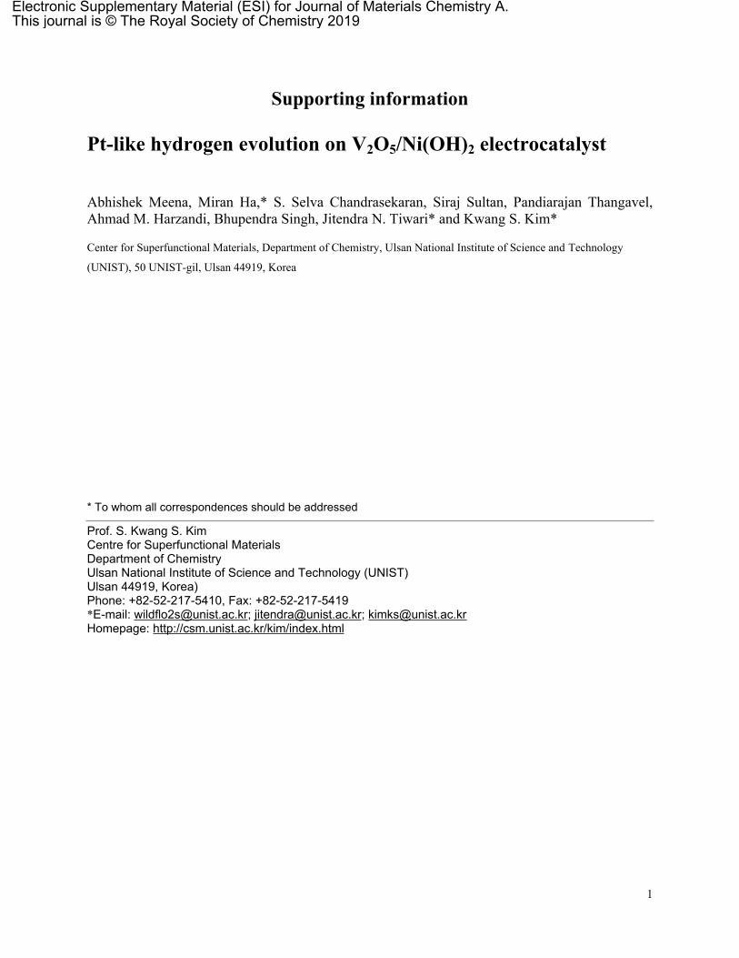
Fig. S3 XPS survey spectrum of the electrocatalyst V2O5/Ni(OH)2@NF.
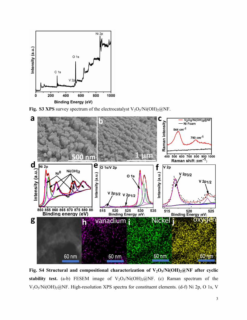
Fig. S4 Structural and compositional characterization of V2O5/Ni(OH)2@NF after cyclic
stability test. (a-b) FESEM image of V2O5/Ni(OH)2@NF. (c) Raman spectrum of the
V2O5/Ni(OH)2@NF. High-resolution XPS spectra for constituent elements. (d-f) Ni 2p, O 1s, V
4
2p, and (f) zoom on V 2p core level spectra. (g-j) HRTEM images of V2O5/Ni(OH)2@NF and the
corresponding elemental mapping images of oxygen, vanadium, and nickel.
Fig. S5 OER polarization curves of V2O5/Ni(OH)2@NF and bare NF in 1 M KOH. (a)
Polarization curves of NF and V2O5/Ni(OH)2@NF at a scan rate of 2mV s−1. (b) Polarization
curves derived Tafel plots of NF and V2O5/Ni(OH)2@NF. As expected, the V2O5/Ni(OH)2@NF
exhibits much higher OER activity and lower Tafel slope than NF.
5
Fig. S6 Set up of water splitting by a solar panel (Two electrodes are connected to a solar panel
and multi-meter).
Theoretical calculations
From the experimental findings, the theoretical model of V2O5/Ni(OH)2@NF was designed to
study HER properties using DFT. In order to make this structure, the (111) surface of Ni slab with
three-layer thickness was cleaved from the FCC-Ni, and the 8×8 surface supercell was constructed
in a hexagonal lattice (Fig. 6A) and it acted as a substrate of NF. At the top layer of Ni, the Ni(OH)2
and V2O5 structures were deposited to construct the V2O5/Ni(OH)2@Ni interfaces as shown in Fig.
6A. Before making this material, we carried out extensive studies of H adsorption property on
(111) Ni-surface, NiO, Ni(OH)2, and V2O5, separately (Fig. S7 to S11). With that, a piece of
(010)V2O5 slab (breadth and width of 1.2 and 1.0 nm, respectively) was cleaved by considering a
minimum energy configuration to deposit it on Ni surface along with optimization of the Ni(OH)2
structure which leads to experimentally synthesized local structures such as V2O5@NF and
Ni(OH)2@NF (Fig. 6). In addition, we calculated the band structure of Ni(OH)2 including various
types of hydrogen coverage using the PBE+U method (U=6.2 eV for Ni) (Fig. S13).
In the synthesized V2O5/Ni(OH)2@Ni, V2O5 was found in orthorhombic structure with
Pmmn (Space group No. 59), and this structure was optimized using DFT calculations. The obtained
lattice constant (a = 11.52 Å, b = 4.378 Å, and c = 3.527 Å) matches well with experiment (a =
11.51 Å, b = 4.37 Å, and c = 3.56 Å). The optimized structure of V2O5 is cleaved along the (100),
(010), and (001) facets to calculate the surface energy. Here, is defined, where 𝛾=
𝐸𝑠𝑢𝑟𝑓 ‒ 𝑛 × 𝐸𝑏𝑢𝑙𝑘2𝐴
, , and are the ground state energy of cleaved structure, the single formula unit of bulk 𝐸𝑠𝑢𝑟 𝐸𝑏𝑢𝑙𝑘 𝑛
structure, and the number of formula units in the cleaved structure, respectively. The calculated 𝛾
for the (100), (010), and (001) surfaces are 0.23, 0.17, and 0.41 eV/Å2, respectively. It expresses
that the (010) surface stands energetically in favor of forming stable structure. Higher index facets
tend to be less stable and the hydrogen adsorption energies on these less stable surfaces tend to be
stronger [Z. Quan, Y. Wang, J. Fang, Acc. Chem. Res. 2013, 46, 191-202]. Indeed, the ΔGH* on
O atoms of V2O5 (001) and V2O5 (310) were too strong (<-0.6 eV), which makes them inactive
surface for HER. We easily expect that any higher index surfaces of V2O5 would be inactive (Fig.
S7, Fig. S8). Similarly, it is expected that any higher index surfaces of nickel foam would not be
active.
6
The (010) surface of V2O5 with two atomic layer thickness (0.9 nm), as shown in Fig. S7
(a), was undertaken to study the HER activity. Four different sites of H binding at the top of the
slab are represented in Figs. S7 (a). With respect to the coordination number of oxygen atoms at
the surface, four active sites are distinguished. Sites S1, S2, and S3 are oxygen atoms with
coordination numbers, of one, two, and three, respectively, while S4 is a surface V site. The
calculated ΔGH* values for all four sites show that S1 and S2 tend to adsorb H, while S3 and S4 (a
more positive value which is not shown in the Fig. S7 (c)) are ready to desorb H. Among these
four sites, S2 is highly active with near-zero ΔGH* of -0.14 eV, and the next active site is S1 with
ΔGH* of -0.32 eV. The same calculations were repeated for three atomic-layer-thick (010) surface
slab (1.38 nm) and the obtained ΔGH* at S1 and S2 are -0.31 and -0.15 eV, respectively. This verifies
the consistency in the calculations. In addition, (001) surface of V2O5 which was analyzed from
TEM was also considered to calculate ΔGH* on three different O atom sites (S5-S7) and one V atom
site (S8) of surface (Figs. S7 (b)). H atom on S8 atom was optimized to be on S6 site. The calculated
ΔGH* values for S5-S7 sites tend to adsorb H strongly and they are shown in Fig. S7 (c) also.
7
Fig. S7 Ball and stick models of the V2O5(010) and V2O5(001) surface of V2O5 slab: (a) top
and side views of (010), and (b) top and side views of (001). The four different H adsorption sites
are indicated by numbers 1 to 8. (c) H-adsorption Free energy (ΔGH*) profile for hydrogen
adsorption sites S1-S3 in the (010) surface and S5-S7 in the (001) surface (the high ΔGH* value at
S4 =1.2 eV is not drawn here).
Fig. S8 Ball and stick models of the V2O5(310) surface on V2O5 slab as a side view and the free
energies (ΔGH*) at seven different H adsorption sites denoted as S1-S7. All sites give large ΔGH*
except for only S6 which is still not small.
8
Fig. S9 Ball and stick models of side and top views of (a) H adsorbed on a surface Ni atom of
Ni(OH)2, (b) H adsorbed on a surface O atom of Ni(OH)2-½Hd (i.e., NiOH(OH1.75)) and (c) H
adsorbed on a surface O atom of Ni(OH)2-¼Hd (i.e., NiOH(OH0.75)) with three layers
thickness. Blue, red, and grey colors denote H, O, and Ni atoms, respectively. From the optimized
bulk structure of Ni(OH)2, a surface with 2×2 supercell along [100] crystallographic direction is
cleaved and the three layers thick surface slab is modelled as shown in the Fig.. The H atom is
attached on the Ni atom. The calculated H adsorption energy is 0.843 eV, which was not changed
substantially when the thickness is reduced from three layers to one layer. Therefore, a piece of
one layer thick Ni(OH)2 is deposited on Ni surface for V2O5/Ni(OH)2/Ni.
Fig. S10 Ball and stick models of side and top view of NiO(111) with a 4×4 supercell. Red and
grey colors indicate O and Ni atoms, respectively.
9
The optimized structure of NiO is cleaved in (111) plane with 5L-thickness. The bottom surface
is terminated with Ni atoms and it is kept fixed in order to achieve the bulk nature. Then, the top
surface terminated with O atoms, which are allowed to relax and find the optimized structure. In
this structure, there are two different H adsorption sites of Ni and O with H adsorption energies of
-1.15 and -0.72 eV, respectively. The calculated values are very close to the values in a previous
work.[5]
Fig. S11 Ball and stick models of Ni(111) surface. The optimized fcc Ni is cleaved along the
(111) plane and a 4×4 supercell with 5 layers thick surface slab is constructed. In this surface, three
different H adsorption sites are available on Ni atom (H bonded with one Ni atom), Ni-Ni bonding
site (H bonded with two Ni atoms), and fcc site (H bonded with three Ni atoms). Among three
sites, fcc site is energetically more stable and it produces adsorption energy of -0.535 eV.
Fig. S12.Modified Ni-Ni bond lengths (in Å) at various H adsorption sites shown in Fig. 6A.
(1) modified NF-surface, (2) Ni(OH)2@Ni interface, (3) between V2O5@Ni and Ni(OH)2@Ni, and
(4) V2O5@Ni. The original Ni-Ni bond length at the pristine Ni-surface is 2.50 Å.
10
Fig. S13 V2O5@Ni(OH)2 with H adsorption site at the interface. (a) Top view. (b) Side view
(H: blue, O: red, V: green, Ni: light black).
Fig. S14 Band structure of 2 bottom layers of Ni(OH)2 and 1 top layer of (a) Ni(OH)2-½Hd,
(b) Ni(OH)2-¼Hd, and (c) Ni(OH)2. (The black solid lines and blue dashed lines in the band
structures images represent the spin up bands and spin down bands, respectively). The structure
with 3 layers of pure Ni(OH)2 shows a high band gap of 2.81 eV. This value increases to ~5 eV
towards the experimental value of 4 eV when GW calculation is performed. If H atoms are not
fully covered on the surface of Ni(OH)2, the band gap becomes smaller than that on its pure state.
11
Fig. S15 Projected density of states (PDOS) (solid lines) and d-band center relative to fermi energy
(εd-εF, dotted lines) of Ni top surface in pristine Ni, V2O5/Ni, and V2O5/Ni(OH)2@Ni.
Fig. S16 Images of the electrode surface of V2O5/Ni(OH)2@NF (a) before and (b) after the
hydrothermal process. The color of NF changes from gray to black. This color change suggests
12
that the V2O5/Ni(OH)2@NF particles are uniformly coated on NF and no uncoated portion is left
on NF.
Fig. S17 Electrocatalytic HER activity of V2O5/Ni(OH)2@NF catalysts synthesized with different
precursor concentrations (NH4VO3).
Fig. S18 The XPS survey scans of Ni(OH)2@NF electrode (A), the corresponding Ni 2p (B)
and O 1s (C) core level spectra. The Ni 2p spectrum shows two major peaks at 855.72 and
873.38eV, corresponding to Ni 2p3/2 and Ni 2p1/2, respectively, with a spin-energy separation of
17.6 eV, which is a characteristic signature of Ni2+ of Ni(OH)2. Two shake up satellites peaks of
Ni 2p3/2 and Ni 2p1/2 was located at around at 861.55 eV and 879.67 eV, respectively. The high
resolution spectrum of O 1s contains a peak at 530.50 eV which usually is attributed to oxygen-
metal bond, and the other fitted peaks at 531.14 and 532.04 eV are attributed to oxygen in
hydroxide group, and a surface-adsorbed water molecule.
13
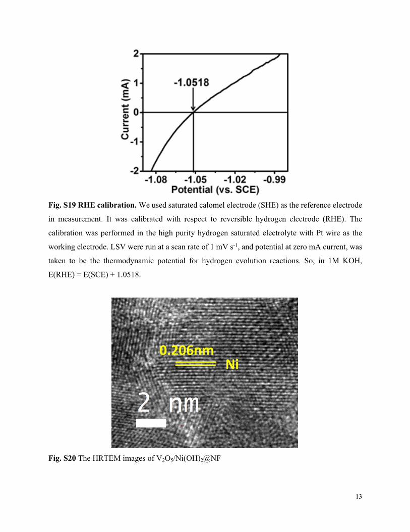
Fig. S19 RHE calibration. We used saturated calomel electrode (SHE) as the reference electrode
in measurement. It was calibrated with respect to reversible hydrogen electrode (RHE). The
calibration was performed in the high purity hydrogen saturated electrolyte with Pt wire as the
working electrode. LSV were run at a scan rate of 1 mV s-1, and potential at zero mA current, was
taken to be the thermodynamic potential for hydrogen evolution reactions. So, in 1M KOH,
E(RHE) = E(SCE) + 1.0518.
Fig. S20 The HRTEM images of V2O5/Ni(OH)2@NF
14
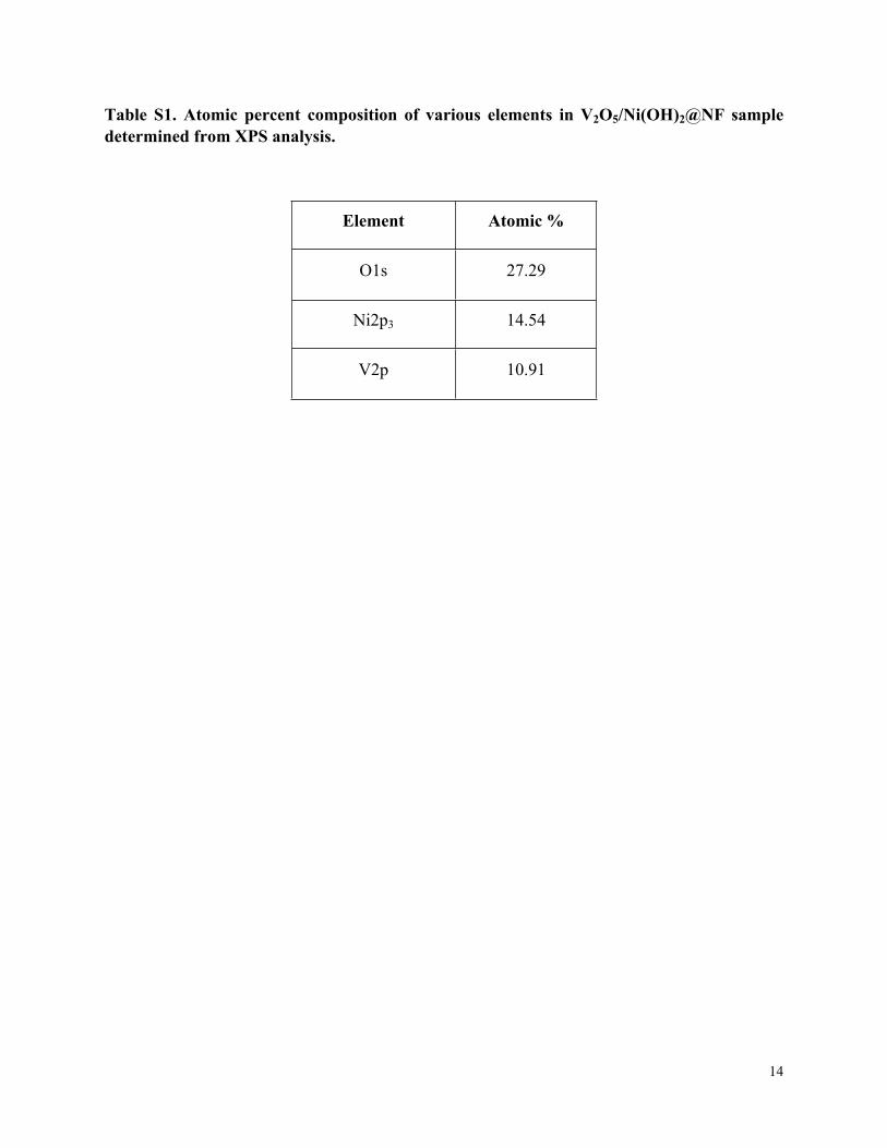
Table S1. Atomic percent composition of various elements in V2O5/Ni(OH)2@NF sample determined from XPS analysis.
Element Atomic %
O1s 27.29
Ni2p3 14.54
V2p 10.91
15
Table S2. Overpotentials of recently developed highly active non-novel metal catalysts.
Overpotential at 10mA/cm2
(mV)
Conditions(Electrolyte,
Scan Rate, Gas Used)
Refs.
V2O5/Ni(OH)2@NF 39 1M KOH This work
20% Pt/C@NF 35 1M KOH This work
VOOH Hollow Nanospheres 164 1M KOH 1
Pt NWs/SL-Ni(OH)2 98 mV @5mA/cm2
1 M KOH 2
Nanocrystalline Ni5P4 50 1M KOH 3
NiSe/NF 96 1M KOH 4
MoCx nano-octahedrons 151 1M KOH 5
NiCo2Px/CF 58 1 M KOH 6
NiO/Ni-CNT 80 1M KOH, 7
np(Co052Fe0.48)2P 79 1M KOH 8
Mn-CoP/Ti 76 1M KOH 9
Zn0.30Co2.70S4 85 1M KOH 10
(CoP)0.54-(FeP) 0.46-NRs/G 98 1M KOH 11
c-CoSe2 200 1M KOH 12
Co-P/Cu foil 94 1M KOH 13
N, P-doped Mo2C@carbon nanospheres 47 1M KOH 14
WC nanocrystals/CNTs 150 0.1M KOH 15
Cu NDs/Ni3S2 NTs-CFs 128 1M KOH 16
Ni0.89Co0.11Se2 MNSN/NF 85 1M KOH 17
NiFeOx/CFP 88 1M KOH 18
CoNx/C 170 0.1M KOH 19
np-CuTi 47 0.1M KOH 20
Ni3N@CQDs 69 1M KOH 21
W‐SAC 53 0.1M KOH 22
N-doped Ni3S2 155 1M KOH 23
N–NiCo2S4 41 1M KOH 24
Co-Ex-MoS2 89 1M KOH 25
MoP nanosheets 49 1M KOH 26
20% Pt/C@NF(1 mg) 31 1M KOH 27
16
References1. H. Shi, H. Liang, F. Ming and Z. Wang, Angew. Chem. Int. Ed., 2017, 56, 573-577.2. H. Yin, S. Zhao, K. Zhao, A. Muqsit, H. Tang, L. Chang, H. Zhao, Y. Gao and Z. Tang, Nat. Commun., 2015,
6, 6430.3. A. B. Laursen, K. R. Patraju, M. J. Whitaker, M. Retuerto, T. Sarkar, N. Yao, K. V. Ramanujachary, M.
Greenblatt and G. C. Dismukes, Energ. Environ. Sci., 2015, 8, 1027-1034.4. C. Tang, N. Cheng, Z. Pu, W. Xing and X. Sun, Angew. Chem. Int. Ed., 2015, 54, 9351-9355.5. H. B. Wu, B. Y. Xia, L. Yu, X.-Y. Yu and X. W. Lou, Nat. Commun., 2015, 6, 6512.6. R. Zhang, X. Wang, S. Yu, T. Wen, X. Zhu, F. Yang, X. Sun, X. Wang and W. Hu, Adv.Mater, 2017, 29,
1605502-n/a.7. M. Gong, W. Zhou, M.-C. Tsai, J. Zhou, M. Guan, M.-C. Lin, B. Zhang, Y. Hu, D.-Y. Wang, J. Yang, S. J.
Pennycook, B.-J. Hwang and H. Dai, Nat. Commun., 2014, 5, 4695.8. Y. Tan, H. Wang, P. Liu, Y. Shen, C. Cheng, A. Hirata, T. Fujita, Z. Tang and M. Chen, Energ. Environ. Sci.,
2016, 9, 2257-2261.9. T. Liu, X. Ma, D. Liu, S. Hao, G. Du, Y. Ma, A. M. Asiri, X. Sun and L. Chen, ACS. Catal., 2017, 7, 98-102.10. Z.-F. Huang, J. Song, K. Li, M. Tahir, Y.-T. Wang, L. Pan, L. Wang, X. Zhang and J.-J. Zou, J. Am. Chem.
Soc, 2016, 138, 1359-1365.11. B. Liu, L. Huo, Z. Gao, G. Zhi, G. Zhang and J. Zhang, Small, 2017, 13, 1700092-n/a.12. P. Chen, K. Xu, S. Tao, T. Zhou, Y. Tong, H. Ding, L. Zhang, W. Chu, C. Wu and Y. Xie, Adv.Mater, 2016,
28, 7527-7532.13. N. Jiang, B. You, M. Sheng and Y. Sun, Angew. Chem. Int. Ed., 2015, 54, 6251-6254.14. Y.-Y. Chen, Y. Zhang, W.-J. Jiang, X. Zhang, Z. Dai, L.-J. Wan and J.-S. Hu, ACS Nano, 2016, 10, 8851-
8860.15. X. Fan, H. Zhou and X. Guo, ACS Nano, 2015, 9, 5125-5134.16. J.-X. Feng, J.-Q. Wu, Y.-X. Tong and G.-R. Li, J. Am. Chem. Soc, 2018, 140, 610-617.17. B. Liu, Y.-F. Zhao, H.-Q. Peng, Z.-Y. Zhang, C.-K. Sit, M.-F. Yuen, T.-R. Zhang, C.-S. Lee and W.-J. Zhang,
Adv.Mater, 2017, 29, 1606521-n/a.18. H. Wang, H.-W. Lee, Y. Deng, Z. Lu, P.-C. Hsu, Y. Liu, D. Lin and Y. Cui, Nat. Commun., 2015, 6, 7261.19. H.-W. Liang, S. Brüller, R. Dong, J. Zhang, X. Feng and K. Müllen, Nat. Commun., 2015, 6, 7992.20. Q. Lu, G. S. Hutchings, W. Yu, Y. Zhou, R. V. Forest, R. Tao, J. Rosen, B. T. Yonemoto, Z. Cao, H. Zheng,
J. Q. Xiao, F. Jiao and J. G. Chen, Nat. Commun., 2015, 6, 6567.21. M. Zhou, Q. Weng, Z. I. Popov, Y. Yang, L. Y. Antipina, P. B. Sorokin, X. Wang, Y. Bando and D. Golberg,
ACS Nano, 2018, 12, 4148-4155.22. W. Chen, J. Pei, C. T. He, J. Wan, H. Ren, Y. Wang, J. Dong, K. Wu, W. C. Cheong and J. Mao, Adv.Mater,
2018, 30, 1800396.23. T. Kou, T. Smart, B. Yao, I. Chen, D. Thota, Y. Ping and Y. Li, Adv. Ener. Mater., 2018, 8, 1703538.24. Y. Wu, X. Liu, D. Han, X. Song, L. Shi, Y. Song, S. Niu, Y. Xie, J. Cai, S. Wu, J. Kang, J. Zhou, Z. Chen, X.
Zheng, X. Xiao and G. Wang, Nat. Commun., 2018, 9, 1425.25. Y. Luo, X. Li, X. Cai, X. Zou, F. Kang, H.-M. Cheng and B. Liu, ACS Nano, 2018, 12, 4565-4573.26. G. Li, Y. Sun, J. Rao, J. Wu, A. Kumar, Q. N. Xu, C. Fu, E. Liu, G. R. Blake, P. Werner, B. Shao, K. Liu, S.
Parkin, X. Liu, M. Fahlman, S.-C. Liou, G. Auffermann, J. Zhang, C. Felser and X. Feng, Adv. Ener. Mater., 2018, 8, 1801258.
27. B. Liu, Y.-F. Zhao, H.-Q. Peng, Z.-Y. Zhang, C.-K. Sit, M.-F. Yuen, T.-R. Zhang, C.-S. Lee and W.-J. Zhang, Adv.Mater, 2017, 29, 1606521.