127
By Riham Hazem Raafat Lecturer of Chest Diseases Ainshams University
| Date post: | 22-Jan-2018 |
| Category: |
Health & Medicine |
| Upload: | dr-riham-hazem-raafat |
| View: | 658 times |
| Download: | 0 times |
By
Riham Hazem RaafatLecturer of Chest Diseases
Ainshams University
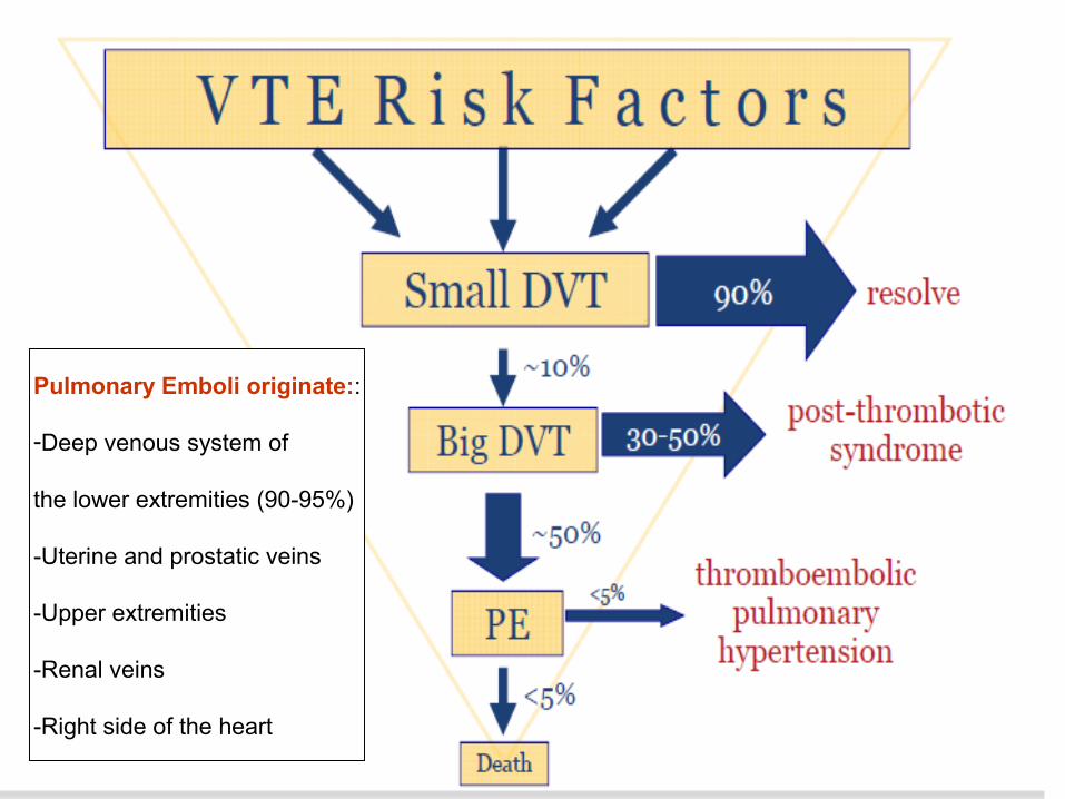
Pulmonary Emboli originate::
-Deep venous system of
the lower extremities (90-95%)
-Uterine and prostatic veins
-Upper extremities
-Renal veins
-Right side of the heart

Risk Factors

• Alteration of blood flow: – Prolonged immobilisation, – Obesity, – Pregnancy, – Cancer
• Factors in blood vessel wall: – Surgery (risk in 1st 2 wks up to 3 Ms), – Catheterisation,– Trauma
• Hypercoagulable states: – Estrogen containing OCP, – Genetic thrombophilia (Factor V Leiden deficiency, Protein C and
Protein S deficiency, antithrombin III deficiency etc.), – Acquired thrombophilia (antiphospholipid syndrome, nephrotic
syndrome, paroxysmal nocturnal hemoglobinuria)

What is PE?
- A pulmonary embolism refers to the obstruction of
a pulmonary artery or its branches
Incidence: 600,000/year
Mortality rate: 50,000 to 200,000/yr
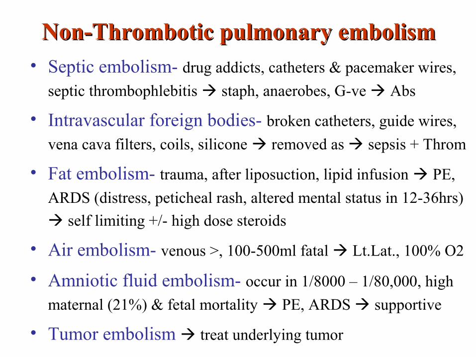
Non-Thrombotic pulmonary embolismNon-Thrombotic pulmonary embolism• Septic embolism- drug addicts, catheters & pacemaker wires,
septic thrombophlebitis staph, anaerobes, G-ve Abs
• Intravascular foreign bodies- broken catheters, guide wires,
vena cava filters, coils, silicone removed as sepsis + Throm
• Fat embolism- trauma, after liposuction, lipid infusion PE,
ARDS (distress, peticheal rash, altered mental status in 12-36hrs)
self limiting +/- high dose steroids
• Air embolism- venous >, 100-500ml fatal Lt.Lat., 100% O2
• Amniotic fluid embolism- occur in 1/8000 – 1/80,000, high
maternal (21%) & fetal mortality PE, ARDS supportive
• Tumor embolism treat underlying tumor

PathophysiologyPathophysiology
• Increased pulmonary vascular resistance
• Impaired gas exchange
• Alveolar hyperventilation
• Increased airway resistance
• Decreased pulmonary compliance
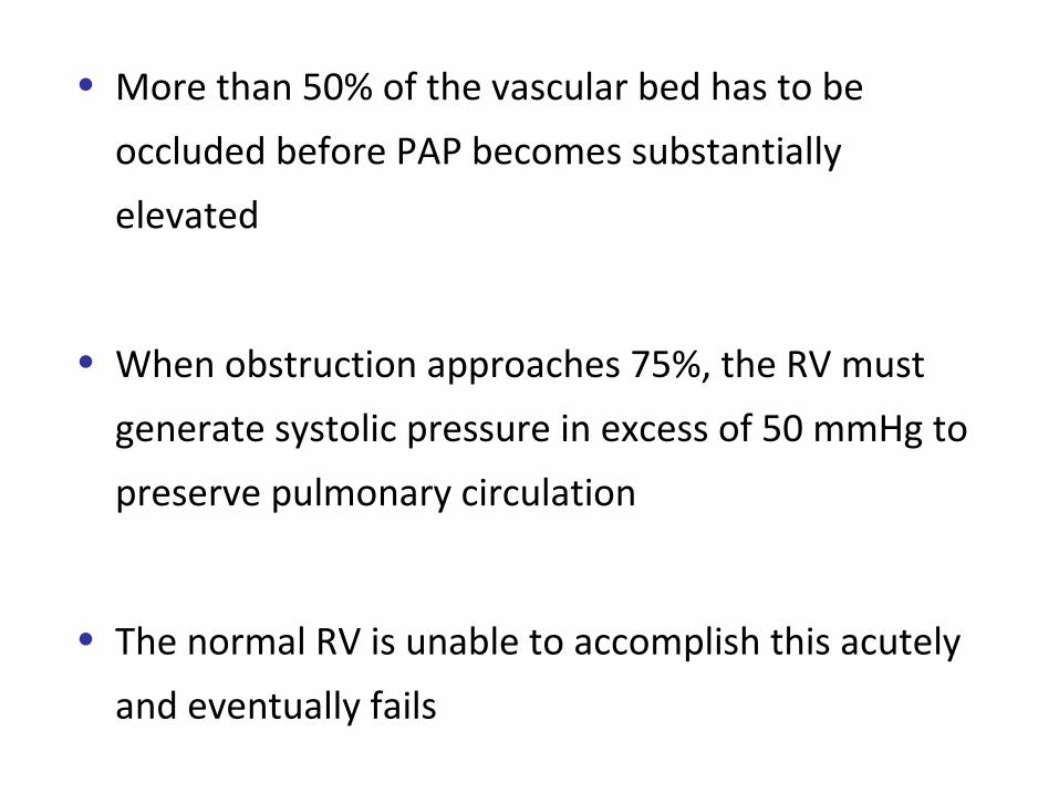
• More than 50% of the vascular bed has to be
occluded before PAP becomes substantially
elevated
• When obstruction approaches 75%, the RV must
generate systolic pressure in excess of 50 mmHg to
preserve pulmonary circulation
• The normal RV is unable to accomplish this acutely
and eventually fails

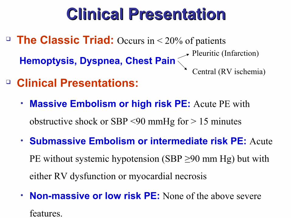
Clinical PresentationClinical Presentation The Classic Triad: Occurs in < 20% of patients
Hemoptysis, Dyspnea, Chest Pain
Clinical Presentations:
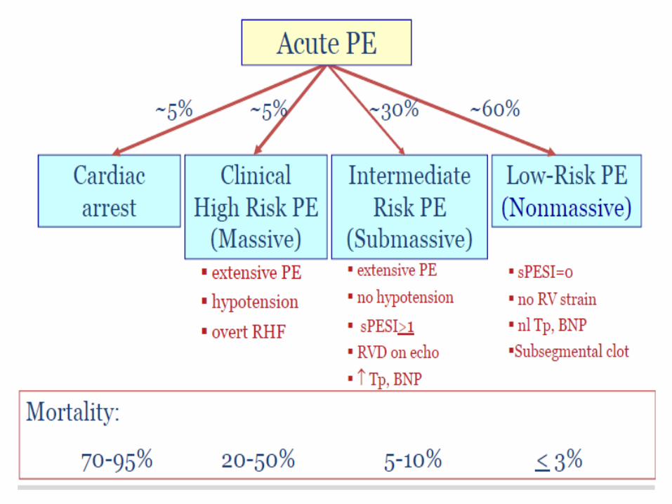
• Massive Embolism or high risk PE: Acute PE with
obstructive shock or SBP <90 mmHg for > 15 minutes
• Submassive Embolism or intermediate risk PE: Acute
PE without systemic hypotension (SBP ≥90 mm Hg) but with
either RV dysfunction or myocardial necrosis
• Non-massive or low risk PE: None of the above severe
features.
Pleuritic (Infarction)
Central (RV ischemia)
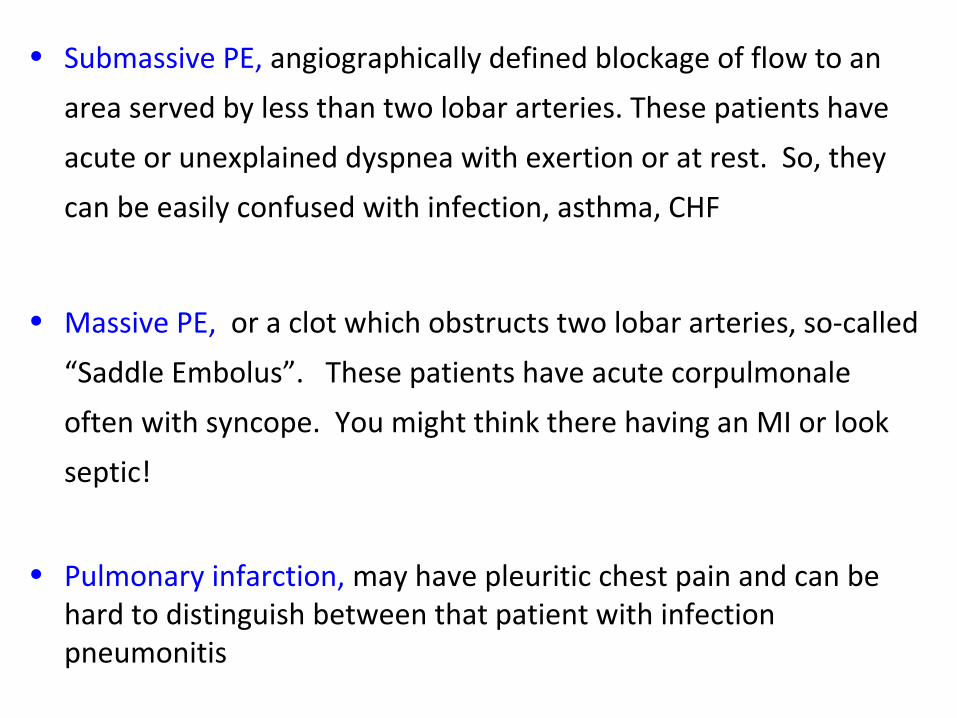
• Submassive PE, angiographically defined blockage of flow to an
area served by less than two lobar arteries. These patients have
acute or unexplained dyspnea with exertion or at rest. So, they
can be easily confused with infection, asthma, CHF
• Massive PE, or a clot which obstructs two lobar arteries, so-called
“Saddle Embolus”. These patients have acute corpulmonale
often with syncope. You might think there having an MI or look
septic!
• Pulmonary infarction, may have pleuritic chest pain and can be hard to distinguish between that patient with infection pneumonitis

Symptom Percent
Dyspnea 84
Chest Pain, pleuritic 74
Anxiety 59
Cough 53
Hemoptysis 30
Sweating 27
Chest Pain, non-pleuritic 14
Syncope 13
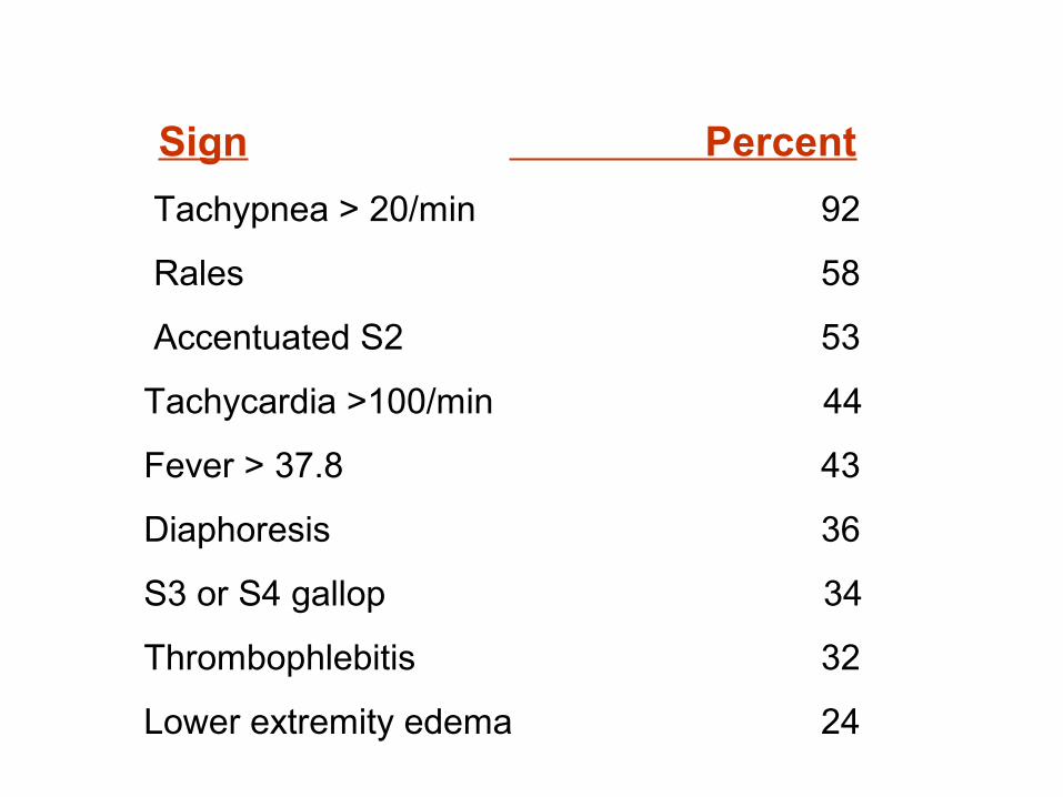
Sign Percent
Tachypnea > 20/min 92
Rales 58
Accentuated S2 53
Tachycardia >100/min 44
Fever > 37.8 43
Diaphoresis 36
S3 or S4 gallop 34
Thrombophlebitis 32
Lower extremity edema 24

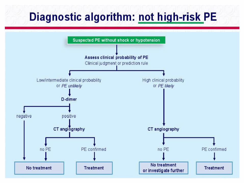
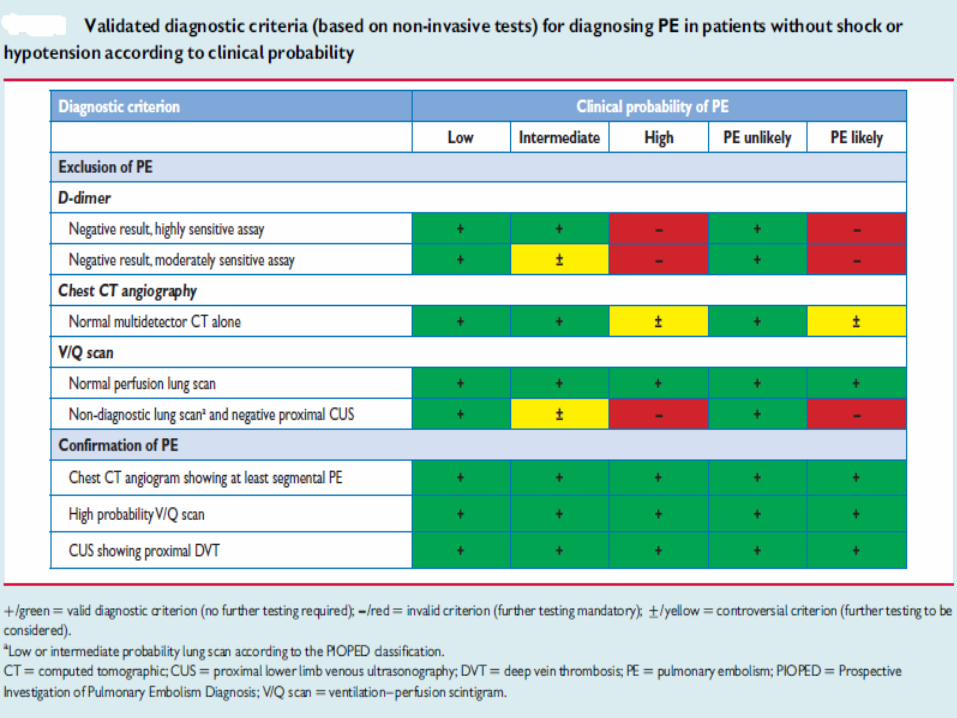
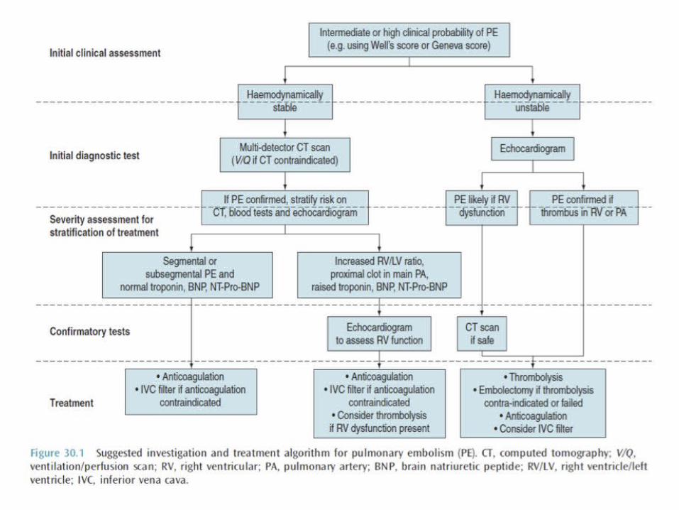
Diagnosis
• Risk stratification
• Clinical examination
• Bed side tests (ECG, ABG)
• Laboratory tests (D-dimer, Cardiac biomarkers)
• Imaging techniques (Ultrasound/ Doppler scan, CXR, CTPA, V/Q scan, Echocardiogram)


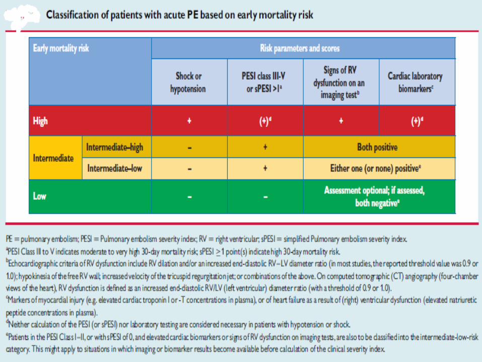
Risk stratification
• PERC Rule
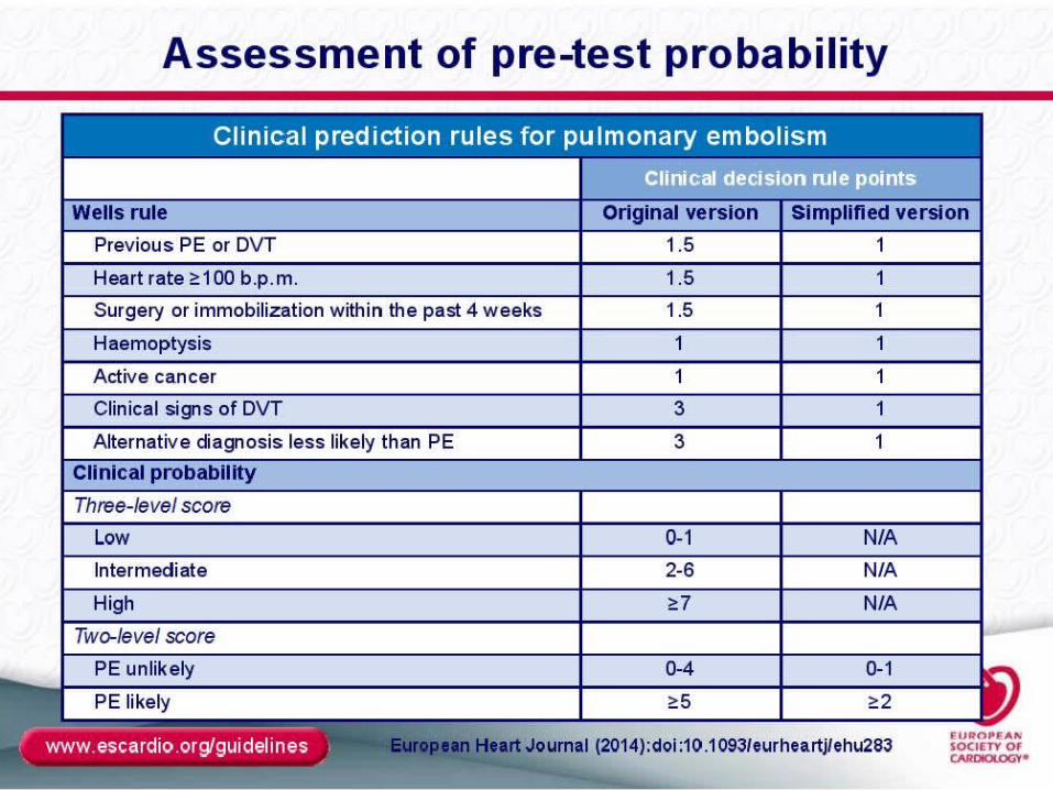
• Wells score for PE
• Modified Geneva score for PE
• PESI Index
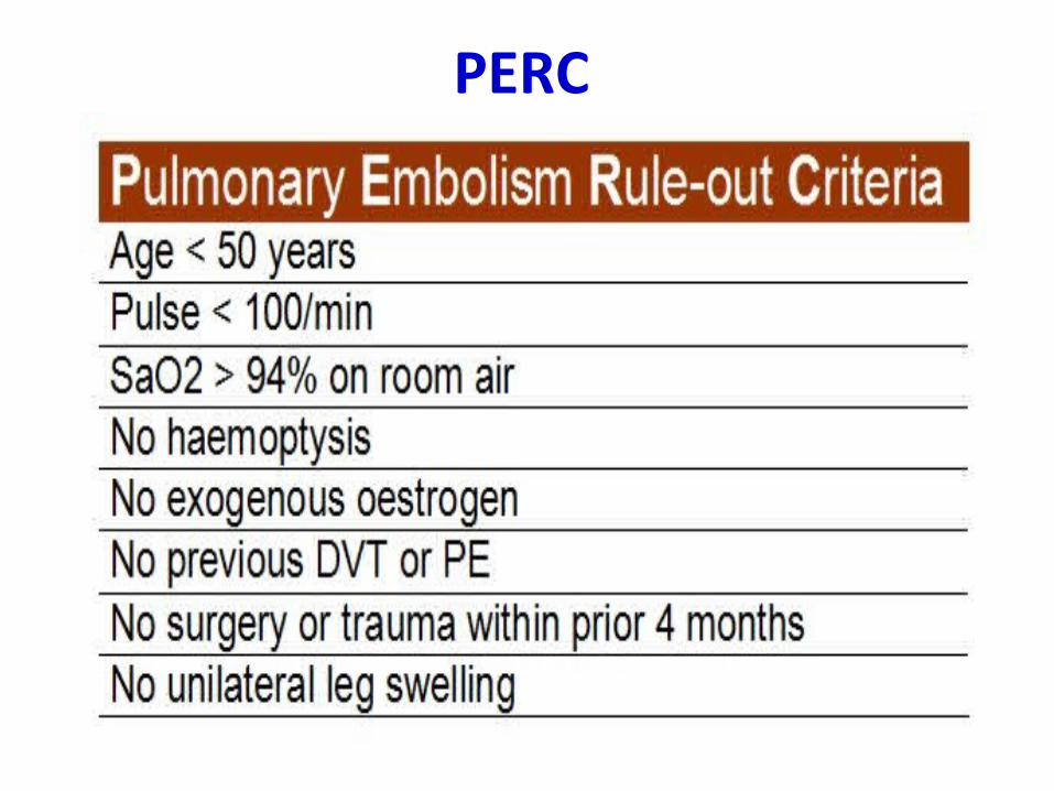
PERC
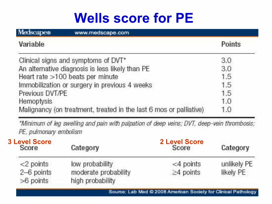
Wells score for PE
3 Level Score 2 Level Score
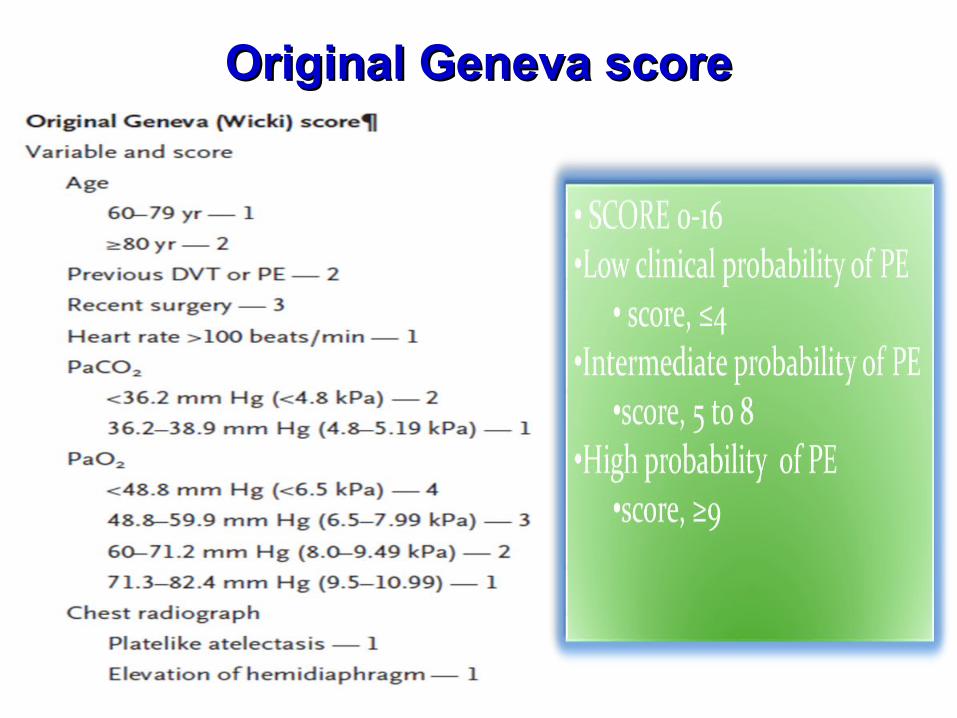
Original Geneva scoreOriginal Geneva score

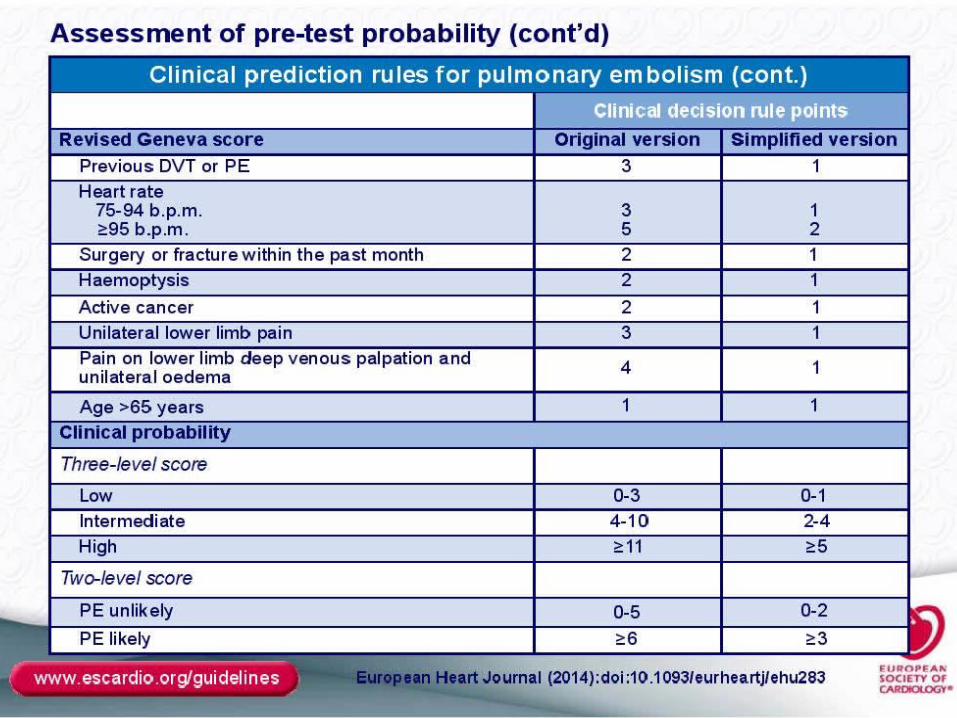
Revised Geneva scoreRevised Geneva score
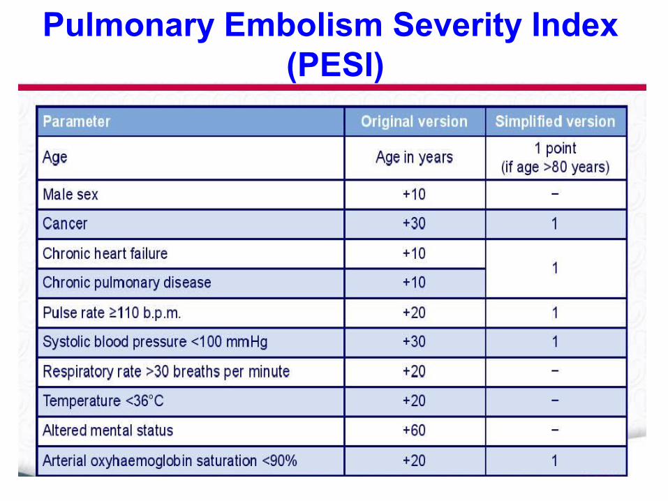
Pulmonary Embolism Severity Index (PESI)

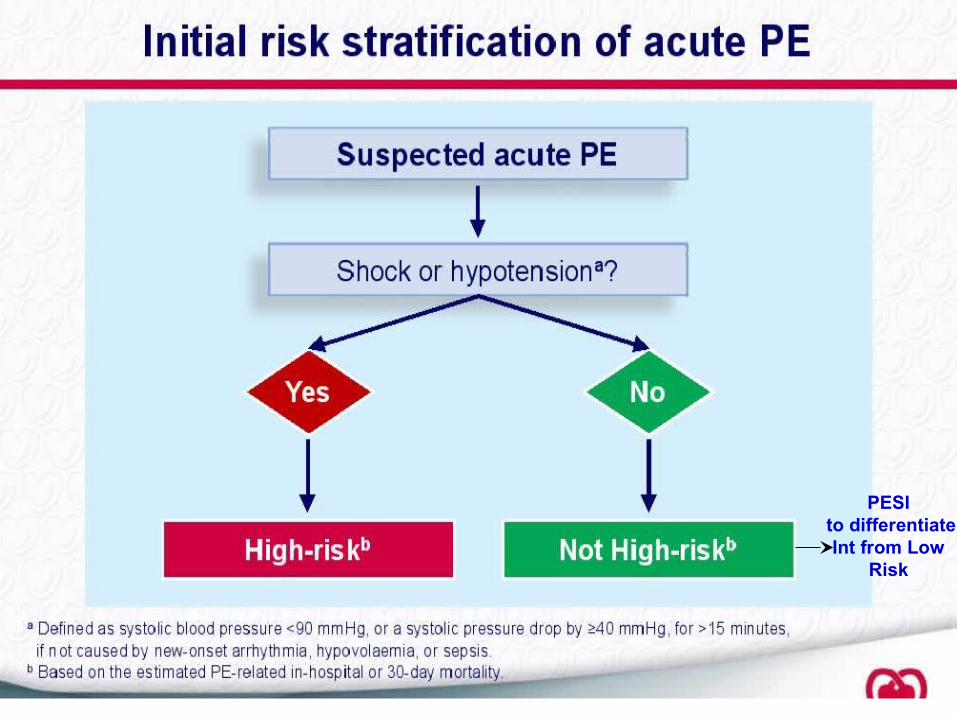
PESI to differentiate Int from Low
Risk


Biomarkers (cardiac & non-cardiac)• Troponin ‐ released from right ventricle Injury
• Cardiac BNP ‐ released from cardiac myocytes in response
to elevated pressures RVD
*A normal troponin and BNP can safely exclude high risk
patients with a negative predictive value of 97-100%
• H-FABP (heart type fatty acid binding protein) – early
marker for injury (good for prognosis as well)
• NGAL (neutrophil gelatinase associated lipocalin) &
Cystatin C – both indicating kidney injury, also shown to
have prognostic value

ABG findings in PE
• pH= ↑
• PaO2= ↓
• PaCO2= ↓
• HCO3= Normal
• Aa gradient= Large
Aa gradient= PAO2- PaO2

Chest x-ray
• Mostly normal findings
• Done to exclude other pathology
• Pleural effusion/ Atelectatic bands
• Specific signs:- Hampton’s hump (in infarction)- Westermark sign
- Palla’s sign - Fleischner sign (in massive embolism or PHTN)


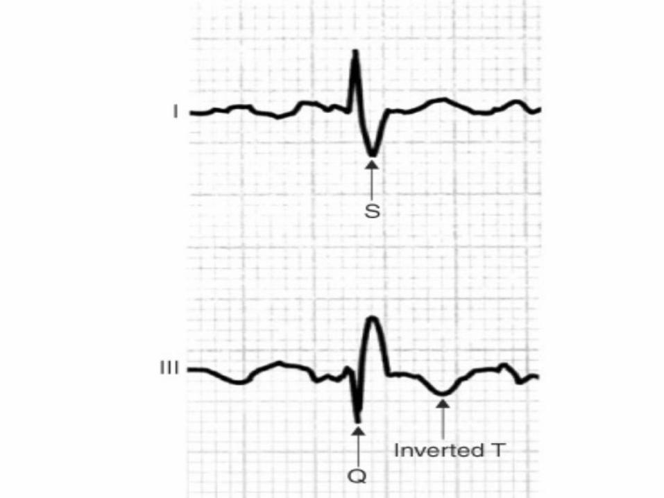
ECG findings in PE
• Normal sinus rhythm
• Sinus tachycardia
• Tall peaked T waves in V1- V4
• S1Q3T3 pattern: Not specific. Can be seen in any Cor pulmonale syndrome
• RBBB

S1Q3T3 pattern ECG
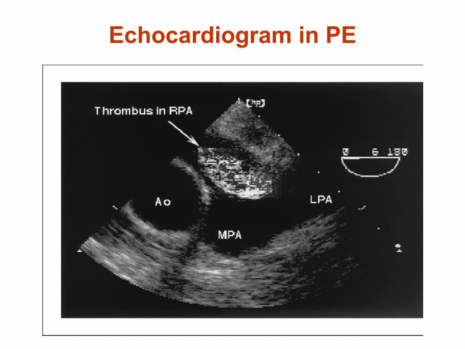
Echocardiogram in PE

- RV Strain
• An RV/LV ratio of >0.9 was shown to be and independent
predictive factor for HOSPITAL MORTALITY
• RV hypokinesia on baseline echo following PE with a ~40% higher
mortality rate
- Used also to detect TAPSE & Tricuspid Jet Velocity to
detect CTEPH
- TOE used to detect pulmonary artery emboli & its
branches
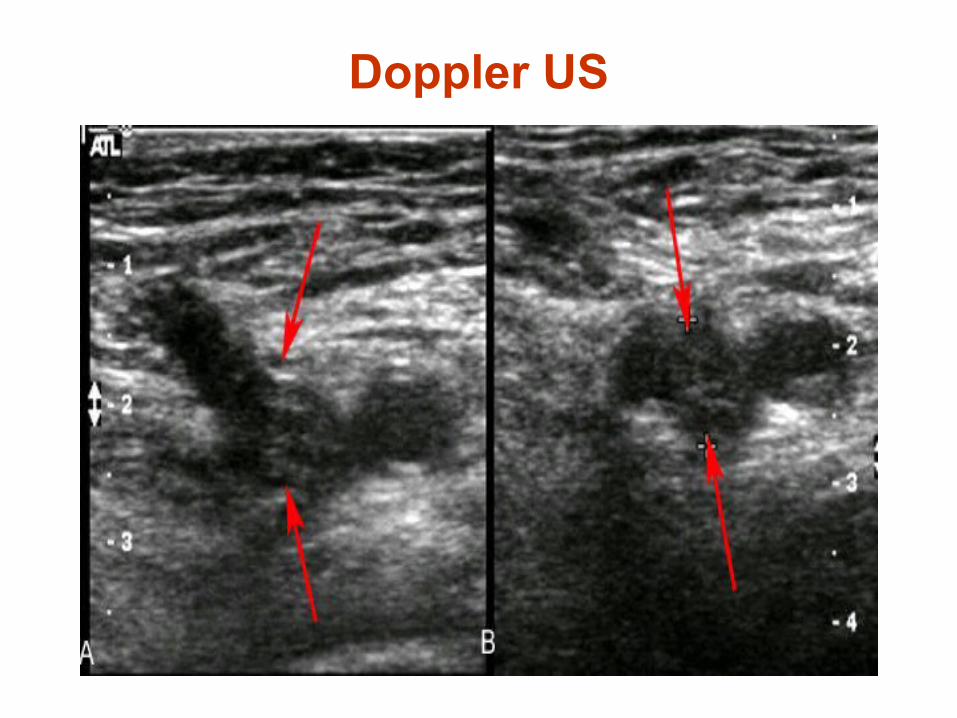
Doppler US

D-dimer in PE
• D-dimer is a type of Fibrin degradation product
• Can be raised due to a number of reasons
• Negative D-dimer rules out PE/DVT in 98% cases
• False positive D-dimer: infection, pregnancy, renal failure, post-operative

– Qualitative
• Bed side RBC agglutination test
• Low Specificity and Sensitivity
– “SimpliRED D-dimer”
– Quantitative
• Enzyme linked immunosorbent assay “Dimertest”
• Positive assay is > 500ng/ml
• VIDAS D-dimer, 2nd generation ELISA test
• Specificity decreases with age above 80 to 10% so
age-adjusted cut-off points are used for that

CTPA
Indications:
- Suspected PE
Contra-indications:- Renal failure
- Pregnancy
- Allergy to radio-contrast
Procedure:
- Radioactive iodine administered IV
- CT scan performed

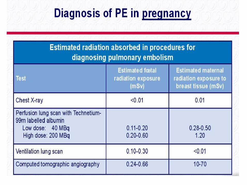
Pitfalls of CTPA:
• Average radiation exposure is 12.4-31.8 mSV.
• This was estimated to increase the risk of breast cancer by 1.004 to 1.042 and lung cancer from 1.005 to 1.076.
• The excess risk of cancer for individuals over 55 would be less than 1%;
• In a young 20-year-old woman this would be estimated to increase the relative lifetime risk of breast or lung cancer by 1.7 to 5.5%.
(Hurwitz et al. 2007)
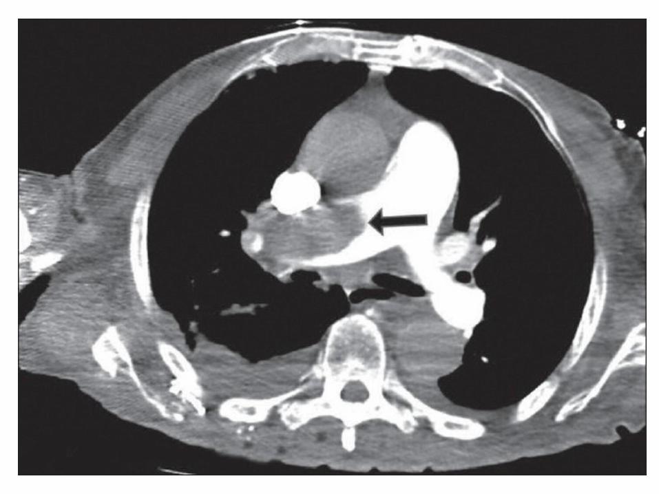
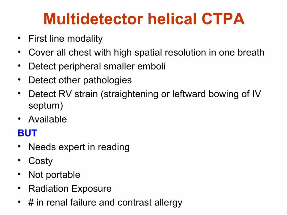
Multidetector helical CTPA• First line modality
• Cover all chest with high spatial resolution in one breath• Detect peripheral smaller emboli• Detect other pathologies• Detect RV strain (straightening or leftward bowing of IV
septum)
• Available
BUT• Needs expert in reading• Costy• Not portable
• Radiation Exposure
• # in renal failure and contrast allergy


Ventilation-perfusion scan
Indications:
- Renal failure
- Pregnancy
Procedure:
- Ventilation scan with Xenon inhalation
- Perfusion scan with Tc99m labelled radioactive dye infusion
- Scan V/Q
- Result: unmatched V/Q


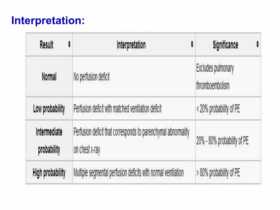
Interpretation:

Ancillary TestAncillary Test
WBC Poor sensitivity and nonspecific (Can be as high
as 20,000 in some patients)
Hgb/Hct PTE does not alter count but if extreme, consider
polycythemia, a known risk factor
ESR Don’t get one, terrible test in regard to any
predictive value

DDXCondition Differentiating signs/symptoms
MI Retrosternal pressure radiating to the jaw, arm, or neck.Risk factors include long-standing hypertension, diabetes, or hypercholesterolaemia.
Pneumonia Cough, purulent sputum.Fever above 39.0°C generally higher than in PE.
Pneumothorax History of recent trauma to the chest.Decreased breath sounds unilaterally Hyperresonance on percussion of affected side.Deviation of the trachea away from the affected lung.
CHF, acute exacerbation
Orthopnoea, paroxysmal nocturnal dyspnoea,Increased bilateral lower extremity swelling.Diffuse crackles on pulmonary auscultation.Elevated jugular venous pressure.
Pericarditis Chest pain improves when sitting up and worsens when supine.
Tamponade, cardiac Beck's triad of hypotension, muffled heart sounds, and elevated jugular venous pressure
Panic disorder Sudden-onset anxiety, feeling faint, and palpitations.Recurrent, discrete period of intense fear/discomfort.

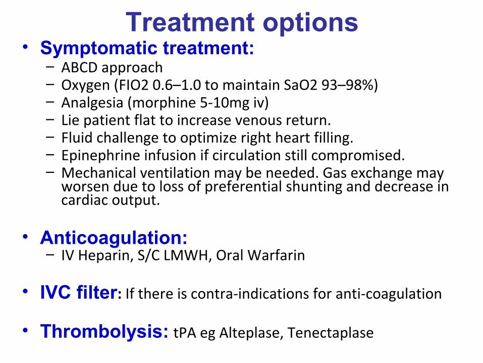
Treatment options• Symptomatic treatment:
– ABCD approach– Oxygen (FIO2 0.6–1.0 to maintain SaO2 93–98%) – Analgesia (morphine 5-10mg iv)– Lie patient flat to increase venous return. – Fluid challenge to optimize right heart filling. – Epinephrine infusion if circulation still compromised. – Mechanical ventilation may be needed. Gas exchange may
worsen due to loss of preferential shunting and decrease in cardiac output.
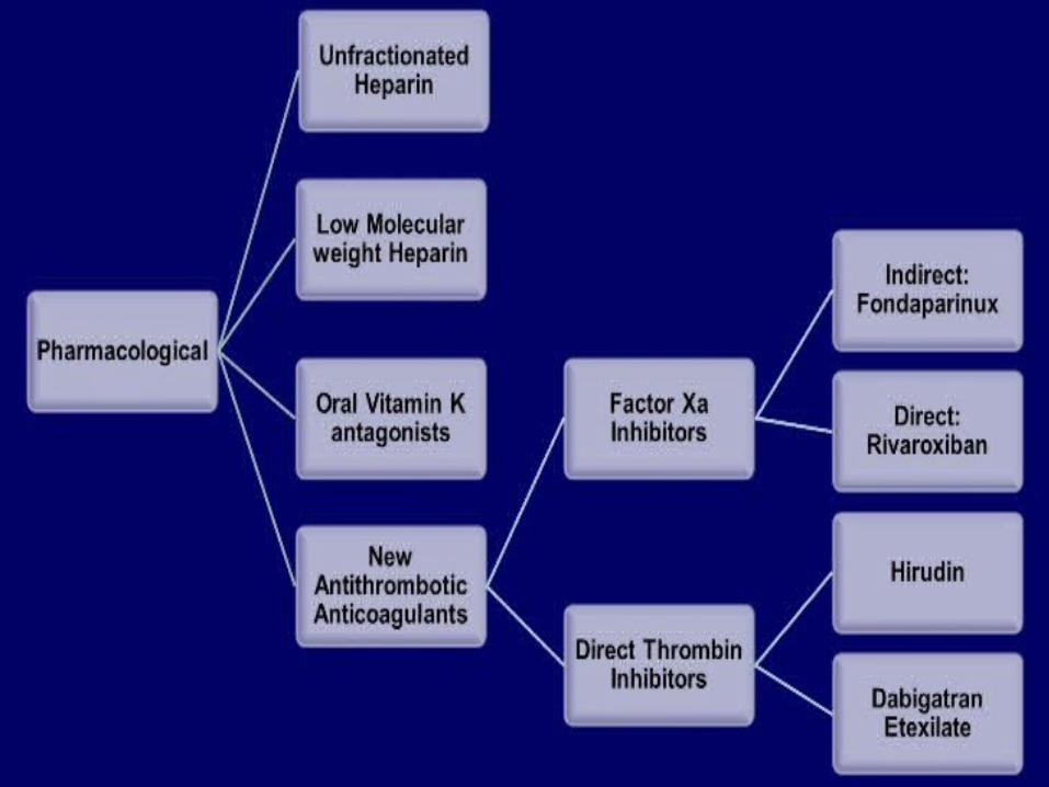
• Anticoagulation:– IV Heparin, S/C LMWH, Oral Warfarin
• IVC filter: If there is contra-indications for anti-coagulation
• Thrombolysis: tPA eg Alteplase, Tenectaplase
• Surgical procedures: Pulmonary embolectomy
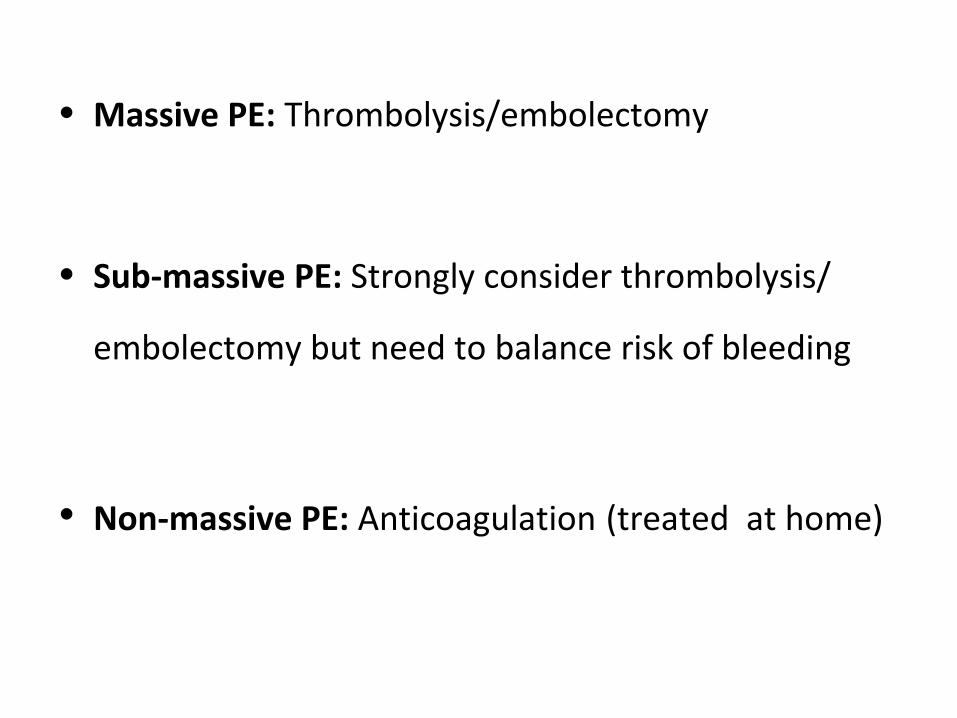
• Massive PE: Thrombolysis/embolectomy
• Sub-massive PE: Strongly consider thrombolysis/
embolectomy but need to balance risk of bleeding
• Non-massive PE: Anticoagulation (treated at home)

Intermediate Risk (Submassive) PE
• Definition: Acute PE without systemic hypotension (systolic blood pressure 90 mm Hg) but with either:
-- RV dysfunction: Hypokinesia Or Dilatation: RV/LV diameter >0.9 on MDCT or Echo
-- BNP >90 pg/ml
-- Appropriate ECG Changes (RBBB, etc.)
-- Myocardial necrosis: Troponin I > 0.14ng/ml
• Intermediate Risk PE therapy:
-- ANTICOAGULATION (AC)- HEPARIN
AC therapy prevents further clot growth
Consider Single Drug NOAC’s for stable patients
-- Oxygen supplementation
-- Telemetry monitoring


High-Risk (Massive) PE
• Definition: Acute PE with:
– Cardiac arrest / hemodynamic instability
– Sustained hypotension (systolic blood pressure 90 mm Hg for at least 15 minutes OR requiring inotropic support not due to a secondary cause (arrhythmia, sepsis)
*Remember: The presence of “lots” of PE isn’t enough to call it “massive
• High-Risk PE therapy:
– Systemic Anticoagulation ASAP
– Supplemental oxygen for O2 sat <90%
– Admit to the intensive care unit: Significant hypoxemia, Hemodynamic compromise thrombolytic therapy
– Mechanical ventilation for respiratory failure
– For Hypotension: IVF, Vasopressor Support


AnticoagulationIV Heparin
– 80 units/kg bolus followed by – 18 units/kg infusion
• Monitor APTT 60-90 sec
• Side effects: – HITS (Heparin induced thrombocytopenia
syndrome): paradoxical hypercoagulable state leads to clots
– Bleeding

• Raschke Nomogram:Raschke Nomogram:

• Use of Heparin Before and After Thrombolysis:Use of Heparin Before and After Thrombolysis:
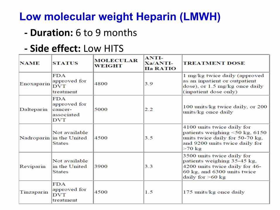
Low molecular weight Heparin (LMWH)
- Duration: 6 to 9 months
- Side effect: Low HITS

FondaparinuxFondaparinux• Anticoagulant pentasaccharide that specifically
inhibits activated factor X• By selectively binding to antithrombin, it
potentiates (about 300 times) the neutralization of factor Xa by antithrombin
• It does not cross-react with heparin-induced antibodies
• FDA has approved fondaparinux for initial treatment of acute PE and acute DVT as a bridge to oral anticoagulation with warfarin

Vitamin K antagonist
• Warfarin: – 5mg PO initial dose– Check regular INR 2-3
• Side effects:– Bleeding– Unusual bruises– Headache

Novel Oral Anticoagulants (NOACs)Novel Oral Anticoagulants (NOACs)
• Promise immediate onset of action and administration
in fixed doses without routine laboratory coagulation
monitoring but not used in severe renal impairment
• Few interactions, making them more “user friendly”
Dabigatran: direct thrombin inhibitor (150mg bid)
Rivaroxaban: factor Xa inhibitor (15mg bid 3wks 20mg o.d)
Apixaban: factor Xa inhibitor (10mg bid 1wk 5mg bid)
Edoxaban: factor Xa inhibitor (60mg o.d, 30mg if low cr.cl. Or
wt < 60 kg)

*N.B.:
The results of the trials (RE-COVER, RECORD-3, EINSTEIN-PE,
AMPLIFY, Hokusai-VTE) using NOACs in the treatment of VTE
indicate that these agents are non-inferior (in terms of efficacy) and
possibly safer (particularly in terms of major bleeding) than the
standard heparin/VKA regimen.
High TTR values were achieved under VKA treatment in all trials; on the
other hand, the study populations included relatively young patients,
very few of whom had cancer.

Recommendations for Initial Anticoagulation forRecommendations for Initial Anticoagulation for Acute PE Acute PE (AHA/ASC 2011, ACCP 2012) (AHA/ASC 2011, ACCP 2012)
• Therapeutic anticoagulation with SC LMWH, IV or SC UFH with
monitoring, unmonitored weight-based SC UFH, or SC
fondaparinux + VKA (till INR >2 for 24 hr) should be given to pts with
objectively confirmed PE and no # to anticoagulation (1B)
• Therapeutic parenteral anticoagulation during the diagnostic
workup should be given to pts with intermediate (if diag. delay >4hrs)
or high clinical probability of PE & no # to anticoagulation (2C)
• Therapeutic parenteral anticoagulation during the diagnostic
workup is not given in low probability (if diag. not delayed than 24 hrs)
(2C) Preferred than UFH except if # (renal impairment, with thrombolysis or can’t afford)

Optimal Duration of Anticoagulation Optimal Duration of Anticoagulation
ACCP 2012
According to bleeding risk (3Ms or Extended)

Thrombolysis
• Indications:
– Massive PE
– Sub-massive PE where risk of bleeding low (in RVD?!)
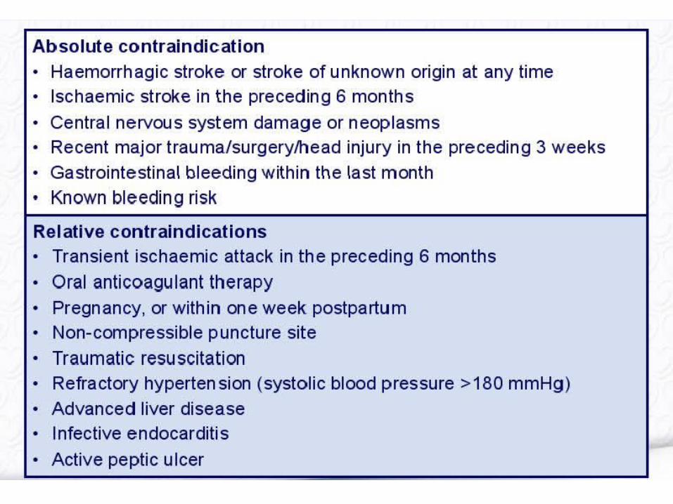
• Contraindications:
– Bleeding, recent stroke, HI, current GI bleeding,
bleeding PUD, surgery within 7 day, prolonged CPR

•Drugs:
(Most rapid)

• About administration About administration
Intravenous route
-- primary method of delivery
Rapid infusion
-- Shorter regimens may not only prove efficacious
but also reduce the risk of hemorrhagic
complications
Catheter-directed therapy
-- for massive PE, may induce major bleeding

• General guidelines for administration General guidelines for administration

before after

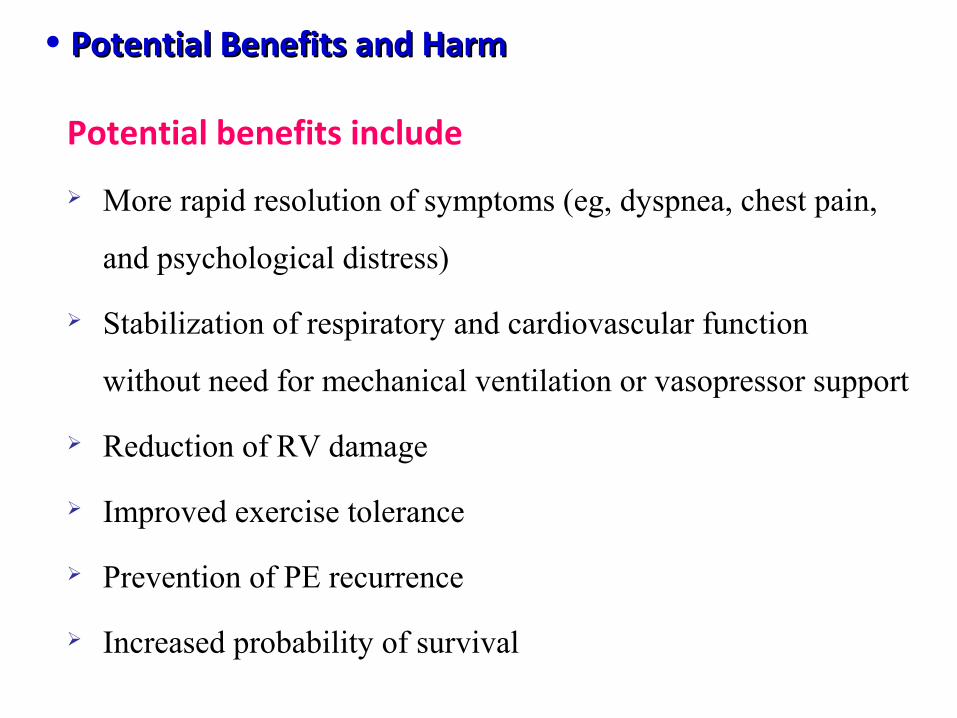
• Potential Benefits and HarmPotential Benefits and Harm
Potential benefits include
More rapid resolution of symptoms (eg, dyspnea, chest pain,
and psychological distress)
Stabilization of respiratory and cardiovascular function
without need for mechanical ventilation or vasopressor support
Reduction of RV damage
Improved exercise tolerance
Prevention of PE recurrence
Increased probability of survival
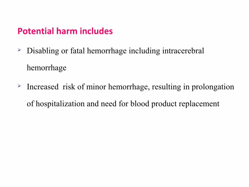
Potential harm includes
Disabling or fatal hemorrhage including intracerebral
hemorrhage
Increased risk of minor hemorrhage, resulting in prolongation
of hospitalization and need for blood product replacement

ACC/AHA & ACCP RecommendationsACC/AHA & ACCP Recommendations
Through Peripheral Vein is better than Pulmonary Artery Catheter
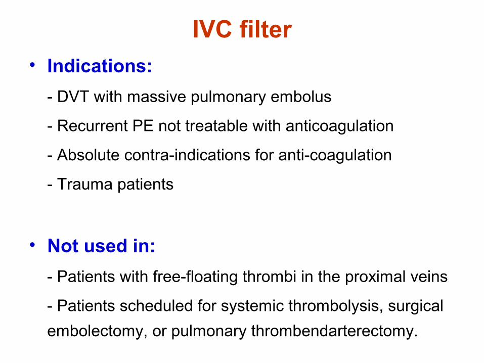
IVC filter• Indications:
- DVT with massive pulmonary embolus
- Recurrent PE not treatable with anticoagulation
- Absolute contra-indications for anti-coagulation
- Trauma patients
• Not used in:
- Patients with free-floating thrombi in the proximal veins
- Patients scheduled for systemic thrombolysis, surgical
embolectomy, or pulmonary thrombendarterectomy.

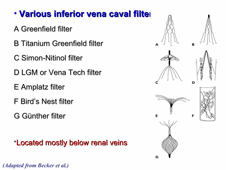
• Various inferior vena caval filtersVarious inferior vena caval filters::
A Greenfield filterA Greenfield filter
B Titanium Greenfield filter B Titanium Greenfield filter
C Simon-Nitinol filter C Simon-Nitinol filter
D LGM or Vena Tech filter D LGM or Vena Tech filter
E Amplatz filter E Amplatz filter
F Bird’s Nest filterF Bird’s Nest filter
G Günther filterG Günther filter
**Located mostly below renal veinsLocated mostly below renal veins
(Adapted from Becker et al.)
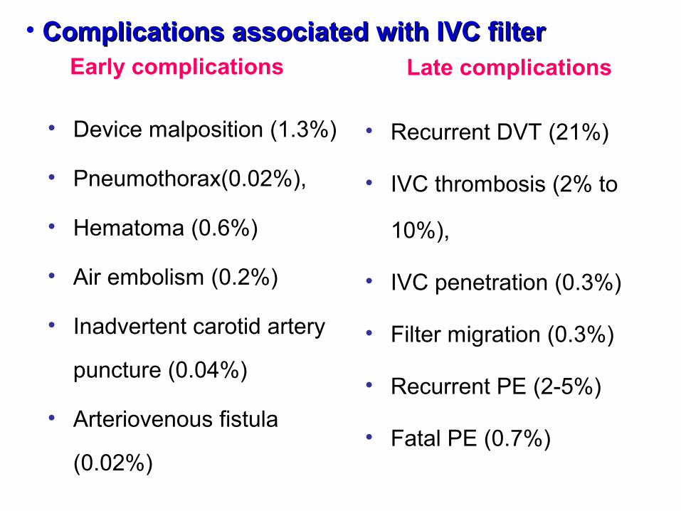
• Complications associated with IVC filterComplications associated with IVC filterEarly complications
• Device malposition (1.3%)
• Pneumothorax(0.02%),
• Hematoma (0.6%)
• Air embolism (0.2%)
• Inadvertent carotid artery
puncture (0.04%)
• Arteriovenous fistula
(0.02%)
Late complications
• Recurrent DVT (21%)
• IVC thrombosis (2% to
10%),
• IVC penetration (0.3%)
• Filter migration (0.3%)
• Recurrent PE (2-5%)
• Fatal PE (0.7%)

Recommendations on IVC Filters in the Recommendations on IVC Filters in the Setting of Acute PESetting of Acute PE


Catheter-Based InterventionsCatheter-Based Interventions• Performed as an alternative to thrombolysis When there are contraindications When emergency surgical thrombectomy is unavailable
or contraindicated Hybrid therapy that includes both catheter-based clot
fragmentation and local thrombolysis is an emerging strategy
• Goals of catheter-based therapy include Rapidly reducing pulmonary artery pressure, RV strain,
and pulmonary vascular resistance (PVR) Increasing systemic perfusion Facilitating RV recovery

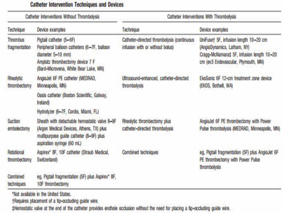
• Categories of percutaneous intervention
Suction thrombectomy with aspiration catheters
Thrombus fragmentation with pigtail or balloon catheters
Rheolytic thrombectomy with hydrodynamic catheters
(saline jet or drug)
Rotational thrombectomy
Conventional catheter directed thrombolysis (drugs)
U/S accelerated thrombolysis with CDT (inc permeability)
Pharmaco-mechanical thrombolysis (combined technique)

• Catheter-directed therapyCatheter-directed therapy
Local delivery of streptokinase
-- Extensive lysis (by perfusion scan and pulmonary
arteriography at 12 to 24 hour follow-up)
Intrapulmonary versus peripheral alteplase
-- no advantage over the intravenous route
Direct delivery into clot
--Enhanced thrombolysis, relatively low doses (in an
animal model of PE)
-- Could prove advantageous over the intravenous route

• Side Effects (2%)
Death from worsening RV failure,
Distal embolization,
Pulmonary artery perforation with lung hemorrhage,
Systemic bleeding complications,
Pericardial tamponade,
Heart block or bradycardia,
Haemolysis,
Contrast-induced nephropathy, and
Puncture-related complications
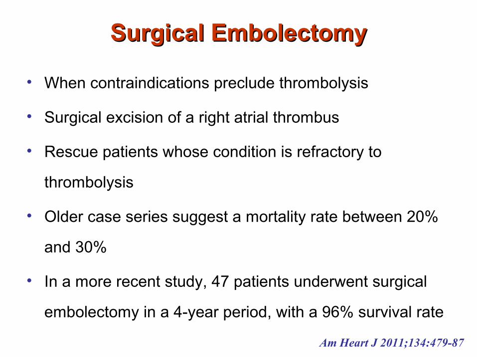
Surgical EmbolectomySurgical Embolectomy
• When contraindications preclude thrombolysis
• Surgical excision of a right atrial thrombus
• Rescue patients whose condition is refractory to
thrombolysis
• Older case series suggest a mortality rate between 20%
and 30%
• In a more recent study, 47 patients underwent surgical
embolectomy in a 4-year period, with a 96% survival rate
Am Heart J 2011;134:479-87

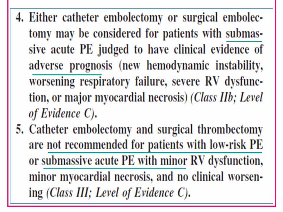
Recommendations (AHA – ACCP)Recommendations (AHA – ACCP)
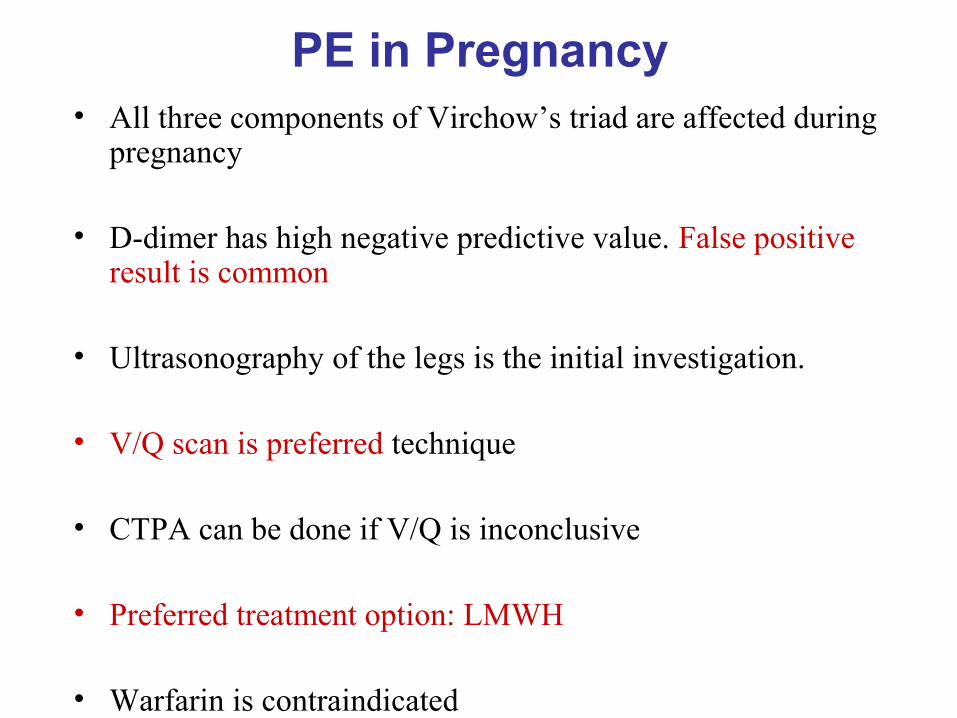
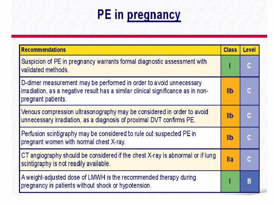
PE in Pregnancy• All three components of Virchow’s triad are affected during
pregnancy
• D-dimer has high negative predictive value. False positive result is common
• Ultrasonography of the legs is the initial investigation.
• V/Q scan is preferred technique
• CTPA can be done if V/Q is inconclusive
• Preferred treatment option: LMWH
• Warfarin is contraindicated
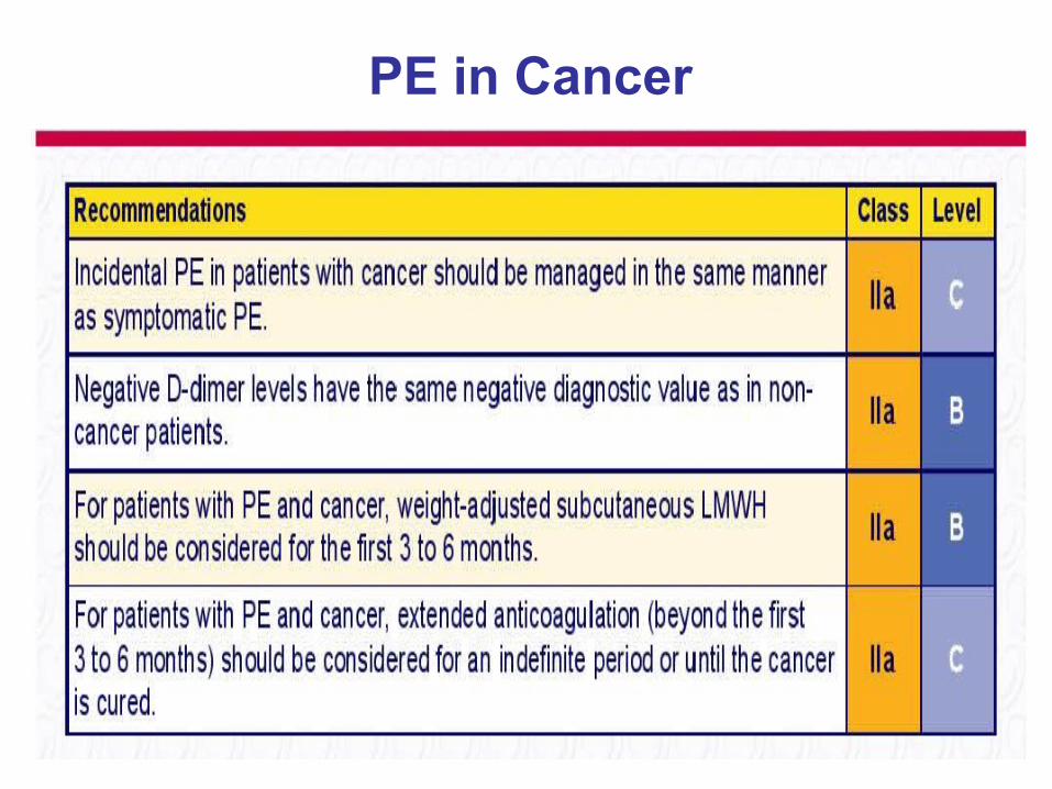
PE in Cancer
Prevention of PE
• Control of obesity
• Stop smoking
• Stockings
• Heparin: 5000 units/day SC bid or tid
• Enoxaprin: 40 mg/day SC

1 Hospitalization with medical illness
Enoxaparin 40 mg SC qd orDalteparin 5000 units SC qd orFondaparinux 2.5 mg SC qd (in patients with a heparin allergy such as heparin-induced thrombocytopenia) orGraduated compression stockings or intermittent pneumatic compression
2 General surgery Unfractionated heparin 5000 units SC bid or tid orEnoxaparin 40 mg SC qd orDalteparin 2500 or 5000 units SC qd
3 Major orthopedic surgery Warfarin (target INR 2 to 3) orEnoxaparin 30 mg SC bid orEnoxaparin 40 mg SC qd orDalteparin 2500 or 5000 units SC qd orFondaparinux 2.5 mg SC qdRivaroxaban 10 mg qd (in Canada and Europe)Dabigatran 220 mg bid (in Canada and Europe)

4 Oncologic surgery Enoxaparin 40 mg SC qd
5 Neurosurgery Unfractionated heparin 5000 units SC bid orEnoxaparin 40 mg SC qd andGraduated compression stockings or intermittent pneumatic compressionConsider surveillance lower extremity ultrasonography
6 Thoracic surgery Unfractionated heparin 5000 units SC tid andGraduated compression stockings or intermittent pneumatic compression

CTEPH
It is an important cause of pulmonary hypertension (Class IV-
mean PAP greater than 25 mm Hg) that either after PE is diagnosed
(0.1-9.1% in 2yrs after PE) or even without previous PE.
The only identifiable RFs for persistent pulmonary HTN: Age 70
years & Systolic PAP 50 mm Hg at the initial presentation ??
Unresolved residual thrombus becomes organized and fibrosed,
leading to ongoing obstruction to pulmonary blood flow.
Untreated, this leads to progressive pulmonary hypertension,
RV dysfunction, failure and death even on anticoagulation.


“Not a disease, but a
syndrome in which the
pressure in the pulmonary
circulation is raised”
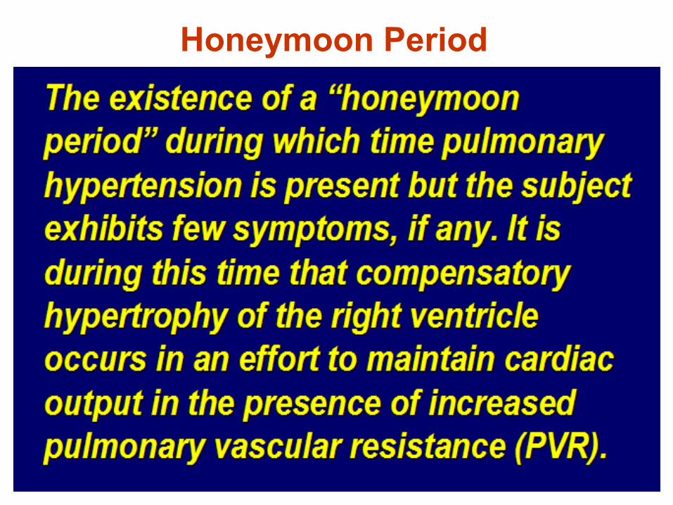
Honeymoon Period
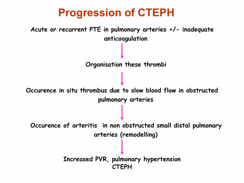
Progression of CTEPH
Acute or recurrent PTE in pulmonary arteries +/- inadequate anticoagulation
Organisation these thrombi
Occurence in situ thrombus due to slow blood flow in obstructed pulmonary arteries
Occurence of arteritis in non obstructed small distal pulmonary arteries (remodelling)
•
Increased PVR, pulmonary hypertensionCTEPH

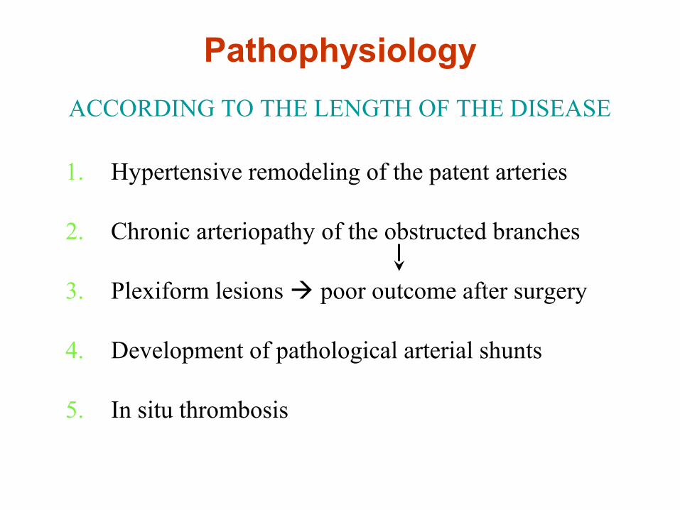
1. Hypertensive remodeling of the patent arteries
2. Chronic arteriopathy of the obstructed branches
3. Plexiform lesions poor outcome after surgery
4. Development of pathological arterial shunts
5. In situ thrombosis
ACCORDING TO THE LENGTH OF THE DISEASE
Pathophysiology


Diagnosis of CTEPH
To confirm diagnosis, support ttt decisions &
Measure PVR pre and postop

Echo Probability of PHTN

Other Echo Findings
Echo signs from at least two different categories (A/B/C) from the list should be present to alter the level of echo probability of pulmonary hypertension.
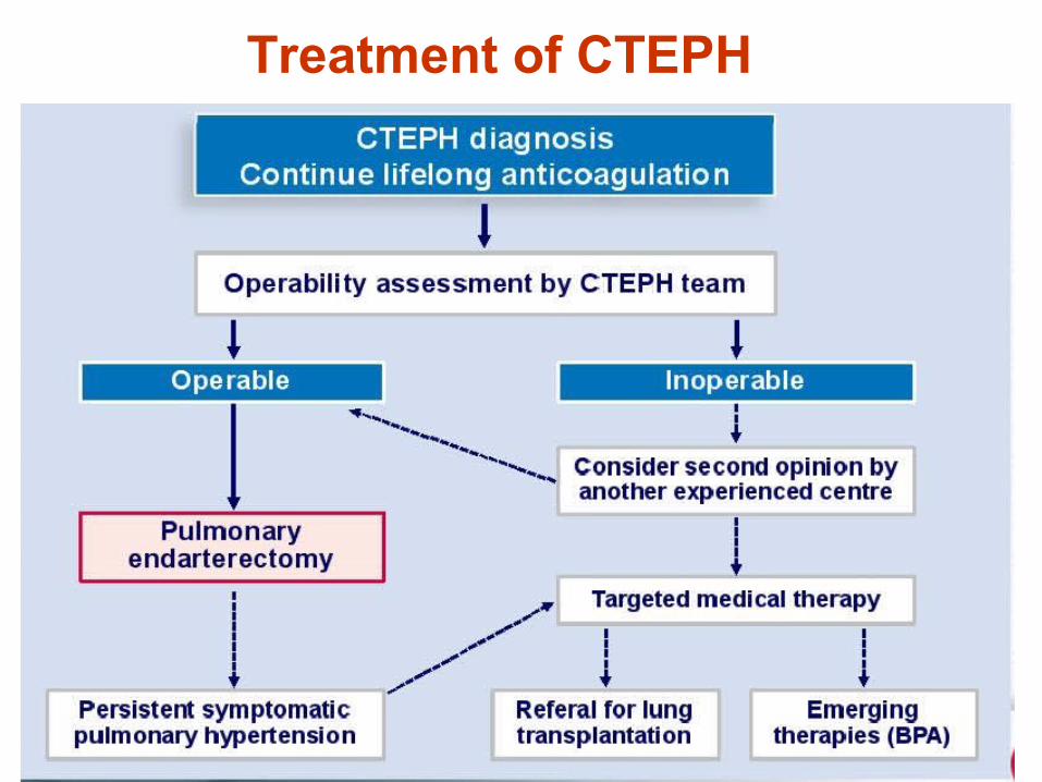
Treatment of CTEPH
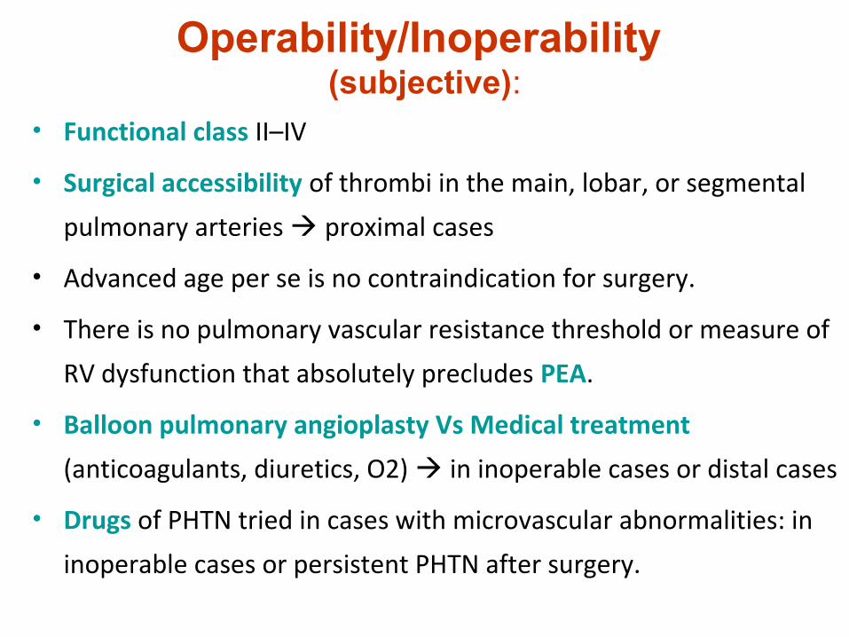
Operability/Inoperability (subjective):
• Functional class II–IV
• Surgical accessibility of thrombi in the main, lobar, or segmental
pulmonary arteries proximal cases
• Advanced age per se is no contraindication for surgery.
• There is no pulmonary vascular resistance threshold or measure of
RV dysfunction that absolutely precludes PEA.
• Balloon pulmonary angioplasty Vs Medical treatment
(anticoagulants, diuretics, O2) in inoperable cases or distal cases
• Drugs of PHTN tried in cases with microvascular abnormalities: in
inoperable cases or persistent PHTN after surgery.

PEA
BPA
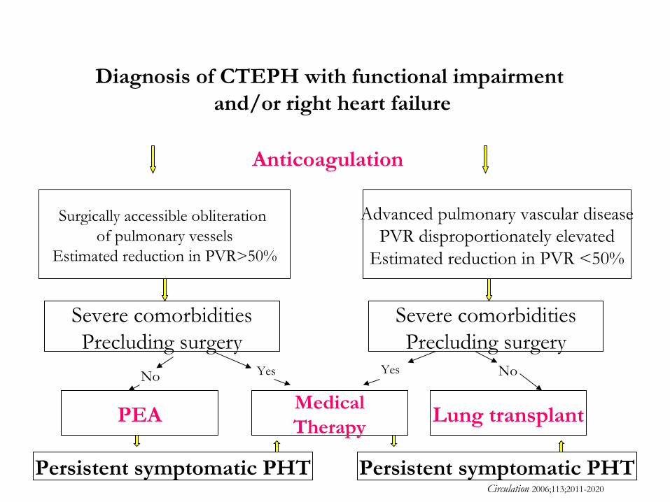
Diagnosis of CTEPH with functional impairment and/or right heart failure
Surgically accessible obliteration of pulmonary vessels
Estimated reduction in PVR>50%
Advanced pulmonary vascular diseasePVR disproportionately elevated
Estimated reduction in PVR <50%
Severe comorbiditiesPrecluding surgery
Severe comorbiditiesPrecluding surgery
PEAMedicalTherapy
Lung transplant
Persistent symptomatic PHT Persistent symptomatic PHT
No Yes No
Circulation 2006;113;2011-2020
Anticoagulation
Yes

Humbert M et al. N Engl J Med. 2004;351:1425-1436.
Targets for Current or Emerging TherapiesTargets for Current or Emerging Therapies
Big EndothelinBig Endothelin
Endothelin-Endothelin-convertingconverting
EnzymeEnzyme
EndothelinEndothelinReceptor AReceptor A
EndothelinEndothelinReceptor BReceptor B
VasoconstrictionVasoconstrictionandand
ProliferationProliferation
EndothelinReceptor
Antagonists
Endothelin-1Endothelin-1
Endothelin PathwayEndothelin Pathway
ArginineArginine
Nitric OxideNitric OxideSynthaseSynthase
VasodilatationVasodilatationandand
AntiproliferationAntiproliferation
Nitric OxideNitric Oxide
cGMPcGMP ExogenousNitric Oxide
Phosphodiesterase Type-5Phosphodiesterase Type-5
PhosphodiesteraseType-5 Inhibitors
Nitric Oxide PathwayNitric Oxide Pathway
Arachidonic AcidArachidonic Acid
ProstacyclinProstacyclinSynthaseSynthase
VasodilatationVasodilatationandand
AntiproliferationAntiproliferation
ProstacyclinProstacyclin
cAMPcAMP
ProstacyclinProstacyclinDerivativesDerivatives
ProstacyclinDerivatives
Prostacyclin PathwayProstacyclin Pathway
Guanylate cyclase stimulator
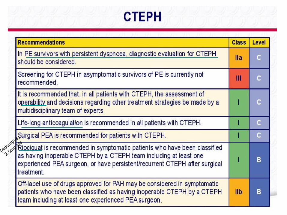
(Adempus)
2.5mg tds


