64
Pulmonary Embolism DR. ANISH CHOUDHARY JR III, MD ( RADIOLOGY) BVDUMC PUNE
| Date post: | 21-Jan-2018 |
| Category: |
Health & Medicine |
| Upload: | anish-choudhary |
| View: | 292 times |
| Download: | 1 times |
Pulmonary Embolism
D R . A N I S H C H O U D H A R Y
J R I I I , M D ( R A D I O L O G Y )
B V D U M C P U N E
Pulmonary embolism (PE) is a blockage of the main artery of the lung, or one of its branches by a substance that has travelled from elsewhere in the body through the bloodstream (embolism).
PE most commonly results from deep vein thrombosis (a blood clot in the deep veins of the legs or pelvis) that breaks off and migrates to the lung, a process termed venous thromboembolism (VTE).
A small proportion of cases are caused by the embolization of air, fat, or talc in drugs of intravenous drug abusers.
The obstruction of the blood flow through the lungs and the resultant pressure on the right ventricle of the heart leads to the symptoms and signs of PE. The risk of PE is increased in various situations, such as cancer or prolonged bed rest.
Most Common Symptoms of PE (PIOPED Study)
Dyspnea (73%)
Pleuritic chest pain (66%)
Coughing (37%)
Hemoptysis (13%)
Atypical Presentations of PE Abdominal/pelvic pain
Decreasing level of consciousness
Fever
Productive cough
Seizures
Syncope
Wheezing
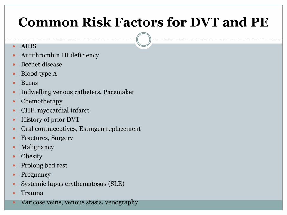
Common Risk Factors for DVT and PE
AIDS
Antithrombin III deficiency
Bechet disease
Blood type A
Burns
Indwelling venous catheters, Pacemaker
Chemotherapy
CHF, myocardial infarct
History of prior DVT
Oral contraceptives, Estrogen replacement
Fractures, Surgery
Malignancy
Obesity
Prolong bed rest
Pregnancy
Systemic lupus erythematosus (SLE)
Trauma
Varicose veins, venous stasis, venography
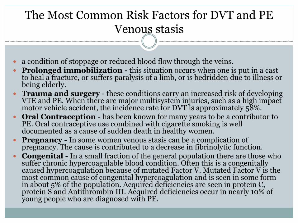
The Most Common Risk Factors for DVT and PE Venous stasis
a condition of stoppage or reduced blood flow through the veins. Prolonged immobilization - this situation occurs when one is put in a cast
to heal a fracture, or suffers paralysis of a limb, or is bedridden due to illness or being elderly.
Trauma and surgery - these conditions carry an increased risk of developing VTE and PE. When there are major multisystem injuries, such as a high impact motor vehicle accident, the incidence rate for DVT is approximately 58%.
Oral Contraception - has been known for many years to be a contributor to PE. Oral contraceptive use combined with cigarette smoking is well documented as a cause of sudden death in healthy women.
Pregnancy - In some women venous stasis can be a complication of pregnancy. The cause is contributed to a decrease in fibrinolytic function.
Congenital - In a small fraction of the general population there are those who suffer chronic hypercoagulable blood condition. Often this is a congenitally caused hypercoagulation because of mutated Factor V. Mutated Factor V is the most common cause of congenital hypercoagulation and is seen in some form in about 5% of the population. Acquired deficiencies are seen in protein C, protein S and Antithrombin III. Acquired deficiencies occur in nearly 10% of young people who are diagnosed with PE.
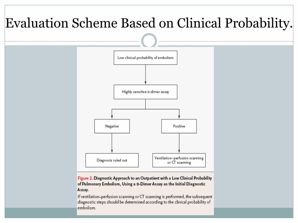
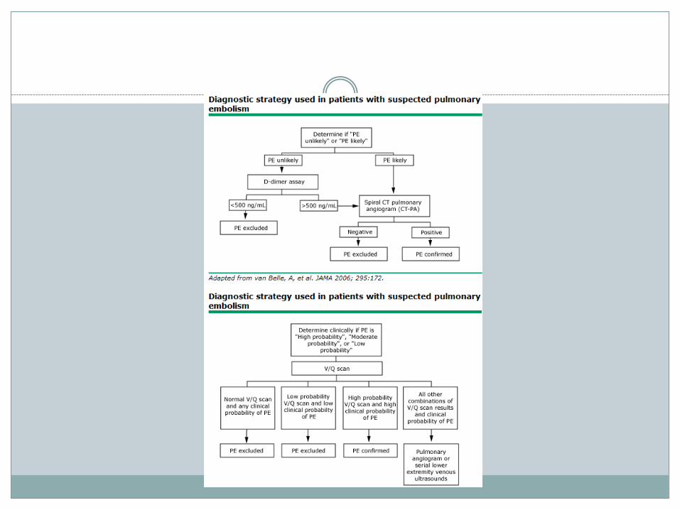
Evaluation Scheme Based on Clinical Probability.
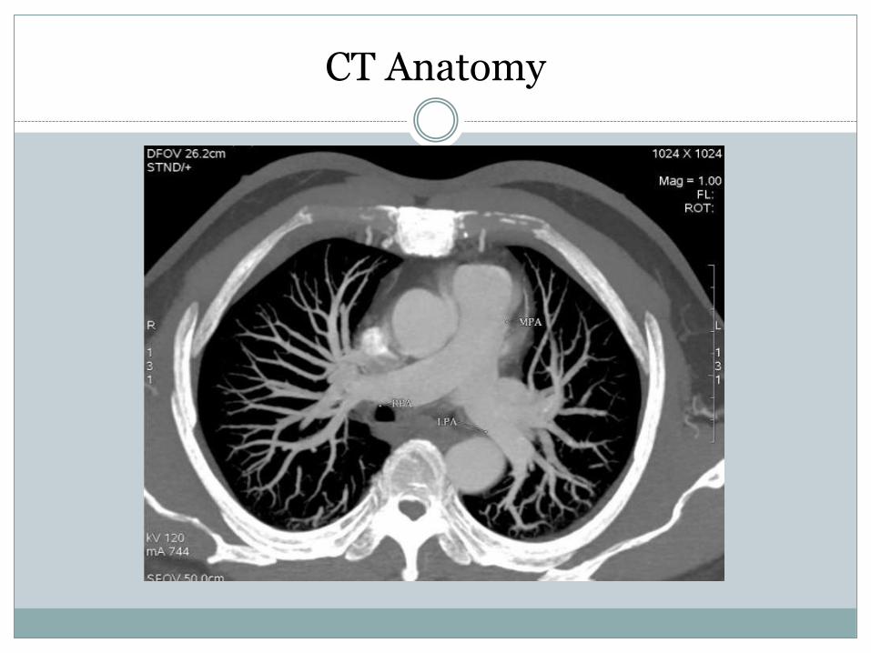
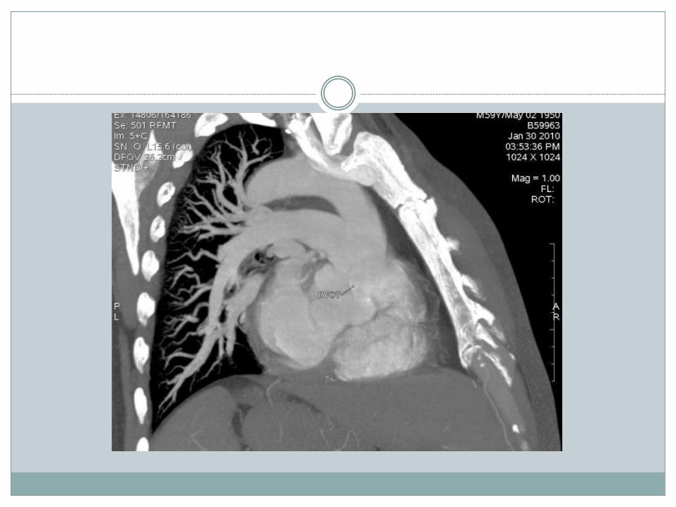
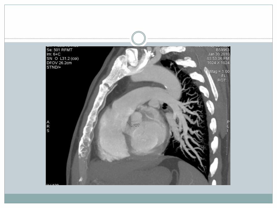
CT Anatomy
Types of pulmonary embolism.
Thrombotic pulmonary embolism.
Acute pulmonary embolism.
Chronic pulmonary embolism
Non-thrombotic pulmonary embolism.
High Sensitivity D-Dimer.
• High negative predictive value for PE (based on pulmonary angiography)
• For D-dimer <500ng/mL, negative predictive value (NPV) 91-99%
• For D-dimer >500ng/mL, sens=93%, spec=25%, and positive predictive value (PPV) = 30%
• PPV and NPV are affected by prevalence.• Test is also useful for DVT rule out
(<500ng/mL): NPV 92% • If pretest probability is intermediate (27.8) you
are supposed to image, but if you order a D-Dimer what do the results mean.
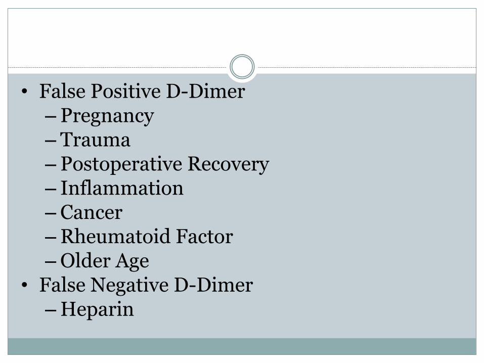
• False Positive D-Dimer– Pregnancy– Trauma– Postoperative Recovery– Inflammation– Cancer–Rheumatoid Factor–Older Age
• False Negative D-Dimer–Heparin
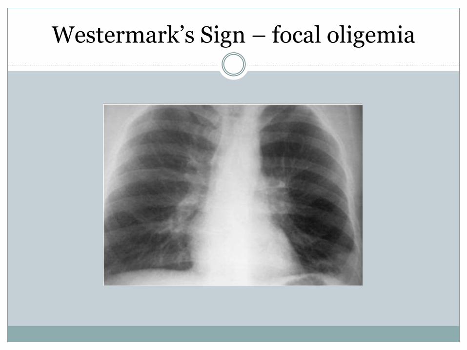
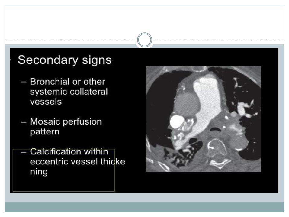
Westermark’s Sign – focal oligemia
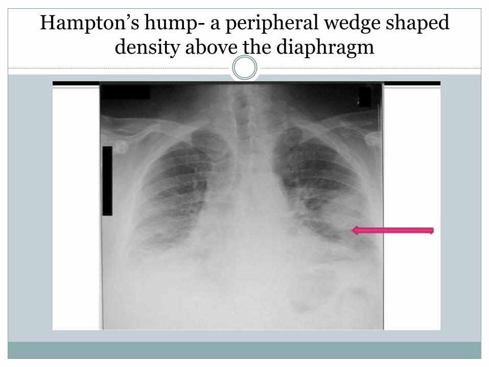
Hampton’s hump- a peripheral wedge shaped density above the diaphragm
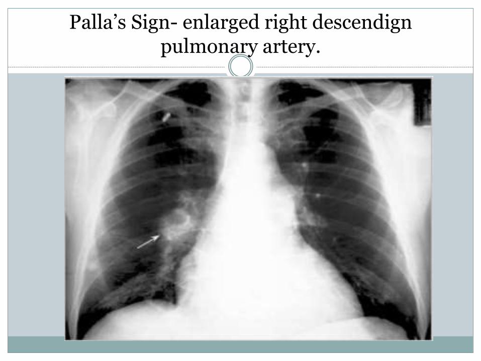
Palla’s Sign- enlarged right descendignpulmonary artery.
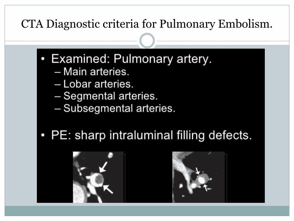
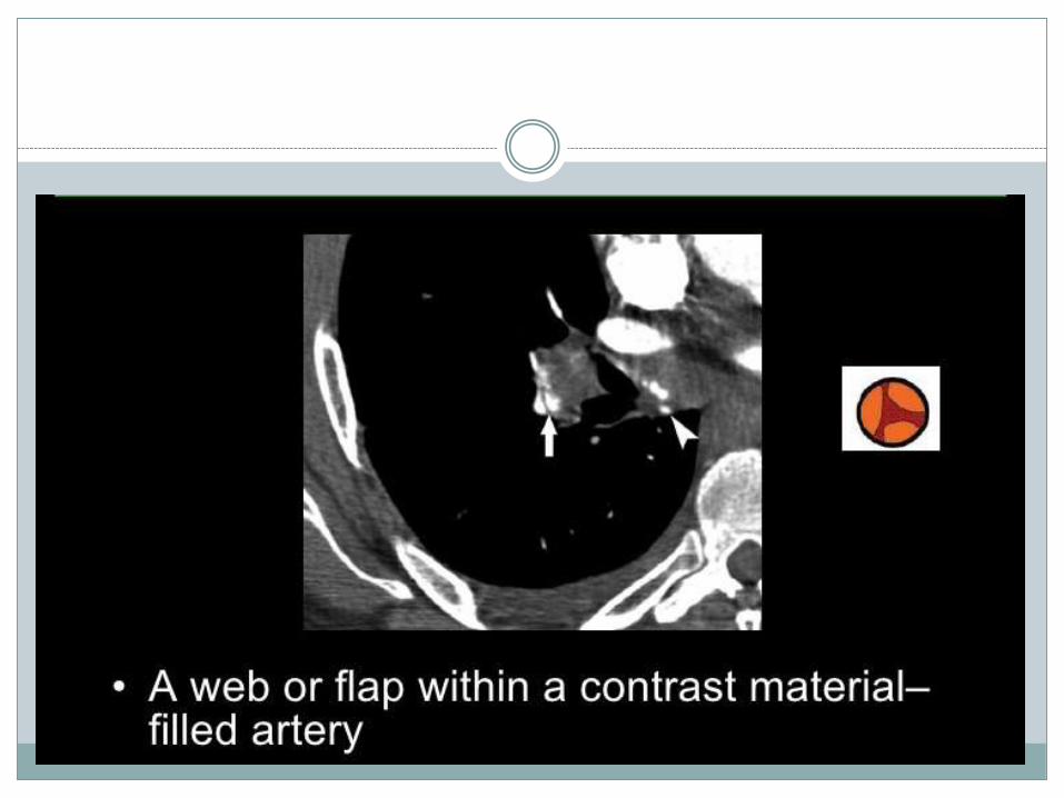
CTA Diagnostic criteria for Pulmonary Embolism.
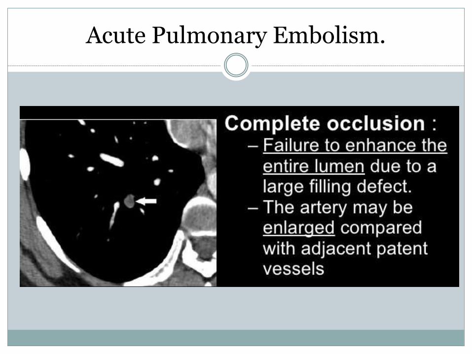
Acute Pulmonary Embolism.
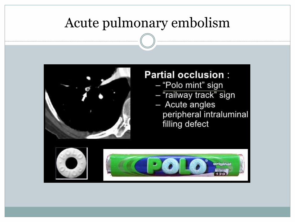
Acute pulmonary embolism
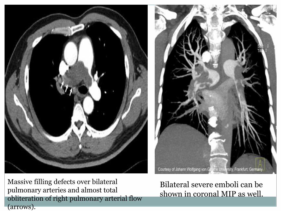
Massive filling defects over bilateral pulmonary arteries and almost total obliteration of right pulmonary arterial flow (arrows).
Bilateral severe emboli can be shown in coronal MIP as well.
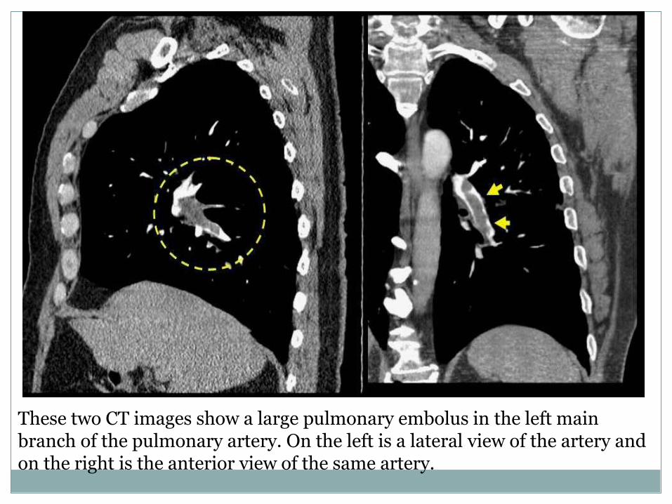
These two CT images show a large pulmonary embolus in the left main branch of the pulmonary artery. On the left is a lateral view of the artery and on the right is the anterior view of the same artery.
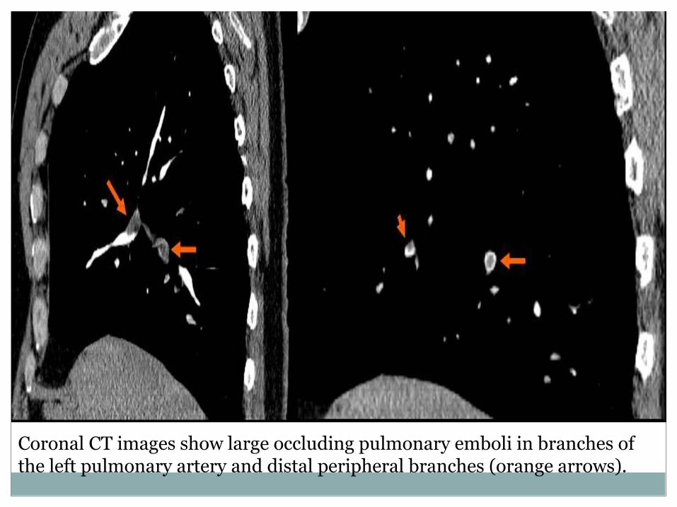
Coronal CT images show large occluding pulmonary emboli in branches of the left pulmonary artery and distal peripheral branches (orange arrows).
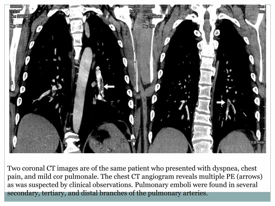
Two coronal CT images are of the same patient who presented with dyspnea, chest pain, and mild cor pulmonale. The chest CT angiogram reveals multiple PE (arrows) as was suspected by clinical observations. Pulmonary emboli were found in several secondary, tertiary, and distal branches of the pulmonary arteries.
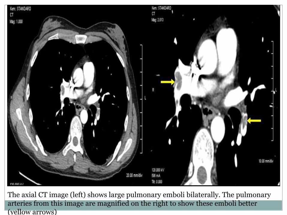
The axial CT image (left) shows large pulmonary emboli bilaterally. The pulmonary arteries from this image are magnified on the right to show these emboli better (yellow arrows)
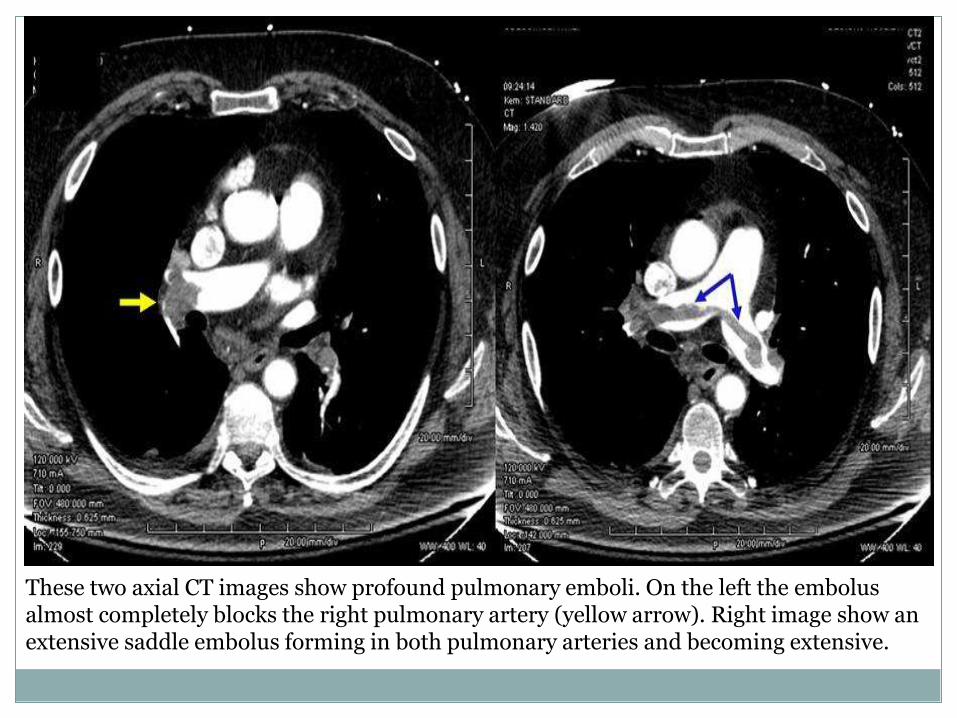
These two axial CT images show profound pulmonary emboli. On the left the embolus almost completely blocks the right pulmonary artery (yellow arrow). Right image show an extensive saddle embolus forming in both pulmonary arteries and becoming extensive.
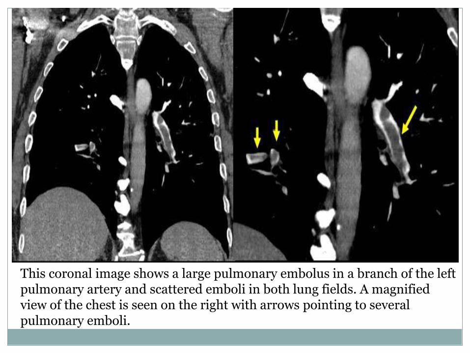
This coronal image shows a large pulmonary embolus in a branch of the left pulmonary artery and scattered emboli in both lung fields. A magnified view of the chest is seen on the right with arrows pointing to several pulmonary emboli.
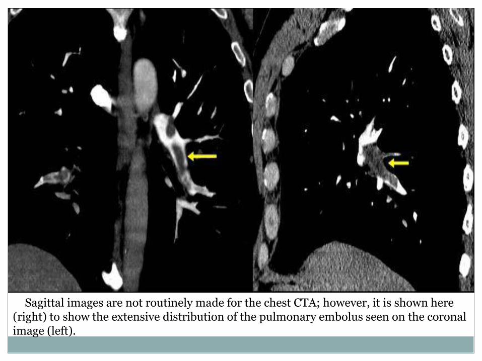
Sagittal images are not routinely made for the chest CTA; however, it is shown here (right) to show the extensive distribution of the pulmonary embolus seen on the coronal image (left).
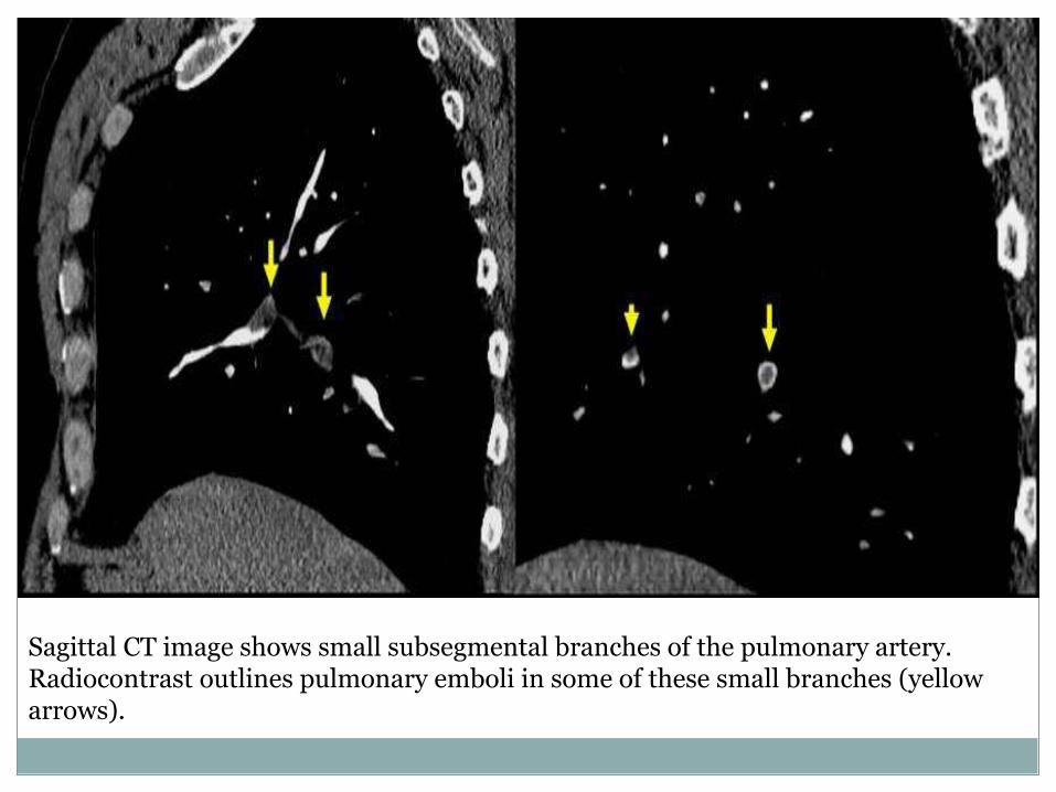
Sagittal CT image shows small subsegmental branches of the pulmonary artery. Radiocontrast outlines pulmonary emboli in some of these small branches (yellow arrows).
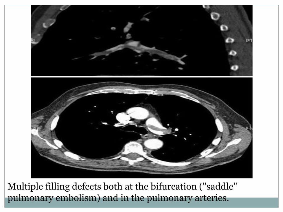
Multiple filling defects both at the bifurcation ("saddle" pulmonary embolism) and in the pulmonary arteries.

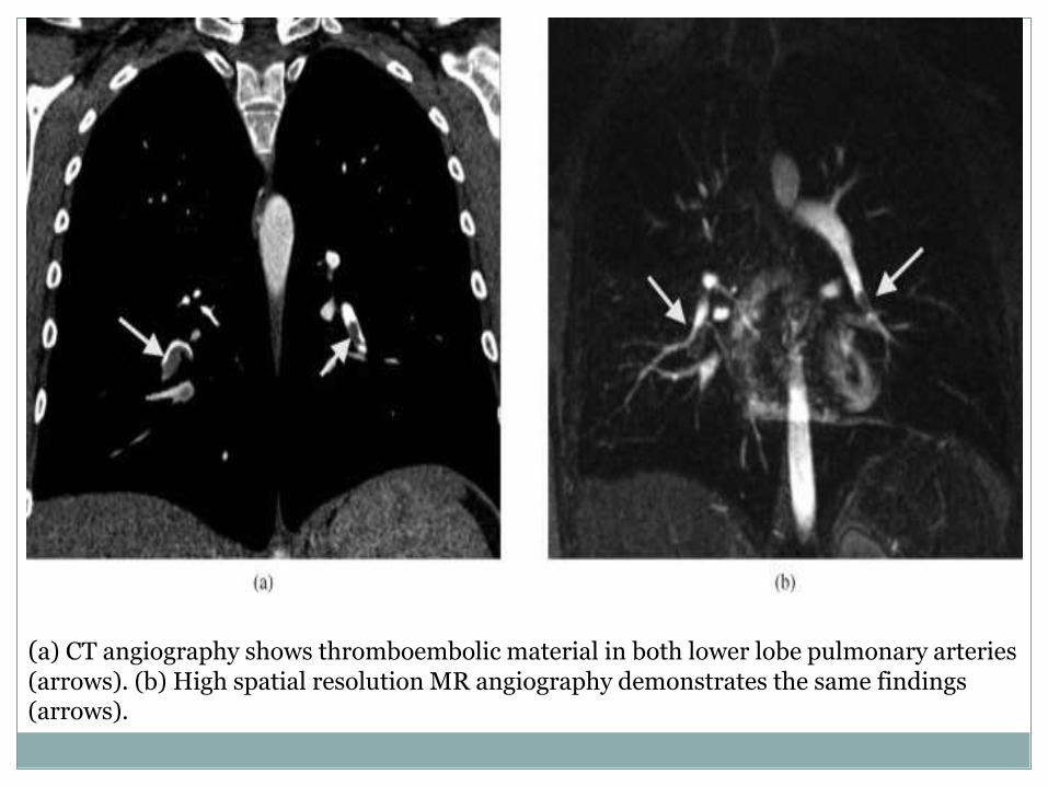
(a) CT angiography shows thromboembolic material in both lower lobe pulmonary arteries (arrows). (b) High spatial resolution MR angiography demonstrates the same findings (arrows).

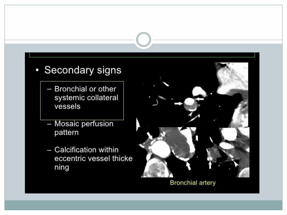
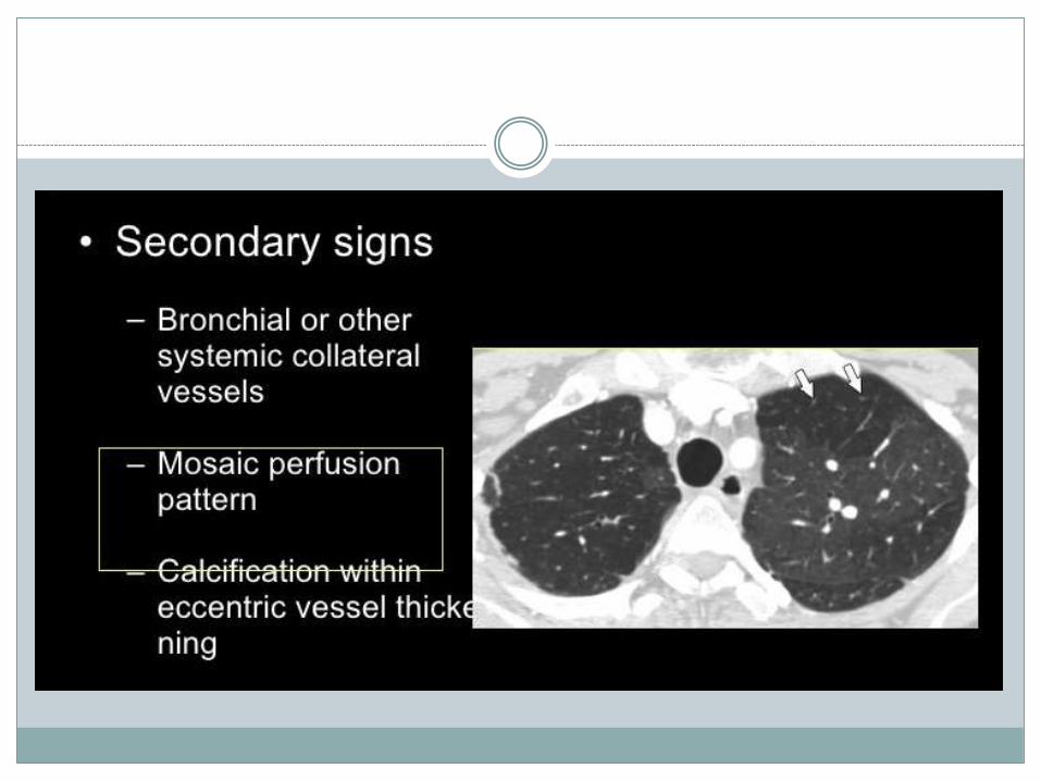
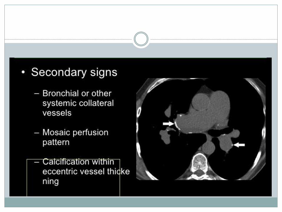
Chronic pulmonary embolism
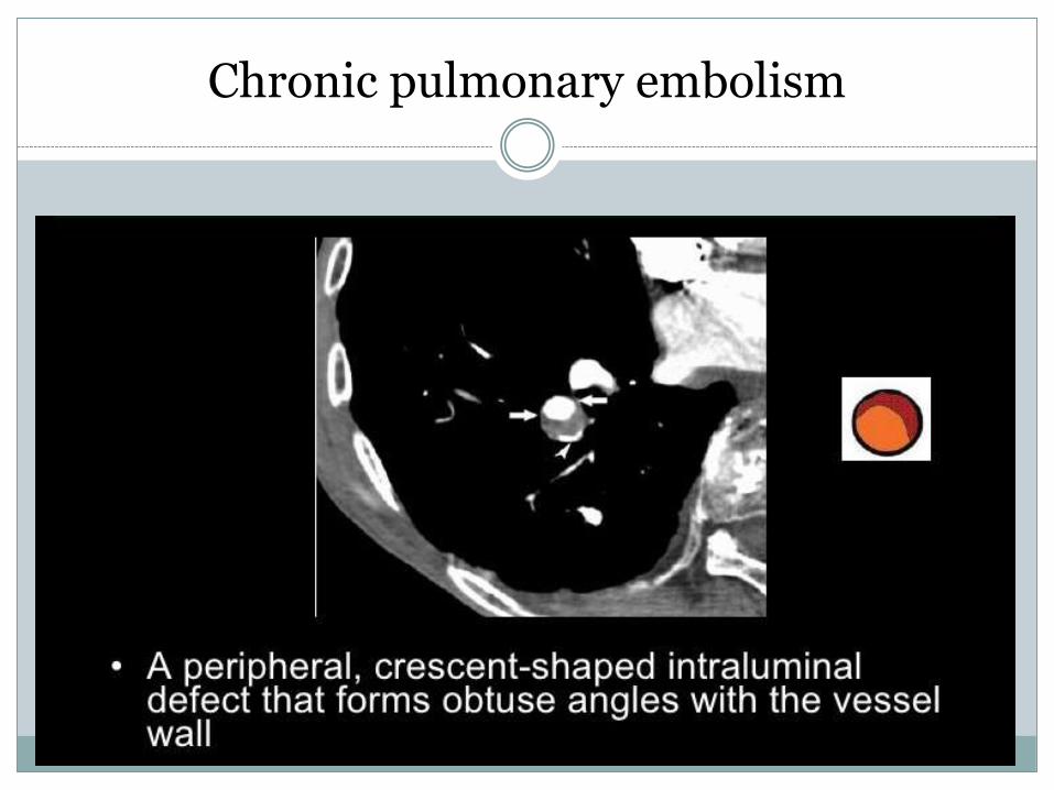
Chronic pulmonary embolism
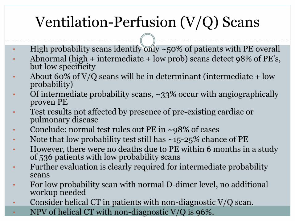
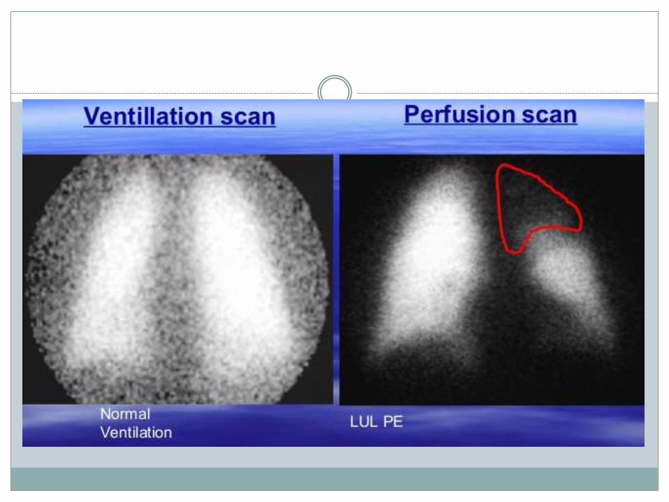
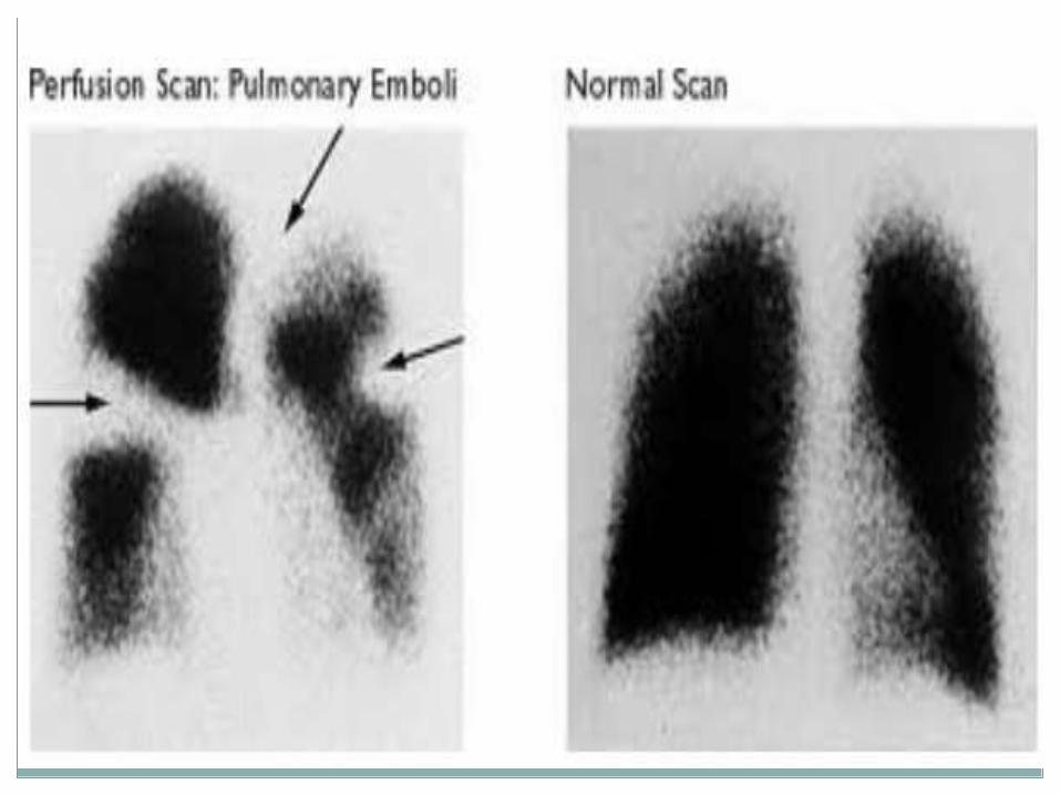
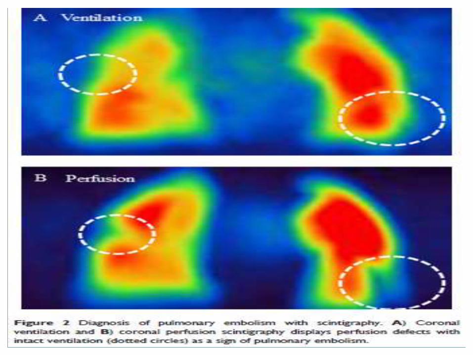
Ventilation-Perfusion (V/Q) Scans
• High probability scans identify only ~50% of patients with PE overall • Abnormal (high + intermediate + low prob) scans detect 98% of PE's,
but low specificity • About 60% of V/Q scans will be in determinant (intermediate + low
probability) • Of intermediate probability scans, ~33% occur with angiographically
proven PE • Test results not affected by presence of pre-existing cardiac or
pulmonary disease • Conclude: normal test rules out PE in ~98% of cases • Note that low probability test still has ~15-25% chance of PE • However, there were no deaths due to PE within 6 months in a study
of 536 patients with low probability scans • Further evaluation is clearly required for intermediate probability
scans • For low probability scan with normal D-dimer level, no additional
workup needed • Consider helical CT in patients with non-diagnostic V/Q scan. • NPV of helical CT with non-diagnostic V/Q is 96%.
Lower Extremity Doppler US.
• To evaluate for DVT as possible cause of PE or to help rule in PE
• Up to 40% of patients with DVT without PE symptoms will HAVE a PE by angiography
• Serial US should be probably be performed in patients with abnormal (or non-diagnostic) V/Q scans and positive D-Dimers.
• These US should be carried out on days 1, 3, 7, and 14.
• A positive US on any of these days with abnormal V/Q rules IN a PE.
• Negative serial US scans reduce likelihood of PE to <2%.
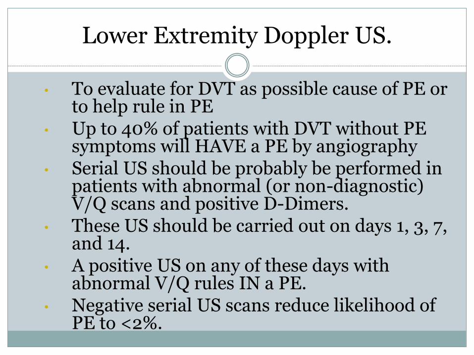
Normal US and Color Doppler
DVT Location of Vein Marked
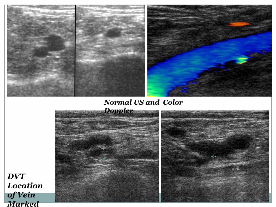
A subtotal occlusive thrombus in the right popliteal vein is shown in this colour Doppler image.
Right lower extremity (A) transverse and (B) sagittal images from color Doppler ultrasound demonstrates blood flow in the femoral artery but not in the common femoral vein (arrows). This is an indirect finding that suggests common femoral DVT.
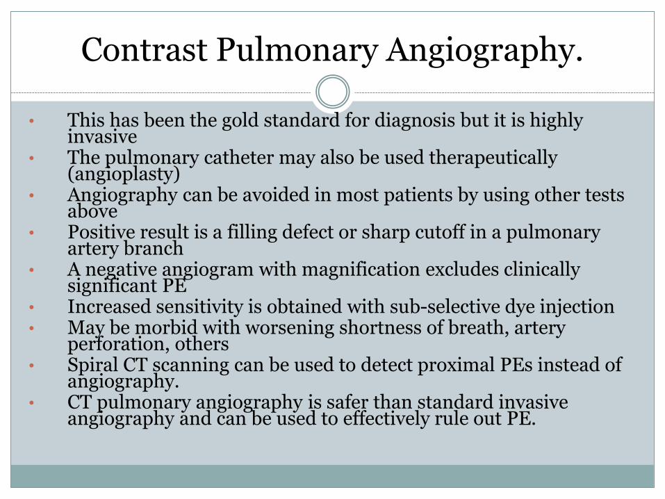
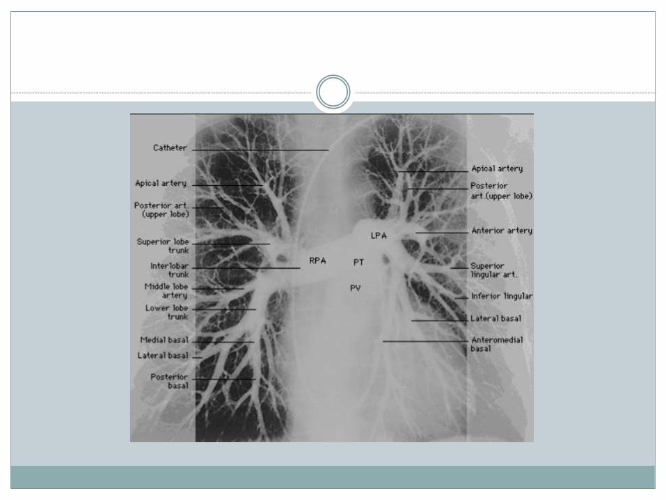
Contrast Pulmonary Angiography.
• This has been the gold standard for diagnosis but it is highly invasive
• The pulmonary catheter may also be used therapeutically (angioplasty)
• Angiography can be avoided in most patients by using other tests above
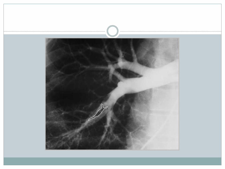
• Positive result is a filling defect or sharp cutoff in a pulmonary artery branch
• A negative angiogram with magnification excludes clinically significant PE
• Increased sensitivity is obtained with sub-selective dye injection • May be morbid with worsening shortness of breath, artery
perforation, others • Spiral CT scanning can be used to detect proximal PEs instead of
angiography. • CT pulmonary angiography is safer than standard invasive
angiography and can be used to effectively rule out PE.
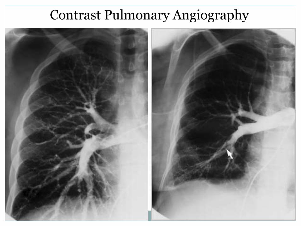
Contrast Pulmonary Angiography
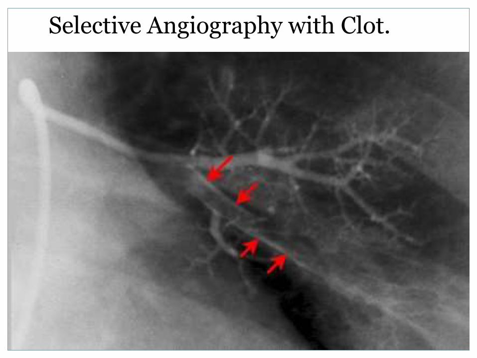
Selective Angiography with Clot.
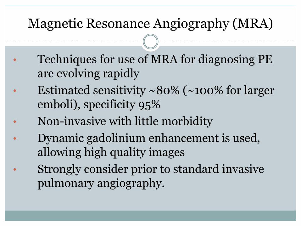
Magnetic Resonance Angiography (MRA)
• Techniques for use of MRA for diagnosing PE are evolving rapidly
• Estimated sensitivity ~80% (~100% for larger emboli), specificity 95%
• Non-invasive with little morbidity
• Dynamic gadolinium enhancement is used, allowing high quality images
• Strongly consider prior to standard invasive pulmonary angiography.
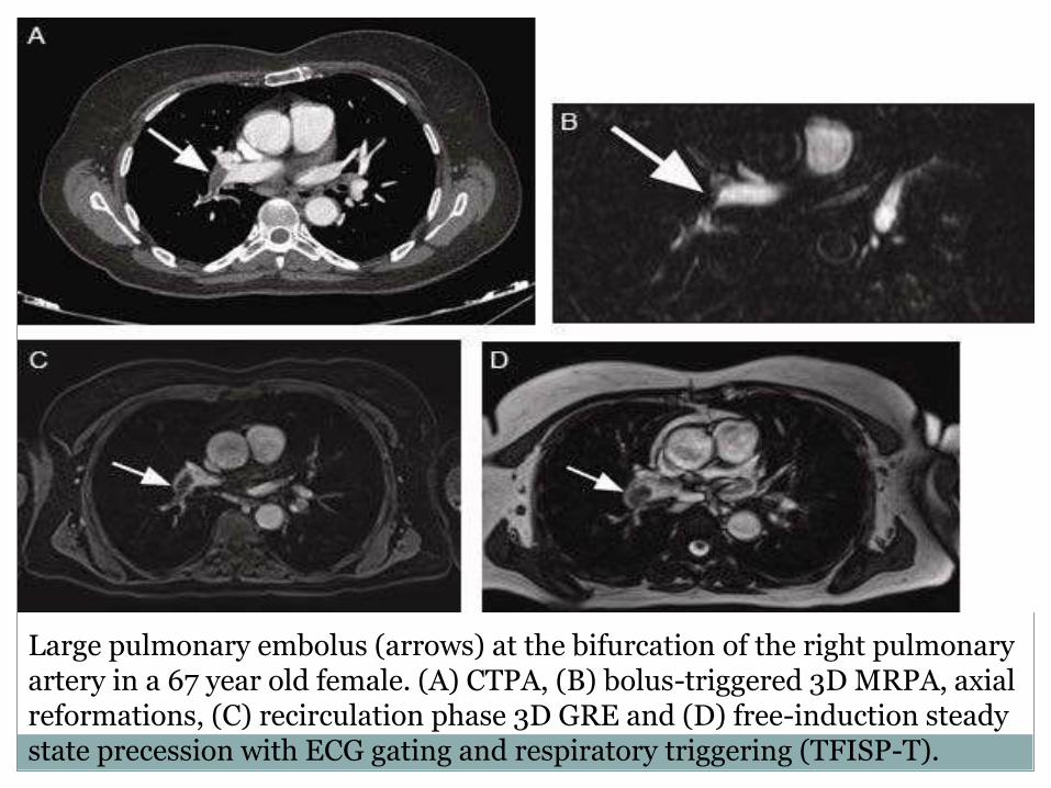
Large pulmonary embolus (arrows) at the bifurcation of the right pulmonary artery in a 67 year old female. (A) CTPA, (B) bolus-triggered 3D MRPA, axial reformations, (C) recirculation phase 3D GRE and (D) free-induction steady state precession with ECG gating and respiratory triggering (TFISP-T).
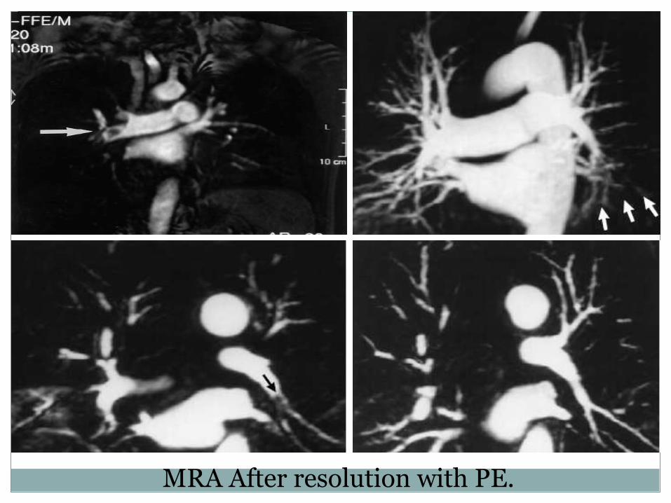
MRA After resolution with PE.
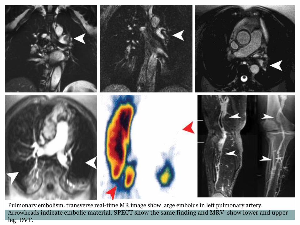
Pulmonary embolism. transverse real-time MR image show large embolus in left pulmonary artery.
Arrowheads indicate embolic material. SPECT show the same finding and MRV show lower and upper leg DVT.
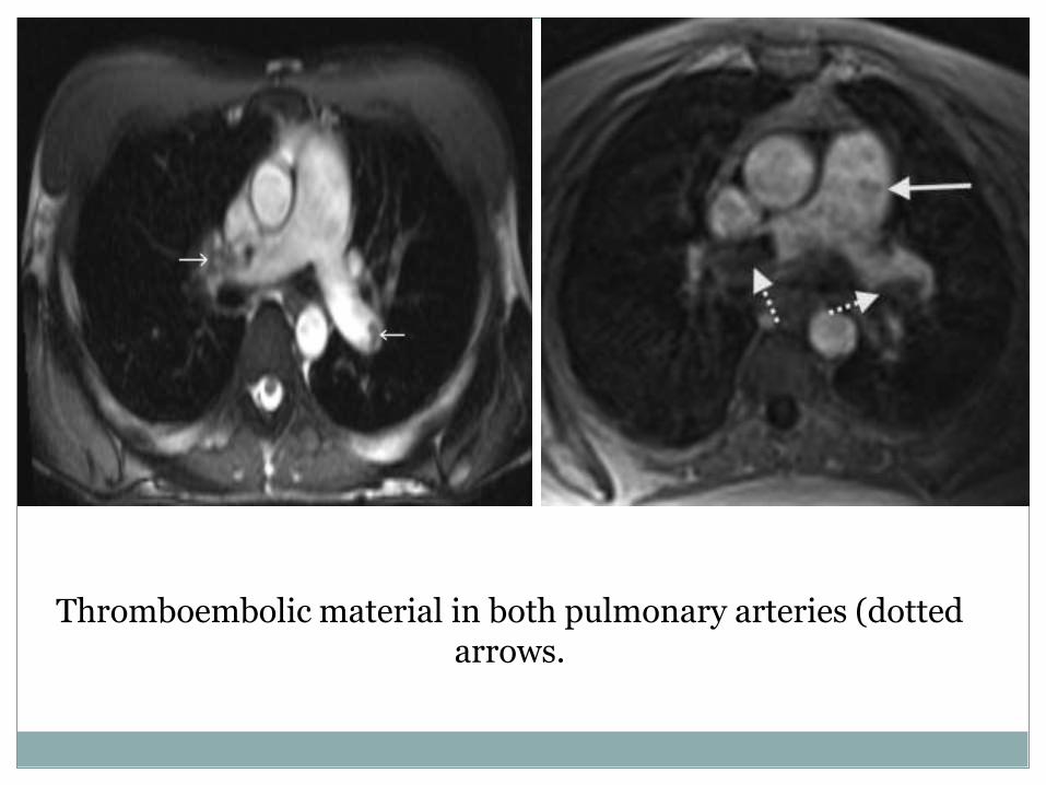
Thromboembolic material in both pulmonary arteries (dotted arrows.
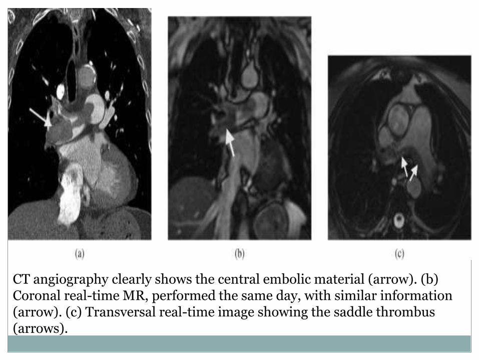
CT angiography clearly shows the central embolic material (arrow). (b) Coronal real-time MR, performed the same day, with similar information (arrow). (c) Transversal real-time image showing the saddle thrombus (arrows).
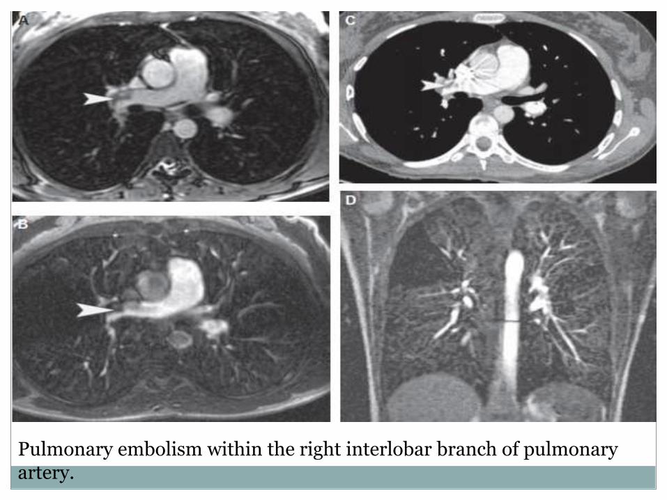
Pulmonary embolism within the right interlobar branch of pulmonary artery.
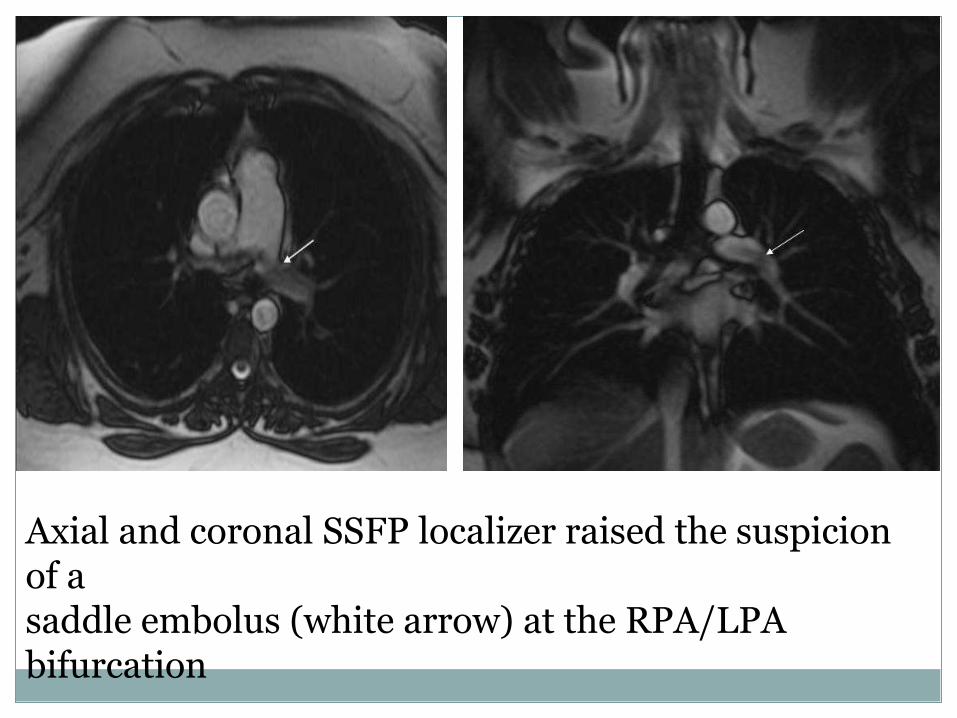
Axial and coronal SSFP localizer raised the suspicion of a saddle embolus (white arrow) at the RPA/LPA bifurcation
Pulmonary embolus is a major health problem that is highly treatable when diagnosed, but carries high risk for morbidity and mortality is undiagnosed. Radiography is a major diagnostic tool in finding pulmonary embolus. There are several diagnostic studies like CT, nuclear V/Q scan and angiographic procedures that diagnose pulmonary embolus. The main cause of PE is venous thromboembolus that is diagnosed with ultrasound. Treatment for both PE and VTE can reduce the risk of recurring disease to below 2%, which is medically acceptable.

THANK YOU


![Acute Pulmonary Embolism [Radiology North Amer Clinics 2010]](https://static.documents.pub/doc/80x56/577d35bd1a28ab3a6b91457c/acute-pulmonary-embolism-radiology-north-amer-clinics-2010.jpg)


