Page 1
1
Pulmonary Hypertension Case
Studies
Chad Link, DO FACC
Sparrow Hospital
Thoracic and Cardiovascular Institute
Chairman- TCI Cardiology Section
.
Disclosures
Speakers Bureau – Actelion Pharmaceuticals, Pfizer and BMS
Clinical Research Support– Sanofi Aventis
Pulmonary Hypertension
Page 2
2
What is Pulmonary Hypertension?
• Pulmonary Hypertension is increased pressure in the pulmonary
arteries.
• Pulmonary Hypertension causes symptoms such as shortness of
breath during routine activity (for example, climbing two flights of
stairs), tiredness, chest pain, and a racing heartbeat. As the
condition worsens, its symptoms may limit all physical activity.
• Pulmonary hypertension (PH) was previously classified into 2
categories: 1) primary pulmonary hypertension; or 2) secondary
pulmonary hypertension according to the presence of identified
causes or risk factors.
1. Sitbon O et al. Circulation 2005
2. D’Alonzo GE et al. Ann Intern Med 1991
What is Pulmonary Hypertension?
• Since the second World Symposium on pulmonary hypertension
held in Evian, in 1998, a clinical classification was established in
order to individualize different categories of PH sharing similar
pathological findings, similar hemodynamic characteristics and,
similar management
• Pulmonary Hypertension is divided into five groups based on its
causes and treatment options.
1. Sitbon O et al. Circulation 2005
2. D’Alonzo GE et al. Ann Intern Med 1991
1. PAH
– Idiopathic PAH
– Familial (Heritable) PAH
– Associated with:
• Connective tissue disease
• BMPR2, ALK1, endoglin
• CHD (shunts)
• Portal hypertension
• HIV infection
• Sickle cell disease
• Drugs and toxins
• Other (thyroid)
– Persistent pulmonary
hypertension of the newborn
2. PH associated with left heart
disease- Diastolic, Systolic or
Valvular disease
3. PH associated with respiratory
disease
– COPD
– Interstitial lung diseases
4. PH due to chronic thrombotic
and/or embolic disease
5. Miscellaneous (sarcoidosis,
glycogen storage disease)
PH: WHO Group Classification1
1. Simonneau G et al. JACC 2004
Page 3
3
1. PAH
– Idiopathic PAH
– Familial (Heritable) PAH
– Associated with:
• Connective tissue disease
• BMPR2, ALK1, endoglin
• CHD (shunts)
• Portal hypertension
• HIV infection
• Sickle cell disease
• Drugs and toxins
• Other (thyroid)
– Persistent pulmonary
hypertension of the newborn
2. PH associated with left heart
disease- Diastolic, Systolic or
Valvular disease
3. PH associated with respiratory
disease
– COPD
– Interstitial lung diseases
4. PH due to chronic thrombotic
and/or embolic disease
5. Miscellaneous (sarcoidosis,
glycogen storage disease)
PH: WHO Group Classification1
1. Simonneau G et al. JACC 2004
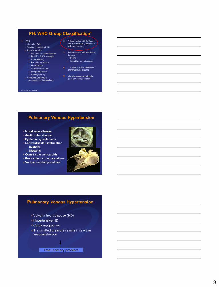
Pulmonary Venous Hypertension
• Mitral valve disease
• Aortic valve disease
• Systemic hypertension
• Left ventricular dysfunction
– Systolic
– Diastolic
• Constrictive pericarditis
• Restrictive cardiomyopathies
• Various cardiomyopathies
Pulmonary Venous Hypertension:
• Valvular heart disease (HD)
• Hypertensive HD
• Cardiomyopathies
• Transmitted pressure results in reactive
vasoconstriction
Treat primary problem
Page 4
4
1. PAH
– Idiopathic PAH
– Familial (Heritable) PAH
– Associated with:
• Connective tissue disease
• BMPR2, ALK1, endoglin
• CHD (shunts)
• Portal hypertension
• HIV infection
• Sickle cell disease
• Drugs and toxins
• Other (thyroid)
– Persistent pulmonary
hypertension of the newborn
2. PH associated with left heart
disease- Diastolic, Systolic or
Valvular disease
3. PH associated with respiratory
disease
– COPD
– Interstitial lung diseases
4. PH due to chronic thrombotic
and/or embolic disease
5. Miscellaneous (sarcoidosis,
glycogen storage disease)
PH: WHO Group Classification1
1. Simonneau G et al. JACC 2004
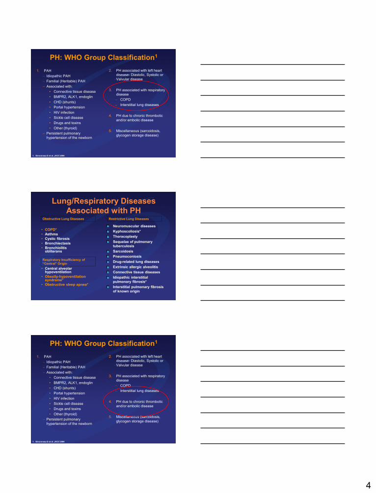
Lung/Respiratory Diseases
Associated with PH
• COPD*
• Asthma
• Cystic fibrosis
• Bronchiectasis
• Bronchiolitis obliterans
• Central alveolar hypoventilation
• Obesity-hypoventilation syndrome*
• Obstructive sleep apnea*
Neuromuscular diseases
Kyphoscoliosis*
Thoracoplasty
Sequelae of pulmonary
tuberculosis
Sarcoidosis
Pneumoconiosis
Drug-related lung diseases
Extrinsic allergic alveolitis
Connective tissue diseases
Idiopathic interstitial
pulmonary fibrosis*
Interstitial pulmonary fibrosis
of known origin
Respiratory Insufficiency of
“Central” Origin
Obstructive Lung Diseases Restrictive Lung Diseases
1. PAH
– Idiopathic PAH
– Familial (Heritable) PAH
– Associated with:
• Connective tissue disease
• BMPR2, ALK1, endoglin
• CHD (shunts)
• Portal hypertension
• HIV infection
• Sickle cell disease
• Drugs and toxins
• Other (thyroid)
– Persistent pulmonary
hypertension of the newborn
2. PH associated with left heart
disease- Diastolic, Systolic or
Valvular disease
3. PH associated with respiratory
disease
– COPD
– Interstitial lung diseases
4. PH due to chronic thrombotic
and/or embolic disease
5. Miscellaneous (sarcoidosis,
glycogen storage disease)
PH: WHO Group Classification1
1. Simonneau G et al. JACC 2004
Page 5
5
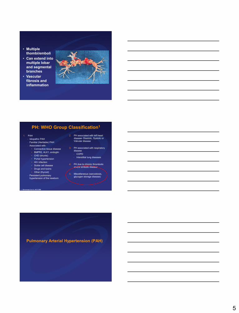
• Multiple
thombi/emboli
• Can extend into
multiple lobar
and segmental
branches
• Vascular
fibrosis and
inflammation
1. PAH
– Idiopathic PAH
– Familial (Heritable) PAH
– Associated with:
• Connective tissue disease
• BMPR2, ALK1, endoglin
• CHD (shunts)
• Portal hypertension
• HIV infection
• Sickle cell disease
• Drugs and toxins
• Other (thyroid)
– Persistent pulmonary
hypertension of the newborn
2. PH associated with left heart
disease- Diastolic, Systolic or
Valvular disease
3. PH associated with respiratory
disease
– COPD
– Interstitial lung diseases
4. PH due to chronic thrombotic
and/or embolic disease
5. Miscellaneous (sarcoidosis,
glycogen storage disease)
PH: WHO Group Classification1
1. Simonneau G et al. JACC 2004
Pulmonary Arterial Hypertension (PAH)
Page 6
6
What is PAH?
• PAH is a syndrome characterised by a progressive increase in
pulmonary vascular resistance (PVR)
– leads to right ventricular overload
– eventually leads to right ventricular failure and premature
death1
– If untreated, the median survival is 2.8 years2 which is
comparable with some malignancies
• Increased PVR is related to progressive changes in the
pulmonary arterioles
– vasoconstriction
– obstructive remodelling of the pulmonary vessel wall
– inflammation
– in-situ thrombosis1. Sitbon O et al. Circulation 2005
2. D’Alonzo GE et al. Ann Intern Med 1991
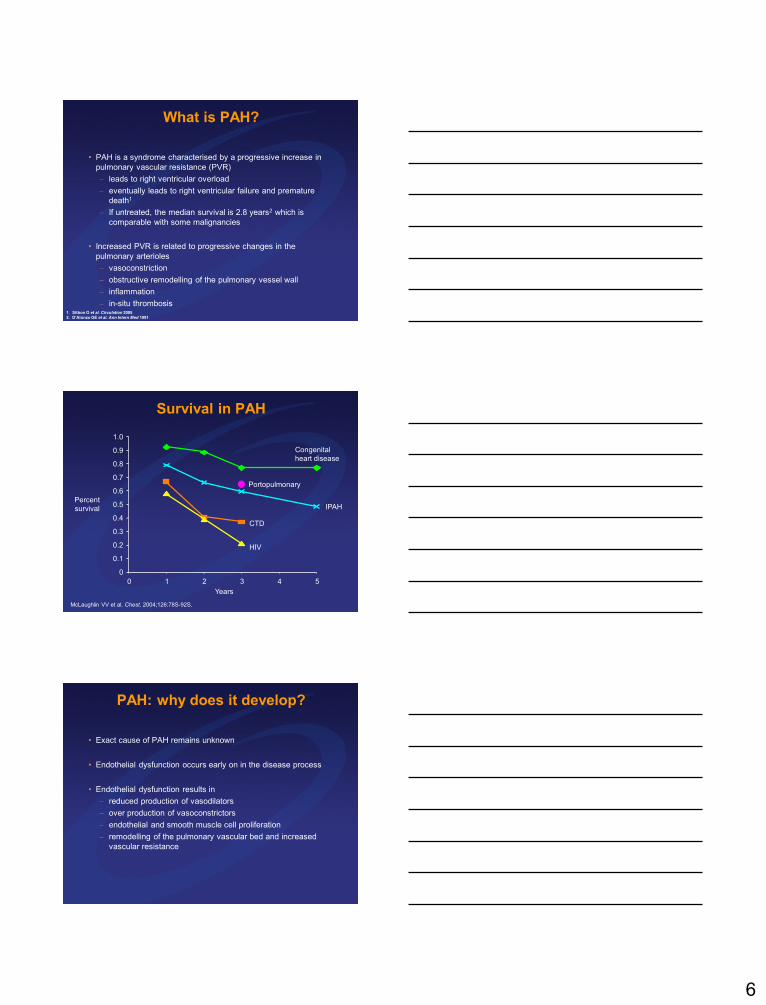
0
0.1
0.2
0.3
0.4
0.5
0.6
0.7
0.8
0.9
1.0
0 1 2 3 4 5
Percent
survival
McLaughlin VV et al. Chest. 2004;126:78S-92S.
Congenital
heart disease
Portopulmonary
IPAH
CTD
HIV
Years
Survival in PAH
PAH: why does it develop?
• Exact cause of PAH remains unknown
• Endothelial dysfunction occurs early on in the disease process
• Endothelial dysfunction results in
– reduced production of vasodilators
– over production of vasoconstrictors
– endothelial and smooth muscle cell proliferation
– remodelling of the pulmonary vascular bed and increased
vascular resistance
Page 7
7
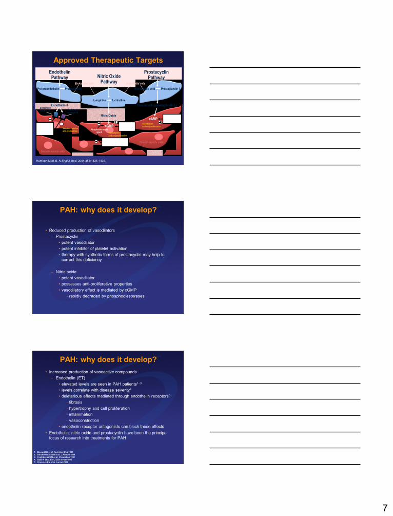
Approved Therapeutic Targets
Humbert M et al. N Engl J Med. 2004;351:1425-1436.
cGMP
cAMP
Vasoconstriction
and proliferation
Endothelin receptor A
Exogenous
nitric oxideEndothelin-
receptor
antagonists
Endothelin receptor B
Phosphodiesterase
type 5 inhibitor
Vasodilation
and antiproliferation
Phosphodiesterase type 5
Vasodilation
and antiproliferation
Prostacyclin
derivatives
Nitric Oxide
Endothelin-1
Pre-proendothelin
L-arginine
Prostaglandin I2
L-citrulline
Nitric OxidePathway
EndothelinPathway
ProstacyclinPathway
Endothelial cells
Proendothelin
Endothelial cells
Arachidonic acid
Smooth muscle cells
Prostacyclin (prostaglandin I2)
Smooth muscle cells
PAH: why does it develop?
• Reduced production of vasodilators
– Prostacyclin
• potent vasodilator
• potent inhibitor of platelet activation
• therapy with synthetic forms of prostacyclin may help to
correct this deficiency
– Nitric oxide
• potent vasodilator
• possesses anti-proliferative properties
• vasodilatory effect is mediated by cGMP
– rapidly degraded by phosphodiesterases
PAH: why does it develop?
• Increased production of vasoactive compounds
– Endothelin (ET)
• elevated levels are seen in PAH patients13
• levels correlate with disease severity4
• deleterious effects mediated through endothelin receptors5
– fibrosis
–hypertrophy and cell proliferation
– inflammation
–vasoconstriction
• endothelin receptor antagonists can block these effects
• Endothelin, nitric oxide and prostacyclin have been the principal
focus of research into treatments for PAH
1. Stewart DJ et al. Ann Inter Med 1991
2. Vancheeswaran R et al. J Rheum 1994
3. Yoshibayashi M et al. Circulation 19914. Galiè N et al. Eur J Clin Invest 1996
5. Channick RN et al. Lancet 2001
Page 8
8
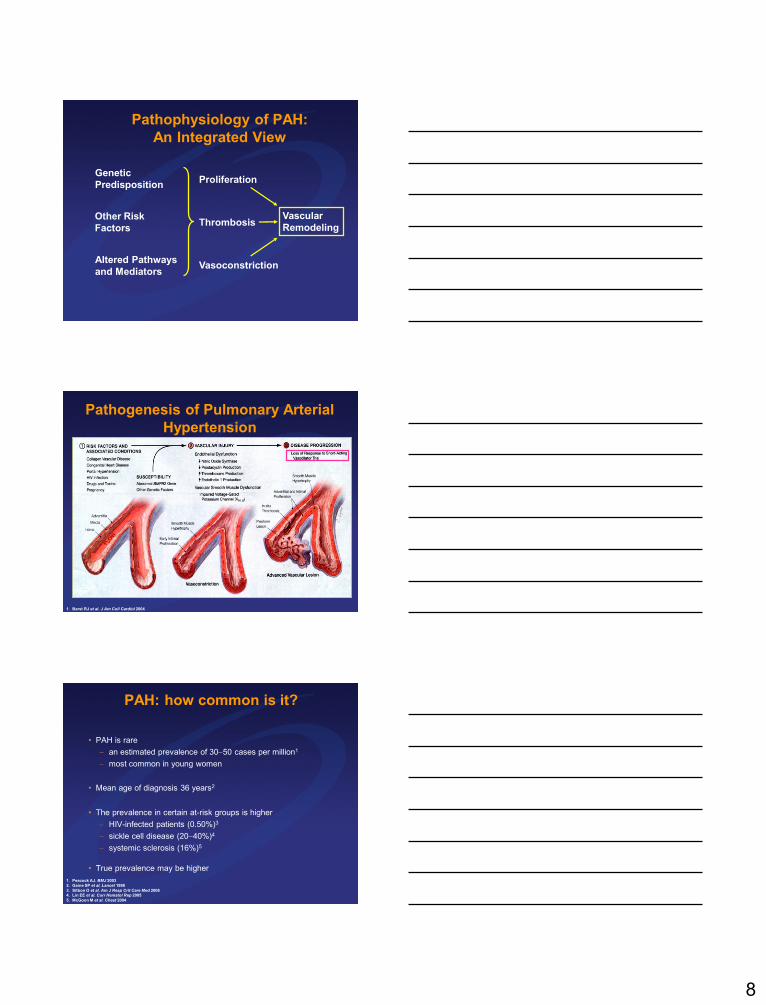
Vascular
RemodelingOther Risk
Factors
Altered Pathways
and Mediators
Genetic
Predisposition
Pathophysiology of PAH:
An Integrated View
Proliferation
Vasoconstriction
Thrombosis
Pathogenesis of Pulmonary Arterial
Hypertension
1. Barst RJ et al. J Am Coll Cardiol 2004
PAH: how common is it?
• PAH is rare
– an estimated prevalence of 3050 cases per million1
– most common in young women
• Mean age of diagnosis 36 years2
• The prevalence in certain at-risk groups is higher
– HIV-infected patients (0.50%)3
– sickle cell disease (2040%)4
– systemic sclerosis (16%)5
• True prevalence may be higher
1. Peacock AJ. BMJ 2003
2. Gaine SP et al. Lancet 1998
3. Sitbon O et al. Am J Resp Crit Care Med 20084. Lin EE et al. Curr Hematol Rep 2005
5. McGoon M et al. Chest 2004
Page 9
9
PAH Related to Connective
Tissue Disease• Connective tissue diseases
– scleroderma (most common)
– systemic lupus erythematosus
– Sjogren’s syndrome
– rheumatoid arthritis
– MCTD
• PH is one of the top causes of death in scleroderma
• Similar to IPAH pathology
• Medical treatment same as for IPAH, but benefits less than for IPAH
Hachulla E et al. Rheumatology. 2009;48:304-308.
Survival in Pulmonary Arterial
Hypertension
• Survival rates (patients with IPAH) at 1, 3 and 5 years were 68%, 48% and 34% respectively
• PAH mortality contributed to
• Right heart failure 47%
• Sudden Death 26%
• Other (pneumonia) 27%
• Although new treatments have improved mortality rates, there is little evidence to support reversal of aberrant remodeling
D’Alonzo GE, et al. Ann Intern Med 1991;115:343-349.
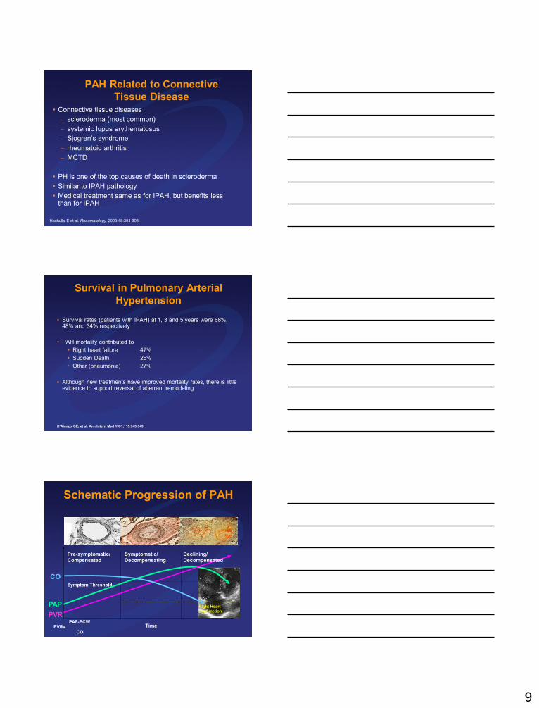
Schematic Progression of PAH
Time
PAP
PVR
CO
Pre-symptomatic/
Compensated
Symptomatic/
Decompensating
Symptom Threshold
Right Heart
Dysfunction
Declining/
Decompensated
PVR=PAP-PCW
CO
Page 10
10
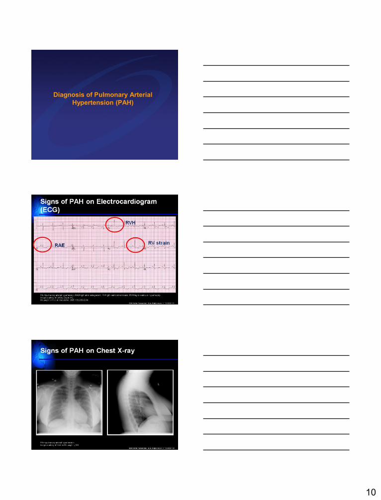
Diagnosis of Pulmonary Arterial
Hypertension (PAH)
Page 11
11
Diagnosing & Assessing PAH
PAH: Early diagnosis is crucial
• If untreated, the median survival is 2.8 years which is comparable with
some malignancies1-3
• diagnosis can be delayed for months or years and frequently occurs
when disease is relatively advanced4
• mean time from onset to diagnosis is estimated to be approximately
2 years5
• Although patients progress at different rates; early stage PAH is still a
devastating condition and can rapidly deteriorate
• Early diagnosis and intervention is therefore crucial
• patients who begin targeted therapy in less severe PAH (WHO
FCI/II) demonstrate a better prognosis than those in a more severe
stage (WHO FCIII/IV)6
1. D'Alonzo GE et al. Ann Intern Med 1991
2. Kato I et al. Cancer 2001
3. Bjoraker JA et al. Am J Respir Crit Care Med 19984. Gaine SP et al. Lancet 1998
5. Humbert M et al. Am J Respir Crit Care Med 2006
6. Sitbon O et al. J Am Coll Cardiol 2002
Page 12
12
Echocardiogram
PFT’s
Polysomnography
VQ Scan
• Sleep Disorder
• Chronic PE
Functional Test
(6MWT, CPET)
Overnight
Oximetry
History
Exam
CXR
ECG
HIV
ANA
LFT’s
RH Cath
TTE
Pulmonary Angiography
Chest CT Angiogram
Coagulopathy Profile
Vasodilator Test
Exercise RH Cath
Volume Loading
ABG’s
• Index of Suspicion of PH
• RVE, RAE, RVSP, RV
Function
• Left Heart Disease
• VHD, CHD
• Ventilatory Function
• Gas Exchange
Other CTD Serologies
• HIV Infection
• Scleroderma, SLE, RA
• Portopulmonary Htn
• Establish Baseline
• Prognosis
• Confirmation of PH
• Hemodynamic Profile
• Vasodilator Response
Pivotal Tests Contingent Tests Contribute to
Assessment of:
Left Heart CathMcLaughlin VV et al. J Am Coll Cardiol.
2009;53:1573-1619.
AC
CF
/AH
A D
iag
no
stic A
lgo
rith
m
Echocardiogram
PFT’s
Polysomnography
VQ Scan
•Sleep Disorder
•Chronic PE
Functional Test
(6MWT, CPET)
Overnight
Oximetry
HIV
ANA
LFT’s
RH Cath
TEE
Exercise Echo
Pulmonary Angiography
Chest CT Angiogram
Coagulopathy Profile
Vasodilator Test
Exercise RH Cath
Volume Loading
ABG’s
•Index of Suspicion of PH
• RVE, RAE, RVSP, RV
Function
• Left Heart Disease
• VHD, CHD
•Ventilatory Function
•Gas Exchange
Other CTD Serologies
•HIV Infection
•Scleroderma, SLE, RA
•Portopulmonary Htn
•Establish Baseline
•Prognosis
•Confirmation of PH
•Hemodynamic Profile
•Vasodilator Response
Contingent Tests Contribute to
Assessment of:
Left Heart Cath
ECG
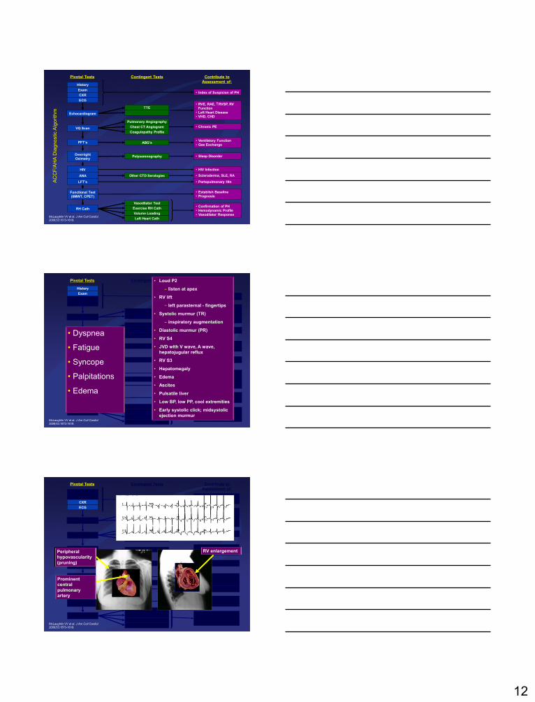
Pivotal Tests • Loud P2
– listen at apex
• RV lift
− left parasternal - fingertips
• Systolic murmur (TR)
– inspiratory augmentation
• Diastolic murmur (PR)
• RV S4
• JVD with V wave, A wave,
hepatojugular reflux
• RV S3
• Hepatomegaly
• Edema
• Ascites
• Pulsatile liver
• Low BP, low PP, cool extremities
• Early systolic click; midsystolic
ejection murmur
• Dyspnea
• Fatigue
• Syncope
• Palpitations
• Edema
CXR
History
Exam
McLaughlin VV et al. J Am Coll Cardiol.
2009;53:1573-1619.
Echocardiogram
PFT’s
Polysomnography
VQ Scan
•Sleep Disorder
•Chronic PE
Functional Test
(6MWT, CPET)
Overnight
Oximetry
HIV
ANA
LFT’s
RH Cath
TEE
Exercise Echo
Pulmonary Angiography
Chest CT Angiogram
Coagulopathy Profile
Vasodilator Test
Exercise RH Cath
Volume Loading
ABG’s
•Index of Suspicion of
PH
• RVE, RAE, RVSP, RV
Function
• Left Heart Disease
• VHD, CHD
•Ventilatory Function
•Gas Exchange
Other CTD Serologies
•HIV Infection
•Scleroderma, SLE, RA
•Portopulmonary Htn
•Establish Baseline
•Prognosis
•Confirmation of PH
•Hemodynamic Profile
•Vasodilator Response
Contingent Tests Contribute to
Assessment of:
Left Heart Cath
Pivotal Tests
History
Exam
Prominent
central
pulmonary
artery
Peripheral
hypovascularity
(pruning)
RV enlargement
CXR
ECG
McLaughlin VV et al. J Am Coll Cardiol.
2009;53:1573-1619.
Page 13
13
Screening Patients With Symptoms
• Echocardiogram
• High clinical suspicion based on clinical exam, etc
• CTD (Systemic Sclerosis (SSc) ,Lupus, RA, Scleroderma)
• Liver transplant candidates
• Shunts
• Amphetamine Derivatives
• Family members of a patient with familial Pulmonary
Arterial Hypertension (FPAH)
• Patients with HIV
PFT’s
Polysomnography
VQ Scan
•Sleep Disorder
•Chronic PE
Functional Test
(6MWT, CPET)
Overnight
Oximetry
HIV
ANA
LFT’s
RH Cath
TEE
Exercise Echo
Pulmonary Angiography
Chest CT Angiogram
Coagulopathy Profile
Vasodilator Test
Exercise RH Cath
Volume Loading
ABG’s
•Index of Suspicion of
PH
• RVE, RAE, RVSP, RV
Function
• Left Heart Disease
• VHD, CHD
•Ventilatory Function
•Gas Exchange
Other CTD Serologies
•HIV Infection
•Scleroderma, SLE, RA
•Portopulmonary Htn
•Establish Baseline
•Prognosis
•Confirmation of PH
•Hemodynamic Profile
•Vasodilator Response
Contingent Tests Contribute to
Assessment of:
Left Heart Cath
Pivotal Tests
History
Exam
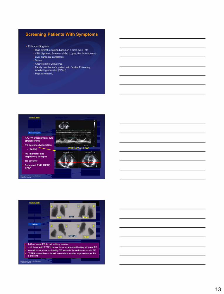
• CXR
• ECG
v
RVSP= 4(VTR)2 + RAP
Echocardiogram
LV RV LV RV
• RA, RV enlargement, IVS
straightening
• RV systolic dysfunction
– TAPSE
• IVC diameter and
inspiratory collapse
• TR severity
• Estimated PVR, MPAP,
DPAP
McLaughlin VV et al. J Am Coll Cardiol.
2009;53:1573-1619.
Echocardiogram
PFT’s
Polysomnography •Sleep Disorder
•Chronic PE
Functional Test
(6MWT, CPET)
Overnight
Oximetry
HIV
ANA
LFT’s
RH Cath
TEE
Exercise Echo
Pulmonary Angiography
Chest CT Angiogram
Coagulopathy Profile
Vasodilator Test
Exercise RH Cath
Volume Loading
ABG’s
•Index of Suspicion of
PH
• RVE, RAE, RVSP, RV
Function
• Left Heart Disease
• VHD, CHD
•Ventilatory Function
•Gas Exchange
Other CTD Serologies
•HIV Infection
•Scleroderma, SLE, RA
•Portopulmonary Htn
•Establish Baseline
•Prognosis
•Confirmation of PH
•Hemodynamic Profile
•Vasodilator Response
Contingent Tests Contribute to
Assessment of:
Left Heart Cath
CXR
ECG
Pivotal Tests
History
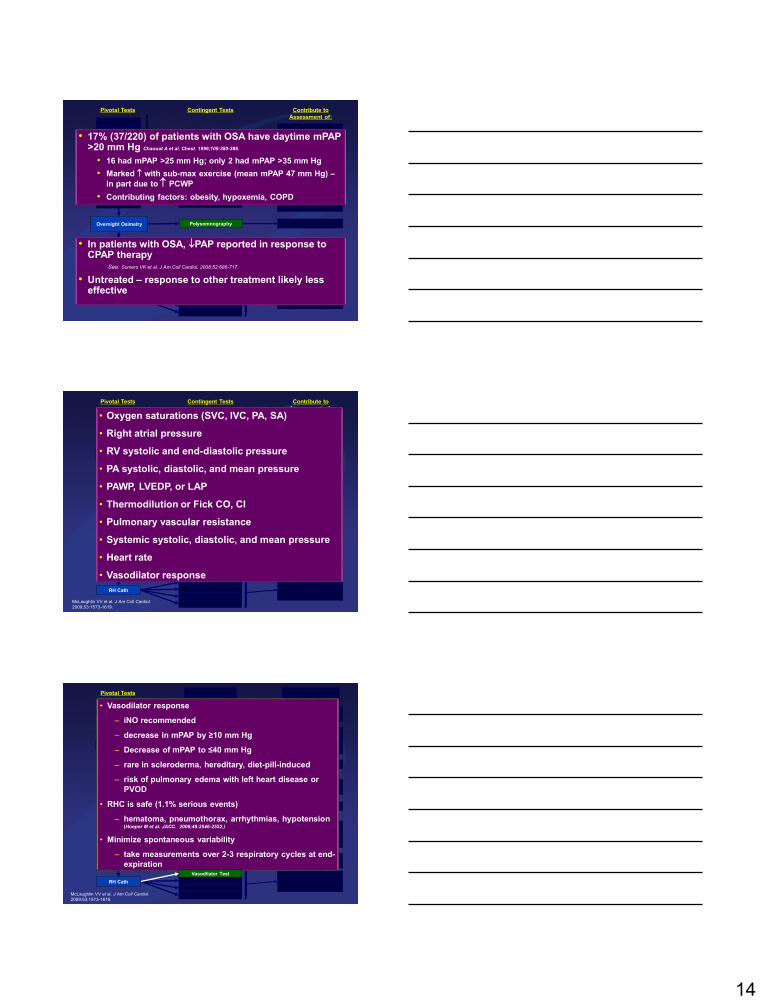
ExamR L L R
Ant PostIPAH
R L L R
Ant PostCTEPH
• 3-4% of acute PE do not entirely resolve
• ½ of those with CTEPH do not have an apparent history of acute PE
• Normal or very low probability VQ essentially excludes chronic PE
• CTEPH should be excluded, even when another explanation for PH is present
VQ Scan
McLaughlin VV et al. J Am Coll Cardiol.
2009;53:1573-1619.
Page 14
14
Echocardiogram
PFT’s
VQ Scan
•Sleep Disorder
•Chronic PE
Functional Test
(6MWT, CPET)
HIV
ANA
LFT’s
RH Cath
TEE
Exercise Echo
Pulmonary Angiography
Chest CT Angiogram
Coagulopathy Profile
Vasodilator Test
Exercise RH Cath
Volume Loading
ABG’s
•Index of Suspicion of
PH
• RVE, RAE, RVSP, RV
Function
• Left Heart Disease
• VHD, CHD
•Ventilatory Function
•Gas Exchange
Other CTD Serologies
•HIV Infection
•Scleroderma, SLE, RA
•Portopulmonary Htn
•Establish Baseline
•Prognosis
•Confirmation of PH
•Hemodynamic Profile
•Vasodilator Response
Contingent Tests Contribute to
Assessment of:
Left Heart Cath
CXR
ECG
Pivotal Tests
History
Exam
• 17% (37/220) of patients with OSA have daytime mPAP>20 mm Hg Chaouat A et al. Chest. 1996;109:380-386.
• 16 had mPAP >25 mm Hg; only 2 had mPAP >35 mm Hg
• Marked with sub-max exercise (mean mPAP 47 mm Hg) –
in part due to PCWP
• Contributing factors: obesity, hypoxemia, COPD
• In patients with OSA, PAP reported in response to CPAP therapy
See: Somers VK et al. J Am Coll Cardiol. 2008;52:686-717.
• Untreated – response to other treatment likely less effective
PolysomnographyOvernight Oximetry
Echocardiogram
PFT’s
Polysomnography
VQ Scan
•Sleep Disorder
•Chronic PE
Functional Test
(6MWT, CPET)
Overnight
Oximetry
HIV
ANA
LFT’s
TEE
Exercise Echo
Pulmonary Angiography
Chest CT Angiogram
Coagulopathy Profile
Vasodilator Test
Exercise RH Cath
Volume Loading
ABG’s
•Index of Suspicion of
PH
• RVE, RAE, RVSP, RV
Function
• Left Heart Disease
• VHD, CHD
•Ventilatory Function
•Gas Exchange
Other CTD Serologies
•HIV Infection
•Scleroderma, SLE, RA
•Portopulmonary Htn
•Establish Baseline
•Prognosis
•Confirmation of PH
•Hemodynamic Profile
•Vasodilator Response
Contingent Tests Contribute to
Assessment of:
Left Heart Cath
CXR
ECG
Pivotal Tests
History
Exam• Oxygen saturations (SVC, IVC, PA, SA)
• Right atrial pressure
• RV systolic and end-diastolic pressure
• PA systolic, diastolic, and mean pressure
• PAWP, LVEDP, or LAP
• Thermodilution or Fick CO, CI
• Pulmonary vascular resistance
• Systemic systolic, diastolic, and mean pressure
• Heart rate
• Vasodilator response
RH Cath
McLaughlin VV et al. J Am Coll Cardiol.
2009;53:1573-1619.
Echocardiogram
PFT’s
Polysomnography
VQ Scan
•Sleep Disorder
•Chronic PE
Functional Test
(6MWT, CPET)
Overnight
Oximetry
HIV
ANA
LFT’s
TEE
Exercise Echo
Pulmonary Angiography
Chest CT Angiogram
Coagulopathy Profile
ABG’s
•Index of Suspicion of
PH
• RVE, RAE, RVSP, RV
Function
• Left Heart Disease
• VHD, CHD
•Ventilatory Function
•Gas Exchange
Other CTD Serologies
•HIV Infection
•Scleroderma, SLE, RA
•Portopulmonary Htn
•Establish Baseline
•Prognosis
•Confirmation of PH
•Hemodynamic Profile
•Vasodilator Response
Contingent Tests Contribute to
Assessment of:
Left Heart Cath
CXR
ECG
Pivotal Tests
History
Exam• Vasodilator response
– iNO recommended
– decrease in mPAP by ≥10 mm Hg
– Decrease of mPAP to ≤40 mm Hg
– rare in scleroderma, hereditary, diet-pill-induced
– risk of pulmonary edema with left heart disease or
PVOD
• RHC is safe (1.1% serious events)
– hematoma, pneumothorax, arrhythmias, hypotension (Hoeper M et al. JACC. 2006;48:2546-2552.)
• Minimize spontaneous variability
– take measurements over 2-3 respiratory cycles at end-
expiration
Exercise RH Cath
Volume Loading
Vasodilator Test
RH Cath
McLaughlin VV et al. J Am Coll Cardiol.
2009;53:1573-1619.
Page 15
15
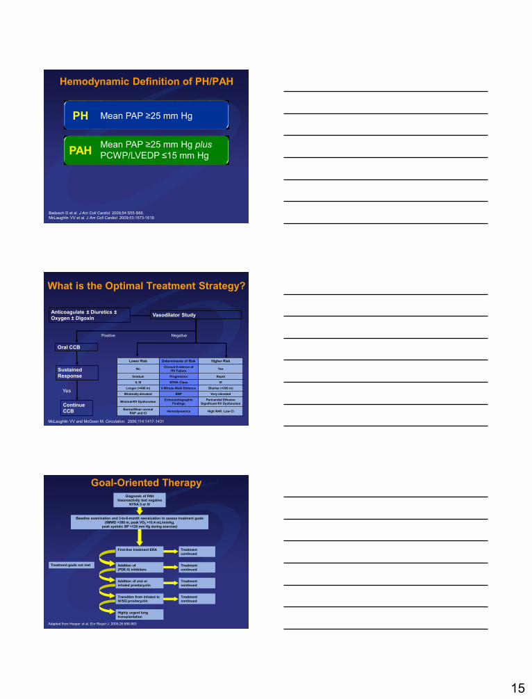
Badesch D et al. J Am Coll Cardiol. 2009;54:S55-S66.
McLaughlin VV et al. J Am Coll Cardiol. 2009;53:1573-1619.
Hemodynamic Definition of PH/PAH
PH
PAHMean PAP ≥25 mm Hg plus
PCWP/LVEDP ≤15 mm Hg
Mean PAP ≥25 mm Hg
Vasodilator StudyAnticoagulate ± Diuretics ±
Oxygen ± Digoxin
Sustained
Response
Positive
Oral CCB
Continue
CCB
Yes
Negative
Lower Risk Determinants of Risk Higher Risk
NoClinical Evidence of
RV FailureYes
Gradual Progression Rapid
II, III NYHA Class IV
Longer (>400 m) 6 Minute Walk Distance Shorter (<300 m)
Minimally elevated BNP Very elevated
Minimal RV DysfunctionEchocardiographic
Findings
Pericardial Effusion
Significant RV Dysfunction
Normal/Near normal
RAP and CIHemodynamics High RAP, Low CI
What is the Optimal Treatment Strategy?
McLaughlin VV and McGoon M. Circulation. 2006;114:1417-1431.
Adapted from Hoeper et al. Eur Respir J. 2005;26:858-863.
Goal-Oriented TherapyDiagnosis of PAH
Vasoreactivity test negative
NYHA II or IV
Baseline examination and 3-to-6-month reevaluation to assess treatment goals
(6MWD >380 m, peak VO2 >10.4 mL/min/kg,
peak systolic BP >120 mm Hg during exercise)
Treatment goals not met
First-line treatment ERA Treatment
continued
Addition of
(PDE-5) inhibitors
Treatment
continued
Addition of oral or
inhaled prostacyclin
Treatment
continued
Transition from inhaled to
IV/SQ prostacyclin
Treatment
continued
Highly urgent lung
transplantation
Page 16
16
Case Studies
Case Study
DE is a 66 year old male who presented to his
primary care physicians office with complaints
of cough, increased fatigue and dyspnea on
exertion. Past medical history includes
hypertension, smoking and osteoarthritis. He
had a recent Cardiolite stress test which was
negative. BP in office is 160/90. An
echocardiogram was ordered.
Height 64 inches. Weight 275 lbs.
Physical Exam- III/VI SEM, 1+ lower extremity
edema
Case Study
2D Echo results:
LVEF: 65%
Normal RV function
Stage I diastolic dysfunction
Mild LVH
Mild biatrial chamber size enlargement
Normal left and right ventricular size
Severe tricuspid regurgitation
RVSP 60 mmHg
Page 17
17
Case Study
Based on the initial presentation and
echocardiogram your next step would be
as follows? BP in office is 160/90. Weight 275 lbs.
A. No further testing or treatment is indicated
B. Start on oral diuretics
C. Set up for a PFT, VQ Scan and Sleep Study
D. Place on anti-hypertensive medications
E. Referral to TCI Cardiology
Case Study
You decided to place DE on HCTZ 25 mg and lisinopril 5 mg
daily. He returns one month later with continued dyspnea. BP
in office is improved at 138/80. An ECG was performed which
was essentially normal. He denes CP.
Height 64 inches. Weight 220 (225) lbs.
Physical Exam- III/VI SEM, 1+ lower extremity edema
Case Study
Given his continued symptoms, what
would be your next step?
A. No further testing or treatment is indicated
B. Refer to weight loss management center
C. Set up for a PFT, VQ Scan and Sleep Study
D. Increase anti-hypertensive medications
E. Referral to TCI Cardiology
Page 18
18
Case Study
Comprehensive Evaluation
Review previous echocardiograms
Pulmonary Function Study with DLCO
Apnea Link Monitor / Sleep study
VQ Scan
Labs- ANA, RF (if no history of CTD), BNP, LFT’s, TSH, HIV
Echo with saline contrast r/o shunt
BP logs
6MWT
Case Study
Severe Obstructive Sleep Apnea/Stage I diastolic dysfunction
Other studies normal.
CPAP for 3 months then re-evaluate
Blood pressure control
6MWT
2D echo limited check RVSP
Echocardiogram
PFT’s
VQ Scan
•Sleep Disorder
•Chronic PE
Functional Test
(6MWT, CPET)
HIV
ANA
LFT’s
RH Cath
TEE
Exercise Echo
Pulmonary Angiography
Chest CT Angiogram
Coagulopathy Profile
Vasodilator Test
Exercise RH Cath
Volume Loading
ABG’s
•Index of Suspicion of
PH
• RVE, RAE, RVSP, RV
Function
• Left Heart Disease
• VHD, CHD
•Ventilatory Function
•Gas Exchange
Other CTD Serologies
•HIV Infection
•Scleroderma, SLE, RA
•Portopulmonary Htn
•Establish Baseline
•Prognosis
•Confirmation of PH
•Hemodynamic Profile
•Vasodilator Response
Contingent Tests Contribute to
Assessment of:
Left Heart Cath
CXR
ECG
Pivotal Tests
History
Exam
• 17% (37/220) of patients with OSA have daytime mPAP>20 mm Hg Chaouat A et al. Chest. 1996;109:380-386.
• 16 had mPAP >25 mm Hg; only 2 had mPAP >35 mm Hg
• Marked with sub-max exercise (mean mPAP 47 mm Hg) –
in part due to PCWP
• Contributing factors: obesity, hypoxemia, COPD
• In patients with OSA, PAP reported in response to CPAP therapy
See: Somers VK et al. J Am Coll Cardiol. 2008;52:686-717.
• Untreated – response to other treatment likely less effective
PolysomnographyOvernight Oximetry
Page 19
19
Case Study
HG is a 62 year old female who presented to your office for a
hospital follow up after not having been seen for 4 years. She
has a known history of HTN, hyperlipidemia and fibromyalgia.
She was recently hospitalized for shortness of breath and
underwent an echocardiogram which demonstrated an EF of
35% with no significant valvular abnormalties. A catheterization
was performed which demonstrated normal coronaries and a
PCWP of 23 mmHg (normal 8-10 mmHg) with a mPAP of 30
mmHg (normal 12-15 mmHg). She was diagnosed with a non
ischemic CM and placed on medical therapy. Her
echocardiogram demonstrated an RVSP of 65 mmHg.
Height 66 inches. Weight 165 lbs.
Physical Exam- III/VI SEM, 1+ lower extremity edema
Case Study
2D Echo results:
LVEF: 35%
Normal RV function
Stage I diastolic dysfunction
Mild LVH
Mild biatrial chamber size enlargement
RVSP 65 mmHg
Case Study
Based on her hospitalization and
echocardiogram with abnormal RVSP what
would be her diagnosis?
A. Pulmonary Arterial Hypertension
B. PH WHO Group II secondary to LH disease
C. PH WHO Group III secondary to Intrinsic lung disease
D. CTEPH
E. More information is needed to make this diagnosis
Page 20
20
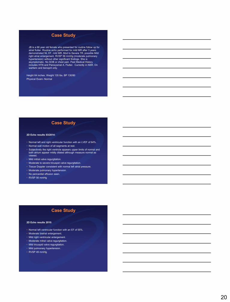
Case Study
JB is a 68 year old female who presented for routine follow up for
atrial flutter. Routine echo performed for mild MR after 3 years
demonstrated NL EF, mild MR, Mod to Severe TR, possible Mild
right atrial enlargement, RVSP 56 mmHg (moderate pulmonary
hypertension) without other significant findings. She is
asymptomatic. No SOB or chest pain. Past Medical History
includes HTN and Paroxysmal A. Flutter. Currently in NSR. On
warfarin and lisinopril only.
Height 64 inches. Weight 135 lbs. BP 130/80
Physical Exam- Normal
Case Study
2D Echo results 03/2014:
• Normal left and right ventricular function with an LVEF of 64%.
• Normal wall motion of all segments at rest.
• Subjectively the right ventricle appears upper limits of normal and
both atrium appear mildly dilated although measure normal as
viewed.
• Mild mitral valve regurgitation.
• Moderate to severe tricuspid valve regurgitation.
• Tissue Doppler consistent with normal left atrial pressure.
• Moderate pulmonary hypertension.
• No pericardial effusion seen.
• RVSP 56 mmHg
Case Study
2D Echo results 2010:
• Normal left ventricular function with an EF of 55%.
• Moderate biatrial enlargement.
• Mild right ventricular enlargement.
• Moderate mitral valve regurgitation.
• Mild tricuspid valve regurgitation.
• Mild pulmonary hypertension.
• RVSP 49 mmHg
Page 21
21
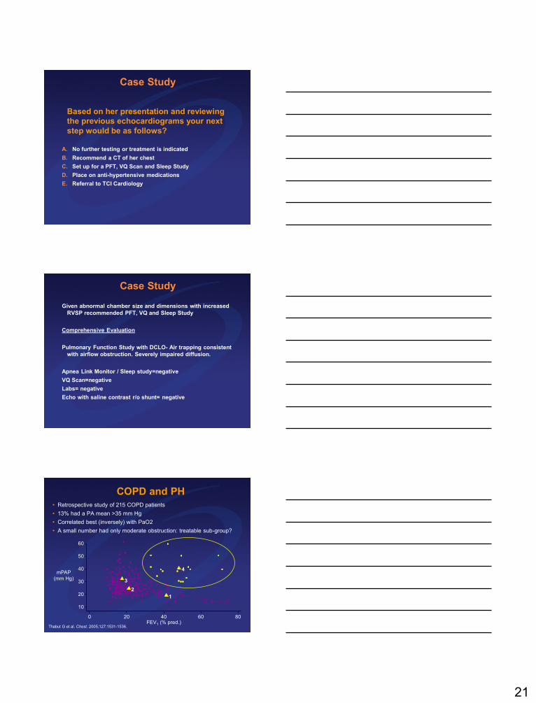
Case Study
Based on her presentation and reviewing
the previous echocardiograms your next
step would be as follows?
A. No further testing or treatment is indicated
B. Recommend a CT of her chest
C. Set up for a PFT, VQ Scan and Sleep Study
D. Place on anti-hypertensive medications
E. Referral to TCI Cardiology
Case Study
Given abnormal chamber size and dimensions with increased
RVSP recommended PFT, VQ and Sleep Study
Comprehensive Evaluation
Pulmonary Function Study with DCLO- Air trapping consistent
with airflow obstruction. Severely impaired diffusion.
Apnea Link Monitor / Sleep study=negative
VQ Scan=negative
Labs= negative
Echo with saline contrast r/o shunt= negative
COPD and PH• Retrospective study of 215 COPD patients
• 13% had a PA mean >35 mm Hg
• Correlated best (inversely) with PaO2
• A small number had only moderate obstruction: treatable sub-group?
Thabut G et al. Chest. 2005;127:1531-1536.FEV1 (% pred.)
mPAP
(mm Hg)
10
20
30
40
60
50
0 20 40 60 80
4
1
2
3
Page 22
22
Case Study
VG is a 43 year old female who presented with lower extremity
swelling and dyspnea on exertion which has been worsening
over the last 6 months. 2D echocardiogram demonstrated an
RVSP of 71 mmHg. Known history of Scleroderma. 30 plus pack
year smoking history. No history of CAD with recent negative
stress test.
2014 2D Echo results:
LVEF: 55%
Reduced RV function
Mild biatrial chamber size enlargement
Mild right ventricular enlargement
Severe tricuspid regurgitation
RVSP 71 mmHg
2007 Echo- Normal LV and RV function RVSP- 34 mmHg
Case Study
Based on her presentation and reviewing
the previous echocardiograms your next
step would be as follows?
A. No further testing or treatment is indicated
B. Place patient on sildenafil therapy
C. Set up for a PFT, VQ Scan and or a Sleep Study
D. Refer to Rheumatology
E. Referral to TCI Cardiology
Diagnostic Confirmation:
Right Heart Catheterization
In patients with suspected PAH:
• Right heart catheterization is required to confirm
the presence of PAH, establish the specific
diagnosis and determine the severity[Strength of recommendation: A]
• Right heart catheterization is required to guide
therapy[Strength of recommendation: B]
McGoon M et al for the American College of Chest Physicians. Chest.
2004;126:14S-34S.
Page 23
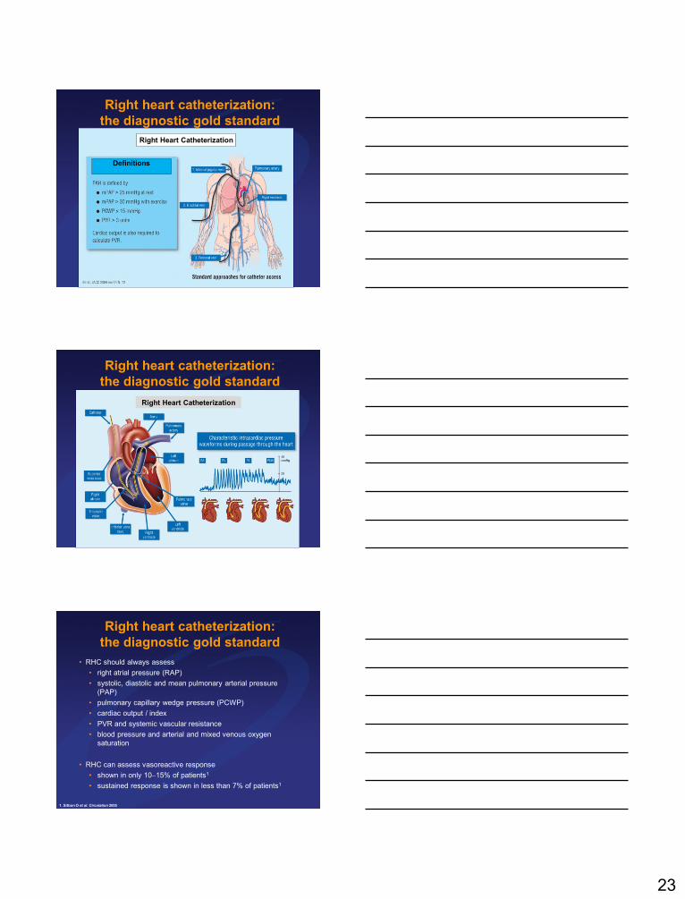
23
Right heart catheterization:
the diagnostic gold standard
Right Heart Catheterization
Definitions
Right heart catheterization:
the diagnostic gold standard
Right Heart Catheterization
Right heart catheterization:
the diagnostic gold standard
• RHC should always assess
• right atrial pressure (RAP)
• systolic, diastolic and mean pulmonary arterial pressure
(PAP)
• pulmonary capillary wedge pressure (PCWP)
• cardiac output / index
• PVR and systemic vascular resistance
• blood pressure and arterial and mixed venous oxygen
saturation
• RHC can assess vasoreactive response
• shown in only 1015% of patients1
• sustained response is shown in less than 7% of patients1
1. Sitbon O et al. Circulation 2005
Page 24
24
Case Study
Right Heart Catheterization
RA-8 mmHg
mPAP- 49 mmHg
PCWP- 4 mmHg
Transpulmonary gradient-45 mmHg
Cardiac output- 5.13 l/m
PVR-8.775 woods units
Negative Vasodilator challenge
Case Study
VT is a 26 year old female who presented to her primary care
physicians office in February 2010 with complaints of cough,
increased fatigue and significant dyspnea on exertion. Otherwise
healthy female, no significant past medical history, has one
healthy child. No findings were noted on her physical exam. She
was given Singulair and albuterol MDI and recommend follow up
in 1 week. She continued to have SOB and she proceeded to the
ER on day three.
During her ER visit an ECG was ordered. All labs were normal.
CXR performed and ER notes no significant findings.
Case Study
Page 25
25
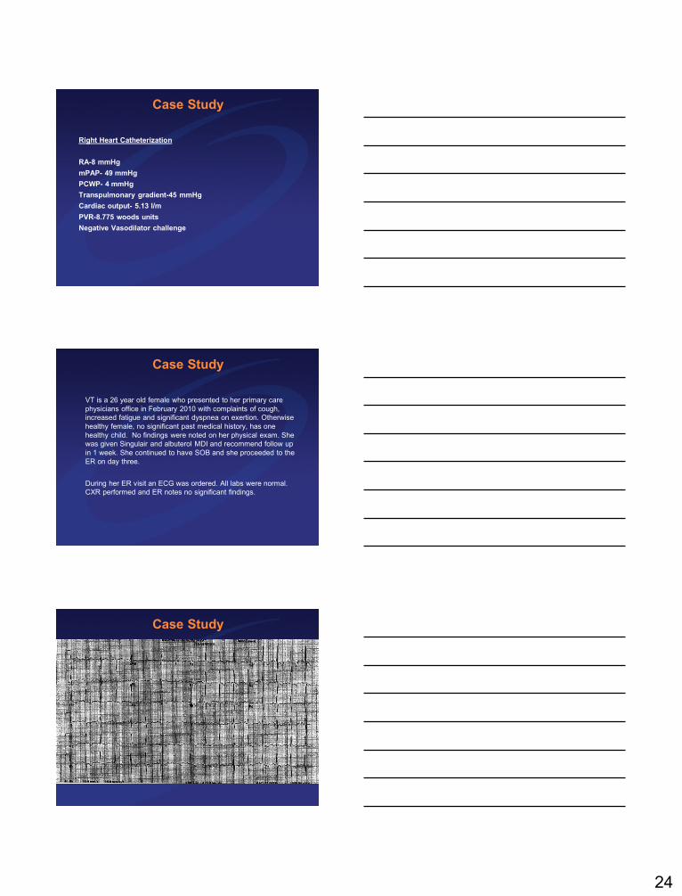
Case StudyWhat is the main abnormality on the EKG?
A. ST Segment Depression
B. Right Ventricular Hypertrophy
C. Left Ventricular Hypertrophy
D. Right Bundle Branch Block
E. Left Bundle Branch Block
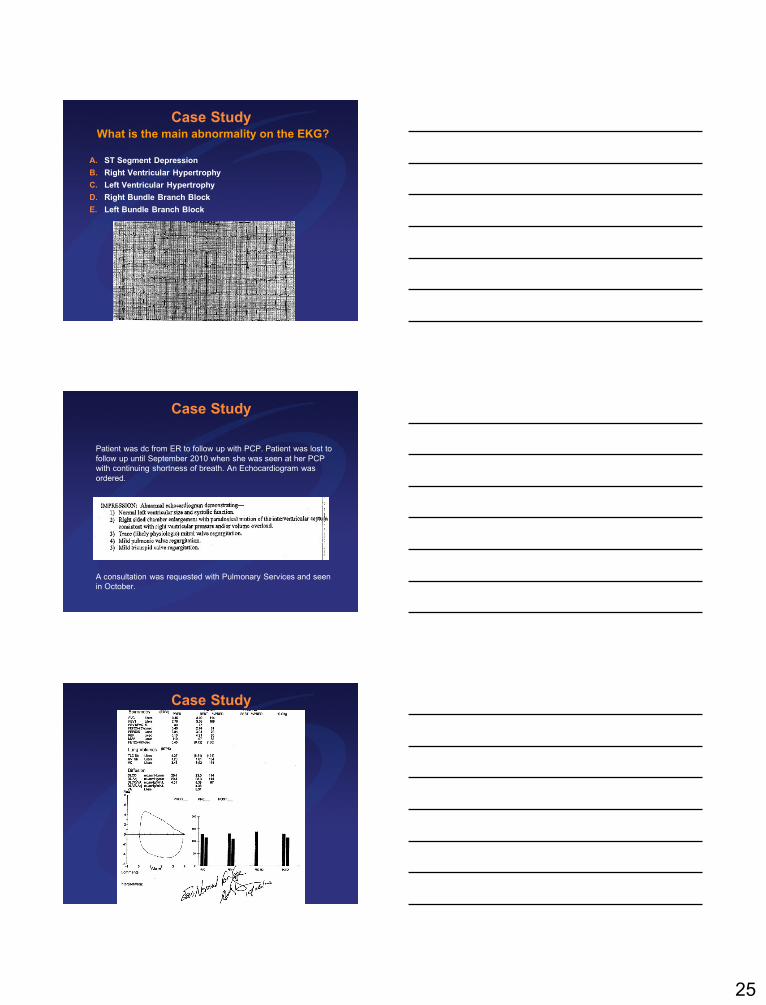
Case Study
Patient was dc from ER to follow up with PCP. Patient was lost to
follow up until September 2010 when she was seen at her PCP
with continuing shortness of breath. An Echocardiogram was
ordered.
A consultation was requested with Pulmonary Services and seen
in October.
Case Study
Page 26
26
Case Study
Based on her presentation and reviewing
the previous echocardiograms your next
step would be as follows?
A. No further testing or treatment is indicated
B. Place patient on sildenafil therapy
C. Set up for a PFT, VQ Scan and or a Sleep Study
D. Start her on albuterol and atrovent nebulizer
E. Referral to TCI Cardiology
Case Study
Cardiology consult was requested and was seen in November.
Right heart catheterization was performed within a few days
which demonstrated a mean pulmonary artery pressure of 64
mmHg consistent with severe pulmonary hypertension.
© 2011 Actelion Pharmaceuticals US, Inc. All rights reserved. 11 172 01 00 0611 78
Page 27
27
© 2011 Actelion Pharmaceuticals US, Inc. All rights reserved. 11 172 01 00 0611 79
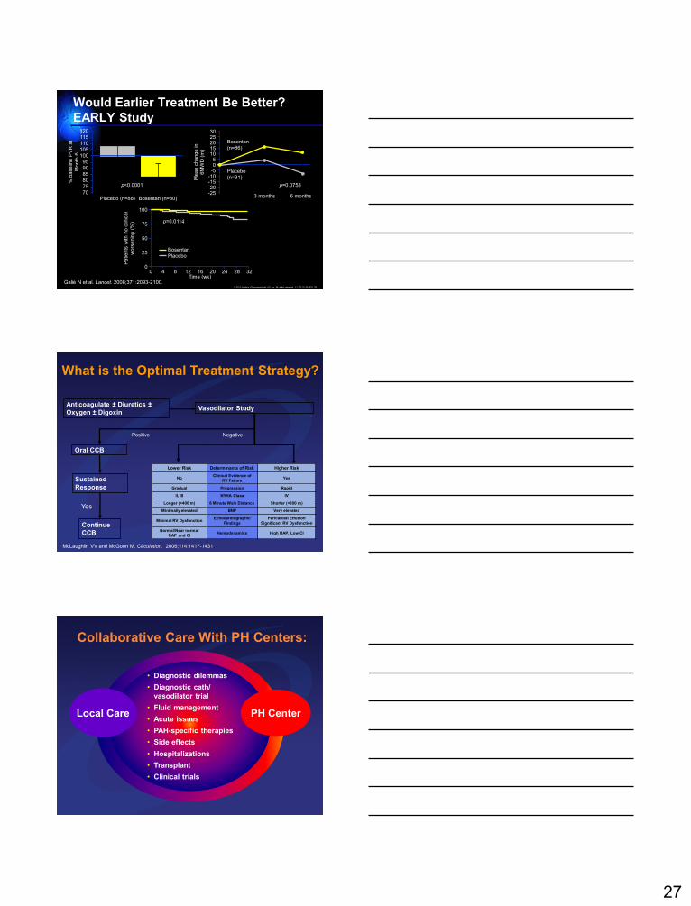
Would Earlier Treatment Be Better?
EARLY Study
Galiè N et al. Lancet. 2008;371:2093-2100.Time (wk)
0
25
50
75
100
Patients
with n
o c
linic
al
wors
enin
g (
%)
Bosentan
Placebo
0 4 8 12 16 20 24 28 32
p=0.0114
115
110
120
105
95
90
100
85
75
70
80% b
aselin
e P
VR
at
Month
6
Placebo (n=88) Bosentan (n=80)
2520
30
15
0-5
10
-10
-20-25
-15
5
3 months 6 months
Bosentan
(n=86)
Placebo
(n=91)Mean c
hange in
6M
WD
(m
)
p<0.0001 p=0.0758
Vasodilator StudyAnticoagulate ± Diuretics ±
Oxygen ± Digoxin
Sustained
Response
Positive
Oral CCB
Continue
CCB
Yes
Negative
Lower Risk Determinants of Risk Higher Risk
NoClinical Evidence of
RV FailureYes
Gradual Progression Rapid
II, III NYHA Class IV
Longer (>400 m) 6 Minute Walk Distance Shorter (<300 m)
Minimally elevated BNP Very elevated
Minimal RV DysfunctionEchocardiographic
Findings
Pericardial Effusion
Significant RV Dysfunction
Normal/Near normal
RAP and CIHemodynamics High RAP, Low CI
What is the Optimal Treatment Strategy?
McLaughlin VV and McGoon M. Circulation. 2006;114:1417-1431.
Local Care PH Center
Collaborative Care With PH Centers:
• Diagnostic dilemmas
• Diagnostic cath/
vasodilator trial
• Fluid management
• Acute issues
• PAH-specific therapies
• Side effects
• Hospitalizations
• Transplant
• Clinical trials
Page 28
28
Final Thoughts
• Comprehensive history and physical is foundation for diagnosis
• Noninvasive screening as indicated
• Treat any identified factor(s) that could contribute to or exacerbate pulmonary hypertension
• Invasive hemodynamics are crucial
• Refer early
Thank You for your attention!
References
Sitbon O et al. Circulation 2005
D’Alonzo GE et al. Ann Intern Med 1991
Galie N et al. Eur Heart J 2004.
Gaine SP et al. Lancet 1998
Barst RJ et al. J Am Coll Cardiol 2004
Simonneau G et al. JACC 2004
Barst RJ et al. J Am Coll Cardiol 2004
D’Alonzo GE, et al. Ann Intern Med 1991;115:343-349.
Peacock AJ. BMJ 2003
Gaine SP et al. Lancet 1998
Sitbon O et al. Am J Resp Crit Care Med 2008
Lin EE et al. Curr Hematol Rep 2005
McGoon M et al. Chest 2004
Stewart DJ et al. Ann Inter Med 1991
Vancheeswaran R et al. J Rheum 1994
Yoshibayashi M et al. Circulation 1991
Galiè N et al. Eur J Clin Invest 1996
Channick RN et al. Lancet 2001
Kato I et al. Cancer 2001
Bjoraker JA et al. Am J Respir Crit Care Med 1998
Humbert M et al. Am J Respir Crit Care Med 2006
Sitbon O et al. J Am Coll Cardiol 2002
Galie N et al. Eur Heart J 2004
Hachulla E et al Ann Rheum Dis 2004
McGoon M et al. Chest 2004
ATS. Am J Crit Care Med 2002
Galiè N et al. Lancet 2008
Humbert H et al. N Engl J Med 2004
Channick RN et al. Lancet 2001
Humbert H et al. N Engl J Med 2004
Galiè N et al. N Engl J Med 2005