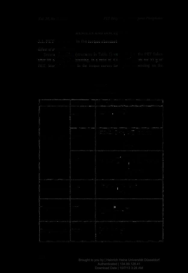
POLYETHYLENE TEREPHTHALATE) RECYCLING WITH ORGANIC PHOSPHITES -1. INCREASE IN MOLECULAR WEIGHT. Christine R. Nascimento and Marcos L. Dias* Instituto de Macromolicalas Professora Eloisa Mano, Universidade Federal do Rio de Janeiro, CP 68525, 21945-970 Rio de Janeiro, RJBrazil ABSTRACT Recycling of poly(ethylene terephthalate) by processing flakes obtained from post-consumer PET bottles was investigated. Flakes were additivated with organic phosphites during the processing in an internal mixer and the torque and temperature of the melt were monitored in each run. The molecular weight and polydispersity of the recycled PET were measured by viscometry and SEC. Some of the phosphites acted as chain extenders increasing the polymer M w . Best chain extension results were obtained with triarylphosphites, while dialkyl and trialkylphosphites promoted PET degradation. The study showed that triphenylphosphite was the best chain extender used in this work. Key words: PET bottles, recycling, phosphites. 1. INTRODUCTION The importance of post-consumer material recycling has grown recently. On the other hand, it is often difficult to match the ecological requirements with the technical and economical viability of recycled products. During the mechanical recycling of PET bottles, the polymer is submitted to several degradative processes like thermal and hydrolytic degradation. The presence * Corresponding author 143 Brought to you by | Heinrich Heine Universität Düsseldorf Authenticated | 134.99.128.41 Download Date | 10/7/13 3:28 AM
Transcript
Toxicology and Applied Pharmacology 269 (2013) 250–262
Contents lists available at SciVerse ScienceDirect
Toxicology and Applied Pharmacology
j ourna l homepage: www.e lsev ie r .com/ locate /ytaap
Pulmonary instillation of low doses of titanium dioxide nanoparticles in mice leads toparticle retention and gene expression changes in the absence of inflammation
Mainul Husain a, Anne T. Saber b, Charles Guo a, Nicklas R. Jacobsen b, Keld A. Jensen b, Carole L. Yauk a,Andrew Williams b, Ulla Vogel b,c, Hakan Wallin b,d, Sabina Halappanavar a,⁎a Environmental Health Science and Research Bureau, Health Canada, Ottawa, Ontario K1A 0K9, Canadab The Danish NanoSafety Centre, National Research Centre for the Working Environment, Copenhagen DK-2100, Denmarkc Department of Micro- and Nanotechnology, Technical University of Denmark, Kgs. Lyngby DK-2800, Denmarkd Institute of Public Health, University of Copenhagen, Copenhagen DK-1014, Denmark
We investigated gene expression, protein synthesis, and particle retention in mouse lungs following intratrachealinstillation of varying doses of nano-sized titanium dioxide (nano-TiO2). Female C57BL/6 mice were exposed to ru-tile nano-TiO2 via single intratracheal instillations of 18, 54, and 162 μg/mouse.Micewere sampled 1, 3, and 28 dayspost-exposure. The deposition of nano-TiO2 in the lungs was assessed using nanoscale hyperspectral microscopy.Biological responses in the pulmonary system were analyzed using DNA microarrays, pathway-specific real-timeRT-PCR (qPCR), gene-specific qPCR arrays, and tissue protein ELISA. Hyperspectral mapping showed dose-dependent retention of nano-TiO2 in the lungs up to 28 days post-instillation. DNA microarray analysis revealedapproximately 3000 genes that were altered across all treatment groups (±1.3 fold; p b 0.1). Several inflammatorymediators changed in a dose- and time-dependent manner at both themRNA and protein level. Although no influxof neutrophilswas detected at the lowdose, changes in the expression of several genes and proteins associatedwithinflammationwere observed. Resolving inflammation at themedium dose, and lack of neutrophil influx in the lungfluid at the low dose, were associated with down-regulation of genes involved in ion homeostasis and muscle reg-ulation. Our gene expression results imply that retention of nano-TiO2 in the absence of inflammationover timemaypotentially perturb calcium and ion homeostasis, and affect smooth muscle activities.
Titanium dioxide (TiO2) is generally considered to be chemically inertbecause it is highly insoluble, thermally stable, and non-flammable. How-ever, rats exposed to high doses of pigment-grade and ultrafine TiO2 dustby inhalation or instillation develop lung cancer (Heinrich et al., 1995). Inaddition, the International Agency for Research on Cancer has classifiedpigment-grade TiO2 (microparticles with size range of 100–200 nm) asa possible human carcinogen (group 2B) (IARC, 2010; Iavicoli et al.,2011) based on animal and cell culture data, but there is little evidencefor TiO2-mediated carcinogenic effects in humans. TiO2 is produced pri-marily as aggregates or agglomerates of nano-sized particles. It accountsfor 70%of the total productionof pigmentsworldwide. TiO2-nanoparticles(TiO2-NPs) are extensively used in a wide range of products including
ce and Research Bureau, ERHSD,03A), 50 Columbine Driveway,.ain), [email protected] (A.T. Saber),obsen), [email protected]@hc-sc.gc.cak (H. Wallin),
13 Published by Elsevier Inc. All rig
paints, coatings, cosmetics, and sunscreens (Liao et al., 2008; Madl andPinkerton, 2009; Park et al., 2009). As a result of the high productionvolume and extensive applications of powdered TiO2, reasonable risksfor exposures through inhalation or via other routes are anticipated.TiO2-NPs exhibit unique physico-chemical properties associated withtheir nano-size, which alters their biological behaviors at the cellular, sub-cellular, and protein levels resulting in adverse health effects (Liu et al.,2009; Nemmar et al., 2011; Oberdorster et al., 2000; Warheit et al.,2007). Thus, a detailed characterization of the tissue-level effects andmechanisms of action of TiO2-NPs is required in order to establish accept-able exposure limits.
Lung is a primary target organ of NP exposure via inhalation in oc-cupational settings. Pulmonary responses are largely driven by thephysico-chemical properties of the NPs, and it is well establishedthat inhalation of TiO2-NPs induces pulmonary responses that primarilyinclude inflammation. Ferin et al. (1992) were the first to demonstratethat pulmonary responses in rats exposed to TiO2-NPs are greatlyinfluenced by particle size (Ferin et al., 1992). These authors also showedthat intratracheal instillation or inhalation of TiO2-NPs induces profoundneutrophilic inflammation/infiltration, translocation of particles to thedeeper regions of lungs, and longer retention of particles compared totheir microparticulate counterpart. Similar particle size-associated effects
a Estimate of the average crystalline size along the shortest and longest crystallographicdirection.
b Calculated by difference from 100 wt.%.
251M. Husain et al. / Toxicology and Applied Pharmacology 269 (2013) 250–262
on pulmonary inflammation in rats were also observed by Renwick et al.(2004). Additional evidence suggests that the adverse pulmonary effectsassociated with TiO2-NPs are further amplified if the NPs are surface-coated with alumina or amorphous silica (Warheit et al., 2007). Inflam-matory response to TiO2-NPs varies depending on the crystalline form(Grassian et al., 2007), with mixed rutile/anatase forms exhibiting in-creased toxicity over other types of TiO2-NPs driven by the larger sur-face areas of mixed TiO2-NP types (Warheit et al., 2007). Thus, theseand other studies (reviewed in (Johnston et al., 2009)) firmly establishthat TiO2-NPs elicit pulmonary toxicity and tissue injury, and that thetoxicity is based on several physical and chemical properties.
Several studies have shown that ultrafine particles or NPs effec-tively evade primary phagocyte-mediated clearance mechanisms inthe lungs and hence migrate to the deeper interstitial regions of thelungs where they are retained over long periods of time (Ferin et al.,1992; Katsnelson et al., 2010). Other studies have suggested that exces-sive pulmonary retention and accumulation of particlesmay lead to lunginjury and/or enhanced risks of developing cancer (Bermudez et al.,2002, 2004; Heinrich et al., 1995). However, most of these studies usedphysiologically irrelevant doses and thus, the biological implication oflow dose exposure to TiO2-NPs, and the underlying molecular mecha-nisms of the observed pulmonary responses, remain largely unknown.Additionally, very few studies have examined the effects of retained par-ticles following low dose exposures on pulmonary systems.
We (Saber et al., 2012) recently compared pulmonary responsein mice following single instillation doses of 18, 54 and 162 μgof nano-TiO2/mouse, corresponding to 1.5, 5 and 15 working daysat the Danish occupational exposure level for TiO2 (6.0 mg Ti/m3–
9.75 mg TiO2/m3) at three different post-exposure time points, andshowed dose- and time-dependent pulmonary neutrophilic inflam-mation. Interestingly, the lowest dose in this study did not elicit anypulmonary inflammation as measured by inflammatory cellular influxin the bronchoalveolar lavage fluid (BAL). Based on these results a NoObserved Effect Level (NOEL) for nano-TiO2 of 19 cm2 was derived formice, corresponding to the lowest tested dose (Saber et al., 2012).
It could be hypothesized that the lack of inflammatory cell influx inthe BAL at the low doses observed in the study described above is a re-sult of first-order rates of clearance in the low dose range. In the presentstudy we rationalized that the lack of observable changes in inflamma-tory cell influx reported by Saber et al. (2012), is actually the result of di-minished induction of clearance mechanisms at low dose NP exposures,and that NPswill thus still be present in the lungs even in the absence ofinflammation. To test our hypothesis we used nanoscale hyperspectralmicroscopy to measure nano-TiO2 retention in the lungs of the samemice used by Saber et al. (2012), in parallel with pulmonary gene andprotein expression profiling, to study dose and time-related responses.We expected to find significant retention of particles in lungs at alldoses over extended periods of time resulting in perturbations of normalbiological functions. An additional objective for this study was to gener-ate testable hypotheses for novel mechanisms involved in pulmonaryeffects induced by low doses of nano-TiO2.
Materials and methods
The experimental samples used in this study were generated in anearlier study; the animals, characterization of particles, exposure, andexposure monitoring are previously described in Saber et al. (2012)and are briefly outlined below.
Animals
Five-to-seven week old female C57BL/6 mice were purchased fromTaconic (Ry, Denmark) and were acclimatized for 1–3 weeks priorto the commencement of the experiment. Mice were maintained ingroups in polypropylene cages with sawdust bedding and enrichment,at 20–22 °C room temperature and relative humidity of 40–60% with
a 12 h light-to-dark cycle. All mice received food and water ad libitumthroughout the experiment. All animal experiments were approved bythe Danish “Animal Experiments Inspectorate” and performed accordingto their guidelines for ethical conduct in the care and use of animals inresearch.
Particle characterization and preparation of exposure stock
The nanomaterial used in this study was an ultrafine powder ofsurface coated rutile TiO2 also known as UV-Titan L181 (Kemira, Pori,Finland) with an average crystallite size of 20.6 nm and surface area of107.7 m2/g, and is termed ‘nano-TiO2’ throughout this article. Details ofthe particle characterization were described previously (Halappanavaret al., 2011; Hougaard et al., 2010), and are summarized in Table 1.
Naïve C57BL/6 mice were flushed twice with 0.6 ml of 0.9% NaCl toobtain approximately 1 ml of bronchoalveolar lavage fluid (BAL). BALwas centrifuged at 400 g for 10 min at 4 °C to remove cells. The super-natantwas used in the dispersant liquid. A total of 4.05 mgof nano-TiO2
was suspended in 1 ml ofMilliQwater containing 0.9%NaCl and 10% v/vacellular BAL. Particle suspensions were prepared by sonicating thesamples using an S-450D sonifier (Branson Ultrasonics Corp., Danbury,CT, USA) at 10% amplitude for a total of 16 min on ice with alternating10 s pulses and pauses. Three different dilutions of nano-TiO2were pre-pared and used in this experiment: 162 μg, 54 μg (1:3 dilution of the162 μg), and 18 μg (1:3 dilution of the 54 μg). Control vehicle solutionswere prepared without the nano-TiO2 (MilliQ water containing 90%of 0.9% NaCl and 10% acellular BAL fluid). The doses 18, 54, and162 μg/mouse are equivalent to 1.5, 5, and 15working days at the Danishoccupational exposure level for TiO2 (6.0 mg Ti/m3–9.75 mg TiO2/m3),respectively (Saber et al., 2012).
Exposure of mice and harvesting of tissue
Each treatment group consisted of a minimum of 6 animals. Mice inthe experimental groups received a single intratracheal instillation of 18(low dose), 54 (medium dose), or 162 μg (high dose) of nano-TiO2 ina 40 μl suspension as described above, followed by 150 μl air with a250 μl SGE glass syringe (250F-LT-GT, MicroLab, Aarhus, Denmark).Intratracheal instillation was performed at 37 °C on heating plates,under anesthesia using Hypnorm® (fentanyl citrate 0.315 mg/mland fluanisone 10 mg/ml from Janssen Pharma) and Dormicum®(Midazolam 5 mg/ml from Roche). Control mice received 40 μl of disper-sant vehicle only. Following instillation, mice were under observationuntil they recovered from anesthesia. Mice were allowed to recover for
252 M. Husain et al. / Toxicology and Applied Pharmacology 269 (2013) 250–262
24 h, 3 days, and 28 days following instillation before being euthanizedunder anesthesia (Hypnorm/Dormicum). Blood, BAL fluid, and tissueswere collected. Lung tissues from experimental and control mice werecut into small pieces, stored in cryogenic vials, and snap frozen immedi-ately in liquid nitrogen. All tissues were stored at −80 °C until analysis.All analyses including microarray, validation of microarray results, pro-tein analysis, and microscopic imaging for detection of particles wereconducted on the left lobe of the lung tissue.
Detection of nano-TiO2 in lung tissue
Frozen lung tissue samples (n = 2–3) from low and medium dosegroups sampled on day 1 and day 28 andmatched controlswere embed-ded in paraffin. Paraffin-embedded tissues were sliced into 5 μm thicksections and stained with hematoxylin-eosin (H-E). H-E stained lungtissue sections (two sections per sample) were sent to CytoViva, Inc.(Auburn, AL, USA) for hyperspectral imaging and quantitative mappingof nano-TiO2 particles in the lungs. The proprietary hyperspectral imag-ing technology uses a nano-scalemicroscope and a darkfield-based opti-cal illuminator to focus light on samples, yielding images with improvedcontrast and signal-to-noise ratio. Such imaging technique takes advan-tage of the intrinsic light scattering properties of the particle of interest(e.g., nano-TiO2 particles). The hyperspectral imager consists of a con-centric imaging visible and near-infrared (VNIR) spectrophotometer(400–1000 nm)with integrated CCD camera, and is controlled by Envi-ronment for Visualization (ENVI 4.8) software for hyperspectral imageanalysis. The images (100× magnification) were captured using a DageExcel Color Cooled-M camera that is attached to an Olympus BX 43 opti-cal microscope. In order to aid the identification and image analysisprocess, a reference spectral library of nano-TiO2 was created prior tothe analysis of the actual sample images using a customized softwareanalysis program, and coded experimental samples were compared tothose in the reference library. Samples were decoded after the comple-tion of analysis to examine particle-related retention.
Total RNA extraction and purification
The total tissue for RNA extraction varied between 10 and 15 mg fromeach animal. In order to ensure unbiased analysis of tissue response, totalRNA was isolated from randomly sectioned (weighing 10–15 mg) leftlung lobe. RNA was isolated from 5 to 6 individual animals from eachof the treatment and control groups using TRIzol reagent (Invitrogen,Carlsbad, CA, USA) and purified using RNeasy Plus Mini kits (Qiagen,Mississauga, ON, Canada) according to the manufacturer's instruction.Total RNA concentration was measured using a NanoDrop 2000 spectro-photometer (Thermo Fisher Scientific Inc., Wilmington, DE, USA), andRNA quality and integrity were assessed using an Agilent 2100Bioanalyzer (Agilent Technologies, Inc., Mississauga, ON, Canada)according to the manufacturer's instruction. All samples showed RNAintegrity numbers of 7 and above, indicating high quality RNA, andwere used to conduct microarray and qPCR experiments.
Microarray hybridization
Total RNA (250 ng) samples from individual mice (n = 5–6 perexperimental and control group) and from universal reference totalRNA (Agilent Technologies, Inc., Mississauga, ON, Canada) were used tosynthesize double-stranded cDNA and cyanine labeled cRNA usingQuick Amp Labeling Kit (Agilent Technologies Inc., Mississauga, ON, Can-ada) according to themanufacturer's instructions. Control and nano-TiO2
treated cRNAswere labeledwith cyanine 5-CTP, and reference RNAswerelabeledwith cyanine 3-CTP using a T7 RNA polymerase in vitro transcrip-tion kit (Agilent Technologies Inc., Mississauga, ON, Canada) and purifiedusing RNeasy Mini kits (Qiagen, Mississauga, ON, Canada). Each experi-mental cRNA sample was mixed with an equimolar amount of referencecRNA and was hybridized to Agilent mouse 4 × 44 oligonucleotide
microarrays (Agilent Technologies Inc., Mississauga, ON, Canada) for17 h in a hybridization chamber at 65 °C with a rotation speed of10 rpm. Arrays were washed according to manufacturer's protocols(Agilent Technologies Inc., Mississauga, ON, Canada) and scanned on anAgilent G2505B scanner. Data from the scanned images were extractedusing Agilent Feature Extraction software version 9.5.3.1. All microarraydata from this study are available at theNational Center for BiotechnologyInformation (NCBI) and Gene Expression Omnibus (GEO) databases. GEOaccession number for these files is GSE41041.
Statistical analysis of microarray data
A reference randomized block design (Kerr, 2003; Kerr andChurchill, 2007) was used to analyze gene expression microarray data.Data were normalized using LOcally WEighted Scatterplot Smoothing(LOWESS) (Cleveland, 1979) regression modeling method and statisti-cal significance of the differentially expressed genes was determinedusing MicroArray ANalysis Of VAriance (MAANOVA) (Wu et al., 2003)in R statistical software (http://www.r-project.org). The Fs statistic(Cui et al., 2005) was used to test the treatment effects and p-valueswere estimated by the permutation method using residual shuffling.The false discovery rate (FDR) multiple testing correction (Benjaminiand Hochberg, 1995) was applied to minimize any false positives. Foldchange calculationswere based on the least-squaremeans. Genes show-ing expression changes of at least 1.3 fold in either direction comparedto their matched controls and having p-values of less than or equal to0.1 (p ≤ 0.1) were considered as significantly differentially expressedand were used in the downstream analysis. We applied a liberal ap-proach since we were looking for effects in the low dose range in theabsence of any observable phenotype.
Functional and pathway analysis of differentially expressed genes
Functional gene ontology (GO) analysis of the differentially ex-pressed genes was performed using the Database for Annotation, Vi-sualization and Integrated Discovery (DAVID) v6.7 (Huang da et al.,2009). Benjamini–Hochberg corrected GO processes with a Fisher'sexact p ≤ 0.05 were considered to be significantly enriched (over-represented). Specific biological pathways associated with the differ-entially expressed genes were identified using Ingenuity PathwayAnalysis (IPA, Ingenuity Systems, Redwood City, CA, USA) andMetaCore(Thomson Reuters Scientific Inc., Philadelphia, PA, USA) software. Path-ways with a Fisher's exact p-value of≤0.05 (p ≤ 0.05)were consideredfor discussion. The pathway analysis methods employed in the presentstudy enable the extraction of biologicallymeaningful information froma long list of differentially expressed genes. The quality and annotationof different pathway analysis packages vary. Therefore, the methodsemployed in our study are considered a starting point, and are used todirect future research. For a detailed review on the limitations of thepresent pathway analysis approaches please refer to Khatri et al. (2012).
Real-time PCR (qPCR) validation of microarray data
Mouse inflammatory cytokines and receptors (PAMM-011A,SABiosciences, Frederick, MD, USA) and custom PCR arrays wereused to validate 126 genes in total. Genes for the custom PCR array(CAPM11274, SABiosciences, Frederick, MD, USA) were selected basedon their established roles in biological processes such as acute phaseresponse, ion homeostasis, lipid metabolism, and muscle regulation,and exhibited fold changes of at least ±1.3 (p ≤ 0.1). Approximately800 ng of the total RNA (n = 3 per group) from each of the experimen-tal and control groups ofmicewas reverse transcribed using an RT2 firststrand cDNA synthesis kit (SABiosciences, Frederick, MD, USA). qPCRswere conducted using RT2 SYBR Green qPCR Master Mix in a CFX96Real-Time System (BioRad Laboratories, Mississauga, ON, Canada)according to the manufacturer's instructions. Threshold cycle (Ct) values
253M. Husain et al. / Toxicology and Applied Pharmacology 269 (2013) 250–262
were normalized using Hprt, Hsp90ab1, and Gapdh as internal controlgenes, and relative expression of the differentially expressed genes wasdetermined using online PCR array data analysis software (SABiosciences,Frederick, MD, USA). The correlation between the qPCR and microarrayfold changes of the validated genes was determined using Spearman'srank correlation analysis (Spearman, 1904) in R statistical software. AShapiro–Wilk normality test (Shapiro andWilk, 1965) for the fold changedatasets was performed prior to the correlation analysis to identify if thedata were normally distributed.
Expression analysis of inflammatory proteins
Total protein from the frozen lung tissues (n = 3) from experimen-tal and control micewas extracted using Bio-Plex Cell Lysis Kits (BioRadLaboratories, Mississauga, ON, Canada) and quantified using Bradfordprotein assay kits (Bio-Rad Laboratories, Mississauga, ON, Canada).Expression of mouse pro-inflammatory cytokines was assessed usingProMouse Cytokine 23-plex and 9-plex assay kits (BioRad Laboratories,Mississauga, ON, Canada) according to the manufacturer's instructionsin a Bio-Plex 200 system (BioRad Laboratories, Mississauga, ON, Canada).Briefly, 100 μg of each lysate was diluted with themanufacturer suppliedsample diluents. Anti-cytokine/chemokine antibody-conjugatedmagnet-ic beads were immobilized in each individual well of the 96 well filterplates using a vacuum manifold. Plates were washed twice, and 50 μlof pre-diluted standards and sample lysateswas added to each of the des-ignated well. Plates were shaken at room temperature for 30 min at350 rpm. Following 3 washes, plates were incubated for 30 min with25 μl of pre-diluted multiplex detection antibody. Plates were washed 3times and incubated with 50 μl of pre-diluted streptavidin-conjugatedphycoerythrin for 10 min. Following a final 3-step wash, plates were in-cubated with 125 μl of assay buffer in each well and were analyzedusing the Bio-Plex protein assay system. The concentration of each cyto-kine and chemokine was determined using Bio-Plex Manager Version6.0 software.
Results
The pulmonary inflammatory response was assessed to investigatethe impact of nano-TiO2 deposition inmouse lungs bymeasuring BAL dif-ferential cell counts and the results were published (Saber et al., 2012).Since the inflammatory phenotype reported in Saber et al. (2012) pro-vides context to the present study, the results are summarized below.Briefly, exposure to nano-TiO2 induced a significant inflammatory re-sponse in the 54 μg dose group on days 1 and 3, and in the 162 μg dosegroup across all the post-exposure time points studied. Inflammationwas mainly characterized by an increase in the total cellular count andinflammatory neutrophils in BAL. Inflammation was pronounced onday 1 for both of these dose groups and was partially resolved by28 days post-exposure time. No significant changes were observed inthe BAL cellular profiles of the 18 μg dose group compared to the controls.
Detection of nano-TiO2 in lung tissue
In order to understand if lack of inflammation in the low dose groupis attributed to the efficient clearance of particles, the lungs of miceexposed to low (18 μg) or high (162 μg) doses of nano-TiO2 sampledon days 1 and 28 post-exposure were examined for particle retentionusing the CytoViva patented VNIR hyperspectral imaging system anddarkfieldmicroscopy. Nanoparticles in general have a significantly differ-ent refractive index from the surrounding tissue, stains, and mountingmedium used to process the tissues, which leads to greater scatteringof light in the sections containing NPs and hence their easy detection.Two reference spectral libraries were made. The first library establisheda complete spectral profile of the nano-TiO2 used in this study (Fig. 1a).The second library cataloged a set of bright non-specific inclusions thatwere fluorescing as strongly as the nano-TiO2, but were also found in
the untreated control samples (Fig. 1b). These non-specific inclusionsare the result of endogenous cellularmaterials fluorescing in all samples.Images from the treated samples were overlaid with the reference spec-tral library for nano-TiO2 for accurate and precise detection of nano-TiO2
particles in the exposed samples.Fig. 1c shows dark field (upper panel), dark field hyperspectral
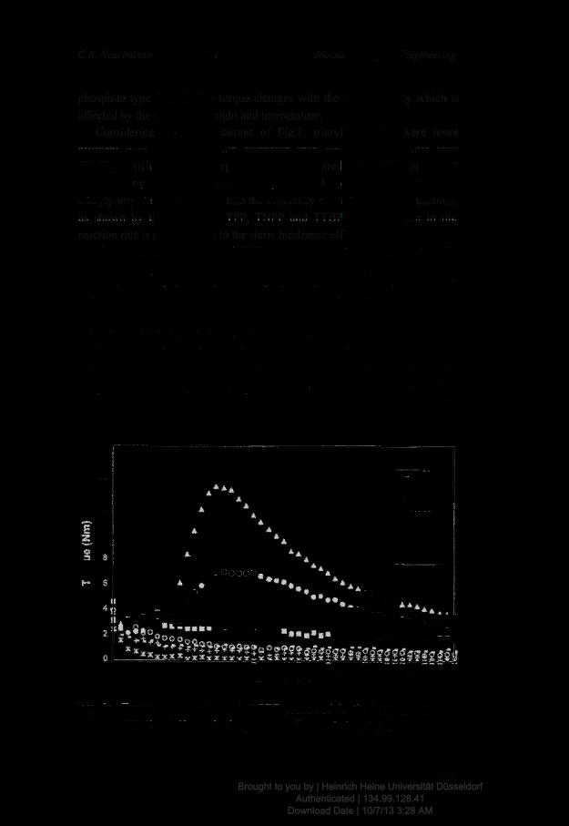
(middle panel), and hyperspectral mapping (bottom panel, overlayof dark field hyperspectral imageswith nano-TiO2 reference spectra) im-ages. Qualitative analysis of particle-laid lung tissues showed aggregatedand agglomerated nano-TiO2 (white inclusions indicated bywhite arrowheads) in randomparts of the lung tissue increasing in a dose-dependentmanner. A significant amount of nano-TiO2was observed in day 1 aswellas in day 28 post-exposure samples (Fig. 1c, upper panel). We did notapply a quantitative approach tomeasure the exact numbers of particlesor aggregates. Quantitative analyses would require highly precise local-ized sectioning to compare the exact same lung regions across the sam-ples, which was not possible in this study. Thus, it was not possible todetermine if the amount of particles retained in lungs decreasedwith in-creasing recovery time. Red dots in the bottom panel (Fig. 1c, indicatedby arrow heads) show areas of inclusions that were matched to thespecific hyperspectral reference library created for nano-TiO2. Brightinclusions in the control samples did not match with the spectra ofnano-TiO2 (Figs. 1a and b). These results demonstrate dose-dependentdeposition and sustained retention of nano-TiO2 over 28 days followingthe exposure for both the lowest and the highest doses.
Gene expression analysis
In order to understand the biological implications of tissue particleretention in the absence of inflammation, whole lung gene expressionprofiling was conducted on samples from all the doses and time points.MAANOVA analysis revealed a total of 3452 probes representing 3070unique genes significantly differentially expressed in response to low,medium and high doses of nano-TiO2 instillation on post-exposuredays 1, 3 and 28. Changes in gene expression were considered signifi-cant if they showed p-values of ≤0.10 and a fold change ≥1.3 in eitherdirection (Supplementary Table 1). Since the present study focused onfinding the effects following very low dose exposures that did notshow any observable biological response as measured by inflammatorycell counts in BAL fluid, we selected a liberal fold change of ±1.3 andp-value cut-off of p ≤ 0.1. These thresholds are aligned with therecommended cut-offs by Shi et al. (2008). These authors suggested theuse of fold change ranking in conjugation with a flexible (non-stringent)p-value threshold in order to generate reproducible differentially ex-pressed gene lists (Shi et al., 2008).
A principal component analysis (PCA) on all of the differentiallyexpressed genes revealed a dose and time effect (Supplementary Fig.1). A total of 1708 genes were significantly differentially expressed onday 1 in at least one of the dose groups compared to controls. Specifical-ly, 462, 691, and 1015 transcripts were affected by nano-TiO2 treatmentin the low, medium, and high dose groups respectively. There were only60 genes in common across the dose groups (Fig. 2a). Most of the genesonday1were up-regulated. A total of 1255 transcriptswere differential-ly expressed relative to controls on day 3 including 693, 652, and 620 inthe low, medium, and high dose groups respectively. There were 209transcripts that were similarly affected across all the dose groups onday 3 (Fig. 2b), and the majority of the genes on day 3 were down-regulated. On day 28, a total of 1198 genes were differentially expressedwith 325, 279, and 805 genes changing in the low, medium, and highdose respectively (Fig. 2c), andwith a fairly equal number of transcriptsbeing both up- and down-regulated.
Biological functions of differentially expressed genes
GO functional classification was used to identify specific themesin the gene expression patterns following exposure to nano-TiO2. The
Fig. 1. Identification of nano-TiO2 particles in lung tissues. Lung tissues frommice harvested 1 and 28 days following a single intratracheal instillation of low (18 μg) and high (162 μg)doses of nano-TiO2 were subjected to visible and near-infrared (VNIR) hyperspectral imaging to identify particle retention in these tissues. (a) Reference spectral library from nano-TiO2
exposed tissue. (b) Reference spectral library from control tissue. (c) Dark field images from nano-TiO2 exposed tissues (upper panel). Dark field hyperspectral images from nano-TiO2
exposed tissues identifying these nanoparticles, which appeared as aggregates of white inclusions (middle panel). Hyperspectral mapping of nano-TiO2 in these tissues appeared asred dots or aggregates (bottom panel).
254 M. Husain et al. / Toxicology and Applied Pharmacology 269 (2013) 250–262
differentially expressed genes were analyzed using DAVID; biologicalprocesses with a Fisher's exact p-value ≤0.05 (Benjamini–Hochbergcorrected) were considered significantly enriched. The common andunique biological processes significantly enriched in different dose groupsacross the time points are shown in Fig. 3.
Biological processes affected on day 1. The top common biologicalprocess altered on day 1 in all three doses consisted of 252 genes(Supplementary Table 2) implicated in immune/inflammatory responses,and was associated with five specific GO terms: immune response[GO:0006955], response to wounding [GO:0009611], inflammatoryresponse [GO:0006954], response to organic substance [GO:0010033],and chemotaxis [GO:0006935] (Fig. 3; day 1, common GO processes).In addition to the immune response, genes in the medium dose groupwere implicated in various types of metabolism including: fatty acidmetabolic process [GO:0006631], and lipid [GO:0006638] and glycerol[GO:0006662]metabolic processes (Fig. 3; day 1, unique GO processes).Genes over-represented in these GO processes were predominantlydown-regulated. Unique biological processes affected in the high dosegroup included: lipid biosynthetic process [GO:0008610], and choles-terol metabolic process [GO:0008203], cell proliferation [GO:0008283],DNA replication [GO:0006260], and extracellular matrix organization[GO:0030198] (Fig. 3; day 1, unique GO processes).
Biological processes affected on day 3. Genes expressed on day 3weremainly involved inmuscle development and/ormuscle contractionprocesses, as indicated by 5 closely related GO biological pro-cesses commonly enriched in all 3 dose groups: cytoskeletonorganization [GO:0007010], muscle organ development [GO:0007517],actin filament-based process [GO:0030029], muscle contraction[GO:0006936], and striated muscle tissue development [GO:0014706](Fig. 3; day 3, common GO processes). A complete list of 66 genes thatwere over-represented on day 3 implicated in muscle developmentand function is provided in Supplementary Table 3. Unique processesaltered in the medium dose group included microtubule-based move-ment [GO:0007017, GO:0007018]. Uniquely altered processes in thelow dose group included homeostatic process [GO:0042592], ion ho-meostasis [GO:0050801], and regulation of body fluid levels[GO:0050878] (Fig. 3; day 3, unique GO processes).
Biological processes affected on day 28. Muscle regulation (musclecontraction [GO:0006936] and striated muscle tissue development[GO:0060537]) were commonly affected in the low and medium dosegroups (Fig. 3; day 28, common GO processes) and consisted of 27 af-fected genes in these groups (Supplementary Table 4). Some of theseprocesses were also altered on day 3. Inflammatory response (Fig. 3;day 28, unique GO processes) was uniquely altered in the high dosegroup; calcium ion homeostasis [GO:0055074], homeostatic process
Fig. 2. Venn diagrams showing the overlap of genes that were significantly differentially expressed in response to low (18 μg), medium (54 μg), and high (162 μg) doses ofnano-TiO2 exposure on (a) day 1, (b) day 3, and (c) day 28 post-instillation.
255M. Husain et al. / Toxicology and Applied Pharmacology 269 (2013) 250–262
[GO:0042592], and cation homeostasis [GO:0055080] were unique tomedium dose group.
Biological pathway analysis
Canonical pathway analysis was conducted using IPA tools. A total of14 pathways were commonly significantly enriched across all treat-ment groups (Table 2). There were 7 uniquely affected pathways on3 days post-exposure, and 4 uniquely perturbed pathways on day 28.Consistent with affected GO processes, the immune-inflammatory re-sponse pathway was commonly affected on day 1. Calcium signaling,actin cytoskeleton, and fatty acidmetabolismpathwayswere perturbedon day 3. The hepatic fibrosis/hepatic stellate cell activation pathwaywas significantly affected on day 28. A total of 56 genes were relatedto the hepatic fibrosis/hepatic stellate cell activation pathway andthese were affected across all dose groups.
qPCR validation of microarray results
A large subset of genes spanning major biological processes/path-ways altered by the treatmentwas selected for qPCRvalidation. These fo-cused primarily on genes that are associated with inflammation, acutephase response, ion transport, calcium ion binding, and muscle contrac-tion. The qPCR results are presented in Fig. 4 and Supplementary Table 5.Out of the 84 pro-inflammatory genes present on the inflammatorychemokine and cytokine array, 74 genes were differentially expressed(1.3 fold in either direction; p ≤ 0.1) in at least one dose within at leastone time point. Of the 42 genes analyzed by customarray, 38were signif-icantly altered in treated relative to control mice. A complete list of the112 validated genes from qPCR experiments is provided in Supplemen-tary Table 5. Sixty-two of the 112 genes were common to both microar-ray and qPCR (Fig. 4). A correlation analysis performed on the foldchanges of 62 validated genes showed high correlation as indicated by
the Spearman's rank correlation rho (r) value of 0.89 (p ≤ 0.0001)(Supplementary Fig. 2).
Tissue expression of pro-inflammatory chemokines and cytokines
Most of the inflammatory modulators are synthesized and stored inan endogenous pool; many of them require activation at the post-transcriptional and translational level. Upon stimulation, these endoge-nous pools are mobilized therefore the cells/tissues do not immediatelyrequire fresh synthesis of mRNA. Therefore, in order to be sure that wedid not miss anything in the low dose group by focusing on trans-criptional changes we assessed protein levels of 31 chemokines, cyto-kines, and their receptors. Due to the limited availability of biologicalmaterial, we used a multiplex ELISA assay and therefore not all selectedchemokines and cytokines for protein analysis are matched with thosethat showed changes in expression at the gene level. Nineteen of the31 proteins examined were more expressed on day 1 (T helper (Th)-1and Th-2 cytokines; e.g., IL-1b, IL-2, IL-5, IL-9, IL-10, IL-12, IL-13, IL-17,G-CSF, and GM-CSF) in at least one of the dose groups. Seven proteinswere significantly up-regulated on day 3 (CCL3, CCL4, FGF Basic, MCSF,CXCL9, CXCL2, and VEGF) and 8 on day 28 (IL-12(p40), CCL3, CCL4,FGF Basic, MCSF, CXCL9, CXCL2, and VEGF) compared to their matchedcontrols (Fig. 5). None of the up-regulated proteins in the low dosegroup from day 1 samples showed corresponding increases at themRNA level. Only four proteins (CCL2, CCL3, CCL4, and CXCL1) showedcorresponding increases in mRNA levels in the medium and high dosegroups.
Comparison of nano-TiO2 whole body inhalation and intra-trachealinstillation studies
In a previous study we analyzed pulmonary gene expression pro-files of C57BL/6 mice that were subjected to whole body inhalation of
Fig. 3. Gene ontology (GO) analysis of differentially expressed genes. The upper panel shows the GO biological processes that were enriched and were in common among the low, medium,and high dose groups. The lower panel indicates GO biological processes that were uniquely enriched in the low, medium, and high dose groups. Columns from left to right present theenrichment of GO biological processes on days 1, 3, and 28, respectively. Results from low, medium, and high dose groups are shown in blue, green, and red bars respectively.
256 M. Husain et al. / Toxicology and Applied Pharmacology 269 (2013) 250–262
42 mg/m3 of nano-TiO2 1 h/day for 11 consecutive days (Halappanavaret al., 2011; Hougaard et al., 2010). Global gene expression was assessed5 days following the last exposure. The phenotype was characterized byneutrophilic inflammation as measured by BAL cellular differentialcounts. A parallel analysis of pulmonary gene expression profiles showeddifferential expression of 355 genes with a fold change of at least ±1.3(p ≤ 0.1). These genes mainly belonged to immune-inflammatory re-sponse, acute phase response, chemotaxis, and complement activationprocesses. In the present study, similar neutrophil-dominant inflamma-tionwas observed following intratracheal instillation.Moreover, approx-imately 57% (201/355, p ≤ 0.1 and fold change ±1.3; SupplementaryTable 6) of those genes were commonly differentially regulated in thelungs in one or more of the treatment conditions. In addition to takingpart in immune-inflammatory responses, these common genes alsoplay a role in muscle contraction, ion/calcium ion homeostasis, and fattyacid/lipid metabolic processes. For example, some of these commonlyaffected genes include Il1b, Saa3, cxcl5, Ly6f, Slc26a4, Lcn2, Noxo1,cxcl12, ccl7, ccl9, ccl2, ccl17, ccl12, Atp1a2, Myom2, Tnnt2, Fasn, Aacs,and Ch25h.
Discussion
In this study lung gene expression profiles of mice exposed to 3different doses of nano-TiO2 were analyzed at three different post-exposure time points in parallel with the analysis of particle retentionin the lungs.
Enhanced darkfield imaging of lung tissue sections demonstratedthat intratracheal instillation of nano-TiO2 resulted in non-uniform de-position of particles (seen as white inclusions; Fig. 1c) throughout thelung regions at all of the examined doses and post-exposure time points.
Near-infrared hyperspectral image analysis confirmed that the highly il-luminated white inclusions observed in treated samples correspondedspecifically to nano-TiO2 (shown in red; Fig. 1c). Particle retention wasobserved more in the high dose group than the low doses. Interestingly,mice exposed to the low dose did not exhibit any signs of pulmonaryneutrophilic inflammation, but noticeable particle retention was ob-served. Moreover, particles were also observed in the lungs of miceexposed to the high dose group at 28 days post-exposure when inflam-mation is primarily resolving. These findings suggest: (1) the low dose(18 μg) was not sufficient to induce neutrophil influx in BAL, or (2)acute inflammation as measured by increased number of inflammatorycells in BAL induced immediately after a low dose of particles was re-solved within 24 h.
Relatively few studies have shown the retention of NPs in the lung.Hougaard et al. (2010) found that 21% of the deposited pulmonarydose inmice exposed by inhalation to the same nano-TiO2 for 11 consec-utive days at 42 mg/m3 for 1 h was retained 26 days after terminatingthe exposure. Each day of this dosing regimen corresponds to a single4-hour daily exposure at the current Danish occupational exposurelimit of 9.75 mg/m3 TiO2. It is suggested that phagocytic uptake and/orphagocyte-mediated translocation are the key clearance mechanismsfor particles deposited in airways and alveoli. However, various studieshave found that nano-scale particles evade these mechanisms andpenetrate interstitial regions of lungs. For example, Geiser et al. (2008)demonstrated that 20 nm TiO2-NPs are not recognized and cleared byphagocytes in rat lungswithin thefirst 24 h following exposure. Instead,NPs are engulfed within the first 24 h via non-specific processes occur-ring during the phagocytosis of the surrounding material (Geiser et al.,2008). These findings suggest that most of the TiO2-NPs escape theimmediate and primary clearance mechanisms, and are thus free to
Table 2Significantly enriched biological pathways in the low, medium and high doses of nano-TiO2 exposed mice sampled 1, 3 or 28 days following the single initial exposure.
Pathways Low dose Medium dose High dose
No. of genes Fisher's p-value No. of genes Fisher's p-value No. of genes Fisher's p-value
Day 1Acute phase response signaling 8 0.040 21 0.000 44 0.000LXR/RXR activation 7 0.019 21 0.000 40 0.000Atherosclerosis signaling 8 0.005 10 0.015 27 0.000LPS/IL-1 mediated inhibition of RXR function 9 0.047 15 0.015 26 0.000Hepatic fibrosis/hepatic stellate cell activation 9 0.005 16 0.000 24 0.000Communication between innate and adaptive immune cells 10 0.000 8 0.004 14 0.000Role of pattern recognition receptors in recognition of bacteriaand viruses
10 0.000 8 0.037 13 0.005
IL-17A signaling in fibroblasts 4 0.009 4 0.049 12 0.000Crosstalk between dendritic cells and natural killer cells 5 0.029 7 0.028 10 0.013TNFR2 signaling 5 0.000 4 0.026 7 0.001MIF regulation of innate immunity 7 0.000 5 0.021 6 0.032MIF-mediated glucocorticoid regulation 6 0.000 4 0.037 5 0.041Role of IL-17A in psoriasis 2 0.023 3 0.007 5 0.000Pathogenesis of multiple sclerosis 2 0.014 2 0.040 4 0.001
Day 28Hepatic fibrosis/hepatic stellate cell activation 13 0.000 12 0.000 12 0.013Signaling by Rho family GTPases 9 0.020 7 0.047 17 0.017Cdc42 signaling 6 0.025 5 0.036 11 0.019Differential regulation of cytokine production in intestinal epithelialcells by IL-17A and IL-17F
2 0.050 2 0.035 4 0.010
257M. Husain et al. / Toxicology and Applied Pharmacology 269 (2013) 250–262
translocate to interstitial regions. Zhu et al. (2008) compared 22 nmand 280 nm iron oxide particles and found that 22 nm particles weremore readily embedded in the lung interstitium (Zhu et al., 2008).Size-dependent particle uptake bymacrophageswas also demonstratedin rats using iridium and polystyrene particles (Semmler-Behnke et al.,2007). Despite the fact that ~50% of inhaled particles were immediatelyavailable for uptake by phagocytes in BAL, this study revealed that lessthan 20% of them were associated with phagocytic macrophages.Hougaard et al. (2010) showed that ~25% of the deposited pulmonarydose is retained up to 5 days, and 21% up to 26 days post-exposure fol-lowing nano-TiO2 inhalation in mice. These results collectively suggestthat the observed pulmonary particle retention in our present study inall dose groups across the different post-exposure time points couldbe the combined result of three potential factors: (1) inability to gener-ate sufficient chemotactic signaling to induce phagocytic infiltration;(2) failed phagocytic recognition; and (3) timely engulfment and rapidtranslocation to interstitial lung spaces and inefficient clearance.
Inflammation is routinely used as a measure of pulmonary responseto particle exposures. We thus sought to examine whether the lack ofinflammation (low dose) or diminished inflammation (late time point)observed in our study is truly reflective of a lack of biological or toxico-logical response in the lungs following nano-TiO2 exposure. To do this,we quantified changes in global pulmonary gene expression for alldoses and post-exposure time points in our experiment. As expected,we observed large changes in the expression of genes associated with in-flammatory processes, which were both dose- and time-dependent.GO ontology analysis of differentially expressed genes revealed en-richment in four main functional groups; inflammation and immuneresponse, lipid metabolism, muscle regulation, and ion homeostasis.Several genes encoding complements, cytokines, chemokines, andacute phase responders, including C3, Il1b, Ccl4, Ccl17, Cxcl5, Saa1,Saa2, Saa3, S100a8, S100A9, and Nfkbia were significantly up-regulated
in this study, indicating the recruitment of leukocytes, predominantlyneutrophils (Saber et al., 2012) and induction of acute phase signaling.Pathway specific PCR arrays containing 84 different cytokines andchemokines were used to confirm the majority of these changes(Fig. 4 and Supplementary Table 6). Although the low dose group didnot show any evidence of cellular infiltration in BAL (Saber et al.,2012), various genes associated with inflammation were perturbed inthese samples, albeit to a lesser extent than the higher doses. This find-ing is consistentwith the hypothesis that inflammatory signaling occurseven at low doses, but is insufficient to cause measurable influx ofinflammatory cells in lung lavages.
Despite the lack of neutrophilic infiltration in BAL and the somewhatsubtle changes in the expression of inflammatory genes in lung tissuesof mice in the low dose group, we observed significant increases in pro-tein synthesis of 17 inflammatorymediators in these samples includingIL-2, IL-5, IL-9, IL-10, IL-12, IL-13, IL-17, CCL11, GM-CSF, IFN-g, CXCL1,CCL2, CCL3, CCL4, FGF-basic, MCSF, and VEGF. These results again sug-gest that exposure to the low dose led to the initiation of transientchemotactic signaling, but that this signaling may have been belowthe threshold level required to initiate a respiratory burst leading toneutrophil influx in BAL. Alternately, the rapid interstitialization ofparticles may have resulted in activation of interstitial macrophages,creating a chemotactic gradient in lungs that shifts the inflammatoryinflux to the interstitium leading to decreased cellular influx in the alve-olar space (Ferin et al., 1992; Oberdorster et al., 1992).
Differential cellular count of inflammatory cells in BAL fluid is an ac-ceptedmarker of lung response following exposure to particles (Warheitet al., 1991). Based on lack of inflammatory cell influx in BAL fluid, manystudies have claimed that TiO2 particles are not toxic. Our results showno increases in inflammatory cell influx in BAL fluid following exposureto low doses of TiO2. However, our microarray results revealed signifi-cant alteration in the expression of a few inflammatory genes. ELISA
Fig. 4. Validation of genes identified as significantly differentially expressed in microarray experiment by qPCR. A total of 62 genes from the microarray experiment were alsoexamined in the qPCR experiment in either of the low, mediumor high dose groups in samples from: (a) day 1, (b) day 3, and (c) day 28 post-instillation periods. All genes frommicroarrayand qPCR experiments had the same directional fold changes of at least±1.3with a p-value of≤0.1. Please note that no genes from the low dose group on day 1met this threshold. The barsin yellow represent transcripts measured in microarray experiments and the bars in brown represent transcripts measured with qPCR.
258 M. Husain et al. / Toxicology and Applied Pharmacology 269 (2013) 250–262
assays revealed upregulated protein synthesis of several inflammatorymodulators including CXCL1 and CXCL2. These results suggest that dif-ferential BAL counts are not sensitive enough to pick up effects followinglow dose exposures, and that assessment of genes and proteins associat-ed with inflammation may provide a more sensitive measure of the pul-monary response.
Importantly, many of the genes in the perturbed pathways noted inthis studywere also up-regulated inmouse lungs followingwhole-bodyinhalation (1 h/day for 11 consecutive days) of 40 mg UV-Titan/m3 ofthe same nano-TiO2 particles tested by Halappanavar et al. (2011).The 40 mg TiO2/m3/h exposure corresponds to the 8-hr time weightedaverage (TWA) Danish occupational exposure limit (At-vejledning,2007). If it is assumed that 9% of the inhaled particle mass ends up inthe respiratory system, then the total volume of inhaled air per hourwould be 1.8 l/h (Hougaard et al., 2010) in an 8 hworking day. In com-parison, the doses 18, 54 and 162 μg/mouse used in the present studyare the equivalent of 1.5, 5 and 15 working days at the Danish occupa-tional exposure level for TiO2 (6.0 mg Ti/m3–9.75 mg TiO2/m3), respec-tively. Thus, the inhalation dose is similar to the 54 μg dose used in thepresent study and indicates that the responses observed are similar tothose observed following exposure to the 54 μg dose on day 1. These re-sults demonstrate that the inflammatory responses following bolusadministration of nano-TiO2 are comparable to those observed follow-ing inhalation, a physiological route of exposure to particles in the envi-ronment. Thus, it is reasonable to argue that the low-dose instillation
results observed in this study are relevant to those from inhalationstudy by Halappanavar et al. (2011).
Chronic persistent inflammation, pulmonary fibrosis, and lung tu-mors have been documented in rats following chronic and subchronicinhalation of low toxicity ultrafine titanium dioxide particles (reviewedin (Johnston et al., 2009)). However, it has been argued that these studiesuse very high doses that lead to excessive accumulation of particles andsaturation of phagocyte-mediated clearance mechanisms in lungs, andare thus less physiologically relevant. Although very little is knownabout the effects of prolonged retention of low levels of particles in thelungs, retention and accumulation of particles over extended periods oftime are postulated to lead to increased lung particle burden and en-hanced risk of developing small airway cancer. In the present study weextend our dose down tomuch lower thanwhat has been used in earlierstudies (Ferin et al., 1992; Grassian et al., 2007; Renwick et al., 2004;Warheit et al., 2007) and reviewed in Johnston et al. (2009) and demon-strate significant retention of particles in the lungs even at this low dose.We hypothesize that low levels of particle retention and accumulationover time (leading to persistent particle presence in alveolar or inter-stitial spaces) might hinder air movement, alter lung smooth musclecontraction activities, and induce tissue foreign body reaction leadingto tissue scaring and tissue fibrosis. In support of this hypothesis geneexpression and pathway analysis revealed muscle regulation and ionhomeostasis to be significantly altered in the nano-TiO2 exposed mouselungs. Specifically, we observed down-regulation of several genes
Fig. 5. Analysis of proteins involved in pro-inflammatory response in the lungs of experimental mice exposed to nano-TiO2 compared to controls on (a) and (b) day 1, (c) day 3, and(d) day 28 post-instillation periods. The bars in blue, green, and red represent proteins expressed in response to low (18 μg), medium (54 μg), and high (162 μg) doses of nano-TiO2
exposure respectively. The y-axis shows the fold change ± SE of exposed relative to control mice. Statistical significance was calculated using Student's t-test. *p ≤ 0.1; **p ≤ 0.05;***p ≤ 0.01; and ****p ≤ 0.001.
259M. Husain et al. / Toxicology and Applied Pharmacology 269 (2013) 250–262
Fig. 6. Theassociations andpathways that are perturbed following low-dose exposure to nano-TiO2 that linkparticle retention and response to potential longer-termhealth outcomes. Genesthat are shownwith a red or green circle were up- or down-regulated, respectively. Genes with circles that are colored both red and green circles hadmixed expression (up-regulated at onetime point and down-regulated in another time point). NF-KB, c-Fos and cyclic AMP dependent PKA are indirectly connected to the genes shown; however, are not specifically altered in ourdata set. The green arrows connecting genes/proteins represent positive regulation/activation; the brown arrows connecting genes/proteins represent negative regulation/inhibition; thegrey arrows connecting genes/proteins represent unspecified regulation.
260 M. Husain et al. / Toxicology and Applied Pharmacology 269 (2013) 250–262
(Tnni3, Tnnt2, Tpm1, Myh4, Myh6, Actc1, S100a8, and Actn2) that regu-late muscle contraction and calcium homeostasis 3 and 28 days post-instillation. The response was much more pronounced on day 28, withdramatic down-regulation of several genes implicated in ion homeosta-sis, calcium binding, muscle contraction and development, and cytoskel-eton organization (Fig. 4). Of note, the perturbations in the expression ofgenes associated with ion homeostasis and muscle functions were inde-pendent of the inflammatory status. The effects on ion homeostasis wereprominent only on days 3 and 28 following low andmedium dose expo-sures; in contrast, these time points and doses showed little or no inflam-mation. Similar resolution of inflammation in the presence of substantialparticle burden was also reported in a study conducted by Ferin et al.(1992). These authors suggested that inflammatory reaction could bean indication of lung responses occurring during the exposure process
and does not necessarily reflect the lung burden of particles. Our resultstogether with those of Ferin et al. (1992) suggest that inflammationmaynot be the underlying factor influencing the gene expression changes as-sociated with ion homeostasis; rather, these effects are potentially theresults of persistent and prolonged retention of particles in the lungs.Thus, we speculate that the absence of inflammation resulting in theprolonged confinement of particles to one place is a prerequisite to se-questration of ions around the particle deposition sites, leading to imbal-ances in ion homeostasis and deregulation ofmuscle functions. Althougha clear evidence in support of this hypothesis could not be provided inthe present study, our results are indicative of particle-specific effects.
Calcium influx and signaling regulate neutrophil activation and neu-trophil mediated inflammatory processes in the lungs (Burgos et al.,2011; Tintinger et al., 2005). Imbalance in calciumhomeostasis following
261M. Husain et al. / Toxicology and Applied Pharmacology 269 (2013) 250–262
pulmonary inflammation severely affects contraction of airway smoothmuscle in the lungs (Perez-Zoghbi et al., 2009). Gatti et al. (2008) useda rodent implantation model to evaluate the effects of differentnanomaterials and corresponding bulk materials implanted for 6–12 months on subcutaneous ormuscular tissues and found that sites im-planted with TiO2-NPs developed intramuscular foreign body granulo-mas and moderate fibrosis surrounded by macrophages and giant cells(Gatti et al., 2008). Moreover, nano-TiO2 exposure caused an increasein the generation of Fe, Na, S, and P ions close to the particle depositionsite and spherical Ca–P precipitates containing Fe and Zn, specifically ob-served at 12 months post-implantation period. Sequestration of ions,such as Ca and P, renders them biologically unavailable (Gatti et al.,2008). Since these ions play an important role in many signal transduc-tion pathways, their precipitation will lead to altered homeostasis andimbalance in cellular functioning. Althoughwe could not confirm the for-mation of granulomas or analyze ion sequestration at the site of particledeposition, substantive alterations in the expression of several genes in-volved in ion homeostasis andmuscle contraction suggest that prolongedaccumulation of particles could trigger airway smooth muscle pathology,and increase the risk of developing lung conditions such as asthma, chron-ic obstructive pulmonary disease, and pulmonary fibrosis (Byrne andBaugh, 2008).
In conclusion (Fig. 6), our combined data indicate that a significantamount of nano-TiO2 particles is deposited in the lungs even followingexposure to very low doses, and that particles are retained in the lungsfor at least 28 days. Although further experiments are needed to confirmthe ideas presented in the present study, our results imply that particleaccumulation over an extended timeframe together with alterations inthe expression of several genes associatedwith ionhomeostasis andmus-cle functionmay potentially interferewith calcium, ion, and lipid homeo-stasis, and affect pulmonary smooth muscle contraction. Prolongeddisturbances in ion homeostasis and airway smooth muscle functioningcan potentially contribute to the development of lung diseases such aspulmonaryfibrosis, asthma, and even lung cancers.Moreover, statisticallysignificant changes in the expression of several genes and proteins in-volved in vital lung functions in the absence of any observable inflamma-tion call for a careful re-evaluation of particle induced lung toxicity.
Funding
Thisworkwas supported by theHealth Canada's Genomics Researchand Development Initiative, Chemicals Management Plan 2-Nano,and Internal A-base Research Funds, as well as the Danish NanoSafetyCentre co-funded by the Danish Work Research Foundation.
Conflicts of interest
The authors declare that there are no conflicts of interest.
Appendix A. Supplementary data
Supplementary data to this article can be found online at http://dx.doi.org/10.1016/j.taap.2013.03.018.
References
At-vejledning, 2007. Grænseværdier for stoffer og materialer. At-vejledning C.0.1.København. Arbejdstilsynet 1–84.
Benjamini, Y., Hochberg, Y., 1995. Controlling the false discovery rate: a practical andpowerful approach to multiple testing. J. R. Stat. Soc. Ser. B Methodol. 57, 289–300.
Bermudez, E., Mangum, J.B., Asgharian, B., Wong, B.A., Reverdy, E.E., Janszen, D.B., Hext, P.M.,Warheit, D.B., Everitt, J.I., 2002. Long-term pulmonary responses of three laboratoryrodent species to subchronic inhalation of pigmentary titanium dioxide particles.Toxicol. Sci. 70, 86–97.
Burgos, R.A., Conejeros, I., Hidalgo, M.A., Werling, D., Hermosilla, C., 2011. Calcium influx,a new potential therapeutic target in the control of neutrophil-dependent inflamma-tory diseases in bovines. Vet. Immunol. Immunopathol. 143, 1–10.
Byrne, J.D., Baugh, J.A., 2008. The significance of nanoparticles in particle-induced pulmonaryfibrosis. Mcgill J. Med. 11, 43–50.
Ferin, J., Oberdorster, G., Penney, D.P., 1992. Pulmonary retention of ultrafine and fineparticles in rats. Am. J. Respir. Cell Mol. Biol. 6, 535–542.
Gatti, A.M., Kirkpatrick, J., Gambarelli, A., Capitani, F., Hansen, T., Eloy, R., Clermont, G.,2008. ESEM evaluations of muscle/nanoparticles interface in a rat model. J. Mater.Sci. Mater. Med. 19, 1515–1522.
Geiser, M., Casaulta, M., Kupferschmid, B., Schulz, H., Semmler-Behnke, M., Kreyling,W., 2008. The role ofmacrophages in the clearance of inhaledultrafine titaniumdioxideparticles. Am. J. Respir. Cell Mol. Biol. 38, 371–376.
Grassian, V.H., O'Shaughnessy, P.T., Adamcakova-Dodd, A., Pettibone, J.M., Thorne, P.S.,2007. Inhalation exposure study of titanium dioxide nanoparticles with a primaryparticle size of 2 to 5 nm. Environ. Health Perspect. 115, 397–402.
Halappanavar, S., Jackson, P., Williams, A., Jensen, K.A., Hougaard, K.S., Vogel, U.,Yauk, C.L., Wallin, H., 2011. Pulmonary response to surface-coated nanotitaniumdioxide particles includes induction of acute phase response genes, inflammatorycascades, and changes in microRNAs: a toxicogenomic study. Environ. Mol. Mutagen.52, 425–439.
Heinrich, U., Fuhst, R., Rittinghausen, S., Creutzenberg, O., Bellmann, B., Koch, W.,Levsen, K., 1995. Chronic inhalation exposure of Wistar rats and two different strainsof mice to diesel engine exhaust, carbon black, and titanium dioxide. Inhal. Toxicol. 7,533–556.
Hougaard, K.S., Jackson, P., Jensen, K.A., Sloth, J.J., Loschner, K., Larsen, E.H., Birkedal,R.K., Vibenholt, A., Boisen, A.M., Wallin, H., Vogel, U., 2010. Effects of prenatal exposureto surface-coated nanosized titanium dioxide (UV-Titan). A study in mice. Part. FibreToxicol. 7, 16.
Huang da, W., Sherman, B.T., Lempicki, R.A., 2009. Systematic and integrative anal-ysis of large gene lists using DAVID bioinformatics resources. Nat. Protoc. 4,44–57.
IARC, 2010. IARC monographs on the evaluation of carcinogenic risks to humans: carbonblack, titanium dioxide, and talc. IARC Monogr. Eval. Carcinog. Risks Hum. 93, 1–413.
Iavicoli, I., Leso, V., Fontana, L., Bergamaschi, A., 2011. Toxicological effects of titaniumdioxide nanoparticles: a review of in vitro mammalian studies. Eur. Rev. Med.Pharmacol. Sci. 15, 481–508.
Johnston, H.J., Hutchison, G.R., Christensen, F.M., Peters, S., Hankin, S., Stone, V., 2009.Identification of the mechanisms that drive the toxicity of TiO(2)particulates: thecontribution of physicochemical characteristics. Part. Fibre Toxicol. 6, 33.
Katsnelson, B., Privalova, L.I., Kuzmin, S.V., Degtyareva, T.D., Sutunkova, M.P., Yeremenko,O.S., Minigalieva, I.A., Kireyeva, E.P., Khodos, M.Y., Kozitsina, A.N., Malakhova, N.A.,Glazyrina, J.A., Shur, V.Y., Shishkin, E.I., Nikolaeva, E.V., 2010. Some peculiarities ofpulmonary clearance mechanisms in rats after intratracheal instillation of magnetite(Fe3O4) suspensions with different particle sizes in the nanometer and micrometerranges: are we defenseless against nanoparticles? Int. J. Occup. Environ. Health 16,508–524.
Kerr, M.K., 2003. Design considerations for efficient and effective microarray studies.Biometrics 59, 822–828.
Kerr, M.K., Churchill, G.A., 2007. Statistical design and the analysis of gene expressionmicroarray data. Genet. Res. 89, 509–514.
Khatri, P., Sirota, M., Butte, A.J., 2012. Ten years of pathway analysis: current approachesand outstanding challenges. PLoS Comput. Biol. 8, e1002375.
Liao, C.M., Chiang, Y.H., Chio, C.P., 2008. Model-based assessment for human inhalationexposure risk to airborne nano/fine titanium dioxide particles. Sci. Total Environ.407, 165–177.
Liu, R., Yin, L., Pu, Y., Liang, G., Zhang, J., Su, Y., Xiao, Z., Ye, B., 2009. Pulmonary toxicityinduced by three forms of titanium dioxide nanoparticles via intra-tracheal instil-lation in rats. Prog. Nat. Sci. 19, 573–579.
Madl, A.K., Pinkerton, K.E., 2009. Health effects of inhaled engineered and incidentalnanoparticles. Crit. Rev. Toxicol. 39, 629–658.
Nemmar, A., Melghit, K., Al-Salam, S., Zia, S., Dhanasekaran, S., Attoub, S., Al-Amri, I., Ali,B.H., 2011. Acute respiratory and systemic toxicity of pulmonary exposure to rutileFe-doped TiO(2) nanorods. Toxicology 279, 167–175.
Oberdorster, G., Ferin, J., Gelein, R., Soderholm, S.C., Finkelstein, J., 1992. Role of thealveolar macrophage in lung injury: studies with ultrafine particles. Environ. HealthPerspect. 97, 193–199.
Oberdorster, G., Finkelstein, J.N., Johnston, C., Gelein, R., Cox, C., Baggs, R., Elder, A.C.,2000. Acute pulmonary effects of ultrafine particles in rats and mice. Res. Rep.Health Eff. Inst. 5–74 (disc 75–86).
Park, E.J., Yoon, J., Choi, K., Yi, J., Park, K., 2009. Induction of chronic inflammation in micetreated with titanium dioxide nanoparticles by intratracheal instillation. Toxicology260, 37–46.
Perez-Zoghbi, J.F., Karner, C., Ito, S., Shepherd, M., Alrashdan, Y., Sanderson, M.J., 2009.Ion channel regulation of intracellular calcium and airway smooth muscle function.Pulm. Pharmacol. Ther. 22, 388–397.
Renwick, L.C., Brown, D., Clouter, A., Donaldson, K., 2004. Increased inflammation andaltered macrophage chemotactic responses caused by two ultrafine particle types.Occup. Environ. Med. 61, 442–447.
262 M. Husain et al. / Toxicology and Applied Pharmacology 269 (2013) 250–262
dioxide toxicity in mouse lung is reduced in sanding dust from paint. Part. FibreToxicol. 9, 4.
Semmler-Behnke, M., Takenaka, S., Fertsch, S., Wenk, A., Seitz, J., Mayer, P., Oberdorster, G.,Kreyling, W.G., 2007. Efficient elimination of inhaled nanoparticles from the alveolarregion: evidence for interstitial uptake and subsequent reentrainment onto airwaysepithelium. Environ. Health Perspect. 115, 728–733.
Shapiro, S.S., Wilk, M.B., 1965. An analysis of variance test for normality (complete samples).Biometrika 52, 591–611.
Spearman, C., 1904. The proof and measurement of association between two things.Am. J. Psychol. 15, 72–101.
Tintinger, G., Steel, H.C., Anderson, R., 2005. Taming the neutrophil: calcium clearanceand influx mechanisms as novel targets for pharmacological control. Clin. Exp.Immunol. 141, 191–200.
Warheit, D.B., Carakostas, M.C., Hartsky, M.A., Hansen, J.F., 1991. Development of ashort-term inhalation bioassay to assess pulmonary toxicity of inhaled particles:comparisons of pulmonary responses to carbonyl iron and silica. Toxicol. Appl.Pharmacol. 107, 350–368.
Warheit, D.B., Webb, T.R., Reed, K.L., Frerichs, S., Sayes, C.M., 2007. Pulmonary toxicitystudy in rats with three forms of ultrafine-TiO2 particles: differential responsesrelated to surface properties. Toxicology 230, 90–104.
Wu, H., Kerr, K.M., Cui, X., Churchill, G.A., 2003. MAANOVA: a software package for theanalysis of spotted cDNA microarray experiments. Anal. Gene Expr. Data 313–341.
Zhu,M.T., Feng,W.Y.,Wang, B., Wang, T.C., Gu, Y.Q.,Wang,M.,Wang, Y., Ouyang, H., Zhao,Y.L., Chai, Z.F., 2008. Comparative study of pulmonary responses to nano- andsubmicron-sized ferric oxide in rats. Toxicology 247, 102–111.