Page 1
1
PULMONARY PATHOLOGY JOURNAL CLUB
(March 2021 Articles)
Presented by Kristine Konopka, M.D.
Michigan Medicine
April 26, 2021
Table of Contents
Discussion articles
Page 5 Han YB et al. Tumor spread through air spaces (STAS): prognostic significance
of grading in non-small cell lung cancer. Mod Pathol 2021; 34:549-61.
Page 7 Li Y et al. Progression to fibrosing diffuse alveolar damage in a series of 30
minimally invasive autopsies with COVID-19 pneumonia in Wuhan, China.
Histopathology 2021; 78:542-55.
Page 8 Zhang Y et al. Excellent prognosis of patients with invasive lung
adenocarcinomas during surgery misdiagnosed as atypical adenomatous
hyperplasia, adenocarcinoma in situ, or minimally invasive adenocarcinoma by
frozen section. CHEST 2021; 159:1265-72.
Page 9 Kunnath-Velayudhan S et al. Masson trichrome and sulfated Alcian blue stains
distinguish light chain deposition disease from amyloidosis in the lung. Am J
Surg Pathol 2021; 45:405-13.
Articles for notation
Page 10 Neoplastic lung disease
Alidousty C et al. Prevalence and potential biological role of TERT amplifications
in ALK translocated adenocarcinoma of the lung. Histopathology 2021; 78:578-
85.
Amemiya R et al. Prognostic impact of the tumor immune microenvironment in
pulmonary pleomorphic carcinoma. Lung Cancer 2021; 153:56-65.
Boland JM et al. Molecular genetic landscape of sclerosing pneumocytomas:
evidence of aberrant mTOR pathway signaling and lack of recurrent
translocations. Am J Clin Pathol 2021; 155:397-404.
Chia PL et al. Expression of EGFR and conformational forms of EGFR in
malignant pleural mesothelioma and its impact on survival. Lung Cancer 2021;
153:35-41.
Hashemi S et al. Surprising impact of stromal TIL’s on immunotherapy efficacy
in a real-world lung cancer study. Lung Cancer 2021; 153:81-9.
Page 2
2
Ikeda T et al. The epithelial-mesenchymal transition phenotype is associated with
the frequency of tumor spread through air spaces (STAS) and a high risk of
recurrence after resection of lung carcinoma. Lung Cancer 2021; 153:49-55.
Li R et al. Pseudo-small cell transformation in EGFR-mutant adenocarcinoma.
Lung Cancer 2021; 153:120-5.
Suzuki J et al. Relationship between podoplanin-expressing cancer-associated
fibroblasts and the immune microenvironment of early lung squamous cell
carcinoma. Lung Cancer 2021; 153:1-10.
Takagi H et al. Delta-like 1 homolog (DLK1) as a possible therapeutic target and
its application to radioimmunotherapy using 125I-labelled anti-DLK1 antibody in
lung cancer models (HOT1801 and FIGHT004). Lung Cancer 2021; 153:134-42.
Page 12 Non-neoplastic lung disease
Barisione E et al. Fibrotic progression and radiologic correlation in matched lung
samples from COVID-19 post-mortems. Virchows Arch 2021; 478:471-85.
Gallob F et al. Senescence and autophagy in usual interstitial pneumonia.
Virchows Arch 2021; 478:479-506.
McMullen PD et al. A descriptive and quantitative immunohistochemical study
demonstrating a spectrum of platelet recruitment patterns across pulmonary
infections including COVID-19. Am J Clin Pathol 2021; 155:354-63.
Valdivia-Mazeyra MF et al. Increased number of pulmonary megakaryocytes in
COVID-19 patients with diffuse alveolar damage: an autopsy study with clinical
correlation and review of the literature. Virchows Arch 2021; 478:487-96.
List of review articles, case reports, and letters to the editor
Review articles
Beasley MB et al. Pleural mesothelioma classification update. Virchows Arch 2021;
478:59-72.
Bohnenberger H et al. Recent advances and conceptual changes in the classification of
neuroendocrine tumors of the thymus. Virchows Arch 2021; 478:129-35.
Bösmüller H et al. The pulmonary pathology of COVID-19. Virchows Arch 2021;
478:137-50.
Canberk S et al. Cytology samples and molecular biomarker testing in lung cancer –
advantages and challenges. Virchows Arch 2021; 478:45-57.
Page 3
3
Chatzopoulos K et al. Update on genetically defined lung neoplasms: NUT carcinoma
and thoracic SMARCA4-deficient undifferentiated tumors. Virchows Arch 2021; 478:21-
30.
Fernandez-Cuesta L et al. Challenges in lung and thoracic pathology: molecular advances
in the classification of pleural mesotheliomas. Virchows Arch 2021; 478:73-80.
Marx A et al. Molecular pathology of thymomas: implications for diagnosis and therapy.
Virchows Arch 2021; 478:101-10.
Metovic J et al. Morphologic and molecular classification of lung neuroendocrine
neoplasms. Virchows Arch 2021; 478:5-19.
Oramas DM et al. Major pathologic response in patients treated for non-small cell
carcinoma of the lung: is there a magic number in the histologic sections to be evaluated?
Adv Anat Pathol 2021; 28:67-71.
Roden AC et al. Common and rare carcinomas of the thymus. Virchows Arch 2021;
478:111-28.
Smith ML et al. Vaping-related lung injury. Virchows Arch 2021; 478:81-8.
Uruga H et al. Predictive biomarkers for response to immune checkpoint inhibitors in
lung cancer: PD-L1 and beyond. Virchows Arch 2021; 478:31-44.
Verleden SE et al. Molecular approach to the classification of chronic fibrosing lung
disease – there and back again. Virchows Arch 2021; 478:89-99.
Case reports
Günther G et al. Pulmonary alveolar microlithiasis complicated by tuberculosis. N Engl J
Med 2021; 384:e36.
Lazzaro MC et al. A potential case of asbestos-related granulomatosis due to adulterant
contamination in a drug user. Virchows Arch 2021; 478:361-6.
Lee MH et al. Histopathological correlation of acute on chronic eosinophilic pneumonitis
caused by vaporized cannabis oil inhalation. CHEST 2021; 153:e137-9.
Letters to the editor
Bain WG et al. Lower respiratory tract myeloid cells harbor SARS-CoV-2 and display an
inflammatory phenotype. CHEST 2021; 159:963-6.
Edupuganti S et al. Organizing pneumonia as a manifestation of coronavirus disease
2019. Pathol Int 2021; 71:210-2.
Page 4
4
Ren P et al. A novel TNIP2-RET fusion identified in a patient with mucinous
adenocarcinoma of the lung. Lung Cancer 2021; 153:179-81.
Wang B et al. PLB1-ALK: A novel head-to-head fusion gene identified by next-
generation sequencing in a lung adenocarcinoma patient. Lung Cancer 2021; 153:176-8.
Zhai X et al. OFCC1-ALK (Ointergenic: A20): A novel OFCC1 intergenic region-ALK
fusion identified from a lung adenocarcinoma patient. Lung Cancer 2021; 153:171-3.
Page 5
5
Discussion articles
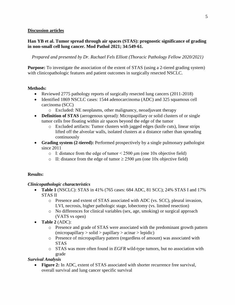
Han YB et al. Tumor spread through air spaces (STAS): prognostic significance of grading
in non-small cell lung cancer. Mod Pathol 2021; 34:549-61.
Prepared and presented by Dr. Rachael Fels Elliott (Thoracic Pathology Fellow 2020/2021)
Purpose: To investigate the association of the extent of STAS (using a 2-tiered grading system)
with clinicopathologic features and patient outcomes in surgically resected NSCLC.
Methods:
Reviewed 2775 pathology reports of surgically resected lung cancers (2011-2018)
Identified 1869 NSCLC cases: 1544 adenocarcinoma (ADC) and 325 squamous cell
carcinoma (SCC)
o Excluded: NE neoplasms, other malignancy, neoadjuvant therapy
Definition of STAS (aerogenous spread): Micropapillary or solid clusters of or single
tumor cells free floating within air spaces beyond the edge of the tumor
o Excluded artifacts: Tumor clusters with jagged edges (knife cuts), linear strips
lifted off the alveolar walls, isolated clusters at a distance rather than spreading
continuously
Grading system (2-tiered): Performed prospectively by a single pulmonary pathologist
since 2011
o I: distance from the edge of tumor < 2500 μm (one 10x objective field)
o II: distance from the edge of tumor 2500 μm (one 10x objective field)
Results:
Clinicopathologic characteristics
Table 1 (NSCLC): STAS in 41% (765 cases: 684 ADC, 81 SCC); 24% STAS I and 17%
STAS II
o Presence and extent of STAS associated with ADC (vs. SCC), pleural invasion,
LVI, necrosis, higher pathologic stage, lobectomy (vs. limited resection)
o No differences for clinical variables (sex, age, smoking) or surgical approach
(VATS vs open)
Table 2 (ADC):
o Presence and grade of STAS were associated with the predominant growth pattern
(micropapillary > solid > papillary > acinar > lepidic)
o Presence of micropapillary pattern (regardless of amount) was associated with
STAS
o STAS was more often found in EGFR wild-type tumors, but no association with
grade
Survival Analysis
Figure 2: In ADC, extent of STAS associated with shorter recurrence free survival,
overall survival and lung cancer specific survival
Page 6
6
o 5-year RFS: No STAS (91.8%), STAS1 (79%), STAS 2 (60.5%)
o 5-year OS: No STAS (95%), STAS1 (88%), STAS 2 (74%)
No differences in survival for SCC
Subgroup analysis: Stage IA non-mucinous ADC
o STAS II associated with shorter recurrence free survival and lung cancer specific
survival
o STAS II was an independent risk factor for recurrence in limited and radical
resection
o STAS I had no bearing on recurrence in multivariate analysis
Take-home message: STAS II was an independent poor prognostic factor in low stage non-
mucinous ADC, regardless of the extent of resection. This data suggests that including presence
and grade of STAS in pathology reports could be useful; however, large-scale, multi-institutional
studies are needed to establish a global standard for grading the extent of STAS.
Page 7
7
Li Y et al. Progression to fibrosing diffuse alveolar damage in a series of 30 minimally
invasive autopsies with COVID-19 pneumonia in Wuhan, China. Histopathology 2021;
78:542-55.
Purpose: Document the evolution of diffuse alveolar damage (DAD) in patients that die of
COVID-19 pneumonia and correlate the progression to fibrosing DAD with patient age, duration
of clinical course, hospitalization, and mechanical ventilation
Methods:
Consecutively reviewed lung tissue and medical records on minimally invasive autopsies
performed at a Wuhan, China, hospital from patients who tested positive for SARS-CoV-
2 or had positive antibodies prior to death
o Minimally invasive autopsy: Ultrasound-guided core needle biopsies performed
on bilateral lungs to obtain at least 4 to 5 tissue samples
Slides reviewed by 3 pathologists
DAD classified as acute, organizing, or fibrosing based upon prominent component, and
other pathologic findings noted (detailed in Supplemental Table 1)
Results:
30 patients with COVID-19 underwent minimally invasive autopsy between February
and March 2020
Table 1 details clinical characteristics, duration of illness, hospitalization, treatment(s),
and days of ventilatory support
o Mean age 69 years (39-91 years)
o 20 males; 10 females
o Most patients had at least 1 significant underlying illness
‒ Hypertension > malignancy > CAD; only 1 with respiratory disease
(COPD)
o Chest CT only available for 1 patient (refer to Supplemental Information)
DAD diagnosed in 28 of 30 cases (93%)
o DAD cause of death in 28 patients; acute pneumonia and gastric cancer in 2
o Most cases showed multiple patterns, but predominant patterns were as follows:
‒ Acute, n = 9
‒ Organizing, n = 7
‒ Fibrosing, n = 12 (refer to Figure 2 for pattern of fibrosis)
Table 2 details clinical correlations with predominant DAD patterns
o Fibrosing DAD patients on average younger and ventilated longer than patients
with acute or organizing DAD
o Patients with organizing or fibrosing DAD had longer days of illness and
hospitalization than those with acute DAD
Take-home message: Patients with prolonged hospitalization and ventilation, who die of
COVID-19-related, may develop fibrosing DAD that is morphologically characterized by diffuse
alveolar septal thickening by collagenous fibrosis. This begs the question – Will we see an
increase in fibrosing lung disease as a result of the COVID-19 pandemic?
Page 8
8
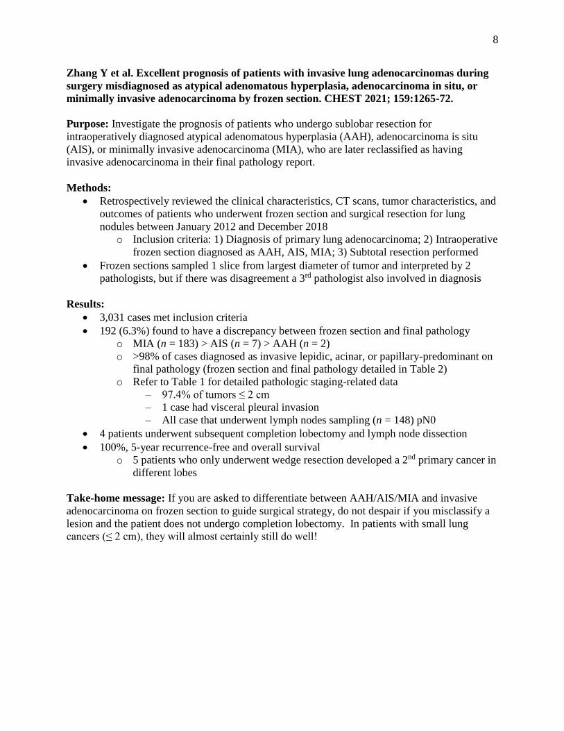
Zhang Y et al. Excellent prognosis of patients with invasive lung adenocarcinomas during
surgery misdiagnosed as atypical adenomatous hyperplasia, adenocarcinoma in situ, or
minimally invasive adenocarcinoma by frozen section. CHEST 2021; 159:1265-72.
Purpose: Investigate the prognosis of patients who undergo sublobar resection for
intraoperatively diagnosed atypical adenomatous hyperplasia (AAH), adenocarcinoma is situ
(AIS), or minimally invasive adenocarcinoma (MIA), who are later reclassified as having
invasive adenocarcinoma in their final pathology report.
Methods:
Retrospectively reviewed the clinical characteristics, CT scans, tumor characteristics, and
outcomes of patients who underwent frozen section and surgical resection for lung
nodules between January 2012 and December 2018
o Inclusion criteria: 1) Diagnosis of primary lung adenocarcinoma; 2) Intraoperative
frozen section diagnosed as AAH, AIS, MIA; 3) Subtotal resection performed
Frozen sections sampled 1 slice from largest diameter of tumor and interpreted by 2
pathologists, but if there was disagreement a 3rd pathologist also involved in diagnosis
Results:
3,031 cases met inclusion criteria
192 (6.3%) found to have a discrepancy between frozen section and final pathology
o MIA (n = 183) > AIS (n = 7) > AAH (n = 2)
o >98% of cases diagnosed as invasive lepidic, acinar, or papillary-predominant on
final pathology (frozen section and final pathology detailed in Table 2)
o Refer to Table 1 for detailed pathologic staging-related data
‒ 97.4% of tumors ≤ 2 cm
‒ 1 case had visceral pleural invasion
‒ All case that underwent lymph nodes sampling (n = 148) pN0
4 patients underwent subsequent completion lobectomy and lymph node dissection
100%, 5-year recurrence-free and overall survival
o 5 patients who only underwent wedge resection developed a 2nd primary cancer in
different lobes
Take-home message: If you are asked to differentiate between AAH/AIS/MIA and invasive
adenocarcinoma on frozen section to guide surgical strategy, do not despair if you misclassify a
lesion and the patient does not undergo completion lobectomy. In patients with small lung
cancers (≤ 2 cm), they will almost certainly still do well!
Page 9
9
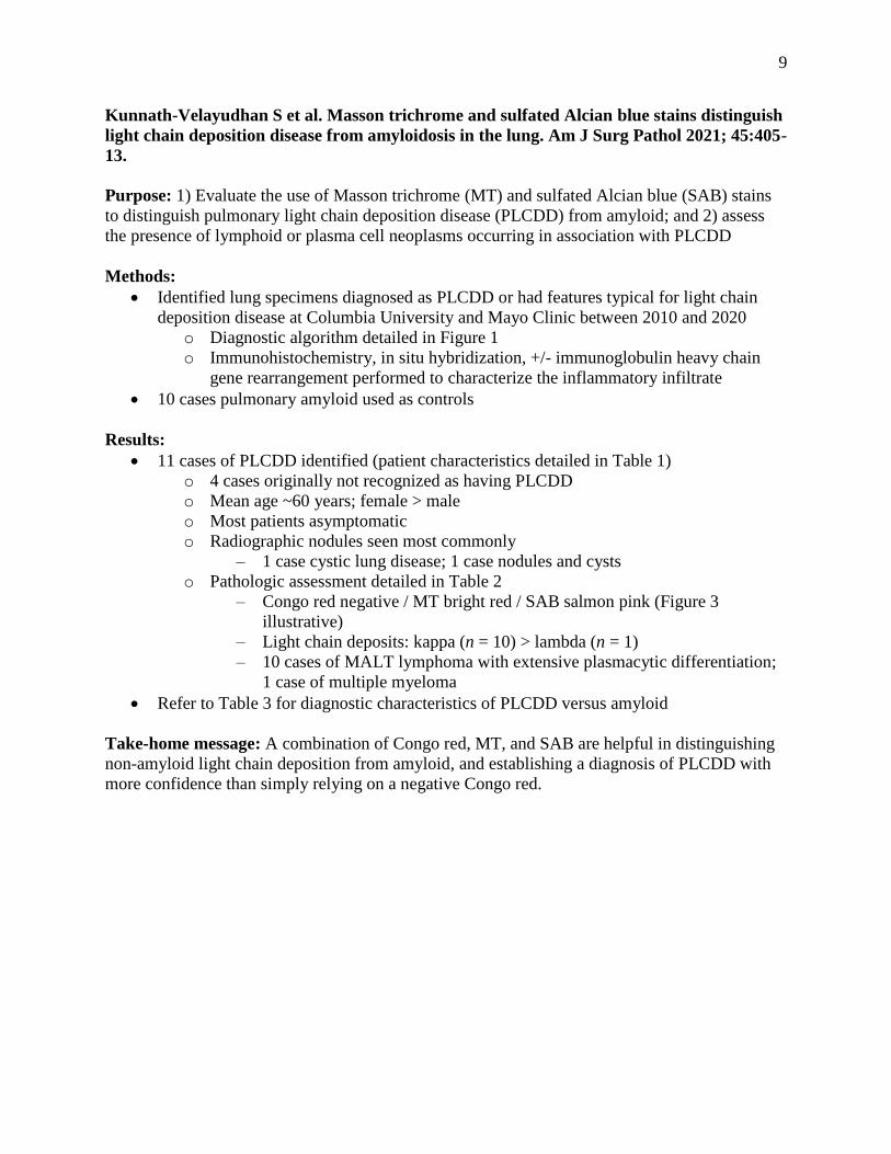
Kunnath-Velayudhan S et al. Masson trichrome and sulfated Alcian blue stains distinguish
light chain deposition disease from amyloidosis in the lung. Am J Surg Pathol 2021; 45:405-
13.
Purpose: 1) Evaluate the use of Masson trichrome (MT) and sulfated Alcian blue (SAB) stains
to distinguish pulmonary light chain deposition disease (PLCDD) from amyloid; and 2) assess
the presence of lymphoid or plasma cell neoplasms occurring in association with PLCDD
Methods:
Identified lung specimens diagnosed as PLCDD or had features typical for light chain
deposition disease at Columbia University and Mayo Clinic between 2010 and 2020
o Diagnostic algorithm detailed in Figure 1
o Immunohistochemistry, in situ hybridization, +/- immunoglobulin heavy chain
gene rearrangement performed to characterize the inflammatory infiltrate
10 cases pulmonary amyloid used as controls
Results:
11 cases of PLCDD identified (patient characteristics detailed in Table 1)
o 4 cases originally not recognized as having PLCDD
o Mean age ~60 years; female > male
o Most patients asymptomatic
o Radiographic nodules seen most commonly
‒ 1 case cystic lung disease; 1 case nodules and cysts
o Pathologic assessment detailed in Table 2
‒ Congo red negative / MT bright red / SAB salmon pink (Figure 3
illustrative)
‒ Light chain deposits: kappa (n = 10) > lambda (n = 1)
‒ 10 cases of MALT lymphoma with extensive plasmacytic differentiation;
1 case of multiple myeloma
Refer to Table 3 for diagnostic characteristics of PLCDD versus amyloid
Take-home message: A combination of Congo red, MT, and SAB are helpful in distinguishing
non-amyloid light chain deposition from amyloid, and establishing a diagnosis of PLCDD with
more confidence than simply relying on a negative Congo red.
Page 10
10
Articles for notation
Neoplastic lung disease
Alidousty C et al. Prevalence and potential biological role of TERT amplifications in ALK
translocated adenocarcinoma of the lung. Histopathology 2021; 78:578-85.
Take-home message: In this study, 109 ALK-translocated lung adenocarcinomas were
screened for TERT amplification using FISH. Five cases (4.6%) showed TERT amplification and
4 of these subsequently underwent genetic further interrogation, which showed genetic
instability. The histologic patterns associated with TERT amplification included papillary (n = 2)
and solid (n = 3) growth patterns.
Amemiya R et al. Prognostic impact of the tumor immune microenvironment in pulmonary
pleomorphic carcinoma. Lung Cancer 2021; 153:56-65.
Take-home message: Immunohistochemical stains for E-cadherin, vimentin, PD-L1,
CA-IX, CD204, Foxp3, CD8, and CD20 were applied to 80 cases of pleomorphic carcinoma to
characterize the immune microenvironment. The aim was to correlate the scores of these
immunostains with patient outcomes. High (≥50%) PD-L1 expression in adenocarcinomas and
large cell carcinomas was associated with longer 5-year overall survival and recurrence-free
survival; however, this did not hold true for squamous cell carcinomas and PD-L1 showed no
prognostic significance.
Boland JM et al. Molecular genetic landscape of sclerosing pneumocytomas: evidence of
aberrant mTOR pathway signaling and lack of recurrent translocations. Am J Clin Pathol
2021; 155:397-404.
Take-home message: In this work from the Mayo Clinic, 10 cases of sclerosing
pneumocytoma from 8 patients (1 with two tumors; 1 with multiple, bilateral tumors) underwent
next-generation sequencing and RNA-seq. Mutations in AKT were identified in 7 of 9 specimens
and 1 had mutations in both PTEN and PIK3R1, suggesting that abnormal activation of
PIK3/AKT/mTOR pathway is a consistent oncologic events in sclerosing pneumocytomas. No
recurrent fusion genes were identified.
Chia PL et al. Expression of EGFR and conformational forms of EGFR in malignant
pleural mesothelioma and its impact on survival. Lung Cancer 2021; 153:35-41.
Take-home message: To expand on the title, the aim of this study was to determine the
prevalence of wtEGFR expression and EGFR amplification, including conformational forms, by
immunohistochemistry and FISH, and correlate those findings with survival in 329 tissue
microarrays from patients with malignant mesothelioma. EGFR overexpression was most
common in epithelioid mesothelioma; however, overexpression was not associated with true
EGFR amplification. A conformational form of EGFR was detected in 8.2% of cases, 84.6% of
which were the epithelioid subtype, and seemed to be associated with poorer outcomes; however,
this finding was not statistically significant.
Page 11
11
Hashemi S et al. Surprising impact of stromal TIL’s on immunotherapy efficacy in a real-
world lung cancer study. Lung Cancer 2021; 153:81-9.
Take-home message: Here, the authors retrospectively assessed the impact of various
clinicopathologic variables in patients with metastatic non-small cell lung cancer, who were
treated with immunotherapy agents, nivolumab or pembrolizumab. The entire cohort comprised
366 patients all of whom had PD-L1 testing on their tumors, while 141 of these also had PD-L1
expression reported in the stroma and assessment of the CD8+ infiltrate in the tumor and stroma.
The strongest pathologic predictor of progression free survival and overall survival was the
presence of CD8+ stromal lymphocytes.
Ikeda T et al. The epithelial-mesenchymal transition phenotype is associated with the
frequency of tumor spread through air spaces (STAS) and a high risk of recurrence after
resection of lung carcinoma. Lung Cancer 2021; 153:49-55.
Take-home message: The authors retrospectively assessed 635 lung resections for
adenocarcinoma and squamous cell carcinoma and discovered that STAS was present in 44% of
cases, more commonly seen in adenocarcinomas, particularly those with micropapillary and solid
types, and patients with higher pathologic stage, lymphovascular invasion, and pleural invasion.
Immunohistochemistry for markers of EMT (E-cadherin, vimentin, and β-catenin) were applied
to the tumors to evaluate the associated between an epithelial (E-cadherin+/vimentin-),
intermediate (E-cadherin+/vimentin+ or E-cadherin-/vimentin-, and mesenchymal E-cadherin-
/vimentin+) phenotype and the presence of STAS. STAS was associated with nuclear
translocation of β-catenin and more often observed in tumors with an intermediate or
mesenchymal phenotype than an epithelial phenotype, and the mesenchymal phenotype was
found to be an independent predictor of high risk for recurrence.
Li R et al. Pseudo-small cell transformation in EGFR-mutant adenocarcinoma. Lung
Cancer 2021; 153:120-5.
Take-home message: For purposes of this study, small cell transformation is defined by
the authors as patients originally diagnosed as EGFR-mutant adenocarcinoma on lung biopsy or
resection, who went on to have small cell carcinomas identified on re-biopsy of the lung or
biopsies of distant metastases, indicating that the tumor transformed to small cell carcinoma.
Fourteen cases of “small cell transformation” were identified, including 1 case reported as
combined adenocarcinoma and small cell carcinoma on initial biopsy; however, re-review of the
initial biopsies revealed previously not identified small cell components in the majority of these
cases (up to 90%) and those cases were re-categorized as “pseudo-small cell transformation.”
The authors also found that mutated EGFR expression by immunohistochemistry was limited to
the adenocarcinoma component, leading them to conclude that only the adenocarcinoma
component would respond to EGFR tyrosine kinase inhibitors.
Suzuki J et al. Relationship between podoplanin-expressing cancer-associated fibroblasts
and the immune microenvironment of early lung squamous cell carcinoma. Lung Cancer
2021; 153:1-10.
Page 12
12
Take-home message: The authors investigated the relationship between podoplanin-
expressing tumor-associated fibroblasts and the tumor microenvironment in stage I lung
squamous cell carcinoma through gene expression profiles and the use of immunohistochemistry.
In vitro studies of podoplanin-high squamous cell carcinoma fibroblasts showed higher secretion
of immunosuppressive cytokines. Patient sample testing also seemed to suggest that podoplanin-
expression resulted in an immunosuppressive tumor microenvironment.
Takagi H et al. Delta-like 1 homolog (DLK1) as a possible therapeutic target and its
application to radioimmunotherapy using 125I-labelled anti-DLK1 antibody in lung cancer
models (HOT1801 and FIGHT004). Lung Cancer 2021; 153:134-42.
Take-home message: The expression of DLK1 was evaluated in 112 cases of small cell
lung carcinoma (SCLC) and 101 cases of non-small cell lung carcinoma (NSCLC) and examined
to learn if there is prognostic significance. Twenty-one percent of SCLC and 17% of NSCLC
expressed DLK1; however, expression was only associated with lymph node metastases in
NSCLC and therefore perhaps not surprisingly poorer recurrence-free survival, but was
otherwise of no special significance. The authors further showed that DLK1 might serve as a
novel therapeutic target for immunotherapy using in vitro and animal models.
Non-neoplastic lung disease
Barisione E et al. Fibrotic progression and radiologic correlation in matched lung samples
from COVID-19 post-mortems. Virchows Arch 2021; 478:471-85.
Take-home message: In this Italian study, 8 patients who were ventilated and SARS-
CoV2-positive, underwent cyrobiopsy immediately after death with the aim of correlating
histologic findings with duration of illness and radiographic findings. Two patients with
groundglass opacities had early diffuse alveolar damage (DAD) and positive SARS-CoV-2
immunostains. The remaining 6 patients had proliferative (n = 3) or fibrotic (n = 3) DAD,
negative immunostains, and died 24 to 42 days after symptom onset; however, I suspect that at
least 1 of the cases of fibrotic DAD was potentially a patient with underlying UIP - - This patient
was a 75-year-old male with lower lobe honeycombing on chest CT and microscopic honeycomb
change on biopsy.
Gallob F et al. Senescence and autophagy in usual interstitial pneumonia. Virchows Arch
2021; 478:479-506.
Take-home message: As the title implies, the aim of this study was to evaluate the
presence or absence of senescence and autophagy in usual interstitial pneumonia (UIP) through
the use of immunohistochemistry. The cohort included 23 patient samples, including 10 cases of
idiopathic pulmonary fibrosis (IPF) and 13 cases of connective tissue disease-associated UIP
(CTD-UIP). There was no difference in the staining patterns between IPF and CTD-UIP cases
with both groups showing expression of cell cycle arrest (senescence) markers and upregulation
of autophagy markers, the latter being particularly prominent in fibroblastic foci.
Page 13
13
McMullen PD et al. A descriptive and quantitative immunohistochemical study
demonstrating a spectrum of platelet recruitment patterns across pulmonary infections
including COVID-19. Am J Clin Pathol 2021; 155:354-63.
Take-home message: CD61 immunohistochemical staining was quantitated in lung
tissue obtained from autopsy cases (n = 27) to assess platelet deposition. The cohort comprised 3
cases with histologically normal lungs, 4 cases of DAD without primary lung infections, 9
patients with COVID-19, 2 with influenza, 4 each with different types of bacterial and invasive
fungal pneumonias, and 1 with bilateral pulmonary thromboembolic disease. Infectious causes
of lung disease resulted in increased CD61 expression compared to controls and the level of
staining in COVID-19 was similar to that seen in non-infectious causes of DAD.
Valdivia-Mazeyra MF et al. Increased number of pulmonary megakaryocytes in COVID-
19 patients with diffuse alveolar damage: an autopsy study with clinical correlation and
review of the literature. Virchows Arch 2021; 478:487-96.
Take-home message: In this observational study, the authors compare the number of
megakaryocytes seen in lung tissue from COVID-19 patients who underwent autopsy (n = 18) to
a control group comprised of patients with diffuse alveolar damage (DAD) who died of other
causes (n = 14) and a second control group of histologically normal lung tissue obtained from
lobectomy specimens performed for tumor resection (n = 14). The number of megakaryocytes
counted in 25 HPF in COVID-19 patients was elevated (7.61 ± 5.59) compared to non-COVID-
19 DAD (4 ± 4.17) and normal (1.14 ± 0.86) controls. The authors propose that the increase in
pulmonary megakaryocytes may be implicated in the thrombotic events seen in patients with
severe COVID-19.