Regular paper Purification and characterization of an antibacterial protein from dried fruiting bodies of the wild mushroom Clitocybe sinopica Suyue Zheng 1,2 , Qinghong Liu 1 , Guoqing Zhang 1 , Hexiang Wang 1and Tzi Bun Ng 31 State Key Laboratory for Agrobiotechnology and Department of Microbiology, China Agricultural University, Beijing, China; 2 Hebei Engineering University, Handan, China; 3 School of Biomedical Sciences, Faculty of Medicine, The Chinese University of Hong Kong, Shatin, New Territories, Hong Kong, China A novel antibacterial protein with a molecular mass of 44 kDa has been isolated from dried fruiting bodies of the wild mushroom Clitocybe sinopica. Sodium dodecyl sulfate/polyacrylamide gel electrophoresis showed that the protein was composed of two subunits each with a molecular mass of 22 kDa. Its N-terminal amino-acid se- quence, SVQATVNGDKML, has not been reported for other antimicrobial proteins. The purification protocol included ion exchange chromatography on DEAE-cellu- lose, CM-cellulose and Q-Sepharose, and gel filtration by fast protein liquid chromatography on Superdex 75. The antibacterial protein was adsorbed on all three ion exchangers. The antimicrobial activity profile of the pro- tein against tested bacterial and fungal strains disclosed that it possessed potent antibacterial activity against Agrobacterium rhizogenes, A. tumefaciens, A. vitis, Xan- thomonas oryzae and X. malvacearum with a minimum inhibitory concentration mostly below 0.6 μM. However, it had no antibacterial activity against Pseudomonas batatae, Erwinia herbicola, Escherichia coli, and Staphy- lococcus aureus, and no antifungal activity against Set- osphaeria turcica, Fusarium oxysporum, Verticillium dahl- iae, Bipolaris maydis, and B. sativum. The antibacterial antivity against A. tumefaciens was stable after exposure to 20–60 °C for 30 min and to pH 4–9 for 1 h. Keywords: mushroom, fruiting bodies, antibacterial protein, Clito- cybe sinopica Received: 01 August, 2009; revised: 12 January, 2010; accepted: 31 January, 2010; available on-line: 02 March, 2010 INTRODUCTION Mushrooms have long been appreciated for their fla- vor and texture. They need to produce antibacterial and antifungal compounds in order to survive in their natural environment. It is therefore not surprising that antimicrobial compounds could be isolated from many mushrooms (Lindequist et al., 2005). Antimicrobial pro- teins (AMPs), as important antibacterial and antifungal compounds, have attracted attention of a large number of investigators. AMPs are largely distributed among living organisms including plants, animals, fungi and some single-celled microorganisms (Jenssen, 2005). AMPs, which directly interfere with the growth, multiplication and spread of microbial organisms and permit plants and animals to resist infection by environmental microbes, represent important components of the defense system (Lehrer & Ganz, 1999). To date, different proteins with anti- bacterial or antifungal activity have been reported. Most of these proteins were isolated from animals (Steiner et al., 1981; Lemaître et al., 1996; Destoumieux et al., 1997; Krishnakumari & Nagaraj, 1997; Torres-Larios et al., 2000), plants (Huynh et al., 1992; Cammue et al., 1995; Talas, 2004) and bacteria (Barja et al., 1989; James et al., 1996; Longeon et al., 2004). Only few of them came from fungi (Mygind et al., 2005; Hu et al., 2006), espe- cially macrofungi. Antifungal peptides and proteins have been isolated from the fungi Aspergillus giganteus (Lacadena et al., 1995), A. niger (Lee et al., 1999), Zygosaccharomyces bailii (Weiler et al., 2003), Hypsizigus marmoreus (Lam & Ng, 2001b), Pleu- rotus erygii (Wang & Ng, 2004), Lyophyllum shimeji (Lam & Ng, 2001a), and Tricholoma giganteum (Guo et al., 2005). However, the literature pertaining to antibacterial pep- tides and proteins is scanty. Clitocybe sinopica is a well-known wild edible mushroom species found in China. To the best of our knowledge, no research has been reported on the chemical composi- tion and biological activities of C. sinopica extract. There- fore, the aim of the present work was to evaluate the antimicrobial potential of this mushroom extract on sev- eral phytopathogenic microorganisms. MATERIALS AND METHODS Materials. The fruiting bodies of the wild mushroom Clitocybe sinopica were collected from Heilongjiang prov- ince, in the northeast region of China. Fifteen bacterial pathogenic strains were used in this study: Agrobacterium rhizogenes (NL24-2, NL5-4, MG 12-1, HBT 6-1, SX073, NL12-2) A. vitis (MI3-2, MI23-1), A. tumefaciens (MG10- 1), Xanthomonas oryzae, X. malvacearum, Pseudomonas batatae, Erwinia herbicola, Escherichia coli and Staphylococcus aureus. Five fungal strains were also tested: Setosphaeria turcica, Bi- polaris maydis, Fusarium oxysporum, Verticillium dahliae and Bipolaris sativum. All tested bacterial and fungal strains were obtained from the Department of Plant Pathology of the China Agriculture University (Beijing, China PR). Isolation procedure. Dried fruiting bodies of C. sinopica were homogenized in 0.15 M NaCl at 4 °C overnight. The supernatant was centrifuged at 8000 × g e-mail: Tzi Bun Ng: [email protected]; Hexiang Wang: [email protected]Abbreviations: AMP, antimicrobial proteins; cfu, colony-forming unit; FPLC, fast protein liquid chromatography; MIC, minimum in- hibitory concentration. Vol. 57, No 1/2010 43–48 on-line at: www.actabp.pl
Transcript
Regular paper
Purification and characterization of an antibacterial protein from dried fruiting bodies of the wild mushroom Clitocybe sinopicaSuyue Zheng1,2, Qinghong Liu1, Guoqing Zhang1, Hexiang Wang1 and Tzi Bun Ng3
1State Key Laboratory for Agrobiotechnology and Department of Microbiology, China Agricultural University, Beijing, China; 2Hebei Engineering University, Handan, China; 3School of Biomedical Sciences, Faculty of Medicine, The Chinese University of Hong Kong, Shatin, New Territories, Hong Kong, China
A novel antibacterial protein with a molecular mass of 44 kDa has been isolated from dried fruiting bodies of the wild mushroom Clitocybe sinopica. Sodium dodecyl sulfate/polyacrylamide gel electrophoresis showed that the protein was composed of two subunits each with a molecular mass of 22 kDa. Its N-terminal amino-acid se-quence, SVQATVNGDKML, has not been reported for other antimicrobial proteins. The purification protocol included ion exchange chromatography on DEAE-cellu-lose, CM-cellulose and Q-Sepharose, and gel filtration by fast protein liquid chromatography on Superdex 75. The antibacterial protein was adsorbed on all three ion exchangers. The antimicrobial activity profile of the pro-tein against tested bacterial and fungal strains disclosed that it possessed potent antibacterial activity against Agrobacterium rhizogenes, A. tumefaciens, A. vitis, Xan-thomonas oryzae and X. malvacearum with a minimum inhibitory concentration mostly below 0.6 μM. However, it had no antibacterial activity against Pseudomonas batatae, Erwinia herbicola, Escherichia coli, and Staphy-lococcus aureus, and no antifungal activity against Set-osphaeria turcica, Fusarium oxysporum, Verticillium dahl-iae, Bipolaris maydis, and B. sativum. The antibacterial antivity against A. tumefaciens was stable after exposure to 20–60 °C for 30 min and to pH 4–9 for 1 h.
Mushrooms have long been appreciated for their fla-vor and texture. They need to produce antibacterial and antifungal compounds in order to survive in their natural environment. It is therefore not surprising that antimicrobial compounds could be isolated from many mushrooms (Lindequist et al., 2005). Antimicrobial pro-teins (AMPs), as important antibacterial and antifungal compounds, have attracted attention of a large number of investigators.
AMPs are largely distributed among living organisms including plants, animals, fungi and some single-celled microorganisms (Jenssen, 2005). AMPs, which directly interfere with the growth, multiplication and spread of microbial organisms and permit plants and animals to resist infection by environmental microbes, represent important components of the defense system (Lehrer
& Ganz, 1999). To date, different proteins with anti-bacterial or antifungal activity have been reported. Most of these proteins were isolated from animals (Steiner et al., 1981; Lemaître et al., 1996; Destoumieux et al., 1997; Krishnakumari & Nagaraj, 1997; Torres-Larios et al., 2000), plants (Huynh et al., 1992; Cammue et al., 1995; Talas, 2004) and bacteria (Barja et al., 1989; James et al., 1996; Longeon et al., 2004). Only few of them came from fungi (Mygind et al., 2005; Hu et al., 2006), espe-cially macrofungi.
Antifungal peptides and proteins have been isolated from the fungi Aspergillus giganteus (Lacadena et al., 1995), A. niger (Lee et al., 1999), Zygosaccharomyces bailii (Weiler et al., 2003), Hypsizigus marmoreus (Lam & Ng, 2001b), Pleu-rotus erygii (Wang & Ng, 2004), Lyophyllum shimeji (Lam & Ng, 2001a), and Tricholoma giganteum (Guo et al., 2005). However, the literature pertaining to antibacterial pep-tides and proteins is scanty.
Clitocybe sinopica is a well-known wild edible mushroom species found in China. To the best of our knowledge, no research has been reported on the chemical composi-tion and biological activities of C. sinopica extract. There-fore, the aim of the present work was to evaluate the antimicrobial potential of this mushroom extract on sev-eral phytopathogenic microorganisms.
MATErIALS AND METHoDS
Materials. The fruiting bodies of the wild mushroom Clitocybe sinopica were collected from Heilongjiang prov-ince, in the northeast region of China. Fifteen bacterial pathogenic strains were used in this study: Agrobacterium rhizogenes (NL24-2, NL5-4, MG 12-1, HBT 6-1, SX073, NL12-2) A. vitis (MI3-2, MI23-1), A. tumefaciens (MG10-1), Xanthomonas oryzae, X. malvacearum, Pseudomonas batatae, Erwinia herbicola, Escherichia coli and Staphylococcus aureus. Five fungal strains were also tested: Setosphaeria turcica, Bi-polaris maydis, Fusarium oxysporum, Verticillium dahliae and Bipolaris sativum. All tested bacterial and fungal strains were obtained from the Department of Plant Pathology of the China Agriculture University (Beijing, China PR).
Isolation procedure. Dried fruiting bodies of C. sinopica were homogenized in 0.15 M NaCl at 4 °C overnight. The supernatant was centrifuged at 8000 × g
e-mail: Tzi Bun Ng: [email protected]; Hexiang Wang: [email protected]: AMP, antimicrobial proteins; cfu, colony-forming unit; FPLC, fast protein liquid chromatography; MIC, minimum in-hibitory concentration.
Vol. 57, No 1/201043–48
on-line at: www.actabp.pl
S. Zheng and others44 2010
for 15 min before (NH4)2SO4 was added to 80% satu-ration. The precipitate was collected by centrifugation at 8 000 × g for 15 min again. Then the precipitate was dissolved and dialyzed to remove (NH4)2SO4 before ion exchange chromatography on a DEAE-cellulose (Sigma) column (2.5 cm × 20 cm) in 10 mM sodium phosphate buffer (pH 7.5). Unadsorbed proteins (frac-tion D1) were eluted with the buffer while bound ones were desorbed sequentially with 100 mM NaCl, 200 mM NaCl and 1 M NaCl to form fractions D2, D3 and D4, respectively. Fraction D3 with antibacte-rial activity was dialyzed and then chromatographed on a column of CM-cellulose (Sigma) (1.5 × 20 cm) in 10 mM sodium phosphate buffer (pH 6.2). After elution of unadsorbed proteins (CM1) with the same buffer, the bound fractions CM2, CM3 and CM4 were eluted with the same buffer containing 50 mM NaCl, 150 mM NaCl and 1 M NaCl, respectively. The ac-tive peak (CM3) was then subjected to ion exchange chromatography on a column of Q-Sepharose (GE Healthcare) (1 cm × 10 cm) in 10 mM sodium phos-phate buffer (pH 6.5). After removal of unadsorbed protein (fraction Q1), the bound material was eluted with a linear NaCl concentration gradient (0–500 mM) in the same buffer. The active fraction (Q2) was then gel-filtered on a Superdex 75 HR 10/30 column (GE Healthcare) by fast protein liquid chromatography (FPLC) in 0.2 M NH4HCO3 buffer (pH 8.5) using an AKTA Purifier (GE Healthcare). Two peaks were ob-tained. The first peak (SU1) with antibacterial activ-ity was subjected to sodium dodecyl sulfate-polyacr-ylamide gel electrophoresis (SDS/PAGE) which was conducted according to the method of Laemmli and Favre (1973). N-terminal sequencing was carried out using a Hewlett-Packard (HP) Edman degradation unit and an HP 1000 HPLC System.
Assay of antibacterial activity. The assay for anti-bacterial activity was conducted using sterile Petri plates (90 mm × 15 mm) containing 15 ml suitable medium agar (1.8% agar). Five milliliters of warm nutrient agar (0.7 %) containing the bacterium were poured into the plates. A sterile blank paper disk (6.25 mm in diameter) or Oxford cup was placed on the agar. The antibacterial protein (40 μl of a 1 mg/ml solution) in 10 mM sodium phos-phate buffer (pH 7.0) was added to one of the disks. Only sterile buffer was added to the control disk. The plate was incubated at 28 °C for 24 h. A transparent ring around the paper disk signifies antibacterial activity (Lam et al., 2000).
Assay of minimum inhibitory concentration (MIC). The MIC for each bacterial strain tested was measured by the liquid culture medium dilution method using Luria Bertani or modified 523 medium, depending on the strain. Nine hundred milliliters of medium were placed in a sterilized test tube to which 90 μl of the tested sample and 10 μl of the cultured bacterial solu-tion (final bacterial count of 1 × 10⁶ cfu/ml) were added. The tube was cultured under suitable conditions. The minimum concentration of added samples in which no bacterial growth was observed was defined as the MIC (Hirasawa et al., 1999).
Assay of antifungal activity. The assay was conduct-ed as detailed by Wang et al. (2004).
Test for thermostability and pH stability. The antibacterial protein was exposed to various tempera-tures ranging from 20 °C to 80 °C at 10 °C intervals for 30 min. After heat treatment, samples were centrifuged (12 000 × g, 5 min) and the supernatants were used for assay of antibacterial activity. The antibacterial activity of
each sample was compared with that of a control treated at 4 °C for 30 min.
The test for pH stability was conducted as described above. The antibacterial protein was treated at various pH values from 3 to 9 for 1 h.
Determination of ribonuclease activity. The activ-ity of the antibacterial protein toward yeast tRNA was determined as described by Wang and Ng (1999).
Determination of haemagglutinating activity. The assay was conducted as described by Wang et al. (1996).
rESuLTS
Protein purification
Antibacterial activity was located mainly in fraction D3 adsorbed on DEAE-cellulose which was eluted with 0.2 M NaCl. The larger unadsorbed fraction D1 and other adsorbed fractions were devoid of antibacte-rial activity. D3 was fractionated on CM-cellulose into an unadsorbed fraction CM1 and three adsorbed frac-tions CM2, CM3, and CM4. Antibacterial activity resided in fraction CM3. Fraction CM3 was collected for fur-ther purification on a Q-Sepharose column. The activ-ity was eluted with a linear gradient of 0–0.5 M NaCl in 10 mM phosphate buffer (pH 6.5). Two peaks were obtained. Antibacterial activity was detected in fraction Q2 (Fig. 1). Fraction Q2 was resolved on Superdex 75 into two fractions, SU1 and SU2 (Fig. 2). The first and larger fraction, SU1, was enriched in antibacterial activity. It demonstrated a molecular mass of 44 kDa in gel filtration (Fig. 2) and 22 kDa in SDS/PAGE (Fig. 3). Thus the antibacterial protein is most likely composed of two subunits with the same molecular mass of 22 kDa. The yields of the various chromatographic fractions are presented in Table 1. The N-terminal sequence of the protein, SVQATVNGDKML, is not found in any re-ported antimicrobial protein. It is dissimilar to those of previously isolated mushroom antimicrobial proteins (Ta-ble 2).
Assays of antimicrobial and other activities
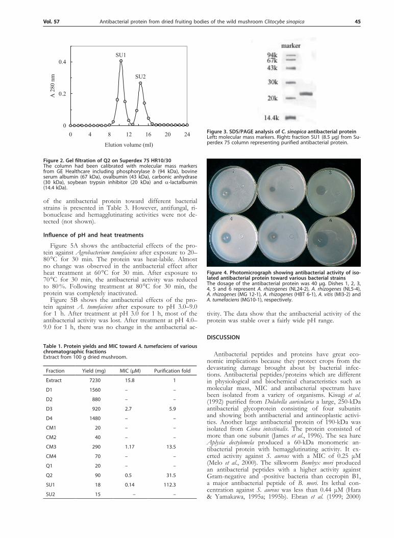
The antimicrobial activity of the isolated protein was tested against different bacterial and fungal strains. The antimicrobial activity was tested at the dose of 40 μg, it showed potent antimicrobial activity against most bacte-ria (Fig. 4). The minimal inhibitory concentration (MIC)
0
0.2
0.4
0.6
0 30 60 90 120
Elution volume (ml)
A28
0 nm
0
0.1
0.2
0.3
0.4
0.5
Q1
Q2
NaC
l (M
)
Figure 1. Cation exchange chromatography of fraction CM3 on Q-SepharoseStarting buffer: 10 mM sodium phosphate buffer (pH 6.5).
Antibacterial protein from dried fruiting bodies of the wild mushroom Clitocybe sinopicaVol. 57 45Vol. 57 45
of the antibacterial protein toward different bacterial strains is presented in Table 3. However, antifungal, ri-bonuclease and hemagglutinating activities were not de-tected (not shown).
Influence of pH and heat treatments
Figure 5A shows the antibacterial effects of the pro-tein against Agrobacterium tumefaciens after exposure to 20–80 °C for 30 min. The protein was heat-labile. Almost no change was observed in the antibacterial effect after heat treatment at 60 °C for 30 min. After exposure to 70 °C for 30 min, the antibacterial activity was reduced to 80 %. Following treatment at 80 °C for 30 min, the protein was completely inactivated.
Figure 5B shows the antibacterial effects of the pro-tein against A. tumefaciens after exposure to pH 3.0–9.0 for 1 h. After treatment at pH 3.0 for 1 h, most of the antibacterial activity was lost. After treatment at pH 4.0–9.0 for 1 h, there was no change in the antibacterial ac-
tivity. The data show that the antibacterial activity of the protein was stable over a fairly wide pH range.
DISCuSSIoN
Antibacterial peptides and proteins have great eco-nomic implications because they protect crops from the devastating damage brought about by bacterial infec-tions. Antibacterial peptides/proteins which are different in physiological and biochemical characteristics such as molecular mass, MIC and antibacterial spectrum have been isolated from a variety of organisms. Kisugi et al. (1992) purified from Dolabella auricularia a large, 250-kDa antibacterial glycoprotein consisting of four subunits and showing both antibacterial and antineoplastic activi-ties. Another large antibacterial protein of 190-kDa was isolated from Ciona intestinalis. The protein consisted of more than one subunit (James et al., 1996). The sea hare Aplysia dectylomela produced a 60-kDa monomeric an-tibacterial protein with hemagglutinating activity. It ex-erted activity against S. aureus with a MIC of 0.25 μM (Melo et al., 2000). The silkworm Bombyx mori produced an antibacterial peptides with a higher activity against Gram-negative and -positive bacteria than cecropin B1, a major antibacterial peptide of B. mori. Its lethal con-centration against S. aureus was less than 0.44 μM (Hara & Yamakawa, 1995a; 1995b). Ebran et al. (1999; 2000)
0
0.2
0.4
0 4 8 12 16 20 24
Elution volume (ml)
A 2
80 n
m SU2
SU1
Figure 2. Gel filtration of Q2 on Superdex 75 Hr10/30The column had been calibrated with molecular mass markers from GE Healthcare including phosphorylase b (94 kDa), bovine serum albumin (67 kDa), ovalbumin (43 kDa), carbonic anhydrase (30 kDa), soybean trypsin inhibitor (20 kDa) and a-lactalbumin (14.4 kDa).
Figure 3. SDS/PAGE analysis of C. sinopica antibacterial proteinLeft: molecular mass markers. Right: fraction SU1 (8.5 μg) from Su-perdex 75 column representing purified antibacterial protein.
Table 1. Protein yields and MIC toward A. tumefaciens of various chromatographic fractions Extract from 100 g dried mushroom.
Fraction Yield (mg) MIC (μM) Purification fold
Extract 7230 15.8 1
D1 1560 – –
D2 880 – –
D3 920 2.7 5.9
D4 1480 – –
CM1 20 – –
CM2 40 – –
CM3 290 1.17 13.5
CM4 70 – –
Q1 20 – –
Q2 90 0.5 31.5
SU1 18 0.14 112.3
SU2 15 – –
Figure 4. Photomicrograph showing antibacterial activity of iso-lated antibacterial protein toward various bacterial strainsThe dosage of the antibacterial protein was 40 μg. Dishes 1, 2, 3, 4, 5 and 6 represent A. rhizogenes (NL24-2), A. rhizogenes (NL5-4), A. rhizogenes (MG 12-1), A. rhizogenes (HBT 6-1), A. vitis (MI3-2) and A. tumefaciens (MG10-1), respectively.
S. Zheng and others46 2010
reported antibacterial proteins with a molecular mass of 27-kDa and 31-kDa and activity against P. aerugino-sa, P. fluorescens, Aeromonas hyarophila and S. aureus from carp mucus. Their MIC ranged from 0.16 μM to 4.8 μM against S. aureus. The bullfrog produced a 1865-Da peptide and another 3691-Da one both with antibacte-rial activity. They exerted antibacterial activity against B. subtilis and E. coli with a MIC of 1.6–10.7 μM (Minn et al., 1998). A 35-kDa antibacterial protein with a MIC of 14.3 μM to 28.6 μM was isolated from Cordyceps sinensis (Hu et al., 2006).
In this study, we describe the isolation and properties of an antibacterial protein with a molecular mass of 44 kDa from dried fruiting bodies of the wild mushroom C. sinopica. This protein displayed a strong antibacterial
activity on Agrobacterium spp. and Xanthomonas spp., but there was no obvious inhibitory effect against fungi. A comparison of the N-terminal sequence and the molecu-lar mass of the C. sinopica antibacterial protein with the aforementioned antibacterial proteins reveals that they are distinctly different (Table 3). So it can be regarded as a novel antibacterial protein.
The behavior of the C. sinopica protein on the vari-ous ion exchange chromatographic media employed in the isolation procedure was different from that reported for other antimicrobial proteins. It was adsorbed on DE-AE-cellulose whereas other antimicrobial proteins were not (Lam & Ng, 2001a; Wang & Ng, 2004; Wang et al., 2004; Guo et al., 2005), but they were all adsorbed on CM-cellulose.
Table 2. Comparison of N-terminal sequence of C. sinopica antibacterial protein with those of other antimicrobial proteins
Name Source N-terminal sequence Da ReferenceA
ntib
acte
rial p
rote
inC. sinopica SVQATVNGDKML 44 000 This study
Marine bacterium MNLKIHPSVGAXLGNRQM 90 000 Barja et al., 1989
Cordyceps sinensis ALATQHGAP 35 000 Hu et al., 2006
Bullfrog GVVKVSLRKGESLRARL 1 865 Minn et al., 1998
Bullfrog IIKVPLKKFKSMRGVMRDHGIKAPVV 3 691 Minn et al., 1998
Scylla serrata GQALNKLMPKIVSAIIYMVG 10 800 Huang et al., 2006
Figure 5. Effect of heat and pH treatments on the antibacterial activity of isolated antibacterial protein against A. tumefaciens(A) Heat treatments (30 min). A 20 μg sample of the protein was applied to each paper disk in sterile water. After treatment at 20 °C, 30 °C, 40 °C, 50–60 °C, 70 °C and 80 °C for 30 min (A–G), (CK) represents control incubated at 4 °C for 30 min. (B) pH treatment (1 h). A 20 μg sample of the protein was applied to each paper disk in sterile water. After treatment at pH 3.0, 4.0, 5.0, 6.0, 7.0, 8.0 and 9.0 for 1 h (A–G), (CK) represents control of sterile water.
Antibacterial protein from dried fruiting bodies of the wild mushroom Clitocybe sinopicaVol. 57 47
The C. sinopica antibacterial protein was devoid of ri-bonuclease or lectin activities. This is important in view of the reports that some ribonucleases (Ng & Wang, 2000; Wang & Ng, 2000) and lectins (Broekaert et al., 1989; Gozia et al., 1995; Ye et al., 2001) exhibit antimi-crobial activity.
In conclusion, our research demonstrated that C. si-nopica fruiting bodies produce an antibacterial protein that shows a broad spectrum of activity against a number of plant pathogenic bacteria, including strains of the genera Agrobacterium and Xanthomonas. Its high potency and the broad spectrum of antibacterial activity suggest that the expression of its gene in transgenic plants may confer protection against bacterial pathogens.
Further work aims at the elucidation of the mecha-nism of action and cloning of the gene encoding the an-tibacterial protein for possible pratical application in the future.
Acknowledgements
This work was financially supported by National Grants of China (nyhyzx07-008, 2007BAD89B00 and 2010CB732202).
rEFErENCES
Barja JL, Lemos ML, Toranzo A (1989) Purification and characteriza-tion of an antibacterial substance produced by a marine Alteromonas species. Antimicrob Agents Chemother 33: 1674–1679.
Broekaert WF, Van Parijs J, Leyns F, Joos H, Peumans WJ (1989) A chitin-binding lectin from stinging nettle rhizomes with antifungal properties. Science 245: 1100–1102.
Cammue BPA, Thevissen K, Hendriks M, Eggermont K, Goderis IJ, Proost P, Damme JV, Osborn RW, Guerbette F, Kader JC, Broekaert WF (1995) A potent antimicrobial protein from onion seeds showing sequence homology to plant lipid transfer proteins. Plant Physiol 109: 445–455.
Destoumieux D, Bulet P, Loew D, Dorsselaer AV, Rodriguez J, Bachère E (1997) Penaeidins, a new family of antimicrobial peptides isolated from the shrimp Penaeus vannamei (Decapoda). J Biol Chem 272: 398–406.
Ebran N, Julien S, Orange N, Saglio P, Lemaitra C, Molle G (1999) Pore-forming properties and antibacterial activity of proteins ex-tracted from epidermal mucus of fish. Comp Biochem Physiol Part A 122: 181–189.
Ebran N, Julien S, Orange N, Auperin B, Molle G (2000) Isolation and characterization of novel glycoproteins from fish epidermal mu-cus: correlation between their pore-forming properties and their an-tibacterial activities. Biochim Biophys Acta 1467: 271–280.
Gozia O, Ciopraga J, Bentia T, Lungu M, Zamfirescu I, Tudor R, Ro-seamu A, Nitu F (1995) Antifungal properties of lectin and new chitinase from potato tuber. FEBS Lett 370: 245–249.
Guo YX, Wang HX, Ng TB (2005) Isolation of trichogin, an antifun-gal protein from fresh fruiting bodies of the edible mushroom Tri-choloma giganteum. Peptides 26: 575–580.
Hara S, Yamakawa M (1995a) A novel antibacterial peptide family iso-lated from the silkworm Bombyx mori. Biochem J 310: 651–656.
Hara S, Yamakawa M (1995b) Moricin, a novel type of antibacterial peptide isolated from the silkworm Bombyx mori. J Biol Chem 270: 29923–29927.
Hirasawa M, Shouji N, Neta T, Fukushima K, Takada K (1999) Three kinds of antibacterial substances from Lentinus edodes (Berk.) Sing. (Shiitake, an edible mushroom). Int J Antimicrob Agents 11: 151–157.
Hu Z, Ye MQ, Xia LQ, Tu WJ, Li L, Zou GL (2006) Purification and characterization of an antibacterial protein from the cultured mycelia of Cordyceps sinensis. Wuhan University Journal of Natural Sciences 3: 709–714.
Huynh QK, Bergmeyer JR, Zobel JF (1992) Isolation and characteri-zation of a 22 kDa protein with antifungal properties from maize seeds. Biochem Biophys Res Commun 182: 1–5.
James SG, Holmstrom C, Kjelleberg S (1996) Purification and char-acterization of a novel antibacterial protein from the marine bacte-rium D2. Appl Environ Microbiol 62: 2783–2788.
Jenssen H (2005) Anti herpes simplex virus activity of lactoferrin/lactoferricin — an example of antiviral activity of antimicrobial pro-tein/peptide. Cell Mol Life Sci 62: 3002–3013.
Krishnakumari V, Nagaraj R (1997) Antimicrobial and hemolytic ac-tivities of crabrolin, a 13-residue peptide from the venom of the European hornet, Vespa crabro, and its analogs. J Pept Res 50: 88–93.
Kisugi J, Ohye H, Kamiya H, Yamazaki M (1992) Biopolymers from marine invertebrates. XIII. Characterization of an antibacterial pro-tein, dolabellanin A, from the albumen gland of the sea hare, Dola-bella auricularia. Chem Pharm Bull 40: 1537–1539.
Lacadena J, Pozo AM, Gasset M, Patiño, B, Campos-Olivas R, Vázquez C, Martínez-Ruiz A, Mancheño JM, Oñaderra M, Gavi-lanesa JG (1995) Characterization of the antifungal protein secreted by the mould Aspergillus giganteus. Arch Biochem Biophys 324: 273–281.
Laemmli UK, Favre M (1973) Gel electrophoresis of proteins. J Mol Biol 80: 575–599.
Lam SK, Ng TB (2001a) First simultaneous isolation of a ribosome inactivating protein and an antifungal protein from a mushroom (Lyophyllum shimiji) together with evidence for synergism of their an-tifungal effects. Arch Biochem Biophys 393: 271–280.
Lam SK, Ng TB (2001b) Hypsin, A novel thermostable ribosome inac-tivating protein with antifungal and antiproliferative activities from fruiting bodies of the edible mushroom Hypsizigus marmoreus. Biochem Biophys Res Commun 285: 1071–1075.
Lam YW, Wang HX, Ng TB (2000) A robust cysteine-deficient chiti-nase-like antifungal protein from inner shoots of the edible chive Allium tuberosum. Biochem Biophys Res Commun 279: 74–80.
Lee DG, Shin SY, Maeng CY, Jin ZZ, Kim KL Hahm KS (1999) Iso-lation and characterization of a novel antifungal peptide from Asper-gillus niger. Biochem Biophys Res Commun 263: 646–651.
Lehrer RI, Ganz T (1999) Antimicrobial peptides in mammalian and insect host defence. Curr Opinion Immunol 11: 23–27.
Lemaître C, Orange N, Saglio P, Saint N, Gagnon J, Molle G (1996) Characterization and ion channel activities of novel antibacterial proteins from the skin mucosa of carp (Cyprinus carpio). Eur J Bio-chem 240: 143–149.
Lindequist U, Niedermeyer THJ, Jülich WD (2005) The pharmacologi-cal potential of mushrooms. eCAM 2: 285–299.
Longeon A, Peduzzi J, Barthélemy M, Corre S, Nicolas JL, Guyot M (2004) Purification and partial identification of novel antimicrobial protein from marine bacterium Pesudoalteromonas species strain X153. Mar Biotechno 6: 633–641.
Melo VMM, Duarte ABG, Carvalho AFFU, Siebra EA, Vasconcelos IM (2000) Purification of a novel antibacterial and haemagglutinat-ing protein from the purple gland of the sea hare, Aplysia dactylomela Rang, 1828. Toxicon 38: 1415–1427.
Minn II, Kim HS, Kim SC (1998) Antimicrobial peptides derived from pepsinogens in the stomach of the bullfrog, Rana catesbeiana. Biochim Biophys Acta 1407: 31–39.
Mygind PH, Fischer RL, Schnorr KM, Hansen MT, Sönksen CP, Ludvigsen S, Raventós D, Buskov S, Christensen B, De Maria L, Taboureau O, Yaver D, Elvig-Jørgensen SG, Sørensen MV, Chris-tensen BE, Kjaerulff S, Frimodt-Moller N, Lehrer RI, Zasloff M, Kristensen HH (2005) Plectasin is a peptide antibiotic with thera-peutic potential from a saprophytic fungus. Nature 437: 975–980.
Ng TB, Wang HX (2000) Panaxagin, a new protein from Chinese gin-seng possesses antifungal, antiviral, translation-inhibiting and ribos-ome inactivating activities. Life Sci 68: 739–749.
Steiner H, Hultmark D, Engström A, Bennich H, Boman HG (1981) Sequence and specificity of two antibacterial proteins involved in insect immunity. Nature 292: 246–248.
Table 3. Antimicrobial activity of C. sinopica antibacterial protein
Strain MIC (μM)
A. rhizogenes (NL24-2) 0.28
A. rhizogenes (NL5-4) 2.27
A. rhizogenes (MG 12-1) 0.28
A. rhizogenes (HBT 6-1) 0.28
A. rhizogenes (SX073) 0.14
A. rhizogenes (NL12-2) 0.28
A. vitis (MI3-2) 0.28
A. vitis (MI23-1) 1.14
A. tumefaciens (MG10-1) 0.14
X. oryzae 0.56
X. malvacearum 0.56
S. Zheng and others48 2010
Talas T (2004) Screening antimicrobial activities of basic protein frac-tions from dry and germinated wheat seeds. Biologia Plantarum 48: 583–588.
Torres-Larios A, Gurrola GB, Zamudio FZ, Possani LD (2000) Hadru-rin, a new antimicrobial peptide from the venom of the scorpion Hadrurus aztecus. Eur J Biochem 267: 5023–5031.
Wang HX, Ng TB, Liu WK, Ooi VEC, Chang ST (1996) Isolation and characterization of two distinct lectins with antiproliferative activity from the cultured mycelia of the mushroom Tricholoma mongolicum. Int J Pept Protein Res 46: 508–513.
Wang HX, Ng TB (1999) Isolation of a new ribonuclease from fresh fruiting bodies of the straw mushroom. Biochem Biophys Res Commun 264: 714–718.
Wang HX, Ng TB (2000) Quinqueginsin, a novel protein with antihu-man immunodeficiency virus, antifungal, ribonuclease and cell-free
translation-inhibitory activities from American ginseng roots. Biochem Biophys Res Commun 269: 203–208.
Wang HX, Ng TB (2004) Eryngin, a novel antifungal peptide from fruiting bodies of the mushroom Pleurotus eryngii. Peptides 25: 1–5.
Wang HX, Ng TB, Liu QH (2004) Alveolarin, a novel antifungal polypeptide from the wild mushroom Polyporus alveolaris. Peptides 25: 693–696.
Weiler F, Schmitt MJ, Zygocin A (2003) Secreted antifungal toxin of the yeast Zygosaccharomyces bailii and its effect on sensitive fungal cells. FEMS Yeast Res 3: 69–76.
Ye XY, Ng TB, Tsang PWK, Wang J (2001) Isolation of a ho-modimeric lectin with antifungal and antiviral activities from red kidney bean (Phaseolus vulgaris). J Protein Chem 20: 367–375.