Purification and Long-Term Expansion of Multipotent Endothelial-Like Cells with Potential Cardiovascular Regeneration Juan A. Marchal, 1,2, * Manuel Pico ´n, 1,2, * Macarena Pera ´n, 1,3 Clara Bueno, 4 Manuel Jime ´ nez-Navarro, 5 Esmeralda Carrillo, 1,2 Houria Boulaiz, 1,2 Noela Rodrı ´guez, 5 Pablo A ´ lvarez, 1 Pablo Menendez, 4 Eduardo de Teresa, 5 and Antonia Ara ´ nega 1,2 Endothelial progenitor cells (EPC) represent a relatively rare cell population, and expansion of sufficient cell numbers remains a challenge. Nevertheless, human adipose-derived stem cells (hASC) can be easily isolated and possess the ability to differentiate into endothelial cells. Here, we propose the isolation and characterization of multipotent endothelial-like cells (ME-LC) with the capacity to maintain their vascular progenitor properties for long periods. hASC were isolated from lipoaspirates and cultured through distinct consecutive culture stages for 2 months to enrich ME-LC: first in Dulbecco’s modified Eagle’s medium–fetal bovine serum (stage I), followed by a stage of culture in absent of fetal bovine serum (stage II), a culture in SFO3 medium (stage III), and, finally, the culture of ME-LC into collagen IV-coated flasks in endothelial growth medium (EGM-2) (stage IV). ME-LC display increased expression levels of endothelial and hematopoietic lineage markers (CD45, KDR, and CXCR4) and EPC markers (CD34 and CD133), whereas the expression of CD31 was barely detectable. Reverse tran- scription (RT)-polymerase chain reaction assays showed expression of genes involved in early stages of EPC differentiation and decreased expression of genes associated to differentiated EPC (TIE-2, DLL4, and FLT-1). ME- LC formed capillary-like structures when grown on Matrigel, secreted increased levels of stromal cell-derived factor-1 (SDF-1), and showed the ability to migrate attracted by SDF-1, vascular endothelial growth factor, and hematopoietic growth factor cytokines. Importantly, ME-LC retained the capacity to differentiate into cardiomyocyte- like cells. We present a simplified and efficient method to generate large numbers of autologous ME-LC from lipoaspirates-derived hASC, opening up potential cell-based therapies for cardiovascular regenerative medicine. Introduction I n the adult, different mesodermal stem/progenitor cells have been identified, including endothelial progenitor cells (EPC), which are involved in the angiogenesis and vasculo- genesis processes [1]. Upon vascular injury, EPC are mobi- lized from the bone marrow (BM) by cytokine secretion such as vascular endothelial growth factor (VEGF) and stromal cell-derived factor-1 (SDF-1) to migrate and regenerate the damaged tissue [2]. EPC are commonly characterized by the expression of the surface markers CD34 and CD133, lack of the hematopoietic marker CD45, and co-expression of CXCR4 and VEGFR2 (KDR/flk-1) [2,3]. EPC differentiation into mature endothe- lial cells (ECs) is accompanied by a loss of expression of CD133 and a concomitant increase in CD31 expression, CD144 (VE-cadherin), and other markers [4]. Furthermore, expression of certain genes is also used during the charac- terization of bona fide ECs. For instance, FLT-1, TIE-2, CCR7, and C-KIT are expressed on EPC, among others cell types, whereas CDK2, a cyclin-dependent kinase, is overexpressed on later stages of EPC differentiation and absent on early phases of EPC differentiation [5]. Blood-derived EPC or BM-derived stem cells have been used to improve myocardial perfusion and contractile func- tion and enhance limb perfusion in patients [6,7]. However, there are still some drawbacks for their clinical utility such as the extremely low number of EPC in the bloodstream and the 1 Biopathology and Regenerative Medicine Institute (IBIMER), Centro de Investigacio ´n Biome ´dica, Universidad de Granada, Granada, Spain. 2 Department of Human Anatomy and Embryology, Facultad de Medicina, Universidad de Granada, Granada, Spain. 3 Department of Health Sciences, Universidad de Jae ´n, Jae ´n, Spain. 4 Andalusian Stem Cell Bank, Centro de Investigacio ´n Biome ´dica, Consejerı ´a de Salud-Universidad de Granada, Granada, Spain. 5 Heart Unit, Hospital Universitario ‘‘Virgen de la Victoria,’’ Ma ´laga, Spain. *These authors contributed equally to this work. STEM CELLS AND DEVELOPMENT Volume 21, Number 4, 2012 ȑ Mary Ann Liebert, Inc. DOI: 10.1089/scd.2011.0072 562
Transcript
Purification and Long-Term Expansion of MultipotentEndothelial-Like Cells with Potential
Cardiovascular Regeneration
Juan A. Marchal,1,2,* Manuel Picon,1,2,* Macarena Peran,1,3 Clara Bueno,4 Manuel Jimenez-Navarro,5
Esmeralda Carrillo,1,2 Houria Boulaiz,1,2 Noela Rodrıguez,5 Pablo Alvarez,1 Pablo Menendez,4
Eduardo de Teresa,5 and Antonia Aranega1,2
Endothelial progenitor cells (EPC) represent a relatively rare cell population, and expansion of sufficient cellnumbers remains a challenge. Nevertheless, human adipose-derived stem cells (hASC) can be easily isolated andpossess the ability to differentiate into endothelial cells. Here, we propose the isolation and characterization ofmultipotent endothelial-like cells (ME-LC) with the capacity to maintain their vascular progenitor properties forlong periods. hASC were isolated from lipoaspirates and cultured through distinct consecutive culture stages for2 months to enrich ME-LC: first in Dulbecco’s modified Eagle’s medium–fetal bovine serum (stage I), followedby a stage of culture in absent of fetal bovine serum (stage II), a culture in SFO3 medium (stage III), and, finally,the culture of ME-LC into collagen IV-coated flasks in endothelial growth medium (EGM-2) (stage IV). ME-LCdisplay increased expression levels of endothelial and hematopoietic lineage markers (CD45, KDR, and CXCR4)and EPC markers (CD34 and CD133), whereas the expression of CD31 was barely detectable. Reverse tran-scription (RT)-polymerase chain reaction assays showed expression of genes involved in early stages of EPCdifferentiation and decreased expression of genes associated to differentiated EPC (TIE-2, DLL4, and FLT-1). ME-LC formed capillary-like structures when grown on Matrigel, secreted increased levels of stromal cell-derivedfactor-1 (SDF-1), and showed the ability to migrate attracted by SDF-1, vascular endothelial growth factor, andhematopoietic growth factor cytokines. Importantly, ME-LC retained the capacity to differentiate into cardiomyocyte-like cells. We present a simplified and efficient method to generate large numbers of autologous ME-LC fromlipoaspirates-derived hASC, opening up potential cell-based therapies for cardiovascular regenerative medicine.
Introduction
In the adult, different mesodermal stem/progenitor cellshave been identified, including endothelial progenitor cells
(EPC), which are involved in the angiogenesis and vasculo-genesis processes [1]. Upon vascular injury, EPC are mobi-lized from the bone marrow (BM) by cytokine secretion suchas vascular endothelial growth factor (VEGF) and stromalcell-derived factor-1 (SDF-1) to migrate and regenerate thedamaged tissue [2].
EPC are commonly characterized by the expression of thesurface markers CD34 and CD133, lack of the hematopoieticmarker CD45, and co-expression of CXCR4 and VEGFR2(KDR/flk-1) [2,3]. EPC differentiation into mature endothe-
lial cells (ECs) is accompanied by a loss of expression ofCD133 and a concomitant increase in CD31 expression,CD144 (VE-cadherin), and other markers [4]. Furthermore,expression of certain genes is also used during the charac-terization of bona fide ECs. For instance, FLT-1, TIE-2, CCR7,and C-KIT are expressed on EPC, among others cell types,whereas CDK2, a cyclin-dependent kinase, is overexpressedon later stages of EPC differentiation and absent on earlyphases of EPC differentiation [5].
Blood-derived EPC or BM-derived stem cells have beenused to improve myocardial perfusion and contractile func-tion and enhance limb perfusion in patients [6,7]. However,there are still some drawbacks for their clinical utility such asthe extremely low number of EPC in the bloodstream and the
1Biopathology and Regenerative Medicine Institute (IBIMER), Centro de Investigacion Biomedica, Universidad de Granada, Granada, Spain.2Department of Human Anatomy and Embryology, Facultad de Medicina, Universidad de Granada, Granada, Spain.3Department of Health Sciences, Universidad de Jaen, Jaen, Spain.4Andalusian Stem Cell Bank, Centro de Investigacion Biomedica, Consejerıa de Salud-Universidad de Granada, Granada, Spain.5Heart Unit, Hospital Universitario ‘‘Virgen de la Victoria,’’ Malaga, Spain.*These authors contributed equally to this work.
STEM CELLS AND DEVELOPMENT
Volume 21, Number 4, 2012
� Mary Ann Liebert, Inc.
DOI: 10.1089/scd.2011.0072
562
low availability and harvesting difficulties of BM-derivedstem cells. This may be severely hampered in elderly patientsor due to high morbidity associated with vascular disease [8].
In contrast, human adipose-derived stem cells (hASC) canbe isolated in a greater number through a safe noninvasiveroutinely liposuction procedure. These hASC can also beexpanded in culture and differentiate into different cell types,including ECs [9]. However, to use these progenitor cellsclinically in regeneration of vascular and/or heart lesions, itis necessary to develop reliable and reproducible methods toisolate and expand these cells [10].
In the present study we propose a new approach of easy-to-derive large number of multipotent endothelial-like cells(ME-LC) from human adipose tissue with the capacity todisplay endothelial and cardiomyocytes-like properties inculture for long periods.
Methods
Isolation and culture of hASC from humanadipose tissue
Subcutaneous adipose tissue was obtained from 15 dif-ferent patients by a minimally invasive procedure aftersigned informed consent from all patients and approval fromthe Ethics Committee of the Clinic University Hospital ofMalaga (Spain). This study conformed to the principlesoutlined in the Declaration of Helsinki. In each experimentwe used at least 4 lipoaspirates. Isolation and culture ofhASC was performed as described previously [11,12]. hASCwere cultured in Dulbecco’s modified Eagle’s medium(DMEM; Sigma, St Louis, MO) containing 10% fetal bovineserum (FBS; Lonza, Basel, Switzerland) and 1% Penicillin–Streptomycin solution (Sigma) (DMEM-FBS).
Differentiation assays of hASC
hASC were plated at 2 · 103 cells/cm2 in DMEM-FBS andwere allowed to adhere for 24 h. Culture medium was thenreplaced with specific differentiation inductive media. Foradipogenic and osteogenic differentiation, cells were cul-tured for 2 weeks in Adipogenic Mesenchymal Stem Cells(MSC) Differentiation Bullet Kit and Osteogenic MSC Dif-ferentiation Bullet Kit (Lonza), respectively. For chondro-genic differentiation, cells growing in monolayer werecultured in NH ChondroDiff Medium (Miltenyi Biotec,Auburn, CA) for 3 weeks. Recent articles published by ourgroup have demonstrated the capability of chondrogenicdifferentiation of hASC in monolayer cultures [12,13]. Dif-ferentiated cell cultures were stained with Oil Red O (Am-resco, Solon, OH) for adipogenic differentiation, Alizarin Red(Lonza) for osteogenic differentiation, or Toluidine Blue(Sigma) for chondrogenic differentiation [14].
ME-LC isolation and expansion
hASC were split and seeded at 3 · 106 cells/T-75 tissueculture flask (BD Falcon, Franklin Lakes, NJ) in DMEM-FBS(stage I). After third or fourth cell-culture passage (2weeks), the culture medium was replaced by serum-freemedium (DMEM) to induce the development of multicel-lular aggregates that were termed ‘‘sphere cluster forma-tions’’ (SCF) (stage II) (Fig. 1). SCF were scraped off after 3
weeks and seeded in a 6-well plate at a concentration of 10–15 SCF per well in DMEM-FBS for 48 h. Isolated cells werethen subcultured in medium SFO3 [RPMI-1640: DMEM:F12, 0.1% bovine serum albumin, 50mM 2-mercaptoethanol,and 1% Penicillin-Streptomycin (Sigma)] (stage III) [15].Within the next 3 weeks, cells were scraped off and seededinto collagen IV-coated flasks and grown in EC medium,endothelial basal medium-2 (EBM-2; Lonza) containing 5%FBS, and human recombinant VEGF, hydrocortisone, hu-man recombinant epidermal growth factor, human recom-binant long R insulin-like growth factor-1 (R3-IGF-1),ascorbic acid, human recombinant basic fibroblast growthfactor, and gentamicin sulfate-amphotericin-B (EGM-2;Lonza) (stage IV) (Fig. 1). Cells obtained from stages III andIV were considered as ME-LC. Human umbilical vein ECs(HUVEC) were also cultured in endothelial growth medium(EGM-2) as a control.
FIG. 1. Cartoon representing the ME-LC isolation and pu-rification procedure. hASC were isolated by enzymatic di-gestion from lipoaspirates obtained from patients by aminimally invasive surgery. Stage I: cells cultured in DMEM-FBS for 10–14 days; stage II: cells cultured in DMEM-FBS for10–14 days and then 2–3 weeks in DMEM; stage III: cellsscrapped off from the stage II and seeded in SFO3 for about 3weeks; stage IV: cells scrapped off from the SFO3 culturemedium and cultured into collagen IV-coated flasks in EGM-2 for at least 1 week. ME-LC, multipotent endothelial-likecell; hASC, hASC, human adipose-derived stem cell; DMEM,Dulbecco’s modified Eagle’s medium; FBS, fetal bovine se-rum; SCF, sphere cluster formations.
ENDOTHELIAL-LIKE CELLS FOR CARDIOVASCULAR REPAIR 563
Flow cytometry
Cells were trypsinized, washed, and resuspended inphosphate-buffered saline (PBS) with 2% bovine serum al-bumin (Sigma), and 2 mM ethylenediaminetetraacetic acid(EDTA, Sigma). Cells were incubated in the dark at 4�C for45 min with the following fluorochrome-conjugated mono-clonal antibodies: CD133-PE (Miltenyi), CD105-APC, CD90-FITC (eBioscience Inc., San Diego, CA), KDR-APC (R&DSystem, Minneapolis, MN), CD34-FITC, CD45-APC-Cy7, CD73-PE, and CXCR4-PE (BD Biosciences, San Jose, CA). Cellswere then washed in PBS and analyzed in a fluorescence-activated cell sorting (FACS) Canto II cytometer equipped withthe FACS Diva analysis software (BD Biosciences) [16]. Dataobtained were expressed as mean – standard error (SE) from 4independent experiments performed in triplicate (P < 0.05).
Gene expression profile
For reverse transcription-polymerase chain reaction (RT-PCR) analysis, total RNA was extracted from cells using theRNeasy Mini Kit (Qiagen, Valencia, CA) according to themanufacturer’s instructions. The cDNA reaction was per-formed using 0.5–2 mg of total RNA with primers fromSuperScript II-kit (Invitrogen, Carlsbad, CA) according tothe manufacturer’s instructions. Forward and reverse pri-mer sequences and expected PCR product sizes for eachspecific gene are shown in Table 1. The PCR was performedwith Reddy Mix PCR Master Mix (Thermo Fischer Scien-tific, Epsom, United Kingdom). After initial denaturation(2 min at 94�C) 35 cycles were performed (20 s 94�C, fol-lowing by 1 min annealing and 51�C for FLT-1, TIE-2, DLL4,CDK2, and b-actin and 43�C for CXCR4, CD133, and Ccr-7followed by 1 min extension at 72�C). The PCR productswere run on 1% agarose gel and photographed under UVlight [17,18].
Functional capillary formation assays
The ability to form capillaries in semisolid medium wastested by culturing trypsinized cells on Matrigel�-coated96-well plates (BD Biosciences) in EGM-2 medium.Matrigel� was thawed, used to cover the culture plastic(50 mL per well of a 96-well plate), and allowed to solidifyfor 1 h at 37�C. Cells from stage I to stage IV cultures wereindependently seeded. Outgrowths obtained from culturesat different stages of the endothelial isolation process wereseeded on Matrigel�-containing plates at 5 to 20 · 103 cellsper well and cultured in EGM-2 medium for 7 days. Fourhours, 24 h and 7 days after the initial plating photographswere taken using a Leica DM 5500B (Leica, Solms, Ger-many) microscope equipped with the Meta Systems soft-ware [19]. Figures were processed with Adobe Photoshop7.0. Cells were counted for the formation of capillarystructures. The number of capillary-like structures wasmeasured after 24 h and each cord portion between theramifications was considered 1 capillary unit. Mean – SEvalues were obtained by evaluating the whole cultures ofeach well under the same conditions from 3 independentexperiments performed in duplicate. A semiquantitativemeasurement of capillary formation on MatrigelTM wasperformed as described elsewhere (capillary formation in-dex) [20,21], using HUVEC-like control.
Cytokine determination
The production of the chemokine SDF-1 in different cul-tures was assayed by enzyme-linked immunosorbent assay(ELISA). First, serum and other supplements were removedfrom the culture medium to avoid potential interference withthe measurements. Cell culture supernatants were collected24 h later and were used for the assay. ELISA was performedusing the Human SDF-1 Kit (R&D System), according to themanufacturer’s protocol, and the measurements of emittedsignal at 450 nm were taken with the ELx800TM microplateabsorbance reader (Bio-Tek Instruments GmbH, Bad Frie-drichshall, Germany). All data about SDF-1 secretion werecompared taking account the same number of cells in eachculture stage (5 · 105 cells) and were obtained from 4 inde-pendent experiments performed in duplicate.
Migration assays
To determine cell migration, a modified Boyden chamberassay was performed using a 24-well microchemotaxischamber (BD Biosciences). About 105 cells growing in colla-gen IV-coated flasks in EGM-2 medium (stage IV) wereseeded onto the upper Boyden chamber in EBM-2 mediumsupplemented with 10% FBS. In the lower chamber, a culture
Table 1. Sequences of the Primers Used in the
Reverse Transcription-Polymerase Chain Reactions
Gene Primer sequencesProductsize (bp)
CD133 Fw: 5¢-CTA GAT ACT GCTGTT GAT GTC-3¢
361
Rev: 5¢-TCC TTG TAG ACCCAG AAA CT-3¢
Ccr7 Fw: 5¢-CAG CCT TCC TGTGTG GTT-3¢
219
Rev: 5¢-AGG AAC CAG GCTTTA AAG T-3¢
Tie-2 Fw: 5¢-AAC TCT GTG TGCAAC TGG TCC-3¢
181
Rev: 5¢-AAG TCA TCT TCCGAG CTT GG-3¢
CXCR4 Fw: 5¢-AGA ACC AGC GGTTAC CAT-3¢
174
Rev: 5¢-ATG CCA GTT AAGAAG ATG AT-3¢
Dll4 Fw: 5¢-ACT ACT GCA CCCACC ACT CC-3¢
359
Rev: 5¢-CCT GTC CAC TTTCTT CTC GC-3¢
Cdk2 Fw: 5¢-CCT GGC ACT GAGACT GAG GG-3¢
516
Rev: 5¢-CTC AGA ATC TCCAGG GAA CAG G-3¢
Flt-1 Fw: 5¢-CAC CAA GAG CGACGT GTG-3¢
196
Rev: 5¢-TTT TGG GTC TCTGTG CCA G-3¢
b-actin Fw: 5¢-ATC ATG TTT GAGACC TTC AA-3¢
318
Rev: 5¢-CAT CTC TTG CTCGAA GTC CA-3¢
564 MARCHAL ET AL.
medium containing 50 ng/mL of VEGF, 25 ng/mL of he-matopoietic growth factor (HGF), and 100 ng/mL of SDF-1was added. Cells were labelled with 5mM of calcein AM(Invitrogen) and they were photographed using a confocalmicroscope (Leica DMI6000). The nonmigrating cells in theupper chamber were scraped off using blunt-ended forcepsand swabs, and washed with PBS. Moreover, after 1–12 hincubation at 37�C in a 5% CO2 atmosphere, the cells werefixed in 2% paraformaldehyde for 5 min and washed in PBS.The fluorescence from the cells migrated to the lowerchamber was measured using a fluorescence microplatereader (FLx800; Bio-Tek Instruments, Inc., Winooski, VT)from the bottom at 485/535 nm wavelength. The migratedcells were represented by the ratio of fluorescence as com-pared to the control.
Cardiac differentiation
ME-LC (stages III and VI) were seeded at 5–20 · 103 cellsper well of a Matrigel�-coated 96-well plate in EGM-2medium. Culture medium was replaced 2 weeks later byEBM-2 containing 5–10 or 15 mM of 5-azacytidine (5-aza) for24–48 h. Culture medium was changed back to EGM-2 andcells were cultured for 3–4 weeks. Cells were then detachedfrom Matrigel� with dispase (BD Biosciences) and seededin a 8-well chamber slide (Nunc, Rochester, NY) at 5–10 · 103 cells per well for 4 days in EGM-2 for immunoflu-orescence analysis.
Immunofluorescence
Cells were washed 3 times with PBS and fixed with 4%paraformaldehyde in PBS for 30 min at room temperature.Then, cells were permeabilized with 0.1% Triton X-100 for15 min, washed 3 times with PBS, and blocked in 2%blocking buffer solution (Roche, Barcelona, Spain) for 1 h atroom temperature. Cells were then incubated overnight withprimary antibodies diluted 1:100 in blocking buffer solutionat 4�C, washed 3 times in PBS, and then incubated for 2 hwith secondary (FITC- or TRITC-conjugated) antibodies di-luted 1:200 in blocking buffer solution. Afterward, they werewashed 3 times in PBS and slides were mounted with 4¢,6-diamidino-2-phenylindole-containing mounting solution(Ultra Cruz TM Mounting Medium; Santa Cruz Biotechnol-ogy, Santa Cruz, CA). Cells treated in the same way butincubated with isotype-matched control antibodies wereused like negative control. Antibodies used were as follows:human desmin (Goat monoclonal; Sigma); human cardiac-specific troponin T (mouse monoclonal; Research Diag-nostics, Flanders, NJ); and human sarcomeric a-actinin(mouse monoclonal; Sigma). Photographs were taken with aLeica DM 5500B fluorescent microscope equipped with MetaSystems software.
Statistical analysis
Statistical analysis was performed using the nonpara-metric Kruskal–Wallis H-test for independent experiments.Significant differences between groups were estimatedusing the Mann–Whitney U-test. For the statistical analy-sis SPSS 15.0 software program was used. Data are pre-sented as mean – SE. P < 0.05 was considered as statisticallysignificant.
Results
hASC obtained from lipoaspirates possessMSC properties
hASC isolated from human lipoaspirates were maintainedin culture for > 10 weeks with no signs of senescence. FACScharacterization showed that ex vivo cultured hASC ex-pressed the surface markers CD105 ( > 99%), CD90 ( > 90%),and CD73 ( > 99%) but lacked expression for both hemato-poietic and EC markers CD45, CD34, CD133, CXCR4, andKDR (Fig. 2A). Cells cultured in adipogenic medium ac-quired typical morphology of lipid-laden cells containingintracellular lipid-filled droplets, which stained positive forOil Red O. Alizarin Red S staining demonstrated the pres-ence of osteogenic differentiation, with the presence ofmineralized nodules as shown in Fig. 2B. Chondrogenicdifferentiation was confirmed by toluidine blue stainingshowing accumulation of proteoglycans (Fig. 2B).
ME-LC isolation and expansion
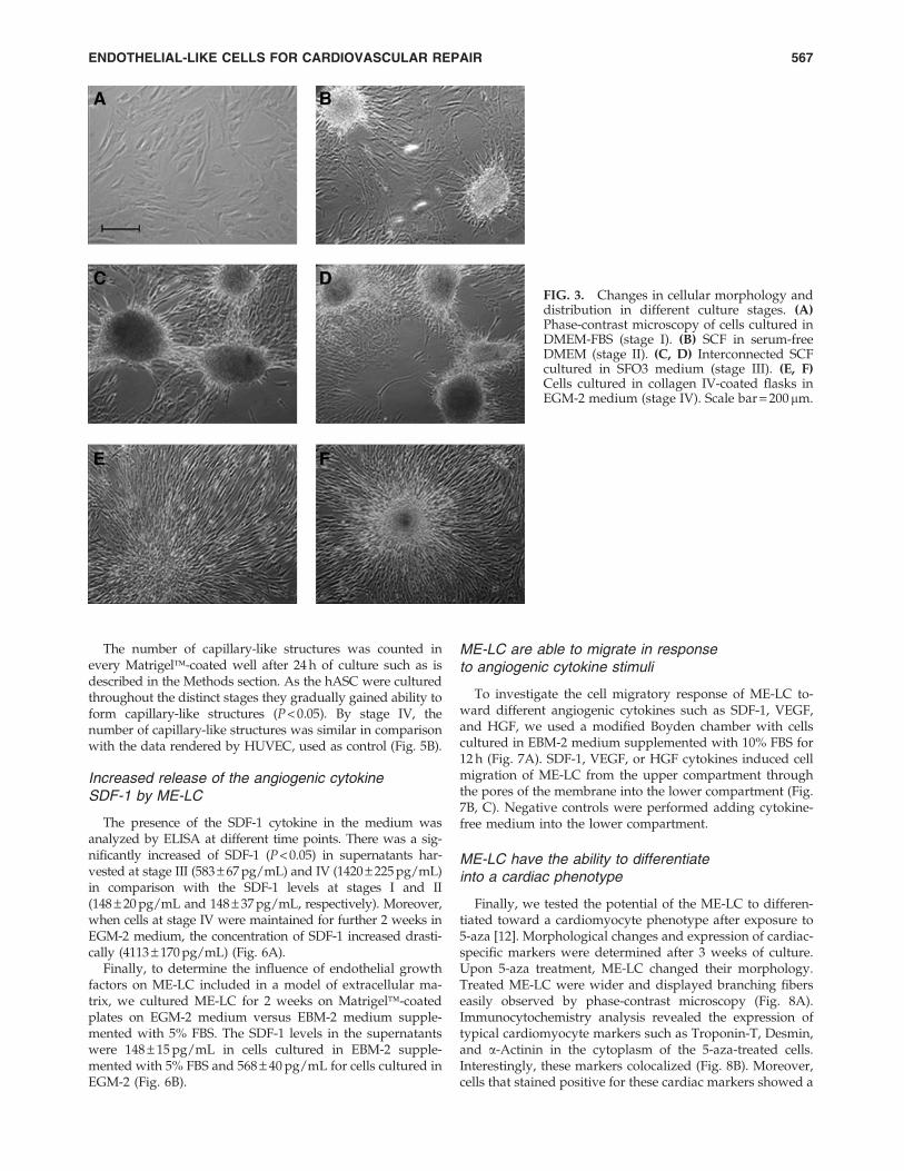
ME-LC were isolated through a series of consecutivestages I to IV detailed in the methods section (see Fig. 1 fordetails). hASC obtained from lipoaspirates grew as amonolayer and displayed a fibroblast-like and spindle-shaped morphology when they were cultured in DMEM-FBS(stage I, Fig. 3A). When the culture medium was replaced byDMEM without serum, the cells began to form 3-dimensionalcellular aggregates (termed SCF), which increased in sizeover time (stage II; Fig. 3B). We obtained between 50 and 70SCF in each T-75 tissue culture flask. When these SCF werescraped-off and seeded in SFO3 medium, SCF began toconnect each other and showed a variable morphology (stageIII; Fig. 3C, D). At the end of stage III were obtained3.9 – 2.1 · 105 cells in each well-plate from 10–15 SCF. For thestage IV, cells scraped-off from 2 wells were seeded in a T-75tissue culture flask. Cells displayed an elongated morphol-ogy and were arranged in parallel or grew attached sur-rounding the SCF (stage IV; Fig. 3E, F). At this stage thedoubling time of ME-LC was shorter than hASC, 2.7 – 0.1days and 3.8 – 0.3 days, respectively.
ME-LC isolated after several culture stagesexpress markers associated to vascularprogenitors coupled to a decreased expressionof the mesenchymal marker CD90
Flow cytometry was carried out in each of these culturestages to assess the presence or absence of hematopoietic,endothelial, and mesenchymal markers. The expression ofthe hematopoietic markers CD133, CD34, CD45, KDR, andCXCR4 significantly varied throughout the isolation process(P < 0.05, Mann–Whitney U-test) (Fig. 4A). Initially, the cul-tures were negative for all markers during stages I and II.When the cells were cultured in SFO3 medium (stage III),they slightly upregulated these markers to a some extend:14.1% – 1.7% for CD133, 18.3% – 4.9% for CD34, 21% – 3.7%for CD45, 27.3% – 5.5% for KDR, and 13% – 1.65% forCXCR4. In stage IV, when the cells had been cultured inEGM-2 medium, the expression of these markers dropped(Fig. 4A).
ENDOTHELIAL-LIKE CELLS FOR CARDIOVASCULAR REPAIR 565
High levels of expression of the MSC markers CD105,CD73, and CD90 were observed throughout the differentstages of the ME-LC isolation procedure (Fig. 4B). A Mann–Whitney U-test indicates significant (P < 0.05) differencesamong the expression of the 3 markers (Fig. 4B). Interest-ingly, CD90 expression decreased significantly in cells cul-tured in SFO3 medium (stage III: 41% – 15.8%) in comparisonwith cells in the stage I (94.1% – 2.2%), stage II (80 – 8.6), andstage IV (87.5% – 2.7%).
Figure 4C shows a comparison between marker expres-sion of cells at stages I, III, and IV versus HUVEC, whichwere used as control of mature ECs. HUVEC and ME-LCshowed expression of the progenitor markers (CXCR4 andKDR). CD133 and CD34 were found expressed in ME-LC butnot in HUVEC. Expression of the MSC marker CD90 de-creased in both ME-LC cultured in SFO3 and HUVEC ascompared with cells at stage I. Finally, the endothelial mar-ker CD31 was highly expressed in mature HUVEC(63% – 11.4%) but it was barely expressed in stages I, III, andIV (Fig. 4C). All together, these data suggest that ME-LCisolated from hASC cultures express markers resembling avascular progenitor phenotype.
Expression levels of genes related with EPC were as-sessed by RT-PCR. Cells cultured from stage I to IVmaintained the expression of genes such as CD133,CXCR4, CDK2, and FLT1. A weak expression of CCR7 and
CDK2 genes was detected in cells at stage III (Fig. 4D).Expression of TIE2 was only detected at stage I, but not insubsequent stages. Finally, expression of DLL4 (a Notchligand) was found highly expressed at stage I. Its expres-sion, however, decreased at from stage II onward and wascompletely lost at stages III and IV (Fig. 4D). These geneexpression data suggest the endothelial progenitor phe-notype of ME-LC.
ME-LC enhance functional capillary-likestructures formation in a Matrigel assay
As shown in Fig. 5A, cells from cultures at stage I and IIwere not able to form any capillaries over 7 days. On theother hand, cells previously grown in SFO3 or EGM-2 (stagesIII and IV, respectively) displayed a large number of capillary-like structures as early as 4 h after being seeded onMatrigel� and the appearance of capillary-like structuresincreased overtime. After 7 days in culture, a well-establishedcellular network was present in all the cultures (Fig. 5A). As apositive control, the capability of HUVEC to form capillary-like structures in Matrigel� was also assessed. The resultsshowed the appearance of these capillary-like structures after 4and 24 h in Matrigel�. However, these structures disappearedwhen HUVEC were cultured for 7 days likely due to their verymature nature (Fig. 5A).
FIG. 2. Phenotypic characterization anddifferentiation potential of hASC. (A) Flowcytometry characterization of hASC. Grayhistograms identify the isotype controls(negative). (B) Differentiation potential ofhASC toward adipogenic, chondrogenic, andosteogenic lineage. Adipogenesis was con-firmed by Oil Red O staining, chondrogen-esis by Toluidine Blue staining, andosteogenesis by Alizarin Red S staining.Upper pictures show negative controls,hASCs cultured in normal medium for 2weeks, and then histochemically stained.Scale bar = 200 mm.
566 MARCHAL ET AL.
The number of capillary-like structures was counted inevery Matrigel�-coated well after 24 h of culture such as isdescribed in the Methods section. As the hASC were culturedthroughout the distinct stages they gradually gained ability toform capillary-like structures (P < 0.05). By stage IV, thenumber of capillary-like structures was similar in comparisonwith the data rendered by HUVEC, used as control (Fig. 5B).
Increased release of the angiogenic cytokineSDF-1 by ME-LC
The presence of the SDF-1 cytokine in the medium wasanalyzed by ELISA at different time points. There was a sig-nificantly increased of SDF-1 (P < 0.05) in supernatants har-vested at stage III (583 – 67 pg/mL) and IV (1420 – 225 pg/mL)in comparison with the SDF-1 levels at stages I and II(148 – 20 pg/mL and 148 – 37 pg/mL, respectively). Moreover,when cells at stage IV were maintained for further 2 weeks inEGM-2 medium, the concentration of SDF-1 increased drasti-cally (4113 – 170 pg/mL) (Fig. 6A).
Finally, to determine the influence of endothelial growthfactors on ME-LC included in a model of extracellular ma-trix, we cultured ME-LC for 2 weeks on Matrigel�-coatedplates on EGM-2 medium versus EBM-2 medium supple-mented with 5% FBS. The SDF-1 levels in the supernatantswere 148 – 15 pg/mL in cells cultured in EBM-2 supple-mented with 5% FBS and 568 – 40 pg/mL for cells cultured inEGM-2 (Fig. 6B).
ME-LC are able to migrate in responseto angiogenic cytokine stimuli
To investigate the cell migratory response of ME-LC to-ward different angiogenic cytokines such as SDF-1, VEGF,and HGF, we used a modified Boyden chamber with cellscultured in EBM-2 medium supplemented with 10% FBS for12 h (Fig. 7A). SDF-1, VEGF, or HGF cytokines induced cellmigration of ME-LC from the upper compartment throughthe pores of the membrane into the lower compartment (Fig.7B, C). Negative controls were performed adding cytokine-free medium into the lower compartment.
ME-LC have the ability to differentiateinto a cardiac phenotype
Finally, we tested the potential of the ME-LC to differen-tiated toward a cardiomyocyte phenotype after exposure to5-aza [12]. Morphological changes and expression of cardiac-specific markers were determined after 3 weeks of culture.Upon 5-aza treatment, ME-LC changed their morphology.Treated ME-LC were wider and displayed branching fiberseasily observed by phase-contrast microscopy (Fig. 8A).Immunocytochemistry analysis revealed the expression oftypical cardiomyocyte markers such as Troponin-T, Desmin,and a-Actinin in the cytoplasm of the 5-aza-treated cells.Interestingly, these markers colocalized (Fig. 8B). Moreover,cells that stained positive for these cardiac markers showed a
FIG. 3. Changes in cellular morphology anddistribution in different culture stages. (A)Phase-contrast microscopy of cells cultured inDMEM-FBS (stage I). (B) SCF in serum-freeDMEM (stage II). (C, D) Interconnected SCFcultured in SFO3 medium (stage III). (E, F)Cells cultured in collagen IV-coated flasks inEGM-2 medium (stage IV). Scale bar = 200mm.
ENDOTHELIAL-LIKE CELLS FOR CARDIOVASCULAR REPAIR 567
parallel and interconnected distribution with the presence ofbi- and multinucleated cells. Control ME-LC nontreated with5-aza were negative for all the cardiomyocyte markers ex-amined (Fig. 8C).
Discussion
Cardiovascular diseases, such as the ischemic heart dis-ease and peripheral arterial occlusive disease, cause an ele-vated morbidity and mortality in developed countries.Currently, several clinical trials use different strategies forcell delivery and a diverse cell sources for transplantation [6–8]. In this respect, regenerative therapy to treat endothelialtissue damage has focused on the use of autologous stem
cells, mainly EPC harvested from blood, BM, or umbilicalcord blood [22]. Nevertheless, the scarcity of EPC in adulttissues makes its therapeutic use a challenge. As an alter-native, the endothelial differentiation potential of MSCs hasbeen explored by stimulating MSCs with angiogenic growthfactors and it was proved that the differentiated MSCs wereable to integrate into new blood vessels in vivo [23].
Recently, it has been shown that adipose tissue contains apopulation of adult multipotent cells with extensive prolif-erative capacity in vitro which are able to differentiate intoseveral lineages, including ECs, smooth muscle cells, andcardiomyocytes [13,24,25]. In fact, there are preclinicalstudies supporting the capability of MSCs obtained fromBM, umbilical cord blood, or adipose tissue to differentiate
FIG. 4. Fluorescence-activatedcell sorting and RT-PCR analysisof endothelial, hematopoietic, andmesenchymal markers through-out ME-LC isolation stages. (A)hASC cultured in different mediawere tested for hematopoietic andendothelial markers (CD133,CD34, KDR, CD45, and CXCR4)and (B) mesenchymal surfacemarkers (CD105, CD73, andCD90) by flow cytometry. (C)Comparative expression of sur-face antigens determined betweenHUVEC (gray bars), ME-LC cul-tured in EGM-2 (stage IV; stripedbars) and SFO3 (stage III; blackbars), and hASC cultured inDMEM-FBS (stage I; white bars) byflow cytometry. All data are ex-pressed as mean – SE of 4 inde-pendent experiments performedin triplicate (P < 0.05). (D) The ex-pression of CD133, CCR7, TIE-2,CXCR4, DLL4, CDK2, and FLT-1was evaluated by RT-PCR in cellsat different stages (I to IV) cul-tured in different media (DMEM-FBS; DMEM; SFO3 and EGM-2).b-actin was used as a housekeep-ing gene. Experiments were per-formed in triplicate and werecarried out at least twice yieldingidentical results. HUVEC, humanumbilical vein endothelial cell; SE,standard error.
568 MARCHAL ET AL.
into endothelial mature cells [26,27]. However, it was re-ported that mature ECs can proliferate in vitro although theygradually loss their proliferative potential hampering theirclinical application [28]. From a therapeutic standpoint, itbecomes necessary the isolation of sufficient numbers ofprogenitor cells capable of maintaining their angiogenic po-tential in vitro for long periods. Here, we present a simpleand reproducible approach to maintain ME-LC isolated fromsubcutaneous adipose tissue. Moreover, we tried to inducecardiomyocytic differentiation to demonstrate the capacity ofME-LC to differentiate into both endothelial and cardio-
myocyte-like cells, which could have advantages in the stemcell-based cardiovascular therapy.
Phenotypic characterization of hASC isolated from li-poaspirates showed a high expression of mesenchymal-specific surface markers such as CD105, CD73, and CD90,and barely expressed hematopoietic stem cells (HSC) or EPCmarkers (CD45, CD34, CD133, CXCR4, or KDR). Moreover,hASC possessed the ability to differentiate into various lin-eages as previously shown [29]. hASC were cultured alongseveral stages (stage I to IV, Fig. 1). Culture in serum-freemedia for 3 weeks resulted in the appearance of SCF that
FIG. 5. Capillary network formationassay. (A) Representative light micros-copy analysis of cells at different cul-ture stages and HUVEC grown onMatrigel�- coated wells with EGM-2medium. Pictures were taken at 4 h,24 h, and 7 days of culture. Picturesfrom 1 representative experiment of 3independent experiments are shown.Scale bar = 200mm. (B) Semiquantifica-tion of the capillary formation index.Bars correspond to the percentage ofthe number of capillary-like structurescomparatively to control (HUVEC)measured after 24 h of culture on Ma-trigel�. All data from 3 independentexperiments performed in duplicateare expressed as mean – SE (**P < 0.05vs. HUVEC).
ENDOTHELIAL-LIKE CELLS FOR CARDIOVASCULAR REPAIR 569
increased in number and size throughout the subsequentculture stages. Previously, Hirashima et al. [15] demon-strated that a chemically defined serum-free culture system,including 2-mecarptoethanol, had the ability to support theproliferation of ECs and their progenitors from mesodermcells. When cells were cultured in SFO3 medium (stage III)and in EC medium (EGM-2; stage IV), both termed ME-LC,increased their expression of EPC and hematopoietic mark-ers (CD34, CD133, KDR, CXCR4, and CD45) [30]. The co-existence of hematopoietic and endothelial markers in theME-LC is indicative of a phenotype resembling early vas-cular progenitors since it has been reported the existence of abipotent precursor cell, termed the hemangioblast, capable ofgiving rise to both hematopoiesis and vascular endothelium[31]. In contrast, mature ECs HUVEC were negative forCD34 and CD133 progenitor markers and strongly positivefor CD31, a mature endothelial marker. These antigens(CD34 and CD133) are lost upon differentiation of endothe-lial progenitors to endothelium [30]. CD34 expression inhASC is correlated with replicative capacity, differentiationpotentials, expression profiles of angiogenesis-related genes,and immaturity or stemness of these cells [32]. Similar resultswere obtained by Howson et al. [33] using postnatal aorta to
develop culture conditions for the isolation of nonendothelialmesenchymal cells with long-term maintenance in an un-differentiated state. Under serum-free conditions vascularprogenitor cells obtained were CD34 + /CD31 - , grewforming spheroids, and were identified as pericyte progeni-tor cells [33]. However, another study showed that using thesame serum-free media, cells obtained from lipoaspirates
FIG. 6. SDF-1 detection in culture supernatants. (A) SDF-1levels were measured by enzyme-linked immunosorbentassay in the medium surpenatant harvested in differentculture stages. (B) SDF-1 concentration released by ME-LCcultured on Matrigel-coated plates grown on EGM-2 me-dium versus EBM-2 medium supplemented with 5% FBS. Alldata are expressed as mean – SE of 4 independent experi-ments performed in duplicate (**P < 0.05). EBM-2, endothelialbasal medium-2; SDF-1, stromal cell-derived factor-1; EGM-2, endothelial growth medium.
FIG. 7. Migration capacity of ME-LC. (A) Illustrative car-toon of the modified Boyden chamber experiment. (B) Con-focal microscopy image of calcein AM-labeled cells thatmigrated through the filter using SDF-1 as chemoactractivecytokine after 12 h. Scale bar = 100 mm. (C) Migratory effect ofSDF-1, VEGF, and hematopoietic growth factor cytokines inME-LC after 1 and 12 h. The migration index was estimateddividing fluorescence data of ME-LC migration toward dif-ferent cytokines respect to the control, which represent ME-LC migration when cytokine-free medium was added intothe lower compartment. All data are expressed as mean – SEof 3 independent experiments performed in triplicate(**P < 0.05 vs. the control group).
570 MARCHAL ET AL.
had increased proportion of Flk-1 + (KDR) marker and werenegative for CD34, CD45, and CD133. When these cells wereseeded in EC differentiation medium for 3 days, the ex-pression of Flk-1 + decreased over time, whereas the ex-pression of mature EC markers increased [27]. In contrast toour study, they cultured the cells during shorter period inserum-free media and in EC medium, likely acceleratingendothelial maturation due to serum components, whichhave been proved to induce cell maturation [34].
Interestingly, both ME-LC and HUVEC expressed highlevels of CXCR4 and KDR. Phenotypically, MSCs are iden-tified by the absence of CD45, CD34, and other hematopoietic-associated markers [3]. In addition, the MSC marker CD90,whose expression was practically absent in HUVEC, was the
marker whose expression most significantly decreasedwhen ME-LC were cultured in SOF3 medium (stage III),suggesting endothelial differentiation. In agreement, it hasbeen shown an annihilation of CD90 antigen expressionon mesenchymal stromal cells by angiogenic stimulation invitro [35].
In contrast, CD90-positive cells recovered at stage IV inwhich cells reached a high rate of proliferation. It has beenshown that the expression of CD90 on EPC and pericytesmay be indicative of their angiogenic potential and capacityfor proliferation [36].
Gene expression profile confirmed the endothelial pro-genitor phenotype with the expression and maintenance ofspecific genes involved in self-renewal, cell cycle promotion,
FIG. 8. Cardiomyocyte differ-entiation potential of ME-LC. (A)Phase-contrast microscopy ofME-LC treated with 10 mM of 5-azacytidine for 24 h and thencultured in EGM-2 for 3 weeks.Image shows a cell with an in-creased size and the presence ofbranching fibers that are charac-teristics of myotube-like cells.Scale bar = 100 mm. (B) Immuno-fluorescence staining for the ex-pression of cardiac markers incells cultured in EGM-2 for 3weeks after induction with 5-azacytidine. Top panels show thecytoplasmatic expression of Tro-ponin-T-TRICT (red) and Des-min-FITC (green) in treated cells.Scale bar = 50mm. Bottom panelsshow a double staining with thecardiac markers a-Actinin-TRICT(red) and Desmin-FITC (green),which colocated in the cyto-plasm. Scale bar = 50 mm. (C)Parallel and interconnected dis-tribution of cardiomyocyte-likecells stained with different anti-bodies displaying the presence ofbi and multinucleated cells. Thesmall picture in the upperright corner represents the nega-tive control. Nuclei are stainedwith 4¢,6-diamidino-2-pheny-lindole (blue). Scale bar = 50mm.
ENDOTHELIAL-LIKE CELLS FOR CARDIOVASCULAR REPAIR 571
and antiapoptotic such as has been recently showed in cordblood-derived EPC [5]. Both CXCR4 and CD133 vasculargenes were expressed at similar levels throughout the dis-tinct culture stages. Nevertheless, genes expressed in differ-entiated EPC such as Cdk2 and Flt-1[5] showed a weakexpression level in ME-LC cultured in SOF3 medium. An-other interesting result was the disappearance of Tie-2 geneexpression, which has been previously reported as angio-genic factor clearly induced in the differentiated ECs [37].Constitutive Ang1–Tie2 signaling is thought to maintain thequiescent endothelial phenotype in vivo [38]. In addition toECs, Tie2 is expressed in a subpopulation of HSC being, inpart, responsible of maintaining a quiescent state in the BMniche [39]. Also, our results showed that the expression of theNotch ligand, delta-like 4 (DLLl4), was downregulated inME-LC, which correlated with the early formation of largenumber of capillary-like structures and the late formation ofa vascular network. Recent studies have demonstrated thatDLLl4 limits the angiogenic potential in developing bloodvessels and the loss of DLL4 results in an arterial hyper-branching phenotype [40]. Interestingly, in contrast to HU-VEC, only ME-LC cultured in SFO3 (stage III) and EGM-2(stage IV) media were able to strongly enhance capillarytubes formation after 7 days. It has been shown that VEGFstimulation of HUVEC induces Dll4 expression, which re-duced vessel sprout length in a 3D tubulogenesis assay,confirming that DLL4 signaling inhibits angiogenesis. DLL4expression seems to acts as a switch blocking EC prolifera-tion and allowing induction of a more mature differentiatedphenotype [41].
Several studies have identified several molecules such asVEGF and SDF-1 as key regulators of the proliferation,chemotaxis toward ischemic tissues, and differentiation ofEPC [42]. hASC secrete multiple potentially synergisticproangiogenic growth factors, including VEGF, HGF, andchemokine SDF-1, which are likely to play a pivotal role forthe hASC-mediated angiogenesis [43]. In fact, SDF-1 hasbeen shown to enhance neovascularization by acceleratingEPC recruitment into ischemic foci [42]. In our study, ME-LCshowed an increased secretion of SDF-1 in comparison withhASC, even when they were seeded into Matrigel�. In ad-dition, the role of SDF-1 to induce migration of CD133 + /CD34 + /KDR + cells has been shown [30]. In the presentstudy, we demonstrate how the ME-LC, which express theSDF-1 receptor, CXCR4, were able to migrate toward anSDF-1 gradient. These data suggest the potential homing ofME-LC to sites of vascular injury for tissue repair.
Most importantly, we show that after exposure to 5-azacytidine ME-LC retained the capacity to differentiate intocardiomyocyte-like cells with the acquisition of a cardiogenicphenotype and the expression of cardiomyocyte-specificmarkers. Morphological changes consisted of the appearanceof wide, branching, and multinucleated cells resemblingcardiac muscle cells [44]. The expression of cardiomyocytemarkers such as troponin-T and a-sarcomericactinin [44] andthe presence of a desmin filaments network, which take partin regulating the mesodermal specification into cardiomyocytes[45], support the cardiomyocyte differentiation from ME-LC.
In summary, our studies indicate that subcutaneous adi-pose tissue may be a useful source of autologous ME-LCwith the capacity to maintain their vascular progenitorproperties. The culture method described in the present
study may be used to isolate, maintain, and propagate thesecells with an increased expression of specific endothelialprogenitor markers. This methodology could be applied toMSCs from other origins such as BM-derived stromal/stemcells. However, the interest of our study was the use of hASCisolated from liposuction, which is a less invasive methodthan BM aspiration and allows the collection of a high rate ofprogenitor cells. This can overcome the limited proliferationpotential of mature ECs and EPC, which hampers theirclinical use. ME-LC increased the secretion of SDF-1, formedvascular-like structures, and displayed the ability to migratetoward a cytokine gradient. Moreover, ME-LC retained thecapacity to differentiate into cardiomyocyte-like cells,showing expression of typical cardiomyocyte markers. Thisproperty suggests the potential of ME-LC to recellularizedamaged tissue or strengthen the post-infarct scar as well asinducing neovascularization of the affected area, whichcould have advantages in the stem cell-based cardiovasculartherapy. It has been demonstrated the ability of cardiac stemcells to differentiate into the ECs, contributing to neovascu-larization in the process of tissue remodeling and/or re-generation [46]. Furthermore, recent studies showed interestin the cardiac and endothelial capacity of stem cell, provid-ing important tools for the study of differentiation in vitroand future stem cell therapy for ischemic cardiomyopathy[47,48]. Future studies should explore further the functionalin vitro and in vivo mechanisms of ME-LC in cardiovasculardiseases along with their potential therapeutic impact, andour experimental data suggest that these cells may prove tobe a valuable tool for vascular and cardiac repair.
Funding
This work was supported in part by grants from the In-stituto de Salud Carlos III (Fondo de Investigacion Sanitariagrant number PI10/02295), the Consejerıa de Salud ( Junta deAndalucıa grant number PI-0384/2008), the Consejeria deEconomıa, Innovacion y Ciencia ( Junta de Andalucıaexcellence project number CTS-6568) and the Universityof Granada (GREIB translational project number GREIB.PT_2010_09). Research in P.M.’s Lab was supported by grantsfrom the Instituto de Salud Carlos III (grant numberPI070026), Ministry of Science and Innovation (MICINN;grant number PLE-2009-0111), and by the Junta de Andalu-cıa (grant number CICE-P08-CTS-3678). C.B. is supported bythe ISCIII-FIS (grant number CP07/00059).
Acknowledgments
We gratefully acknowledge Manuela Exposito from FI-BAO for excellent technical assistance with statistical studies.We also thank Emma Gutierrez Gonzalez for helping in theartwork.
Author Disclosure Statement
No competing financial interests exist.
References
1. Asahara T, T Murohara, A Sullivan, M Silver, R van der Zee,T Li, B Witzenbichler, G Schatteman and JM Isner. (1997).Isolation of putative progenitor endothelial cells for angio-genesis. Science 275:964–967.
572 MARCHAL ET AL.
2. S Kim and von Recum H. (2008). Endothelial stem cells andprecursors for tissue engineering: cell source, differentiation,selection, and application. Tissue Eng Part B Rev 14:133–147.
3. Campioni D, A Lo Monaco, F Lanza, S Moretti, L Ferrari, MFotinidi, R La Corte, A Cuneo and F Trotta. (2008). CXCR4pos circulating progenitor cells coexpressing monocytic andendothelial markers correlating with fibrotic clinical featuresare present in the peripheral blood of patients affected bysystemic sclerosis. Haematologica 93:1233–1237.
4. Krenning G, BW van der Strate, M Schipper, XJ van Seijen,BC Fernandes, MJ van Luyn and MC Harmsen. (2009).CD34 + cells augment endothelial cell differentiation ofCD14 + endothelial progenitor cells in vitro. J Cell Mol Med13:2521–2533.
5. Igreja C, R Fragoso, F Caiado, N Clode, A Henriques, LCamargo, EM Reis and S Dias. (2008). Detailed molecularcharacterization of cord blood-derived endothelial progeni-tors. Exp Hematol 36:193–203.
6. Taljaard M, MR Ward, MJ Kutryk, DW Courtman, NJ Ca-mack, SG Goodman, TG Parker, AJ Dick, J Galipeau and DJStewart. (2010). Rationale and design of Enhanced Angio-genic Cell Therapy in Acute Myocardial Infarction (ENACT-AMI): the first randomized placebo-controlled trial ofenhanced progenitor cell therapy for acute myocardial in-farction. Am Heart J 159:354–360.
7. Tateishi-Yuyama E, H Matsubara, T Murohara, U Ikeda, SShintani, H Masaki, K Amano, Y Kishimoto, K Yoshimoto,H Akashi, K Shimada, T Iwasaka and T Imaizumi. Ther-apeutic Angiogenesis using Cell Transplantation (TACT)Study Investigators. (2002). Therapeutic angiogenesis forpatients with limb ischaemia by autologous transplantationof bone-marrow cells: a pilot study and a randomised con-trolled trial. Lancet 360:427–435.
8. Dzau VJ, M Gnecchi, AS Pachori, F Morello and LG Melo.(2005). Therapeutic potential of endothelial progenitor cellsin cardiovascular diseases. Hypertension 46:7–18.
9. Iwashima S, T Ozaki, S Maruyama, Y Saka, M Kobori, KOmae, H Yamaguchi, T Niimi, K Toriyama, Y Kamei, STorii, T Murohara, Y Yuzawa, Y Kitagawa and S Matsuo.(2009). Novel culture system of mesenchymal stromal cellsfrom human subcutaneous adipose tissue. Stem Cells Dev18:533–543.
10. Foresta C, L De Toni, A Ferlin and A Di Mambro. (2010).Clinical implication of endothelial progenitor cells. ExpertRev Mol Diagn 10:89–105.
11. Jin XB, YS Sun, K Zhang, J Wang, XD Ju and SQ Lou. (2007).Neocartilage formation from predifferentiated human adi-pose derived stem cells in vivo. Acta Pharmacol Sin 28:663–671.
12. Rodrıguez-Serrano F, P Alvarez, O Caba, M Picon, JAMarchal, M Peran, J Prados, C Melguizo, AR Rama, HBoulaiz and A Aranega. (2010). Promotion of humanadipose-derived stem cell proliferation mediated by exoge-nous nucleosides. Cell Biol Int 34:917–924.
13. Peran M, JA Marchal, E Lopez, M Jimenez-Navarro, HBoulaiz, F Rodrıguez-Serrano, E Carrillo, G Sanchez-Espin, Ede Teresa, D Tosh and A Aranega. (2010). Human cardiactissue induces transdifferentiation of adult stem cells to-wards cardiomyocytes. Cytotherapy 12:332–337.
14. Rubio R, J Garcıa-Castro, I Gutierrez-Aranda, J Paramio, MSantos, P Catalina, PE Leone, P Menendez and R Rodrıguez.(2010). Deficiency in p53 but not retinoblastoma induces thetransformation of mesenchymal stem cells in vitro and ini-tiates leiomyosarcoma in vivo. Cancer Res 70:4185–4194.
15. Hirashima M, M Ogawa, S Nishikawa, K Matsumura, KKawasaki, M Shibuya and S Nishikawa. (2003). A chemi-cally defined culture of VEGFR2 + cells derived from em-bryonic stem cells reveals the role of VEGFR1 in tuning thethreshold for VEGF in developing endothelial cells. Blood101:2261–2267.
16. Menendez P, JA Perez-Simon, MV Mateos, MD Caballero, MGonzalez, San-Miguel JF and A Orfao. (2002). Influence ofthe different CD34 + and CD34- cell subsets infused onclinical outcome after non-myeloablative allogeneic periph-eral blood transplantation from human leucocyte antigen-identical sibling donors. Br J Haematol 119:135–143.
17. Montes R, G Ligero, L Sanchez, P Catalina, T de la Cueva, ANieto, GJ Melen, R Rubio, J Garcıa-Castro, C Bueno and PMenendez. (2009). Feeder-free maintenance of hESCs inmesenchymal stem cell-conditioned media: distinct require-ments for TGF-beta and IGF-II. Cell Res 19:698–709.
18. Ramos-Mejia V, GJ Melen, L Sanchez, Gutierrez-Aranda I, GLigero, JL Cortes, PJ Real, C Bueno and P Menendez. (2010).Nodal/activin signaling predicts human pluripotent stemcell lines prone to differentiate toward the hematopoieticlineage. Mol Ther DOI:10.1038/mt.2010.179
19. Catalina P, C Bueno, R Montes, A Nieto, G Ligero, L San-chez, M Jara, A Rasillo, A Orfao, J Cigudosa, O Hovatta, MGreaves and P Menendez. (2009). Genetic stability of humanembryonic stem cells: A first-step toward the developmentof potential hESC-based systems for modeling childhoodleukemia. Leuk Res 33:980–990.
20. Soares R, G Balogh, S Guo, F Gartner, J Russo and F Schmitt.(2004). Evidence for the notch signaling pathway on the roleof estrogen in angiogenesis. Mol Endocrinol 18:2333–2343.
21. Lopes FC, A Rocha, A Pirraco, LO Regasini, DH Silva, VSBolzani, I Azevedo, IZ Carlos and R Soares. (2009). Anti-angiogenic effects of pterogynidine alkaloid isolated fromAlchornea glandulosa. BMC Complement Altern Med 9:15.
22. Ingram DA, LE Mead, H Tanaka, V Meade, A Fenoglio, KMortell, K Pollok, MJ Ferkowicz, D Gilley and MC Yoder.(2004). Identification of a novel hierarchy of endothelialprogenitor cells using human peripheral and umbilical cordblood. Blood 104:2752–2760.
23. Dai W, SL Hale, BJ Martin, JQ Kuang, JS Dow, LE Wold andRA Kloner. (2005). Allogeneic mesenchymal stem celltransplantation in postinfarcted rat myocardium: short- andlong-term effects. Circulation 112:214–223.
24. Madonna R and R De Caterina. (2010). Adipose tissue: anew source for cardiovascular repair. J Cardiovasc Med(Hagerstown) 11:71–80.
25. Bayes-Genis A, C Soler-Botija, J Farre, P Sepulveda, A Raya,S Roura, C Prat-Vidal, C Galvez-Monton, JA Montero, DBuscher and JC Belmonte. (2010). Human progenitor cellsderived from cardiac adipose tissue ameliorate myocardialinfarction in rodents. J Mol Cell Cardiol 49:771–780.
26. Chen MY, PC Lie, ZL Li and X Wei. (2009). Endothelialdifferentiation of Wharton’s jelly–derived mesenchymalstem cells in comparison with bone marrow–derived mes-enchymal stem cells 7:629–640.
27. Martınez-Estrada OM, Y Munoz-Santos, J Julve, M Reinaand S Vilaro. (2005). Human adipose tissue as a source ofFlk-1 + cells: new method of differentiation and expansion.Cardiovasc Res 65:328–333.
28. Prasad Chennazhy K and LK Krishnan. (2005). Effect ofpassage number and matrix characteristics on differentiationof endothelial cells cultured for tissue engineering. Bioma-terials 26:5658–5667.
ENDOTHELIAL-LIKE CELLS FOR CARDIOVASCULAR REPAIR 573
29. Marchal JA, H Boulaiz, M Peran, J Prados, J Campos, FGonzalez, F Rodrıguez-Serrano, C Melguizo, C Velez, ECarrillo, F Hita, R Ortiz, A Martınez-Amat, O Caba, CVentura and A Aranega. Eds. (2009). Therapeutic Potential ofDifferentiation in Cancer and Normal Stem Cells. Nova SciencePublishers, Inc., New York.
30. Peichev M, AJ Naiyer, D Pereira, Z Zhu, WJ Lane, M Wil-liams, MC Oz, DJ Hicklin, L Witte, MA Moore and S Rafii.(2000). Expression of VEGFR-2 and AC133 by circulatinghuman CD34( + ) cells identifies a population of functionalendothelial precursors. Blood 95:952–958.
31. Wang L, Li L, F Shojaei, C Cerdan, P Menendez, T Martin, ARouleau and M Bhatia. (2004). Endothelial and hematopoi-etic cell fate of human embryonic stem cells originates fromprimitive endothelium with hemangioblastic properties.Immunity 21:31–41.
32. Suga H, D Matsumoto, H Eto, K Inoue, N Aoi, H Kato,J Araki and K Yoshimura. (2009). Functional Implicationsof CD34 Expression in Human Adipose–Derived Stem/Progenitor Cells. Stem Cells Dev 18:1201–1210.
33. Howson KM, AC Aplin, M Gelati, G Alessandri, Parati EAand RF Nicosia. (2005). The postnatal rat aorta containspericyte progenitor cells that form spheroidal colonies insuspension culture. Am J Physiol Cell Physiol 289:1396–1407.
34. Landerholm TE, XR Dong, J Lu, NS Belaguli, RJ Schwartzand MW Majesky. (1999). A role for serum response factor incoronary smooth muscle differentiation from proepicardialcells. Development 126:2053–2062.
35. Campioni D, F Lanza, S Moretti, L Ferrari and A Cuneo.(2008). Loss of Thy-1 (CD90) antigen expression on mesen-chymal stromal cells from hematologic malignancies is in-duced by in vitro angiogenic stimuli and is associated withpeculiar functional and phenotypic characteristics. Cy-totherapy 10:69–82.
36. Bagley RB, W Weber, C Rouleau and BA Teicher. (2005).Pericytes and Endothelial Precursor Cells: Cellular Interac-tions and Contributions to Malignancy. Cancer Res 65:9741–9750.
37. Furuhata S, K Ando, M Oki, K Aoki, S Ohnishi, K Aoyagi, HSasaki, H Sakamoto, T Yoshida and S Ohnami. (2007). Geneexpression profiles of endothelial progenitor cells by oligo-nucleotide microarray analysis. Mol Cell Biochem 298:125–138.
38. Saharinen P, M Bry and K Alitalo. (2010). How do angio-poietins Tie in with vascular endothelial growth factors?Curr Opin Hematol 17:198–205.
39. Gomei Y, Y Nakamura, H Yoshihara, K Hosokawa, HIwasaki, T Suda and F Arai. (2010). Functional differencesbetween two Tie2 ligands, angiopoietin-1 and -2, in regula-tion of adult bone marrow hematopoietic stem cells. ExpHematol 38:82–89.
40. Hogan BM, R Herpers, M Witte, H Helotera, K Alitalo, HJDuckers and Schulte-Merker S. (2009). Vegfc/Flt4 signalling
is suppressed by Dll4 in developing zebra fish interseg-mental arteries. Development 136:4001–4009.
41. Harrington LS, RC Sainson, CK Williams, JM Taylor, W Shi,JL Li and AL Harris. (2008). Regulation of multiple angio-genic pathways by Dll4 and Notch in human umbilical veinendothelial cells. Microvasc Res 75:144–154.
42. Yamaguchi J, KF Kusano, O Masuo, A Kawamoto, M Silver,S Murasawa, M Bosch-Marce, H Masuda, DW Losordo, JMIsner and T Asahara. (2003). Stromal cell-derived factor-1effects on ex vivo expanded endothelial progenitor cell re-cruitment for ischemic neovascularization. Circulation107:1322–1328.
43. Kondo K, S Shintani, R Shibata, H Murakami, R Murakami,M Imaizumi, Y Kitagawa and T Murohara. (2009). Im-plantation of adipose-derived regenerative cells enhancesischemia-induced angiogenesis. Arterioscler Thromb VascBiol 29:61–66.
44. Makino S, K Fukuda, S Miyoshi, F Konishi, H Kodama,J Pan, M Sano, T Takahashi, S Hori, H Abe, J Hata, AUmezawa and S Ogawa. (1999). Cardiomyocytes can begenerated from marrow stromal cells in vitro. J Clin Invest103:697–705.
45. Hollrigl A, M Hofner, M Stary and G Weitzer. (2007). Dif-ferentiation of cardiomyocytes requires functional serineresidues within the amino-terminal domain of desmin. Dif-ferentiation 75:616–626.
46. Mohri T, Y Fujio, M Maeda, T Ito, T Iwakura, Y Oshima, YUozumi, M Segawa, H Yamamoto, T Kishimoto andJ Azuma. (2006). Leukemia inhibitory factor induces endo-thelial differentiation in cardiac stem cells. J Biol Chem 281:6442–6447.
47. Li SC, J Acevedo, PH Schwartz, L Wang, H Jiang, J Luo, RGPestell, WG Loudon and AC Chang. (2011). Mechanisms forprogenitor cell-mediated repair for ischemic heart injury.Curr Stem Cell Res Ther PMID:2l466480
48. Choi SC, WJ Shim and DS Lim. (2008). Specific monitoringof cardiomyogenic and endothelial differentiation by dualpromoter-driven reporter systems in bone marrow mesen-chymal stem cells. Biotechnol Lett 30:835–843.
Address correspondence to:Dr. Juan A. Marchal
Biopathology and Regenerative Medicine Institute (IBIMER)Centro de Investigacion Biomedica