Membranes are widely used for protein bioseparation(Przybycien et al., 2004), the main advantages beinghigh productivity, ease of scale-up and easy cleaningand sanitization of equipment. The main limitation withordinary membrane based separations is the generallylow selectivity, i.e. poor resolution. Current research onmembrane based bioseparations is focused on selectivityimprovement (van Reis and Zydney, 2001), oneapproach being the integration of membrane separationwith other separation techniques (Ghosh, 2004). The
resultant techniques could be referred to as hybridbioseparations (Kim and Cramer, 1991).
Humanized monoclonal antibodies (mAbs) holdsignificant promise as biopharmaceuticals (Vaughan etal., 1998). mAbs are usually purified from mammaliancell culture media by protein-A or protein-G basedaffinity chromatography (Roque et al., 2004). Whileaffinity chromatography gives excellent separation, it isexpensive, comparatively slow and difficult to scale-upon account of the use of soft gel-based media. Bothprotein-A and protein-G are immunotoxins and theseprotein ligands are known to leach out from chromato-graphic media (Carlson, 2005). Affinity media-boundmAbs are usually desorbed by acidic buffers and thismay lead to some protein aggregation (Arakawa et al.,2004). Membrane based separation techniques have
2 L. Wang et al. / Journal of Immunological Methods 314 (2006) 1–8
been proposed for purification of mAbs: high-resolutionultrafiltration (Wan et al., 2005) and membranechromatography (Ghosh, 2001). In a recent paper ahybrid bioseparation technique for the fractionation ofplasma proteins human serum albumin and humanimmunoglobulin G has been described (Ghosh, 2004).The current paper discusses the use of a similar hybridbioseparation method for the purification of humanizedmAbs Alemtuzumab and hIgG1-CD from mammaliancell culture media.
Alemtuzumab or Campath-1H is a humanized IgG1type mAb against lymphocyte antigen CD52 (Hale etal., 2002). It is used for the treatment of autoimmunediseases including rheumatoid arthritis, multiple sclero-sis (MS) and vasculitis as well as in bone marrow andorgan transplantation. This mAb is usually produced byculturing genetically engineered Chinese hamster ovary(CHO) cells (Phillips et al., 2001). hIgG1-CD4 is ahumanized IgG1 type mAb against CD4 antigen whichhas been investigated for its therapeutic properties inpatients with refractory psoriasis and rheumatoidarthritis (Isaacs et al., 1997) and is also produced byCHO cell culture. Media used for mammalian cellculture traditionally contained 10% foetal calf serum(FCS). In recent years there has been a move away fromsuch conventional serum based cell culture media,particularly in biopharmaceutical manufacture, due tothe potential risk associated with prion proteins. Most
Fig. 1. Monoclonal antibody retention in
serum-free media used now typically contain definedproteins such as bovine albumin, insulin, ferritin andtransferrin. The main challenge involved in purifyingmAbs is their separation from these media proteins.
In this paper we describe a rapid, scalable andinexpensive hybrid bioseparation technique for purifica-tion of humanized monoclonal antibodies from mam-malian cell culture supernatant. The technique involvesthe selective and reversible retention of the monoclonalantibody within a membrane device by a combination ofsieving and hydrophobic interaction-based membranechromatography using the same microfiltration mem-brane (hydrophilic PVDF membrane having 0.22 μmaverage pore size). The principle of the hybridbioseparation technique is shown in Fig. 1, this beingcyclic in nature i.e. of the capture and release type. Afraction of the mAb in a feed sample is precipitatedusing high ammonium sulphate concentration and this isretained by sieving using a microfiltration membrane.This membrane also serves as a selective adsorptionmedia and binds the mAb remaining in solution byhydrophobic interaction, at the same ammoniumsulphate concentration. Using this combination ofmechanisms the antibody is selectively and quantita-tively retained within the membrane module. Theretained mAb is subsequently released by reducing theammonium sulphate concentration which results in thesimultaneous dissolution of the precipitated mAb
the hybrid bioseparation technique.
Table 1Binding capacity values for humanized mAbs hIgG1-CD4 andAlemtuzumab on microporous PVDF membrane at differentammonium sulphate concentrations
Ammonium sulphateconcentration (M)
hIgG1-CD4(mg/ml)
Alemtuzumab(mg/ml)
1.65 49.01 31.321.7 64.29 35.44
MAb concentration in feed: 0.1 mg/ml.Capture buffer: 20 mM sodium phosphate (pH 7.0) having appropriateammonium sulphate concentration.Flow rate: 1 ml/min.Membrane: 0.2 μm PVDF.Effective membrane disc diameter: 10 mm.Number of discs: 2.
3L. Wang et al. / Journal of Immunological Methods 314 (2006) 1–8
fraction and elution of the membrane bound fraction.The results obtained are discussed.
2. Experimental
2.1. Material
Alemtuzumab (or Campath-1H, batch #37) andhIgG1-CD4 (batch #10) were kindly donated by theTherapeutic Antibody Centre, Oxford University, UK.Serum-free CHO cell culture media (catalogue numberC1707) was purchased from Sigma. PVDF microfiltra-tion membrane (hydrophilic PVDF, 0.22 μm pore size,GVWP) was kindly donated by Millipore. All samplesolutions used in the hybrid bioseparation were preparedusing 20 mM sodium phosphate (pH 7.0) as basebuffer which in turn was prepared using ultra-purewater (18.2 MΩ cm) obtained from a NanopureDiamond (Barnstead) water purification unit. Allchemical used in the experiments e.g. sodium phosphate,ammonium sulphate, sodium citrate were purchasedfrom Sigma.
2.2. Experimental set-up
An AKTAprime liquid chromatography system(Amersham Biosciences) was used for carrying outthe hybrid bioseparation. A syringe filter holder(polycarbonate, 25 mm diameter, product number16517 E, Sartorius) was integrated with this system(in place of the chromatographic column) usingappropriate PEEK tubings and connectors. A stackof five membrane discs each having effective diameterof 20 mm was housed within the filter holder and thisdevice is referred to as the membrane module in thispaper. A 5 ml sample loop was used for injecting feedsamples for purification. The UV absorbance (at280 nm), pH and conductivity of the effluent streamfrom the module along with the system pressure werecontinuously recorded and logged into a computerusing PrimeView (Amersham Biosciences).
2.3. Experimental methods
All experiments were carried out at ambienttemperature i.e. 24 °C. The mAb binding capacity ofthe PVDF membrane was determined using a smallersyringe filter (stainless steel, 13 mm diameter, cataloguenumber 1980-001, Whatman) within which a stack oftwo membrane discs was housed. The effective diameterof the membrane discs were 10 mm. mAb solutionsprepared in appropriate capture solutions were used to
carry out breakthrough binding and elution experimentsat 1 ml/min flow rate using the AKTAprime system. Themembrane was first saturated with the mAb, theunbound material was removed and the membrane-bound mAb was then eluted using the base buffer. Theamount of mAb bound on the membrane was calculatedfrom the area under the curve of the eluted peak usingappropriate calibrations for hIgG1-CD4 and Alemtuzu-mab. The hybrid bioseparation experiments were carriedout using a stack of five PVDF membrane discs withinthe polycarbonate syringe filter holder. The feedsolutions were prepared by spiking the CHO cell culturemedia with the appropriate mAb such that its concen-tration was 0.5 mg/ml, this value being typical of wellexpressed mAbs. The capture solution was pumpedthrough the membrane module until stable UV absor-bance, pH, conductivity and pressure readings wereobtained. Just prior to injection into the membranemodule, the feed solution was mixed at 1:1 ratio withstock ammonium sulphate solutions to obtain the samesalt concentration as that of the respective capturesolution. 5 ml of this mixture was injected into themembrane module and the flow of capture solution wasresumed after this and continued until all media proteins(i.e. impurities) were removed from the module asindicated by the return of the UVabsorbance value to itsbaseline. The captured monoclonal antibody was thenreleased from the membrane module by switching overto the base buffer which resulted in the lowering of theammonium sulphate concentration within the membranemodule.
The feed, flow-through and purified mAb sampleswere analysed by affinity chromatography using aHiTrap™ rProtein-A FF affinity column (AmershamBiosciences). Both mAbs bound to this column whileother media proteins did not. The mAbs bound to theaffinity column from 20 mM sodium phosphate buffer
Table 2Solubility values for hIgG1-CD4 and Alemtuzumab at differentammonium sulphate concentrations
hIgG1-CD4 Alemtuzumab
Solubility at 1.65 M concentration(mg/ml)
0.191 0.209
Solubility at 1.7 M concentration(mg/ml)
0.143 0.178
4 L. Wang et al. / Journal of Immunological Methods 314 (2006) 1–8
(pH 7.0) and were eluted by 100 mM sodium citratebuffer (pH 3.0). The samples for affinity chromatogra-phy were desalted using centrifugal ultrafilters (Nano-sep 3 kDaMWCO, Pall, catalogue number OD003C). Ineach case 100 μl of sample was injected and the affinitychromatography was carried out at 1 ml/min flow rate.The mAb purity was determined based on appropriatecalibrations. The feed, flow-through and purified mAbsamples were also analysed by non-reducing SDS-PAGE (7.5% gel) and the protein bands were visualizedby using Coomassie blue dye (Laemmli, 1970).
3. Results and discussion
Membrane chromatography on its own can givehigh-resolution mAb separation but the processingcapacity is limited by the amount that can be boundon the membrane. On the other hand ammoniumsulphate induced precipitation gives high processingcapacity but the resolution is extremely poor. The mainlimitation of using ammonium sulphate induced pre-
Fig. 2. Separation of hIgG1-CD4 at two different ammonium sulphate conceflow rate=1 ml/min).
cipitation for mAb purification is the trade-off that existsbetween purity and recovery. As the precipitatingammonium sulphate concentration is increased, theantibody recovery increases due to more precipitationbut the purity decreases due to more contaminatingprotein precipitation. Therefore, precipitation alonewould not result in a selective and total separation ofthe mAbs from mammalian cell culture media. Thehybrid bioseparation technique combines sieving basedretention of ammonium sulphate precipitated mAb andhydrophobic interaction based membrane adsorption ofthe dissolved mAb on the same microfiltration mem-branes at the same solution condition. The mAb is firstpartially precipitated using a particular ammoniumsulphate concentration, this fraction being retained bya microfiltration membrane. The mAb remaining insolution at that point is then captured by adsorption onthe same microfiltration membrane.
Hydrophilic PVDF membrane (0.22 μm pore size,GVWP, Millipore) was selected for this study sincepreliminary studies showed that this membrane had agood compromise of high antibody binding capacityand low fouling tendency. Table 1 lists the bindingcapacities of hIgG1-CD4 and Alemtuzumab undersaturating conditions on the PVDF membrane at twodifferent ammonium sulphate concentrations in thecapture solution i.e. 1.65 M and 1.7 M. With hIgG1-CD4, the binding capacity increased significantly withincrease in ammonium sulphate concentration. On theother hand, the binding capacity of Alemtuzumab wasrelatively less responsive to the increase in salt
ntrations using hybrid bioseparation technique (A: 1.65 M, B: 1.7 M;
Fig. 4. Non-reducing SDS-PAGE (7.5%) obtained with feed and
5L. Wang et al. / Journal of Immunological Methods 314 (2006) 1–8
concentration. Table 2 lists the solubility values for thetwo mAbs at 1.65 and 1.7 M ammonium sulphateconcentration. As would be expected from the salting-out theory the solubility of both mAbs decreased withincrease in salt concentration.
Data shown in Tables 1 and 2 suggest that higheramounts of mAbs would be retained by hybridbioseparation at 1.7 M salt concentration due to thecombination of more precipitation and more adsorption.However, this has to be weighed against the possibilityof more impurity retention at the higher salt concentra-tion. Preliminary experiments showed that at ammo-nium sulphate concentrations greater or equal to 1.75 Msignificant amounts of media proteins precipitated alongwith the mAb. However, lowering of ammoniumsulphate concentration below 1.6 M reduced the amountof mAb precipitated as well as that bound on themembrane. Two ammonium sulphate concentrationswere therefore identified for carrying out the hybridseparation experiments: 1.65 M and 1.7 M.
purified hIgG1-CD4 samples from hybrid bioseparation experimentcarried out at 1.65 M ammonium sulphate concentration.
Fig. 3. Affinity chromatograms obtained with feed, flow-through andpurified hIgG1-CD4 samples from hybrid bioseparation experimentcarried out at 1.65 M ammonium sulphate concentration.
Fig. 2 shows the hybrid bioseparation of hIgG1-CD4from CHO cell culture media using 1.65 M (A) and1.7 M (B) ammonium sulphate concentrations in thecapture solution. The first peak in each case correspondsto the unbound UV-absorbing media componentsincluding media proteins, peptides, amino acids andother low molecular weight compounds. On loweringthe ammonium sulphate concentration, the retainedmAb was released from the membrane module andappeared as a peak. Fig. 3 shows the affinitychromatograms for the feed, flow-through and purifiedmAb samples obtained using the 1.65 M ammoniumsulphate capture solution. Prior to affinity chromato-graphy, the samples were desalted using centrifugalultrafilters. The absence of mAb in the flow-throughindicates its high recovery. The hIgG1-CD4 purity in the
Table 3Hybrid bioseparation of hIgG1-CD4: purity and recovery values atdifferent ammonium sulphate concentrations
Effective mAb concentration in feed: 0.25 mg/ml.Capture buffer: 20 mM sodium phosphate (pH 7.0) containingappropriate ammonium sulphate concentration.Flow rate: 1 ml/min.Membrane: 0.2 μm PVDF.Effective membrane diameter: 20 mm.Stack: 5 discs.
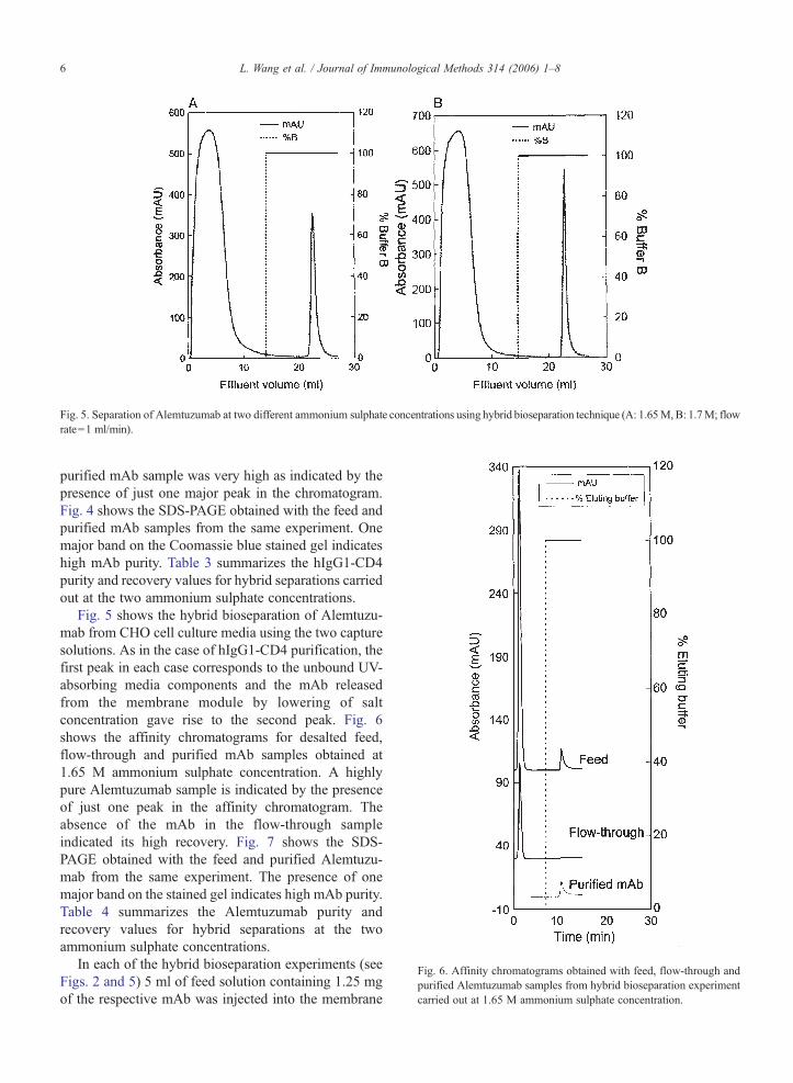
Fig. 5. Separation of Alemtuzumab at two different ammonium sulphate concentrations using hybrid bioseparation technique (A: 1.65M, B: 1.7M; flowrate=1 ml/min).
Fig. 6. Affinity chromatograms obtained with feed, flow-through andpurified Alemtuzumab samples from hybrid bioseparation experimentcarried out at 1.65 M ammonium sulphate concentration.
6 L. Wang et al. / Journal of Immunological Methods 314 (2006) 1–8
purified mAb sample was very high as indicated by thepresence of just one major peak in the chromatogram.Fig. 4 shows the SDS-PAGE obtained with the feed andpurified mAb samples from the same experiment. Onemajor band on the Coomassie blue stained gel indicateshigh mAb purity. Table 3 summarizes the hIgG1-CD4purity and recovery values for hybrid separations carriedout at the two ammonium sulphate concentrations.
Fig. 5 shows the hybrid bioseparation of Alemtuzu-mab from CHO cell culture media using the two capturesolutions. As in the case of hIgG1-CD4 purification, thefirst peak in each case corresponds to the unbound UV-absorbing media components and the mAb releasedfrom the membrane module by lowering of saltconcentration gave rise to the second peak. Fig. 6shows the affinity chromatograms for desalted feed,flow-through and purified mAb samples obtained at1.65 M ammonium sulphate concentration. A highlypure Alemtuzumab sample is indicated by the presenceof just one peak in the affinity chromatogram. Theabsence of the mAb in the flow-through sampleindicated its high recovery. Fig. 7 shows the SDS-PAGE obtained with the feed and purified Alemtuzu-mab from the same experiment. The presence of onemajor band on the stained gel indicates high mAb purity.Table 4 summarizes the Alemtuzumab purity andrecovery values for hybrid separations at the twoammonium sulphate concentrations.
In each of the hybrid bioseparation experiments (seeFigs. 2 and 5) 5 ml of feed solution containing 1.25 mgof the respective mAb was injected into the membrane
Table 4Hybrid bioseparation of Alemtuzumab: purity and recovery values atdifferent ammonium sulphate concentrations
Effective mAb concentration in feed: 0.25 mg/ml.Capture buffer: 20 mM sodium phosphate (pH 7.0) containingappropriate ammonium sulphate concentration.Flow rate: 1 ml/min.Membrane: 0.2 μm PVDF.Effective membrane diameter: 20 mm.Stack: 5 discs.
Fig. 7. Non-reducing SDS-PAGE (7.5%) obtained with feed andpurified Alemtuzumab samples from hybrid bioseparation experimentcarried out at 1.65 M ammonium sulphate concentration.
7L. Wang et al. / Journal of Immunological Methods 314 (2006) 1–8
module. This gave an overall feed mAb concentration of0.25 mg/ml, a fraction of this being in the precipitatedform and the balance remaining in soluble form. For aparticular mAb, the fraction retained by sievingincreased with increase in salt concentration. The mAbpurity and recovery results summarized in Tables 3 and4 indicate that 1.65 M ammonium sulphate concentra-tion resulted in purer mAb samples. At 1.7 M saltconcentration the recovery retention increased due tomore impurity precipitation and possibly more impurityadsorption. The recoveries were high at both solutionconditions for both mAbs. It must be noted that theamount of membrane used in the membrane module wasin excess of that required to completely bind Alemtu-zumab remaining in solution at 1.65 M ammoniumsulphate concentration.
The mAb capturing capacity could be increased bypre-concentration of the feed sample by ultrafiltration orany other appropriate technique. By doing so, theamount of mAb retained by sieving would be sig-nificantly increased while keeping that retained byadsorption more or less constant. However, this wouldincrease the complexity of the overall separation processand potentially decrease the mAb recovery due to lossesin the additional separation step. Moreover, such a pre-concentration step would also increase the concentra-tions of the media proteins in the feed sample thuspotentially increasing their chances of co-retentionultimately leading to lower mAb purity. The affinity
chromatograms shown in Figs. 3 and 6 and the non-reducing SDS-PAGE results shown in Figs. 4 and 7clearly show that the mAb purities obtained at 1.65 Mammonium sulphate concentration were very high.Also, the absence of mAb in the flow through samples(see Figs. 3 and 6) indicated that the mAb recovery atthis salt concentration was very high. Thus an efficientsingle-step hybrid bioseparation technique combininghigh recovery and high resolution separation of mAbs isdemonstrated. The separation process as discussedtypically took 20–30 min to carry out. By using largerdiameter membranes, the processing time could besignificantly further cut down. This technique usesstandard laboratory reagents such as ammoniumsulphate and of-the-shelf microfiltration membranesand is significantly less expensive than techniques suchas protein-A or protein-G based affinity chromatogra-phy. The purified antibody solution obtained using thistechnique would have a high ammonium sulphateconcentration. This salt would have to be removed bydiafiltration or a similar protein-salt separation techni-que. Such a step is common to almost all forms ofchromatographic separations such as ion-exchange oraffinity or hydrophobic interaction and is usuallyconsidered a polishing step.
In the hybrid bioseparation experiments discussed inthis paper the observed transmembrane pressure was inthe range of 20–30 kPa. Therefore similar separationscould easily be carried out in a facile manner in thelaboratory using syringes for pushing appropriatevolumes of capture, feed and release solutions sequen-tially through the syringe filter holder. At this scale,problems such as membrane fouling and pressure build-up due to cake formation would be negligible. At alarger scale, some of these problems would exist. Thesecould easily be overcome by using a stirred-cellmembrane module such as that described in Ghosh(2004).
8 L. Wang et al. / Journal of Immunological Methods 314 (2006) 1–8
Acknowledgements
We thank Dr. Geoff Hale, Dr. Pru Bird and othermembers of the Therapeutic Antibody Centre, OxfordUniversity, UK for donating the Alemtuzumab andhIgG1-CD4 humanized monoclonal antibodies. A NewOpportunities Grant from Canada Foundation andInnovation and Ontario Innovations Trust enabled thepurchase of equipment used in this study. We also thankMillipore for donating the 0.22 μm hydrophilic PVDFmembranes. RG holds a Canada Research Chair inBioseparations Engineering.
References
Arakawa, T., Philo, J.S., Tsumoto, K., Yumioka, R., Ejima, D., 2004.Elution of antibodies from a Protein-A column by aqueousarginine solutions. Protein Expression and Purification 36, 244.
Ghosh, R., 2001. Separation of proteins using hydrophobic interactionmembrane chromatography. Journal of Chromatography A 923,59.
Ghosh, R., 2004. Separation of human albumin and IgG by amembrane-based integrated bioseparation technique involvingsimultaneous precipitation, microfiltration and membrane adsorp-tion. Journal of Membrane Science 237, 109.
Hale, G., Slavin, S., Goldman, J.M., Mackinnon, S., Giralt, S.,Waldmann, H., 2002. Alemtuzumab (Campath-1H) for treatmentof lymphoid malignancies in the age of anmnyeloablativeconditioning? Bone Marrow Transplantation 30, 797.
Kim, Y.J., Cramer, S.M., 1991. Metal affinity displacement of proteins.Journal of Chromatography 549, 89.
Laemmli, U.K., 1970. Clevage of structural proteins during theassembly of the head of bacteriophage T4. Nature 227, 680.
Phillips, J., Drumm, A., Harrison, P., Bird, P., Bhamra, K., Berrie, E.,Hale, G., 2001. Manufacture and quality control of CAMPATH-1antibodies for clinical trials. Cytotherapy 3, 233.
Przybycien, T.M., Pujar, N.S., Steele, L.M., 2004. Alternativebioseparation operations: life beyond packed-bed chromatography.Current Opinion in Biotechnology 15, 469.
Roque, A.C.A., Lowe, C.R., Taipa, M.A., 2004. Antibodies andgenetically engineered related molecules: production and purifica-tion. Biotechnology Progress 20, 639.
van Reis, R., Zydney, A., 2001. Membrane separations in biotechnol-ogy. Current Opinion in Biotechnology 12, 208.