Page 1/7 Purtscher-like retinopathy presented a honeycomb- like pattern in optical coherence topography angiography Bing Li Chinese Academy of Medical Sciences and Peking Union Medical College Hospital, Department of Ophthalmology https://orcid.org/0000-0001-8958-2972 Youxin Chen ( [email protected]) https://orcid.org/0000-0002-7231-5058 Donghui Li Chinese Academy of Medical Sciences & Peking Union Medical College Hospital, Department of Ophthalmology Case report Keywords: Purtscher-like retinopathy, fundus uorescein angiography, optical coherence topography angiography; optical coherence topography Posted Date: November 5th, 2019 DOI: https://doi.org/10.21203/rs.2.9797/v4 License: This work is licensed under a Creative Commons Attribution 4.0 International License. Read Full License Version of Record: A version of this preprint was published on November 21st, 2019. See the published version at https://doi.org/10.1186/s12886-019-1233-8.
Transcript
Page 1/7
Purtscher-like retinopathy presented a honeycomb-like pattern in optical coherence topographyangiographyBing Li
Chinese Academy of Medical Sciences and Peking Union Medical College Hospital, Department ofOphthalmology https://orcid.org/0000-0001-8958-2972Youxin Chen ( [email protected] )
https://orcid.org/0000-0002-7231-5058Donghui Li
Chinese Academy of Medical Sciences & Peking Union Medical College Hospital, Department ofOphthalmology
License: This work is licensed under a Creative Commons Attribution 4.0 International License. Read Full License
Version of Record: A version of this preprint was published on November 21st, 2019. See the publishedversion at https://doi.org/10.1186/s12886-019-1233-8.
AbstractBackground: To report a case of Purtscher-like retinopathy (PUR) and the optical coherence tomography(OCT) and OCT angiography (OCT-A) �ndings before and after treatment. Case presentation: A 65-year-old male presented with acute onset of vision loss for 2 weeks. Fundus examination revealed cotton-woolspots, retinal haemorrhage, and Purtscher �ecken spread around the optic disc in the right eye. He wasdiagnosed with Purtscher-like retinopathy because he lacked any traumatic medical history. OCTpresented some band-like hyperre�ective lesions at the inner nuclear layer, which are indicative ofparacentral acute middle maculopathy (PAMM). OCT-A revealed apparent reduction in blood �ow signalat the deep retina and choriocapillaris layers with a honeycomb-like hypointense signal pattern. After 3months of follow-up, OCT revealed resolution of retinal oedema, but PAMM lesions remained visible.Based on OCT-A, the honeycomb-like pattern turned into a homogeneous reduction in blood �ow withsmall patches of hypointense signal areas in the choriocapillaris. Conclusion: This case presented a newOCT-A sign in PUR with a honeycomb-like hypointense signal at the choriocapillaris layer, indicating theinvolvement and ischaemia of the choroid during the pathological process.
BackgroundPurtscher’s retinopathy was �rst described in 1910 by Otmar Purtscher in a middle-aged man who fell offa tree and suffered cranial trauma [1]. When non-traumatic aetiologies are present, the correct de�nition isPurtscher-like retinopathy (PUR). The exact mechanism of Purtscher’s retinopathy/PUR remainsunknown. The most accepted theory for its pathogenesis is attributed to an embolic phenomenonresulting in occlusion of the precapillary arterioles [2]. Here, we report a case of PUR with some notablesigns in optical coherence tomography (OCT) and OCT angiography (OCT-A), which may provide moreinformation about the pathological features.
Case PresentationA 65-year-old male presented to our clinical center with the chief complaint of acute onset of vision lossin the right eye for 2 weeks without a clear cause before the onset. He reported neither trauma nor aspecial systemic medical history. Ophthalmic inspections showed that the best-corrected visual acuity(BCVA) was hand movements in the right eye and 20/20 in the left eye. The affected right eye presented apositive result of a relative afferent pupillary defect (RAPD). Other signs of anterior segment andintraocular pressure were unremarkable. Dilated fundus examination revealed extensive spreading ofcotton-wool spots con�ned to the peripapillary and posterior pole with slight retinal haemorrhage, as wellas Purtscher �ecken around the macular fovea in the right eye. The contralateral eye was almost normalexcept for mild arteriosclerosis. Fluorescence angiography (FA) indicated a slight delayed arteriovenouscirculation time (14 s) and mottled hypo�uorescence corresponding to cotton-wool spots and Purtscher�ecken. OCT presented retinal thickening and oedema, especially in the inner layer. In addition, OCTrevealed lesions of the hyperre�ective band at the inner nuclear layer (INL) and on either side of the foveacorresponding to perifoveal wedge-shaped white-gray lesions, which is similar to paracentral acute
Page 3/7
middle maculopathy (PAMM). OCT-A revealed reduced blood �ow in both the inner and deep retinalvascular plexuses and a honeycomb-like hypointense signal pattern at the choriocapillaris layer. (Figure1)
He was diagnosed with PUR due to the lack of trauma or other speci�c systemic diseases. We orderedlaboratory testing to exclude common aetiologies, such as systemic lupus erythaematosus (SLE) andthrombotic thrombocytopenic purpura (TTP). The results, including antinuclear antibodies (ANA),antineutrophil cytoplasmic antibody (ANCA), erythrocyte sedimentation rate (ESR), C-reactive protein(CRP) and routine blood examination, were all within normal limits. He was also referred to the internalmedical department for further investigation. Only obsolete lacunar infarctions were detected after athorough review of the whole body. Alprostadil (P�zer, ATTN: CAVERJECT®) 10 U intravenous injection(Q.D. for 10 days) was prescribed as vascular dilation therapy to improve retinal blood �ow and preventfurther damage. After 3 months of rehabilitation, his visual acuity recovered to 20/400. The retinalhaemorrhage, cotton wood spots and Purtscher �ecken were mostly resolved. OCT indicated remission ofretinal oedema, but the PAMM lesions (the hyperre�ective band at the INL) remained visible. OCT-Arevealed that the honeycomb-like pattern in the choriocapillaris had turned into a homogeneous reductionin blood �ow with some small patches of hypointense signal areas. (Figure 2)
Discussion And ConclusionsAccording to a recent systemic review of PUR, the most frequent aetiologies are trauma and acutepancreatitis [3]. A previous report presented a patient who developed PUR after a myocardial infarctionwith a concomitant transient ischaemic attack [4]. Although the exact mechanism of PUR remainsunknown, currently, the most accepted theory for its pathogenesis is an embolic phenomenon resulting inocclusion of the precapillary arterioles [2]. Air, fat, platelets, �brin, leukocyte aggregates and exogenousparticles are all potential emboli. In this case, the presence of lacunar infarction hinted that fundusdisorders share the same aetiology as the retinal vasculature has embryologic origins similar to those ofthe cerebral vasculature. Hypertensive retinopathy is an important differential diagnosis. However, hisblood pressure was well controlled at that time and was unilaterally involved without apparentpapilloedema; thus, this diagnosis seems less likely.
The diagnosis of PUR is mostly clinical depending on speci�c medical histories, including sudden visionloss after trauma or other special systemic diseases and typical signs, including retinal haemorrhage,cotton-wool spots and Purtscher �ecken. Multimodal-image inspections, including OCT, OCT-A and FA,may provide more information for diagnosis and follow-up. Previous researchers have reported commonsigns of FA �ndings in PURs, including areas of non-perfusion, retinal ischaemia and slower �lling ofvessels [2]. OCT-A may reveal extensive non-perfusion in the macular area in both the inner and deepcapillary plexuses [5, 6]. OCT mostly indicates retinal oedema at the macula. Recent studies havereported cystoid macular oedema or even subretinal �uid in patients with PUR [7]. PAMM lesions, aspecial OCT feature of PUR discovered in recent years, present as hyperre�ective bands at the INL in OCT,indicating an ischaemic condition at the outer retinal capillary plexus [8].
Page 4/7
This patient also presented low perfusion in the retina layer. However, the presentation of low perfusion inthe choriocapillaris was characteristic with a notable sign of a honeycomb-like hypointense signal patternin OCT-A, indicating the ischaemic involvement of the choroid. The anatomical features of the lobularblood supply in the choroid from the short posterior ciliary artery may partly explain this phenomenonsince the areas of low perfusion are localized and well de�ned. The reduction in �ow in thechoriocapillaris is easily considered a projection artefact of cotton-wool spots and �ecken of thesuper�cial layer. However, the low perfusion area is more extensive and shows clearer boundaries in D4than in D2. The projection artefact would not have such sharp edges for low perfusion areas.
Many patients with PUR can regain their visual acuity to normal levels spontaneously after the aetiologyis resolved, while some patients have a poor prognosis despite various treatments. There is still no exactprognostic factor for PUR. In this case, some special circumstances should be taken into consideration topredict visual outcomes. Because the patient delayed consultation for 2 weeks, the exact process of thedisease was unknowable, and the best opportunity for intervention might have been lost. Additionally, thepresence of PAMM lesions surrounding the fovea indicated a poor prognosis as this sign represented theischaemia of both the inner and deep retinal capillary plexuses at the fovea [9]. Furthermore, OCT-Ashowed a honeycomb-like hypointense signal pattern in choriocapillaris that corresponds to anatomicstructures of choroidal lobules. This �nding provided a sign of choroidal lobular infarction and explainedthe restricted recoverability of this patient. At present, there is still no consensus on the treatment of PUR.Most viewpoints consider observation and treatment of the underlying aetiology to be the mostreasonable therapeutic option without the risk of adverse drug effects. In this case, we prescribedAlprostadil injections, which have been proven to attenuate immunohistochemical and histologicalrepercussions in renal tissue [10]. In addition, we referred him to the internal medicine department forfurther examination for cerebral infarction and hypertension, which may be the underlying aetiology ofPUR.
In conclusion, PURs are reportedly mostly related to trauma or other special systemic conditions. PUR islargely an embolic occlusion disease. This case provides another perspective to detect potential embolicaetiologies, which is especially reasonable for patients in their 60s if no trauma or other speci�c systemicdiseases are reported. FA and OCT-A provide more information for diagnosis and follow-up visits. Thepresence of PAMM lesions on OCT scans and a honeycomb-like hypointense signal pattern in OCT-A atthe choriocapillaris layer may indicate a poor visual prognosis because of the ischaemia in the macularfovea and choroid.
DeclarationsEthics approval and Consent for participate
This study was approved by the Review Board of Peking Union Medical College Hospital. Writteninformed consent to participate was obtained from the patient.
Consent for publication
Written informed consent for publication was obtained from the patient. A copy of the written consent isavailable for review by the editor of this journal.
Availability of data and material
Some datasets generated and/or analyzed during the current study are not publicly available due to thearticle word limit, but are available from the corresponding author on reasonable request.
Competing interests
One of the authors, Youxin Chen, is a member of the editorial board of BMC ophthalmology. No othercompeting interests exist.
Funding
Chinese Academy of Medical Sciences (CAMS), Beijing, China.
Non-pro�t Central Research Institute Fund of Chinese Academy of Medical Sciences, No. 2018PT32029
This funding supports the cost of the English language improvement.
Page 6/7
Authors' contributions
BL contributed to the collection of the medical record and clinical data of the case and the writing of themanuscript. YC contributed to the whole management and medical care of the patient’s diagnosis,ophthalmic and laboratory examination, and follow-up clinic. DL contributed to the acquirement ofophthalmic images including fundus, FFA, OCT images. All authors have read and approved the �nalmanuscript.
References1. Purtscher O. Noch unbekannte befunde nach schadeltrauma. Ber Dtsch Ophthalmol
Ges.1910.;36:294-301.
2. Agrawal A, McKibbin MA: Purtscher's and Purtscher-like retinopathies: a review. Surv Ophthalmol2006, 51(2):129-136.
3. Miguel AI, Henriques F, Azevedo LF, Loureiro AJ, Maberley DA: Systematic review of Purtscher's andPurtscher-like retinopathies. Eye (London, England) 2013, 27(1):1-13.
4. Ang L, Chang BCM: Purtscher-like retinopathy - A rare complication of acute myocardial infarctionand a review of the literature. Saudi J Ophthalmol 2017, 31(4):250-256.
5. Hamoudi H, Nielsen MK, Sorensen TL: Optical Coherence Tomography Angiography of PurtscherRetinopathy after Severe Tra�c Accident in 16-Year-Old Boy. Case Rep Ophthalmol Med 2018,2018:4318354.
�. Xiao W, He L, Mao Y, Yang H: Multimodal Imaging in Purtscher Retinopathy. Retina (Philadelphia, Pa)2018, 38(7):e59-e60.
7. Onaran Z, Akbulut Y, Tursun S, Ogurel T, Gokcinar N, Alpcan A: Purtscher-Like Retinopathy Associatedwith Synthetic Cannabinoid (Bonzai) Use. Turk J Ophthalmol 2019, 49(2):114-116.
�. Rahimy E, Kuehlewein L, Sadda SR, Sarraf D: Paracentral Acute Middle Maculopathy: What We KnewThen and What We Know Now. Retina (Philadelphia, Pa) 2015, 35(10):1921-1930.
9. Nakashima H, Iwama Y, Tanioka K, Emi K: Paracentral Acute Middle Maculopathy followingVitrectomy for Proliferative Diabetic Retinopathy: Incidence, Risk Factors, and ClinicalCharacteristics. Ophthalmology 2018.
10. Soares BL, Freitas MA, Montero EF, Pitta GB, Miranda F, Jr.: Alprostadil attenuates in�ammatoryaspects and leucocytes adhesion on renal ischemia and reperfusion injury in rats. Acta Cir Bras2014, 29 Suppl 2:55-60.
Figures
Figure 1
Page 7/7
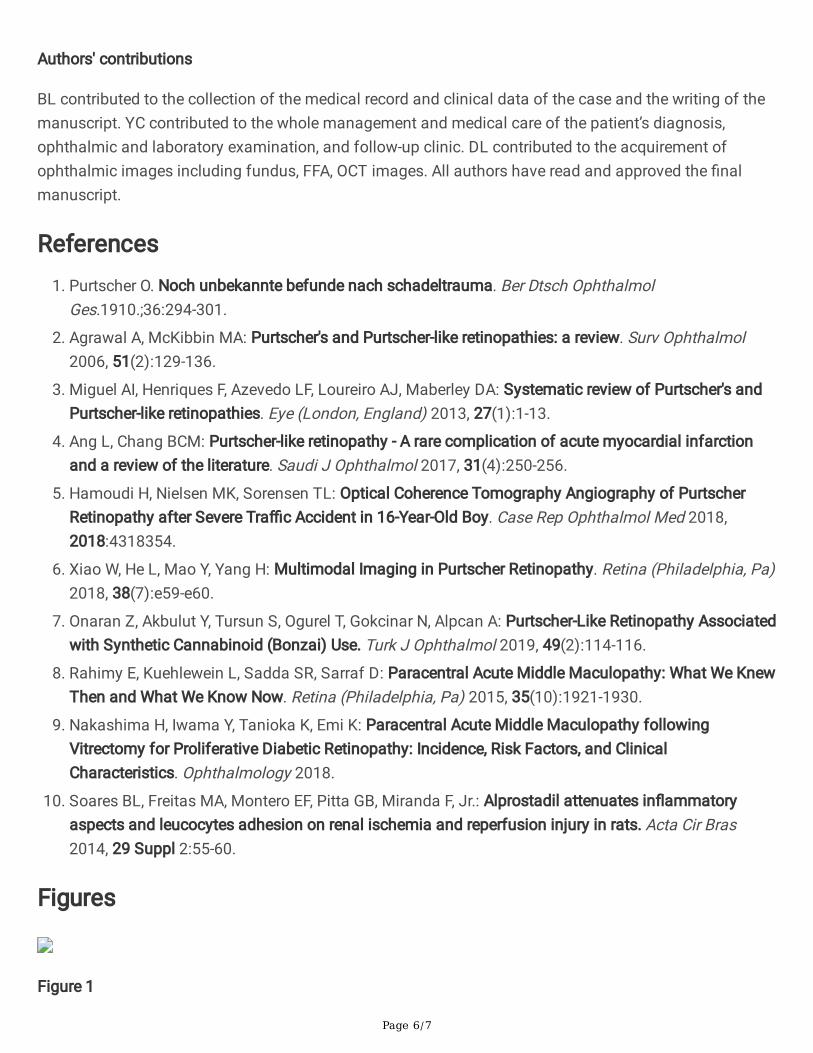
Multimodal imaging of the affected right eye. Fundus photography showed extensive spreading ofcotton-wool spots con�ned to the peripapillary and posterior pole with slight retinal haemorrhage, as wellas Purtscher �ecken around the macular fovea in the right eye. (A). FA presented delayed arteriovenouscirculation time (B1, B2). OCT B-scan of the fovea noted retinal oedema and PAMM-like lesion at INL (redarrows)(C). OCT-A of the fovea (6×6mm) indicates the condition of blood supplement in super�cial retina(D1), outer retina (D2), deep retina (D3) and choriocapillaris (D4). The blood �ow reduced apparently inboth the inner and deep retinal vascular plexuses and a honeycomb-like hypointense signal pattern at thechoriocapillaris layer.
Figure 2
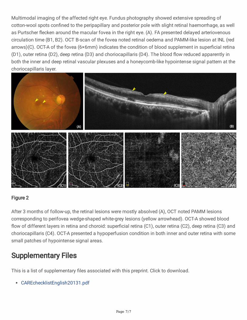
After 3 months of follow-up, the retinal lesions were mostly absolved (A), OCT noted PAMM lesionscorresponding to perifovea wedge-shaped white-grey lesions (yellow arrowhead). OCT-A showed blood�ow of different layers in retina and choroid: super�cial retina (C1), outer retina (C2), deep retina (C3) andchoriocapillaris (C4). OCT-A presented a hypoperfusion condition in both inner and outer retina with somesmall patches of hypointense signal areas.
Supplementary Files
This is a list of supplementary �les associated with this preprint. Click to download.


![The Guide - Diabetic Retinopathy - Vision Lossvisionloss.org.au/wp-content/uploads/2016/05/The... · the guide [diabetic retinopathy] What is Diabetic Retinopathy? Diabetic Retinopathy](https://static.documents.pub/doc/80x56/5e3ed00bf9c32e41ea6578a8/the-guide-diabetic-retinopathy-vision-the-guide-diabetic-retinopathy-what.jpg)


