263
Quantitative structure activity relationships for the biotransformation and toxicity of halogenated benzene-derivatives Implications for enzyme catalysis and reaction mechanisms
Quantitative structure activity relationships for the biotransformation and toxicity
of halogenated benzene-derivatives
Implications for enzyme catalysis and reaction mechanisms
Promotor: Dr. C. Veeger
Hoogleraar in de Biochemie
Co-promotor: Dr. Ir. I.M.C.M. Rietjens
Universitair hoofddocent
Vakgroep Biochemie
()no%^ r2o9^
Quantitative structure activity relationships for the biotransformation and toxicity
of halogenated benzene-derivatives
Implications for enzyme catalysis and reaction mechanisms
Proefschift
Ter verkrijging van de graad van
doctor in de landbouw- en milieuwetenschappen,
op gezag van de Rector Magnificus,
Dr. C.M. Karssen, in het openbaar te verdedigen
op vrijdag 7 juni 1996 des ochtends om elf uur in de Aula
van de Landbouwuniversiteit te Wageningen.
door
Nicole Hubertine Pauline Cnubben geboren 13 februari 1967, te Meerssen.
i/..-._
CIP-DATA KONINKLIJKE BIBLIOTHEEK, DEN HAAG
Cnubben, Nicole Hubertine Pauline Cnubben
Quantitative structure activity relationships for the biotransformation and toxicity of halogenated benzene-derivatives: implications for enzyme catalysis and reaction mechanisms / Nicole Hubertine Pauline Cnubben. -[S.I. : s.n.] Thesis University Wageningen. - With summary in Dutch. ISBN 90-5485-520-7 Subject headings: QSAR / biotransformation / toxicity / benzene-derivatives.
IWO '* WV »
The studies described in this thesis were carried out at the Department of
Biochemistry, Agricultural University Wageningen, Dreijenlaan 3, 6703 HA
Wageningen, The Netherlands.
This work was supported by the Netherlands Organization for Scientific Research
(NWO), section Medical Sciences.
Ter nagedachtenis aan mijn vader
Voor mijn familie
Voor Peter
Contents
Chapter Page
Introduction. 11
Biotransformation enzymes involved in the detoxification and 17 bioactivation of amino- and nitrobenzene derivatives.
Study on the regioselectivity and mechanism of the aromatic 69 hydroxylation of monofluoroanilines. (Chemico-Biological Interactions 85, 151-172, 1992)
Molecular orbital based quantitative structure-activity 93 relationship for the cytochrome P450-catalyzed 4-hydroxylation of halogenated anilines. (Chemical Research in Toxicology 7, 590-598, 1994)
A spectrophotometric assay for the detection of 2- 115 aminophenols in biological samples. (Analytical Biochemistry 220, 165-171, 1994)
The effect of varying halogen substituent patterns on the 131 cytochrome P450 catalyzed dehalogenation of 4-halogenated anilines to 4-aminophenol metabolites. (Biochemical Pharmacology 49, 1235-1248, 1995)
Relationships between the regioselectivity of the hydroxylation 157 of C4-substituted 2-fluoroaniline derivatives and their toxic endpoints. (Accepted for publication in Toxicology and Applied Pharmacology, 1996)
Chapter Page
Different metabolic pathways of 2,5-difluoronitro- and 2,5- 181 difluoroaminobenzene compared to molecular orbital substrate characteristics. (Chemico-Biological Interactions 94,49-75, 1995)
Influence of the halogen-substituent pattern of 210 fluoronitrobenzenes on their biotransformation and capacity to induce methemoglobinemia. (Accepted for publication in Toxicology and Applied Pharmacology, 1996)
10 Comparative MO-QSAR studies in various species including 235 man. (Accepted for publication in Chemico-Biological Interactions , 1996)
H Summary and conclusions. 253
Samenvatting voor niet-vakgenoten 261 Curriculum Vitae 267 List of publications 269 Dankwoord 271
Abbreviations and symbols
AO atomic orbital
ARNT aromatic hydrocarbon receptor nuclear translocator
ATP adenosine 5'-triphosphate
BD benzodioxole
BS A bovine serum albumine
BUN blood urea nitrogen
CO carbonmonoxide
DMA dimethylamine
DMG dimethylglycine
DMSO dimethylsulphoxide
Ea activation energy
EDTA ethylenediaminetetraacetate
E(HOMO) energy of the highest occupied molecular orbital
E(LUMO) energy of the lowest unoccupied molecular orbital
E(SOMO) energy of the single occupied molecular orbital
FAD flavin adenine dinucleotide
FMN flavin mononucleotide (riboflavin-5'-phosphate)
FMO flavin-containing monooxygenase
GC-MS gas chromatography - mass spectrometry
GSH reduced glutathione
GST glutathione S-transferase
GS-X glutathione conjugate of compound X.
7GT y-glutamyl transpeptidase
HOMO highest occupied molecular orbital
HPLC high performance liquid chromatography
HXO hypohalous acid
IR infra red
IS internal standard
Kms apparent Michaelis constant Km
K(js apparent Kj of the cytochrome P450-substrate complex
In natural logarithm
log P logarithm of experimental octanol/water partition coefficient
LUMO lowest unoccupied molecular orbital
Me2SO dimethylsulphoxide
MO
MP-8
NAD(P)H
NAT
NMR
PAPS
PCB PCN
PPAR
QSAR
SEM
SOMO
S
SO
ST
tBuOOH
TCA
TLC
TMAO
TSP
UDPG
UV
Ex
^•max
molecular orbital
microperoxidase 8
reduced nicotinamide-adenine dinucleotide
N-acetyltransferase
nuclear magnetic resonance
3'-phosphoadenosine5'-phosphosulphate
polychloorbiphenyl pregnenolone-16oc-carbonitrile
peroxisome proliferator-activated receptor
quantitative structure activity relationship
standard error of the mean
single occupied molecular orbital
substrate
oxygenated substrate
sulphotransferase
tertiair butyl hydroperoxide
trichloroacetic acid
thin layer chromatography
trimethylamine N-oxide
sodium 3-trimethylsilyl-(2,2,3,3-2H4)-propionate
uridine 5'-diphospho-glucuronyltransferase
ultra violet light
extinction coefficient at x nm
position of absorption maximum
CHAPTER 1
Introduction.
General aspects of biotransformation.
Living organisms are exposed to a great variety of natural and man-made
substances, such as pharmacological agents, industrial chemicals, environmental
pollutants, food additives etcetera. These chemical compounds may enter the body
accidently or on purpose through the skin, the gastrointestinal tract or the respiratory
tract. The fate of these compounds in the body is generally dependent on their
physico-chemical characteristics. For instance, highly lipophilic compounds will
accumulate in fat tissues due to their affinity for a hydrophobic environment, whereas
reactive compounds may react with cellular macromolecules, thereby altering their
structure and/or function.
Fortunately, organisms are equipped with a special defense system able to
deal with most of the low molecular weight compounds. This system consists of a
complex enzymatic machinery aiming to modify these body-foreign compounds
(xenobiotica) into more hydrophilic products (metabolites), thereby enhancing
efficient excretion into the bile or urine. This sophisticated process of metabolic
transformation of molecules, catalyzed by several enzyme systems, is called
biotransformation. For most xenobiotics, metabolic processes are not just one-step
events, but occur via multiple competing and sequential pathways. Classically,
biotransformation reactions are categorized essentially into three groups; phase I, II
and III. Phase I reactions involve the introduction of a polar functional group into the
molecule by oxidation, reduction or hydrolysis. In phase II reactions, a reactive
centre of a polar xenobiotic or of a metabolite from the phase I reaction is conjugated
generally with a more hydrophilic moiety. This can be for example a glutathione,
sulphate, glucuronide or acetate moiety. Finally, phase III consists of elimination
systems like the ATP-dependent P-glycoprotein and the glutathione S-conjugate
export pumps [Ishikawa, 1992].
-12- Introduction
Initially, biotransformation was only regarded as a detoxification mechanism
for exogenous as well as endogenous substances. However, biotransformation is often
associated with the introduction of a chemically reactive function into the molecule.
As a consequence, the ultimate or intermediate metabolites may elicit adverse effects
through interaction with critical cellular targets, eventually leading to disturbance of
biochemical and physiological processes. This aspect of biotransformation is called
bioactivation. The bioactivating properties of the biotransformation system, however,
may also serve as a basis for the generation of pharmacologically active compounds
within specific areas of the body to produce the actual therapeutic effect of the
administered drug. The balance between detoxification and bioactivation eventually
determines the expression of toxicity upon xenobiotic exposure.
Insight into the relationships between the structure and metabolism of
xenobiotics is urgently needed for the costly design and safety assessment of new
compounds in drug and agrochemical industries. Organisms are frequently exposed to
an almost unlimited number of natural chemicals and other xenobiotics, and data on
the biological effects of all these compounds are far from complete or even lacking.
This requires the definition of molecular structural features in order to classify the
biological activities of compounds. The recognition that substrate characteristics are
often related to biological activity of xenobiotics, has prompted the development of
QSARs or Quantitative Structure Activity Relationships. Initially, much studies were
devoted to obtain relationships between biological effects and substituent parameters
like log P or n, Hammett a and/or Taft Ea values to describe hydrophobic, electronic
and steric influences [for review see: Hansch and Zang, 1993; Lewis, 1990]. In those
studies relationships were primarily described for transport of compounds through
multicompartmental systems, substrate-receptor interactions and the ability to reach
the site of action. Later, it became increasingly evident that for many xenobiotics
biotransformation plays a critical role in the development of chemically induced
toxicities, because of the formation of reactive intermediates and metabolites. The
enzymatic generation of reactive species is often the response-determining step.
Hence, knowledge on the metabolic fate of xenobiotics and the molecular mechanisms
of biotransformation enzymes involved, is of crucial importance to the understanding
of the nature, site and mechanism of action of these compounds. Therefore, more and
more attention has been devoted to quantitative structure metabolism relationships. In
this way, molecular and biochemical toxicology aim to contribute to the setting of
priorities in toxicological research and to the development of safer chemicals used as
drugs, food additives and agrochemicals.
Chapter 1 -13-
Scope of this thesis.
The main objective of the studies presented in this thesis was the description
of QSARs for the cytochrome P450 and glutathione S-transferase mediated
biotransformation and toxicity of small aromatic molecules. The superfamilies of
cytochromes P450 and glutathione S-transferases are the most important and
numerous phase I and phase II biotransformation enzymes, respectively, involved in
the detoxification and bioactivation of a great variety of chemicals. The aim of the
present thesis was to describe QSARs on the basis of insight in the molecular
mechanisms of the enzymes involved. This insight is used to define the specific
substrate characteristic(s) that influence the reaction and can, thus, provide the
appropriate QSAR parameter to explain or even predict the outcomes of a
biotransformation reaction. Ultimately, the QSARs may provide a basis for the
prediction of the rate and regioselectivity of enzymatic conversion and
biotransformation-related toxicity. Besides, QSARs based on insight in the molecular
mechanisms of biotransformation enzymes may be helpfull in a more rational design
of new biocatalysts.
A general characteristic feature of biotransformation enzymes, especially
when compared to other enzymes, is their relatively, broad and partially overlapping
substrate specificity and regioselectivity. This property enables the biotransformation
system to metabolize an almost unlimited number of compounds by a limited number
of enzymes. The broad substrate specificity can be primarily attributed to the
relatively large active sites of the enzymes. In order to elucidate molecular
mechanisms of enzymes and determine the specific substrate characteristics that
influence biotransformation reactions, the contribution of the active site of the
enzymes in directing the juxtaposition of the substrates by steric factors have to be
minimalized. Therefore, the investigations of this thesis were focussed on relatively
small aromatic molecules.
General outline of this thesis.
Halogenated aromatic compounds are widely used in industry, commerce and
agriculture and are a major contribution to nowadays environmental pollution.
Modern agrochemical and pharmaceutical industries are known to synthesize wide
varieties of new halogenated aromatics, to be used as agrochemicals, dyes, drugs or
building blocks in the synthesis of new industrially relevant compounds. Organisms
-14- Introduction
are frequently exposed to these compounds or their degradation products with
possible toxicological consequences. Clearly, toxicological testing of all these newly
synthesized compounds is a time- and money-consuming problem.
Based on these considerations, the studies of this thesis were undertaken, in
order to investigate whether insight into the molecular mechanisms of
biotransformation enzymes is helpfull in defining QSARs for the metabolic fate of
aromatic compounds. Insight into factors that direct the rates and regioselectivities of
biotransformation processes will help to gain insight in factors that direct processes
of, and chances on, bioactivation or detoxification of xenobiotics to which the
organism is exposed.
In the present study the research was predominantly focussed on nitrogen
(amino or nitro) substituted halogenated aromatics, like anilines and nitrobenzenes.
These compounds were chosen, because -as outlined above- they are of industrial
relevance and used on a large scale, but also because their biotransformation is
known to include pathways leading to both detoxification and the generation of
reactive intermediates assumed to contribute to the toxicity of amino- and
nitrobenzene derivatives (bioactivation). In the present studies in particular, fluorine
containing compounds were investigated, due to the unique characteristics of this
type of halogen. Introduction of fluorines into a molecule influences the electron
distribution in the molecule and its lipophilicity, without introducing any major steric
chances since the van der Waals radius of fluorine (1.35A) is almost similar to that of
a hydrogen (1.20Â). Furthermore, fluorine substitution is frequently used for site-
specific blocking of metabolic pathways, since the strong C-F bond is thought to
prevent reactivity at a fluorinated center. Thus, the introduction of fluorines into a
molecule can be used to alter the rate, route and extent of drug metabolism with
minimal effect on steric influences [Park and Kitteringham, 1994]. Besides, the
metabolism of fluorine containing compounds can be elegantly monitored by 19F-
NMR.
In chapter 2 of this thesis, a general description of the enzymes primarily
involved in the biotransformation of the amino- and nitrobenzene derivatives is
given. For cytochromes P450 and glutathione S-transferases special attention is
focussed on the reaction mechanisms involved, because the studies of the present
thesis aim to base QSARs on the chemical substrate characteristics and the way in
which these characteristics may influence the supposed reaction mechanism.
In chapter 3 and 4 clear examples of QSAR studies are presented directed at
understanding either the regioselectivity (chapter 3) or the rate (chapter 4) of the
Chapter 1 -15-
cytochrome P450 catalyzed aromatic hydroxylation of a series of aniline derivatives.
The QSARs obtained are based on the reaction mechanism of the enzyme system
involved and computer calculated chemical reactivity parameters of the substrates.
To fully describe the biotransformation pathways of not only fluorinated
anilines (identified on the basis of 19F-NMR analysis), but also of other anilines with
chlorine, bromine or iodine substituents, methods to quantify the metabolites from
aromatic or N-hydroxylation were developed, as far as they were not available from
the literature. Chapter 5 describes the development of a new and sensitive chemical
assay for the detection of halogenated 2-aminophenols in biological samples. In
further studies of the thesis this method could be used for the identification and
quantification of 2-aminophenol derivatives in addition to HPLC and NMR analysis.
Using the available methods for the analysis of metabolites of aniline
derivatives, studies were carried out to investigate the influence of the nature, number
and position of halogen substituents at the aromatic ring of aniline derivatives on
metabolic pathways, with special emphasis on the bioactivation to toxic metabolites,
and, thus on the toxicity of the respective aromatic compounds. In chapter 6 the effect
of halogen substituent patterns on possibilities for bioactivation through the
cytochrome P450 catalyzed oxidative dehalogenation, leading to reactive
benzoquinoneimine metabolites, was investigated and quantified. In chapter 7 the
relationship between the regioselectivity of the hydroxylation of a series of aniline
derivatives and their actual toxic endpoint (nephrotoxicity and methemoglobinemia)
was studied, providing some general rules for the way in which biotransformation of
these compounds is influencing their toxicity.
Finally, attention was focussed on the changes in metabolic profiles observed
upon full oxidation of the amino moiety to a nitro substituent. In chapter 8 it is
outlined that the molecular orbital based QSARs obtained in chapter 4, but also in
recent studies with glutathione S-transferases [Rietjens et ai, 1995], can actually
explain why the amino derivatives are mainly metabolized through cytochrome P450
dependent metabolic pathways, whereas the nitro analogues are not, and, instead are
rather metabolized through glutathione S-transferase catalyzed reactions. Chapter 9
describes a molecular orbital based quantitative structure activity relationship for
both the biotransformation and reactivity of a series of fluoronitrobenzenes with
nucleophiles. This study provides a clear example of how chemical reactivity
parameters of fluoronitrobenzenes direct biotransformation processes and, as a
consequence, toxicity in rats.
-16- Introduction
A final study (chapter 10) actually demonstrates that the QSARs obtained for
the regioselectivity and rate of cytochrome P450 catalyzed conversions of the
relatively small halogenated benzene derivatives, can be extrapolated from rats to
various species, including man.
Altogether, the results of the present thesis fully support that the use of
computer calculated molecular orbital parameters are a useful additional tool to
study, not only the mechanisms of the enzymes involved in biotransformation of
xenobiotics, but also, to explain, or even predict the outcomes of biotransformation
pathways in mammalian systems exposed to these chemicals. Finally, the findings
described in this thesis are summarized and discussed in chapter 11.
REFERENCES.
Hansch C. and Zhang L., Quantitative structure activity relationships of cytochrome P450, Drug
Metabolism Reviews 25, 1-48, 1993.
Ishikawa T., The ATP-dependent glutathione S-conjugate export pump, Trends Biochem. Sei. 17, 463-
468,1992.
Lewis D.F.V., MO-QSARs: a review of molecular orbital-generated quantitative structure activity
relationships, Progress in drug metabolism, (Ed. G.G. Gibson), Taylor & Francis, Chapter 5,
205-255, 1990.
Park B.K. and Kitteringham N.R., Effects of fluorine substitution on drug metabolism:
pharmacological and toxicological implications, Drug Metabolism Reviews 26, 605-643,
1994.
Rietjens I.M.C.M., Soffers A.E.M.F., Hooiveld G.J.E.J., Veeger C. and Vervoort J., Quantitative
structure activity relationships based on computer calculated parameters for the overall rate of
glutathione S-transferase catalyzed conjugation of a series of fluoronitrobenzenes, Chem. Res.
Tox. 8,481-488, 1995.
CHAPTER 2
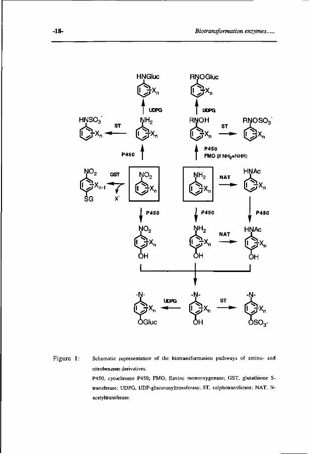
Biotransformation enzymes involved in the detoxification and bioactivation of amino- and nitrobenzene derivatives.
The phase I enzymes play an important role in the enzymatic oxidation of
xenobiotic chemicals. A number of these enzymes, contain a heme-cofactor (e.g.
cytochromes P450) or a FAD-cofactor (e.g. flavin-containing monooxygenase,
monoamine oxidase) as the site of dioxygen activation [Gibson and Skett, 1989;
Guengerich, 1990]. The phase II enzymes are involved in the conjugation of
compounds, generally yielding water-soluble metabolites, which can be excreted in
bile or urine. In this section the phase I enzymes cytochrome P450 and flavin-
containing monooxygenase as well as the phase II enzymes glutathione S-transferase,
N-acetyltransferases, UDP-glucuronyltransferase and sulphotransferase will be
discussed, because they are relevant for the biotransformation reactions of the present
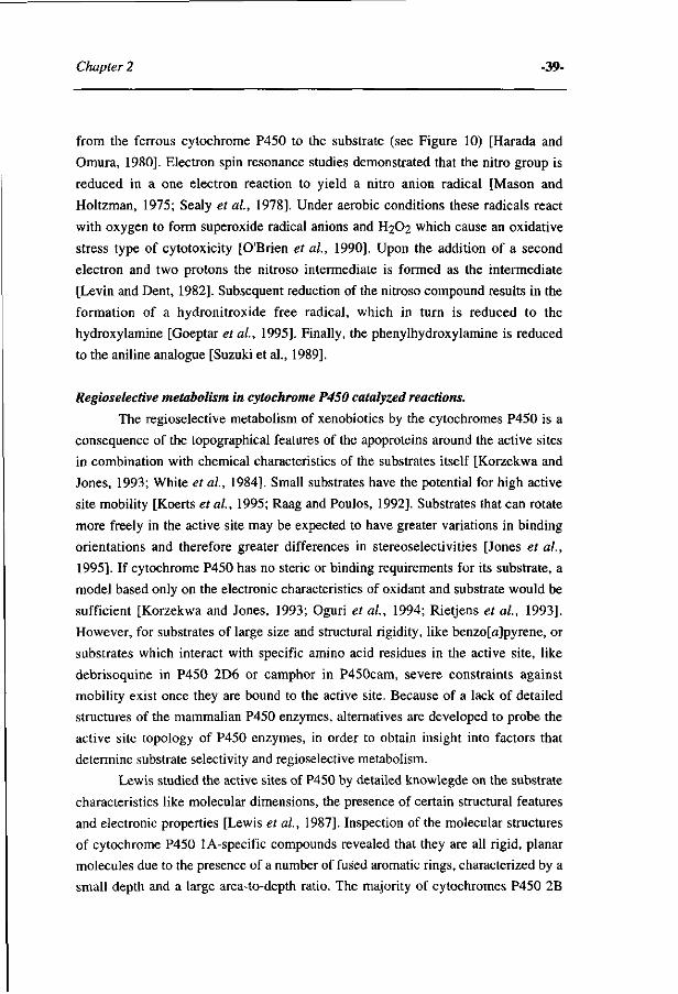
thesis (see Figure 1).
2.1. Phase 1 biotransformation enzymes.
2.1.1. Cytochrome P450 enzyme system.
General.
Cytochromes P450 constitute a family of enzymes that play a key role in the
oxidative, peroxidative and reductive biotransformation of a surprisingly large
number of structurally unrelated compounds. The cytochrome P450-dependent
monooxygenases are involved in many steps of the biosynthesis and biodégradation
of endogenous compounds such as steroids, fatty acids and prostaglandins, as well as
in the conversion of exogenous compounds such as drugs and environmental
-18- Biotransformation enzymes..
NO
HNGluc
t lls^rXn ~* l|s^ï'Xn
t
HNSO3" NHP ST '
P450
2 GST
^ X n - i "" r
P450
l " ^ x n
)H
-N-UDPG
H .-fxn - * -
)Gluc
RNOGluc
6x» t
RljL.. ST . ^ _ „
l|^jrXn *"~ ([^JXn
t
RNOSOo
P450 FMO (if NH2=NHR)
N02 NH2
0*» NAT
P450
NH,
i :TX„
NAT
)H
t ST
HNAc
óx" P450
HNAc
I '^Xn
-N-
I' J*n
iso,-
Figure 1: Schematic representation of the biotransformation pathways of amino- and
nitrobenzene derivatives.
P450, cytochrome P450; FMO, flavine monooxygenase; GST, glutathione S-
transferase; UDPG, UDP-glucuronyltransferase; ST, sulphotransferase; NAT, N-
acetyltransferase.
Chapter 2 -19-
pollutants, allowing their elimination from living organisms [Guengerich, 1993].
Furthermore, due to their ability to convert a vast array of substrates, cytochromes
P450 and also other heme-containing enzymes are recognized as powerfull
biocatalysts for industrial and environmental applications such as for example the
production of fine-chemicals or the introduction of the genes of these enzymes into
microorganisms that can be used for the bioremediation of contaminated sites. As a
result, there is great and broad interest in the relationship between P450 structure and
the mechanisms of substrate recognition and catalysis [Hasemann et al, 1995].
Another reason underlying the considerable interest in P450 proteins, originates from
the fact that their actions are not always benificial. Cytochromes P450 are frequently
involved in chemical carcinogenesis, since they catalyze the oxidative activation of
carcinogens to reactive electrophilic intermediates, that bind covalently to DNA, thus
damaging the genetic material and activating oncogenes. In addition the reactive
metabolites may induce the activation of the protein kinase C cascade leading to the
phosphorylation of key nuclear transcription factors involved in the regulation of
DNA replication [Parke, 1994].
Cytochromes P450 are found ubiquitously in animal and plant kingdoms
[Nelson et al, 1993]. The P450 enzymes inherite their name from the Soret
absorption maximum around 450 nm of the reduced Fe2+-CO complex which is
characteristic for the axial thiolate ligation, provided by the cysteinyl residue [Omura
and Sato, 1964]. In mammalia, these enzymes are found within almost every cell type
of the body. The liver is the organ, which not only expresses most of the different
cytochrome P450 genes, but also contains the highest P450 amounts, and, is thereby
the major site of the metabolism of xenobiotics, drugs and endogenous compounds.
The unique property of the cytochrome P450 enzyme system to metabolize
such a large variety of compounds originates from the existance of several types of
cytochrome P450 enzymes (the so-called multiplicity), their inducibility as well as
their broad and partially overlapping substrate specificity [Gonzalez, 1989;
Guengerich, 1993].
The cytochrome P450 gene family.
More than 250 genes of the so-called superfamily of cytochromes P450 can
be found in mammalia, birds, reptiles, fish, yeast, bacteria and plants. This
superfamily is composed of families and subfamilies of enzymes that have been
sequenced, characterized and classified on the basis of their primary amino acid
-20- Biotransformation enzymes..
sequence homology [Nelson et ai, 1993; Nebert, 1989; Nebert, 1991]. In general,
one gene family exhibits up to 36% resemblance to a cytochrome P450 of another
family, whereas within a single subfamily the genes always share greater than 59%
sequence similarity [Gonzales, 1994; Paine, 1995]. Two general functional classes of
P450 exist. One class containing P450s for the conversion of fatty acids (CYP4),
steroids (CYP17, 19, 21 and 27) and cholesterol (CYP7 and 11), which are well
conserved with respect to their catalytic activities and display a rather rigid substrate
and product specificity, due to their critical role in steroid and bile acid synthesis
[Gonzalez et al, 1994]. The other class of P450s consists of the enzymes encoded for
by genes of the CYP1, 2, 3 and 4 families, which are predominantly involved in the
metabolism of exogenous compounds [Paine, 1995]. The levels of the individual
cytochromes P450 may vary considerably among species and individuals dependent
on age, hormonal status, dietary factors (caffeine, charbroiled meat, alcohol),
xenobiotic exposure (barbiturates, smoking, drugs) and genetic polymorphisms.
Polymorphism in cytochrome P450 gene expression in humans has been described
for the drug nifedipine (CYP3A4) [Paine, 1995], S-mephenytoin (CYP2C19) [de
Morais et al, 1994] and debrisoquine (CYP2D6) [Gonzalez and Idle, 1994; Gonzalez
and Meyer, 1991; Gouch et al, 1990]. Differences in the overall cytochrome P450
patterns among individuals, but also differences in the catalytic activities and
regulatory pathways of cytochromes P450 may result in altered metabolism and
hence can lead to different pharmacological or toxic responses. This aspect of
variability is of present concern in pharmacological industries [Nedelcheva and Gut,
1994]. For instance, the CYP2C subfamilies are the most abundantly expressed
P450s in the rat liver, whereas in humans the CYP3A subfamily predominates
[Gonzalez, 1992; Paine, 1995], indicating that extrapolation of biotransformation and
toxicity data from rodents to humans in some cases is very difficult or even
impossible.
The best known forms of the cytochromes P450 involved in the
biotransformation and bioactivation of drugs and other xenobiotics belong to the
CYP 1 A, 2A, 2B, 2D, 2E and 3A gene families. Cytochromes P450 of the 1A1 and
1A2 subfamilies are mainly involved in the metabolism of respectively polycyclic
hydrocarbons and arylamines, amino acid and protein pyrolysates [Gonzalez, 1992;
Shimada and Nakamura, 1987]. CYP2A6 exhibits activity towards activation of
benzo(a)pyrene, aflatoxin Bi and nitroso compound metabolism [Crespi et al, 1990;
Guengerich, 1992]. CYP2B6 is involved in the activation of the prodrug
cyclophoshamide [Chang and Waxman, 1993]. The CYP2D6 is well known due to its
Chapter 2 -21-
polymorphic expression in man and is involved in the biotransformation of
debrisoquine and sparteine. CYP2E1 plays an important role in the metabolism of
numerous low molecular weight halogenated hydrocarbon species (e.g. CCI4,
CHCI3), benzenes and dialkylnitrosamines [Guengerich et al, 1991; Yang et al,
1990]. The most abundantly expressed hepatic enzyme CYP3A4 has been
demonstrated to catalyze the conversion of numerous large compounds like aflatoxin
B1, nifedipine and testosteron [Gallagher et al, 1994].
Regulation and induction.
The cytochrome P450 patterns are extremely variable among individuals
(genetic polymorphism) and are partially dependent on xenobiotic exposure (P450
induction or inhibition) [Nedelcheva etal, 1994]. Induction leads to variation in the
cytochrome P450 enzyme patterns, and thereby to modulation of biotransformation
activities. The regulation of the cytochromes P450 is very complex, involving
increase of transcription and translation as well as mRNA- and protein stabilisation
[Nedelcheva et al, 1994; Nelson et al, 1993; Okey et al, 1990]. The mechanism of
induction of cytochromes P450 is dependent on the type of inducer.
In general, cytochrome P450 inducers are classified into five classes, named
after the prototype inducer, which are the polycyclic aromatic hydrocarbon-, the
phénobarbital-, the ethanol-, the glucocorticoid- and the peroxisome-proliferator-
inducible cytochromes P450. The aromatic hydrocarbon (Ah) receptor is involved in
the regulation of the CYP1A1 and 1A2 genes. This receptor is composed of two
heterodimeric subunits consisting of a ligand binding subunit and a subunit called the
Ah receptor nuclear translocator (ARNT). Inducers like TCDD (2,3,7,8-
tetrachlorodibenzo-p-dioxin) or other polycyclic aromatic hydrocarbons, bind to the
Ah receptor and this couple binds to the ARNT protein. The complex is translocated
to the cell nucleus, where it binds to a specific region of the CYP1 Al gene, the so-
called xenobiotic regulatory element. The transcription of the CYP1A1 gene is
stimulated by this interaction by a yet not fully understood mechanism [Guengerich,
1993]. CYP1A2 is to a minor extent coinduced with CYP1 Al, although an additional
mechanism for CYP1A2 induction has been demonstrated to exist. This alternative
mechanism is mediated by benzodioxole (BD) compounds such as safrole, isosafrole
and myristicin,which are involved in the regulation of the CYP1A2 gene, and act by
mechanisms which are independent of the Ah receptor [Adams et al, 1993; Ryu et
al, 1995]. It has been suggested that formation of a BD metabolite-cytochrome P450
-22- Biotransformation enzymes..
complex is asssociated with decreased turnover and accumulation of distinct P450
enzymes [Cook and Hodgson, 1985; Steward et ai, 1985], The barbiturates constitute
the second type of P450 inducers, and primarily induce the CYP2B subfamilies and
to a minor extent the CYP2A, 3A and 2C subfamilies [Waxman and Azaroff, 1992].
The exact mechanism by which the cell recognizes this group of inducers is not yet
established. One has speculated that a phenobarbital-type receptor exists that
eventually leads to increased transcription of the CYP2B gene and as a result
increased levels of mRNA. In the presence of barbiturates a positive regulator protein
is produced that displaces the repressor allowing the enhanced production of P450
mRNA [Shaw and Fulco, 1992]. Molecular shape has been suggested to be a factor in
the ability of compounds to induce P450s, with globular compounds of specified
molecular dimensions being better inducers of CYP2B1, and compounds with planar
configurations inducing P4501A [Lewis et al, 1986; Lewis et ai, 1987]. The ethanol
inducible CYP2E subfamily is regulated by substrates such as ethanol, aceton or
isoniazide, primarily through protein and mRNA stabilization. Binding of the
substrate or its metabolite to CYP2E1 protects the protein from rapid degradation.
Pregnenolone-16a-carbonitrile (PCN) or dexamethasone act as inducers of the
glucocorticoid inducible CYP3A subfamily, predominantly due to activation of gene
transcription and consequently mRNA increase [Simmons et al, 1987]. Finally, the
genes of the enzymes belonging to the P4504A subfamily are transcriptionally
activated by a structurally diverse groups of chemicals known as the peroxisome
proliferators (e.g. clofibric acid, linoleic acid). Induction has been reported to be
mediated by a receptor named PPAR, peroxisome proliferator-activated receptor
[Issemann and Green, 1990]. The PPARs are activated by peroxisome proliferators in
a process in which the ligand-receptor complex is thought to bind to the regulatory
regions of responsive genes, thereby enhancing transcription [Muerhoff et al, 1992].
Crystal structures of cytochromes P450.
The mammalian cytochrome P450 enzymes are membrane-bound and as such
have been resistant to crystallization and detailed structural characterization. Due to
the hydrophobic character, the protein has a tendency to aggregate rather than form
distinct crystals. The relationship between the amino acid sequences of P450s and
their three-dimensional structures is crucial for a better understanding of their
function, such as substrate selectivity, stereospecificity of hydroxylation reactions
and interactions with electron-transfer partners. Until the crystal structure of a
Chapter 2 -23-
mammalian cytochrome P450 is available, three-dimensional models of eukaryotic
P450s constructed from amino acid sequence alignments with P450RM3> P450cam.
P450ery and P450terp are helpful in identifying potential residues in cytochromes
P450 that might be important in the function of these enzymes and these residues are
candidates for site-directed mutagenesis [Hasemann et al, 1995; Lewis, 1995;
Nelson and Strobel, 1989; Ouzounis andMelvin, 1991; Zvelebil etal., 1991].
The cytochromes P450 are divided into two classes based on their sequence
homology and the identity of their redox-partner protein. The class I P450s comprise
the soluble bacterial and membrane-bound mitochondrial P450 systems which consist
of three components, namely FAD reductase, a non-heme iron-sulfur protein,
putidaredoxin (bacterial) or adrenodoxin (mitochondrial), and the cytochrome P450
protein. The microsomal cytochromes P450 belong to the class II enzymes and
receive their NADPH-derived electrons directly from a FAD/FMN containing
reductase [Guengerich, 1993; Hasemann etal., 1995; Li and Poulos, 1994].
The soluble class I cytochrome P450cam (product of the CYP101 gene),
which catalyzes the conversion of camphor to 5-exo-hydroxycamphor, has served for
some time as the only prototypical cytochrome P450 in providing detailed chemical,
physical, and structural information in order to understand detailed structure-function
relationships for the superfamily. Recently, the structure of additional class I
enzymes cytochrome P450terp (product of the CYP108 gene) and cytochrome
P450eryp (product of the eryF gene) as well as the first structure of a class II enzyme,
the hemoprotein domain of cytochrome P450RM3 (product of the CYP102 gene)
became available [Boddupalli et ai, 1992; Cupp-Vickery and Poulos, 1995;
Ravichandran et al, 1993]. P450terp is a bacterial enzyme which catalyzes the
oxidation of a-terpineol. P450eryF catalyzes the 6S-hydroxylation of the large
substrate 6-deoxyerythronolide B, the initial reaction in a multistep pathway to
convert 6-deoxyerythronolide B into the antibiotic, erythromycin. P450BM3 ' S a
flavocytochrome monooxygenase isolated from Bacillus Megaterium and catalyzes
the hydroxylation and epoxidation of several fatty acid substrates. Although BM3 is
also a soluble bacterial enzyme, it is considered to be a better prototype for
microsomal P450s than the soluble P450cam. P450terp and P450eryF as it is grouped
within the class II P450s based on both sequence homology and its requirements for a
FAD/FMN containing reductase domain as a source of electrons. Therefore, BM3
more closely resembles the sequence and functional properties of eukaryotic
microsomal P450s. BM3 is unique among P450s in that the holoenzyme is
catalytically self-sufficient, since it consists of both a FAD/FMN containing
-24- Biotransformation enzymes..
reductase domain and a heme-containing P450 domain fused into a single 119000
dalton polypeptide.
Although the sequence identity between the (crystallized) P450s is generally
low, these proteins have a similar overall topology [Cupp-Vickey and Poulos, 1995;
Hasemann et al, 1995]. The most highly conserved region of the P450 structure is
the a-helical domain located in the protein core, namely the I and L helices and the
heme binding region. The long I helix which extends across the entire molecule is a
structural feature characteristic of all P450s. Much attention has been focussed on the
role of the conserved threonine, a residue of the I helix, in oxygen scission during
catalysis. This threonine has been proposed to act as the proton donor to bound
dioxygen during the reaction cycle [Gerber and Sligar, 1992; Raag et al, 1990].
However, in cytochrome P450eryF, where an alanine is located at a position
homologous to that of threonine in the other P450s, a possible participation of the
substrate in a network of hydrogen bonds, providing a proton shuttle pathway to the
iron linked dioxygen, has been suggested [Cupp-Vickery and Poulos, 1995]. The
underlying reason for the threonine to alanine substitution in P450eryF might be, that
its large substrate would be sterically hindered by the side-chain methyl group of a
threonine residue.
The heme prosthetic group is embedded between the I and L helices of the a
domain. The cysteine pocket is the most significant feature in all cytochromes P450
as it provides the axial heme ligand, a conserved cysteine residue. The residue
tryptophan 96 of cyt P450BM3 is virtually invariant in most microsomal P450s, and is
thought to be involved in electron transfer from the FMN moiety of reductase to the
heme [Lewis, 1995]. In P450BM3 the active site heme is accessible through a long
hydrophobic channel formed primarily by the ß-domain and the B' and F helices of
the a domain, lined with mostly non-aromatic hydrophobic residues. At the entrance
of the substrate binding pocket an exposed hydrophobic patch is formed, that may be
important for the initial docking of a lipophilic substrate, since these residues are
solvent exposed, mobile and located adjacent to the binding pocket [Ravichandran et
al, 1993].
Substantial differences in structure are observed for the B', F and G helices,
which together form the majority of the substrate-access channel, and for the regions
of importance in redox partner binding. The structural differences around the
substrate binding pocket could confer different substrate specificities. For instance,
the extended structure of the B' helix of P450terp and the F-G loop of both P450terp
and P450ßM3 provide a much more open or accessible binding pocket as that seen in
Chapter 2 -25-
P450cam [Hasemann et al, 1995]. Due to the large structural variation of the
substrate-binding pocket of the different P450s, the possibility to predict the potential
residues that determine substrate specificity of other P450s whose three-dimensional
structures have not been determined, is limited. Indeed, situations are known where
very small residue changes (even one residue) are known to change catalytic
specificity in a remarkable way [Lindberg and Negishi, 1989].
Catalytic cycle
The cytochromes P450 possess a so-called monooxygenase activity,
indicating that one oxygen atom combines with two electrons and two protons to
form H20, whereas the other oxygen is inserted into the substrate, following the
stoichiometry, where S represents the substrate;
NAD(P)H + H+ + 0 2 + S > NAD(P)+ + H20 + SO.
The catalytic mechanism of monooxygenation can be divided in two events; oxygen
activation and substrate oxidation. The most important steps of the catalytic
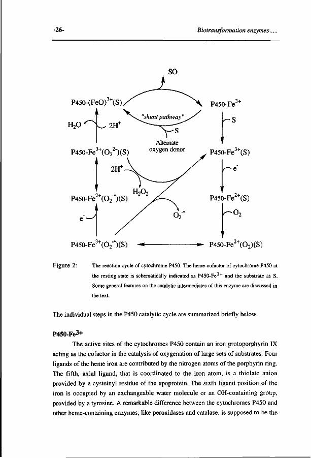
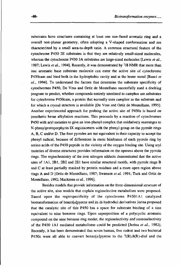
cytochrome P450 cycle are presented in Figure 2.
The cytochrome P450 system is associated with the membrane of the smooth
endoplasmatic reticulum and consists of several components which are involved in
the electron transport from NADPH or NADH to the cytochrome P450 enzyme, the
protein which ultimately catalyzes the final electron transfer to molecular oxygen and
the insertion of an oxygen atom into the substrate. The components that take part in
the electron transfer to cytochrome P450 are the flavoproteins NADPH- or NADH-
cytochrome reductase (the first and second electron) and the hemoprotein cytochrome
b5 (the second electron). For an optimal catalytic activity, the cytochrome P450
multi-enzyme system also requires the presence of phospholipids, important
components of membranes [Lu et al., 1976]. The phospholipids have been suggested
to assist in complex formation between P450 and its redox partners [Strobel et al,
1970]. The presence of such a hydrophobic environment has been assumed to target
hydrophobic endogenous compounds or xenobiotica to these proteins, resulting in a
higher effective concentration in the neighbourhood of the active site [Ebel et al,
1978; Parry et al, 1976].
-26- Biotransformation enzymes..
P450-(FeO)J+(S)
HoO
P450-Fe3+(O22")(S)
Alternate oxygen donor
2tT
P450-Fe2+(O2"')(S) n
P450-Fe3+(S)
P450-Fe3+(O2"')(S)
P450-Fe2+(S)
•O,
P450-Fe2+(O2)(S)
Figure 2: The reaction cycle of cytochrome P450. The heme-cofactor of cytochrome P450 at
the resting state is schematically indicated as P450-Fe-'+ and the substrate as S.
Some general features on the catalytic intermediates of this enzyme are discussed in
the text.
The individual steps in the P450 catalytic cycle are summarized briefly below.
P450-Fe3+
The active sites of the cytochromes P450 contain an iron protoporphyrin IX
acting as the cofactor in the catalysis of oxygenation of large sets of substrates. Four
ligands of the heme iron are contributed by the nitrogen atoms of the porphyrin ring.
The fifth, axial ligand, that is coordinated to the iron atom, is a thiolate anion
provided by a cysteinyl residue of the apoprotein. The sixth ligand position of the
iron is occupied by an exchangeable water molecule or an OH-containing group,
provided by a tyrosine. A remarkable difference between the cytochromes P450 and
other heme-containing enzymes, like peroxidases and catalase, is supposed to be the
Chapter 2 -27-
presence of a cysteine instead of a histidine or tyrosine residue respectively as the
axial ligand. This cysteine in cytochromes P450 is responsible for the characteristic
absorption at 450 nm of the reduced Fe2+-CO complex. Upon occupation of the sixth
ligand, the iron atom is in the plane of the porphyrin ring in the hexacoordinated low
spin (S=l/2) Fe3+ form, where the five 3d electrons of the iron are maximally paired.
When the sixth ligand position is unoccupied, the iron atom is out of the plane of the
porphyrin ring and in the pentacoordinated, high spin (S=5/2) Fe3+ form, where the
five 3d electrons of the iron are maximally unpaired.
P450-Fe3+S
Substrates may interact in three ways with the active site centre of
cytochromes P450. The type 1 substrates show high affinity for the high spin state
and have been proposed to interact with a hydrophobic site near the heme group.
Reversed type 1 substrates have a higher affinity for the low spin state. Finally, type
2 substrates such as the nitrogen containing compounds, have been assumed to
occupy the sixth axial ligand position, which is reflected by a high to low spin shift.
For some P450s (e.g. P450cam) the substrate binding, shifts the high spin-low
spin equilibrium in favor of the high spin configuration and results in an increase in
the redox-potential, thereby facilitating the first one-electron reduction of the
enzyme-substrate complex. However, the iron spin-state of most of the mammalian
P450s is not correlated with the redox-potential or a faster reduction rate [Kominami
and Takemori, 1982]. Despite the lack of a correlation between the degree of shift of
the spin equilibrium, the change in redox-potential and the rate of cytochrome P450
reduction [Guengerich, 1983], it is generally assumed that substrate-binding
facilitates the uptake of the first electron. Besides a possible increase in the redox-
potential, substrate binding to cytochrome P450 may also enhance both the rate of
association and the affinity of the reductase-P450 complex and consequently the
efficiency of electron flow from reductase to P450 [Backes and Eyer, 1989].
P450-Fe2+S For the mammalian microsomal P450s, the first electron comes from
NADPH, transferred by the flavoprotein NADPH-cytochrome P450 reductase via
their FAD and FMN cofactors to the heme complex of cytochromes P450 converting
its ferric to the ferrous state. The reductase functions as a brigde between the
NADPH, a two electron donor, and cytochrome P450, an one electron acceptor, via a
multistep reaction cycle proposed by Iyanagi et al. [1981]. Cytochromes P450 are
-28- Biotransformation enzymes.
also known to possess reducing activity, and this may occur at this stage of the
catalytic cycle. Under anaerobic conditions, certain xenobiotics such as halogenated
alkanes, azo dyes, nitro compounds and quinones, may accept electrons directly from
the reduced P450-Fe2+ complex [Goeptar et al., 1995].
P450-Fe2+(O2)-S <-> P450-Fe3+(O2->S
After the one-electron reduction, dioxygen can be coordinated to the sixth
position, thus forming the hexacoordinated P450-Fe2+(O2)-S.
Mossbauer spectroscopy on this one-electron reduced cytochrome P450cam substrate
dioxygen complex indicated that the electron resides on the oxygen molecule instead
of on the iron; P450-Fe3+(O2"')S [Sharrock et ai, 1976]. At this point, a reactive
superoxide anion can dissociate returning the enzyme to its Fe3+-S state.
P450-Fe2+(O2")S <-> P450-Fe3+(O22-)S.
The second electron can be provided by either NADPH via NADPH-
cytochrome P450 reductase as well as by NADH via NADH-cytochrome reductase
and cytochrome b5. For mitochondrial and bacterial cytochrome P450 the first and
second electron are both donated by the NADH-flavoprotein ferredoxin system. The
transfer of a second electron and of a proton leads to a transient intermediate
structure, which is assumed to be formally equivalent to a peroxide like complex,
cytochrome P450-Fe3+(OOH~)-S, although this is yet not unambigously proven. At
this stage the reactive hydrogen peroxide may be released, leading to uncoupling of
the P450 catalytic cycle. The P450-Fe3+(OOH~) species has been implicated in the
oxidation of substrates that contain a carbonyl-group. When the carbonyl group is
correctly positioned in the active site, the P450-Fe3+(OOH~) is intercepted to produce
a carbonyl-adduct, rather than decompose to the usual cytochrome P450 high-valent
iron-oxo species (see below) [Akthar and Wright, 1991].
Cleavage of the dioxygen bond Cytochromes P450 are capable of cleaving the dioxygen bond in two ways,
namely the heterolytic or homolytic bond scission. Organic hydroperoxides and
peroxyacids are frequently used to model the putative P450-Fe3+(OOH") complex
formed immediately prior to dioxygen bond cleavage during the reaction cycle of this
enzyme, the so-called "peroxide shunt" pathway [Mansuy et ai, 1989]. As shown in
Figure 3, heterolytic cleavage of the dioxygen bond generates the high-valent iron-
oxo porphyrin cation radical species (homologous to compound I of peroxidases),

Chapter 2 -29-
together with a ROH molecule, while homolytic cleavage results in the formation of
an iron-oxo species (homologous with compound II of peroxidases) and reactive
alkoxy radicals (RO) capable of hydrogen abstraction from the substrate.
A main function of the cysteinyl ligand is presumed to be the assistance in the
heterolytic cleavage of the dioxygen bond, through electron donation. This is based
for instance on the observation that the ratio of heterolysis to homolysis increases
substantially, when the proximal histidinyl ligand in myoglobin is replaced by a
cysteine [Adachi et al, 1993]. The observed preference for heterolysis is probably
due to a greater electron push provided by the cysteine thiolate relative to that of the
histidine imidazole [Adachi et al, 1993; Allentoff et al 1992; Higuchi et al, 1990].
Additionally, the cleavage-mode of the dioxygen bond has been demonstrated to
depend on the polar nature of the peroxy bond [Lee and Bruice, 1985], the
architecture of the P450 active site and the structural dimensions of the
hydroperoxides. The latter two factors are related to the degree of water present in the
active site, which assists in the highly regulated delivery of protons into the active
site by way of a hydrogen-bonding network [Correia et al, 1995].
heterolytic
Por-Fe3+ + ROOH
•*• Por+'FeIV=0 + ROH
PorFe iv=0 + H+ + RO homolytic
Figure 3: Hetero- versus homolytic dioxygen bond scission in cytochrome P450. The
porphyrin of P450 is schematically represented as "Por".
P450 (FeO)3+ S. Upon heterolytic cleavage of the dioxygen bond, the so-called cytochrome
P450 high-valent iron-oxo porphyrin cation radical complex is formed [Larroque et
al. 1990]. The porphyrin radical cation thus produced is stabilized by resonance over
the extensive network of conjugated double bonds of the porphyrin. Knowledge on
the exact nature of the active species is based on indirect evidence obtained from
studies on other hemo-proteins (peroxidases) and iron-porphyrin model systems
[Mansuy, 1987; Mansuy et ai, 1989]. The active species, for convenience
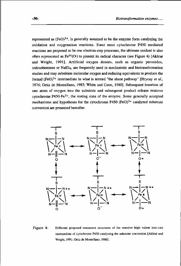
-30- Biotransformation enzymes.
represented as (FeO)3+, is generally assumed to be the enzyme form catalyzing the
oxidation and oxygenation reactions. Since most cytochrome P450 mediated
reactions are proposed to be one electron-step processes, the ultimate oxidant is also
often represented as Fe IV(0) to present its radical character (see Figure 4) [Akhtar
and Wright, 1991]. Artificial oxygen donors, such as organic peroxides,
iodosobenzene or NaIC>4, are frequently used in mechanistic and biotransformation
studies and may substitute molecular oxygen and reducing equivalents to produce the
formal (FeO)3+ intermediate in what is termed "the shunt pathway" [Hrycay et al,
1976; Ortiz de Montellano, 1985; White and Coon, 1980]. Subsequent insertion of
one atom of oxygen into the substrate and subsequent product release restores
cytochrome P450-Fe3+, the resting state of the enzyme. Some generally accepted
mechanisms and hypotheses for the cytochrome P450 (FeO)3+ catalyzed substrate
conversion are presented hereafter.
s s s
M | _ N N — I — N N-— —N
N — 1 | N O
N —| N N —| N 0~ O»
! ! t T s
N - N + . N — — N + . N — —N+«
: / Il % . . M -M M — I r> N ' _ | | _ ^ N N - | — N N - | — N
o o o .
Figure 4 : Different proposed resonance structures of the reactive high valent iron-oxo
intermediate of cytochrome P450 catalyzing the substrate conversion [Akhtar and
Wright, 1991; Ortiz de Montellano, 1986].
Chapter 2 -31-
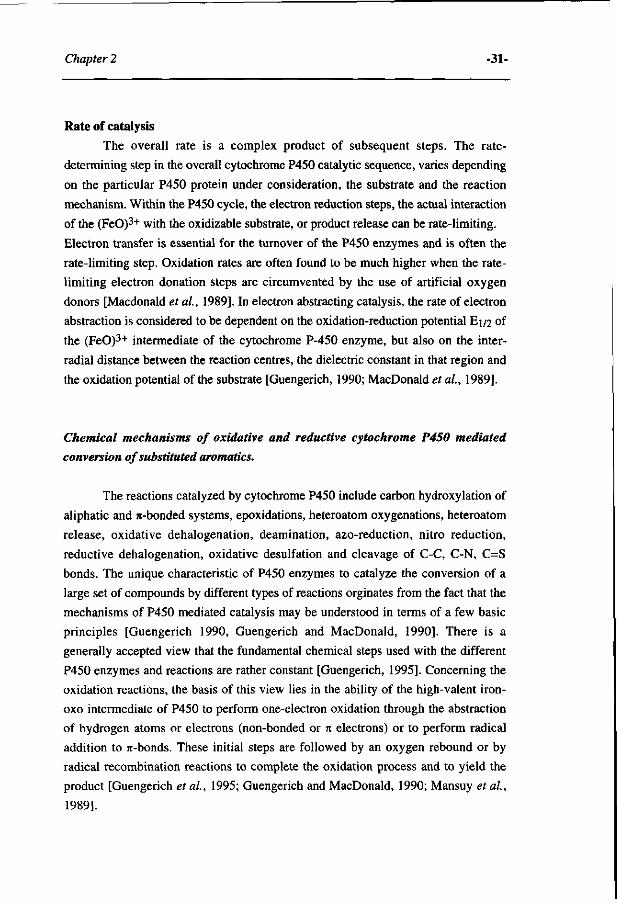
Rate of catalysis
The overall rate is a complex product of subsequent steps. The rate-
determining step in the overall cytochrome P450 catalytic sequence, varies depending
on the particular P450 protein under consideration, the substrate and the reaction
mechanism. Within the P450 cycle, the electron reduction steps, the actual interaction
of the (FeO)3+ with the oxidizable substrate, or product release can be rate-limiting.
Electron transfer is essential for the turnover of the P450 enzymes and is often the
rate-limiting step. Oxidation rates are often found to be much higher when the rate-
limiting electron donation steps are circumvented by the use of artificial oxygen
donors [Macdonald etal, 1989]. In electron abstracting catalysis, the rate of electron
abstraction is considered to be dependent on the oxidation-reduction potential E1/2 of
the (FeO)3+ intermediate of the cytochrome P-450 enzyme, but also on the inter-
radial distance between the reaction centres, the dielectric constant in that region and
the oxidation potential of the substrate [Guengerich, 1990; MacDonald et al, 1989].
Chemical mechanisms of oxidative and reductive cytochrome P450 mediated
conversion of substituted aromatics.
The reactions catalyzed by cytochrome P450 include carbon hydroxylation of
aliphatic and w-bonded systems, epoxidations, heteroatom oxygenations, heteroatom
release, oxidative dehalogenation, deamination, azo-reduction, nitro reduction,
reductive dehalogenation, oxidative desulfation and cleavage of C-C, C-N, C=S
bonds. The unique characteristic of P450 enzymes to catalyze the conversion of a
large set of compounds by different types of reactions orginates from the fact that the
mechanisms of P450 mediated catalysis may be understood in terms of a few basic
principles [Guengerich 1990, Guengerich and MacDonald, 1990], There is a
generally accepted view that the fundamental chemical steps used with the different
P450 enzymes and reactions are rather constant [Guengerich, 1995]. Concerning the
oxidation reactions, the basis of this view lies in the ability of the high-valent iron-
oxo intermediate of P450 to perform one-electron oxidation through the abstraction
of hydrogen atoms or electrons (non-bonded or n electrons) or to perform radical
addition to rc-bonds. These initial steps are followed by an oxygen rebound or by
radical recombination reactions to complete the oxidation process and to yield the
product [Guengerich et al., 1995; Guengerich and MacDonald, 1990; Mansuy et al,
1989].
-32- Biotransformation enzymes..
Only the basic molecular mechanisms of the types of metabolic transformations that
are within the scope of this thesis (aliphatic and aromatic hydroxylation, epoxidation,
heteroatom oxidation, oxidative dehalogenation and nitroreduction) will be
considered here in more detail.
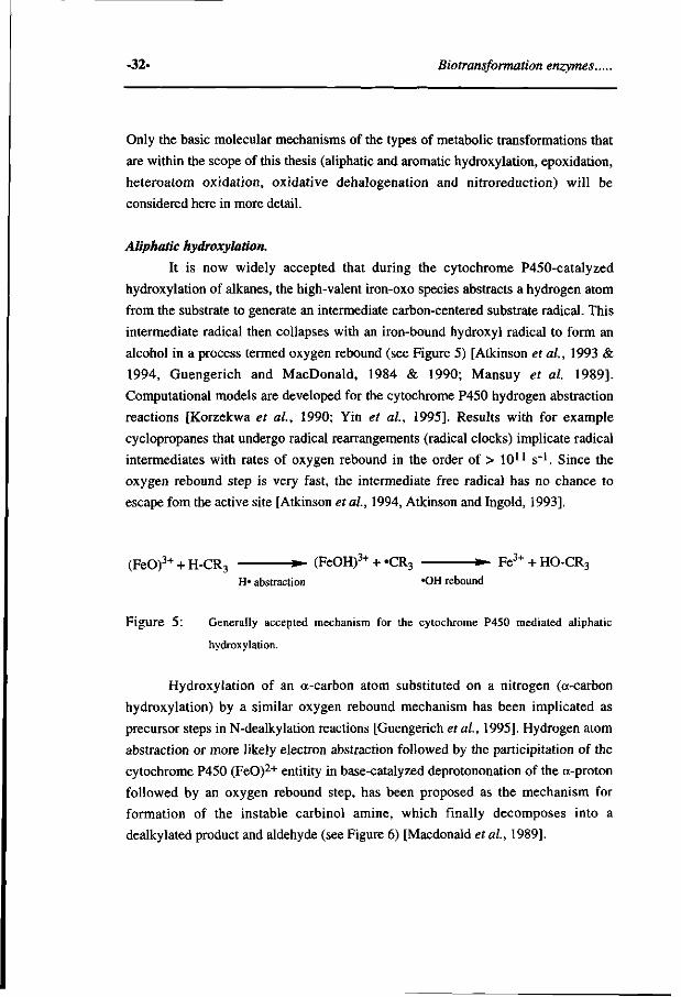
Aliphatic hydroxylation.
It is now widely accepted that during the cytochrome P450-catalyzed
hydroxylation of alkanes, the high-valent iron-oxo species abstracts a hydrogen atom
from the substrate to generate an intermediate carbon-centered substrate radical. This
intermediate radical then collapses with an iron-bound hydroxyl radical to form an
alcohol in a process termed oxygen rebound (see Figure 5) [Atkinson et ai, 1993 &
1994, Guengerich and MacDonald, 1984 & 1990; Mansuy et al. 1989].
Computational models are developed for the cytochrome P450 hydrogen abstraction
reactions [Korzekwa et ai, 1990; Yin et ai, 1995]. Results with for example
cyclopropanes that undergo radical rearrangements (radical clocks) implicate radical
intermediates with rates of oxygen rebound in the order of > 1011 s"1. Since the
oxygen rebound step is very fast, the intermediate free radical has no chance to
escape fom the active site [Atkinson et ai, 1994, Atkinson and Ingold, 1993].
(FeO)3+ + H-CR3 • (FeOH)3++ «CR3 * • Fe3++HO-CR3
H« abstraction 'OH rebound
Figure 5: Generally accepted mechanism for the cytochrome P450 mediated aliphatic
hydroxylation.
Hydroxylation of an a-carbon atom substituted on a nitrogen (a-carbon
hydroxylation) by a similar oxygen rebound mechanism has been implicated as
precursor steps in N-dealkylation reactions [Guengerich et ai, 1995]. Hydrogen atom
abstraction or more likely electron abstraction followed by the participitation of the
cytochrome P450 (FeO)2+ entitity in base-catalyzed deprotononation of the a-proton
followed by an oxygen rebound step, has been proposed as the mechanism for
formation of the instable carbinol amine, which finally decomposes into a
dealkylated product and aldehyde (see Figure 6) [Macdonald et ai, 1989].
Chapter 2 -33-
H ,3+
OH I
(FeO)3+ + RHCXRn • (FeOH)J+ + RHCXRn • FeJ+ + RHCXRn
H« abstraction .QH rebound
a in
X) H
\2+ '•+ (FeO)z+ + RHCXRn
I S t RC=0 + HXRn
H
Figure 6: Proposed mechanism for heteroatom dealkylation (X represents O, N or S).
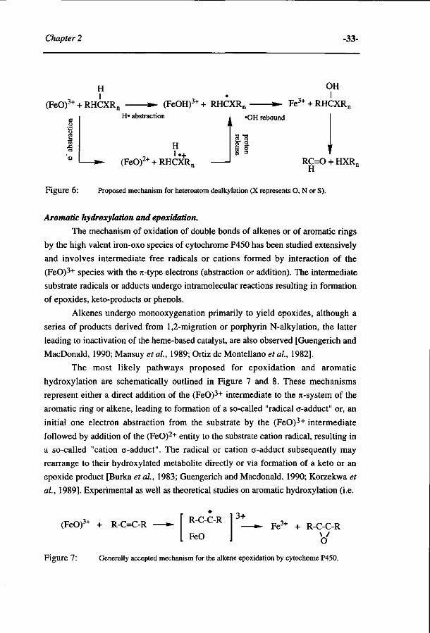
Aromatic hydroxylation and epoxidation.
The mechanism of oxidation of double bonds of alkenes or of aromatic rings
by the high valent iron-oxo species of cytochrome P450 has been studied extensively
and involves intermediate free radicals or cations formed by interaction of the
(FeO)3+ species with the 7t-type electrons (abstraction or addition). The intermediate
substrate radicals or adducts undergo intramolecular reactions resulting in formation
of epoxides, keto-products or phenols.
Alkenes undergo monooxygenation primarily to yield epoxides, although a
series of products derived from 1,2-migration or porphyrin N-alkylation, the latter
leading to inactivation of the heme-based catalyst, are also observed [Guengerich and
MacDonald, 1990; Mansuy et al, 1989; Ortiz de Montellano et al, 1982].
The most likely pathways proposed for epoxidation and aromatic
hydroxylation are schematically outlined in Figure 7 and 8. These mechanisms
represent either a direct addition of the (FeO)3+ intermediate to the re-system of the
aromatic ring or alkene, leading to formation of a so-called "radical a-adduct" or, an
initial one electron abstraction from the substrate by the (FeO)3+ intermediate
followed by addition of the (FeO)2+ entity to the substrate cation radical, resulting in
a so-called "cation a-adduct". The radical or cation a-adduct subsequently may
rearrange to their hydroxylated metabolite directly or via formation of a keto or an
epoxide product [Burka et al, 1983; Guengerich and Macdonald, 1990; Korzekwa et
al, 1989]. Experimental as well as theoretical studies on aromatic hydroxylation (i.e.
(FeO)
Figure 7
3+ + R-C=C-R R-C-C-R
FeO
3+ -»- Fe 3+ + R-C-C-R
\ / O
Generally accepted mechanism for the alkene epoxidation by cytochome P450.
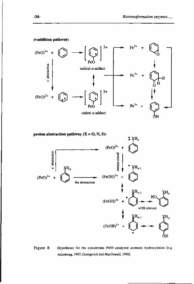
-34- Biotransformation enzymes..
a-addition pathway:
(FeO)3+ + O
(FeO)2+ + O
rad
[9] FeO
ical o-add i
[91 Kor»
3+
uct
3+
»-
»-
* • Fe3+ +
Fe 3+
0 ^ o
I
cation a-adduct
Fe3+ + (T " ^
proton abstraction pathway (X = O, N, S): 3.XH + -fnn
2+ _ ^ (FeOr + |
8 w t n-1
(FeO)3+ + | H» abstraction
(FeOH)^ + |
' XHn-l
(FeOH)3+ + | ^ ^ t5
m
T F"
'OH rebound
i XHn
(FeOH)3+ + l | H - » - - » - [I
Figure 8: Hypotheses for the cytochrome P450 catalyzed aromatic hydroxylation [e.j
Armstrong, 1987; Guengerich and MacDonald, 1990].
Chapter 2 -35-
phenol formation) indicate that an initial electrophilic attack of the (FeO)3+ species
on the substrate -without formation of epoxides as the precursor- is the preferred
mechanism [Guengerich and Macdonald, 1990; Korzekwa et ai, 1989; Rietjens et
ai, 1995; Riley and Hanzlik, 1994]. The results of Korzekwa suggest that the extent
of charge transfer and spin density on the substrate resulting in either a radical or
cation o-adduct intermediate species may be substituent dependent, with electron
donating groups favoring the cation species. It has been proposed that the "radical 0-
adduct" favors epoxide formation, whereas the "cation o-adduct" favors direct phenol
or ketone formation. [Korzekwa et ai, 1985]. The aromatic epoxides instead of
giving rise to phenolic metabolites may rather result in formation of rather catechol
metabolites, upon epoxide hydrolase catalyzed conjugation with water, followed by
aromatization, or of glutathione conjugation derived metabolites [Rietjens et al.,
1993].
Aromatic hydroxylation of substrates containing heteroatoms with a relatively
low oxidation potential (N, S, P, O) have been proposed to proceed by either
hydrogen atom abstraction, or electron abstraction and proton release from the
heteroatoms, followed by rearrangement of the radicals and oxygen rebound to form
Jhe product [Armstrong, 1987]. For ^-substituted anisoles the cytochrome P450
oxidative metabolism -also aromatic hydroxylation- could be predicted qualitatively
on the basis of spin distributions, energy differences between substrates, metabolic
intermediates and products taken only hydrogen abstraction mechanisms into account
[Groot et ai, 1995, Koymans et ai, 1993]. However, a direct addition of a
cytochrome P450 (FeO)3+ intermediate to a jt-bond or to a lone pair [e.g. Riley and
Hanzlik, 1994] as hypothetical mechanism for aromatic hydroxylation was not taken
into account in those studies. Recently, a mechanistic study on the oxidative
metabolism of [3,5-2H2]-4-iodoanisole, explained aromatic hydroxylation via direct
aromatic hydroxylation. The formation of 2-hydroxy-4-iodoanisole was proposed to
occur almost entirely by direct aromatic hydroxylation with no epoxide or
cyclohexadienone as the intermediates, whereas the 3-hydroxylation could proceed
via direct aromatic hydroxylation or formation of cyclohexadienone intermediates,
without epoxide intermediates [Rizk and Hanzlik, 1995]. The hypothesis that 3-
hydroxylation occurs without epoxide formation, is in accordance with the
observation that the 2,3- or 3,4-chlorobenzene epoxides rearrange to respectively 2-
and 4-chlorophenol, without formation of 3-chlorophenol [Selander et ai, 1975]. To
suggest a completely different mechanism for OH incorporation at different sites in
one molecule seems unlikely.
-36- Biotransformation enzymes..
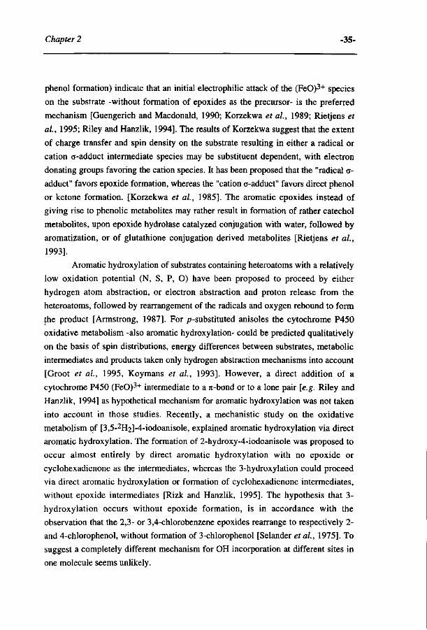
N-oxygenation.
N-oxygenation is a process that is mechanistically related to, cc-carbon
hydroxylation and subsequent N-dealkylation (see above) (Figure 9). Understanding
the processes of the partitioning between N-dealkylation and N-oxygenation is
important, because in some cases the two pathways can lead to quite different
products. In N-oxygenation oxidation occurs at the nitrogen, whereas in N-
dealkylation the oxygen is transferred to the carbon adjacent to the heteroatom. It has
been proposed that N-oxygenation/N-dealkylation proceeds via an initial one-electron
oxidation at the nitrogen by the high-valent iron-oxo intermediate of cytochrome
P450. Support for an one-electron oxidation pathway for N-oxygenation and N-
dealkylation was provided by several lines of evidence: cycloalkylamines undergo
rearrangements characteristic of one-electron transfer processes [Bondon et al, 1989;
Hanzlik and Tullman, 1982; Macdonald et al, 1982], the rate of the cytochrome
P450 mediated dimethylaniline oxidation was found to correlate with the substrate
oxidation-reduction potential [Macdonald et ai, 1989], low isotope effects were
observed for N-dealkylation reactions [Guengerich et al., 1995] and the formation of
alkyl radicals was reported upon oxidation of l,4-dihydro-4-alkylpyridines [Augusto
et al., 1982; Lee et al, 1988]. The partitioning of the resulting putative aminium
radicals between radical recombination and a-deprotonation (i.e. between N-
oxygenation and N-dealkylation) is influenced by the ease of deprotonation and
aminium radical stability. N-oxygenation of amines does not occur unless 1) no <x-
heteroatom oxygenation O"
(FeO)3++ CR3XRn • (FeO)2++ CR3XRn » - Fe3+ + CR3X+Rn
e" abstraction
N-hydroxylation OH
(FeO)3++ R-NH2 • (FeOH)3+ + R-NH • Fe J ++ R-NH •OH rebound
(FeO)2+ + R-NH2 '
Figure 9: Proposed general mechanisms for the cytochrome P450-catalyzed N-dealkylation
and N-hydroxylation.
Chapter 2 -37-
protons are available as is the case for the primary arylamines (see Figure 9), 2) <x-
protons are inaccesible due to Bredt's rule as is the case for quinidine (Bredt's rule
prevents abstraction of a proton at the individual bridgehead nitrogen for steric
reasons) [Guengerich et al, 1986] or 3) radical stability is provided by neighbouring
electron donating groups [Guengerich, 1990].
Finally, for heteroatoms with a relatively high ionization potential, a hydrogen
abstraction pathway, instead of a single electron transfer pathway with subsequent
proton release, may be preferred [Armstrong, 1987; Guengerich and Macdonald,
1984].
Oxidative dehalogenation.
Cytochromes P450 catalyze reductive as well as oxidative dehalogenation of
halogenated and aliphatic compounds, and the respective reactions are modulated by
oxygen tension but also the chemical nature of the substrate [Lefever and Wackett,
1994]. Here, attention is focussed on the mechanisms with respect to the oxidative
dehalogenation of aromatic compounds.
Cytochrome P450-mediated oxidative dehalogenation of aromatic compounds
to phenolic derivatives has been observed for fluorinated [Daly et al, 1968; Den
Besten et al, 1993; Li et al 1985, Rietjens et al, 1992], chlorinated [Daly et al,
1968], brominated [Zheng and Hanzlik, 1992] and recently iodinated [Rizk and
Hanzlik, 1995] compounds. Several pathways are thought to be involved in the
oxidative dehalogenation of aromatics. For 4-fluorinated phenols and anilines as well
as pentafluorochlorobenzene, elimination of the fluoride as an anion results in
formation of respectively benzoquinone(imine) and a benzohaloquinone cation as the
primary reaction products, which can be reduced subsequently to give the phenolic
derivative [Den Besten et al, 1993; Rietjens and Vervoort; 1991; Rietjens et al,
1992]. The P450-catalyzed deiodination of 4-iodoanisole to 4-methoxyphenol is
suggested to involve C-O bond formation via direct attack of the (FeO)3+
intermediate on the aromatic ring followed by reductive cleavage of the C-iodine
bond, with electrons coming from P450 reductase. The observation that no 1 80 from
H2180 is incorporated into 4-methoxyphenol indicates that the phenolic oxygen
originates from molecular oxygen via the cytochrome (FeO)3+ species [Rizk and
Hanzlik, 1995].
Noteworthy, it has been proposed that halogenated aromatics may also be
metabolized by direct oxidation of the halogen on the aromatic ring to an
intermediate that produces the phenolic derivative and eliminates the halogen as
-38- Biotransformation enzymes..
(HXO) [Macdonald, 1983; Rietjens et al., 1992; Van Ommen and Van Bladeren P.J.,
1989]. This hypothetical mechanism was based on the observation that cytochrome
P450 oxidizes iodobenzene to iodosobenzene [Burka et al, 1980]. Later, Guengerich
demonstrated that except iodine, also bromine substituents can be oxidized by using
the 4-tórt-butyl-2,5-bis[l-hydroxy-l-(trifluoromethyl)-2,2,2-trifluoroethyl] halo-
benzenes of which the oxidized reaction products are relatively stable [Guengerich,
1989]. In terms of understanding mechanisms of P450 catalysis this type of reaction
is interesting, however the physiological importance remains to be established yet.
Finally, during the cytochrome P450-mediated monooxygenation a halogen
can be lost from its position via an intramolecular migration to the carbon position
adjacent to the hydroxylated position. This phenomenon, the so-called "NIH shift"
(named after the National Institute of Health), has been reported especially for
chloro- and bromo-substituents [Daly et al., 1972].
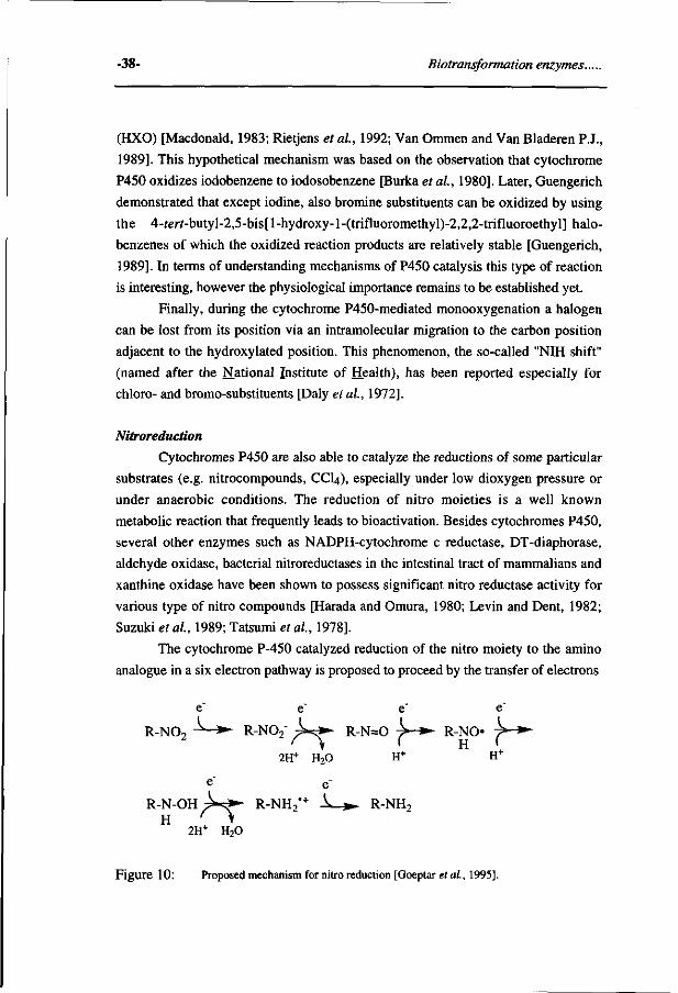
Nitroreduction
Cytochromes P450 are also able to catalyze the reductions of some particular
substrates (e.g. nitrocompounds, CCI4), especially under low dioxygen pressure or
under anaerobic conditions. The reduction of nitro moieties is a well known
metabolic reaction that frequently leads to bioactivation. Besides cytochromes P450,
several other enzymes such as NADPH-cytochrome c reductase, DT-diaphorase,
aldehyde oxidase, bacterial nitroreductases in the intestinal tract of mammalians and
xanthine oxidase have been shown to possess significant nitro reductase activity for
various type of nitro compounds [Harada and Omura, 1980; Levin and Dent, 1982;
Suzuki etal, 1989; Tatsumi et al, 1978].
The cytochrome P-450 catalyzed reduction of the nitro moiety to the amino
analogue in a six electron pathway is proposed to proceed by the transfer of electrons
R-N02 : k > - R-N=0 y »• R-NO > »»
2H+ H20 H+ H+
R - N - O H ^ - ^ R-NH2*+ ^ . » . R-NH2
2H+ H20
Figure 10: Proposed mechanism for nitro reduction [Goeptar et ai, 1995].
Chapter 2 -39-
from the ferrous cytochrome P450 to the substrate (see Figure 10) [Harada and
Omura, 1980]. Electron spin resonance studies demonstrated that the nitro group is
reduced in a one electron reaction to yield a nitro anion radical [Mason and
Holtzman, 1975; Sealy et ai, 1978]. Under aerobic conditions these radicals react
with oxygen to form superoxide radical anions and H2O2 which cause an oxidative
stress type of cytotoxicity [O'Brien et al, 1990]. Upon the addition of a second
electron and two protons the nitroso intermediate is formed as the intermediate
[Levin and Dent, 1982]. Subsequent reduction of the nitroso compound results in the
formation of a hydronitroxide free radical, which in turn is reduced to the
hydroxylamine [Goeptar et al, 1995]. Finally, the phenylhydroxylamine is reduced
to the aniline analogue [Suzuki et al., 1989].
Regioselective metabolism in cytochrome P450 catalyzed reactions.
The regioselective metabolism of xenobiotics by the cytochromes P450 is a
consequence of the topographical features of the apoproteins around the active sites
in combination with chemical characteristics of the substrates itself [Korzekwa and
Jones, 1993; White et al, 1984]. Small substrates have the potential for high active
site mobility [Koerts et ai, 1995; Raag and Poulos, 1992]. Substrates that can rotate
more freely in the active site may be expected to have greater variations in binding
orientations and therefore greater differences in stereoselectivities [Jones et al,
1995]. If cytochrome P450 has no steric or binding requirements for its substrate, a
model based only on the electronic characteristics of oxidant and substrate would be
sufficient [Korzekwa and Jones, 1993; Oguri et al, 1994; Rietjens et al, 1993].
However, for substrates of large size and structural rigidity, like benzo[«]pyrene, or
substrates which interact with specific amino acid residues in the active site, like
debrisoquine in P450 2D6 or camphor in P450cam, severe constraints against
mobility exist once they are bound to the active site. Because of a lack of detailed
structures of the mammalian P450 enzymes, alternatives are developed to probe the
active site topology of P450 enzymes, in order to obtain insight into factors that
determine substrate selectivity and regioselective metabolism.
Lewis studied the active sites of P450 by detailed knowlegde on the substrate
characteristics like molecular dimensions, the presence of certain structural features
and electronic properties [Lewis et al, 1987]. Inspection of the molecular structures
of cytochrome P450 lA-specific compounds revealed that they are all rigid, planar
molecules due to the presence of a number of fused aromatic rings, characterized by a
small depth and a large area-to-depth ratio. The majority of cytochromes P450 2B
-40- Biotransformation enzymes.
substrates have structures containing at least one non-fused aromatic ring and a
overall non-planar geometry, often adopting a V-shaped conformation and are
characterized by a small area-to-depth ratio. A common structural feature of the
cytochrome P450 2E substrates is that they are relatively small-sized molecules,
whereas the cytochrome P450 3A substrates are large-sized molecules [Lewis et ai,
1987; Lewis et al, 1994]. Recently, it was demonstrated by ÏH-NMR that more than
one aromatic base substrate molecule can enter the active site of cytochrome
P450cam and bind both in the hydrophobic cavity and at the heme metal [Banci et
al, 1994]. To understand the factors that determine the substrate specificity of
cytochromes P450, De Voss and Ortiz de Montellano succesfully used a docking
program to predict, whether compounds entirely unrelated to camphor are substrates
for cytochrome P450cam, a protein that normally uses camphor as the substrate and
for which a crystal structure is available [De Voss and Ortiz de Montellano, 1995].
Another experimental approach for probing the active site of P450s is based on
prosthetic heme alkylation reactions. This proceeds by a reaction of cytochromes
P450 with aryl moieties to give an iron-phenyl complex that oxidatively rearranges to
N-phenylprotoporphyrin IX regioisomers with the phenyl group on the pyrrole rings
A, B, C and/or D. The four pyrroles are not equivalent in their capacity to accept the
phenyl radical, because of differences in steric hindrance of each pyrrole ring by
amino acids of the P450 peptide in the vicinity of the oxygen binding site. Using aryl
moieties of diverse structures provides information on the openess above the pyrrole
rings. The regiochemistry of the iron nitrogen adducts demonstrated that the active
sites of 1A1, 2B1, 2B2 and 2E1 have similar structural motifs, with pyrrole rings B
and C at least partially masked by protein residues and a more open region above
rings A and D [Ortiz de Montellano, 1987; Swanson et al. 1991; Tuck and Ortiz de
Montellano, 1992; Mackman et ai, 1996].
Besides models that provide information on the three-dimensional structure of
the active site, also models that explain regioselective metabolism were proposed.
Based upon the regiospecificity of the cytochrome P4501A1 catalyzed
biotransformation of benzo[a]pyrene and its di-hydrodiol derivatives Jerina proposed
that the catalytic site of this P450 has a space for substrate binding of a size
equivalent to nine benzene rings. Upon superposition of a polycyclic aromatic
compound on the nine benzene ring model, the regioselectivity and stereoselectivity
of the P450 1A1 mediated metabolisme could be predicted [Jerina et al., 1982].
Recently, it has been demonstrated that seven human, five rodent and two bacterial
P450s were all able to convert benzo[a]pyrene to the 7(R),8(R)-diol and the
Chapter 2 -41-
9(R),10(R)-diol with an extremely high degree of stereoselectivity, which is likely
due to the large size of the substrate molecule and its constraints in the active site
[Jones et al., 1995]. For smaller molecules, however, it was proposed that more
orientations in the active site are possible. NMR relaxation studies were used to
investigate the preferred orientation in the binding of acetaminophen to P450 1A1
and 2B1 isoforms. Using 15N and 13C NMR Tj relaxation rate measurements, it was
suggested that P450 1A1 produces the N-acetyl-p-benzoquinoneimine preferentially,
because of closer proximity of the heme iron to the amide nitrogen, whereas P450
2B1 produces 3-hydroxyacetaminophen preferentially because of closer proximity of
the heme iron to the phenolic oxygen [Myers et al., 1994], Remarkably however,
using !H NMR Tj relaxation rate measurements, the nitrogen of the acetamido group
was reported to be closer to the heme iron in P450 2B1, than in P4501A1 [Van de
Straat et ai, 1987].
X-ray crystal structures and site-directed mutagenesis of P450cam gave
valuable information on the substrate-P450 interactions. The camphor is bound
through its carbonyl group via a hydrogen bridge to Tyr 96 so that oxygen insertion
can occur at C5. However, the absolute regiospecificity of camphor hydroxylation is
lost when camphor is metabolized by the Tyr96Phe mutant enzyme which is unable
to form a hydrogen bond with the substrate. The five different products observed,
when camphor is hydroxylated by the Tyr96Phe mutant enzyme suggests that
camphor itself can bind in multiple orientations when it is not anchored through a
hydrogen bond [Raag and Poulos, 1992]. X-ray crystal structures have been
determined for complexes of cytochrome P450cam with the substrates camphane,
adamantane and thiocamphor. Unlike the natural substrate camphor, which hydrogen
bonds to Tyr 96 and is metabolized to a single product, camphane, admantane and
thiocamphor do not form a hydrogen bond with the enzyme and all are hydroxylated
at multiple positions. Again it was concluded that the lack of a substrate-enzyme
hydrogen bond allows substrates greater mobility in the active site explaining this
lower regiospecificity of metabolism [Raag and Poulos, 1991].
Current advances in computational chemistry allow the modelling of the more
relevant cytochrome P450 (FeO)3+ intermediate. Molecular dynamic studies on the
orientation of thiocamphor in the active site of P450cam indicated, that the
orientation in the active site is different in the presence of the reactive high-valent
iron-oxo species, than in the absence of that oxygen and in addition is more
consistent with the formation of the 5-alcohol as being the major product [Paulsen
and Ornstein, 1993]. Moreover, on the basis of molecular dynamic simulations with
-42- Biotransformation enzymes..
inclusion of all atoms within a 16Â "belly" region surrounding the reactive iron-oxo
species of P450cam, the predicted regioselectivities of the hydroxylation of d-
camphor, d-camphane, d-thiocamphor [Harris and Loew, 1995] and the
stereochemistry of the styrene and ß-methylstryrene epoxides formed by P450cam
[Fruetel et al, 1992] were in excellent agreement with experimental results.
In conclusion, more knowledge on the active site structure of the different
P450 enzymes in the reactive (FeO)3+ state, specific interactions of substrates with
amino-acid residues or the heme in the active site, insight into molecular reaction
mechanisms and chemical substrate characteristics that determine the reaction
chemistry, may eventually lead to a good prediction of substrate specificity and
stereoselective metabolism of xenobiotics.
2.1.2. Flavin-containing monooxygenase.
The flavin-containing monooxygenases (FMO) are a family of enzymes that
metabolize a variety of structurally diverse pesticides, therapeutic agents and other
xenobiotics. The mammalian FMO enzymes are catalogued into five different groups
FMOl, FM02, FM03, FM04 and FM05 on the basis of their amino acid sequence
identities [Lawton, 1994]. The expression of the FMOs is regulated by hormonal and
dietary factors, but the effects on induction appear to be species- and tissue dependent
[Cashman, 1995].
These endoplasmatic reticulum-bound flavoproteins utilize NADPH and
molecular oxygen to catalyze the monooxygenation of nucleophilic heteroatoms like
nitrogen, sulphur, phosphorous [Ziegler, 1980; Ziegler 1988; Cashman, 1995], but
also the selenium atom [Ziegler, 1992]. FMOs are able to N-oxidize virtually all
lipophilic secondary and tertiary amines. Remarkably, FMOs also tend to
hydroxylate aromatic carbon atoms which are located ortho or para to an hydroxyl
or amine moiety, such as the 2-hydroxylation of 1-naphtylamine, the 1-hydroxylation
of 2-naphtylamine and the 4-hydroxylation of 4-fluoro-N-methylaniline, probably via
rearrangement of N-hydroxy-metabolites [Boersma et al, 1993; Hammonds et al,
1985].
The physiological function of the FMOs remains undefined, although some
potential endogenous substrates have been identified. In human, substantial evidence
has been obtained indicating that FMO serves an important role in the detoxication of
the vast majority of naturally occurring alkaloids and medicinal tertairy amines to
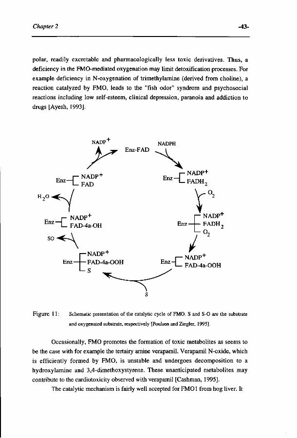
Chapter 2 -43-
polar, readily excretable and pharmacologically less toxic derivatives. Thus, a
deficiency in the FMO-mediated oxygenation may limit detoxification processes. For
example deficiency in N-oxygenation of trimethylamine (derived from choline), a
reaction catalyzed by FMO, leads to the "fish odor" syndrom and psychosocial
reactions including low self-esteem, clinical depression, paranoia and addiction to
drugs [Ayesh, 1993].
NADPn
y~ NADPH
NADP + E n Z _ C FAD
_ j - NADP+
E n z — L FAD-4a-OH
NADP+
i—NADP-'-Enz—UFAD-43 -OOH
l - s
E n z I_FADH2
r. I— o,
NADP"" Enz—I— FADH,
Enz-| NADP"1" FAD-4a-OOH
Figure 11 : Schematic presentation of the catalytic cycle of FMO. S and S-0 are the substrate
and oxygenated substrate, respectively [Poulsen and Ziegler, 1995],
Occasionally, FMO promotes the formation of toxic metabolites as seems to
be the case with for example the tertairy amine verapamil. Verapamil N-oxide, which
is efficiently formed by FMO, is unstable and undergoes decomposition to a
hydroxy lamine and 3,4-dimethoxystyrene. These unanticipated metabolites may
contribute to the cardiotoxicity observed with verapamil [Cashman, 1995].
The catalytic mechanism is fairly well accepted for FMOl from hog liver. It
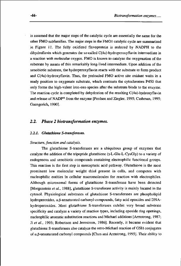
-44- Biotransformation enzymes..
is assumed that the major steps of the catalytic cycle are essentially the same for the
other FMO subfamilies. The major steps in the FMOl catalytic cycle are summarized
in Figure 11. The fully oxidized flavoprotein is reduced by NADPH to the
dihydroflavin which generates the so-called C(4a)-hydroperoxyflavin intermediate in
a reaction with molecular oxygen. FMO is known to catalyze the oxygenation of the
substrate by means of this remarkably long-lived intermediate. Upon addition of the
xenobiotic substrate, the hydroperoxyflavin reacts with the substrate to form product
and C(4a)-hydroxyflavin. Thus, the preloaded FMO active site oxidant waits in a
ready position to oxygenate substrate, which contrasts the cytochromes P450 that
only forms the high-valent iron-oxo species after the substrate binds to the enzyme.
The reaction cycle is completed by dehydration of the resulting C(4a)-hydroxyflavin
and release of NADP+ from the enzyme [Poulsen and Ziegler, 1995; Cashman, 1995;
Guengerich, 1990].
2.2. Phase 2 biotransformation enzymes.
2.2.1. Glutathione S-transferases.
Structure, function and catalysis.
The glutathione S-transferases are a ubiquitous group of enzymes that
catalyze the addition of the tripeptide glutathione (y-L-Glu-L-CysGly) to a variety of
endogenous and xenobiotic compounds containing electrophilic functional groups.
This reaction is the first step in mercapturic acid pathway. Glutathione is the most
prominent low molecular weight thiol present in cells, and competes with
nucleophilic entities in cellular macromolecules for reaction with electrophiles.
Although microsomal forms of glutathione S-transferase have been detected
[Morgenstein et al, 1988], glutathione S-transferase activity is mainly located in the
cytosol. Physiological substrates of glutathione S-transferases are phospholipid
hydroperoxides, a,ß-unsaturated carbonyl-compounds, fatty acid epoxides and DNA-
hydroperoxides. Most glutathione S-transferases exhibit very broad substrate
specificity and catalyze a variety of reaction types, including epoxide ring openings,
nucleophilic aromatic substitution reactions and Michael additions [Armstrong, 1987;
Ji et al, 1993; Robertson and Jernstrom, 1986]. Recently, it became evident that
glutathione S-transferases also catalyze the retro-Michael reaction of GSH conjugates
of a,ß-unsaturated carbonyl compounds [Chen and Armstrong, 1995]. Their ability to
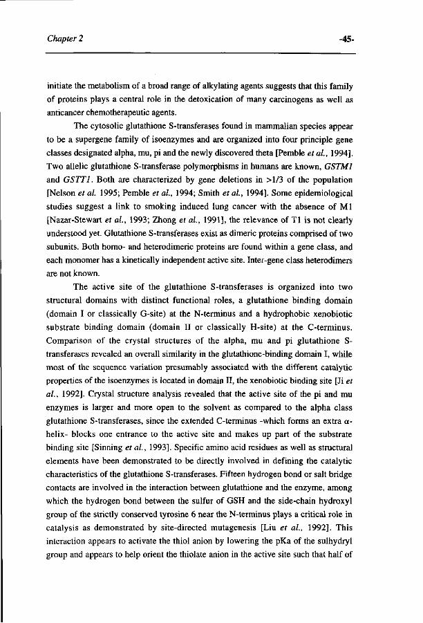
Chapter 2 -45-
initiate the metabolism of a broad range of alkylating agents suggests that this family
of proteins plays a central role in the detoxication of many carcinogens as well as
anticancer chemotherapeutic agents.
The cytosolic glutathione S-transferases found in mammalian species appear
to be a supergene family of isoenzymes and are organized into four principle gene
classes designated alpha, mu, pi and the newly discovered thêta [Pemble et ai, 1994].
Two allelic glutathione S-transferase polymorphisms in humans are known, GSTM1
and GSTT1. Both are characterized by gene deletions in >l/3 of the population
[Nelson et al. 1995; Pemble et ai, 1994; Smith et ai, 1994]. Some epidemiological
studies suggest a link to smoking induced lung cancer with the absence of Ml
[Nazar-Stewart et ai, 1993; Zhong et ai, 1991], the relevance of Tl is not clearly
understood yet. Glutathione S-transferases exist as dimeric proteins comprised of two
subunits. Both homo- and heterodimeric proteins are found within a gene class, and
each monomer has a kinetically independent active site. Inter-gene class heterodimers
are not known.
The active site of the glutathione S-transferases is organized into two
structural domains with distinct functional roles, a glutathione binding domain
(domain I or classically G-site) at the N-terminus and a hydrophobic xenobiotic
substrate binding domain (domain II or classically H-site) at the C-terminus.
Comparison of the crystal structures of the alpha, mu and pi glutathione S-
transferases revealed an overall similarity in the glutathione-binding domain I, while
most of the sequence variation presumably associated with the different catalytic
properties of the isoenzymes is located in domain II, the xenobiotic binding site [Ji et
ai, 1992]. Crystal structure analysis revealed that the active site of the pi and mu
enzymes is larger and more open to the solvent as compared to the alpha class
glutathione S-transferases, since the extended C-terminus -which forms an extra cc-
helix- blocks one entrance to the active site and makes up part of the substrate
binding site [Sinning et ai, 1993]. Specific amino acid residues as well as structural
elements have been demonstrated to be directly involved in defining the catalytic
characteristics of the glutathione S-transferases. Fifteen hydrogen bond or salt bridge
contacts are involved in the interaction between glutathione and the enzyme, among
which the hydrogen bond between the sulfur of GSH and the side-chain hydroxyl
group of the strictly conserved tyrosine 6 near the N-terminus plays a critical role in
catalysis as demonstrated by site-directed mutagenesis [Liu et ai, 1992]. This
interaction appears to activate the thiol anion by lowering the pKa of the sulhydryl
group and appears to help orient the thiolate anion in the active site such that half of
-46- Biotransformation enzymes..
its surface is shielded by hydrophobic residues and the other half is exposed for
reaction with the electrophilic substrate [Ji et al, 1992]. However, the equivalent
residue of Tyr 6 in the theta-class structure is not present in the active site, but its role
appears to have been replaced by either a nearby serine or another tyrosine residue
located in the C-terminal domain of the enzyme [Wilce et ah, 1995]. Domain II
provides a great deal of the hydrophobic surface that interacts with the xenobiotics
and the products of the reactions. Using molecular modeling techniques a predictive
substrate model for class mu rat glutathione S-transferase 4-4 was constructed,
containing three interaction sites responsible for Lewis acid-Lewis base interaction as
well as a region responsible for aromatic interactions with the substrates [Groot et al,
1995].
The rate of the glutathione S-transferase catalyzed reaction can be limited by
a number of different chemical or physical steps in the mechanism, such as formation
of the Meisenheimer complex in a glutathione S-transferase catalyzed nucleophilic
aromatic substitution reaction, assistance of specific amino-acid residues in substrate
activation, stabilisation of the transition state or dissociation of the product. The mu
class isoenzyme 3-3, which is particularly efficient in catalyzing aromatic
nucleophilic substitution reactions, appears to stabilize the transition state of the
Meisenheimer complex by hydrogen bonding interactions between the pro-R o-
nitrogroup of l-chloro-l-(S-glutathionyl)-2,4-dinitrocyclohexadienate anion and the
hydroxyl groups of Tyr6 and Tyrl 15 and between the p-nitro group and an enzyme-
bound water [Graminski et al, 1989; Ji et al, 1993]. Further structural and
mechanistic analysis on the functional properties of Tyr 115 in mu class isoenzyme
3-3 revealed that the hydroxyl group of this residue provides electrophilic assistance
in addition of glutathione to arene oxide substrates [Johnson et al, 1993]. Interaction
of the flexible C-terminal tail with the N-terminal domain helps to limit the rate of
product release from the active site of mu class isoenzymes in nucleophilic aromatic
substitution reactions [Zhang et al, 1992; Johnson et al, 1993]. Recently, it was
demonstrated that the chemical reactivity for nucleophilic attack of
fluoronitrobenzenes in the glutathione S-transferase catalyzed reaction might also be
rate-limiting [Rietjens et al, 1995].
Bioactivation and detoxification via glutathione conjugation.
In general, the conjugation of an electrophilic compound with glutathione is a
detoxification reaction, but there are several examples where glutathione conjugation
plays an important role in the bioactivation of chemicals by different types of

Chapter 2 -47-
mechanisms. For example, glutathione conjugation of quinone- and quinoneimine-
derivatives targets the metabolites to the kidneys, where subsequent processing by
the proximal tubular brush border enzyme y-glutamyltranspeptidase are thought to be
involved in the selective toxicity of these compounds by a yet unknown mechanism
[Fowler et al, 1991; Fowler et al, 1994, Lock et al, 1993]. Conjugation to
glutathione has been demonstrated to be the initial step in the bioactivation of
haloalkenes. After subsequent degradation to the corresponding cysteine conjugates,
ß-lyase mediated formation of reactive thiol compounds are proposed to be
responsible for the nephrotoxicity caused by haloalkenes [Commandeur et al, 1995].
Glutathione is also involved in the conjugative activation of vicinal dihaloalkanes to
the reactive episulfonium ion intermediate, which forms adducts with DNA
[Guengerich, 1992].
Glutathione S-transferases are of particular interest because they have been
implicated in the development of resistance of cells and organisms towards
pesticides, herbicides and chemotherapeutic alkylating agents [Tew, 1994]. Increased
conjugation with glutathione as well as an increased efflux of the GS-conjugates
from the cell by ATP-dependent GS-X pumps, is a mechanism frequently associated
with this type of drug resistance [Ishikawa, 1992].
2.2.2. N-acetyltransferases
N-acetylation is a major biotransformation pathway for compounds with an
amino group. N-acetyltransferases catalyze the transfer of the acetyl group from
endogenous acetyl coenzyme A to molecules that contain a primary amine function.
Besides drugs and other chemicals like arylamine and hydrazine derivatives, also
certain biogenic amines such as histamine and serotonin are substrates for the N-
acetyltransferases [Weber et al, 1990]. Structure activity studies of a series aniline-
derivatives with substituents that differ in hydrophobicity, position, number, size and
charge demonstrated that some orf/zo-substituted derivatives are not acetylated,
probably due to steric hindrance [Andres et al, 1987].
At least two groups of N-acetyltransferases are identified, one being
monomorphic (NAT1) and the other being polymorphic (NAT2) [Blum et al, 1990].
The polymorphism in N-acetyltransferase activity leads to the existance of the so-
called 'fast' and 'slow' acetylator phenotypes. N-acetylation has been recognized to
play a modulatory role in the metabolic activation of mutagenic and carcinogenic
aromatic amines and various food pyrosylates that are classified as carcinogenic
[Weber et al, 1990]. From patterns of epidemiology slow acetylators appear to be
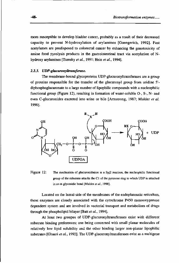
-48- Biotransformation enzymes..
more susceptible to develop bladder cancer, probably as a result of their decreased
capacity to prevent N-hydroxylation of arylamines [Guengerich, 1992]. Fast
acetylators are predisposed to colorectal cancer by enhancing the genotoxicity of
amine food pyrolysis products in the gastrointestinal tract via acetylation of N-
hydroxy arylamines [Turesky et al, 1991; Hein et al, 1994].
2.2.3. UDP-glucuronyltransferase.
The membrane-bound glycoproteins UDP-glucuronyltransferases are a group
of proteins responsible for the transfer of the glucuronyl group from uridine 5'-
diphosphoglucuronate to a large number of lipophilic compounds with a nucleophilic
functional group (Figure 12), resulting in formation of water-soluble O-, S-, N- and
even C-glucuronides excreted into urine or bile [Armstrong, 1987; Mulder et al
1990].
OH OH
— O—P — O—P — O*,
O O
UDPGA
OH
Figure 12: The mechanism of glucuronidation is a S]vj2 reaction, the nucleophilic functional
group of the substrate attacks the CI of the pyranose ring to which UDP is attached
in an a-glycosidic bond [Mulder et al, 1990].
Located on the lumal side of the membranes of the endoplasmatic reticulum,
these enzymes are closely associated with the cytochrome P450 monooxygenase
dependent system and are involved in vectorial transport and metabolism of drugs
through the phospholipid bilayer [Batt et al, 1994].
At least two groupes of UDP-glucuronyltransferases exist with different
substrate binding preferences; one being concerned with small planar molecules of
relatively low lipid solubility and the other binding larger non-planar lipophilic
substrates [Ghauri et al, 1992]. The UDP-glucuronyltransferases exist as a multigene
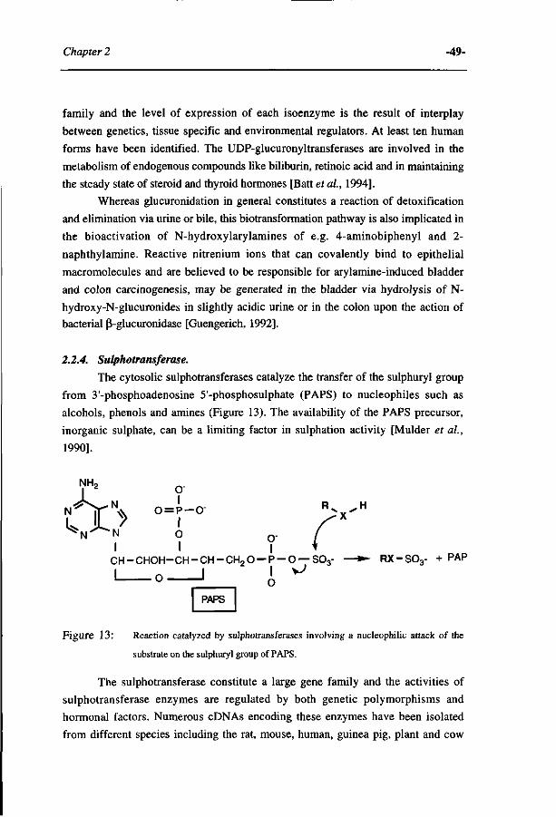
Chapter 2 -49-
family and the level of expression of each isoenzyme is the result of interplay
between genetics, tissue specific and environmental regulators. At least ten human
forms have been identified. The UDP-glucuronyltransferases are involved in the
metabolism of endogenous compounds like biliburin, retinoic acid and in maintaining
the steady state of steroid and thyroid hormones [Batt et al, 1994].
Whereas glucuronidation in general constitutes a reaction of detoxification
and elimination via urine or bile, this biotransformation pathway is also implicated in
the bioactivation of N-hydroxylarylamines of e.g. 4-aminobiphenyl and 2-
naphthylamine. Reactive nitrenium ions that can covalently bind to epithelial
macromolecules and are believed to be responsible for arylamine-induced bladder
and colon carcinogenesis, may be generated in the bladder via hydrolysis of N-
hydroxy-N-glucuronides in slightly acidic urine or in the colon upon the action of
bacterial ß-glucuronidase [Guengerich, 1992].
2.2.4. Sulphotransferase.
The cytosolic sulphotransferases catalyze the transfer of the sulphuryl group
from 3'-phosphoadenosine 5'-phosphosulphate (PAPS) to nucleophiles such as
alcohols, phenols and amines (Figure 13). The availability of the PAPS precursor,
inorganic sulphate, can be a limiting factor in sulphation activity [Mulder et al,
1990].
O" N ' '\v o = p - o -
/ I N O 0 . I I I
CH-CHOH-CH-CH-CH 2 0-P-0 —S03- — • RX-S03- + PAP
i u —' o
I PAPS Figure 13: Reaction catalyzed by sulphotransferases involving a nucleophilic attack of the
substrate on the sulphuryl group of PAPS.
The sulphotransferase constitute a large gene family and the activities of
sulphotransferase enzymes are regulated by both genetic polymorphisms and
hormonal factors. Numerous cDNAs encoding these enzymes have been isolated
from different species including the rat, mouse, human, guinea pig, plant and cow
-50- Biotransformation enzymes.
[Kong et ai, 1994]. In the past, these enzymes were most often classified on the basis
of their substrate specificities toward simple phenols, phenolic monoamines and
hydroxysteroids. Recently, however, the sulphotransferase genes have been grouped
into distinct gene families, since the substrate-based classification of these enzymes
appears to be inappropriate due to the broad and overlapping substrate specificity of
the individual sulphotransferases [Yamazoe et al, 1994].
In general, the sulphotransferase enzymes exhibit a high affinity and a low
capacity, whereas glucuronidation has a low affinity and a high capacity for the same
substrates [Temellini et al, 1991].
Sulphation of endogenous (e.g. bile salts, steroids, catecholamines) or foreign
chemicals has been recognized as an important pathway of metabolic inactivation,
through enhancing the polarity and solubility of compounds and thereby hasten their
excretion from the body. However, for a number of chemicals, such as N-
hydroxylamines, arylhydroxamic acids and benzylic alcohols, sulphation has been
implicated into the bioactivation to reactive intermediates [Miller, 1994].
3. Frontier orbital theory.
Frontier orbital theory has been already frequently used in the field of organic
chemistry to explain the regioselectivity in nucleophilic and electrophilic aromatic
substitutions [Fleming, 1989; Fukui et al, 1954]. This section gives a short
introduction which is mainly based on the monograph by Fleming, in order to
provide a basis to understand how the frontier orbital theory can be used to study and
predict outcomes of enzyme catalysis and, thus, outcomes of biotransformation
processes.
Atomic and molecular orbitals
Atoms consist of a nucleus containing protons and neutrons surrounded by
electrons which are localized in so-called atomic orbitals. Each orbital can contain
two electrons with opposite electron spin. The chemical characteristics of an atom
depend on especially the electrons in the outer-shell orbitals, the so-called valence
shell orbitals. When molecules are formed from atoms, atomic orbitals (AO's) from
one atom are combined with those of another atom to give the molecular orbitals
(MO) of the newly formed molecule. These MO's are not, as the AO's localised
around one atomic nucleus, but can be delocalised over all atomic nuclei of the
molecule.
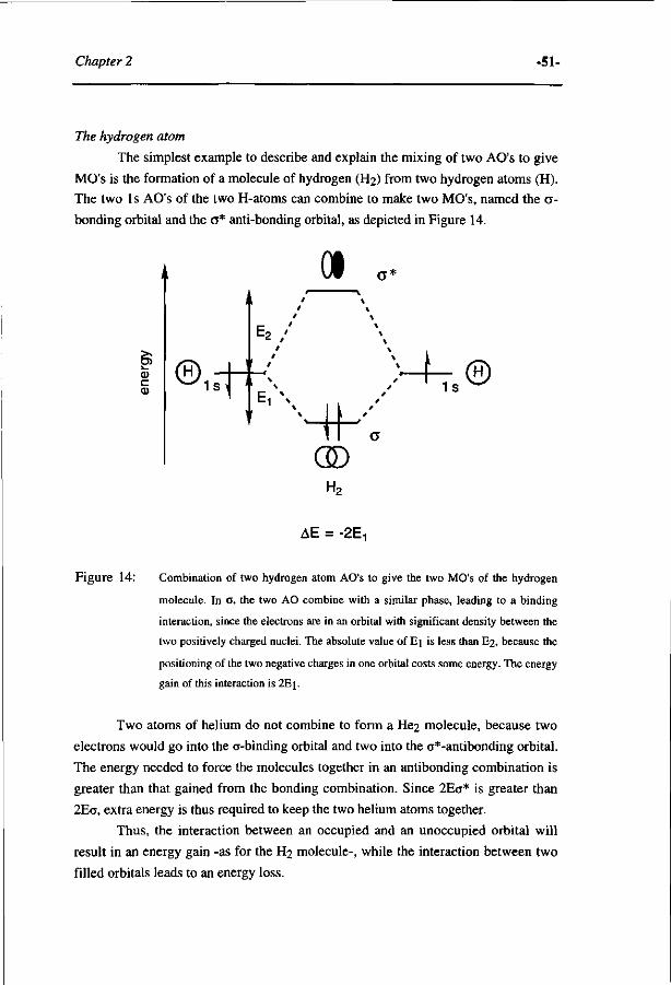
Chapter 2 -51-
The hydrogen atom
The simplest example to describe and explain the mixing of two AO's to give
MO's is the formation of a molecule of hydrogen (H2) from two hydrogen atoms (H).
The two 1 s AO's of the two H-atoms can combine to make two MO's, named the <j-
bonding orbital and the o* anti-bonding orbital, as depicted in Figure 14.
CI
u , s T v : 1 *
OD H2
1s ®
AE = -2E
Figure 14: Combination of two hydrogen atom AO's to give the two MO's of the hydrogen
molecule. In <J, the two AO combine with a similar phase, leading to a binding
interaction, since the electrons are in an orbital with significant density between the
two positively charged nuclei. The absolute value of Ej is less than E2, because the
positioning of the two negative charges in one orbital costs some energy. The energy
gain of this interaction is 2Ei.
Two atoms of helium do not combine to form a He2 molecule, because two
electrons would go into the 0-binding orbital and two into the a*-antibonding orbital.
The energy needed to force the molecules together in an antibonding combination is
greater than that gained from the bonding combination. Since 2Ec* is greater than
2Ea, extra energy is thus required to keep the two helium atoms together.
Thus, the interaction between an occupied and an unoccupied orbital will
result in an energy gain -as for the H2 molecule-, while the interaction between two
filled orbitals leads to an energy loss.
-52- Biotransformation enzymes.
Intermolecular MO-interactions: contributions to the energy of activation for a
reaction
When two reacting species start to interact, the orbitals of one reactant will
start to interact with the orbitals of the other reactant. Combinations between empty
orbitals will not have effect on the energy of the system, as there are no electrons
involved. However, the interaction between the filled orbitals of the one reactant
(HOMO) with the filled orbitals of the other reacant (HOMO) will all require energy
(Figure 15A). The energy required is a reason for the existance of an activation
energy for a reaction. The interaction between a filled MO of the nucleophile
(HOMO) and an unoccupied MO of the electrophile (LUMO) will give rise to an
energy gain as schematically presented in Figure 15. The interaction of an orbital
below the HOMO, for example HOMO-1, with an orbital above the LUMO, for
example LUMO+1, will also contribute to the energy of the interaction and hence to
lowering the energy of the transition state, but the effect is usually less than that of
the HOMO/LUMO interaction. The HOMO/LUMO interaction thus contributes most
to the energy gained, that can be used to overcome the activation barrier of the
reaction.
- f t
i X >
'F >ft-MK' -ft
- f t
%E, \ m \ *
AE = 2E2 - 2E, >0 AE = - 2E
Figure 15: The interaction of the HOMO of one molecule with the HOMO of another (A); The
interaction of the HOMO of one molecule with the LUMO of another (B)
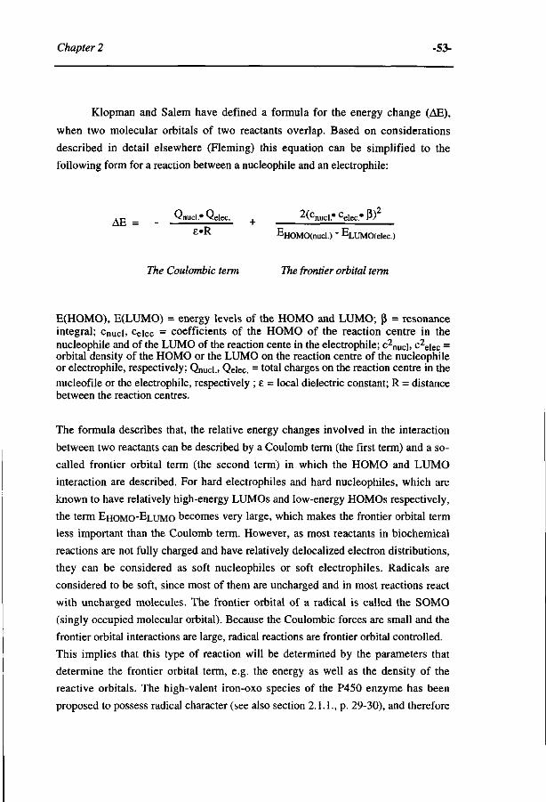
Chapter 2 -53-
Klopman and Salem have defined a formula for the energy change (AE),
when two molecular orbitals of two reactants overlap. Based on considerations
described in detail elsewhere (Fleming) this equation can be simplified to the
following form for a reaction between a nucleophile and an electrophile:
A p _ Qnucl.* Qelec. + 2(cnucl.# celec." ß)
e*^ EHOMO(nucl ) - ELUMO(elec )
The Coulombic term The frontier orbital term
E(HOMO), E(LUMO) = energy levels of the HOMO and LUMO; ß = resonance integral; cnucl. celec = coefficients of the HOMO of the reaction centre in the nucleophile and of the LUMO of the reaction cente in the electrophile; c2
nuc], c2
eiec = orbital density of the HOMO or the LUMO on the reaction centre of the nucleophile or electrophile, respectively; Qnucl.. Qelec. = total charges on the reaction centre in the nucleofile or the electrophile, respectively ; E = local dielectric constant; R = distance between the reaction centres.
The formula describes that, the relative energy changes involved in the interaction
between two reactants can be described by a Coulomb term (the first term) and a so-
called frontier orbital term (the second term) in which the HOMO and LUMO
interaction are described. For hard electrophiles and hard nucleophiles, which are
known to have relatively high-energy LUMOs and low-energy HOMOs respectively,
the term EHOMO-ELUMO becomes very large, which makes the frontier orbital term
less important than the Coulomb term. However, as most reactants in biochemical
reactions are not fully charged and have relatively delocalized electron distributions,
they can be considered as soft nucleophiles or soft electrophiles. Radicals are
considered to be soft, since most of them are uncharged and in most reactions react
with uncharged molecules. The frontier orbital of a radical is called the SOMO
(singly occupied molecular orbital). Because the Coulombic forces are small and the
frontier orbital interactions are large, radical reactions are frontier orbital controlled.
This implies that this type of reaction will be determined by the parameters that
determine the frontier orbital term, e.g. the energy as well as the density of the
reactive orbitals. The high-valent iron-oxo species of the P450 enzyme has been
proposed to possess radical character (see also section 2.1.1., p. 29-30), and therefore
-54- Biotransformation enzymes.
this intermediate can be considered to possess a SOMO. As this intermediate acts as a
a electrophilic species, the SOMO might interact with the HOMO of the substrate.
Together these considerations form the basis for the application of frontier
orbital theory in making QSAR's for the relative rate of conversion between different
molecules, and/or the regioselectivity (i.e. the relative rate at various reaction centres
in one molecule) of a biotransformation reaction.
Chapter 2 -55-
REFERENCES.
Adachi S., Nagano S., Ishimori K., Watanabe Y., Morishima I., Egawa T., Kitagawa T. and Makino
R., Roles of proximal ligand in heme proteins: Replacement of proximal histidine of human
myoglobin with cysteine and tyrosine by site-directed mutagenesis as models for P450,
chloroperoxidase, and catalase, Biochemistry 32, 241-252, 1993.
Adams N.H., Levi P.E. and Hodgson E., Regulation of cytochrome P-450 isoenzymes by
methylenedioxyphenyl compounds, Chem.-Biol. Interactions 86, 255-274, 1993.
Akhtar M. and Wright J.N., A unified mechanistic view of oxidative reactions catalysed by P450 and
related Fe-containing enzymes, Nat. Prod. Rep. 8, 527-551, 1991.
Allentoff A.J., Bolton J.L., Wilks A., Thompson J.A. and Ortiz de Montellano P.R., Heterolytic versus
homolytic peroxide bond cleavage by sperm whale myoglobin and myoglobin mutants, J.
Am. Chem. Soc. 114, 9744-9749, 1992.
Andres H.H., Vogel R.S., Tarr G.E., Johnson L. and Weber W.E., Purification, physicochemical, and
kinetic properties of liver acetyl-CoA: arylamine N-acetyltransferase from rapid acetylator
rabbits Molecular Pharmacology 31, 446-456, 1987.
Armstrong R.N., Enzyme catalyzed detoxication reactions: mechanisms and stereochemistry, CRC
Critical Reviewes in Biochemistry 22, 39-88, 1987.
Atkinson J.K., Hollenberg P.F., Ingold K.U., Johnson C.C., Le Tadic M.-H., Newcomb M., Putt D.A.,
Cytochrome P450-catalyzed hydroxylations of hydrocarbons: Kinetic deuterium isotope
effects for the hydroxylation of an ultrafast radical clok, Biochemistry 33, 10630-10637,
1994.
Atkinson J.K. and Ingold K.U., Cytochrome P450 hydroxylation of hydrocarbons: Variation in the rate
of oxygen rebound using cyclopropyl radical clocks including two new ultrafast probes,
Biochemistry 32, 9209-9214, 1993.
Augusto O., Beilan H.S., Ortiz de Montellano P.R., The catalytic mechanism of cytochrome P450:
spin-trapping evidence for one-electron substrate oxidation. J. Biol. Chem. 257, 11288-
11295,1982.
Ayesh R., Mitchell S.C., Zhang A., and Smith R.L., The fish odor syndrome: biochemical, familial
and clinical aspects, Br. Med. J. 307, 655-657, 1993.
BackesW.L. and Eyer CS., Cytochrome P450 LM2 reduction, J. Biol. Chem. 264, 6252-6259, 1989.
Banci L., Bertini I., Marconi S., Pierattelli R. and Sligar S.G., Cytochrome P450 and aromatic bases, A
•H NMR study, J. Am. Chem. Soc. 116, 4866-4873, 1994.
Batt A.M., Magdalou J., Vincent-Viry M., Ouzzine M., Fournel-Gigleux S., Galteau M.M., Siest G.,
Drug metabolizing enzymes related to laboratory medicine: cytochromes P450 and UDP-
glucuronosyltransferases, Clinica Chimica Acta 226, 171-190, 1994.
-56- Biotransformation enzymes..
Blum M., Grant D.M., McBride W., Heim M., and Meyer U.A., Human arylamine N-acetyl-
transferase genes: isolation, chromosomal localization, and functional expression, DNA Cell
Biol. 9,193-203, 1990.
Bock K.W., Metabolic polymorphism affecting activation of toxic and mutagenic arylamines, Trends
Pharmacol. Science 13, 223-226, 1992.
Boddupalli S.S., Hasemann CA., Ravichandran K.G., Lu J.-Y., Goldsmith E.J., Deisenhofer J.,
Peterson J.A., Crystallization and preliminary x-ray diffraction analysis of P450terp a nd the
hemoprotein domain of P450ßM-3. enzymes belonging to two distinct classes of the
cytochrome P450 superfamily, Proc. Natl. Acad. Sei. 89, 5567-5571, 1992.
Boersma M.G., Cnubben N.H.P., van Berkel W.J.H., Blom M., Vervoort J. and Rietjens I.M.C.M.,
Role of cytochrome P450 and flavin containing monooxyenase in the biotransformation of 4-
fluoro-N-methylaniline, Drug Metabolism and Disposition 21, 218-230, 1993.
Bogaards J.J.P., Van Ommen B., Wolf CR. and Van Bladeren P.J., Human cytochrome P450 enzyme
selectivities in the oxidation of chlorinated benzenes, Toxicology and Applied Pharmacology
132, 44-52,1995.
Bondon A., Macdonald T.L., Harris .M. and Guengerich F.P. Oxidation of cycloalkylamines by
cytochrome P450. Mechanism based inactivation, adduct formation, ring expansion and
nitrone formation, J. Biol. Chem. 264, 1988-1997, 1989.
Burka L.T., Plucinski T.M. and Macdonald T.L., Mechanisms of hydroxylation by cytochrome P450;
metabolism of monohalobenzenes by phenobarbital-induced microsomes, Proc. Natl. Acad.
Sei. 80,6680-6684,1983.
Burka L.T., Thorsen A. and Guengerich F.P., Enzymatic monooxygenation of halogen atoms:
Cytochrome P-450 catalyzed oxidation of iodobenzene by iodosobenzene, J. Am. Chem. Soc.
102, 7615-7616, 1980.
Cashman J.R., Structural and catalytic properties of the mammalian flavin-containing monooyxgenase,
Chem. Res. Toxicol. 8, 165-181, 1995.
Chang T K.H., Weber G.F., Crespi C.L. and Waxman D.J., Differential activation of
cyclophosphamide and ifosphamide by cytochromes P450 2B and 3A in human liver
microsomes. Cancer Research 53, 5629-5637, 1993.
Chen J. and Armstrong R.N., Stereoselective catalysis of a Retro-Michael reaction by class Mu
glutathione transferases. Consequences for the internal distribution of products in the active
site, Chem. Res. Toxicol. 8, 580-585, 1995.
Commandeur, J.N.M., Stijntjes G.J., and Vermeulen N.P.E., Enzymes and transport systems involved
in the formation and disposition of glutathione S-conjugates, Role in bioactivation and
detoxication mechanisms of xenobiotics, Pharmac. Rev. 47, 271-330, 1995.
Cook J. and Hodgson E., The induction of cytochrome P450 by isosafrole and related
Chapter 2 -57-
methylenedioxyphenyl compounds, Chem.-Biol. Interactions 54,299-315, 1985.
Coon M.J., Ding X., Pernecky S.J. and Vaz A.D.N. Cytochrome P450: progress and predictions, The
FASEB Journal 6, 669-673, 1992.
Correia M.A., Yao K., Allentoff A.J., Wrighton S.A., Thompson J.A., Interactions of peroxyquinols
with cytochromes P450 2B1, 3A1 and 3A5, Influence of the apoprotein on heterolytic versus
homolytic O-O bond cleavage, Arch. Biochem. Biophys. 317, 471-478, 1995.
Crespi C.L., Penman B.W., Leaky J.A., Arlotto M.P., Stark A., Parkinson A., Turner T. and Steimel
D., Human cytochrome P4501A3: cDNA sequence, role of the enzyme in the metabolic
activation of promutagens, comparison to the nitrosoamine activation by human cyctochrome
P4502E1, Carcinogenesis 3, 1293-1301, 1990.
Cupp-Vickery J.R. and Poulos T.L., Structure of cytochrome P450 eryF involved in erthromycin
biosynthesis, Stuctural Biology 2, 144-153, 1995.
Daly J.W., Guroff G., Udenfriend S. and Witkop B., Hydroxylation of alkyl and halogen substituted
anilines and acetanilides by microsomal hydroxylases, Biochem. Pharmacol. 17, 31-36, 1968.
Daly J.W., Jerina D.M. and Witkop B., Arene oxides and the NIH shift: The metabolism, toxicity and
carcinogenity of aromatic compounds, Experientia 28, 1129-1264, 1972.
Den Besten C , Van Bladeren P.J., Duizer E., Vervoort J. and Rietjens I.M.C.M., Cytochrome P450-
mediated oxidation of pentafluorophenol to tetrafluorobenzoquinone as the primary reaction
product, Chem. Res. Toxicol. 6, 674-680, 1993.
De Voss J.J. and Ortiz de Mpntellano P.R., Computer-assisted, structure-based prediction of substrates
for cytochrome P450cam, J. Am. Chem. Soc. 117, 4185-4186, 1995.
Ebel R.E., O'Keeffe, Peterson J.A.,. Substrate binding to hepatic microsomal cytochrome P450, J.
Biol. Chem. 253, 3888-3897,1978.
Fleming I. (ed.), Radical reactions, in: Frontier Orbitals and Organic Chemical Reactions, John Wiley
& Sons, New York, 182-186, 1989.
Fruetel J.A., Collins J.R., Camper D.L., Loew G.H. and Ortiz de Montellano P.R.O., Calculated and
experimental absolute stereochemistry of the styrene and ß-methylstyrene epoxides formed
by cytochrome P450cam, J. Am. Chem. Soc. 114, 6987-6993, 1992.
Fukui K., Yonezawa T., Nagata C , and Shingu H., Molecular orbital theory of orientation in aromatic,
heteroaromatic and other conjugated molecules, / . Chem. Phys. 22, 1433-1442, 1954.
Gallagher E.P. Wienkers L.C., Stapleton P.L., Kunze K.L., Eaton D.L., Role of CYP1A2 and
CYP3A4 in the bioactivation of alfatoxin BI (AFB1) by human liver microsomes. Cancer
Research 54, 1-8, 1994.
Gerber N.C. and Sligar S.G., Catalytic mechanism of cytochrome P-450: evidence for a distal charge
relay, J. Am. Chem. Soc. 114, 8742-8743, 1992.
Ghauri F.Y., Blackledge CA., Glen R.C., Sweatman B.C., Lindon J.C., Beddell CR., Wilson I.D. and
-58- Biotransformation enzymes.
Nicholson J.K., Quantitative structure-metabolism relationships for substituted benzoic acids
in the rat, Biochem. Pharmacol. 44, 1935-1946, 1992.
Gibson G.G. and Skett P. (Eds.), in: Introduction to drug metabolism, Chapman and Hall, New York,
1989.
Goeptar A.R., Scheerens H. and Vermeulen N.P.E., Oxygen and xeniobiotic reductase activities of
cytochrome P450, Critical Reviews in Toxicology 25, 25-65, 1995.
Gonzalez F.J., Human cytochromes P450: problems and prospects, TIPS Reviews 131, 346-352, 1992.
Gonzalez F.J., The molecular biology of cytochrome P450s, Pharmacological Reviews 40, 243-288,
1989.
Gonzalez F.J. and Idle J.R., Pharmacogenetic phenotyping and genotyping, present status and future
potential, Clin. Pharmacokinet. 26,59-70, 1994.
Gonzalez F.J. and Meyer U.A., Molecular genetics of the debrisoquine-spartein polymorphism, Clin.
Pharm. Ther. 50,233-238, 1991.
Gough A.C., Miles J.S., Spurr N.W., Moss J.E., Gaedigk A., Eichelbaum M. and Wolf CR.,
Identification of the primary gene defect at the cytochrome P450 CYP2D locus, Nature 347,
773-776, 1990.
Graminski G.F., Zhang P., Sesay M.A., Ammon H.L. and Armstrong R.N., Formation of the 1-(S-
glutathionyl)-2,4,6-trinitrocyclohexadienate anion at the active site of glutathione S-
transferase: Evidence for enzymic stabilization of a-complex intermediates in nucleophilic
aromatic substitution reactions, Biochemistry 28, 6252-6258, 1989.
Groot M.J. de, Donné-Op den Kelder G., Commandeur J.N.M., Lenthe J.H. van, Vermeulen N.P.E.,
Metabolite predictions for para-substituted anisoles based on ab initio complete active space
self-consistent field calculations, Chem. Res. Toxicol 8, 437-443, 1995.
Groot M.J. de, van der Aar E.M., Nieuwenhuizen P.J., van der Plas R.M., Donné-Op den Kelder G.,
Commandeur J.N.M, and Vermeulen N.P.E., A predictive substrate model for rat glutathione
S-transferase 4-4, Chem. Res. Toxicol. 8, 649-658, 1995.
Guengerich F.P., Oxidation-reduction properties of rat liver cytochrome P-450 and NADPH-
cytochrome P-450 reductase related to catalysis in reconstituted systems, Biochemistry 22,
2811-2820, 1983.
Guengerich F.P., Oxidation of halogenated compounds by cytochrome P-450, peroxidases and model
metalloporphyrins, J. Biol. Chem. 264, 17198-17205, 1989.
Guengerich F.P., Enzymatic oxidation of xenobiotic chemicals, Biochemisty and Molecular Biology
25,97-153, 1990.
Guengerich F.P., Metabolic activation of carcinogens, Pharmac. Ther. 54, 17-61, 1992.
Guengerich F.P., Cytochrome P450 enzymes, American Scientist 81, 440-447, 1993.
Guengerich F.P., Bell L.C. and Okazaki O., Interpretations of cytochrome P450 mechanisms from
Chapter 2 -59-
kinetic studies, Biochimie 77, 573-580, 1995.
Guengerich F.P. and Macdonald T.L., Chemical mechanisms of catalysis by cytochromes P450 : A
unified view, Ace. Chem. Res. 17, 9-16, 1984.
Guengerich F.P. and Macdonald T.L., Mechanisms of cytochrome P-450 catalysis, The FASEB
Journal 4, 2453-2459, 1990.
Guengerich F.P., Muller-Enoch D., Blair I.A., Oxidation of quinidine by human liver cytochrome
P450., Mol. Pharmacol. 30, 287-.29S, 1986.
Guengerich F.P., Kim P. and Iwasaki M., Role of human cytochrome P450IIE1 in the oxidation of
many low molecular weight cancer suspects, Chem. Res. Toxicol. 4, 168-179, 1991.
Gulick A.M. and Fahl W.E., Mammalian glutathione S-transferases: regulation of an enzyme system
to achieve chemotherapeutic efficacy, Pharmac. Ther. 66, 237-257, 1995.
Hammons G.J., Guengerich F.P., Weis CC, Beland F.A., and Kadbular F.F., Metabolic oxidation of
carcinogenic arylamines by rat, dog and human hepatic microsomes and by flavin-containing
and cytochrome P-450 monooxygenase, Cancer Research 45, 3578-3585, 1985.
Hansch C. and Zhang L., Quantitative structure activity relationships of cytochrome P450, Drug
Metabolism Reviews 25, 1-48, 1993.
Hanzlik R.P. and Tullman R.H., Suicidal inactivation of cytochrome P450 by cyclopropylamines.
Evidence for cation radical intermediates, J. Am. Chem. Soc. 104, 2048-2050, 1982.
Harada N. and Omura T., Participation of cytochrome P450 in the reduction of nitro compounds by rat
liver microsomes, J. Biochem. 87, 1539-1554, 1980.
Harris D. and Loew G., Prediction of regiospecific hydroxylation of camphor analogs by cytochrome
P450cam, J. Am. Chem. Soc. 117, 2738-2746, 1995.
Hasemann CA., Kurumbail R.G., Boddupalli S.S., Peterson J.A., Deisenhof J., Structure and function
of cytochromes P450: a comparative analysis of three crystal structures, Structure 15, 41-62,
1995.
Hein D.W., Rustan T.D., Ferguson R.J., Doll M.A., Gray K., Metabolic activation of aromatic and
heterocyclic N-hydroxyarylamines by wild-type and mutant recombinant human NAT1 and
NAT2 acetyltransferases, Arch. Toxicol. 68, 129-133, 1994.
Higuchi T., Uzu S. and Hirobe M. Synthesis of a highly stable iron porphyrin coordinaetd by
alkylthiolate anion as a model for cytochrome P450 and its catalytic activity in O-O bond
cleavage, J. Am. Chem. Soc. 112, 7051-7053, 1990.
Hlavica P., Regulatory mechanisms in the activation of nitrogenous compounds by mammalian
cytochrome P450 isoenzymes, Drug Metabolism Reviews 26 (1&2), 325-348, 1994.
Hrycay E.G., Gustafsson J.-A., Ingelman-Sundberg M. and Ernster L., The involvement of
cytochrome P450 in hepatic microsomal steroid hydroxylation reactions supported by sodium
periodate, sodium chlorite, and organic hydroperoxides, Eur. J. Biochem. 61, 43-52, 1976.
-60- Biotransformation enzymes.
Ishikawa T., The ATP-dependent glutathione S-conjugate export pump, Trends Biochem. Sei. 17, 463-
468, 1992.
Isseman I. and Green S., Activation of a member of the steroid hormone receptor superfamily by
peroxisome proliferators, Nature 347, 645-650, 1990.
Iyanagi T., Makino R. and Koichi Anan F., Studies on the microsomal mixed-function oxidase system:
Mechanism of action of hepatic NADPH-cytochrome P450 reductase, Biochemistry 20, 1722-
1730, 1981.
Jerina D.M., Michaud D.P., Feldmann R.J., Armstrong R.N., Vyas K.P., Thakker D.R., Yagi H.,
Thomas P.E., Ryan D.E. and Levin W., Stereochemical modeling of the catalytic site of
cytochrome P450c. In: Sato R., Kato R. (eds.) Microsomes, drug oxidations, and drug
toxicity. Tokyo: Japan Scientific Societies Press, 195-201, 1982.
Ji X., Armstrong R.N., and Gilliland G.L., Snapshots along the reaction coordinate of an S]sjAr
reaction catalyzed by glutathione transferase, Biochemistry 32, 12949-12954, 1993.
Ji X., Zhang P., Armstrong R.N., Gilliland G.L., The three-dimensional structure of a glutathione S-
transferase from the Mu gene class. Stuctural analysis of the binary complex of isoenzyme 3-
3 and glutathione at 2.2-Â resolution, Biochemistry 31, 10169-10184, 1992.
Johnson W., Liu S., Ji X., Gilliland G.L. and Armstrong R.N., Tyrosine 115 participates both in
chemical and physical steps of the catalytic mechanism of a glutathione S-transferase, J. Biol.
Chem. 268, 11508-11511, 1993.
Jones J.P., Shou M., and Korzekwa, Stereospecific activation of the procarcinogen benzo[a]pyrene: A
probe for the active sites of the cytochrome P450 superfamily, Biochemistry 34, 6956-6961,
1995.
Koerts J., Rietjens I.M.C.M., Boersma M.G. and Vervoort J., 'H NMR T\ relaxation rate study on
substrate orientation of fluoromethylanilines in the actives sites of microsomal and purified
cytochrome P450 1A1 and 2B1, FEBS Letters 368, 279-284, 1995.
Kominami S. and Takemori S., Effect of spin state on reductions of cytochrome P450 from bovine
adrenocortical microsomes, Biochem. Biophys. Acta 709, 147-153, 1982.
Kong, Ah-Ng Tony and Fei P., Molecular cloning of three sulfotransferase cDNAs from mouse liver,
Chemico-Biological interactions 92, 161-168, 1994.
Korzekwa K.R. and Jones J.P., Predicting the cytochrome P450 mediated metabolism of xenobiotics,
Pharmacogenetics 3, 1-18, 1993.
Korzekwa K.R., Jones J.P., and Gillette J.R., Theoretical studies on cytochrome P450 mediated
hydroxylation: A predictive model for hydrogen atom abstractions, J. Am. Chem. Soc. 112,
7042-7046, 1990.
Korzekwa K„ Swinney D.C., and Trager W.F, Isotopically labelled chlorobenzenes as probes for the
mechanisms of cytochrome P450 catalyzed aromatic hydroxylation, Biochemistry 28, 9019-
Chapter 2 -61-
9027, 1989.
Korzekwa K., Trager W., Gouterman M, Spangler D. and Loew G.H., Cytochrome P450 mediated
aromatic oxidation: A theoretical study, J. Am. Chem. Soc. 107,4273-4279, 1985.
Koymans L., Donné-Op de Kelder G.M., te Koppele J.M., and Vermeulen N.P.E., Generalized
cytochrome P450-mediated oxidation and oxygenation reactions in substituted aromatic
substrates with activated N-H, O-H, C-H or S-H substituents, Xenobiotica 23, 633-648, 1993.
Larroque C , Lange R., Maurin L., Bienvenue A. and van Lier J.E., On the nature of the cytochrome
P450scc "ultimate oxidant": characterization of a productive radical intermediate, Arch.
Biochem. Biophys. 282, 198-201, 1990.
Lawton M.P., Cashman J.R., Cresteil T., Dolphin C , Elfarra A., Hines R.N., Hodgson E., Kimura T.,
Ozols J., Phillips I., Philpot R.M., Poulsen L.L., Rettie A.E., Williams D.E., and Ziegler
D.M., A nomenclature for the mammalian flavin-containing monooxygenase gene family
based on amino acid sequence identities. Arch. Biochem. Biophys. 308, 254-257, 1994.
Lee J.S., Jacobsen N.E., Ortiz de Montellano P.R., 4-Alkyl radical extrusion in the cytochrome P450
catalyzed oxidation of 4-alkyl-l,4-dihydropyridines, Biochemistry 27, 7703-7710, 1988.
Lee W.A. and Bruice T.C., Homolytic and heterolytic oxygen-oxygen bond scissions accompanying
oxygen transfer to iron(III)porphyrins by percarboxylic acids and hydroperoxides. A
mechanistic criterion for peroxidase and cytochrome P450, J. Am. Chem. Soc. 107, 513-514,
1985.
Lefever M.R. and Wackett L.P., Oxidation of low molecular weight chloroalkanes by cytochrome
P450CAM, Biochem. Biophys. Res. Commun. 201, 373-378, 1994.
Levin A.A., and Dent J.G., Comparison of the metabolism of nitrobenzene by hepatic microsomes and
cecal microflora from Fischer-344 rats in vitro and the relative importance of each in vivo.
Drug Metab. Dispos. 10, 450-454, 1982.
Lewis D.F.V., MO-QSARs: a review of molecular orbital-generated quantitative structure activity
relationships, Progress in drug metabolism, (Ed. G.G. Gibson), Taylor & Francis, Chapter 5,
205-255, 1990.
Lewis D.F.V., Three-dimensional models of human and other mammalian microsomal P450s
constructed from an alignment with P450102 (P450t,m3), Xenobiotica 25, 333-366, 1995.
Lewis D.F.V., C. Ioannides and D.V. Parke, Molecular dimensions of the substrate binding site of
cytochrome P-448, Biochem. Pharmacol. 35, 2179-2185, 1986.
Lewis D.F.V., Ioannides C. and Parke D.V., Structural requirements for substrates of cytochromes P-
450 and P-448, Chem.-Biol. Interact. 64, 39-60, 1987.
Lewis D.F.V., Moereels H., Lake B.G., Ioannides C. and Parke D.V., Molecular modeling of enzymes
and receptors involved in carcinogenesis: QSARs and compact-3D, Drug Metabolism
Reviews 26, 261-285, 1994.
-62- Biotransformation enzymes.
Li H. and Poulos T.L., Structural variation in heme enzymes: a comparative analysis of peroxidase and
P450 crystal structures, Structure 2, 461-464, 1992.
Li J.J., Purdy R.H., Appelman E., Klicka J.K. and Li S.A., Catechol formation of fluoro- and bromo-
substituted estradiols by hamster liver microsomes, Molec. Pharmacol. 27, 559-565, 1985.
Lindberg R.L.P. and Negishi M., Alteration of mouse cytochrome P450con substrate specificity by
mutation of a single amino-acid residue, Nature 339, 632-634, 1989.
Liu S., Zhang P., Ji X., Johnson W.W., Gilliland G.L. and Armstrong R.N., Contribution of tyrosine 6
to the catalytic mechanism of isoenzyme 3-3 of glutathione S-transferase, J. Biol. Chem. 267,
4296-4299, 1992.
Lo H.H., Brown P.I., Rankin CO., Acute nephrotoxicity induced by isomeric dichloroanilines in
Fischer 344 rats, Toxicology 63, 215-231, 1990.
Lo H.H., Brown P.I. and Rankin G.O., Trichloroaniline effects on renal function in vivo and in vitro,
Toxicology Utters 57, 319-328, 1991.
Loida P.J., Sligar S.G., Paulsen M.D., Arnold G.E. and Ornstein R.L., Stereoselective hydroxylation of
norcamphor by cytochrome P450cam, /. Biol. Chem. 270, 5326-5330, 1995.
Lu A.Y.H., Levin W., Vore M., Conney A.H., Thakker D.R., Holder D.R., and Jerina D.M.,
Metabolism of benzo(a)pyrene by purified liver microsomal cytochrome P448 and epoxide
hydrase, In: Carcinogenesis 1, Polynuclear Aromatic hydrocarbons: Chemistry, metabolism
and carcinogenesis pp. 115-126. Freudenthal R.I. and Jones P.W. (eds.) Raven Press, New
York., 1976.
Macdonald T.L., Chemical mechanisms of halocarbon metabolism, CRC Critical Reviews in
Toxicology 11, 85-120, 1983.
Macdonald T.L., Gutheim W.G., Martin R.B., and Guengerich F.P., Oxidation of substituted N,N-
dimethylanilines by cytochromes P450: estimation of the effective oxidation-reduction
potential of cytochrome P450, Biochemistry 28, 2071-2077, 1989.
Macdonald T.L., Zirvi K., Burka L.T., Peyman P., Guengerich F.P., Mechanism of cytochrome P450
inhibition by cyclopropylamines, J. Am. Chem Soc. 104, 2050-2052, 1982.
Mackman R., Guo Z., Guengerich F.P. and Ortiz de Montellano P.R., Active site topology of human
cytochrome P450 2E1, Chem. Res. Toxicol. 9, 223-226, 1996.
Mansuy D., Cytochrome P450 and synthetic models, Pure & Applied Chemistry 59, 759-770, 1987.
Mansuy D., Battioni P. and Battioni J.-P., Chemical model systems for drug-metabolizing cytochrome
P450 dependent monooxygenases [review], Eur. J. Biochem. 184, 2647-285, 1989.
Mason R.P. and Holzman J.L., The mechanism of microsomal and mitochondrial nitroreductase.
Electron spin resonance evidence for nitroaromatic free radical intermediates, Biochemistry
14, 1626-1632, 1975.
Miller J.A., Sulfonation in chemical carcinogenesis-history and present status, Chemico-Biological
Chapter 2 -63-
Interactions 92, 329-341, 1994.
Monks T.J., and Lau S.S., Commentary: renal transport processes and glutathione conjugate-mediated
nephrotoxicity, Drug Metab. Dispos. 15, 437-441, 1987.
Morais de S.M.F., Wilkinson G.R., Blaisdell J., Nakamura K., Meyer U.A. and Goldstein J.A., The
major genetic defect responsible for the polymorphism of (S)-mephenytoin metabolism in
humans,/ Biol. Chem. 269, 15419-15422, 1994.
Morgenstein R. and DePierre J.W., Membrane-bound glutathione transferases. In: Glutathione
conjugation. Eds.: Sies H. and Ketterer B., Academic Press, London, 157-175, 1988.
Muerhoff A.S., Griffin K.J. and Johnson E.F., The peroxisome proliferator-activated receptor mediates
the induction of CYP4A6, a cytochrome P450 fatty acid co-hydroxylase, by clofibric acid, J.
Biol. Chem. 267, 19051-19053, 1992.
Mulder G.J., Coughtrie M.W.H. and Burchell B., (ed), Conjugation reactions in drug metabolism,
Taylor & Francis, Chapter 4, Glucuronidation, 1990.
Myers T.G., Thummel K.E., Kalhorn T.F., and Nelson S.D., Preferred orienations in the binding of 4'-
hydroxyacetanilide (acetaminophen) to cytochrome P450 1 Aland 2B1 isoforms as
determined by 13C- and 15N-NMR relaxation studies, J. Med. Chem. 37, 860-867, 1994.
Nazar-Stewart V., Motulsky A.G., Eaton D.L., White E., Hornung S.K., Leng Z.-T„ Stapleton P. and
Weiss N.S., The glutathione S-transferase m polymorphism as a marker for susceptibility to
lung carcinoma, Cancer Research 53, 2313-2318, 1993.
Nedelcheva V. and Gut I., P450 in the rat and man: methods of investigation, substrate specificities
and relevance to cancer, Xenobiotica 24, 1151-1175, 1994.
Nelson D.R., Kamataki T., Waxman D.J., Guengerich F.P., Estabrook R.W., Feyereisen R., Gonzalez
F.J., Coon M.J., Gunsalus I.C., Gotoh O., Okuda K. and Nebert D.W., The P450 superfamily-
update on new sequences, gene mapping, accession numbers, early trivial names of enzymes,
and nomenclature, DAM Cell Biol. 12, 1-51, 1993.
Nelson H.H., Wiencke J.K., Christiani D.C., Cheng T.J., Zuo Z.-F., Schwartz B.S., Lee B.-K., Spitz
M.R., Wang M., Xu X., Kelsey K.T., Ethnic differences in the prevalence of the homozygous
deleted genotype of glutathione S-transferase theta, Carcinogenesis 16, 1243-1245, 1995.
O'Brien P.J., Wong W.C., Silva J. and Skan S., Toxicity of nitrobenzene compounds towards isolated
hepatocytes: dependence on reduction potential, Xenobiotica 20, 945-955, 1990.
Oguri K., Yamada H. and Yoshimura H., Regiochemistry of cytochrome P450 isozymes, Annu. Rev.
Pharmacol. Toxicol. 34, 251-279, 1994.
Okazaki O. and Guengerich F.P., Evidence for specific base catalysis in N-dealkylation reactions
catalyzed by cytochrome P450 and chloroperoxidase, J. Biol. Chem. 268, 1546-1552, 1993.
Omura T. and Sato R., The carbon monoxide binding pigment of liver microomes, J. Biol. Chem. 239,
2370-2378, 1964.
-64- Biotransformation enzymes.
Ortiz de Montellano P.R., Cytochrome P450; structure, mechanism and biochemistry, Plenum Press,
New York and London, 1986.
Ortiz de Montellano P.R., Control of the catalytic activity of prosthetic heme by the structure of
hemoproteins, Ace. ChemRes. 20,289-294, 1987.
Ortiz de Montellano P.R., Kunze K.L., Beilan H.S. and Wheeler C , Destruction of cytochrome P450
by vinyl fluoride, fluroxene and acetylene. Evidence for a radical intermediate in olefin
oxidation, Biochemistry 21, 1331-1339, 1982.
Ouzounis C.A. and Meivin W.T., Primary and secondary structural patterns in eukaryotic cytochrome
P450 families correspond to structures of helix-rich domain of Pseudomonas putida
cytochrome P450cam, Eur. J. Biochem. 198, 307-315, 1991.
Paine A.J., Heterogeneity of cytochrome P450 and its toxicological significance, Human and
Experimental Toxicology 14, 1-7, 1995.
Parry G., Palmer D.N. and Williams D.J., Ligand partitioning into membranes, its significance in
determining Km and Ks values for cytochrome P450 and other membrane bound receptors
and enzymes, FEBS Letters 67, 123-129.
Parke D.V., The cytochromes P450 and mechanims of chemical carcinogenesis, Environmental health
perspectives 102, 852-853, 1994.
Paulsen M.D. and Ornstein R.L., Substrate mobility in thiocamphor-bound cytochrome P450cam: an
explanation of the conflict between the observed product profile and the X-ray structure,
Protein Engineering 6, 359-365, 1993.
Pemble S., Schroeder K.R., Spencer S.R., Meyer D.J., Hallier E., Bolt H.M., Ketterere B. and Taylor
J.B., Human glutathione S-transferase theta (GSTT1): cDNA cloning and the characterization
of a genetic polymorphism, Biochem. J. 300, 271-276, 1994.
Porter T.D. and Coon M.J., Cytochrome P450, multiplicity of isoforms, substrates, and catalytic and
regulatory mechanisms, J. Biol. Chem. 266, 13469-13472, 1991
Poulsen L.L. and Ziegler D.M., Multisubstrate flavin-containing monooxygenase: applications of
mechanism to specificity. Chem.-.Biol. Interact. 96, 57-73, 1995.
Raag R., Martinins S.A., Sligar S.G. and Poulos T.L., Crystal structure of the cytochrome P450cam
active site mutant Thr252Ala, Biochemistry 30, 11420-11429, 1991.
Raag R. and Poulos T.L., Crystal structures of cytochrome P-450cam complexed with camphane,
thiocamphor and adamantane: factors controlling P450 substrate hydroxylations,
Biochemistry 30, 2674-2684, 1991.
Raag R. and Poulos T.L., X-Ray crystallographic structural studies of cytochrome P450cam: Factors
controlling substrate metabolism, pp. 1-44 in: Relationships between structure and function of
cytochrome P450; experiments, calculations, models, Frontiers in Biotransformation, Vol. 7.
(eds. K. Ruckpaul and H. Rein), Akademie Verlag, Berlin, 1992.
Chapter 2 -65-
Rankin CO., Yang D.J., Cressey-Veneziano K., Casto S., Wang R.T. and Brown P.I., In vivo and in
vitro nephrotoxicity of aniline and its monochlorophenylderivatives in the Fischer 344 rat,
Toxicology 38, 269-283, 1986a.
Rankin G.O., Yang D.J., Teets V.J., Lo H.H. and Brown P.I., 3,5-Dichloroaniline-induced
nephrotoxicity in the Sprague-Dawley rat, Toxicology Letters 30, 173-179, 1986b.
Ravichandran K.G., Boddupalli S.S., Hasemann CA., Peterson J.A., Deisenhofer J., Crystal structure
of hemoprotein domain of P450BM-3, a prototype for microsomal P450's, Science 261, 731-
736, 1993.
Reinemer P., Dirr H.W., Ladenstein R., Huber R., Lo Bello M., Federici G. and Parker M.W., Three-
dimensional structure of class 7t glutathione S-transferases from human placenta in complex
with S-hexylglutathione at 2.8 Â resolution, J. Molec. Biol. 227, 214-226, 1992.
Reinemer P., Dirr H.W., Ladenstein R., Schäffer J., Gallay O., and Huber R., The three dimensional
structure of class n glutathione S-transferase in complex with glutathione sulfonate at 2.3 Â
resolution, EMBO J. 10,1997-2005, 1991.
Rietjens I.M.C.M., Soffers A.E.M.F., Hooiveld G.J.E.J., Veeger C. and Vervoort J., Quantitative
structure activity relationships based on computer calculated parameters for the overall rate of
glutathione S-transferase catalyzed conjugation of a series of fluoronitrobenzenes, Chem. Res.
Tox. 8, 481-488, 1995.
Rietjens I.M.C.M., Soffers A.E.M.F., Veeger C. and Vervoort J. Regioselectivity of cytochrome P450
catalyzed hydroxylation of fluorobenzenes predicted by calculated frontier orbital substrate
characteristics, Biochemistry 32, 4801-4812, 1993.
Rietjens I.M.C.M., and Vervoort J., Bioactivation of 4-fluorinated anilines to benzoquinoneimines as
primary reaction products, Chem.-Biol. Interact. 22, 263-281, 1991.
Rietjens I.M.C.M., and Vervoort J., A new hypothesis for the mechanism for the cytochome P450
dependent aerobic conversion of hexahalogenated benzenes to pentahalogenated phenols,
Chem. Res. Toxicol. 5, 10-19, 1992.
Riley P. and Hanzlik R.P., Electron transfer in P450 mechanisms. Microsomal metabolism of
cyclopropylbenzene and/7-cyclopropylanisole, Xenobiotica 24, 1-16, 1994.
Rizk P.N. and Hanzlik R.P., Oxidative and non-oxidative metabolism of 4-iodoanisole by rat liver
microsomes, Xenobiotica 25, 143-150, 1995.
Robertson I.G.C. and Jernstrom B., The enzymatic conjugation of glutathione with bay-region diol-
epoxides of benzo[a]pyrene, benz[a]anthracene and chrysene, Carcinogenesis 7, 1633-1636,
1986.
Ryu D.-Y., Levi P.E. and Hodgson E., Regulation of cytochrome P-450 isoenzymes CYP1A1,
CYP1A2 an CYP2B10 by three benzodioxole compounds, Chem.-Biol. Interact. 96, 235-247,
1995.
-66- Biotransformation enzymes..
Sealy R.C., Swartz H.M. and Olive P.L., Electron spin resonance-spin trapping, detection of
superoxide formation during aerobic microsomal reduction of nitro-compounds, Biochem.
Biophys. Res. Commun. 82, 680-684, 1978.
Sharrock M., Debrunner P.G., Schulz C , Lipscomb J.D., Marshall V. and Gunsalus I.C., Cytochrome
P450cam and its complexes. Mossbauer parameters of the heme iron, Biochim. Biophys. Acta
420, 8-26, 1976.
Shaw G.-C. and Fulco A.J., Barbiturate-mediated regulation of expression of the cytochrome
P450BM-3 gene of Bacillus megaterium by Bm3Rl protein, J. Biol. Chem. 267, 5515-5526,
1992.
Shimada T. and Nakamura S.I., Cytochrome P450 mediated activation of procarcinogens and
promutagens to DNA damaging products by measuring expression of umu-gene in
Salmonella typhimurium TA1535, Biochem. Pharmacol. 36, 1979-1987, 1987.
Simmons D.L., McQuiddy P. and Kasper C.B., Induction of the hepatic mixed function oxidase
system by synthetic glucocorticoids: transcriptional and post-transcriptional regulation, J.
Biol. Chem. 262, 36-332. 1987.
Sinning I., Kleywegt G.J., Cowan S.W., Reinemer P., Dirr H.W., Huber R., Gilliland G.L., Armstrong
R.N., Ji X„ Board P.G., Olin B., Mannervik B. and Jones T.A., Structure determination and
refinement of human alpha class glutathione transferase Al-1, and a comparison with the Mu
and Pi class enzymes., J. Mol. Biol. 232, 192-212, 1993.
Steward A.R., Wrighton S.A., Pasco D.S., Fagan J.B., Li D. and Guzelian P.S., Synthesis and
degradation of 3-methylcholanthrene-inducible cytochromes P-450 and their mRNAs in
primary monolayer cultures of adult rat hepatocytes, Arch. Biochem. Biophys. 241, 494-508,
1985.
Strobel H.W., Lu A.Y.H., Heidema J., and Coon M.J., Phosphatidylcholine requirement in the
enzymatic reduction of hemoprotein P450 and in fatty acid, hydrocarbon, and drug
hydroxylation, J. Biol. Chem. 245,4851-4854, 1970.
Suzuki J. Meguro S., Moriat O., Hirayama S. and Suzuki S., Comparison of in vivo binding of
aromatic nitro and amino compounds to rat hemoglobin, Biochem. Pharmac. 38, 3511-3519,
1989.
Swanson B.A., Dutton D.R. Lunetta J.M., Yang CS. and Ortiz de Montellano P.R., The active sites of
cytochromes P450IA1, IIB1, IIB2, and HEI, J. Biol. Chem. 266, 19258-19264, 1991.
Tatsumi K., Kitamura S., Yoshimura H. and Kawazoe Y. Susceptibility of aromatic nitro compounds
to xanthine oxidase-catalyzed reduction. Chem. Pharm. Bull. 26, 1713-1717, 1978.
Temellini E., Franchi M., Giuliani L. and Pacifici G.M., Human liver sulphotransferase and UDP-
glucuronosyltransferase: structure-activity relationship for phenolic subsrates, Xenobiotica
21, 171-177, 1991.
Chapter 2 -67-
Tew K.D., Gluathione-associated enzymes in anticancer drug resistance, Cancer Research 54, 4313-
4320, 1994.
Tuck S.F. and Ortiz de Montellano P.R., Topological mapping of the active sites of cytochrome
P4502B1 and P4502B2 by in situ rearrangement of aryl-iron complexes, Biochemistry 31,
6911-6916, 1992.
Turesky R.J., Lang N.P., Butler M.A., Teitel C.H., and Kadbular F.F., Metabolic activation of
carcinogenic heterocyclic amines by human liver and colon, Carcinogenesis 12, 1839-1845,
1991.
Valentovic M.A., Ball J.G., Anestis D.K., Beers K.W., Madan E., Hubbard J.L. and Rankin G., Aute
renal and hepatic toxicity of 2-haloanilines in Fischer 344 rats, Toxicology 75, 121-131, 1992.
Van de Straat R., de Vries J., de Boer H.J.R., Vromans R.M. and Vermeulen N.P.E., Relationship
between paracetamol binding to and its oxidation by two cytochrome P-450 isoenzymes - a
proton nuclear magnetic resonance and spectrophotometric study, Xenobiotica 17, 1-9, 1987.
Van Ommen B., and Van Bladeren P.J., Possible reactive intermediates in the oxidative
biotransformation of hexachlorobenzene. Drug Metab. Drug Interact. 7, 213-243, 1989.
Waxman D.J. and Azaroff L., Phénobarbital induction of cytochrome P450 gene expression, Biochem.
J. 281, 577-592, 1992.
Weber W.W., Levy G.N. and Hein D.W., Acetylation. In Conjugation reactions in drug metabolism,
Ed: Mulder G.J., pp 163-191, 1990.
White R.E. and Coon M.J., Oxygen activation by cytochrome P450, Ann. Rev. Biochem. 49, 315-356,
1980.
White R.E., McCarthy M.-B., Egeberg K.D. and Sligar S.G., Regioselectivity in the cytochromes
P450: control by protein constraints and by chemical reactivities, Arch. Biochem. Biophys.
228, 493-502, 1984.
Wilce M.C.J., Board P.G., Feil S.C. and Parker M.W., Crystal structure of a theta-class glutathione
transferase, The EMBO Journal 14, 2133-2143, 1995.
Yamazoe Y., Nagata K., Ozawa S., Kato R., Structural similarity and diversity of sulfotransferases,
Chem.-Biol. Interact. 92, 107-117, 1994.
Yang C.S., Yoo J.-S.H., Ishizaki H. and Hong Y., Cytochrome P450IIE: Roles in nitrosamine
metabolism an mechanisms of regulation, Drug Metabolism Reviews 22, 147-159, 1990.
Yin H., Anders M.W., Korzekwa K.R., Higgins L., Thummel K.E., Kharasch E.D. and Jones J.P.,
Designing safer chemicals: Predicting the rates of metabolism of halogenated alkanes, Proc.
Natl. Acad. Sei. 92, 11076-11080, 1995.
Zhang P., Liu S., Shan S., Ji X., Gilliland G.L. and Armstrong R.N., Modular mutagenesis of exons 1,
2, and 8 of a glutathione S-transferase from the mu class. Mechanistic and structural
consequences for chimeras of isoenzyme 3-3, Biochemistry 31, 10185-10193, 1992.
-68- Biotransformation enzymes.
Zhong S., Howie A.F., Ketterer B., Taylor J., Hayes J.D., and Beckett G.J., Wathen CG., Wolf CR.,
and Spurr N.K., Glutathione S-transferase \l polymorphism as a marker for susceptibility to
lung carcinoma, Cancer Res. 12, 1533-1537, 1991.
Ziegler D.M., Flavin-containing monooxygenases: catalytic mechanisms and substrate specificities,
DrugMetab. Rev. 19, 1-32, 1988.
Ziegler D.M., in Enzymatic basis of detoxication Vol.1, Chapt.9, Microsomal flavin-containing
monooxygenase: oxygenation of nucleophilic nitrogen and sulphur compounds, pp. 201-227
(W.B. Jakoby, ed.) Academic Press, New York, 1980.
Ziegler D.M., Graf P., Poulsen L., Sies H., and Stal W., NADPH-dependent oxidation of reduced
ebselen, 2-selenylbenzanilide, and of 2-(methylseleno)benzanilide catalyzed by pig liver
flavin-containing monooxygenase, Chem. Res. Toxicol. 5, 163-166, 1992.
Zvelebil M.J.J.M., Wolf CR. and Sternberg M.J.E., A predicted three-dimensional structure of human
cytochrome P450: implications for substrate specificity, Protein Engineering 4, 271-282,
1991.
CHAPTER 3
Study on the regioselectivity and mechanism of the aromatic hydroxylation of monofluoroanilines.
N.H.P. Cnubben, J. Vervoort, C. Veeger and I.M.C.M. Rietjens.
Chemico-Biological Interactions 85, 151-172, 1992.
SUMMARY.
The in vitro and in vivo metabolism of monofluoroanilines was investigated.
Special attention was focussed on the regioselectivity of the aromatic hydroxylation
by cytochromes P450 and the mechanism by which this reaction might proceed. The
results clearly demonstrate that the in vitro and in vivo regioselectivity of the
aromatic hydroxylation by cytochromes P450 is dependent on the fluoro-substituent
pattern of the aromatic aniline-ring. Results from experiments with liver microsomes
from differently pretreated rats demonstrate that the observed regioselectivity for the
aromatic hydroxylation is not predominantly determined by the active site of the
cytochromes P450. To investigate the underlying reason for the observed
regioselectivity, semi-empirical molecular orbital calculations were performed.
Outcomes of these calculations show that neither the frontier orbital densities of the
LUMO/LUMO+1 (lowest unoccupied molecular orbital) of the monofluoroanilines
nor the spin-densities in their NH» radicals can explain the observed
regioselectivities. The frontier orbital densities of the HOMO/HOMO-1 (highest
occupied molecular orbital) of the monofluoroanilines however, qualitatively
correlate with the regioselectivity of the aromatic hydroxylation. Based on these
results, it is concluded that the cytochrome P450 dependent aromatic hydroxylation
of monofluoroanilines does not proceed by hydrogen or electron abstraction from the
aniline substrate to give an aniline-NH« radical. The results rather suggest that
cytochrome P450 catalyzed aromatic hydroxylation of monofluoroanilines proceeds
by an electrophilic attack of the (FeO)3+ species of cytochrome P450 on a specific
carbon atom of the aromatic aniline-ring.
-70- Regioselectivity and aromatic hydroxylation..
INTRODUCTION.
Halogenated arylamines are widely used in industry for the synthesis of dyes,
pesticides and herbicides [1]. Mammalian exposure to these compounds might arise
from industrial pollution and from the production of these chemicals as a result of
biological or combustional degradation of agrochemicals.
Cytochrome P450 enzymes catalyze the oxidative biotransformation of these
aromatic compounds. This conversion results in more polar aminophenol
metabolites, which can be conjugated in subsequent reactions followed by excretion
from the body [2-4]. The mechanism by which cytochrome P450 hydroxylates
aromatic compounds is still a matter of debate [5-7].
Based on literature [5,6] several mechanisms for the cytochrome P450
dependent aromatic hydroxylation of arylamines can be proposed, which are
presented in Figure 1A and B, taking 3-fluoroaniline as a model compound.
The first two mechanisms proceed by formation of a fluoroaniline-NH«
radical. This radical results from a reaction in which the cytochrome P450 (FeO)3+
species abstracts a hydrogen atom from the aminogroup of the aniline substrate
(mechanism 1). However, for molecules with heteroatoms of low ionization
potential, like nitrogen, the aniline-NH» radical might as well result from a single
electron transfer to cytochrome P450, followed by the loss of a proton from the
fluoroaniline cation (mechanism 2)[5]. Once the NH» radical is formed, the reaction
pathway proceeds by recombination of the substrate NH» radical with a hydroxide
radical, donated by the (FeOH)3+ moiety of the cytochrome P450 (oxygen rebound
mechanism). This results in the formation of adducts, which rearrange to the
energetically more favorable aminophenols, via keto-enol tautomerization. The third
mechanism, depicted in Figure IB, involves a radical attack of the cytochrome P450
(FeO)3+ intermediate on the aniline-ring to give a so-called a-adduct [6]. The
reaction proceeds by a collapse of this a-complex to give the aminophenols, either
directly or through the formation of epoxides or ketones as an intermediate step [6,7].
The objective of the present study was to investigate which of these reaction
pathways is more likely for the aromatic hydroxylation of arylamines.
Monofluoroanilines were taken as model compounds, because (i) the fluoro-
substituent will not cause significant steric hindrance as its Van der Waals radius
(1.35 Â) almost equals that of a hydrogen substituent (1.20 À); (ii) the metabolism of
fluoroanilines can be studied by 19F-NMR, which implies that all products from
hydroxylation can be detected and quantified in a single run by the same method.
The in vitro and in vivo regioselectivity for the aromatic hydroxylation of the
monofluoroanilines was studied and compared to the outcomes of molecular orbital
calculations. It was expected that, if the aromatic hydroxylation proceeds by
Chapter 3 -71-
(FWH)3 '
,T^^; m&* lEaTlufl* I
NH
6. i
T^v
NH
6-, keto-anol lautomerlsation
NHg
B
ftO H 1 F„3*
Figure 1 Reaction pathway for the hydroxylation of the monofluoroanilines, proceeding by
(A) the so-called "oxygen rebound" mechanism. Formation of the aniline-NH»
radical might result from either abstraction of a hydrogen atom from the aminogroup
of the fluoroaniline (mechanism 1) or from electron abstraction followed by proton
release in two separate steps (mechanism 2) [see Ref. 5], or (B) the attack of the
cytochrome P450 (FeO)^+ intermediate at the aniline-ring to give a O-adduct [see
Refs. 5-7]
-72- Regioselectivity and aromatic hydroxylation..
formation of a NH» radical, the regioselectivity of the reaction depends on the spin
distribution of the radical in the aromatic ring, i.e. the SOMO (single occupied
molecular orbital) density of the NH» radical. However, when the reaction proceeds
by formation of the a-adduct, the site at which the cytochrome P450 (FeO)3+ radical
interacts with the aniline-substrate, will determine the regioselectivity. Because
radical reactions are generally frontier-orbital controlled [8], the site of attack of the
(FeO)3+ SOMO-electron can be expected to be directed by either the HOMO
(highest occupied molecular orbital) or the LUMO (lowest unoccupied molecular
orbital) density of the aniline molecule.
Generally, these assumptions are based on the fact that not only the active site
of the cytochrome P450 and the steric characteristics of the substrates, but also the
electronic characteristics of the substrates will contribute to the regioselectivity of
their aromatic hydroxylation by cytochromes P450 [9]. This paper also describes
experiments carried out to ascertain that regioselectivity for the aromatic
hydroxylation of the monofluoroanilines is indeed not predominantly determined by
steric effects in the active site of the cytochromes P450.
MATERIALS AND METHODS.
Reference compounds.
2-Fluoroaniline, 3-fluoroaniline, 4-fluoroaniline were obtained from Janssen
Chimica (Beerse, Belgium). Potassium fluoride was from Merck (Darmstadt, FRG).
2-Fluoro-4-nitrophenol was synthesized according to the method described by Vogel
[10] for the synthesis of 2- and 4-nitrophenol. Using 2-fluorophenol (Janssen
Chimica) and potassium nitrate (Merck) as starting materials, this procedure resulted
in a reaction mixture containing 2-fluoro-4-nitrophenol and 2-fluoro-6-nitrophenol.
These two products were separated by crystallisation of 2-fluoro-4-nitrophenol in
pentane. The same procedure was followed for the synthesis of 3-fluoro-2-
nitrophenol using 3-fluorophenol (Janssen Chimica) and potassium nitrate as starting
materials.
3-Fluoro-4-aminophenol, 5-fluoro-2-aminophenol, 4-fluoro-2-aminophenol,
3-fluoro-2-aminophenol and 2-fluoro-4-aminophenol were synthesized by chemical
reduction of respectively 3-fluoro-4-nitrophenol (Fluorochem, Derbyshire, UK), 5-
fluoro-2-nitrophenol (Janssen Chimica), 4-fluoro-2-nitrophenol (Fluorochem), 3-
fluoro-2-nitrophenol and 2-fluoro-4-nitrophenol under 3 atm. H2 (24 h) in the
presence of Pd/C (0.1 % w/v) (Janssen Chimica) using ethylacetate as the solvent.
Because the synthesized 3-fluoro-2-nitrophenol and 2-fluoro-4-nitrophenol were not
completely pure, their reduced compounds (3-fluoro-2-aminophenol and 2-fluoro-4-
Chapter 3 -73-
nitrophenol) were further purified on silicagel 60 (70-230 Mesh) (Merck) using a
mixture of ethylacetate-ethanol-acetic acid (15:3:2 by vol.)'as eluent.
2-Fluoro-6-aminophenol was synthesized by the demethylation of 3-fluoro-o-
anisidine (Aldrich, Steinheim, FRG) in hydrobromic acid and acetic acid at 130°C,
essentially as described by Bhatt et al. [11].
2-Fluoracetanilide, 3-fluoro-4-acetamidophenol, 3-fluoracetanilide, 2-fluoro-
4-acetamidophenol, 4-fluoracetanilide and 5-fluoro-2-acetamidophenol were
synthesized by acylation of the corresponding amino-compound following the
procedure described by Vogel [12].
4-Fluoro-N-hydroxyaniline was synthesized by the reduction of 4-
fluoronitrobenzene as described by Coleman et al. [13]. In short, a mixture of 2 g 4-
fluoronitrobenzene (Aldrich) and 1 g ammoniumchloride in 30 ml H2O was stirred
vigorously. Over a period of 15 minutes pulverized zinc was added in small portions,
whereupon the mixture was refluxed at 60°C. After 1 h, the hot suspension was
filtered to remove the zinc oxide residues. The filtrate was saturated with NaCl and
cooled on ice. The yellow precipitate presented 4-fluoro-N-hydroxyaniline and was
isolated.
In vivo exposure to 2-, 3- or 4-fluoroaniline.
Male Wistar rats (300-350 g) were housed individually in metabolism cages.
The animals were given food and water ad libitum. Control urine was collected for
24 h before dosing. After these 24 h, the rats were orally dosed with 50 mg 2-, 3- or
4- fluoroaniline per kg body weight in olive oil. Urine was collected for 24 h after
oral dosing and stored at -20 C.
Preparation of microsomes.
Microsomes were prepared from the perfused livers of male Wistar rats (=
300 g), which were untreated or treated with inducers of the cytochrome P450
enzymes, namely phénobarbital (Brocacef, Maarssen, The Netherlands)(0.1 % in
drinking water for 7 days), acetone (Merck, Darmstadt, FRG)(1.5 ml, 30% in water,
administered by oral gavage, 24 h before sacrificing), dexamethasone (Sigma, St
Louis, MO, USA)(300 mg/kg bodyweight, using a stock solution of 90 mg/ml in
water containing 2% Tween-80, administered by oral gavage, daily for 4 days), 3-
methylcholanthrene (Sigma, St Louis, MO, USA)(30 mg/kg body weight, using a
stock solution of 6 mg/ml in olive oil, i.p., daily for 3 days), and isosafrole (Janssen,
Beerse, Belgium)(150 mg/kg bodyweight, using a stock solution of 50 mg/ml in olive
oil, i.p. daily for 3 days).
Following homogenisation of the livers in Tris-sucrose buffer (50 mM Tris,
0.25 M sucrose, pH 7.4) and centrifugation at 10,000 x g (20 min), the supernatants
-74- Regioselectivity and aromatic hydroxy lotion..
were centrifugea for 75 min. at 105,000 x g. The microsomal pellet was washed once
with Tris-sucrose buffer and finally suspended in potassium phosphate pH 7.25
containing 20% glycerol and 1 mM EDTA. The final preparation was immediately
frozen in liquid nitrogen and stored at -80 C until use.
Cytochrome P450 contents were determined as described by Omura and Sato
[14]. Protein was assayed by the method of Lowry [15] using bovine serum albumin
(Sigma, St Louis, MO, USA) as the standard.
Microsomal incubations.
Microsomal incubations were carried out at 37 C in 0.1 M potassium
phosphate, pH 7.6, containing 1 uM microsomal cytochrome P450 and 10 mM 2-, 3-
or 4-fluoroaniline added as 1% (v/v) of a stock solution in DMSO. The reaction was
started by addition of NADPH (1 mM final concentration) and terminated after 10
min by freezing the reaction mixture in liquid nitrogen for 19F-NMR measurements
or by adding 1.0 ml of the reaction mixture to 0.3 ml 20% trichloroacetic acid (TCA)
for the chemical detection of 4-aminophenol.
Chemical detection of 4-aminophenol.
4-Aminophenol was determined by the method of Brodie and Axelrod [16] as
follows: to 1 ml precipitated supernatant, 100 ul 5% phenol reagent (5% w/v phenol
in 2.5 M NaOH) and 200 ul 2.5 M Na2C03 were added. After 45 min. at room
temperature, the absorbance at 630 nm was measured. The extinction coefficient for
aminophenol in this assay was determined to be 30.5 mM-1 cm"1.
Enzyme hydrolysis of conjugates in urine samples.
To hydrolyse glucuronide conjugates, 8 units (40 ul) of ß-glucuronidase (from
E. coli Rn, Boehringer, Mannheim, Germany) in 1200 ul 0.2 M KH2P04/Na2HP04
pH 6.2 were added to an equal volume of urine sample. The mixture was made
anaerobic by four cycles of evacuating and filling with argon and incubated for 1 h at
37°C.
To hydrolyse the sulphate and glucuronide conjugates 40 ul of the
arylsulphatase/ß-glucuronidase enzyme mixture from Helix pomatia (Boehringer,
Mannheim, Germany) in 1200 ul 0.1 M potassium acetate pH 5.2 were added to an
equal volume of urine sample. The mixture was made anaerobic and incubated for 16
h at 37°C. 4-Fluoro-2-aminophenylsulphate could only be hydrolyzed by
arylsulphatase treatment after its isolation from the urine by Bio-Gel P2 column
chromatography, carried out as described before [17].
Chapter 3 -75-
19F-NMR measurements. 19F-NMR measurements were performed on a Bruker CXP 300 and a Bruker
AMX 300 spectrometer operating at 282.3 MHz as described before [18]. Proton
decoupling (inverse gated decoupling) was achieved with the Waltz-16 pulse
sequence [19] at -16 dB from 20 W (CXP 300) and -22 dB from 50 W (AMX 300).
Norell (Landisville, NJ) 10 mm NMR-tubes were used. The sample volume
was 1.71 ml, containing 100 ul 2H2Û for locking the magnetic field. The samples
were made oxygen free by four cycles of evacuating and filling with argon. Between
60000 and 80000 scans were recorded for spectra from microsomal incubations. For
urine spectra between 2000 and 3000 scans were recorded.
Chemical shifts are reported relative to CFCI3 [20].
Concentrations of the various metabolites observed in the 19F-NMR spectra
were determined by comparison of the integrals of their 19F-NMR resonances to the
integral from the 19F-NMR resonance of parafluorobenzoic acid, added to each
sample as an internal standard.
Molecular orbital computer calculations.
Computer calculations were performed on a Silicon Graphics Iris 4D/85 using
Quanta/Charmm (Polygen inc., U.K.). The semi-empirical molecular orbital method
was used, applying the AMI Hamiltonian from the AMP AC program (Quantum
Chemistry Program Exchange no 506) (Indiana University, Bloomington, IN, USA)
[21]. All calculations were carried out with PRECISE criteria. For all calculations the
self-consistent field was achieved. Geometries were optimized for all bond lengths,
bond angles and torsion angles using the Fletcher-Powell criteria.
The frontier electron densities of the monofluoroanilines were calculated as
indicated by Fukui [22] to assess the contribution of the occupied orbital just below
the HOMO, i.e. HOMO-1, and the unoccupied orbital above the LUMO, i.e.
LUMO+1.
In this study, the outcomes of the in vacuo AMI calculations are related to
the electronic behaviour of these substrates in the relative hydrophobic environment
of the active site pocket of the cytochromes P450. Due to solvation effects and a
different dielectric constant, the intrinsic properties of the compounds might be
influenced. However, it is assumed that this phenomenon not influences the relative
difference of parameters between a series of closely related compounds, like the
monofluoroanilines. The outcomes of these in vacuo computer calculations can thus
be used as an approach to study relative differences within this series of related
compounds.
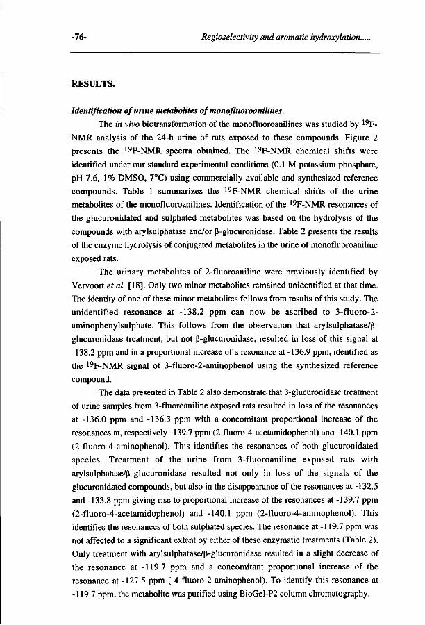
-76- Regioselectivity and aromatic hydroxylation..
RESULTS.
Identification of urine metabolites of monofluoroanilines.
The in vivo biotransformation of the monofluoroanilines was studied by 19F-
NMR analysis of the 24-h urine of rats exposed to these compounds. Figure 2
presents the 19F-NMR spectra obtained. The 19F-NMR chemical shifts were
identified under our standard experimental conditions (0.1 M potassium phosphate,
pH 7.6, 1% DMSO, 7°C) using commercially available and synthesized reference
compounds. Table 1 summarizes the 19F-NMR chemical shifts of the urine
metabolites of the monofluoroanilines. Identification of the 19F-NMR resonances of
the glucuronidated and sulphated metabolites was based on the hydrolysis of the
compounds with arylsulphatase and/or ß-glucuronidase. Table 2 presents the results
of the enzyme hydrolysis of conjugated metabolites in the urine of monofluoroaniline
exposed rats.
The urinary metabolites of 2-fluoroaniline were previously identified by
Vervoort et al. [18]. Only two minor metabolites remained unidentified at that time.
The identity of one of these minor metabolites follows from results of this study. The
unidentified resonance at -138.2 ppm can now be ascribed to 3-fluoro-2-
aminophenylsulphate. This follows from the observation that arylsulphatase/ß-
glucuronidase treatment, but not ß-glucuronidase, resulted in loss of this signal at
-138.2 ppm and in a proportional increase of a resonance at -136.9 ppm, identified as
the 19F-NMR signal of 3-fluoro-2-aminophenol using the synthesized reference
compound.
The data presented in Table 2 also demonstrate that ß-glucuronidase treatment
of urine samples from 3-fluoroaniline exposed rats resulted in loss of the resonances
at -136.0 ppm and -136.3 ppm with a concomitant proportional increase of the
resonances at, respectively -139.7 ppm (2-fluoro-4-acetamidophenol) and -140.1 ppm
(2-fluoro-4-aminophenol). This identifies the resonances of both glucuronidated
species. Treatment of the urine from 3-fluoroaniline exposed rats with
arylsulphatase/ß-glucuronidase resulted not only in loss of the signals of the
glucuronidated compounds, but also in the disappearance of the resonances at -132.5
and -133.8 ppm giving rise to proportional increase of the resonances at -139.7 ppm
(2-fluoro-4-acetamidophenol) and -140.1 ppm (2-fluoro-4-aminophenol). This
identifies the resonances of both sulphated species. The resonance at -119.7 ppm was
not affected to a significant extent by either of these enzymatic treatments (Table 2).
Only treatment with arylsulphatase/ß-glucuronidase resulted in a slight decrease of
the resonance at -119.7 ppm and a concomitant proportional increase of the
resonance at -127.5 ppm ( 4-fluoro-2-aminophenol). To identify this resonance at
-119.7 ppm, the metabolite was purified using BioGel-P2 column chromatography.
Chapter 3 -77-
ÇH,
C-0
ÇH, c-o
- 1
LM i
B
& *! ^ -
UL JU
OH,
C -0
C-O
L. Ji
ÇH,
C-O
ÇH,
C-O
JUE__JL_-> J L.
"1 I -1 20 130
PPM
140
Figure 2 ,9F-NMR spectra of 24-hr urine of monofluoroanilines exposed male Wistar rats.
(A) 2-fluoroaniline, (B) 3-fluoroaniline, and (C) 4-fluoroaniline. Each animal
received 50 mg/kg fluoroaniline dissolved in olive oil.
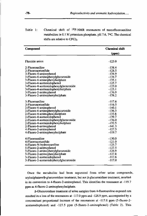
-78- Regioselectivity and aromatic hydroxyiation..
Table 1: Chemical shift of 19F-NMR resonances of monofluoroaniline
metabolites in 0.1 M potassium phosphate, pH 7.6,7°C. The chemical
shifts are relative to CFCI3.
Compound Chemical shift
(ppm)
Fluoride anion -123.0
2-Fluoroaniline -138.4 2-Fluoroacetanilide -128.5 3-Fluoro-4-aminophenol -134.9 3-Fluoro-4-aminophenylglucuronide -134.7 3-Fluoro-4-aminophenylsulphate -135.1 3 -Fluoro-4-acetamidophenol -125.7 3-Fluoro-4-acetamidophenylglucuronide -125.0 3-Fluoro-4-acetamidophenylsulphate -125.1 3-Fluoro-2-aminophenol -136.9 3-Fluoro-2-aminophenylsulphate -138.2
3-Fluoroaniline -117.6 3-Fluoroacetanilide -116.5 2-Fluoro-4-aminophenol -140.1 2-Fluoro-4-aminophenylglucuronide -136.3 2-Fluoro-4-aminophenylsulphate -133.8 2-Fluoro-4-acetamidophenol -139.7 2-Fluoro-4-acetamidophenylglucuronide -136.0 2-Fluoro-4-acetamidophenylsulphate -132.5 2-Fluoro-6-aminophenol -141.3 4-Fluoro-2-aminophenol -127.5 4-Fluoro-2-aminopheny lsulphate -119.7
4-Fluoroaniline -130.0 4-Fluoroacetanilide -121.0 4-Fluoro-N-hydroxy aniline -125.7 5-Fluoro-2-aminophenol -127.5 5-Fluoro-2-aminophenylglucuronide -126.9 5-Fluoro-2-aminophenylsulphate -127.8 5-Fluoro-2-acetamidophenol -117.6 5-Fluoro-2-acetamidophenylglucuronide -117.0
Once the metabolite had been separated from other urine compounds,
arylsulphatase/ß-glucuronidase treatment, but not ß-glucuronidase treatment, resulted
in its conversion to 4-fluoro-2-aminophenol. This identifies the resonance at -119.7
ppm as 4-fluoro-2-aminophenylsulphate.
ß-Glucuronidase treatment of urine samples from 4-fluoroaniline exposed rats
resulted in a loss of the resonances at -117.0 ppm and -126.9 ppm, accompanied by a
concomitant proportional increase of the resonances at -117.6 ppm (5-fluoro-2-
acetamidophenol) and -127.5 ppm (5-fluoro-2-aminophenol) (Table 2). This
Chapter 3 -79-
Table 2: Identification of the 19F-NMR resonances of the glucuronidated and
sulphated metabolites in the urine of Wistar rats dosed with 2-
fluoroaniline, 3-fluoroaniline or 4-fluoroaniline. The urine samples
were either untreated (I), ß-glucuronidase treated (II) or
arylsulphatase/ß-glucuronidase treated (III) (N=2). See section
Materials and Methods for exact procedure.
WF-NMR resonance
(ppm)
Metabolites in urine (% of total 19F-intensity)
I
2-Fluoroaniline -123.0 -125.0 -125.1 -125.7 -134.7 -134.9 -135.1 -135.4 -136.9 -138.2 -138.4
3-Fluoroan -117.6 -119.7 -123.0 -127.5 -132.5 -133.3 -133.6 -133.8 -136.0 -136.3 -139.7 -140.1
4-Fluoroan -117.0 -117.6 -123.0 -126.9 -127.5 -127.8 -130.0 -132.5 -133.9 -134.6 -135.2 -138.0 -139.7 -144.6
0 10.5 15.7 0.9 4.1 0
49.7 4.48
0 6.6 6.4
line 1.2
22.3 15.2
0 37.3 1.8 2.7
20.0 5.5 1.1 0.6 0
line 7.7 0
11.2 3.1 0
69.6 1.5 0.9 0.5 0 1.1 1.0 0 0
II
0 0
17.2 10.6 0
3.5 50.0 2.5 0
6.2 6.6
1.4 21.9 14.9 0.4 36.5 2.1 2.9
20.0 0 0
5.8 1.2
0 8.2 10.9
0 3.0
69.2 1.7 0.9 0.4 0
0.9 0.9 0 0
III
0 0 0
29.5 0
55.1 0
2.2 6.1 0.4 9.6
1.6 14.9 15.8 8.5 0 0 0 0 0 0
40.1 24.1
0 8.4 11.2
0 69.1 1.6 1.7 0 0
0.9 0 0
0.3 1.1
Identified as
Fluoride anion 3-F-4-acetamidophenylglucuronide 3-F-4-acetamidophenylsulphate 3-F-4-acetamidophenol 3-F-4-aminophenylglucuronide 3-F-4-aminophenol 3-F-4-aminophenylsulphate Unknown 3-F-2-aminophenol 3-F-2-aminophenylsulphate 2-Fluoroaniline
3-Fluoroaniline 4-F-2-aminophenylsulphate Fluoride anion 4-F-2-aminophenol 2-F-4-acetamidophenylsulphate Unknown Unknown 2-F-4-aminophenylsulphate 2-F-4-acetamidophenylglucuronide 2-F-4-aminophenylglucuronide 2-F-4-acetamidophenol 2-F-4-aminophenol
5-F-2-acetamidophenylglucuronide 5-F-2-acetamidophenol Fluoride anion 5-F-2-aminophenylglucuronide 5-F-2-aminophenol 5-F-2-aminophenylsulphate 4-Fluoroaniline Unknown Unknown Unknown Unknown Unknown Unknown Unknown
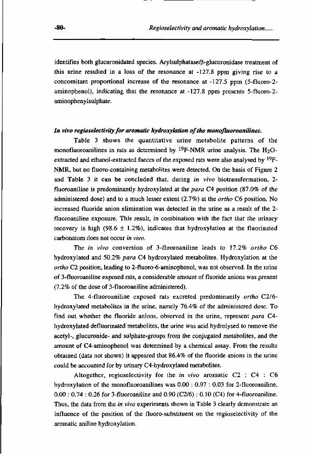
-80- Regioselectivity and aromatic hydroxylation..
identifies both glucuronidated species. Arylsulphatase/ß-glucuronidase treatment of
this urine resulted in a loss of the resonance at -127.8 ppm giving rise to a
concomitant proportional increase of the resonance at -127.5 ppm (5-fluoro-2-
aminophenol), indicating that the resonance at -127.8 ppm presents 5-fluoro-2-
aminophenylsulphate.
In vivo regioselectivity for aromatic hydroxylation of the monofluoroanilines.
Table 3 shows the quantitative urine metabolite patterns of the
monofluoroanilines in rats as determined by 19F-NMR urine analysis. The H2O-
extracted and ethanol-extracted faeces of the exposed rats were also analysed by 19F-
NMR, but no fluoro-containing metabolites were detected. On the basis of Figure 2
and Table 3 it can be concluded that, during in vivo biotransformation, 2-
fluoroaniline is predominantly hydroxylated at the para CA position (87.0% of the
administered dose) and to a much lesser extent (2.7%) at the ortho C6 position. No
increased fluoride anion elimination was detected in the urine as a result of the 2-
fluoroaniline exposure. This result, in combination with the fact that the urinary
recovery is high (98.6 ± 1.2%), indicates that hydroxylation at the fluorinated
carbonatom does not occur in vivo.
The in vivo conversion of 3-fluoroaniline leads to 17.2% ortho C6
hydroxylated and 50.2% para C4 hydroxylated metabolites. Hydroxylation at the
ortho C2 position, leading to 2-fluoro-6-aminophenol, was not observed. In the urine
of 3-fluoroaniline exposed rats, a considerable amount of fluoride anions was present
(7.2% of the dose of 3-fluoroaniline administered).
The 4-fluoroaniline exposed rats excreted predominantly ortho C2/6-
hydroxylated metabolites in the urine, namely 76.4% of the administered dose. To
find out whether the fluoride anions, observed in the urine, represent para C4-
hydroxylated defluorinated metabolites, the urine was acid hydrolysed to remove the
acetyl-, glucuronide- and sulphate-groups from the conjugated metabolites, and the
amount of C4-aminophenol was determined by a chemical assay. From the results
obtained (data not shown) it appeared that 86.4% of the fluoride anions in the urine
could be accounted for by urinary C4-hydroxylated metabolites.
Altogether, regioselectivity for the in vivo aromatic C2 : C4 : C6
hydroxylation of the monofluoroanilines was 0.00 : 0.97 : 0.03 for 2-fluoroaniline,
0.00 : 0.74 : 0.26 for 3-fluoroaniline and 0.90 (C2/6) : 0.10 (C4) for 4-fluoroaniline.
Thus, the data from the in vivo experiments shown in Table 3 clearly demonstrate an
influence of the position of the fluoro-substituent on the regioselectivity of the
aromatic aniline hydroxylation.
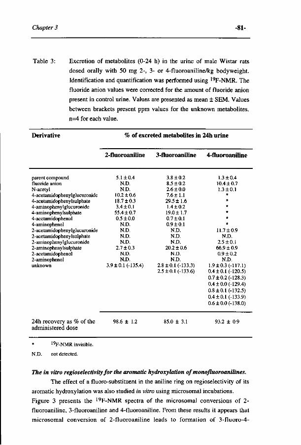
Chapter 3 -81-
Table 3: Excretion of metabolites (0-24 h) in the urine of male Wistar rats
dosed orally with 50 mg 2-, 3- or 4-fluoroaniline/kg bodyweight.
Identification and quantification was performed using 19F-NMR. The
fluoride anion values were corrected for the amount of fluoride anion
present in control urine. Values are presented as mean ± SEM. Values
between brackets present ppm values for the unknown metabolites.
n=4 for each value.
Derivative
parent compound fluoride anion N-acetyl 4-acetamidophenylglucuronide 4-acetamidophenylsulphate 4-aminophenylglucuronide 4-aminophenylsulphate 4-acetamidophenol 4-aminophenol 2-acetamidophenylglucuronide 2-acetamidophenylsulphate 2-aminophenylglucuronide 2-aminophenylsulphate 2-acetamidophenol 2-aminophenol unknown
24h recovery as % of the administered dose
% of excreted metabolites in
2-fluoroaniline
5.1 ±0.4 N.D. N.D.
10.2 ±0.6 18.7 ±0.3 3.4 ±0.1
55.4 ± 0.7 0.5 ± 0.0
N.D. N.D. N.D. N.D.
2.7 ± 0.3 N.D. N.D.
3.9 ±0.1 (-135.4)
98.6 ± 1.2
3-fluoroaniline
3.8 ±0.2 8.5 ±0.2 2.6 ± 0.0 7.6± 1.1 29.5 ±1.6 1.4 ±0.2 19.0 ±1.7 0.7 ±0.1 0.9 + 0.1
N.D. N.D. N.D.
20.2 ±0.6 N.D. N.D.
2.8 ±0.1 (-133.3) 2.5 ±0.1 (-133.6)
85.0 ± 3.1
24h urine
4-fluoroaniline
1.3 ±0.4 10.4 ± 0.7 1.3 ±0.1
* * * * * *
11.7 ±0.9 N.D.
2.5 + 0.1 66.9 + 0.9 0.9 ±0.2
N.D. 1.9+ 0.3 (-117.1) 0.4 ±0.1 (-120.5) 0.7 ±0.2 (-128.3) 0.4 ±0.0 (-129.4) 0.8 + 0.1 (-132.5) 0.4±0.1 (-133.9) 0.6+ 0.0 (-138.0)
93.2 + 0.9
* 19F-NMR invisible.
N.D. not detected.
The in vitro regioselectivity for the aromatic hydroxylation of monofluoroanilin.es.
The effect of a fluoro-substituent in the aniline ring on regioselectivity of its
aromatic hydroxylation was also studied in vitro using microsomal incubations.
Figure 3 presents the 19F-NMR spectra of the microsomal conversions of 2-
fluoroaniline, 3-fluoroaniline and 4-fluoroaniline. From these results it appears that
microsomal conversion of 2-fluoroaniline leads to formation of 3-fluoro-4-
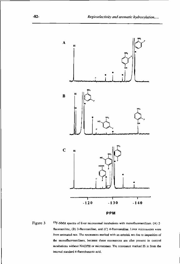
-82- Regioselectivity and aromatic hydroxylation..
-J iM<tayi^flM*V*|MM>^i««*a^
F J I i l
NH2
V**^ W # > # U H
B
i
w > *
l
HO
F"
NH2
OH
^ , .M»*^., .,«*,».. J > . . , ^ X , ' W M / VfWSM**/
, ^^^ -130
""ï 140 120
PPM
Figure 3 '^F-NMR spectra of liver microsomal incubations with monofluoroanilines. (A) 2-
fluoroaniline, (B) 3-fluoroaniline, and (C) 4-fluoroaniline. Liver microsomes were
from untreated rats. The resonances marked with an asterisk are due to impurities of
the monofluoroanilines, because these resonances are also present in control
incubations without NADPH or microsomes. The resonance marked IS is from the
internal standard 4-fluorobenzoic acid.
Chapter 3 -83-
aminophenol as the main metabolite (Figure 3a). No formation of 3-fluoro-2-
aminophenol (-136.9 ppm), resulting from ortho C6 hydroxylation was observed.
Microsomal conversion of 3-fluoroaniline resulted in the metabolites 2-fluoro-4-
aminophenol and 4-fluoro-2-aminophenol (Figure 3b). 2-Fluoro-4-aminophenol is
the main metabolite, indicating that also in vitro para C4-hydroxylation is the
favoured route for the biotransformation of 3-fluoroaniline. Formation of 2-fluoro-6-
aminophenol (-141.3 ppm), resulting from hydroxylation at C2, was not observed at
the detection limit of the measurement (± 1 uM).
The in vitro biotransformation of 4-fluoroaniline resulted in the formation of
5-fluoro-2-aminophenol and fluoride anions. By a chemical assay it was determined
that these fluoride anions represent C4-aminophenol (data not shown).
Thus, analogous to the in vivo conversion, the conversion of
monofluoroanilines in an in vitro microsomal system results in preferential
hydroxylation in the order C4>C6 for 2-fluoroaniline, C4>C6>C2 for 3-fluoroaniline
and C2/6>C4 for 4-fluoroaniline.
Involvement of microsomal cytochromes P450 in the regioselectivity for aromatic
hydroxylation of 3-fluoroaniline.
Further experiments described in this paper were performed to study the
origin of the observed regioselectivities in more detail.
First, the possible involvement of stereoselective influences of the active site
of the cytochromes P450 on the regioselectivity of the aromatic hydroxylation was
investigated. This was done using 3-fluoroaniline as the substrate.
In Table 4 the C2:C4:C6 hydroxylation ratios are presented for the
conversion of 3-fluoroaniline by liver microsomal preparations from rats pretreated
with different cytochrome P450 inducers. Although these ratios slightly change
dependent on the type of microsomes used, the results in Table 4 clearly demonstrate
that C4-hydroxylation of 3-fluoroaniline remains the major route of conversion for
all types of microsomal preparations and ortho C6 hydroxylation is always
significantly favoured over ortho C2 hydroxylation. Only aromatic hydroxylation by
liver microsomes of isosafrole and 3-methylcholanthrene pretreated rats leads to
some ortho C2 hydroxylation of 3-fluoroaniline, however to a lesser extent than
ortho C6 hydroxylation.
In a previous study, the microsomal para C4-hydroxylation of aniline and the
monofluoroanilines was investigated and it was shown that induction of different
cytochromes P450 affects para C4-hydroxylation of all three monofluoroanilines and
aniline in a similar way [23]. For aniline, it was reported that different purified
hepatic cytochrome P450 enzymes are able to hydroxylate this compound, especially
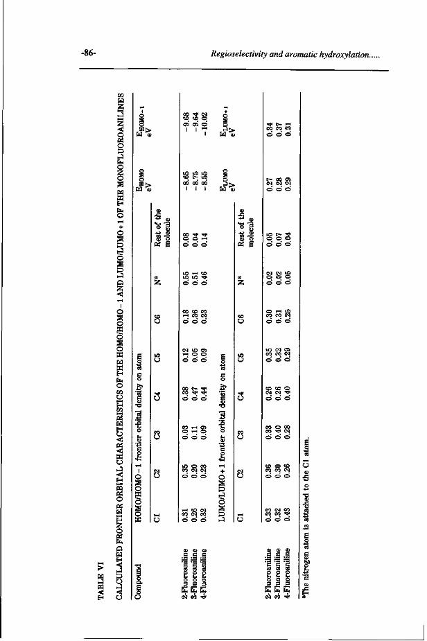
-86- Regioselectivity and aromatic hydroxylation..
« <
W H Z
O BS O & J fa O z o
a H Eb
o + o
o
s
o I
o o o s o a « H fa O CO
o H W
H H
< W O J <u EH 1-4
m ©
OS H H Z
EL.
Q » S3
o
s EC | ^
o S o 5Gî>.
•43 C o
1-1
1 O S O P O s o «
eo U
I N U
T-t
O
*> «
-M $
(S E
OS OJ O I I ^
eo 00 1
00 l
oo l
o s s
o 3
00 t o CO
Ö Ö Ö
i-H O O O O O
oo t - •>» CO ^ TJ* © © ©
I CIS c o
m c
I D
u
CO »"-< CJi O .-H O
m o eo c o CM CM © © ©
i-l tO <N CO CM CO
3
+ o
Ô
&
• H - ^
•M &
0ä g
U
c- oo en « M C4
m t- s o o o ö o ö
M N $
o o o ö ö ö
O H lO eo eo o)
in <M o i
eo co csj © © ©
co co o ( N O ) ' *
'S s
s. E o o
CU 0) V
c c c
ï ï 1 o o o o o o 3 3 3 fa fa Cu
eM co Ti"
co o eo •>* o o
to o co eo o o
CO CM
eo co o o
0> CU
C c
§ g o o L, h o o 3 3
a o
t o CM O
eo •* o
4> G
?: p >-o 3
fa fa fa <M CO T
E 3 rt » H
o 5 o •o J3
3 CIS
.52 E 1 c
£ -U 'S
f
Chapter 3 -87-
al. [25] and Vervoort et al. [18], who dosed rats with 2-fluoroaniline and analysed
the urinary metabolites using respectively HPLC and 19F-NMR. The present study
reports a small amount of ortho hydroxylated product, 2-aminophenylsulphate,
present as 2.7% of all fluorinated metabolites in the urine of a 2-fluoroaniline
exposed rat. Probably one of the unidentified metabolites of Eadsforth et al. [25]
should be ascribed to this 2-aminophenylsulphate.
4-Fluoroaniline appeared to become predominantly C2/6 hydroxylated (82%
of the fluorine intensity present in the urine) and to a minor extent (9%) C4-
hydroxylated. Upon C4-hydroxylation, the fluorine atom is preferentially lost from
the molecule giving rise to fluoride anion formation by a reaction pathway previously
described [26, 27].
3-Fluoroaniline appeared to become hydroxylated at its para C4 and ortho C6
position, but hardly or not at all at its ortho C2 position. For 3-fluoroaniline both in
vitro and in vivo the percentage of metabolites that resulted from ortho C6
hydroxylation (11-20%) was higher than the corresponding percentage for 2-
fluoroaniline (0-2.7%). This implies that a fluoro-substituent at C3 in the aniline
molecule activates the ortho C6 position for aromatic hydroxylation compared to the
situation with a fluoro-substituent at C2. The difference in C2 and C6 hydroxylation
observed for 3-fluoroaniline metabolism is also remarkable. This difference cannot
be ascribed to steric hindrance by the fluorine atom attached to C3, preventing
hydroxylation at C2. This can be concluded from the fact that the Van der Waals
radius of a fluorine substituent (1.35 À), is very close to that of a hydrogen
substituent (1.20 Â).
Additional experiments demonstrated that in in vitro incubations with liver
microsomes from rats pretreated with different cytochrome P450 inducers and, thus,
containing different cytochrome P450 enzyme patterns, the regioselectivity of the
aromatic hydroxylation of 3-fluoroaniline was not significantly influenced, whereas
total conversion was influenced by a factor of only 4.7 at most. This eliminates the
possibility that the regioselectivity is predominantly dictated by the way of substrate
binding to the active site of the cytochrome P450, because it is highly unlikely that
all the different cytochromes P450 dictate the juxtaposition of this relative small
substrate in their hydrophobic active site in a similar way. It also eliminates the
possibility that the various hydroxylated monofluoroaniline products are mainly
formed by different cytochrome P450 enzymes, because in such a case induction of
different P450 patterns would be expected to change the relative contribution of the
various hydroxylated products to the metabolite pattern more significantly than was
observed. This conclusion is further supported by data from Furuya et al. [28] who
reported that C2, C4 and C6 hydroxylated metabolites from acetanilide could all be
formed by one cytochrome P450 (P450IA2) expressed in yeast.
-88- Regioselectivity and aromatic hydroxylotion..
Further experiments described in the present study were undertaken to
investigate whether the origin of the observed differences in regioselectivity for the
monofluoroanilines could be ascribed to intrinsic electronic characteristics of the
substrates. As outlined in Figure 1 the kind of electronic characteristics that will
determine the regioselectivity varies with the mechanism for the cytochrome P450
dependent hydroxylation. When the reaction proceeds by hydrogen or electron
abstaction from the amino moiety, regioselectivity will be influenced by the radical
distribution of the NH« radical formed. Whereas a reaction proceeding by addition of
the (FeO)3+ intermediate in the active site of the P450 will be directed by the density
distribution of one of the frontier orbitals. Results from molecular orbital calculations
clearly demonstrate that nor the spin distribution in the 3-fluoroaniline-NH» radical
nor the LUMO/LUMO+1 frontier orbital density in 3-fluoroaniline can explain the
observed difference in C6 and C2 hydroxylation of the compound. Furthermore, the
data on the spin distribution in the 2-fluoroaniline and 3-fluoroaniline NH» radical
and the LUMO/LUMO+1 frontier orbital density in 2-fluoroaniline and 3-
fluoraniline also do not clearly explain the activation of the C6 position for
hydroxylation when the fluoro-substituent is at C3 instead of C2. Even when an
interaction of the (FeO)3+ SOMO of cytochrome P450 with the aniline
LUMO/LUMO+1, followed by formation of an epoxide as an intermediate is
considered, the LUMO/LUMO+1 densities cannot explain the preferential
hydroxylation of C6 over C2 in 3-fluoroaniline.
Additional results demonstrated that both these phenomena are much better
explained by the calculated HOMO/HOMO-1 frontier orbital density distribution of
the monofluoroaniline molecules. In 3-fluoroaniline, the HOMO/HOMO-1 frontier
orbital density at C6 is 2.0 times higher than the corresponding value in 2-
fluoroaniline, while the energies of the HOMO and HOMO-1 are almost the same for
2-fluoroaniline and 3-fluoroaniline. From this it follows that, based on these
HOMO/HOMO-1 densities, in 3-fluoroaniline compared to 2-fluoroaniline the C6
position is activated for hydroxylation by a a-addition mechanism. The observed
difference in 3-fluoroaniline C2 and C6 hydroxylation can also be elegantly
explained by the difference in HOMO/HOMO-1 density at these positions, being
0.20 and 0.36 respectively.
In vivo formation of N-acetylated aminophenols might proceed through a
cytochrome P450 dependent hydroxylation of the fluoroanilines, followed by N-
acetylation of the hydroxylated product. However, formation of these metabolites can
also result from N-acetylation followed by cytochrome P450 dependent
hydroxylation of the N-acetylfluoroanilines. This implies that the regioselectivity of
the aromatic hydroxylation would also be dictated by molecular orbital
characteristics of the N-acetylfluoroanilines. For this reason, molecular orbital
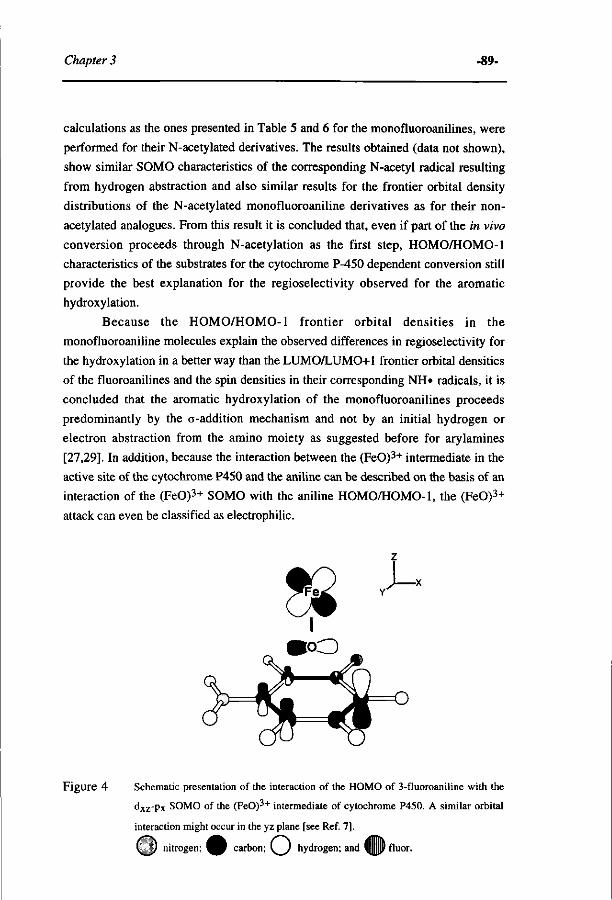
Chapter 3 -89-
calculations as the ones presented in Table 5 and 6 for the monofluoroanilines, were
performed for their N-acetylated derivatives. The results obtained (data not shown),
show similar SOMO characteristics of the corresponding N-acetyl radical resulting
from hydrogen abstraction and also similar results for the frontier orbital density
distributions of the N-acetylated monofluoroaniline derivatives as for their non-
acetylated analogues. From this result it is concluded that, even if part of the in vivo
conversion proceeds through N-acetylation as the first step, HOMO/HOMO-1
characteristics of the substrates for the cytochrome P-450 dependent conversion still
provide the best explanation for the regioselectivity observed for the aromatic
hydroxylation.
Because the HOMO/HOMO-1 frontier orbital densities in the
monofluoroaniline molecules explain the observed differences in regioselectivity for
the hydroxylation in a better way than the LUMO/LUMO+1 frontier orbital densities
of the fluoroanilines and the spin densities in their corresponding NH« radicals, it is
concluded that the aromatic hydroxylation of the monofluoroanilines proceeds
predominantly by the a-addition mechanism and not by an initial hydrogen or
electron abstraction from the amino moiety as suggested before for arylamines
[27,29]. In addition, because the interaction between the (FeO)3+ intermediate in the
active site of the cytochrome P450 and the aniline can be described on the basis of an
interaction of the (FeO)3+ SOMO with the aniline HOMO/HOMO-1, the (FeO)3+
attack can even be classified as electrophilic.
Figure 4 Schematic presentation of the interaction of the HOMO of 3-fluoroaniline with the
d x z -p x SOMO of the (FeO)3+ intermediate of cytochrome P450. A similar orbital
interaction might occur in the yz plane [see Ref. 7].
( J nitrogen; ^ B carbon; \ ) hydrogen; and (||||||P fluor.
-90- Regie/selectivity and aromatic hydroxylation..
Finally, the question can be addressed whether hydroxylation of the
monofluoroanilines at the ortho and para positions requires fast rotation of the
substrate molecule in the active site of the cytochrome P450. Such a rotation would
be in contradiction to results from !H-NMR studies on the longitudinal relaxation
behaviour of protons of paracetamol [30] or acetanilide [31] in the active site of
cytochrome P450.
However, from the molecular orbital calculations of the present study it
follows that the monofluoroaniline HOMO orbitals are rc-type orbitals, as
schematically presented for 3-fluoroaniline in Figure 4.
If the aniline were positioned in the active site with the (FeO)3+ axis
perpendicular to its aromatic ring, the interaction of the aniline HOMO with the
(FeO)3+ dxz-Px as the SOMO orbital [32] might occur as depicted in Figure 4. An
interaction of the aniline HOMO with the (FeO)3+ dyZ-py as the SOMO orbital is
also possible, because the unpaired electron can be present in either of these (FeO)3+
SOMO orbitals. In this case, the interaction of the (FeO)3+ at the ortho and para
positions of the aniline substrate becomes feasible without the requirement for fast
rotation of the substrate in the active site.
In conclusion, the results of the present study indicate that the best working
hypothesis for further research is, that the cytochrome P450 dependent aromatic
hydroxylation of monofluoroanilines proceeds predominantly by the c-addition
mechanism initiated by an electrophilic attack of the (FeO)3+ on the aniline
molecule.
Acknowledgements.
The authors gratefully acknowledge Mr J. Haas for assistance with animal
handling. This study was supported by the Netherlands Organisation for the
Advancement of Scientific Research (N.W.O.).
REFERENCES.
1 J.W. Gorrod and D. Manson, The metabolism of aromatic amines, Xenobiotica 16,933-955,
1986.
2 J.N. Smith and R.T. Williams, Studies in detoxication: the fate of aniline in the rabbit,
Biochem. J. 44, 242-250, 1949.
3 D.V. Parke, Studies in detoxication: the metabolism of [14C]-aniline in the rabbit and other
animals, Biochem. J. 77, 493-503, 1960.
4 D.J. McCarthy, W.R. Waud, R.F. Struck and D.L. Hill, Disposition and metabolism of
Chapter 3 -91-
aniline in Fischer 344 rats and C57BL/6 x C3H V\ mice, Cancer Res. 45, 174-180, 1985.
5 R.N. Armstrong, Enzyme-catalyzed detoxification reactions: mechanism and
stereochemistry, CRC Crit. Rev. Biochem. 22, 39-88, 1987.
6 F.P. Guengerich and T.L. MacDonald, Mechanisms of cytochrome P-450 catalysis, FASEB
J. 4, 2453-2459, 1990.
7 P.R. Ortiz de Montellano (ed.), Oxygen activation and transfer, in: Cytochrome P-450:
Structure, Mechanism and Biochemistry, Plenum Press, New York, 217-271, 1986.
8 I. Fleming (ed.), Radical reactions, in: Frontier Orbitals and Organic Chemical Reactions,
John Wiley & Sons, New York, 182-186, 1989.
9 R.E.White, M-B McCarthy, K.D. Egeberg and S.G. Sligar, Regioselectivity in the
cytochromes P-450: Control by protein constraints and by chemical reactivities, Arch.
Biochem. andBiophys. 228, 493-502, 1984.
10 AJ . Vogel, in: B.S. Furniss, A.J. Hannaford, P.W.G. Smith and A.R. Tatchell (eds.),
Vogel's Textbook of Practical Organic Chemistry, Longman Scientific & Technical,
London, 978, 1989.
11 M.V. Bhatt and S.U. Kulkarni, Cleavage of ethers, Synthesis, 249-282, 1983.
12 A.J. Vogel,in: B.S. Furniss, A.J. Hannaford, P.W.G. Smith and A.R. Tatchell (eds.), Vogel's
Textbook of Practical Organic Chemistry, Longman Scientific & Technical, London, 684,
1978.
13 G.H. Coleman, CM. McClosky and F.A. Stuart, Nitrosobenzene, Org. Synthesis 25, 80-83,
1946.
14 T. Omura and R. Sato, The carbon monoxide pigment of liver microsomes, I. Evidence for
its haemoprotein nature,/ Biol. Chem. 239, 2370-2378, 1964.
15 O.H. Lowry, N.J. Rosebrough , A.L. Farr and A.J. Randall, Protein measurements with
Folin phenol reagent, J. Biol. Chem. 193, 265-275, 1951.
16 B.B. Brodie and J. Axelrod, The estimaton of acetanilide and its metabolic products, aniline,
N-acetyl p-aminophenol and p-aminophenol (free and conjugated) in biological fluids and
tissues, J. Pharmacol. Exp. Ther. 94, 22-28, 1948.
17 I.M.C.M. Rietjens, J. Koerts, S. Boeren and J. Vervoort, Biosynthesis and chemical
determination of polyfluoro-4-hydroxyanilines, Reel. Trav. Chim. Pays-Bas 110, 216-221,
1991.
18 J. Vervoort, P.A. De Jager.J. Steenbergen and I.M.C.M. Rietjens, Development of a 1 9F-
n.m.r. method for studies on the in vivo and in vitro metabolism of 2-fluoroaniline,
Xenobiotica 20, 657-670, 1990.
19 A.J. Shaka, J. Keeler and R. Freeman, Evaluation of a new broad band decoupling
sequence: Waltz 16, / . Magn. Res. 53, 313-340, 1983.
20 V. Wray, in: G.A. Webb (ed.), Annual Reports on NMR Spectroscopy, Vol. 14, Academic
Press, London, 252, 1983.
21 MJ.S. Dewar, E.G. Zoebisch, E.F. Healy and J.J. Stewart, AMI: A new general purpose
-94- MO-QSAR for the cytP450-catalyzed 4-hydroxylation.
electrons of the substrate, this attack is no longer the rate-limiting step of the reaction.
Additional results of the present study demonstrate that the apparent Michaelis constant
Kms of the NADPH/oxygen-supported 4-hydroxylation of the anilines decreases with
increasing hydrophobicity of the aniline derivatives. Because the spectral dissociation
constant K<js of the aniline-cytochrome P450 complex appeared to be severalfold lower
than the Kms, it was concluded that other parameters than binding, influence the Km
s
of the cytochrome P450-catalyzed aniline 4-hydroxylation. In conclusion, the present
paper presents a MO-QSAR for the cytochrome P450 catalyzed conversion of a series
of aniline derivatives. The implications of these findings for the catalytic cycle of
cytochrome P450 are being discussed.
INTRODUCTION.
Cytochromes P450 play an important role in the monooxygenation of aromatic
xenobiotics. The present study focuses on the cytochrome P450-catalyzed conversion
of halogenated anilines. Aniline derivatives are used in industry for the synthesis of
agrochemicals and pigments [1]. Their cytochrome P450-catalyzed conversion is
known to result in both detoxification as well as in bioactivation. Bioactivation can
result, for example, from the cytochrome P450-catalyzed arylamine N-hydroxylation
[2, 3] or from the cytochrome P450-catalyzed oxidative dehalogenation of 4-
halogenated anilines, resulting in formation of electrophilic reactive quinoneimines as
the primary metabolites [4]. Cytochrome P450-dependent monooxygenation of anilines
with a non-halogenated position para with respect to the amino moiety results in
formation of 4-aminophenols as the primary metabolites. 4-Aminophenol can either be
conjugated and be excreted into urine [5, 6] or bile [7] and/or act as an acute
nephrotoxin producing severe necrosis of the proximal tubuli [5, 6, 8, 9].
Regarding the exposure of mammals to halogenated anilines as a consequence
of environmental pollution, it is reasonable to assume that these xenobiotics are present
at a rather low concentration in the mammalian body. The capacity of the cytochrome
'Abbreviations: MO-QSAR, molecular orbital-based quantitative structure-activity relationship;
HXO, hypohalous acid (for example, HCIO = hypochlorous acid); SOMO, singly occupied molecular
orbital; HOMO, highest occupied molecular orbital; HOMO-1, first occupied molecular orbital at the
highest energy level below the HOMO; E(HOMO), energy of the HOMO; Ea, activation energy;
TCA, trichloroacetic acid. Definitions: Kms, apparent Michaelis constant Km for aniline derivatives in
mM; Kds, apparent Kj of the cytochrome P450-substrate complex in mM; kcats, apparent maximum
velocity in nmol converted per nmol cytochrome P450 per minute; e, molar extinction coefficient in
mM"'«cm~l.
Chapter 4 -9 5-
P450 enzyme system for 4-hydroxylation of a homologous series of halogenated
anilines present at subsaturating substrate concentrations can be characterized by and
will be dependent on both kcat and Km [10]. Thus, insight into possibilities for
prediction of kinetic parameters will provide insight in possibilities for prediction of
conversion characteristics of these aniline derivatives.
Therefore, the objective of the present study was to investigate whether
physico-chemical and/or electronic characteristics of a homologous series of aniline
derivatives provide a basis to obtain molecular orbital based quantitative structure-
activity relationships (MO-QSAR's)1 for the kinetic characteristics of their 4-
hydroxylation by the cytochromes P450.
Anilines with a non-halogenated 4-position were used as the model
compounds. Aniline derivatives with a halogenated 4-position were not included,
because the cytochrome P450-catalyzed 4-hydroxylation of an aniline with a
halogenated 4-position was demonstrated previously to proceed by a reaction pathway
different from that of an aniline with a non-halogenated 4-position [4].
MATERIALS AND METHODS.
Chemicals.
4-Aminophenol was obtained from Aldrich (Steinheim, FRG). Phenol and 2-
iodophenol were purchased from Merck (Darmstadt, FRG). 2-Fluoro-, 3-fluoro-, 2-
chloro, 3-chloro-, 2-bromo-, 3-bromo- and 3-iodophenol were from Janssen Chimica
(Beerse, Belgium). Aniline, 2-fluoroaniline, 3-fluoroaniline, 2-chloroaniline, 3-
chloroaniline, 2-bromoaniline, 3-bromoaniline, 2-iodoaniline, 3-iodoaniline, 2,6-
difluoroaniline, 2,5-difluoroaniline, 2,3-difluoroaniline, 3,5-difluoroaniline, 2,3,6-
trifluoroaniline and 2,3,5,6,-tetrafluoroaniline were also purchased from Janssen
Chimica. The purity of all compounds was >98%. Iodosobenzene was synthesized by
the base-catalyzed hydrolysis of diacetoxyiodobenzene (Fluka, Switzerland),
essentially as described by Saltzman and Sharefkin [11], and added to the incubations
from a 20 mM sonicated suspension in 10% Me2SO in demineralized water.
The biosynthesis of polyfluoroaminophenols.
3,5-Difluoro-, 2,6-difluoro-, 2,3-difluoro-, 2,5-difluoro-, 2,3,5-trifluoro- and
2,3,5,6-tetrafluoro-4-aminophenol were prepared by biosynthesis as follows. Male
Wistar rats (300-350 g) were housed individually in metabolism cages. The animals
were given food and water ad libitum. The rats were orally dosed with 100 mg of the
appropriate polyfluoroaniline (respectively 2,6-difluoro-, 3,5-difluoro-, 2,3-difluoro-,
2,5-difluoro-, 2,3,6-trifluoro- and 2,3,5,6-tetrafluoroaniline) per kg body weight in
•96- MO-QSAR for the cytP450-catalyzed 4-hydroxylation.
olive oil. The urine was collected for 48 h after oral dosing. The N-acetyl, glucuronide,
and sulphate moieties were removed from the urinary aminophenol conjugates by acid
hydrolysis, performed by the addition of 1 volume of 12 N HCl to 10 volumes of the
urine sample, followed by heating at 100°C for 30 minutes. After hydrolysis the
solution was neutralized with 12N NaOH and the polyfluoro-4-aminophenols were
isolated from the mixture by extraction into ethyl acetate. The solvent was removed by
evaporation, and the residue was further purified on silicagel 60 (70-230 mesh)
(Merck) using a mixture of ethyl acetate-ethanol-acetic acid (15:3:2 by v/v) as eluent.
The fractions were analyzed using 19F-NMR, the polyfluoro-4-aminophenol-
containing fractions were pooled, and the concentration of the fluorinated 4-
aminophenols was determined using 19F-NMR. The stock solutions of the 4-
aminophenols thus obtained were immediately used for the determination of molar
extinction coefficients in the chemical assay for 4-aminophenol (see hereafter). The
purity of the polyfluoro-4-aminophenols was > 95% as determined by 19F-NMR and >
85 % on the basis of HPLC analysis with UV detection at 240 nm. The identity of the
biosynthetized 4-aminophenols was derived from 19F-NMR and !H-NMR spectral
characteristics. 2,3-Difluoro-4-aminophenol: 19F-NMR (0.1 M potassium phosphate
pH 7.6, relative to CFCl3)(ppm): -160.5 (F3)(dd, 3/F3-F2 = 20.0 Hz, 4 /F3-H5 = 9.0
Hz), -165.3 (F2)(dd, 3 /F2-F3 = 20.0 Hz, 47F2-H6 = 9.6 Hz) and iH-NMR (acetone-
df,, relative to acetone)(ppm): 6.55 (H5)(m, 37H5-H6 = 9.0 Hz, 4 /H5-F3 = 9.0 Hz, 5^H5-F2 = 2.5 Hz), 6.46 (H6)(m, 3/H6-H5 = 9.0 Hz, 47H6-F2 = 9.6 Hz, 5/H6-F3 = 2.5
Hz); 2,5-difluoro-4-aminophenol: 19F-NMR (0.1 M potassium phosphate pH 7.6,
relative to CFCl3)(ppm): -141.4 (F5)(m, 37F5-H6 = H-5 Hz, 47F5-H3 = 8.0 Hz, 5JF5.
F2 = 15.0 Hz,), -146.0 (F2)(m, 3 /F2-H3 = 12.0 Hz, 4JF2-H6 = 8.0 Hz, 57F2-F5 = 15.0
Hz) and iH-NMR (acetone-^6, relative to acetone)(ppm): 6.64 (H3)(dd, 3 /H3-F2 =
12.0 Hz, 4./H3-F5 = 8.0 Hz), 6.61 (H6)(dd, 3/H6-F5 = H-5 Hz, 47H6-F2 = 8.0 Hz);
3,5-difluoro-4-aminophenol: 19F-NMR (0.1 M potassium phosphate pH 7.6, relative
to CFCl3)(ppm): -133.7 (F3/F5)(d, 37F3/F5-H2/H6 = 9.5 Hz) and iH-NMR (acetone
mic, relative to acetone)(ppm): 6.38 (H2/H6)(d, 3^H2/H6-F3/F5 = 9.5 Hz); 2,6-difluoro-
4-aminophenol: 19F-NMR (0.1 M potassium phosphate pH 7.6, relative to
CFCl3)(ppm): -137.8 (F2/F6)(d, 37F2/F6-H3/H5 = 10.0 Hz) and !H-NMR (acetone-d<5,
relative to acetone)(ppm): 6.24 (H3/H5)(d, 3/H3/H5-F2/F6 = 10.0 Hz); 2,3,5-trifluoro-
4-aminophenol: 19F-NMR (0.1 M potassium phosphate pH 7.6, relative to
CFCl3)(ppm): -140.7 (F5)(m, 37F5-H6 = 10.5 Hz, 4/F5-F3 = 10.0 Hz, 5 /F 5 .F 2 =11.0
Hz), -158.5 (F3)(m, 37F3-F2 = 20.5 Hz, 47F3-F5 = 10.0 Hz, 5/F3-H6 = 2.5 Hz),
-171.0 (F2)(m, 37F2-F3 = 20.5 Hz, 47F2-H6 = 8.0 Hz, 5/F2-F5 = HO Hz) and lH-
NMR (acetone-rf(5, relative to acetone)(ppm): 6.54 (H6)(m, 37H6-F5 = 10.5 Hz, 4Ju6-
F2 = 8.0 Hz, 57H6-F3 = 2.5 Hz); 2,3,5,6-tetrafluoro-4-aminophenol: 19F-NMR (0.1 M
potassium phosphate pH 7.6, relative to CFCl3)(ppm): -167.6 (F3/F5)(d, 37F3/F5-
Chapter 4 - 97 -
F2/F6 = 20.0 Hz), -171.4 (F2/F6)(d, 37F2/F6-F3/F5 = 20.0 Hz) and ÏH-NMR (acetone-
d& relative to acetone)(ppm): !H-NMR resonances were not observed in the aromatic
region.
Preparation of microsomes.
Microsomes were prepared from the perfused livers of male Wistar rats (ca.
300 g), which were untreated or treated with inducers of the cytochrome P450
enzymes, namely phénobarbital (Brocacef, Maarssen, The Netherlands), ß-
naphthoflavone (Sigma, St Louis, USA), isosafrole (Janssen, Beerse, Belgium), and
acetone (Merck, Darmstadt, FRG), essentially as described before [12]. Cytochrome
P450 contents were determined as described by Omura and Sato [13]. Protein was
assayed by the method of Lowry [14] using bovine serum albumin (Sigma) as the
standard.
Microsomal incubations.
Microsomal incubations were carried out at 37 C in 0.1 M potassium phosphate
(pH 7.6) containing 1 uM microsomal cytochrome P450 and 0-20 mM (as indicated)
(halogenated) aniline added as 1 % (v/v) of a stock solution in Me2SO. The reaction
was started by the addition of NADPH or iodosobenzene (both at 1 mM final
concentration) and terminated after 10 min (NADPH) or 1 min (iodosobenzene),
respectively, by adding 1.0 mL of the reaction mixture to 0.3 mL 20% (w/v)
trichloroacetic acid (TCA). The cytochrome P450-mediated microsomal conversion of
the aniline derivatives to their 4-aminophenols was observed to be linear in time for at
least 10 minutes in a NADPH/O2-supported reaction and 75 seconds in an
iodosobenzene-supported reaction. For the NADPH series control incubations were
carried out in the absence of NADPH. For the iodosobenzene series control
incubations were carried out for each substrate concentration in the absence of
microsomal cytochrome P450. This was done in order to correct for the chemical
reaction between iodosobenzene and the aniline substrates also resulting in 4-
hydroxylation of the substituted anilines. This correction was 5-40% dependent on the
substrate concentration and aniline derivative used.
Chemical detection of 4-aminophenol.
Halogenated 4-aminophenols were determined essentially by the method of
Brodie and Axelrod [15]. In short, to 1 mL of TCA-precipitated supernatant were
added 100 uL phenol reagent (5% w/v phenol in 2.5 M NaOH) and 200 uL 2.5 M
Na2CC<3. After 60 minutes at room temperature the absorbance at 630 nm was
measured. Introduction of a halogen substituent in the indophenol influences its molar
extinction coefficient at 630 nm [16]. For this reason the £630nm of all halogenated 4-
•98- MO-QSAR for the cytP450-catalyzed 4-hydroxylation.
aminophenol-derived indophenols had to be determined to allow quantification of the
4-aminophenol formation for the various halogenated anilines. For the chlorinated,
brominated and iodinated 4-aminophenols the respective halogenated indophenols were
prepared from the reaction of a suitable halogenated phenol with non-halogenated 4-
aminophenol, instead of by the reaction of the non-substituted phenol with the
halogenated 4-aminophenol as prescribed by the method of Brodie and Axelrod [15].
Figure 1 schematically presents this approach. For the monohalogenated 4-
aminophenols this "reversed" method proved to result in a reliable molar extinction
coefficient of the halogenated indophenols (see Results). In short, the molar extinction
coefficients of the halogenated indophenols derived from the 2- and 3-halogenated 4-
aminophenols were determined by adding respectively 300 uL of 20% TCA, 130 \xL of
3- or 2-halogenated phenol reagent (5% w/v halogenated phenol in 2.5 N NaOH) and
260 uL of 2.5 M Na2C03 to 1 mL sample, containing 0-50 \iM 4-aminophenol in 1%
Me2SO (v/v) in water. The molar extinction coefficients for the polyfluoroindophenols
were determined by the regular method of Brodie and Axelrod [15] using the
polyfluorinated 4-aminophenols obtained from biosynthesis.
NMR measurements. 19F-NMR measurements were performed on a Bruker AMX 300 NMR
spectrometer essentially as described before by Vervoort et al. [17]. Quantification of
the fluorinated compounds in the samples was achieved by comparison of the integrals
of their 19F-NMR resonances to the integral of the resonance of 4-fluorobenzoic acid,
which was added as an internal standard. The splitting patterns of the !H-NMR and 19F-NMR resonances of the biosynthesized 4-aminophenols dissolved in acetone-d<5
were measured on a Bruker AMX 500 MHz. 19F-NMR measurements were performed
using a *H probehead tuned to the 19F frequency (470.5 MHz).
Determination ofKd5.
The apparent spectral dissociation constants (Kds) of the microsomal
cytochromes P450 for a halogenated aniline were determined by spectral analysis of
microsomal protein titrated with the substrate according to the method described by
Schenkman et al. [18]. Liver microsomes from isosafrole-pretreated rats were used,
i.e., the same type of microsomes used for the kinetic experiments, because these
microsomes showed the highest activity with the aniline-substrates (see Results). A
solution of 1 |iM microsomal cytochrome P450 in 0.1 M potassium phosphate buffer
(pH 7.6) containing 20% glycerol was titrated with aliquots (1-5 uL) of halogenated
aniline in 10% Me2SO in demineralized water at 25°C. In a similar way, the reference
sample was titrated with aliquots of a 10% Me2SO solution without the halogenated
aniline. A few minutes after each addition, a difference absorption spectrum was
Chapter 4 -9 9-
recorded from 350 nm to 500 nm with a computer-controlled Aminco DW-2000
spectrophotometer. The scan rate was 2 nm s"1. The apparent spectral dissociation
constants of the substrates (Kas) were determined by fitting the data to the equation AA
= AAmax [S] / (Kds + [S]) (AA = A433nm - A405-410nm)-
Kinetic analysis.
The apparent Michaelis constant Kms (mM) for the aniline substrates and the
apparent maximum reaction rate kcats (nmol halogenated 4-aminophenol per nmol
cytochrome P450 per min) at infinite aniline concentration for the 4-hydroxylation of
the aniline-derivatives by cytochrome P450 were determined from fitting the data to the
standard Michaelis-Menten equation v = Vmax * [S]/(Km + [S]) using the program of
KaleidaGraph, version 2.0.2. (Abelbeck Software). In spite of its multistep reaction
cycle, it appeared possible to analyze the kinetics of the cytochrome P450-catalyzed 4-
hydroxylation of the various anilines by Michaelis-Menten kinetics, the correlation
coefficient "r" of the fits being greater than 0.97 in all cases.
Molecular orbital computer calculations.
Computer calculations were performed on a Silicon Graphics Iris 4D/85 with
Quanta/Charmm (Molecular Simulations, UK). The semi-empirical molecular orbital
method was used, applying the AMI Hamiltonian from the AMPAC program
(Quantum Chemistry Program Exchange, No. 506, Indiana University, Bloomington,
IN). All calculations were carried out with PRECISE criteria. For all calculations the
self-consistent field was achieved. Geometries were optimized for all bond lengths,
bond angles, and torsion angles with the Fletcher-Powell criteria.
The frontier electron densities for electrophilic attack, calculated from the characteristics
of the highest occupied molecular orbital (HOMO) and of the first occupied molecular
orbital at the highest energy level below the HOMO (HOMO-1) as indicated by Fukui
et al. [19], were not significantly different from HOMO densities, because of a
relatively large difference between the energy of the HOMO [E(HOMO)] and the
E(HOMO-l) for all aniline derivatives used in this study. Therefore, the values of the
HOMO were used in this study.
These AMI molecular orbital calculations generate data for compounds in
vacuo, while the outcomes of these molecular orbital calculations are related to the
electronic behaviour of the substrates in the relative hydrophobic environment of the
active site pocket of the cytochromes P450. Solvation effects and a different dielectric
constant in the active site of the enzyme might influence the frontier orbital
characteristics of the substrates compared to the in vacuo situation. However, it is
assumed that this phenomenon will not significantly influence the relative differences
between parameters of a series of closely related compounds, like the halogenated
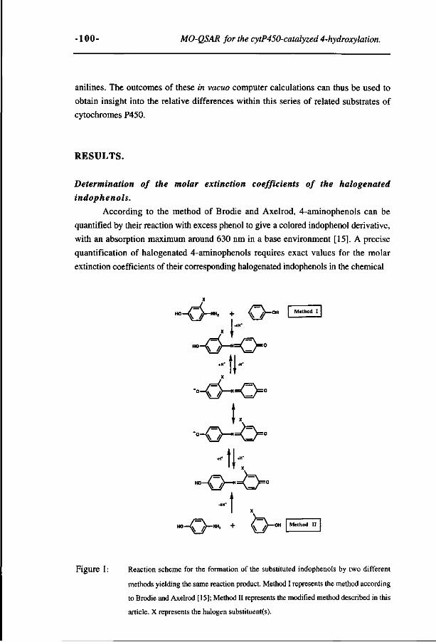
-100- MO-QSAR for the cytP450-catalyzed 4-hydroxylation.
anilines. The outcomes of these in vacuo computer calculations can thus be used to
obtain insight into the relative differences within this series of related substrates of
cytochromes P450.
RESULTS.
Determination of the molar extinction coefficients of the halogenated
indophenols.
According to the method of Brodie and Axelrod, 4-aminophenols can be
quantified by their reaction with excess phenol to give a colored indophenol derivative,
with an absorption maximum around 630 nm in a base environment [15]. A precise
quantification of halogenated 4-aminophenols requires exact values for the molar
extinction coefficients of their corresponding halogenated indophenols in the chemical
X
x (-"•
H O -0 - N = 0 = O
- f J.-X
-°-O-,=O=0
k •°-0—=0=°
:k_ H°-O~~N =O= 0
_ "I ._
Figure 1 : Reaction scheme for the formation of the substituted indophenols by two different
methods yielding the same reaction product. Method I represents the method according
to Brodie and Axelrod [15]; Method II represents the modified method described in this
article. X represents the halogen substituent(s).
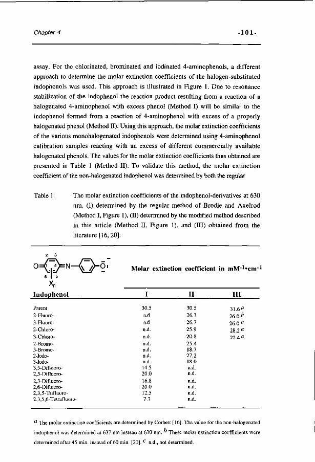
Chapter 4 -101-
assay. For the chlorinated, brominated and iodinated 4-aminophenols, a different
approach to determine the molar extinction coefficients of the halogen-substituted
indophenols was used. This approach is illustrated in Figure 1. Due to resonance
stabilization of the indophenol the reaction product resulting from a reaction of a
halogenated 4-aminophenol with excess phenol (Method I) will be similar to the
indophenol formed from a reaction of 4-aminophenol with excess of a properly
halogenated phenol (Method II). Using this approach, the molar extinction coefficients
of the various monohalogenated indophenols were determined using 4-aminophenol
calibration samples reacting with an excess of different commercially available
halogenated phenols. The values for the molar extinction coefficients thus obtained are
presented in Table 1 (Method II). To validate this method, the molar extinction
coefficient of the non-halogenated indophenol was determined by both the regular
Table 1 : The molar extinction coefficients of the indophenol-derivatives at 630
nm, (I) determined by the regular method of Brodie and Axelrod
(Method I, Figure 1), (II) determined by the modified method described
in this article (Method II, Figure 1), and (III) obtained from the
literature [16, 20].
Xn
Indophenol
Molar extinction coefficient in mM'^cm - 1
I
30.5 n.d n.d n.d. n.d. n.d. n.d. n.d. n.d. 14.5 20.0 16.8 20.0 12.5 7.7
II
30.5 26.3 26.7 25.9 20.8 25.4 18.7 27.2 18.0 n.d. n.d. n.d. n.d. n.d. n.d.
Ill
31.6fl
26.0 e
26.0 e
28.2 ö
22.4«
Parent 2-Fluoro-3-Fluoro-2-Chloro-3-Chloro-2-Bromo-3-Bromo-2-Iodo-3-Iodo-3,5-Difluoro-2,5-Difluoro-2,3-Difluoro-2,6-Difluoro-2,3,5-Trifluoro-2,3,5,6-Tetrafluoro-
a The molar extinction coefficients are determined by Corbett [16]. The value for the non-halogenated
indophenol was determined at 637 nm instead at 630 nm. " These molar extinction coefficients were
determined after 45 min. instead of 60 min. [20], c n.d., not determined.
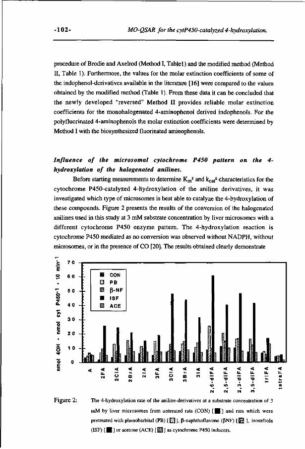
-102- MO-QSAR for the cytP450-catalyzed 4-hydroxylation.
procedure of Brodie and Axelrod (Method I, Table 1) and the modified method (Method
II, Table 1). Furthermore, the values for the molar extinction coefficients of some of
the indophenol-derivatives available in the literature [16] were compared to the values
obtained by the modified method (Table 1). From these data it can be concluded that
the newly developed "reversed" Method II provides reliable molar extinction
coefficients for the monohalogenated 4-aminophenol derived indophenols. For the
polyfluorinated 4-aminophenols the molar extinction coefficients were determined by
Method I with the biosynthesized fluorinated aminophenols.
Influence of the microsomal cytochrome P450 pattern on the 4-
hydroxylation of the halogenated anilines.
Before starting measurements to determine Kms and kcat
s characteristics for the
cytochrome P450-catalyzed 4-hydroxylation of the aniline derivatives, it was
investigated which type of microsomes is best able to catalyze the 4-hydroxylation of
these compounds. Figure 2 presents the results of the conversion of the halogenated
anilines used in this study at 3 mM substrate concentration by liver microsomes with a
different cytochrome P450 enzyme pattern. The 4-hydroxylation reaction is
cytochrome P450 mediated as no conversion was observed without NADPH, without
microsomes, or in the presence of CO [20]. The results obtained clearly demonstrate
o o c
Figure 2:
< u. CM
< — o CM
< ». m CM
< CM
< < — u CO
< k_
ID CO
< _ CO
< u. .— to
CM
< u. «" TJ
i
in
CM
< u. — •o
CO
CM
< u. — lO
CO
< u. a
The 4-hydroxylation rate of the aniline-derivatives at a substrate concentration of 3
mM by liver microsomes from untreated rats (CON) [ H ] and rats which were
pretreated with phénobarbital (PB) [ 0 ] , ß-naphthoflavone (ßNF) [ 0 ], isosafrole
(ISF) [ | ] or acetone (ACE) [ S ] a s cytochrome P450 inducers.
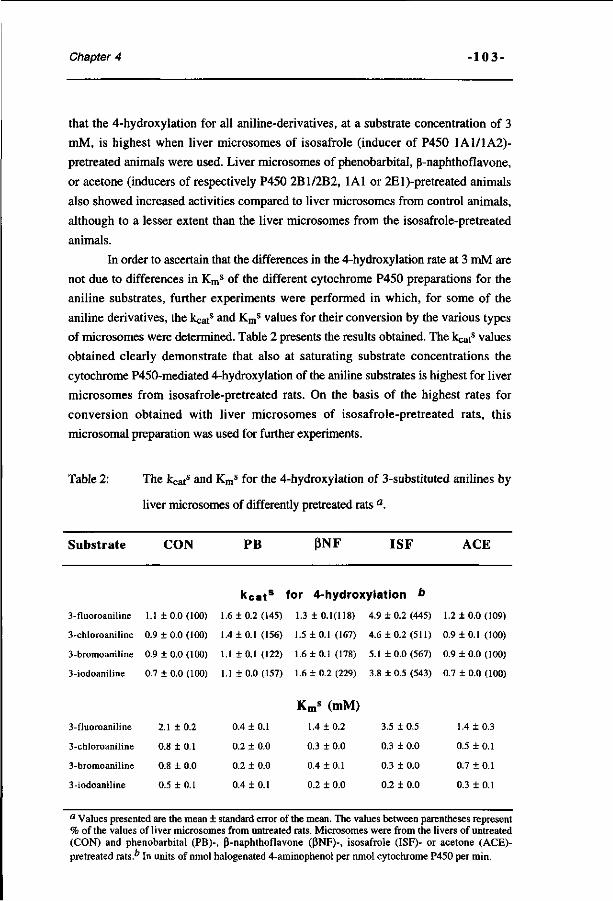
Chapter 4 - 103-
that the 4-hydroxylation for all aniline-derivatives, at a substrate concentration of 3
mM, is highest when liver microsomes of isosafrole (inducer of P450 1A1/1A2)-
pretreated animals were used. Liver microsomes of phénobarbital, ß-naphthoflavone,
or acetone (inducers of respectively P450 2B1/2B2, 1A1 or 2El)-pretreated animals
also showed increased activities compared to liver microsomes from control animals,
although to a lesser extent than the liver microsomes from the isosafrole-pretreated
animals.
In order to ascertain that the differences in the 4-hydroxylation rate at 3 mM are
not due to differences in Kms of the different cytochrome P450 preparations for the
aniline substrates, further experiments were performed in which, for some of the
aniline derivatives, the kcats and Km
s values for their conversion by the various types
of microsomes were determined. Table 2 presents the results obtained. The kcats values
obtained clearly demonstrate that also at saturating substrate concentrations the
cytochrome P450-mediated 4-hydroxylation of the aniline substrates is highest for liver
microsomes from isosafrole-pretreated rats. On the basis of the highest rates for
conversion obtained with liver microsomes of isosafrole-pretreated rats, this
microsomal preparation was used for further experiments.
Table 2: The kcats and Km
s for the 4-hydroxylation of 3-substituted anilines by
liver microsomes of differently pretreated rats a.
Substrate CON PB ßNF ISF ACE
k cat s for 4-hydroxylation b
3-fluoroaniline 1.1 ± 0.0 (100) 1.6 10.2(145) 1.3 + 0.1(118) 4.9 ± 0.2 (445) 1.2 10.0(109)
3-chloroaniline 0.9 10.0(100) 1.4 10.1(156) 1.5 10.1(167) 4.6 10.2(511) 0.9 10.1(100)
3-bromoaniline 0.9 1 0.0 (100) 1.1 1 0.1 (122) 1.6 1 0.1 (178) 5.1 1 0.0 (567) 0.9 1 0.0 (100)
3-iodoaniline 0.7 + 0.0(100) 1.110.0(157) 1.6 10.2(229) 3.8 10.5(543) 0.7 10.0(100)
Km* (mM)
3-fluoroaniline 2.1 1 0.2 0.4 1 0.1 1.4 1 0.2 3.5 1 0.5 1.4 1 0.3
3-chloroaniline 0.8 10.1 0.2 10.0 0.3 10.0 0.3 10.0 0.5 10.1
3-bromoaniline 0.8 10.0 0.2 10.0 0.4 10.1 0.3 10.0 0.7 10.1
3-iodoaniline 0.5 1 0.1 0.4 1 0.1 0.2 1 0.0 0.2 1 0.0 0.3 1 0.1
a Values presented are the mean ± standard error of the mean. The values between parentheses represent % of the values of liver microsomes from untreated rats. Microsomes were from the livers of untreated (CON) and phénobarbital (PB)-, ß-naphthoflavone (ßNF)-, isosafrole (ISF)- or acetone (ACE)-pretreated rats." In units of nmol halogenated 4-aminophenol per nmol cytochrome P450 per min.
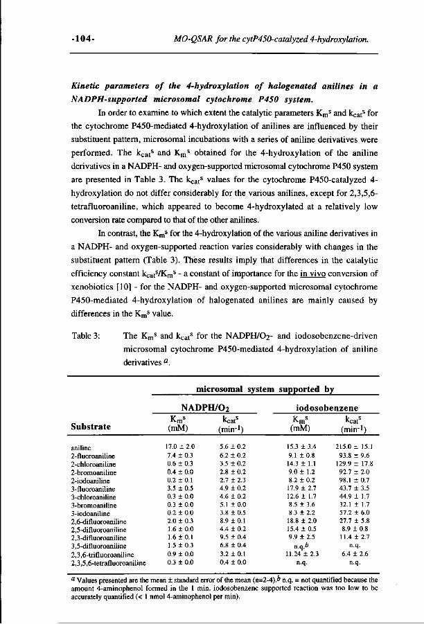
-104- MO-QSAR for the cytP450-catalyzed 4-hydroxylation.
Kinetic parameters of the 4-hydroxylation of halogenated anilines in a
NADPH-supported microsomal cytochrome P450 system.
In order to examine to which extent the catalytic parameters Kms and kcat
s for
the cytochrome P450-mediated 4-hydroxylation of anilines are influenced by their
substituent pattern, microsomal incubations with a series of aniline derivatives were
performed. The kcats and Km
s obtained for the 4-hydroxylation of the aniline
derivatives in a NADPH- and oxygen-supported microsomal cytochrome P450 system
are presented in Table 3. The kcats values for the cytochrome P450-catalyzed 4-
hydroxylation do not differ considerably for the various anilines, except for 2,3,5,6-
tetrafluoroaniline, which appeared to become 4-hydroxylated at a relatively low
conversion rate compared to that of the other anilines.
In contrast, the Kms for the 4-hydroxylation of the various aniline derivatives in
a NADPH- and oxygen-supported reaction varies considerably with changes in the
substituent pattern (Table 3). Thèse results imply that differences in the catalytic
efficiency constant kcats/Km
s - a constant of importance for the in vivo conversion of
xenobiotics [10] - for the NADPH- and oxygen-supported microsomal cytochrome
P450-mediated 4-hydroxylation of halogenated anilines are mainly caused by
differences in the Kms value.
Table 3: The Kms and kcat
s for the NADPH/O2- and iodosobenzene-driven
microsomal cytochrome P450-mediated 4-hydroxylation of aniline
derivatives a.
microsomal system supported by
Substrate
aniline 2-fluoroaniline 2-chloroaniline 2-bromoaniline 2-iodoaniline 3-fluoroaniline 3-chloroaniline 3-bromoaniline 3-iodoaniline 2,6-difluoroaniline 2,5-difluoroaniline 2,3-difluoroaniline 3,5-difluoroaniline 2,3,6-trifluoroaniline 2,3,5,6-tetrafluoroaniline
NADPH/O2 V (mM)
17.0 ± 2.0 7.4 ± 0.3 0.6 ± 0.3 0.4 ± 0.0 0.2 ± 0.1 3.5 ± 0.5 0.3 ± 0.0 0.3 ± 0.0 0.2 ± 0.0 2.0 ± 0.3 1.6 ±0.0 1.6 ±0.1 1.5 ±0.3 0.9 ± 0.0 0.3 ± 0.0
kcatS
(min-1)
5.6 ± 0.2 6.2 ± 0.2 3.5 + 0.2 2.8 ± 0.2 2.7 ± 2.3 4.9 ± 0.2 4.6 ± 0.2 5.1 ±0.0 3.8 ± 0.5 8.9 ±0.1 4.4 ± 0.2 9.5 ± 0.4 6.8 ± 0.4 3.2 ±0.1 0.4 ± 0.0
iodosobenzene Km
s
(mM)
15.3 ± 3.4 9.1 ± 0.8 14.3 ± 1.1 9.0+ 1.2 8.2 ± 0.2 17.9 ± 2.7 12.6 ± 1.7 8.5 ± 3.6 8.3 ± 2.2 18.8 ± 2.0 15.4 ± 0.5 9.9 ± 2.5
n.q.b 11.24 ± 2.3
n.q.
kcatS
(min1)
215.0 ± 15.1 93.8 ± 9.6
129.9 ± 17.8 92.7 ± 2.0 98.1 ± 0.7 43.7 ± 3.5 44.9 ± 1.7 32.1 ± 1.7 57.2 ± 6.0 27.7 ± 5.8 8.9 ± 0.8 11.4 ± 2.7
n.q. 6.4 ± 2.6
n.q.
a Values presented are the mean ± standard error of the mean (n=2-4).* n.q. = not quantified because the amount 4-aminophenol formed in the 1 min. iodosobenzene supported reaction was too low to be accurately quantified (< 1 nmol 4-aminophenol per min).
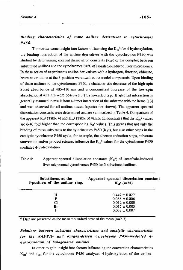
Chapter 4 - 105-
Binding characteristics of some aniline derivatives to cytochromes
P450.
To provide some insight into factors influencing the Kms for 4-hydroxylation,
the binding interaction of the aniline derivatives with the cytochromes P450 was
studied by determining spectral dissociation constants (K<js) of the complex between
substituted anilines and the cytochromes P450 of isosafrole-induced liver microsomes.
In these series of experiments aniline derivatives with a hydrogen, fluorine, chlorine,
bromine or iodine at the 3-position were used as the model compounds. Upon binding
of these anilines to the cytochromes P450, a characteristic decrease of the high-spin
Soret absorbance at 405-410 nm and a concomitant increase of the low-spin
absorbance at 433 nm were observed . This so-called type II spectral interaction is
generally assumed to result from a direct interaction of the substrate with the heme [18]
and was observed for all anilines tested (spectra not shown). The apparent spectral
dissociation constants were determined and are summarized in Table 4. Comparison of
the apparent Kds (Table 4) and Kms (Table 3) values demonstrates that the Km
s values
are 6-40 fold higher than the corresponding K<js values. This means that not only the
binding of these substrates to the cytochromes P450 (Kds), but also other steps in the
catalytic cytochrome P450 cycle, for example, the electron reduction steps, substrate
conversion and/or product release, influence the Kms values for the cytochrome P450
mediated 4-hydroxylation.
Table 4: Apparent spectral dissociation constants (K<js) of isosafrole-induced
liver microsomal cytochromes P450 for 3-substituted anilines.
Substituent at the Apparent spectral dissociation constant 3-position of the aniline ring. Kds (mM)
H 0.447 ± 0.022 F 0.088 ± 0.006 CI 0.012 ± 0.000 Br 0.015 + 0.003 I 0.032 + 0.007
a Data are presented as the mean ± standard error of the mean (n=2-3).
Relations between substrate characteristics and catalytic characteristics
for the NADPH- and oxygen-driven cytochrome P450-mediated 4-
hydroxylation of halogenated anilines.
In order to gain insight into factors influencing the conversion characteristics
Kms and kcat for the cytochrome P450-catalyzed 4-hydroxylation of the aniline-
•106- MO-QSAR for the cytP450-catalyzed 4-hydroxylation.
derivatives relations between catalytic characteristics and substrate characteristics were
investigated.
The decrease of Kms for the cytochrome P450-mediated 4-hydroxylation of
substituted anilines in a NADPH- and oxygen-supported reaction (Table 3) appears to
be linked with an increase of the substrate's lipophilicity. From this observation it
follows that the lipophilicity of the halogenated anilines is an important factor
determining the relative differences in Kms between this series of substrates.
The kcats values were related to other substrate characteristics, i.e., their so-
called frontier orbital characteristics. This can be explained as follows. On the basis of
the frontier orbital theory the relative reactivities of different molecules, and, as a
result, their chances for conversion, i.e., their kcats values, will be dependent on their
frontier orbital characteristics [19, 21]. The frontier orbital characteristics known to
influence relative differences in reactivity are (i) the frontier orbital density for
electrophilic or nucleophilic attack at the reaction centre, and (ii) the energy of the
frontier orbital of interest. When the aromatic hydroxylation of the substrates proceeds
by an electrophilic attack of the cytochrome P450 (FeO)3+ singly occupied molecular
orbital (SOMO) on the aromatic substrate, the HOMO characteristics of the substrate
are of importance. A high HOMO density on the reaction center and a relatively higher
(i.e., less negative) energy of the HOMO can both result in higher reactivity. A
stronger interaction between the substrate HOMO and the cytochrome P450 (FeO)3+
SOMO will result in a stronger reduction of the activation energy (Ea), which is related
to the natural logarithm (In) of kcat. If an electrophilic attack of the cytochrome P450
(FeO)3+ intermediate on the frontier n electrons of the substrate is the rate-limiting
factor in their cytochrome P450-catalyzed conversion, a correlation between HOMO
parameters of the substrates and kcat for 4-hydroxylation can be expected.
Table 5 presents these HOMO characteristics of the various aniline substrates.
The data demonstrate that the E(HOMO) of the anilines varies with the substituent
pattern at the aromatic ring. In Figure 3 the In of the kcats for the cytochrome P450-
catalyzed 4-hydroxylation of the aniline derivatives is plotted against the corresponding
E(HOMO) values. Linear regression analysis reveals the absence of a correlation
between these parameters (r=0.49). Only for 2,3,5,6-tetrafluoroaniline the relatively
low conversion rate qualitatively corresponds with a relatively low E(HOMO). These
results suggest that the electrophilic attack of the cytochrome P450 (FeO)3 +
intermediate on the substrate's frontier K electrons is not the rate-limiting step for
conversion of the anilines in the NADPH/oxygen-supported conversion by
cytochromes P450.
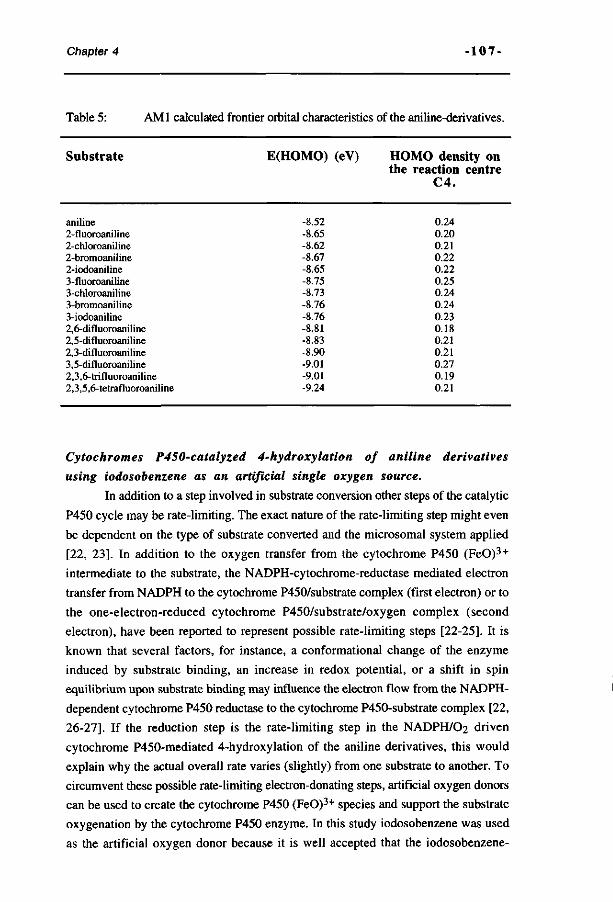
Chapter 4 -107-
Table 5: AMI calculated frontier orbital characteristics of the aniline-derivatives.
Substrate E(HOMO) (eV)
-8.52 -8.65 -8.62 -8.67 -8.65 -8.75 -8.73 -8.76 -8.76 -8.81 -8.83 -8.90 -9.01 -9.01 -9.24
HOMO density on the reaction centre
C4.
0.24 0.20 0.21 0.22 0.22 0.25 0.24 0.24 0.23 0.18 0.21 0.21 0.27 0.19 0.21
aniline 2-fluoroaniline 2-chloroaniline 2-bromoaniline 2-iodoaniline 3-fluoroaniline 3-chloroaniline 3-bromoaniline 3-iodoaniline 2,6-difluoroaniline 2,5-difluoroaniline 2,3-difluoroaniline 3,5-difluoroaniline 2,3,6- trifluoroaniline 2,3,5,6-tetrafluoroaniline
Cytochromes P450-catalyzed 4-hydroxylation of aniline derivatives
using iodosobenzene as an artificial single oxygen source.
In addition to a step involved in substrate conversion other steps of the catalytic
P450 cycle may be rate-limiting. The exact nature of the rate-limiting step might even
be dependent on the type of substrate converted and the microsomal system applied
[22, 23]. In addition to the oxygen transfer from the cytochrome P450 (FeO)3 +
intermediate to the substrate, the NADPH-cytochrome-reductase mediated electron
transfer from NADPH to the cytochrome P450/substrate complex (first electron) or to
the one-electron-reduced cytochrome P450/substrate/oxygen complex (second
electron), have been reported to represent possible rate-limiting steps [22-25]. It is
known that several factors, for instance, a conformational change of the enzyme
induced by substrate binding, an increase in redox potential, or a shift in spin
equilibrium upon substrate binding may influence the electron flow from the NADPH-
dependent cytochrome P450 reductase to the cytochrome P450-substrate complex [22,
26-27]. If the reduction step is the rate-limiting step in the NADPH/O2 driven
cytochrome P450-mediated 4-hydroxylation of the aniline derivatives, this would
explain why the actual overall rate varies (slightly) from one substrate to another. To
circumvent these possible rate-limiting electron-donating steps, artificial oxygen donors
can be used to create the cytochrome P450 (FeO)3+ species and support the substrate
oxygenation by the cytochrome P450 enzyme. In this study iodosobenzene was used
as the artificial oxygen donor because it is well accepted that the iodosobenzene-
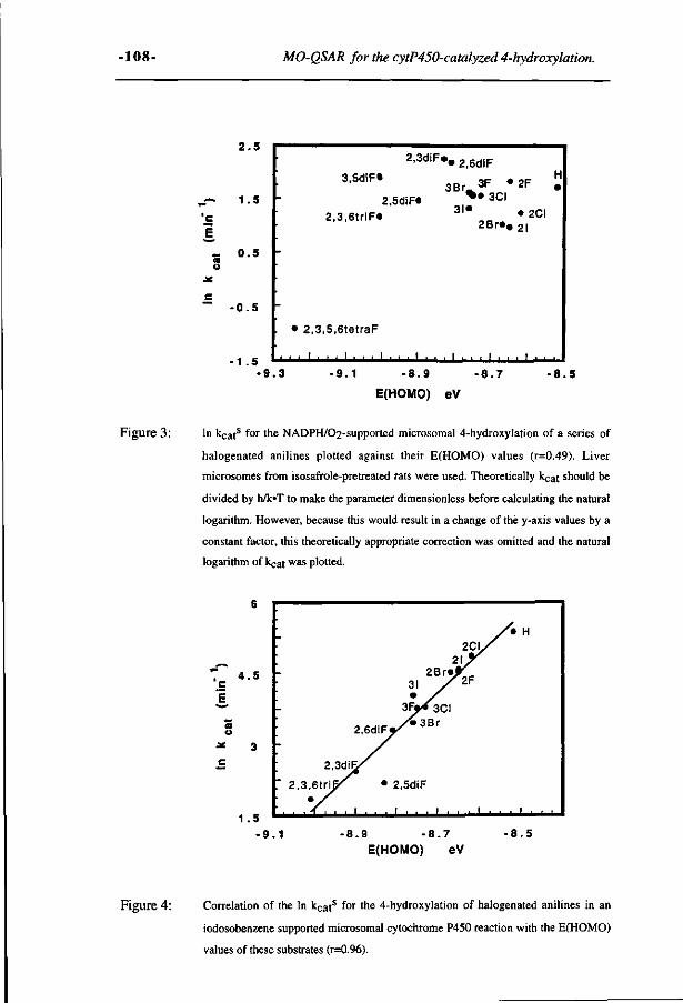
-108- MO-QSAR for the cytP450-catalyzed 4-hydroxylotion.
2 . 5
_ ~ 1.5
'c
I - 0 .5 a o
• 0 . 5
•1 .5
2,3diF«
3,5diF»
2,5diF«
2 ,3,6tr iF«
• 2 ,3,5,6tetraF
_L
'2,6diF
3Br,
3I«
3F « % • 3CI
2F
• 2CI 2 B r « # gl
i_i_i_l_k_i_ j _ 1 _ l _ L j _ 1 _ 1 _
• 9 . 3 • 9 . 1 - 8 . 9
E(HOMO) eV
• 8 . 7 • 8 . 5
Figure 3 : In k c a ts for the NADPH/02-supported microsomal 4-hydroxylation of a series of
halogenated anilines plotted against their E(HOMO) values (r=0.49). Liver
microsomes from isosafrole-pretreated rats were used. Theoretically kçat should be
divided by h/k»T to make the parameter dimensionless before calculating the natural
logarithm. However, because this would result in a change of the y-axis values by a
constant factor, this theoretically appropriate correction was omitted and the natural
logarithm of kçat was plotted.
6
7 4 . 5 c
3 *
1 . 5
•
2CI / '
2Br«r 3I X 2 F • /
- 3Fp< 3CI
2.6diFy*3 B r
2,3diF^/
T 2 , 3 , 6 t r i ^ • 2,5diF
* /
/* H
. . i . , ,
- 9 . 1 - 8 . 9 - 8 . 7 - 8 . 5
E (HOMO) eV
Figure 4: Correlation of the In k c a t s for the 4-hydroxylation of halogenated anilines in an
iodosobenzene supported microsomal cytochrome P450 reaction with the E(HOMO)
values of these substrates (r=0.96).
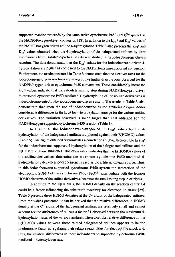
Chapter 4 - 109-
supported reaction proceeds by the same active cytochrome P450 (FeO)3+ species as
the NADPH/oxygen-driven conversion [28]. In addition to the kcats and Km
s values of
the NADPH/oxygen driven aniline 4-hydroxylation Table 3 also presents the kcats and
Kms values obtained when the 4-hydroxylation of the halogenated anilines by liver
microsomes from isosafrole-pretreated rats was studied in an iodosobenzene-driven
reaction. The data demonstrate that the Kms values for the iodosobenzene-driven 4-
hydroxylation are higher as compared to the NADPH/oxygen-supported conversion.
Furthermore, the results presented in Table 3 demonstrate that the turnover rates for the
iodosobenzene-driven reactions are several times higher than the ones observed for the
NADPH/oxygen-driven cytochrome P450 conversions. These considerably increased
kCats values indicate that the rate-determining step during NADPH/oxygen-driven
microsomal cytochrome P450-mediated 4-hydroxylation of the aniline derivatives is
indeed circumvented in the iodosobenzene-driven system. The results in Table 3, also
demonstrate that upon the use of iodosobenzene as the artificial oxygen donor
considerable differences in the kcats for 4-hydroxylation emerge for the various aniline
derivatives. The variation observed is much larger than that obtained for the
NADPH/oxygen-supported cytochrome P450 reaction (Table 3).
In Figure 4, the iodosobenzene-supported In kcats values for the 4-
hydroxylation of the halogenated anilines are plotted against their E(HOMO) values
(Table 5). The figure obtained demonstrates a correlation (r=0.96) between the In kcats
for the iodosobenzene-supported 4-hydroxylation of the halogenated anilines and the
E(HOMO) of these substrates. This observation indicates that the E(HOMO) values of
the aniline derivatives determine the maximum cytochrome P450-mediated 4-
hydroxylation rate, when iodosobenzene is used as the artificial oxygen source. Thus,
in this iodosobenzene-supported cytochrome P450 system the interaction of the
electrophilic SOMO of the cytochrome P450 (FeO)3+ intermediate with the frontier
HOMO electrons of the aniline derivatives, becomes the rate-limiting step in catalysis.
In addition to the E(HOMO); the HOMO density on the reaction center C4
could be a factor influencing the substrate's reactivity for electrophilic attack [29].
Table 5 presents these HOMO densities at the C4 atoms of the halogenated anilines.
From the values presented, it can be derived that the relative differences in HOMO
density at the C4 atoms of the halogenated anilines are relatively small and cannot
account for the differences of at least a factor 31 observed between the maximum 4-
hydroxylation rates of the various anilines. Therefore, the relative difference in the
E(HOMO) values between these related halogenated anilines appears to be the
predominant factor in regulating their relative reactivities for electrophilic attack and,
thus, the relative differences in their iodosobenzene-supported cytochrome P450-
mediated 4-hydroxylation rate.
-110 - MO-QSAR for the cytP450-catalyzed 4-hydroxylation.
DISCUSSION.
In the present study the 4-hydroxylation of the halogenated anilines, catalyzed
by the microsomal cytochrome P450, was investigated. Liver microsomes from
isosafrole-pretreated rats appeared to be most capable of catalyzing the cytochrome
P450-dependent 4-hydroxylation of the aniline derivatives. This suggests a
predominant role for cytochrome P450 1A2 in the 4-hydroxylation of all the anilines
studied. This result is in accordance with the observation of Ryan et al. [30], who
demonstrated that purified reconstituted rat hepatic cytochrome P450 1A2 is one of the
cytochrome P450 enzymes best capable of aniline 4-hydroxylation. Using microsomes
from isosafrole-pretreated rats, further studies were especially focused on the
importance of the physico chemical and electronic characteristics of the aniline
substrates in relation to their cytochrome P450-mediated conversion to the 4-
aminophenol metabolites.
Results of the present study demonstrate a relation between the Kms values for
4-hydroxylation of a series of aniline derivatives and the hydrophobicity of these
substrates. It can be concluded that the hydrophobicity of the substrate is an important
factor in determining the Kms values, of the aniline derivatives. This observation is in
accordance with results obtained by Burka et. al. [31] for the Kms for hydroxylation of
monohalogenated benzenes by phenobarbital-induced microsomes. The Kms was
shown to correlate with the Hansch's K parameter of the substrates [31]. For the
iodosobenzene-driven 4-hydroxylation it was demonstrated that the Kms values are
higher as compared to the Kms values of the NADPH/C^-driven supported conversion.
This observation is best ascribed to the fact that iodosobenzene competes with the
substrate for the binding site of the cytochrome P450 enzymes as has been
demonstrated by Lichtenberg [32].
Additional results of the present study demonstrate that the spectral dissociation
constant K<is, reflecting the binding interaction between the aniline substrate and the
cytochrome P450 enzyme, is 6-40 times less than the Kms value for 4-hydroxylation.
This leads to the conclusion, that not only the binding of the substrate to the
cytochromes P450 (Kds), but also other steps in the catalytic cytochrome P450 cycle,
such as for example electron donation, substrate conversion and/or product release, are
involved in determining the Kms values for the cytochrome P450-mediated 4-
hydroxylation. An alternative explanation could be that the Kjs determined from the
type II binding spectra does not represent the Kd, i.e., the binding, of importance for
substrate conversion. Taking into account that type II spectral characteristics are
generally accepted to result from an interaction of the amino moiety with the Fe3+ of
the cofactor, it is possible that this type of binding is not the one representative for
substrate binding that eventually leads to product formation. Additional support for the
Chapter 4 - I l l -
fact that binding characteristics determined for the resting Fe3+ form of the cytochrome
P450 may not be relevant for the binding interaction characteristics for the activated
(FeO)3+ form comes from a study reported by Paulsen and Ornstein [33]. They
demonstrate that binding of camphor to the Fe3+ form and that to the activated (FeO)3+
form are different indeed. This implies that the Kj of the binding step influencing the
Km might be a different one than the one that can be obtained from type II difference
spectra.
Additional results of the present study clearly demonstrate that reaction steps
preceding the substrate conversion by the cytochrome P450 (FeO)3+ intermediate play
a main role in rate limitation of the NADPH/oxygen-supported aniline 4-hydroxylation.
This follows from the observation that kcats significantly increases when these reaction
steps are surpassed by the use of iodosobenzene instead of NADPH and molecular
oxygen to support the cytochrome P450 reaction. This result indicates that the
interaction of the cytochrome P450 (FeO)3+ intermediate with the aniline substrate is
not the rate-limiting step in the NADPH/oxygen-driven catalysis. The most important
observation of the iodosobenzene experiments, however, was a clear correlation
(r=0.96) observed between the In kcats for the iodosobenzene-supported cytochrome
P450-catalyzed 4-hydroxylation of the various aniline derivatives and their E(HOMO).
This observation implies that the 4-hydroxylation proceeds by the interaction of the
HOMO electrons of the substrate with the cytochrome P450 (FeO)3+ SOMO. For the
iodosobenzene-driven reaction this interaction becomes the rate-limiting step in
catalysis. However, because iodosobenzene is generally accepted to generate the same
reactive cytochrome P450 (FeO)3+ intermediate as formed in the NADPH/O2
supported pathway [28], it can be concluded that the normal NADPH- and oxygen-
driven aniline 4-hydroxylation also proceeds by an electrophilic attack of the SOMO of
the cytochrome P450 (FeO)3+ intermediate on the HOMO TC electrons of the aromatic
aniline ring. Such a mechanism would be in accordance to the one proposed before on
the basis of studies on the regioselectivity of the aromatic hydroxylation of
monofluoroanilines and fluorobenzenes [12, 34]. Only when the reactivity of the
substrate is too low, such as for example for 2,3,5,6-tetrafluoroaniline, the interaction
of the cytochrome P450 (FeO)3+ SOMO with the HOMO electrons of the substrate
might become the rate-limiting step in the NADPH/02-supported reaction cycle.
Acknowledgements.
The authors gratefully acknowledge Mr. J. Haas and Mr. G. van Tintelen for
assistance with animal handling. This study was supported by the Netherlands
Organisation for the Advancement of Scientific Research (N.W.O.).
•112- MO-QSAR for the cytP450-catalyzed 4-hydroxylation.
REFERENCES.
1 Gorrod J.W. and Manson D., (1986) The metabolism of aromatic amines, Xenobiotica 16,
933-955.
2 Eyer P., (1988) Detoxication of N-oxygenated arylamines in erythrocytes. An overview,
Xenobiotica 18, 1327-1333.
3 Uehleke H., (1973) The role of cytochrome P-450 in the N-oxidation of individual amines,
Drug Metabolism and Disposition 1, 299-313.
4 Rietjens I.M.C.M. and Vervoort J., (1991) Bioactivation of 4-fluorinated anilines to
benzoquinoneimines as primary reaction products, Chem. -Biol. Interactions 77, 263-281.
5 Gartland K.P.R., Bonner F.W., Timbrell J.A. and Nicholson J.K., (1989) Biochemical
characterisation of para-aminophenol-induced nephrotoxic lesions in the F344 rat, Arch.
Toxicol. 63, 97-106.
6 Gartland K.P.R., Eason C.T., Bonner F.W. and Nicholson J.K., (1990) Effects of biliary
cannulation and buthionine sulphoximine pretreatment on the nephrotoxicity of para-
aminophenol in the Fisher 344 rat, Arch. Toxicol. 64, 14-25.
7 Klos C , Koob M., Kramer C. and Dekant W., (1992) p-Aminophenol nephrotoxicity:
Biosynthesis of toxic glutathione conjugates, Toxicol. Appl. Pharmacol. 115, 98-106.
8 Newton J.F., Kuo C.-H., Gemborys M.W., Mudge G.H. and Hook J.B., (1982)
Nephrotoxicity of p-aminophenol, a metabolite of acetaminophen, in the Fischer 344 rat,
Toxicol. Appl. Pharmacol. 65, 336-344.
9 Calder I.C., Yong A.C., Woods R.A., Crowe CA., Ham K.N. and Tange J.D., (1979) The
nephrotoxicity of p-aminophenol, II. The effect of metabolic inhibitors and inducers, Chem.-
Biol. Interactions 27, 245-254.
10 Armstrong R.N., (1987) Enzyme-catalyzed detoxification reactions: mechanism and
stereochemistry, CRC Crit. Rev. Biochem. 22, 39-88.
11 Saltzman H. and Sharefkin J.G., (1973) Iodosobenzene, Organic Synthesis, Collective
Volume V, John Wiley & Sons, 658-659.
12 Cnubben N.H.P., Vervoort J., Veeger C. and Rietjens I.M.C.M., (1992) Study on the
regioselectivity and mechanism of the aromatic hydroxylation of monofluoroanilines,
Chem.-Biol. Interactions 85, 151-172.
13 Omura T. and Sato R., (1964) The carbon monoxide pigment of liver microsomes, I.
Evidence for its haemoprotein nature, J. Biol. Chem. 239, 2370-2378.
14 Lowry O.H., Rosebrough N.J., Farr A.L. and Randall A.J., (1951) Protein measurements
with Folin phenol reagent, J. Biol. Chem. 193, 265-275.
15 Brodie B.B. and Axelrod J., (1948) The estimation of acetanilide and its metabolic products,
aniline, N-acetyl p-aminophenol and p-aminophenol (free and total conjugated) in biological
fluids and tissues, J. Pharmac. Exp. Ther. 94, 22-28.
16 Corbett J.F., (1970) Benzoquinone imines Part Vlll. Mechanism and kinetics of the reaction
Chapter 4 - 1 13 -
of p-benzoquinone monoimines with monohydric phenols, J. Chem. Soc, 1503-1508.
17 Vervoort J., De Jager P.A., Steenbergen J. and Rietjens I.M.C.M., (1990) Development of
a ' "F-n.m.r. method for studies on the in vivo and in vitro metabolism of 2-fluoroaniline,
Xenobiotica 20, 657-670.
18 Schenkman J.B., (1970) Studies on the nature of the type I and type II spectral changes in
liver microsomes, Biochemistry 9, 2081-2091.
19 Fukui K., Yonezawa T., Nagata C. and Shingu H., (1954) Molecular orbital theory of
orientation in aromatic, heteroaromatic and other conjugated molecules, J. Chem. Phys. 22,
1433-1442.
20 Rietjens I.M.C.M. and Vervoort J., (1989) Microsomal metabolism of fluoroanilines,
Xenobiotica 19, 1297-1305.
21 Fleming I. (1989), Molecular orbitals and frontier orbitals, in: Frontier Orbitals and Organic
Chemical Reactions (Fleming I., Ed.), pp. 182-186, John Wiley & Sons, New York.
22 Guengerich F.P., (1990) Enzymatic oxidation of xenobiotic chemicals, Biochemistry and
Molecular Biology 25, 97-153.
23 White R.E. and Coon M.J., (1980) Oxygen activation by cytochrome P-450, Ann. Rev.
Biochem. 49, 315-356.
24 Miwa G.T., West S.B., Lu A.Y.H., (1978) Studies on the rate-limiting enzyme component
in the microsomal monooxygenase system, J. Biol. Chem. 253, 1921-1929.
25 Peterson J.A. and Prough R.A. (1986), Cytochrome P-450 reductase and cytochrome b5 in
cytochrome P-450 catalysis, in: Cytochrome P-450: Structure, mechanism and biochemistry
(Ortiz de Montellano P.R., Ed.), pp. 89-117, Plenum Press, New York.
26 Ruckpaul K., Rein H. and Blanck J. (1989), Regulation mechanisms of the activity of the
hepatic endoplasmic cytochrome P-450, in: Frontiers in biotransformation: Basis and
mechanisms of regulation of cytochrome P-450 (Ruckpaul K. and Rein H., Eds.), pp. 1-65,
Taylor & Francis, London.
27 Murray M. and Reidy G.F., (1990) Selectivity in the inhibition of mammalian cytochromes
P-450 by chemical agents, Pharmacol. Rev. 42, 85-101.
28) Ortiz de Montellano P.R. (1986), Oxygen activation and transfer, in: Cytochrome P-450:
Structure, mechanism and biochemistry (Ortiz de Montellano P.R., Ed.), pp. 217-271,
Plenum Press, New York.
29 Vervoort J., Rietjens I.M.C.M., Berkel W.J.H. and Veeger C , (1992) Frontier orbital study
on the 4-hydroxybenzoate-3-hydroxylase-dependent activity with benzoate derivatives, Eur. J.
Biochem. 206, 479-484.
30 Ryan D.E. and Levin W., (1990) Purification and characterization of hepatic microsomal
cytochrome P-450, Pharmac. Ther. Vol. 45, 153-239.
31 Burka L.T., Plucinski T.M. and Macdonald T.L., (1983) Mechanisms of hydroxylation by
cytochrome P-450: Metabolism of monohalobenzenes by phenobarbital-induced
microsomes, Proc. Natl. Acad. Sei. USA 80, 6680-6684.
•114- MO-QSAR for the cytP450-catalyzed 4-hydroxylation.
32 Lichtenberg F., Nastainczyk W. and Ullrich V., (1976) Cytochrome P450 as an oxene
transferase, Biochem. Biophys. Res. Commun. 70, 939-946.
33 Paulsen M.D. and Ornstein R.L., (1993) Substrate mobility in thiocamphor-bound
cytochrome P450cam: an explanation of the conflict between the observed product profile
and the X-ray structure, Protein Engng. 6, 359-365.
34 Rietjens I.M.C.M., Soffers A.E.M.F., Veeger C. and Vervoort J., (1993) Regioselectivity
of cytochrome P-450 catalyzed (poly)fluorobenzene hydroxylation predicted by calculated
frontier orbital substrate characteristics, Biochemistry 32, 4801-4812.
CHAPTER 5
A spectrophotometric assay for the detection of 2-aminophenols in biological samples.
N.H.P. Cnubben, B. Blaauboer, S. Juyn, J. Vervoort and I.M.C.M. Rietjens
Analytical Biochemistry 220, 165-171, 1994.
SUMMARY.
A sensitive and efficient spectrophotometric assay is described for the
determination of 2-aminophenols in biological samples. Using ferric ions as a metal
catalyst, 2-aminophenol dimerizes in an acidic aqueous environment to 2-hydroxy-
isophenoxazin-3-one, an intensively coloured dye. The newly developed assay is
suitable for the detection of 2-aminophenols in the uM range. The paper demonstrates
that this chemical assay is also applicable for the determination of 2-aminophenols
substituted with a halogen at the ortho, meta or para position of the aromatic ring with
respect to the amino moiety, Xmax, and the molar extinction coefficient varying with
the substituent pattern. Results obtained for fluorinated 2-aminophenol detection in
biological samples by either this new method or 19F-NMR are similar. This
observation corroborates that the newly developed assay is suitable for detection of 2-
aminophenols in biological samples.
INTRODUCTION.
Halogenated aniline derivatives are compounds used for the manufacture of
numerous dyes, drugs, pesticides and herbicides [1]. Upon the cytochrome P450-
dependent aromatic hydroxylation of these aniline derivatives, the compounds are
converted into preferentially 2- and 4-aminophenols [2-5]. For the detection of 4-
aminophenols, a sensitive, accurate and efficient chemical assay has been described
- 1 1 8 - Detection of 2-aminophenols.
achieved by the addition of 100 ul 12N HCl to 1 ml of urine sample, followed by
heating at 100°C for 30 minutes. The hydrolyzed samples were adjusted with 12 N
NaOH to pH 7 and extracted three times with 3 ml diethylether to separate the 2-
aminophenols from the mixture. After evaporation of the diethylether, 300 ul 20%
TCA were added to the residue. This was followed by the addition of 1 ml
demineralized water and 100 ul 40 mM ferric ammonium sulfate dodecahydrate. The
2-aminophenol content of these samples was determined as described above. A urine
sample of a rat before exposure to an aniline derivative, treated in a similar way, was
used as the reference.
Determination of 2-aminophenols in microsomal incubations.
Prior to analysis by the chemical assay, samples from microsomal incubations
were frozen in liquid nitrogen and -upon thawing- centrifuged at 13,000 rpm for 10
minutes at 4°C. Diethyl ether extraction of the 2-aminophenols from the supernatant
was carried out as described above for the urine samples, and appeared to be essential,
because the presence of KPj buffer prevents the formation of the yellow colour
initiated by the addition of ferric ammonium sulfate dodecahydrate. The diethyl ether
extraction also liberates 2-aminophenols (partially) bound to the microsomal protein by
hydrophobic interactions, leading to an underestimation of the amount of 2-
aminophenol actually present in the sample.
NMR measurements. l9F-NMR measurements were performed on a Bruker AMX 300 spectrometer
operating at 282.3 MHz essentailly as described before [2]. D2O (Isotec, USA) was
used for locking the magnetic field. The samples were made oxygen-free by four
cycles of evacuating and filling with argon. Concentrations of the various compounds
observed in the 19F-NMR spectra were determined by comparison of the integrals of
their 19F-NMR resonances to the integral from the 19F-NMR resonance of para-
fluorobenzoic acid, added to each sample as an internal standard.
ïH-NMR measurements were performed on a Bruker AMX 500 spectrometer
using 40° pulses and a repetition time of 3s. For JH-NMR measurements acetone-d6
was used as the solvent.
Analysis by infra red.
Fourier Transform infrared spectra of the yellow dye and the synthesized 2-
amino-isophenoxazin-3-one in KBr were recorded on a Biorad FTS-7 Fourier
transform spectrometer.
Chapter 5 - 119 -
Analysis by mass spectrometry
Electron Impact MS was performed using a Finnigan-MAT-95 mass
spectrometer. GC-MS was not applied because of the high melting points of the
compounds.
RESULTS
Calibration curve.
Figure 1 shows the calibration curve for formation of the coloured dye with
increasing concentration of 2-aminophenol using the optimal conditions with respect to
Xmax and reaction time (400 nm, 90 min). Figures 2 and 3 demonstrate that the X,nax of
the 2-aminophenol-derived yellow dye in chloroform is 400 nm (Figure 2) and that the
reaction is completed within 90 minutes (Figure 3). Figure 3 also demonstrates that the
reaction product responsible for the absorbance at 400 nm is stable for at least another
30 minutes beyond the 90 minutes of the assay.
The calibration curve obtained (Figure 1) shows a linear relationship between
the 2-aminophenol concentration and the absorbance at 400 nm (yellow colour)
between 20 and -at least- 100 p.M. From 0-20 |xM a deviation from the linear
relationship between 20-100 uM is observed, pointing at a concentration of about 10
uM that remains undetected. Obviously, the calibration curve for the 20-100 uM region
must be corrected for this apparent "lag". Therefore, the calibration curve, valid
between 20-100 uM, is best described by means of the following equation: A^Onm =
-0.034 + 8.66«[2-aminophenol] with the concentration of 2-aminophenol in mM and at
a value > 0.020 mM.
a o « n <
20 40 6 0 8 0 \itA 2-aminophenol
Figure 1: Calibration curve for the determination of 2-aminophenol.
•120- Detection of 2-aminophenols.
0.5
3 0 0 4 0 0 5 0 0
wavelength [nm]
Figure 2: The visible absorption spectrum of the yellow dye in chloroform formed upon the
reaction of 100 uM 2-aminophenol with ferric ammonium sulfate dodecahydrate.
4 0 6 0 8 0
minutes
Figure 3 : Influence of reaction time on the absorbance at 400 nm of the yellow dye in
chloroform. To a 1-ml sample containing 100 nmol 2-amino-phenol, 0.3 ml 20%
trichloroacetic acid and 0.1 ml 40 mM ferric ammonium sulfate dodecahydrate were
added. The reaction mixtures were extracted with 2 ml chloroform at different time-
intervals.
Identity of the yellow dye.
The identity of the yellow dye, formed upon a reaction of ferric ions with 2-
aminophenol in an acidic aqueous environment, was investigated by means of mass
spectrometry, !H-NMR measurements and Fourier Transform infrared spectrometry.
Initially it was assumed that the yellow dye might be 2-amino-isophenoxazin-3-one [9-
11]. The synthesized 2-amino-isophenoxazin-3-one (m.p 254-256°C) and the yellow
dye (m.p. 262-264°C) appeared to have a mass of respectively m/z 212 (100%), 185
(53%), 184 (27%) and m/z 213 (100%), 185 (80%). The 'H-NMR resonances of the
Chapters - 1 2 1 -
synthesized 2-amino-isophenoxazin-3-one and the yellow dye formed in the chemical
assay show the same characteristics namely two doublets, two triplets and two
singlets, although the resonances of the latter are all shifted to higher field. The IR
spectrum of the synthesized 2-amino-isophenoxazin-3-one confirmed its structure as it
was identical to the IR spectrum of this compound published in the literature [17].
Surprisingly, the IR spectrum of the yellow dye demonstrated to be identical to the IR
spectrum of 2-hydroxy-isophenoxazin-3-one published in the same paper [17].
Finally, comparison of the results from mass analysis, !H-NMR and melting points to
data published in the literature for phenoxazin-3-ones [17-18] further confirms the
identity of the yellow compound as 2-hydroxy-isophenoxazin-3-one.
The influence of halogen substituents at the 2-aminophenol on the
formation of the yellow colour in the chemical assay.
To investigate the effect of halogen substituents at the aromatic ring of the 2-
aminophenol on the formation of the yellow dye as well as the reliability of the newly
developed assay, monofluorinated 2-aminophenols were used. The presence of a
fluorine substituent provides the possibility to analyze 2-aminophenol containing
samples in a quantitative way using 19F-NMR spectroscopy and to compare this to the
results of the newly developed chemical assay.
Table 1 demonstrates that for fluorine-substituted 2-aminophenols the ^max of
the respective coloured reaction products in chloroform and the corresponding molar
extinction coefficients vary significantly with the substituent pattern. However, from
the results in Table 1 it appears that the presence of a fluorine substituent at the ortho,
Table 1: Spectral characteristics of the 2-aminophenol derived dyes in chloroform.
• max e-value ba
(nm) (mM^'cm-1) Compound for C > 0.020 mM«
2-Aminophenol 400 8.66 -0.034 2-Amino-3-fluorophenol 402 3.44 -0.022 2-Amino-4-fluorophenol 433 5.38 -0.026 2-Amino-5-fluorophenol 402 10.14 -0.033
a The calibration curve is described by the equation A = b + e *C* / . A represents the absorbance of the corresponding yellow dye in CHCI3 at A.max, e is the molar extinction coefficient in mM" 1 "cm" ' , C is the concentration of the halogen-subsituted 2-aminophenol in mM, / is the light pathway in cm and b is the correction factor.
•122- Detection of 2-aminophenols.
meta or para position of the aromatic ring with respect to the amino moiety does not
prevent the formation of the yellow dye. The structures of the dimerization products of
the fluorinated 2-aminophenols were identified essentially as described above. The lK-
NMR characteristics, m/z values and corresponding structures of the dimerization
products of the fluorinated 2-aminophenols are presented in Table 2.
Table 2: Spectral characteristics of the dimerization products of the 2-
aminophenol derivatives in the chemical assay.
2-Amino-
phenol
derivative
Dimerization
product
Mass and 'H-NMR spectral
characteristics
NH2
^ - N M R (acetone-d6): 9.01 (OH)(broad s), 7.83 (H9)(d, 3 / H - H = 7.6 Hz), 7.63 (H7)(tr, 3 / H - H = 7.6 Hz, 3-/H-H= 7.5 Hz), 7.52 (H6)(d, 3^H-H= 7.6 Hz), 7.49 (H8)(tr, 37H-H= 7.6, 3yH-H= 7.5 Hz), 6.76 (Hl)(s), 6.42 (H4)(s). m/z: 213 (100%), 185 (80%).
NH2 F F
Main product (70%) F F
, N ^ k ^ N H 2
O I
• H - N M R (acetone-d6): 9.52 (OH)(broad s), 7.71 (H6)(d, 3^H-H= 7.9 Hz), 7.55-7.38 (H7+H8)(m), 6.43 (H4)(s) m/z: 249, compound was not purified from the "minor product".
'H-NMR (acetone-do): 7.68 (H6)(d, 3 iH-H= 7.8 Hz), 7.55-7.38 (H7+H8)(m), 6.51 (NH2)(broad s), 6.48 (H4)(s) m/z: 248, compound was not purified from the "main product".
Minor product (30%)
NH2
^ -NMR (acetone-d6): 7.50 (H6)(dd, 3 7 H - H = 8.8 Hz, 4 7H-F= 4.9 Hz), 7.43 (H9)(dd, 3 7H-F= 9.1 Hz, 4 7 H - H =
2.8 Hz), 7.28 (H7)(m, 37H-H= 8.8 Hz, 37H-F= 8.6 Hz, 47H-H= 2.8 Hz), 6.47 (Hl)(s), 6.42 (NH2)(broad s), 6.30 (H4)(s). m/z: 230 (100%), 203 (39%), 202 (28%).
NH2 -u
'H-NMR (acetone-d6): 9.05 (OH)(broad s), 7.89 (H9)(dd, 37H-H= 8.8 Hz, 4/H-F= 6.1 Hz), 7.38 (H6)(dd, 37H-F= 9.0 Hz, 4 J H - H = 2.6 Hz,), 7.30 (H8)(m, 3/H-H= 8.8 Hz, 3/H-F= 8.8 Hz, 47H-H= 2.6 Hz), 6.75 (Hl)(s), 6.43 (H4)(s). m/z: 231 (100%), 203 (73%).
Chapter 5 -12 3-
Verification of the assay.
Additional experiments were performed to investigate the applicability of the
newly developed 2-aminophenol assay for the detection of 2-aminophenols in
biological samples such as urine samples and microsomal incubations. For this reason,
urine samples from rats exposed to fluoroanilines and samples from microsomal
incubations with fluorinated anilines were analyzed by both the chemical assay and by 19F-NMR.
Urine samples were analyzed after acid hydrolysis, carried out to eliminate
conjugated N-acetyl, sulphate and glucuronic acid residues from the 2-aminophenols.
Table 3 summarizes the values obtained for these urine samples using both the
chemical assay and 19F-NMR. The 19F-NMR spectra of these urine samples have
been published before [12]. It is clear that both methods provide similar results. Thus,
urine components and other monofluoroaniline metabolites which may also be
extracted into the diethylether do not interfere with this chemical assay. Quantification
of 2-aminophenols in the urine of 3-fluoroaniline exposed rats using the chemical
assay may in theory be hampered by the fact that hydroxylation of 3-fluoroaniline can
lead to the formation of two isomeric 2-aminophenols. However, 19F-NMR analysis
of the urine of 3-fluoroaniline-exposed rats revealed that only 2-amino-5-fluorophenol
derived metabolites are present in the urine, thus eliminating this theoretical problem
[12].
Table 3: Comparison of the chemical assay with 19F-NMR analysis for the
estimation of 2-aminophenol in urine samples of monofluoroaniline
exposed rats.
|xmol 2-aminophenol in urine after acid hydrolysis.
Dosed compound 19F-NMR Chemical assay
2-Fluoroaniline 4.8 ± 0.1 4.5 ± 0.5
3-Fluoroaniline 19.6 ± 0.7 19.0 ±0.1
4-Fluoroaniline 97.8 ± 1.1 99.8 + 3.7
Note. Data are presented as means ± standard error of the mean.
Table 4 presents the results of the analysis of samples from microsomal
incubations of monofluoroanilines using 19F-NMR and the chemical assay. In these
microsomal samples the 2-aminophenol metabolites are formed in the |xM range upon
the cytochrome P450-mediated conversion of the aniline derivatives.
-1 2 6 - Detection of 2-aminophenols.
derivative, providing a basis for quantification of the respective 2-aminophenols upon
characterization of their respective calibration curves.
Using the fluorinated analogues, it could be demonstrated that analysis by the
newly developed chemical assay provides similar results for the 2-aminophenol
concentration in urinary and microsomal samples as obtained by 19F-NMR. Thus, the
newly developed chemical assay presented here provides an efficient, reliable and
more sensitive method for the detection of (halogen substituted) 2-aminophenols in
biological samples than the existing colorimetric methods.
Acknowledgements. The authors gratefully acknowledge Mr. A van Veldhuizen and Mr. C. Teunis
from the Department of Organic Chemistry, for help with 'H-NMR measurements,
fourier transform infrared spectrometry and mass spectrometry. The preliminary
results of J. v. Leeuwen on the spectrophotometry of this chemical assay is also
acknowledged. The authors wish to thank Mr. S. Boeren for helpful discussion. This
study was supported by the Netherlands Organisation for the Advancement of
Scientific Research (N.W.O.).
REFERENCES.
1 Gorrod, J.W., and Manson, D., The metabolism of aromatic amines, Xenobiotica 16, 933-
955, 1986.
2 Vervoort, J., Jager, P.A., Steenbergen, J., and Rietjens, I.M.C.M., Development of a ' " F -
n.m.r. method for studies on the in vivo and in vitro metabolism of 2-fluoroaniline,
Xenobiotica 20, 657-670, 1990.
3 Baldwin, M.K., and Hutson, D.H., The metabolism of 3-chloro-4-fluoro-aniline in dog and
rat, Xenobiotica 10, 135-144, 1980.
4 Eadsforth, C.V., Logan, C.J., Morrison, B.J., and Warburton, P.A., 2,4-Difluoroaniline
and 4-fluoroaniline exposure: monitoring by methaemoglobin and urine analyses, Int. Arch.
Occup. Environ. Health 54, 223-232, 1984.
5 Ehlhardt, W.J., and Howbert, J.J., Metabolism and disposition of p-chloroaniline in rat,
mouse and monkey, Drug metabolism and disposition 19, 366-369, 1991.
6 Brodie, B.B., and Axelrod, J., The estimaton of acetanilide and its metabolic products,
aniline, N-acetyl p-aminophenol and p-aminophenol (free and conjugated) in biological
fluids and tissues, / Pharmacol. Exp. Ther. 94, 22-28, 1948.
7 Feigl, F., (Ed.), o-Aminophenol; test with glyoxal and calcium oxide, in: Spot tests in
organic analysis, Elsevier, 2n° edn., 1960, p. 413.
Chapters -12 7-
8 Snell, F.D., and Snell, CT., (Eds.), o-Aminophenol, in: Colorimetric methods of analysis,
Vol. 4, D. v. Nostrand Co. Inc., Princeton, 3 t h edn, 1954, p. 201.
9 King, CM., and Kriek, E., The differential reactivity of the oxidation products of o-
aminophenols toward protein and nucleic acid, Biochimica et Biophysica Acta 111, 147-
153, 1965.
10 Nagasawa, H.T., and Gutmann, H.R., The oxidation of o-aminophenols by cytochrome c
and cytochrome oxidase, I: Enzymatic oxidations and binding of oxidation products to
bovine serum albumine, J. Biol. Chem. 234, 1593-1599, 1959.
11 Nagasawa, H.T., Gutmann, H.R., and Morgan, M.A., The oxidation of o-aminophenols by
cytochrome c and cytochrome oxidase, II: Synthesis and identification of oxidation products,
J. Biol. Chem. 234, 1600-1604, 1959.
12 Cnubben, N.H.P., Vervoort, J., Veeger, C , and Rietjens, I.M.C.M., Study on the
regioselectivity and mechanism of the aromatic hydroxylation of monofluoroanilines,
Chem.-Biol. Interactions, 85, 151-172, 1992.
13 Fischer, O., and Jonas, O., Beitrag zur oxydation der aromatischen orthodiamine und
orthoamidophenole, Berichte der Deutschen Chemischen Gesellschaft, 27, 2782-2785,
1894.
14 Cavill, G.W.K., Clezy, P.S., and Tetaz, J.R., The chemistry of mould metabolites. Part II.
A partial structure for polystictin, J. Chem. Soc, 2646-2651, 1957.
15 Rietjens, I.M.CM., and Vervoort, J., Microsomal metabolism of fluoroanilines,
Xenobiotica 19, 1297-1305, 1989.
16 Omura, T., and Sato, P., The carbon monoxide pigment of liver microsomes, I: Evidence
for its haemoprotein nature, J. Biol. Chem. 239, 2370-2378, 1964.
17 Musso, H., und Matthies, H.-G., Über orceinfarbstoffe, V) IR- und UV-spektren, hydroxy-
und amino-substituierter phenoxazone, Chem. Ber. 90, 1814-1827, 1957.
18 Bolognese, A., and Scherillo G., J. Heterocyclic Chem., 23, 1003-1006, 1986.
19 Miller, D.M., Buettner, G.R., and Aust, S.D., Transition metals as catalysts of
"autoxidation" reactions, Free Radical Biology & Medicine 8, 95-108, 1990.
20 Charles, R.G., and Freiser, H., Structure and behaviour of organic analytical reagents. II.
Stability of chelates of o-aminophenol and of o-aminobenzenethiol, J. Am. Chem. Soc.
74, 1385-1387, 1952.
21 Sims, P., The stability constants of some metal chelates of Ortho aminophenols, J. Chem.
Soc, 3648-3649, 1959.
22 King, CM., Gutmann, H.R., and Chang, S.F., The oxidation of o-aminophenols by
cytochrome c and cytochrome oxidase, IV Interaction of 2-imino-l,2-fluorenoquinone and of
2-imino-2,3-fluorenoquinone with bovine serum albumin, J. Biol. Chem., 238, 2199-
2205, 1963.
23 Butenandt, A., Schiedt, U., und Biekert, E., Über ommochrome, III. Mitteilung: Synthese
des xanthommatins, Ann. Chem. Liebigs 588, 106-116, 1954.
•12 8- Detection of 2-aminophenols.
24 Lenk, W., and Sterzl, H., Peroxidase activity of oxyhaemoglobin in vitro, Xenobiotica 14,
581-588, 1984.
25 Diepolder, E., Ueber Oxydationsproducte des o-Aminophenols, Berichte der Deutschen
Chemischen Gesellschaft SS, 2816-2822, 1902.
CHAPTER 6
The effect of varying halogen substituent patterns on the cytochrome P450-catalyzed
dehalogenation of 4-halogenated anilines to 4-aminophenol metabolites.
N.H.P. Cnubben, J. Vervoort, M. G. Boersma and I.M.C.M. Rietjens.
Biochemical Pharmacology 49, 1235- 1248, 1995.
SUMMARY.
The cytochrome P450-catalyzed biotransformation of 4-halogenated anilines
was studied in vitro with special emphasis on the dehalogenation to 4-aminophenol
metabolites. The results demonstrated that a fluorine substituent at the C4 position was
more easily eliminated from the aromatic ring than a chloro-, bromo- or iodo-
substituent. HPLC analysis of in vitro biotransformation patterns revealed that the
dehalogenation of the C4-position was accompanied by formation of non-halogenated
4-aminophenol, without formation of NIH-shifted metabolites. Changes in the
apparent Vmax for microsomal oxidative dehalogenation appeared to correlate with the
electronegativity of the halogen substituent at C4, the fluorine substituent being the one
most easily eliminated. A similar decrease in the rate of dehalogenation from a fluoro-
to a chloro- to a bromo- to an iodo-substituent was observed in a system with purified
reconstituted cytochrome P450 IIB1, in a tertiair butyl hydroperoxide supported
microsomal cytochrome P450 system as well as in a system with microperoxidase 8.
This microperoxidase 8 is a heme-based mini-enzyme without a substrate binding site,
capable of catalyzing cytochrome P450-like reaction chemistry. Together, these results
excluded the possibility that the difference in the rate of dehalogenation with a varying
C4-halogen substituent arose from a change in the contribution of cytochrome P450
enzymes involved in oxidative dehalogenation with a change in the halogen
•13 0 - Oxidative dehalogenation.
substituent. Rather, they strongly suggested that the difference was indeed due to an
intrinsic electronic parameter of the various C4 halogenated anilines dependent on the
type of halogen substituent. Additional in vitro experiments with polyfluorinated
anilines demonstrated that elimination of the C4-fluorine substituent became more
difficult upon the introduction of additional electron withdrawing fluorine substituents
in the aniline-ring. 19F-NMR analysis of the metabolite patterns showed that the
observed decrease in 4-aminophenol formation was accompanied by a metabolic
switch to 2-aminophenols and N-hydroxyanilines, while products resulting from NIH-
type mechanisms were not observed. For a C4-chloro-, bromo-, or iodo-substituted 2-
fluoroaniline the Vmax for the oxidative dehalogenation was reduced by the additional
electron withdrawing fluorine substituent at the C2 position in a similar way. In
conclusion, the results of the present study strongly indicate that the possibilities for
cytochrome P450 mediated dehalogenation of 4-halogenated anilines to 4-aminophenol
metabolites are dependent on: (i) the characteristics of the halogen that has to be
eliminated, the most electronegative and smallest halogen being the one most easily
eliminated, and (ii) the electron-withdrawing capacities of other substituents in the
aromatic ring, electron-withdrawing substituents decreasing the relative rate of the
reaction. Together these data lead to the conclusion that the halogen is eliminated as a
halogen anion.
INTRODUCTION.
The accumulation of halogen-containing aliphatic and aromatic compounds is a
major factor adding to environmental pollution. The phenomenon originates in the
widespread use of halogenated compounds in industry, commerce and medicine and in
the relatively high persistence of these xenobiotics. Removal of halogen substituents is
considered to be a crucial step in the bioconversion and biodégradation of halogenated
compounds [1-5]. In mammals and microorganisms cytochromes P450 are important
enzymes involved in biodehalogenation processes [6,7].
In our previous studies on the dehalogenation of 4-fluorinated anilines and
phenols a reaction mechanism was proposed for the cytochrome P450-mediated
oxidative defluorination of 4-fluoroanilines to 4-aminophenol metabolites and of
pentafluorophenols to tetrafluorohydroquinone metabolites [8, 9]. Figure 1
schematically presents this pathway. The reaction proceeds by formation of chemically
Abbreviations: MP-8, microperoxidase 8; HXO, hypohalous acid (for example, HCIO =
hypochlorous acid); tBuOOH, tert butyl hydroperoxide; NMR, nuclear magnetic resonance; HPLC,
high performance liquid chomatography.
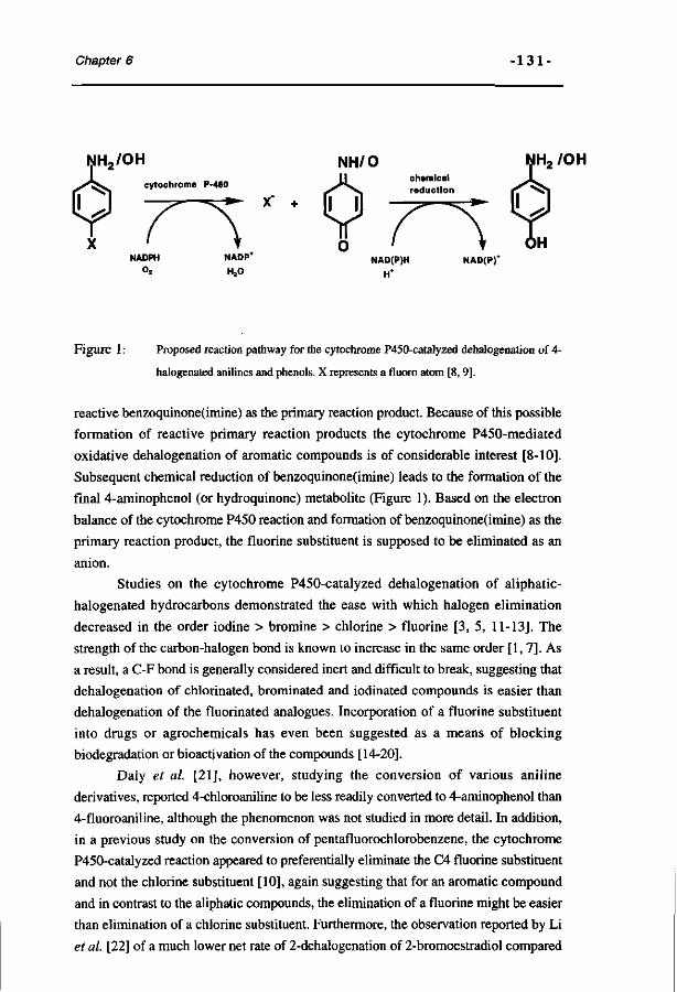
Chapter 6 - 1 3 1 -
NH, /OH
NAD(P)
Figure 1: Proposed reaction pathway for the cytochrome P450-catalyzed dehalogenation of 4-
halogenated anilines and phenols. X represents a fluoro atom [8, 9].
reactive benzoquinone(imine) as the primary reaction product. Because of this possible
formation of reactive primary reaction products the cytochrome P450-mediated
oxidative dehalogenation of aromatic compounds is of considerable interest [8-10].
Subsequent chemical reduction of benzoquinone(imine) leads to the formation of the
final 4-aminophenol (or hydroquinone) metabolite (Figure 1). Based on the electron
balance of the cytochrome P450 reaction and formation of benzoquinone(imine) as the
primary reaction product, the fluorine substituent is supposed to be eliminated as an
anion.
Studies on the cytochrome P450-catalyzed dehalogenation of aliphatic-
halogenated hydrocarbons demonstrated the ease with which halogen elimination
decreased in the order iodine > bromine > chlorine > fluorine [3, 5, 11-13]. The
strength of the carbon-halogen bond is known to increase in the same order [1, 7]. As
a result, a C-F bond is generally considered inert and difficult to break, suggesting that
dehalogenation of chlorinated, brominated and iodinated compounds is easier than
dehalogenation of the fluorinated analogues. Incorporation of a fluorine substituent
into drugs or agrochemicals has even been suggested as a means of blocking
biodégradation or bioactivation of the compounds [14-20].
Daly et al. [21], however, studying the conversion of various aniline
derivatives, reported 4-chloroaniline to be less readily converted to 4-aminophenol than
4-fluoroaniline, although the phenomenon was not studied in more detail. In addition,
in a previous study on the conversion of pentafluorochlorobenzene, the cytochrome
P450-catalyzed reaction appeared to preferentially eliminate the C4 fluorine substituent
and not the chlorine substituent [10], again suggesting that for an aromatic compound
and in contrast to the aliphatic compounds, the elimination of a fluorine might be easier
than elimination of a chlorine substituent. Furthermore, the observation reported by Li
et al. [22] of a much lower net rate of 2-dehalogenation of 2-bromoestradiol compared
•13 2 - Oxidative dehalogenation.
with that of 2-fluoroestradiol, also suggests the relatively easier elimination of an
aromatic fluorine substituent than of other aromatic halogen substituents. These
authors attributed their observation to the inability of the brominated substrate to bind
to the cytochrome P450 enzyme due to steric hindrance, but the phenomenon might
also be due to electronic differences between the fluoro- and bromo-substituent.
The objective of the present study was to investigate the influence of the nature
and number of the halogen substituents on the cytochrome P450-catalyzed
biodehalogenation of aromatic compounds in more detail. The results are expected to
provide additional information on the importance of halogen characteristics for their
possible elimination from an aromatic molecule in a cytochrome P450-catalyzed
reaction and, thus, on the mechanism of the reaction. Furthermore, an extension of the
studies from fluorinated to other halogenated aromatics is of importance considering
the more widespread use of chlorinated and brominated aromatics than of their
fluorinated analogues.
4-Halogenated anilines were taken as the model compounds because
defluorination of 4-fluoroaniline was already demonstrated to occur extensively in
previous studies [23-25].
MATERIALS AND METHODS.
Chemicals
Aniline, 2-fluoro-, 3-fluoro-, 4-fluoro-, 4-chloro-, 4-bromo- and 4-iodoaniline
were purchased from Janssen Chimica (Beerse, Belgium). All di- and
polyfluoroanilines and nitrobenzenes were obtained from Fluorochem (Derbyshire,
UK). 2-Aminophenol and 4-fluoronitrobenzene were from Aldrich and 4-aminophenol
was from Merck (Darmstadt, Germany). The purity of all compounds was > 98%.
Nitrosobenzene was purchased from Aldrich. 4-Fluoro-, 4-chloro-, 4-bromo-
and 4-iodonitrosobenzene were synthesized according to Kennedy and Stock [26]. For
3 min 25 mL 15% (w/v) potassium peroxomonosulphate (minimal 4.5% active
oxygen, Janssen Chimica) were added to a solution of 10 mmol C4-halogenated
aniline in 20 mL glacial acetic acid, cooled in an ice bath. After 2 minutes mixing, the
reaction mixture was immediately extracted twice with 25 mL hexane. The
nitrosobenzene was further purified on a LiChroprep Si60 column using an ISCO
HPLC system with hexane as the eluens. The flow-rate was 6.0 mL/min and fractions
of 10 mL were collected. Detection was at 295 nm. The characteristic green-colored
nitrosobenzene containing fractions were collected and concentrated to a volume of 5
mL by evaporation of the solvent. In order to prevent the decomposition of the
unstable nitrosobenzenes this fraction was not evaporated to dryness. The
Chapter 6 - 133 -
concentration of the C4-halogenated nitrosobenzene was determined by !H-NMR on a
Bruker AC 200 spectrometer using dichloromethane as the internal standard. The stock
solutions of the nitrosobenzene derivatives thus obtained were immediately used for
the determination of molar extinction coefficients in the chemical assay for N-oxidation
products (see below).
Synthesis of the fluorinated N-hydroxyanilines was performed by the chemical
reduction of the nitrobenzene analogue essentially as described by Vogel [27] and
Coleman et al [28] or by the chemical reduction of the synthetized nitrosobenzenes in
0.1 M KPi buffer (pH 7.6) containing 20 mM ascorbic acid.
3-Fluoro-4-aminophenol, 2-fluoro-4-aminophenol and 5-fluoro-2-aminophenol
were synthesized as described before [24],
2-Amino-3,5-difluorophenol, 2-amino-5-chlorophenol, 2-amino-5-
bromophenol and 2-amino-5-iodophenol were synthesized according to Boyland and
Sims [29] using, respectively, 2,4-difluoroaniline, 4-chloroaniline, 4-bromoaniline
and 4-iodoaniline as the starting material. In short, 15 g potassium persulphate
(Janssen Chimica) were added to a solution of 5 g 4-halogenated aniline in 20 mL
ethanol, 250 mL water and 50 mL 2N potassium hydroxide for 8 hr under continuous
stirring. The mixture was kept overnight and filtered. After washing with ether, the
solution was acidified with 2N sulphuric acid and the precipitate separated from the
mixture by filtration. The filtrate was neutralized with 2N potassium hydroxide and the
water-content was reduced to 25-50 mL by freeze-drying. The solution was extracted
three times with 100 mL butanol, the collected butanol phases were evaporated under
reduced pressure and the residue obtained was crystallized from 90% ethanol. The
sulphate esters obtained, were hydrolysed for 45 minutes at 100°C in 3N HCl. After
cooling, the solution was neutralized and the 2-aminophenols were extracted from the
mixture with diethylether. After evaporation of the solvent, the brownish precipitate
was dissolved in 3 mL dichloromethane and applied to a LiChroprep Si60 column (310
mm x 25 mm, particle size 40-63 um) (Merck, Darmstadt, Germany) using an ISCO
HPLC system with 2% (v/v) ethanol in dichloromethane as the eluens. The flow-rate
was 5 mL/min and fractions of 10 mL were collected. Detection was at 295 nm using
an ISCO absorbance detector. The fractions were analysed on 2-aminophenol content
by monitoring their reaction in a chemical assay for 2-aminophenols as described
elsewhere [30]. The 2-aminophenol-containing fractions were collected and after
crystallization the purity of these aminophenols was verified using a Kratos 400 HPLC
system, with a LiChrosorb Cg column (100 mm x 3 mm) (Chrompack, Middelburg,
The Netherlands). After eluting for 5 min with nanopure, a linear gradient from 0 to
80% (v/v) methanol in 22 min, followed by 80% (v/v) methanol for 6 min was
applied. Detection was at 240 nm using a Waters™ 996 photodiode array detector.
From the elution patterns obtained, the purity of the synthesized compounds was
•13 4 - Oxidative dehalogenation.
judged to be >95%.
The identity of the synthetized 2-aminophenols was derived from their 19F-
NMR and/or !H-NMR spectral characteristics. 5-Fluoro-2-aminophenol: 19F-NMR
(0.1 M potassium phosphate pH 7.6, relative to CFCbXppm): -127.5 (F5)(m, 3/F5-H4
= 12.0 Hz, 3/F5-H6 = 12.0 Hz, 47F5-H3 = 9.0 Hz) and iH-NMR (acetone)(ppm): 9.29
(H3)(tr, 3/H3-H4 = 9.0 Hz, 4/H3-F5 = 9.0 Hz), 9.22 (H4)(dd, 37H4-F5 = 12.0 Hz, 3^H4-H3 = 9.0 Hz), 9.07 (H6)(d, 3/H6-F5 = 12.0 Hz); 5-chloro-2-aminophenol: 'H-
NMR (acetoneXppm): 9.42 (H6)(s), 9.33 (H4)(d, 37H4-H3 = 8.3 Hz), 9.29 (H3)(d, 3^H3-H4 = 8.3 Hz); 5-bromo-2-aminophenol: !H-NMR (acetone)(ppm): 9.55 (H6)(s),
9.42 (H4)(d, 3 / H 4 - H 3 = 8.5 Hz), 9.29 (H3)(d, 3/H3-H4 = 8.5 Hz); 5-iodo-2-
aminophenol: !H-NMR (acetone)(ppm): 9.71 (H6)(s), 9.60 (H4)(d, 37H4-H3 = 8.5
Hz), 9.19 (H3)(d, 3 7H3-H4= 8.5 Hz); 3-fluoro-2-aminophenol: ^F-NMR (0.1 M
potassium phosphate pH 7.6, relative to CFCl3)(ppm): -136.9 (F3)(dd, 37F3-H4 =
10.0 Hz, 4 /F3-H5 = 8.5 Hz) and !H-NMR (acetone)(ppm): 9.25 (H5)(m, 37H5-H6 =
8.5 Hz, 37H5-H4 = 8.5 Hz, 47H5-F3 = 8.5 Hz), 9.11 (H6)(d, 3/H6-H5 = 8.5 Hz), 9.02
(H4)(dd, 37H4-H5 = 8.5 Hz, 3/H4-F3 = 10.0 Hz); 3,5-difluoro-2-aminophenol: 19F-
NMR (0.1 M potassium phosphate pH 7.6, relative to CFCbXppm): -134.5 (F3)(d,
37p3.H4 = 9.0 Hz), -127.1 (F5)(tr, 37F5-H6 = 9.0 Hz, 37F5-H4 = 9.0 Hz) and !H-NMR
(acetone)(ppm): 9.17 (H6)(d)(3JH6-F5 = 9.0 Hz), 9.10 (H4)(tr) (3/H4-F3 = 9.0 Hz, 3JH4-F5 = 9.0 Hz).
All di- and polyfluorinated 4-aminophenols were prepared by biosynthesis,
purified and their identity was derived from 19F-NMR and 'H-NMR spectral
characteristics essentially as described previously [31].
Preparation of microsomes
Microsomes were prepared from the perfused livers of male Wistar rats (± 400
g), pretreated with isosafrole (Janssen Chimica) as described previously [24]. For the
in vitro experiments liver microsomes of isosafrole-pretreated rats were used, since
this type of microsome was demonstrated to possess the highest capacity to convert 4-
fluoroaniline to its 4-aminophenol [25]. This was also the case for other 4-halogenated
anilines (unpublished results). Cytochrome P450 content of the microsomes was
measured as described by Omura and Sato [32].
Purification ofmicroperoxidase-8 (MP-8).
MP-8 was purified by proteolytic digestion of horse heart cytochrome c
(Sigma, St. Louis, MO USA) essentially as described in the literature [33]. The sample
was more than 96% pure based on HPLC analysis [33]. The HPLC analysis was
performed on a Waters™ 600 Controller HPLC with a Baker bond WPC 4 column
(25 cm x 4.6 mm). A linear gradient from 0.1% trifluoroacetic acid in water to 50%

Chapter 6 - 135 -
0.1% trifluoroacetic acid in water and 50%, 0.1% trifluoroacetic acid in acetonitril was
applied in 50 min., which was continued isocratically for 5 min. Detection at 214 nm
was performed on an ISCO V4 absorbance detector and detection at 395 nm was
performed on an Applied Biosystems 757 absorbance detector. The heme content was
determined essentially as described previously [34].
Purification of cytochrome P450 IIB I and NADPH-cytochrome P450 reductase.
Cytochrome P450IEB1 and NADPH-cytochrome P450 reductase were purified
from liver microsomes of phénobarbital (Brocacef bv., Maarssen, The
Netherlands)(0.1 % in drinking water for 7 days) pretreated male respectively female
Wistar rats essentially as previously described by Boersma et al. [35].
In vitro incubations
Cytochrome P450-dependent conversion was studied in vitro in microsomal
incubations containing (final concentrations) 0.1 M potassium phosphate pH 7.6,
between 0 and 15 mM of the halogenated aniline (as indicated) added as 1 % (v/v) of a
100 fold concentrated stock solution in dimethylsulphoxide and 1 uM microsomal
cytochrome P450. The reaction was started by the addition of NADPH (1 mM final
concentration) or tBuOOH (10 mM final concentration) and carried out at 37 °C for 10
min. The incubations with tBuOOH as the artificial oxygen donor additionally
contained NADH (1 mM final concentration) in order to reduce benzoquinoneimine to
4-aminophenol. The conversion of halogenated anilines to their 4-aminophenol
metabolites is linear for at least 10 minutes. Incubations with purified, reconstituted
cytochrome P450 IIB 1 were conducted using similar final incubation conditions as
described for the microsomal incubations. Instead of microsomes, the incubations
contained 0.5 nmol cytochrome P450 IIB1, 1.24 units NADPH-cytochrome reductase
and 20 ug dilauroyl phosphatidylcholine (Sigma) per ml incubation mixture and were
preincubated for 6 minutes at 37°C.
The microperoxidase-8 (MP-8) catalyzed conversion was studied in
incubations containing (final concentrations) 0.1 M potassium phosphate pH 7.6,
between 0 and 12.5 mM of the halogenated aniline (as indicated) added as 1% (v/v) of
a 100 fold concentrated stock solution in dimethylsulphoxide, 7.5 uM MP-8 and 1 mM
NADH in order to reduce to primary metabolite 4-benzoquinoneimine to 4-
aminophenol. The reaction was started by the addition of H2O2 (2.5 mM final
concentration) and carried out at 37°C for 1 min.
For chemical analysis of 4-aminophenol or N-hydroxyaniline the reaction was
terminated by the addition of 0.8 mL of the incubation mixture to 0.24 mL of 20%
(w/v) trichloroacetic acid. Upon mixing and centrifugation (5 min, 13000 rpm) the
supernatant was used for the chemical determination of 4-aminophenol or N-
•13 6 - Oxidative dehalogenation.
hydroxyaniline metabolites as described hereafter. For the chemical analysis of 2-
aminophenol metabolites (decribed below) the reaction was terminated by freezing the
sample into liquid nitrogen.
Incubations for HPLC and 19F-NMR analysis additionally contained 1 mM
ascorbic acid to prevent autoxidation of the aminophenols especially during the
overnight 19F-NMR measurement. These incubations were terminated by freezing the
samples into liquid nitrogen. Samples were stored at -20°C, thawed and centrifuged (5
min at 13000 rpm). HPLC analysis of these incubations was performed essentially as
described above for the check on purity of the synthetised aminophenols. The samples
for the 19F-NMR measurements were made oxygen free by four cycles of evacuating
and filling with argon.
Chemical determination of hydroxylated metabolites.
4-Aminophenols were determined essentially as described by Brodie and
Axelrod [36]. In short, 100 uL phenol reagent (5% w/v phenol in 2.5 N NaOH) and
200 uL 2.5 M Na2CC>3 were added to 1 mL trichloroacetic acid precipitated
supernatant. After 60 minutes at room temperature the absorbance at 630 nm was
measured. The presence of a halogen in the indophenol formed in this assay influences
its molar extinction coefficient at 630 nm [37]. For this reason the molar extinction
coefficients of the halogenated aminophenol derived indophenols were determined to
allow quantification of the 4-aminophenol metabolites from the various aniline
derivatives. Molar extinction coefficients were 30.5, 26.7, 26.3, 14.5, 20.0, 16.8,
and 12.5 mM-'cm-1 for the indophenols derived from 4-aminophenol, 3-fluoro-4-
aminophenol, 2-fluoro-4-aminophenol, 3,5-difluoro-4-aminophenol, 2,6-difluoro-4-
aminophenol, 2,3-difluoro-4-aminophenol and 2,3,5-trifluoro-4-aminophenol,
respectively.
Products from C2 hydroxylation of the 4-halogenated anilines were determined
by a chemical assay developed for the detection of 2-aminophenols. This newly-
developed assay is described in detail elsewhere [30]. In short, a 1 mL microsomal
incubation, frozen into liquid nitrogen to stop the reaction, was thawed and centrifuged
at 13000 rpm for 10 min at 4°C. The supernatant was extracted three times with 3 mL
of diethylether. After evaporation of the diethylether, 300 uL of 20% (w/v)
trichloroacetic acid, 1 mL demineralised water and 100 uL 40 mM ammonium
iron(III)sulphate dodecahydrate (Janssen Chimica) were added to the residue. After 45
min at room temperature, the reaction mixture was extracted with 2 mL chloroform and
the absorbance of the chloroform-phase was measured at Xmax of the halogenated 2-
aminophenol derived yellow derivative. The values for the 2-aminophenol
concentrations of the samples were determined from the calibration curves of 2-
aminophenol, 2-amino-5-fluorophenol, 2-amino-5-chlorophenol, 2-amino-5-
Chapter 6 - 137 -
bromophenol and 2-amino-5-iodophenol.
The chemical determination of N-oxidation products (N-hydroxyaniline plus
nitrosobenzene derivatives) was performed essentially as described by Herr and Kiese
[38]. To 1 mL trichloroacetic acid precipitated microsomal supernatant, 0.3 mL 10%
(w/v) potassium ferricyanide in IN HCl and 1 mL demineralized water were added in
order to oxidize the N-hydroxyaniline to the corresponding nitrosobenzene. These
samples were extracted with 2 mL CCI4 and the CCI4 phases were washed twice with
5N H2SO4. To 1.0 mL of the washed CCI4 phases, 1.0 mL of glacial acetic acid and
50 |il 20% (w/v) sodium nitrite were added. After 15 minutes, 100 ul 50% (w/v)
ammonium sulphamate (Janssen Chimica) were added and the samples were mixed for
10 min. Finally, the color formation was started by the addition of 250 ul 80% (v/v)
acetic acid and 50 ul N-(l-naphtyl)ethylenediamine dihydrochloride (Aldrich,
Steinheim, Germany). After 2h in the dark the absorbance of the water phase at 555
nm was measured. Using nitrosobenzene and synthesized 4-fluoro-, 4-chloro-, 4-
bromo- and 4-iodonitrosobenzene molar extinction coefficients of 40.4, 32.9, 41.3,
40.9 and 42.2 mM'^cm-1, respectively, were determined.
Kinetic analysis
The apparent Vmax and Km values for C4-, C2- and N-hydroxylation were
determined by fitting the data to the standard Michaelis-Menten equation: v = Vmax*[S]
/ (Km + [S]) with the program of KaleidaGraph, version 2.0.2 (Abelbeck Software).
The correlation coefficient of the fits was above 0.97 in all cases.
NMR measurements 19F-NMR measurements were performed on a Bruker AMX 300 NMR
spectrometer essentially as described previously by Vervoort et al. [39]. Chemical
shifts are reported relative to CFCI3. Quantification of the fluorinated compounds in
the samples was achieved by comparison of the integrals of their 19F-NMR resonances
to the integral of the resonance of 4-fluorobenzoic acid added as an internal standard.
The splitting patterns of the !H-NMR and 19F-NMR resonances of the
(bio)synthetized 2- and 4-aminophenols dissolved in acetone-d6 were measured on a
Bruker AMX 500 MHz. 19F-NMR measurements were performed using a 'H-
probehead tuned to the 19F frequency (470.5 MHz). !H-NMR chemical shifts are
reported relative to acetone, present in acetone-d6, used for locking the magnetic field.
•138 Oxidative dehalogenation.
RESULTS.
Microsomal cytochrome P450 catalyzed conversion of 4-halogenated
anilines.
The influence of the type of C4 halogen substituent in an aniline molecule on
the cytochrome P450-catalyzed dehalogenation to 4-aminophenol was investigated by
HPLC analysis of microsomal incubations.
Figure 2 presents HPLC chromatograms of the microsomal metabolite patterns
of the four 4-halogenated anilines and -for comparison- aniline itself. The reaction is
cytochrome P450-mediated as no conversion was observed without NADPH, without
TIME (min)
Figure 2: Reversed-phase HPLC chromatograms of the microsomal conversion of A) aniline,
B) 4-fluoroaniline, C) 4-chloroaniline, D) 4-bromoaniline and E) 4-iodoaniline with
detection at 240 nm. The peaks marked with an asterisk were also present in the
chromatograms of control incubations carried out in the absence of NADPH, in the
absence of microsomes, or in the absence of the aniline-derivatives.
Chapter 6 -13 9-
microsomes or in the presence of CO [25]. Peaks were identified on the basis of their
retention times compared to the retention times of the synthetized reference compounds
and the similarity of the absorption spectra provided by the diode array detector. From
these results it follows that the microsomal cytochrome P450-catalyzed conversion of
the anilines results in the formation of 4-aminophenol, (halogenated) 2-aminophenols
and/or N-oxidation products (N-hydroxyaniline and nitrosobenzene derivatives).
Formation of (halogenated) 3-aminophenols or NIH-shifted metabolites is not
observed. In contrast, metabolites resulting from hydroxylation at C4 accompanied by
dehalogenation are readily observed, especially for 4-fluoroaniline and 4-chloroaniline.
These results support the hypothesis that the cytochrome P450-catalyzed C4-
hydroxylation of the 4-halogenated anilines proceeds by dehalogenation of the aromatic
ring rather than by hydroxylation at C4 accompanied by an NIH shift of the halogen to
the adjacent C3 or C5 position.
In addition to the HPLC experiments, metabolite formation in microsomal
incubations was quantified by chemical analysis using specific methods for detection
of 2-aminophenol, 4-aminophenol and N-oxidation products (N-hydroxyaniline- plus
nitrosobenzene-derivatives). Table 1 presents the kinetic parameters obtained for the
microsomal conversion of aniline and the four halogenated anilines. The apparent Vmax
for the formation of 4-aminophenol decreases to 66.8 % of the value of aniline, when
the hydrogen at C4 is replaced by a fluorine. Replacement of the hydrogen at C4 by a
chlorine, bromine or iodine substituent even further decreases the apparent Vmax of
this reaction. However, the apparent Vmax values observed for 4-chloro, 4-bromo and
4-iodoaniline vary only slightly (Table 1).
Table 1 : Kinetic characteristics of the cytochrome P450-catalyzed aromatic C4-
hydroxylation of aniline and its 4-halogenated derivatives determined
using liver microsomes from isosafrole pretreated male Wistar rats
(n=2-4) a.
Compound
Aniline 4-Fluoroaniline 4-Chloroaniline 4-Bromoaniline 4-Iodoaniline
Apparent Vm a x i n
nmol product * min"1 • nmol cyt P450-1
5.55 ± 0.20 (100%) 3.71 ± 0.28 (66.8%) 0.54 ±0.09 (9.7%) 0.39 ±0.10 (7.0%) 0.38 ±0.16 (6.8%)
Apparent Km i n
mM
17.0 ± 2.0 9.3 ± 1.7 1.2 ±0.2 0.7 ± 0.2 0.3 ±0.1
flValues presented are the means + standard error of the mean. Values in parentheses represent the % relative to the value for C4-hydroxylation of aniline.
-1 4 O - Oxidative dehalogenation.
To verify whether the observed change in apparent Vmax for the dehalogenation
to C4 aminophenols was not the result of a general decrease in metabolism from
fluorine > chlorine > bromine > iodine, the conversion rates for formation of
metabolites resulting from C2 and N-hydroxylation were also determined. Table 2
presents the apparent Vmax values for the microsomal C2 and N-hydroxylation and
also the total maximal conversion rate, calculated as the sum of the Vmax values
obtained for the conversion to 2-aminophenol, 4-aminophenol and N-hydroxyaniline
metabolites. The results in Table 2 demonstrate that the total Vmax for the C2, C4 plus
N-hydroxylation is not significantly influenced by the type of C4 halogen substituent.
The decrease in the apparent Vmax for the 4-hydroxylation is accompanied by an
increase in hydroxylation of the 4-halogenated anilines at their C2- and N-position.
Thus, the results in Table 2 indicate that the observed change in dehalogenation is not a
result of a reduced overall capacity of cytochromes P450 to convert the 4-halogenated
anilines.
Table 2: Apparent Vm a x values of the cytochrome P450-catalyzed C2-
hydroxylation and N-hydroxylation and the total conversion of aniline
and its 4-halogenated derivatives determined using liver microsomes
from isosafrole pretreated male Wistar rats (n=2-4) a .
apparent Vmax Vm a x for total
nmol product • min"1 • nmol cyt P450"1 conversion *
Compound 2-Aminophenol N-hydroxyaniline
Aniline 1.13 ±0.12 1.42 + 0.07 8.1010.39 4-Fluoroaniline 2.27 ± 0.07 3.34 ± 0.09 9.32 ± 0.44 4-Chloroaniline 2.93 ±0.12 4.35 ± 0.09 7.82 ± 0.30 4-Bromoaniline 2.67 ± 0.04 4.39 ± 0.09 7.45 ± 0.23 4-Iodoaniline 2.93 ± 0.47 2.93 ± 0.17 6.24 ± 0.80
a Values presented are the mean + standard error of the mean. " Calculated as the sum of apparent
V m a x for C2-, N- and C4-hydroxylation (see Table 1).
Dehalogenation of 4-halogenated anilines by reconstituted cytochrome
P450 IIB1, by a model heme-based catalyst and by a tertiair
hydroperoxide supported microsomal cytochrome P450 system.
To demonstrate that the change in the extent of C4 hydroxylation with a change
in the type of halogen substituent is not due to a change in tcytochrome P450 enzymes
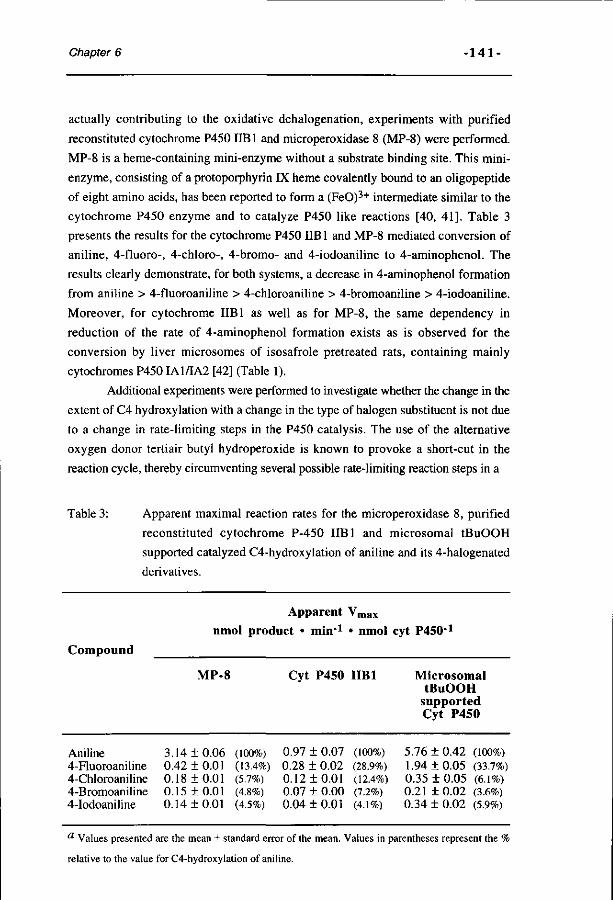
Chapter 6 - 1 4 1 -
actually contributing to the oxidative dehalogenation, experiments with purified
reconstituted cytochrome P450 IIB 1 and microperoxidase 8 (MP-8) were performed.
MP-8 is a heme-containing mini-enzyme without a substrate binding site. This mini-
enzyme, consisting of a protoporphyrin IX heme covalently bound to an oligopeptide
of eight amino acids, has been reported to form a (FeO)3+ intermediate similar to the
cytochrome P450 enzyme and to catalyze P450 like reactions [40, 41]. Table 3
presents the results for the cytochrome P450 IIB 1 and MP-8 mediated conversion of
aniline, 4-fluoro-, 4-chloro-, 4-bromo- and 4-iodoaniline to 4-aminophenol. The
results clearly demonstrate, for both systems, a decrease in 4-aminophenol formation
from aniline > 4-fluoroaniline > 4-chloroaniline > 4-bromoaniline > 4-iodoaniline.
Moreover, for cytochrome IIB1 as well as for MP-8, the same dependency in
reduction of the rate of 4-aminophenol formation exists as is observed for the
conversion by liver microsomes of isosafrole pretreated rats, containing mainly
cytochromes P450IA1/IA2 [42] (Table 1).
Additional experiments were performed to investigate whether the change in the
extent of C4 hydroxylation with a change in the type of halogen substituent is not due
to a change in rate-limiting steps in the P450 catalysis. The use of the alternative
oxygen donor tertiair butyl hydroperoxide is known to provoke a short-cut in the
reaction cycle, thereby circumventing several possible rate-limiting reaction steps in a
Table 3: Apparent maximal reaction rates for the microperoxidase 8, purified
reconstituted cytochrome P-450 IIB1 and microsomal tBuOOH
supported catalyzed C4-hydroxylation of aniline and its 4-halogenated
derivatives.
Apparent Vmax
nmol product • min-1 • nmol cyt P450-1
Compound
Aniline 4-Fluoroaniline 4-Chloroaniline 4-Bromoaniline 4-Iodoaniline
MP-8
3.14 ±0.06 0.42 ± 0.01 0.18 + 0.01 0.15 ±0.01 0.14 ±0.01
(100%) (13.4%) (5.7%) (4.8%) (4.5%)
Cyt P450
0.97 ± 0.07 0.28 ± 0.02 0.12 + 0.01 0.07 ± 0.00 0.04 ± 0.01
IIB1
(100%) (28.9%) (12.4%) (7.2%) (4.1%)
Microsomal tBuOOH
supported Cyt P450
5.76 ± 0.42 (100%) 1.94 ±0.05 (33.7%) 0.35 ± 0.05 (6.1%) 0.21 ± 0.02 (3.6%) 0.34 + 0.02 (5.9%)
a Values presented are the mean ± standard error of the mean. Values in parentheses represent the '
relative to the value for C4-hydroxylation of aniline.
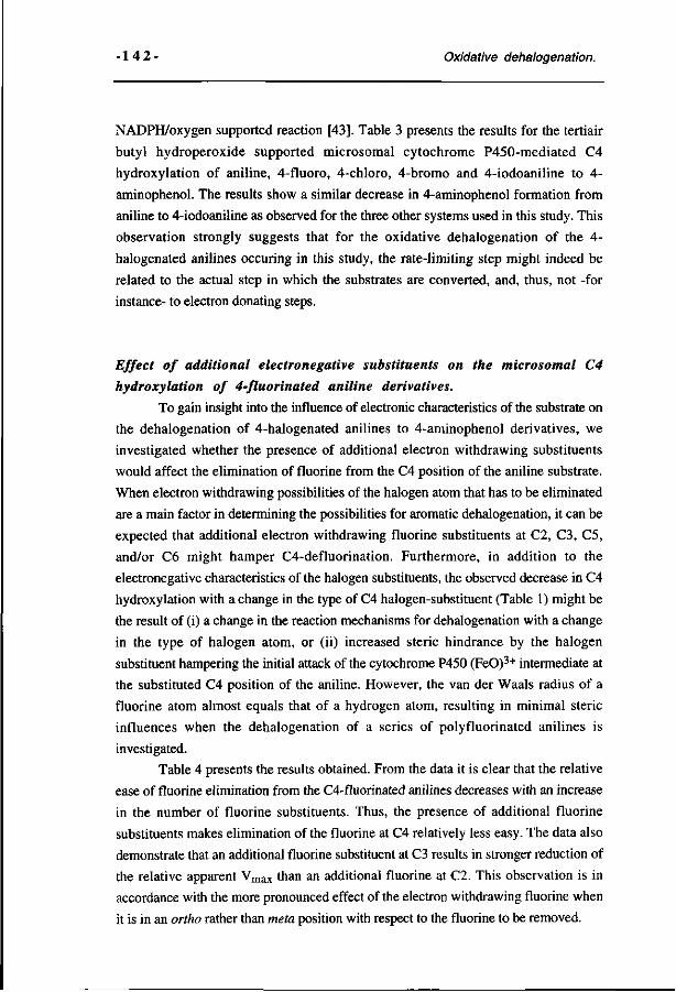
•14 2 - Oxidative dehalogenation.
NADPH/oxygen supported reaction [43]. Table 3 presents the results for the tertiair
butyl hydroperoxide supported microsomal cytochrome P450-mediated C4
hydroxylation of aniline, 4-fluoro, 4-chloro, 4-bromo and 4-iodoaniline to 4-
aminophenol. The results show a similar decrease in 4-aminophenol formation from
aniline to 4-iodoaniline as observed for the three other systems used in this study. This
observation strongly suggests that for the oxidative dehalogenation of the 4-
halogenated anilines occuring in this study, the rate-limiting step might indeed be
related to the actual step in which the substrates are converted, and, thus, not -for
instance- to electron donating steps.
Effect of additional electronegative substituents on the microsomal C4
hydroxylation of 4-fluorinated aniline derivatives.
To gain insight into the influence of electronic characteristics of the substrate on
the dehalogenation of 4-halogenated anilines to 4-aminophenol derivatives, we
investigated whether the presence of additional electron withdrawing substituents
would affect the elimination of fluorine from the C4 position of the aniline substrate.
When electron withdrawing possibilities of the halogen atom that has to be eliminated
are a main factor in determining the possibilities for aromatic dehalogenation, it can be
expected that additional electron withdrawing fluorine substituents at C2, C3, C5,
and/or C6 might hamper C4-defluorination. Furthermore, in addition to the
electronegative characteristics of the halogen substituents, the observed decrease in C4
hydroxylation with a change in the type of C4 halogen-substituent (Table 1) might be
the result of (i) a change in the reaction mechanisms for dehalogenation with a change
in the type of halogen atom, or (ii) increased steric hindrance by the halogen
substituent hampering the initial attack of the cytochrome P450 (FeO)3+ intermediate at
the substituted C4 position of the aniline. However, the van der Waals radius of a
fluorine atom almost equals that of a hydrogen atom, resulting in minimal steric
influences when the dehalogenation of a series of polyfluorinated anilines is
investigated.
Table 4 presents the results obtained. From the data it is clear that the relative
ease of fluorine elimination from the C4-fluorinated anilines decreases with an increase
in the number of fluorine substituents. Thus, the presence of additional fluorine
substituents makes elimination of the fluorine at C4 relatively less easy. The data also
demonstrate that an additional fluorine substituent at C3 results in stronger reduction of
the relative apparent Vmax than an additional fluorine at C2. This observation is in
accordance with the more pronounced effect of the electron withdrawing fluorine when
it is in an ortho rather than meta position with respect to the fluorine to be removed.
Chapter 6 •143-
Table 4: Apparent Vmax values for the C4-hydroxylation of 4-fluorinated anilines
compared to the apparent Vmax values of their non-C4 fluorinated
analogues, determined using liver microsomes from isosafrole
pretreated male Wistar rats (n=2-4).
Substrate
Aniline 4-Fluoroaniline
2-Fluoroaniline 2,4-Difluoroaniline
2,6-Difluoroaniline 2,4,6-Trifluoroaniline
3-Fluoroaniline 3,4-Difluoroaniline
2,3-Difluoroaniline 2,3,4-Trifluoroaniline
3,5-Difluoroaniline 3,4,5-Trifluoroaniline
2,3,6-Trifluoroaniline 2,3,4,6-Tetrafluoroaniline
Apparent Vm a x nmol 4-aminophenol • min"1 •
nmol cyt P450'1
5.55 ± 0.20 3.71 ±0.28
6.23 ± 0.20 2.99 ± 0.07
8.89 ± 0.05 3.21 ±0.16
4.85 ±0.12 0.90 ± 0.03
9.46 ± 0.39 1.18 ±0.04
6.82 ± 0.39 0.42 ± 0.02
3.23 ±0.12 0.66 ±0.16
Relative apparent ' m a x
as % of the non-C4-fluorinated analogue.
66.8
48.0
36.1
18.6
12.5
6.2
20.3
a Values presented are the mean + standard error of the mean.
Metabolite profiles of the microsomal cytochrome P450 catalyzed
conversion of fluorinated aniline-derivatives.
To characterize the overall conversion characteristics of the fluorinated anilines
for the apparent Vm a x studies on C4 hydroxylation 19F-NMR spectra of the
microsomal incubations were determined.
Figure 3 presents the 19F-NMR spectra of the microsomal conversion of the
fluorinated anilines. The 19F-NMR resonances of the metabolites were identified on
the basis of (synthetised) reference compounds. For the metabolites 5,6-difluoro-2-
aminophenol (Figure 3D), 4,5-difluoro-2-aminophenol (Figure 3D), 4,6-difluoro-2-
aminophenol (Figure 3F) and 4,5,6-trifluoro-2-aminophenol (Figure 3F) the 19F-
NMR resonances were identified on the basis of a characteristic shift of the 19F-NMR
resonance known to exist upon the introduction of a hydroxyl moiety at positions
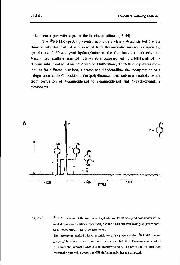
• 1 4 4 - Oxidative dehalogenation.
ortho, meta or para with respect to the fluorine substituent [43,44].
The 19F-NMR spectra presented in Figure 3 clearly demonstrated that the
fluorine substituent at C4 is eliminated from the aromatic aniline-ring upon the
cytochrome P450-catalyzed hydroxylation to the fluorinated 4-aminophenols.
Metabolites resulting from C4 hydroxylation accompanied by a NIH shift of the
fluorine substituent at C4 are not observed. Furthermore, the metabolic patterns show
that, as for 4-fluoro, 4-chloro, 4-bromo and 4-iodoaniline, the incorporation of a
halogen atom at the C4 position in the (poly)fluoroanilines leads to a metabolic switch
from formation of 4-aminophenol to 2-aminophenol and N-hydroxyaniline
metabolites.
P =
»+faÉ0mt*fit0ié/*Ê#***to+r*0m»0m&**^m*tiiti**\Mê***$imfiiA'++mi mS^w******!***,
-120 -140 PPM -160
Figure 3: 19F-NMR spectra of the microsomal cytochrome P450-catalyzed conversion of the
non-C4 fluorinated anilines (upper part) and their 4-fluorinated analogues (lower part);
A) 4-fluoroaniline, B to G, see next pages.
The resonances marked with an asterisk were also present in the 19F-NMR spectra
of control incubations carried out in the absence of NADPH. The resonance marked
IS is from the internal standard 4-fluorobenzoic acid. The arrows in the spectrum
indicate the ppm value where the NIH-shifted metabolites are expected.
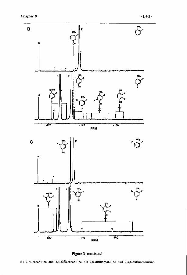
Chapter 6 -145-
B
•L f. .JU/ V-p
HNOH
O'
lJuJt *
m**»M«l*««M4**mM||W*
N H j
N H ,
-120 -140 -160
PPM
. t.ltto. UiirTjViiTr iriuMi K'UVU . VnntW*«W»wiméHt***********P*+>W»»«I• W i up***»*««—M»w«*i»V
u
r F^^-^^F
NH2
iH
A ««HWMi «*| ill t ««nnwwMIMiH»^ I'«V»I'
-120 -140 PPM
-160
Figure 3 -continued-
B) 2-fluoroaniline and 2,4-difluoroaniline, C) 2,6-difluoroaniline and 2,4,6-trifluoroaniline,
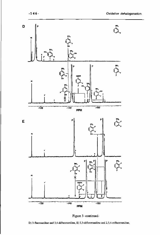
•146- Oxidative dehalogenation.
ii ,,i ;
NH,
-120
^iAM^.>IIH<tii l»*l i i |»»i| i mi JW Uvw. Jjt' L-,— xi
NH,
Ol.
-140 PPM
-160
. i j i nn " T i f f n '"*" r*' r~- ' ••
NHj
LM>I>I I .« •—»»*» wk^VÊÀnfl****
NH2
F ^r F
*K*i+#r \JJ UJ -120 -140 -160
PPM
Figure 3 -continued-
D) 3-fluoroaniline and 3,4-difluoroaniline, E) 2,3-difluoroaniline and 2,3,4-trifluoroaniline,
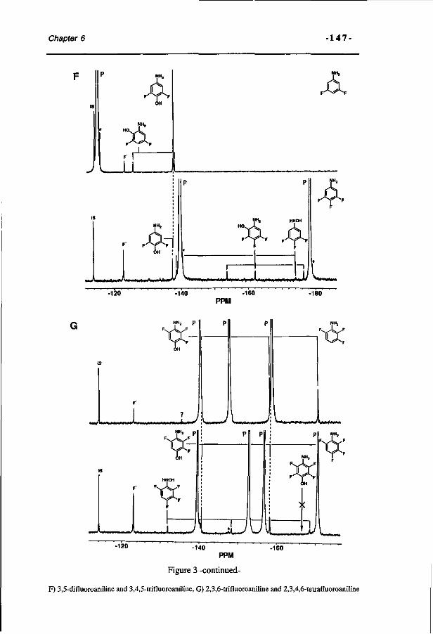
Chapter 6 •147-
NH,
A
NHa HNOH
F^%r F
^«•ww^MMimvMhtV 1 (Mw>*—«w»*% w * ^ 1 J) -120 -140 -160
PPM -180
NHj p
« < W « V i I '• p
I / V M D M M W W M M
u -120 -140 -160
PPM
Figure 3 -continued-
F) 3,5-difluoroaniline and 3,4,5-trifluoroaniline, G) 2,3,6-trifluoroaniline and 2,3,4,6-tetrafluoroaniline
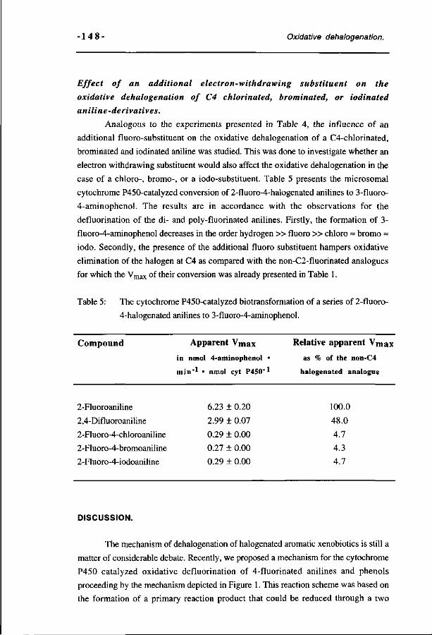
• 1 4 8 - Oxidative dehalogenation.
Effect of an additional electron-withdrawing substituent on the
oxidative dehalogenation of C4 chlorinated, brominated, or iodinated
aniline-derivatives.
Analogous to the experiments presented in Table 4, the influence of an
additional fluoro-substituent on the oxidative dehalogenation of a C4-chlorinated,
brominated and iodinated aniline was studied. This was done to investigate whether an
electron withdrawing substituent would also affect the oxidative dehalogenation in the
case of a chloro-, bromo-, or a iodo-substituent. Table 5 presents the microsomal
cytochrome P450-catalyzed conversion of 2-fluoro-4-halogenated anilines to 3-fluoro-
4-aminophenol. The results are in accordance with the observations for the
defluorination of the di- and poly-fluorinated anilines. Firstly, the formation of 3-
fluoro-4-aminophenol decreases in the order hydrogen » fluoro » chloro = bromo =
iodo. Secondly, the presence of the additional fluoro substituent hampers oxidative
elimination of the halogen at C4 as compared with the non-C2-fluorinated analogues
for which the Vmax of their conversion was already presented in Table 1.
Table 5: The cytochrome P450-catalyzed biotransformation of a series of 2-fluoro-
4-halogenated anilines to 3-fluoro-4-aminophenol.
Compound Apparent V m a x
in nmol 4-aminophenol •
min'* • nmol cyt P450"*
Relative apparent V m a x
as % of the non-C4
halogenated analogue
2-Fluoroaniline
2,4-Difluoroaniline
2-Fluoro-4-chloroaniline
2-Fluoro-4-bromoaniline
2-Fluoro-4-iodoaniline
6.23 ± 0.20
2.99 ± 0.07
0.29 + 0.00
0.27 ± 0.00
0.29 ± 0.00
100.0
48.0
4.7
4.3
4.7
DISCUSSION.
The mechanism of dehalogenation of halogenated aromatic xenobiotics is still a
matter of considerable debate. Recently, we proposed a mechanism for the cytochrome
P450 catalyzed oxidative defluorination of 4-fluorinated anilines and phenols
proceeding by the mechanism depicted in Figure 1. This reaction scheme was based on
the formation of a primary reaction product that could be reduced through a two
Chapter 6 - 1 4 9 -
electron step to the hydroxyaniline or phenol indicating it to be the reactive
benzoquinone(imine) [8, 9]. Elimination of the halogen as a halogen anion then
follows from the net two electron balance of the cytochrome P450 reaction. Results of
the present study demonstrate that the cytochrome P450 catalysed 4-hydroxylation of
4-halogenated anilines proceeds by dehalogenation rather than by hydroxylation
accompanied by an NIH-shift. A fluorine substituent is significantly more easily
eliminated than a chlorine, bromine or iodine substituent, the dehalogenation rate of 4-
chloro-, 4-bromo- and 4-iodoaniline being much more similar. These findings are in
accordance with the observations of Ullrich et al. who studied the conversion of a
comparable group of compounds, the 4-halogenated acetanilides [46].
Using purified reconstituted cytochrome P450 IIB1, tertiair butyl
hydroperoxide supported microsomal cytochrome P450 catalysis and microperoxidase
8, a heme-based model system able to catalyze cytochrome P450 like chemistry [41],
similar changes in the rate of oxidative dehalogenation with a change in the C4 halogen
substituent were observed. Based on these observations it may be concluded that the
observed decrease in dehalogenation with a change in the halogen is not dependent on
(i) different P450 enzymes contributing to the reaction nor on (ii) changes in
orientation of the substrates by the protein core in the large active sites of P450. The
observation of similar effects on the Vmax of the oxidative dehalogenation in the tertiair
butyl hydroperoxide driven reaction with a change in the halogen substituent supports
the hypothesis that the actual rate-limiting step follows formation of the activated
cofactor, and might thus indeed be related to the effect of the halogen substituent on the
actual dehalogenation step. Taken together, these results with the purified reconstituted
cytochrome P450 IIB 1 system, the tertiair butyl hydroperoxide supported microsomal
cytochrome P450 and the MP8 model compound, point to the importance of the
electronic characteristics of the halogen substituents for the reactivity of the C4-
halogenated anilines to participate in cytochrome P450 mediated oxidative
dehalogenation reactions.
For all systems tested, i.e. microsomes, purified reconstituted cytochrome
P450 IIB1, tertiair butyl hydroperoxide supported microsomal cytochrome P450 and
microperoxidase 8, the observed relative apparent Vmax values for dehalogenation of
the anilines correlate (r = 0.95, 0.98, 0.93 and 0.97 respectively) with the
electronegativity of fluorine (4.10), chlorine (2.83), bromine (2.74) and iodine (2.21).
The changes in the apparent Vmax for C4 hydroxylation with a change in the C4
substituent do not correspond to the strength of the carbon halogen bond known to
increase in the opposite order, namely, iodine < bromine < chlorine < fluorine [1,7].
Together, these results point to the importance of electronegativity of the halogen to be
eliminated in the cytochrome P450-catalyzed oxidative dehalogenation. This
observation supports the conclusion that the halogen is eliminated as a halogen anion.
•15 0 - Oxidative dehalogenation.
However, steric hindrance of the initial cytochrome P450 (FeO)3+ attack on C4 by the
larger chloro-, but especially bromo- and iodo-substituents with Van der Waals radius
of respectively 1.80 Â, 1.95 À and 2.15 Â, might provide an additional explanation for
the observed effects of the type of halogen on the apparent Vm a x for the
dehalogenation. Such steric hindrance, in combination with the susceptibility of an
iodo-substituent in particular to become oxidized by cytochromes P450, as
demonstrated by Guengerich using 4-ferf-butyl-2,5-bis[l -hydroxy- l-(trifluoromethyl)-
2,2,2-trifluoro-ethyl]-iodobenzene [47], may even result in a different mechanism. For
iodinated aromatic molecules a mechanism for the cytochrome P450-catalyzed
deiodination has been suggested that proceeds by an interaction of the reactive
cytochrome P450 (FeO)3+ intermediate with non-bonding halogen electrons of the
iodine [47, 48]. This interaction results in the formation of a haloso compound, and,
finally, the formation of the hydroxylated substrate and elimination of the halogen as
HXO [49]. Such a possible change in the actual mechanism of dehalogenation may add
to a change in the rate of the dehalogenation reaction, especially for 4-iodoaniline.
Therefore, additional dehalogenation experiments were performed to provide further
evidence for the mechanism proceeding by elimination of the halogen as an anion. The
effect of additional electron withdrawing fluorine substituents on the cytochrome
P450-catalyzed oxidative dehalogenation of 4-halogenated aniline-derivates to 4-
aminophenol metabolites was studied. The results obtained demonstrate that the
presence of additional electron withdrawing substituents at the aromatic ring is another
factor influencing dehalogenation. The introduction of additional fluorine substituents
in a 4-halogenated aniline molecule lessens the possibility of its oxidative
dehalogenation and, consequently, leads to a metabolic switch to fluorinated 2-
aminophenol and N-hydroxyaniline metabolites. Formation of NIH-shifted metabolites
was not observed. The decreasing effect of a fluorine substituent ortho with respect to
the halogen to be eliminated is larger than that of an aditional meta fluorine substituent.
This observation further lends weight to a mechanism in which the fluorine elimination
is dependent on {i.e. hampered by) additional electron withdrawing substituents in the
aromatic ring and, thus, a mechanism proceeding by elimination of the halogen as
anion.
Altogether, the results of the present paper demonstrate that the possibilities for
the cytochrome P450-mediated oxidative dehalogenation of halogenated anilines do not
depend exclusively on factors previously reported: i.e. the capacity of the amino
moiety to lose a proton and donate electrons to create the quinoneimine primary
metabolite as indicated by the mechanism depicted in Figure 1 [9]. Additional factors
influencing the cytochrome P450-catalyzed oxidative dehalogenation are; (i) the
electronegativity of the halogen to be removed, the more electronegative fluorine being
more easily eliminated than a chlorine, bromine or iodine, and; (ii) the position,
Chapter 6 - 151
number and electron-withdrawing capacities of other substituents in the aromatic ring,
electron withdrawing substituents decreasing the relative rate of the reaction, with an
ortho positioned electron withdrawing substituent being more efficient than one
positioned meta with respect to the reaction centre.
Thus, the results of the present study demonstrate that for the dehalogenation
of aromatic halogenated compounds the electronegativity of the halogen to be
eliminated is more important than the bond energy of the carbon halogen bond, known
to follow the order fluoro > chloro > bromo > iodo. Surprisingly, this implies that the
ease of oxidative dehalogenation of the aromatic 4-halogenated anilines follows the
opposite order than that reported for dehalogenation of aliphatic halogenated
hydrocarbons [3, 5, 11-13]. Previous studies on 2-fluoro- and 2-bromoestradiol [22]
and pentafluorochlorobenzene [10], suggest that for other aromatic halogenated
compounds the order of elimination of the halogen substituents also decreases in the
order fluorine > chlorine > bromine > iodine, and, thus, opposite to the order for
aliphatic halogenated hydrocarbons. This difference between aromatic and aliphatic
dehalogenation might be due to a different reaction mechanism. The dehalogenation of
aliphatic compounds by cytochromes P450 might proceed by one or two electron
reductive pathways [3, 5, 50] or -as proposed for the conversion of dihalomethanes-
by oxidative a-hydroxylation followed by the loss of a halogen as a result of a
nonenzymatic collapse of an unstable intermediate. In contrast, aromatic
dehalogenation most likely proceeds by the mechanism for oxidative dehalogenation as
presented in Figure 1 [8, 9, 51].
Acknowledgments The authors gratefully acknowledge the assistance of Ms. M. Peters and Mr.
G. van Tintelen with animal handling. We also wish to thank Mr. S. Boeren and Mr.
J. de Kok for their help with synthesizing of some halogenated nitrosobenzenes and
HPLC experiments. This study was supported by the Netherlands Organisation for the
Advancement of Scientific Research (N.W.O.). Part of this study was supported by
the E. C. Human Capital and Mobility grant MASIMO no. ERBCHRXCT 920072.
REFERENCES.
1 Anders M.W., Aliphatic halogenated hydrocarbons. In: Metabolic basis of detoxication, (Ed.
Jakoby), pp. 29-49. Acadamic Press, New York, 1982.
2 Wiersma D.A., Schnellmann R.G. and Sipes I.G., Pathways of halogenated hydrocarbon
metabolism. In: Foreign compound metabolism (Eds. Caldwell J. and Paulson G.D.), pp. 53-
64, Taylor & Francis, New York, 1984.

•15 2 - Oxidative dehalogenation.
3 Ahr HJ. , King L.J., Nastainczyk W. and Ullrich V., The mechanism of reductive
dehalogenation of halothane by liver cytochrome P450. Biochem. Pharmacol. 31, 383-390,
1982.
4 Schenk T., Müller R., Mörsberger F., Otto M.K. and Lingens F., Enzymatic dehalogenation
of pentachlorophenol by extracts from Arthrobacter sp. strain ATCC 33790, J. Bacterial.
171, 5487-5491, 1989.
5 Castro CE., Yokoyama W.H. and Belser N.O., Biodehalogenation; reductive reactivities of
microbial and mammalian cytochromes P450 compared with heme and whole-cell models. J.
Agric. Food Chem. 36, 915-919, 1988.
6 Uotila J.S., Kitunen V.H., Saastamoinen T., Coote T., Haggblom M.M. and Salkinoja-
Salonen M.S., Characterization of aromatic dehalogenases of mycobacterium fortuitum CG-2,
J. ofBacteriol. 174, 5669-5675, 1992.
7 MacDonald T.L., Chemical mechanisms of halocarbon metabolism. CRC Crit. Rev.
Toxicol. 11, 85-120, 1983.
8 Rietjens I.M.C.M. and Vervoort J., Bioactivation of 4-fluorinated anilines to
benzoquinoneimines as primary reaction products. Chem.-Biol. Interact. 22, 263-281, 1991.
9 Den Besten C , Van Bladeren P.J., Duizer E., Vervoort J. and Rietjens I.M.C.M.,
Cytochrome P450-mediated oxidation of pentafluorophenol to tetrafluorobenzoquinone as the
primary reaction product. Chem. Res. Toxicol. 6, 674-680, 1993.
10 Rietjens I.M.C.M. and Vervoort J., A new hypothesis for the mechanism for cytochrome
P450 dependent aerobic conversion of hexahalogenated benzenes to pentahalogenated phenols.
Chem. Res. Toxicol. 5, 10-19, 1992.
11 Kubic V.L. and Anders M.W., Metabolism of dihalomethanes to carbon monoxide. II. In
vitro studies. Drug Metab. Disp. 3, 104-112, 1975.
12 Ahmed A.E. and Anders M.W., Metabolism of dihalomethanes to formaldehyde and inorganic
halide. I. In vitro studies. Drug Metab. Disp. 4, 357-361, 1976.
13 Stevens J.L., Ratnayake J.H. and Anders M.W., Metabolism of dihalomethanes to carbon
monoxide. IV. Studies in isolated hepatocytes. Tox. Appl. Pharmacol. 55, 484-489, 1980.
14 Hecht S.S., LaVoie E.J., Bedenko V., Pingaro L., Katayama S., Hoffmann D., Sardella DJ.,
Boger E. and Lehr R.E., Reduction of tumorigenicity and of dihydrodiol formation by fluorine
substitution in the angular rings of dibenzo(a,<)pyrene. Cancer Research, 41, 4341-4345,
1981.
15 Hey M.M., Haaf H., McLachlan J.A. and Metzler M., Indirect evidence for the metabolic
dehalogenation of tetrafluorodiethylstilbestrol by rat and hamster liver and kidney
microsomes. Biochem. Pharmacol. 35, 2135-2139, 1986.
16 Liehr J.G., Modulation of estrogen-induced carcinogenesis by chemical modifications. Arch.
Toxicol, 55, 119-122, 1984.
17 Liehr J.G., 2-Fluoroestradiol: separation of estrogenicity from carcinogenicity. Mol.
Pharmacol. 23, 278-281, 1983.
Chapter 6 - 153 -
18 Morgan P., Maggs J.L., Page P.C.B. and Park B.K., Oxidative dehalogenation of 2-fluoro-
17a-ethynyloestradiol in vivo. Biochem. Pharmacol. 44, 1717-1724, 1992.
19 Oravec C.T., Daniel F.B. and Wong L.K., Comparative metabolism of 7,12-
dimethylbenz[a]anthracene and its non-carcinogenic 2-fluoro analogue by Syrian hamster
embryo cells, Cancer Letters 21, 43-55, 1983.
20 Scribner J.D., Scribner N.K. and Koponen G., Metabolism and nucleic acid binding of 7-
fluoro-2-acetamidofluorene in rats: oxidative defluorination and apparent dissociation fom
hepatocarcinogenesis of 8-(A'-arylamide)guanine adducts on DNA, Chem.-Biol. Interactions
40, 27-43, 1982.
21 Daly J.W., Guroff G., Udenfriend S. and Witkop B., Hydroxylation of alkyl and halogen
substituted anilines and acetanilides by microsomal hydroxylases. Biochem. Pharmacol. 17,
31-36, 1968.
22 Li J.J., Purdy R.H., Appelman E.H., Klicka J.K. and Li S.A., Catechol formation of fluoro-
and bromo-substituted estradiols by hamster liver microsomes. Molec. Pharmacol. 27, 559-
565, 1985.
23 Renson J. and Bourdon V., Hydroxylation et defluorination des dérivés fluorés de l'aniline.
Arch. Int. Pharmacodyn. 171, 240-242, 1968.
24 Cnubben N.H.P., Vervoort J., Veeger C. and Rietjens I.M.C.M., Study on the
regioselectivity and mechanism of the aromatic hydroxylation of monofluoroanilines. Chem.-
Biol. Interact. 85, 151-172, 1992.
25 Rietjens I.M.C.M. and Vervoort J., Microsomal metabolism of fluoroanilines. Xenobiotica
19, 1297-1305, 1989.
26 Kennedy R.J. and Stock A.M., The oxidation of organic substances by potassium
peroxymonosulfate. J. Org. Chem. 25, 1901-1906, 1960.
27 Vogel A.I. In "Vogel's Textbook of Practical Organic Chemistry'" 5 t h ed. (B.S. Furniss, A.J.
Hannaford, P.W.G. Smith, and A.R. Fatchell, eds.), John Wiley & Sons, New York, 1989.
28 Coleman G.H., McCloskey CM. and Stuart F.A., Nitrosobenzene, Organic synthesis 25,
80-83(1946).
29 Boyland E. and Sims P., The oxidation of some aromatic amines with persulphate. J. Chem.
Soc, 980-985, 1954.
30 Cnubben N.H.P., Blaauboer B., Juyn S., Vervoort J. and Rietjens I.M.C.M., A
spectrophotometric assay for the detection of 2-aminophenols in biological samples.
Analytical Biochemistry 220, 165-171, 1994.
31 Cnubben N.H.P., Peelen S„ Borst J.W., Vervoort J., Veeger C. and Rietjens I.M.C.M.,
Molecular orbital based quantitative structure activity relationship for the cytochrome P450
catalyzed 4-hydroxylation of halogenated anilines, Chem. Res. Toxicol., 7, 590-598, 1994 .
32 Omura T. and Sato R. The carbon monoxide-binding pigment of liver microsomes. 1.
Evidence for its hemoprotein nature. / . Biol. Chem. 239, 2370-2378, 1964.
33 Aron J., Baldwin D.A., Marques H.M., Pratt J.M. and Adams P.A., Hemes and
-1 5 4 - Oxidative dehalogenation.
hemoproteins, 1: Preparation and analysis of the heme-containing octapeptide
(microperoxidase 8) and identification of the monomeric form in aqueous solution, J. Inorg.
Biochem., 27, 227-243, 1986.
34 Falk J.E. in Porphyrins and Metalloporhyrins, pp. 181, 240, Elsevier, Amsterdam, 1964.
35 Boersma M.G., Cnubben N.H.P., Van Berkel W.J.H., Blom M., Vervoort J. and Rietjens
I.M.C.M., Role of cytochromes P-450 and flavin-containing monooxygenase in the
biotransformation of 4-fluoro-N-methylaniline, Drug Metab. Dispos., 21, 218-230, 1993.
36 Brodie B.B. and Axelrod J., The estimation of acetanilide and its metabolic products, aniline,
N-acetyl p-aminophenol and p-aminophenol (free and total conjugated) in biological fluids and
tissues. J. Pharmac. Exp. Ther. 94, 22-28, 1948.
37 Corbett J.F., Benzoquinone imimnes Part VII. Mechanisms and kinetics of the reaction of p-
benzoquinone monimines with monohydric phenols. J. Chem. Soc, 1503-1508, 1970.
38 Herr F. and Kiese M., Bestimmung von nitrosobenzol im blute. Naunyn-Schmiedebergs
Arch. Exp. Pathol. Pharmakol. 235, 3510-353, 1959.
39 Vervoort J., De Jager P.A., Steenbergen J. and Rietjens I.M.C.M., Developement of a 1 9F-
n.m.r. method for studies on the in vivo and in vitro metabolism of 2-fluoroaniline,
Xenobiotica 20, 657-670, 1990.
40 Rusvai E., Vègh M., Kramer M. an Horvâth I., Hydroxylation of aniline mediated by heme-
bound oxy-radicals in a heme peptide model system, Biochem. Pharmacol, 37, 4577-4581,
1988.
41 Nakamura S., Mashino T., and Hirobe M., ' ° 0 Incorporation from H2 O2 in the oxidation
of N-methylcarbazole and sulphides catalyzed by microperoxidase-11, Tetrahedron Letters, 33,
5409-5412, 1992.
42 Ryan D.E. and Levin W., Purification and characterization of hepatic microsomal cytochrome
P-450, Pharmacol. Ther.,45, 153-239, 1990.
43 Ortiz de Montellano, P.R. (1986), Oxygen activation and transfer In Cytochrome P-450:
Structure, mechanism and biochemistry (Ortiz de Montellano, P.R., Ed.) pp 217-271,
Plenum Press, New York.
44 Rietjens I.M.C.M., Cnubben N.H.P., van Haandel M., Tyrakowska B., Soffers A.E.M.F.,
and Vervoort J., Different metabolic pathways of 2,5-difluoronitrobenzene and 2,5-
difluoroaminobenzene compared to molecular orbital substrate characteristics, Chem. -Biol.
Interactions, 94, 49-72, 1995.
45 Wray V., Fluorine nuclei in difluorobenzene derivatives. In: Annual reports on NMR
spectroscopy 14, Academic Press, 1983, (Webb, G.A. ed.), 273-281.
46 Ullrich V., Wolf J., Amadori E. and Staudinger H., Hoppe-Seyler's Z. Physiol. Chem., 349,
85-94, 1968.
47 Guengerich F.P., Oxidation of halogenated compounds by cytochrome P450 peroxidases and
model metalloporphyrins. J. Biol. Chem. 264, 17198-17205, 1989.
48 Burka L.T., Thorsen A. and Guengerich F.P., Enzymatic monooxygenation of halogen
Chapter 6 - 1 5 5 -
atoms: cytochrome P450 catalyzed oxidation of iodobenzene by iodosobenzene, J. Am.
Chem. Soc. 102, 7615-7616, 1980.
49 Van Ommen B. and Van Bladeren P.J., Possible reactive intermediates in the oxidative
biotransformation of hexachlorobenzene, Drug Metab. and Drug Interact, 7,214-243, 1989.
50 Nastainczyk W., Ahr H.J. and Ullrich V., The reductive metabolism of halogenated alkanes
by liver microsomal cytochrome P450. Biochem. Pharmacol. 31, 391-396, 1982.
51 Rietjens I.M.C.M., Tyrakowska B., Veeger C. and Vervoort J., Reaction pathways for
biodehalogenation of fluorinated anilines, Eur. J. Biochem., 194, 945-954, 1990.

-156-
CHAPTER 7
Relationships between the regioselectivity of the hydroxylation of C4-substituted 2-fluoroaniline
dérivâtes and their toxic endpoints.
N.H.P. Cnubben, C.L. van den Berg and I.M.C.M. Rietjens.
Accepted for publication in Toxicology and Applied Pharmacology.
SUMMARY.
The in vitro and in vivo metabolic profiles of a series of C4-substituted 2-
fluoroanilines were determined and compared to their capacity to induce
methemoglobinemia and nephrotoxicity in male Wistar rats. Qualitative and
quantitative relationships between the biotransformation and the toxic end-point of
the halogenated anilines could be defined. The rate of in vitro N-hydroxylation of the
aniline-derivatives correlates with the capacity of the compounds to induce
methemoglobinemia (r= 0.96). In the experiments on the nephrotoxicity, attention
was focussed on the relative importance of the C4- and C6-hydroxylated metabolites
of the C4-substituted 2-fluoroanilines. In vivo, the formation of 4-aminophenol
metabolites was demonstrated to vary in the opposite order as the formation of the 6-
aminophenol metabolites. 'H-NMR urinalysis and characterization of a set of
conventional biochemical urinary parameters revealed the occurrence of
nephrotoxicity upon exposure to the aniline-derivatives and were most consistent
with damage at the proximal tubular site. Comparison of the extent of nephrotoxicity
to the extent of formation of respectively the 4-aminophenol and/or 6-aminophenol
metabolites, indicates a predominant role for the C4-hydroxylation route, not the C6-
hydroxylation route, in the induction of nephrotoxic effects. Thus, a qualitative
relationship is observed for the extent of C4-hydroxylation of the aniline-derivatives
and the extent of their in vivo nephrotoxicity. In addition, comparison of the extent of
4-aminophenol formation and nephrotoxicity of both 2-fluoroaniline and 2,4-
-158- Regioselectivity and toxicity
difluoroaniline pointed at a possible role for a bioactivation pathway through
oxidative dehalogenation, resulting in direct formation of a 4-benzoquinoneimine as
the primary metabolite in the case of 2,4-difluoroaniline. Altogether, it is concluded
that reduced possibilities for C4-hydroxylation in the series of aniline-derivatives,
results in increased possibilities for C6- and N-hydroxylation and, consequently, a
shift in the type of toxic end-point observed, i.e. from nephrotoxicity to
methemoglobinemia.
INTRODUCTION.
Halogenated aniline-derivatives are frequently used as industrial
intermediates for the production of dyes, pharmaceuticals and agrochemicals.
Exposure to these type of aromatic amines occurs primarily in the industrial setting,
but also as a result of biotransformation or chemical degradation of their derivatives.
The most frequently reported consequences of acute exposure to aniline-derivatives
are methemoglobinemia [Eadsforth et al, 1984; Harrison and Jollow, 1987; Kiese,
1974; McLean et al, 1969] and nephrotoxicity [Lo et al, 1990; Lo et al, 1991;
Rankin et al, 1986 a & b; Yoshida et al, 1989]. The cytochrome P450-catalyzed
bioconversion of the aniline-derivatives to chemically reactive species, like
nitrosobenzenes, N-hydroxyanilines, benzoquinoneimines and/or ring-hydroxylated
metabolites, is generally accepted to be a prerequisite for the expression of the toxic
effects exerted by these compounds [Eyer, 1983; Rankin et al, 1994; Rietjens et al,
1990; Sabbioni, 1992].
The N-hydroxyaniline metabolites are able to induce methemoglobin
(HbFe3+) formation [Eyer, 1988; Kiese, 1974; Lenk and Riedl, 1989; Maples et al,
1989]. In addition, in in vitro studies 2- and 4-aminophenols were reported to be able
to induce ferrihemoglobin upon their oxidation to quinoneimines within the
erythrocytes [Blaauboer et al, 1976; Eckert and Eyer, 1983], In vivo, however, 2-
and 4-aminophenols may hardly contribute to the aniline-induced
methemoglobinemia. This can best be ascribed to their lower intrinsic
ferrihemoglobin inducing capacity compared to N-oxidation products, in
combination with their efficient conjugation in UDP-glucuronyltransferase and
sulphotransferase-catalyzed reactions [Harrison and Jollow, 1987].
Nephrotoxicity of halogenated anilines, occuring predominantly at the
proximal tubule and to a lesser extent at the distal tubule, is proposed to be linked to
the formation of aminophenol-metabolites [Lo et al, 1991; Rankin et al, 1986a & b;
Valentovic et al, 1992]. The involvement of especially the 4-aminophenol
metabolites in the nephrotoxicity of anilines has been reported, but the present
Chapter 7 -159-
evidence is not conclusive. Rankin et al [1994] demonstrated that the metabolite 2,6-
dichloro-4-aminophenol may contribute to 3,5-dichloroaniline induced
nephrotoxicity, but concluded that other metabolites may also be involved. Oxidation
of 4-aminophenol to the reactive 4-benzoquinoneimine, subsequent conjugation with
glutathione and further processing of the glutathione conjugate by the proximal
tubular brush border enzyme y-glutamyltransferase are thought to be involved in the
selective toxicity of aminophenols [Calder et al., 1975; Calder et al, 1979; Fowler et
al, 1991; Fowler étal, 1993; Fowler étal, 1994, Gartland étal, 1989b; Gartland et
al, 1990a; Josephy e* a/., 1983; Klos étal, 1993; Lockert al, 1993].
Thus, the various aniline-metabolites resulting from N-, C4- and C6-
hydroxylation, vary in their reactivities and toxic characteristics. This implies that the
regioselectivity of the cytochrome P450-dependent hydroxylation is of importance
for the toxic implications of aniline-derivatives, although the two aspects i.e.
biotransformation and toxicity, have not yet been thoroughly investigated in one and
the same study. The objective of the present study was to investigate in one study, for
a series of related aniline-derivatives, possible relationships between the
regioselectivity of their hydroxylation and their capacity to induce
methemoglobinemia and/or nephrotoxicity. C4-Substituted 2-fluoroanilines were
chosen as the substrates in these studies. The fluorine substituent at C2 provides the
possibility for metabolite detection by 19F-NMR, whereas the varying substituent at
C4 (H, F, CI, Br, I) can be expected to influence the regioselectivity of the aromatic
hydroxylation with respect to N-hydroxylation and/or C4 and C2/6 aromatic
hydroxylation [Cnubben etal, 1995].
Qualitative and quantitative relationships between the biotransformation and
the toxic endpoints of the halogenated anilines could be defined.
MATERIALS AND METHODS.
Chemicals.
2-Fluoro-, 2-fluoro-4-chloro- and 2-fluoro-4-bromoaniline were purchased
from Janssen Chimica (Beerse, Belgium). 2-Fluoro-4-iodoaniline and 5-fluoro-2-
nitrophenol were from Aldrich (Steinheim, Germany). 2,4-Difluoroaniline, 3-fluoro-
4-nitrophenol, 3-fluoro-2-nitrophenol were obtained from Fluorochem (Derbyshire,
UK). The purity of these compounds was > 97%. 3-Fluoro-4-aminophenol, 3-fluoro-
2-aminophenol, 5-fluoro-2-aminophenol were synthesized by the chemical reduction
of their nitro analogs in ethylacetate under 4 atm. H2 using palladium on activated
carbon (Janssen Chimica) as the catalyst essentially as described before [Rietjens and
Vervoort, 1989 & 1991]. 3,5-Difluoro-2-aminophenol, 3-fluoro-5-chloro-2-amino-
-160- Regioselectivity and toxicity
phenol, 3-fluoro-5-bromo-2-aminophenol and 3-fluoro-5-iodo-2-aminophenol were
synthesized according to Boyland and Sims [1954] using respectively 2,4-
difluoroaniline, 2-fluoro-4-chloroaniline, 2-fluoro-4-bromoaniline and 2-fluoro-4-
iodoaniline as the starting material. To a solution of 5 g of a halogenated aniline in 20
ml acetone, 250 ml water and 50 ml 2N potassium hydroxide, 15 g potassium
persulphate (Janssen Chimica) were added as an aqueous solution during a time
period of 8 hr under continuous stirring. The mixture was kept overnight and filtered.
After washing with ether, the solution was acidified with 2N sulphuric acid and the
precipitate was separated from the mixture by filtration. The filtrate was neutralized
with 2N potassium hydroxide and the water-content was reduced by freeze-drying.
The solution was extracted 3 times with 100 ml methanol, the solvent was removed
by evaporation under reduced pressure. The sulphate esters were hydrolysed for 45
minutes at 100°C in 3N HCl. After cooling, the solution was neutralized and the 2-
aminophenols were extracted from the mixture with diethylether. After evaporation of
the solvent, the precipitate was dissolved in 3 ml dichloromethane and applied to a
Silicagel 60 column (200 mm x 35 mm) (Merck, Darmstadt, FRG) using 5% (v/v)
ethanol in dichloromethane as the eluens. The flow-rate was 5 ml/min and fractions of
5 ml were collected. The fractions were analyzed on halogenated 2-aminophenol
content using a chemical assay [Cnubben et al, 1994] and 19F-NMR. The 2-
aminophenol containing fractions were collected and after crystallization the purity of
these aminophenols was checked on the basis of 19F-NMR and also on the basis of
their HPLC patterns. HPLC was carried out on a Kratos 400 HPLC system, with a
LiChrosorb C% column (100 mm x 3 mm) (Chrompack, Middelburg, The
Netherlands). After eluting for 5 min with nanopure, a linear gradient from 0-80%
(v/v) methanol in 22 min, followed by 80% (v/v) methanol for 6 min was applied.
Detection was at 240 nm using a Waters™ 996 photodiode array detector. The purity
of the synthesized compounds was determined to be >98%. The identity of the 2-
aminophenols was derived from their !H-NMR and 19F-NMR spectral characteristics:
3,5-difluoro-2-aminophenol: !H-NMR (acetone-d6, relative to acetone)(ppm): 9.17
(H6)(d)(37H6-F5 = 9.0 Hz), 9.10 (H4)(tr)(3/H4-F3 = 9.0 Hz, 3/H4-F5 = 9.0 Hz) and 19F-
NMR (0.1 M potassium phosphate, pH 7.6, relative to CFCl3)(ppm): -134.5 (F3)(d, 3-/F3-H4 = 9.0 Hz), -127.1 (F5)(tr, 3 /F5-H6 = 9.0 Hz, 3/F5-H4 = 9.0 Hz); 3-fluoro-5-
chloro-2-aminophenol: !H-NMR (acetone-d6, relative to acetone)(ppm): 6.69 (H6)(s),
6.67 (H4)(d)(3/H4-F3 = 10-5 Hz) and 19F-NMR (0.1 M potassium phosphate, pH 7.6,
relative to CFCl3)(ppm): -135.4 (F3)(d, 37F3-H4 = 10.5 Hz); 3-fluoro-5-bromo-2-
aminophenol: !H-NMR (acetone-d6, relative to acetone)(ppm): 6.76 (H6)(s), 6.74
(H4)(d)(3/H4-F3 = 10.0 Hz) and 19F-NMR (0.1 M potassium phosphate, pH 7.6,
relative to CFCl3)(ppm): -135.3 (F3)(d, 37F3-H4 = 10.0 Hz); 3-fluoro-5-iodo-2-
aminophenol: 'H-NMR (acetone-d6, relative to acetone)(ppm): 6.92 (H6)(s), 6.88
Chapter 7 -161-
(H4)(d)(37H4-F3 = 10.0 Hz) and 19F-NMR (0.1 M potassium phosphate, pH 7.6,
relative to CFCl3)(ppm): -135.5 (F3)(d, VF3-H4 = 10.0 Hz).
The N-acetylated derivatives of 2-fluoroaniline, 2,4-difluoroaniline, 2-fluoro-
4-chloroaniline, 2-fluoro-4-bromoaniline, 2-fluoro-4-iodoaniline, 3-fluoro-4-
aminophenol, 3-fluoro-2-aminophenol, 3,5-difluoro-2-aminophenol, 3-fluoro-5-
chloro-2-aminophenol, 3-fluoro-5-bromo-2-aminophenol and 3-fluoro-5-iodo-2-
aminophenol were synthesized as described by Vogel [1978]. N-hydroxy-2,4-
difluoroaniline was synthesized by reduction of the nitroanalog 2,4-
difluoronitrobenzene (Fluorochem) essentially as described by Vogel [1989] and
Coleman [1943]. N-hydroxy derivatives of 2-fluoro-, 2-fluoro-4-chloro-, 2-fluoro-4-
bromo- and 2-fluoro-4-iodoaniline were synthesized by the oxidation of the
corresponding aniline to the nitrosobenzene analogue with potassium
peroxymonosulphate in a water-acetic acid mixture using the procedure described by
Kennedy and Stock for the synthesis of 1,4-dinitrosobenzene [Kennedy and Stock,
I960]. The characteristic green nitrosobenzenes were then converted to their N-
hydroxyanilines by chemical reduction with 20 mM ascorbic acid. The 19F-NMR
chemical shifts of the synthesized compounds were as follows (0.1 M potassium
phosphate, pH 7.6, relative to CFCl3)(ppm): 2-fluoro-N-hydroxyaniline -135.3 ppm,
2,4-difluoro-N-hydroxyaniline -121.9 ppm (F4) and -130.6 (F2) ppm, 2-fluoro-4-
chloro-N-hydroxyaniline -132.7 ppm, 2-fluoro-4-bromo-N-hydroxyaniline -132.6
ppm and 2-fluoro-4-iodo-N-hydroxyaniline -132.8 ppm.
Preparation of microsomes.
Microsomes were prepared from the perfused livers of male Wistar rats (ca. 300 g)
[Rietjens and Vervoort, 1989]. Cytochrome P450 contents were determined as
described by Omura and Sato [1964]. Protein was assayed by the method of Lowry et
al. [1951] using bovine serum albumin (Sigma) as the standard.
Microsomal incubations.
Microsomal incubations were carried out at 37 C in 0.1 M potassium
phosphate (pH 7.6) containing 2 uM microsomal cytochrome P450 and 0-10 mM
aniline-derivative added as 1% (v/v) of a stock solution in dimethylsulphoxide. The
reaction was started by the addition of 1 mM NADPH and terminated after 10 min.
by freezing the sample into liquid nitrogen. Samples were stored at -20°C, thawed,
centrifuged (5 min at 13000 rpm) and analysed by 19F-NMR. To 1.6 ml of sample,
100 ui D2O and 10 ul 8.4 mM 4-fluorobenzoic acid as the internal standard were
added. These samples for 19F-NMR analysis were made oxygen free by four cycles
of evacuation and filling with argon. Incubations for 19F-NMR analysis additionally
contained 1 mM ascorbic acid to prevent autoxidation of the aminophenols especially
-162- Regioselectivity and toxicity
during the overnight 19F-NMR measurement.
In vivo exposure of rats
Male Wistar rats (body weight 275-300 g) with a permanent cannulation of
the jugular vein in combination with a head attachment apparatus allowing easy
connection of cannulae for blood sampling were used [Remie et al, 1990]. The rats
were housed individually in metabolism cages. Food and water were allowed ad
libitum. Groups of rats (2-4) were dosed by oral injection with olive oil at 2 ml kg-1
bodyweight (controls), or the desired aniline-derivatives in olive oil at indicated
doses. Urine was collected for 24 hr before dosing and at 0-24 and 24-48 hr after
dosing. The urine samples were stored at -20°C after centrifugation at 13000 rpm for
5 min to remove food partieels and other debris. Blood samples (approximately 250
ul) were collected from the cannulated rats into heparinized tubes at 3 days before
dosing (t=0) and at 1, 2, 4, 8, 24 and 48 hr after dosing and were immediately
analysed for the percentage of methemoglobin present. Additionally, 1 ml blood
samples were collected 3 days before dosing (t=0) and at 24 and 48 hr after dosing
for determination of blood urea nitrogen (BUN) in order to assess the nephrotoxicity.
Enzyme hydrolysis of urinary aniline metabolites.
Enzyme-catalyzed hydrolysis of urine samples was carried out as described
before (Cnubben et al., 1992) using either ß-glucuronidase from Escherichia coli
K12 (Boehringer, Mannheim, Germany) or arylsulphatase/ß-glucuronidase from
Helix pomatia (Boehringer). Samples were made oxygen free by four cycles of
evacuation and filling with argon. Urine samples were analyzed by 19F NMR after
1:1 dilution in 0.2 M potassium phosphate (pH 7.6) containing 20% acetonitrile.
Acetonitrile was added to the urine samples for a better resolution of some of the 19F-NMR resonances [Vervoort et al, 1990].
Determination of methemoglobinemia.
Methemoglobin content was determined by the method of Harrison and
Jollow [1987]. All samples were analysed immediately after they had been collected
in order to prevent decomposition of the unstable methemoglobin.
Determination of biochemical parameters in plasma and urine.
Urines were tested for glucose by the hexokinase method (Sigma kit, No. 16-
UV). Creatinine concentrations in urine were measured by the so-called Jaffé
reaction, based on the reaction between picric acid and creatinine to form a complex
which absorbance is determined at 530 nm [Tietz, 1987]. Protein content in the urine
was determined using the method of Lowry et al. [1951] using bovine serum
Chapter 7 -163-
albumine as the standard. Plasma was tested for urea nitrogen using a diagnostic kit
(BUN, Sigma kit No. 640A).
NMR measurements. 19F-NMR measurements were performed on a Bruker AMX 300
spectrometer as described before [Vervoort et ai, 1990]. Between 1500 and 50000
scans were recorded, depending on the concentrations of the fluorine containing
compounds and the signal to noise ratio required. Chemical shifts are reported
relative to CFCI3. Concentrations of the various metabolites could be calculated by
comparison of the integrals of the 19F-NMR resonances of the metabolites to the
integral of the 19F-NMR resonance of 4-fluorobenzoic acid, added as an internal
standard. The splitting patterns of the !H-NMR and 19F-NMR resonances of the
synthesized 2-aminophenols dissolved in acetone-d6 were measured on a Bruker
AMX 500. 19F-NMR measurements were performed using a *H probehead tuned to
the 19F frequency (470.5 MHz).
Urines were analyzed by !H-NMR measurements on a Bruker AMX 500 after
lyophilization and solvation of the residue in D2O. Fifty free induction decays were
collected using 7 usee pulses with a repetition time of 2.4 sec. Saturation of the
resonance frequency of water was applied to prevent the appearance of an intense
water signal in the spectrum. Chemical shifts are presented relative to sodium 3-
trimethylsilyl-(2,2,3,3-2H4)-propionate (TSP at 0 ppm). TSP also served as an
internal standard for quantitation of urinary marker metabolites indicative for site-
specific renal damage [Gartland, 1989a; Anthony et al, 1994], Such marker
metabolites were trimethylamine N-oxide (TMAO) at 3.27 ppm (singlet, (CH3)3),
dimethylglycine (DMG) at 2.89 ppm (singlet, (CH3)2), dimethylamine (DMA) at
2.71 ppm (singlet, (CH3)2), citrate at 2.55 ppm (AB, (CH2>2), succinate at 2.45 ppm
(singlet, (CH2)2)> acetate at 1.94 ppm (singlet, CH3), lysine at 1.71 ppm (multiplet, ô-
CH2), alanine at 1.48 ppm (doublet, CH3), lactate at 1.33 ppm (doublet, CH3), valine
at 0.99, 1.04, 2.29, 3.62 ppm and isoleucine at 0.92, 1.01, 1.33, 1.70 and 3.68 ppm
and [Anthony et al, 1994, Bales et al, 1984; Iles et al, 1985]. Quantitation by NMR
in samples with high protein contents has been reported to be hampered by the fact
that TSP tends to bind to protein resulting in a broadening and reduction in the
intensity of the TSP resonance as described by Gartland [1989a]. However, in our
system the method was validated by measuring the creatinine concentration in 10
urine samples by another, independent method. Determinations of the amounts of
creatinine in urine by the chemical assay based on the Jaffé reaction compared well
(r = 0.89) with the creatinine concentrations as determined by 'H-NMR spectroscopy
using TSP as the standard.
-164- Regioselectivity and toxicity
Statistical analysis
Statistical analysis of the data was carried out using one-way analysis of
variance (ANOVA), followed by Students /-tests. Ap value of < 0.05 was considered
to be statistically significant.
RESULTS.
In vivo metabolic profiles of the halogenated aniline-derivatives.
First, the effect of variation in the C4-substituent in the C4-substituted 2-
fluoroanilines on the regioselectivity of their cytochrome P450-mediated conversion
was investigated in vivo. The 19F-NMR spectra of the 24 h urines of the rats exposed
to the 2-fluoroaniline-derivatives having different substituents at the C4 position (H,
F, CI, Br or I respectively) are shown in Figure 1. The resonances of the metabolites,
summarized in Table 1, were identified on the basis of synthesized reference
compounds. Enzyme hydrolysis of the urine with ß-glucuronidase and/or
arylsulphatase identifies the resonances as the sulphated or glucuronidated
metabolites in a similar way as described before [Cnubben et al., 1992; Vervoort et
al, 1990]. The quantitative urinary metabolic profiles derived from these spectra are
presented in Table 2. The results demonstrate that the contribution of 4-aminophenol
derived metabolites to the overall metabolite pattern decreases when the C4-
substituent varies and in the order 2-fluoroaniline, 2,4-difluoroaniline, 2-fluoro-4-
chloroaniline, 2-fluoro-4-bromoaniline and 2-fluoro-4-iodoaniline being respectively
86.1, 21.5, 8.5, 6.6 and 5.6% of the total amount of metabolites in the urine. The
contribution of 6-aminophenol derived metabolites to the metabolite pattern
decreases in the opposite order. As a consequence, the total amount of aminophenol
metabolites did not vary significantly for the various aniline derivatives, but the ratio
4-aminophenol to 6-aminophenol-derived metabolites decreases significantly when
the C4 substituent varies from H » F > Cl > Br > I (Table 3). In the urine of 2-
fluoroaniline exposed rats the ratio 4- to 6-aminophenol derived metabolites is 2
orders of magnitude higher than in the urine of rats exposed to the C4-halogenated
derivatives.
Excretion of metabolites in urine resulting from N-hydroxylation was
detected only to a limited extent for 2-fluoro-4-bromoaniline and 2-fluoro-4-
iodoaniline. Finally, it is of importance to note that 4-hydroxylation of the C4-
halogenated 2-fluoroanilines is accompanied by oxidative dehalogenation (Table 2)
and, thus, without formation of NIH-shifted metabolites.
Chapter 7 -165-
Table 1: Chemical shifts of 19F-NMR resonances of identified urinary
metabolites from 2-fluoroaniline (2FA), 2,4-difluoroaniline (2,4-
diFA), 2-fluoro-4-chloroaniline (2F4C1A), 2-fluoro-4-bromoaniline
(2F4BrA) and 2-fluoro-4-iodoaniline (2F4IA) in 0.1 M potassium
phosphate (pH 7.6) containing 20% acetonitrile.
Halogenated derivatives
Parent compound Fluoride anion N-Acetanilide
6-Aminophenol 6-Aminophenylglucuronide 6-AminophenyIsulphate 6-Acetamidophenol 6-Acetamidophenylglucuronide 6-Acetamidophenylsulphate
4-Aminophenol 4-Aminophenylglucuronide 4-Aminophenylsulphate 4-Acetamidophenol 4-Acetamidophenylglucuronide 4-AcetamidophenyIsulphate
N-Hydroxyaniline N-O-glucuronylaniline
2FA
-138.4 -123.0
n.d.
n.d. n.d.
-138.2 n.d. n.d. n.d.
n.d. -135.0 -135.2 -125.4 -124.8 -125.1
-135.3 n.d.
chemical shift (ppm)
2,4-diFA
n.d. -123.0 n.d.
-127.3/-133.7 -127.4/-133.6 -127.7/- 132.6 -113.3/-120.0
n.d. n.d.
n.d. -135.0 -135.2 n.d.
-124.8 -125.1
-121.8/-130.6 n.d.
2F4C1A
n.d. -123.0 n.d.
-135.4 -135.2 -133.6 n.d.
-119.7 -124.3
n.d. n.d.
-135.2 -125.4 -124.8 -125.1
-132.9 -131.3
2F4BrA
n.d. -123.0 n.d.
-135.3 -135.0 -133.6 n.d.
-119.6 -124.2
n.d. n.d.
-135.2 n.d.
-124.8 -125.1
-132.8 -131.3
2F4IA
n.d. -123.0 -126.1
-135.5 -135.2 -133.9 n.d.
-120.5 -124.6
n.d. n.d.
-135.2 n.d.
-124.8 -125.1
-133.3 -131.7
n.d. means not detected.
In vitro N-hydroxylation of the halogenated aniline-derivatives.
As metabolites from N-hydroxylation are generally not efficiently converted
into urine excretable metabolites, but either reactive or reduced to the corresponding
aniline derivatives [Eyer, 1983], the exact contribution of N-hydroxylation to the
metabolic patterns of the C4-substituted 2-fluoroanilines could not be derived from
the in vivo metabolite patterns. Therefore, the extent and rate of N-hydroxylation of
the various aniline derivatives was investigated and characterized in vitro using 19F-
NMR. The apparent Vm a x values for the cytochrome P450-mediated N-
hydroxylation of the aniline-derivatives were determined using microsomes from the
liver of male Wistar rats. The results from these experiments are presented in Table 4
and demonstrate that the reaction rate for N-hydroxylation decreases in the order 2,4-
difluoroaniline > 2-fluoro-4-chloroaniline > 2-fluoro-4-bromoaniline > 2-fluoro-4-
iodoaniline > 2-fluoroaniline.
-166- Regioselectivity and toxicity
Figure 1:
n
XL, V
" ^
r i
NM ' ~°^V'
.1 f5°r
i Lu , .AUL,
(ff
K M NAC
•"Tor' "tjr"
M M
r O O t u e fOSO,"
Ir;
T1
HNOGluC
J—A-L"
o
MAC M M
Y "Y" NAC
T Y MNOOlue
L 'Y"
I •
NAC N M
Y Y y' j — N M nwuuiuc
S T I
Y F
PPM
' 'F-NMR spectra of the urine of rats exposed to (a) 2-fluoroaniline, (b) 2,4-
difluoroaniline, (c) 2-fluoro-4-chloroaniline, (d) 2-fluoro-4-bromoaniline and (e) 2-
fluoro-4-iodoaniline. The resonance marked "IS" is from the internal standard 4-
fluorobenzoic acid. Resonances marked with "?" represent unidentified metabolites.
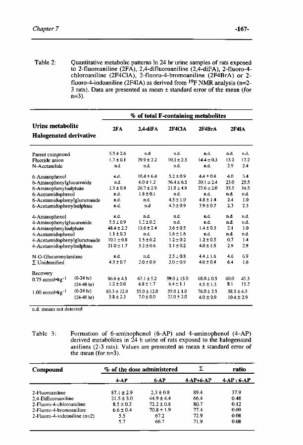
Chapter 7 -167-
Table 2: Quantitative metabolic patterns in 24 hr urine samples of rats exposed to 2-fluoroaniline (2FA), 2,4-difluoroaniline (2,4-diFA), 2-fluoro-4-chloroaniline (2F4C1A), 2-fluoro-4-bromoaniline (2F4BrA) or 2-fluoro-4-iodoaniline (2F4IA) as derived from 19F NMR analysis (n=2-3 rats). Data are presented as mean ± standard error of the mean (for n=3).
Urine metabolite
Halogenated derivative
Parent compound Fluoride anion N-Acetanilide
6-Aminophenol 6-Aminophenylglucuronide 6-Aminophenylsulphate 6-Acetamidophenol 6-Acetamidophenylglucuronide 6-Acetamidophenylsulphate
4-Aminophenol 4-Aminophenylglucuronide 4-Aminophenylsulphate 4-Acetamidophenol 4-Acetamidophenylglucuronide 4-Acetamidophenylsulphate
N-O-Glucuronylaniline X Unidentified
Recovery O ^ m m o l - k g - 1 (0-24 hr)
(24-48 hr)
l .OOmmol'kg-1 (0-24 hr) (24-48 hr)
2FA
5.5 ±2.6
1.7 ±0.1 n.d
n.d. n.d
2.3 + 0.8 n.d. n.d. n.d.
n.d. 5.5 ±0.9 48.4 ± 2.2 1.1 ±0.3 10.1 ±0.8 21.0± 1.7
n.d. 4.5 ±0.7
96.6 ±4.5 1.2 ±0.0
83.3 ± 12.9 5.8 ±2.3
% of total F
2,4-diFA
n.d
29.9 ± 2.2 n.d.
10.4 ± 6.4 6.0 ±1.2 26.7 ± 2.9 1.8 + 0.1
n.d. n.d
n.d. 1.2 + 0.2 13.6 ±2.4
n.d. 1.5 ±0.2 5.2 ±0.6
n.d. 2.0 ±0.9
67.1 ±5.2 6.8 ±1.7
55.0 ±12.0 7.0 ±0.0
-containing
2F4C1A
n.d.
10.3 ±2.5 n.d.
5.2 ±0.9 36.4 ±6.5 21.8 ±4.9
n.d. 4.5 ±1.0 4.3 ±0.9
n.d. n.d.
3.6 ±0.5 1.611.6 1.210.2 2.110.2
2.510.8 2.0 1 0.9
59.0115.0 6.411.1
55.011.0 22.012.0
metabolites
2F4BrA
n.d.
14.410.3 n.d.
4 .410.8 30.112.4 27.612.0
n.d. 4 .811.4 3.910.7
n.d. n.d.
1.410.3 n.d.
1.210.5 4 .011.6
4 .411.6 4 .010.4
68.010.5 4 .511.3
76.013.5 4.0 ± 0.9
2F4IA
n.d.
13.2 2.9
4.0 23.0 35.5 n.d. 2.4 2.3
n.d. n.d. 2.1 n.d. 0.7 2.9
4.6 6.4
60.0 8.1
n.d.
17.2 2.4
3.4 25.5 34.5 n.d. 1.0 2.3
n.d. n.d. 1.0 n.d. 1.4 2.8
6.9 1.6
45.3 15.2
58.514.5 10.412.9
n.d. means not detected
Table 3: Formation of 6-aminophenol (6-AP) and 4-aminophenol (4-AP) derived metabolites in 24 h urine of rats exposed to the halogenated anilines (2-3 rats). Values are presented as mean ± standard error of the mean (for n=3).
Compound
2-Fluoroaniline 2,4-Difluoroaniline 2-Fluoro-4-chloroaniline 2-Fluoro-4-bromoaniline 2-Fluoro-4-iodoaniline (n=2)
% of the dose administered
4-AP
87 . 112 . 9 2 1 . 5 1 3 . 0 8 . 510 . 3 6 . 6 1 0 . 4
5.5 5.7
6-AP
2 . 3 1 0 . 8 4 4 . 914 . 4 7 2 . 210 . 8 7 0 . 811 . 9
67.2 66.7
Z
4-AP+6-AP
89.4 66.4 80.7 77.4 72.9 71.9
ratio
4-AP : 6-AP
37.9 0.48 0.12 0.09 0.08 0.08
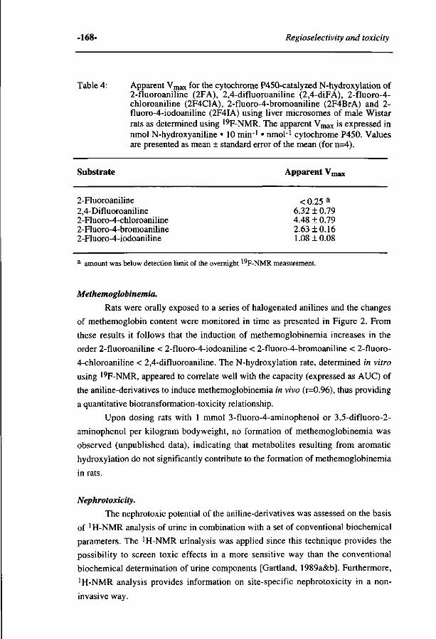
-168- Regioselectivity and toxicity
Table 4: Apparent Vmax for the cytochrome P450-catalyzed N-hydroxylation of 2-fluoroaniline (2FA), 2,4-difluoroaniline (2,4-diFA), 2-fluoro-4-chloroaniline (2F4C1A), 2-fluoro-4-bromoaniline (2F4BrA) and 2-fluoro-4-iodoaniline (2F4IA) using liver microsomes of male Wistar rats as determined using 19F-NMR. The apparent Vmax is expressed in nmol N-hydroxyaniline • 10 min-1 • nmoH cytochrome P450. Values are presented as mean ± standard error of the mean (for n=4).
Substrate Apparent Vmax
2-Fluoroaniline < 0.25 a
2,4-Difluoroaniline 6.32 ± 0.79 2-Fluoro-4-chloroaniline 4.48 ± 0.79 2-Fluoro-4-bromoaniline 2.63 + 0.16 2-Fluoro-4-iodoaniline 1.08 ± 0.08
a amount was below detection limit of the overnight ^F-NMR measurement.
Methemoglobinemia.
Rats were orally exposed to a series of halogenated anilines and the changes
of methemoglobin content were monitored in time as presented in Figure 2. From
these results it follows that the induction of methemoglobinemia increases in the
order 2-fluoroaniline < 2-fluoro-4-iodoaniline < 2-fluoro-4-bromoaniline < 2-fluoro-
4-chloroaniline < 2,4-difluoroaniline. The N-hydroxylation rate, determined in vitro
using 19F-NMR, appeared to correlate well with the capacity (expressed as AUC) of
the aniline-derivatives to induce methemoglobinemia in vivo (r=0.96), thus providing
a quantitative biotransformation-toxicity relationship.
Upon dosing rats with 1 mmol 3-fluoro-4-aminophenol or 3,5-difluoro-2-
aminophenol per kilogram bodyweight, no formation of methemoglobinemia was
observed (unpublished data), indicating that metabolites resulting from aromatic
hydroxylation do not significantly contribute to the formation of methemoglobinemia
in rats.
Nephrotoxicity.
The nephrotoxic potential of the aniline-derivatives was assessed on the basis
of 'H-NMR analysis of urine in combination with a set of conventional biochemical
parameters. The iH-NMR urinalysis was applied since this technique provides the
possibility to screen toxic effects in a more sensitive way than the conventional
biochemical determination of urine components [Gartland, 1989a&b]. Furthermore,
'H-NMR analysis provides information on site-specific nephrotoxicity in a non
invasive way.
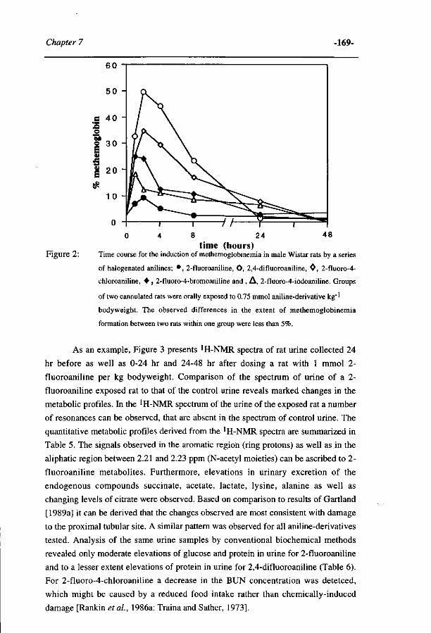
Chapter 7 -169-
T 0 4 8 24
time (hours) Figure 2: Time course for the induction of methemoglobinemia in male Wistar rats by a series
of halogenated anilines; • , 2-fluoroaniline, O, 2,4-difluoroaniline, 0 , 2-fluoro-4-
chloroaniline, • , 2-fluoro-4-bromoaniline and , A , 2-fluoro-4-iodoaniline. Groups
of two cannulated rats were orally exposed to 0.75 mmol aniline-derivative kg"1
bodyweight. The observed differences in the extent of methemoglobinemia
formation between two rats within one group were less than 5%.
As an example, Figure 3 presents 'H-NMR spectra of rat urine collected 24
hr before as well as 0-24 hr and 24-48 hr after dosing a rat with 1 mmol 2-
fluoroaniline per kg bodyweight. Comparison of the spectrum of urine of a 2-
fluoroaniline exposed rat to that of the control urine reveals marked changes in the
metabolic profiles. In the 'H-NMR spectrum of the urine of the exposed rat a number
of resonances can be observed, that are absent in the spectrum of control urine. The
quantitative metabolic profiles derived from the 1 H-NMR spectra are summarized in
Table 5. The signals observed in the aromatic region (ring protons) as well as in the
aliphatic region between 2.21 and 2.23 ppm (N-acetyl moieties) can be ascribed to 2-
fluoroaniline metabolites. Furthermore, elevations in urinary excretion of the
endogenous compounds succinate, acetate, lactate, lysine, alanine as well as
changing levels of citrate were observed. Based on comparison to results of Gartland
[1989a] it can be derived that the changes observed are most consistent with damage
to the proximal tubular site. A similar pattern was observed for all aniline-derivatives
tested. Analysis of the same urine samples by conventional biochemical methods
revealed only moderate elevations of glucose and protein in urine for 2-fluoroaniline
and to a lesser extent elevations of protein in urine for 2,4-difluoroaniline (Table 6).
For 2-fluoro-4-chloroaniline a decrease in the BUN concentration was detetced,
which might be caused by a reduced food intake rather than chemically-induced
damage [Rankin et al, 1986a; Traina and Sather, 1973].
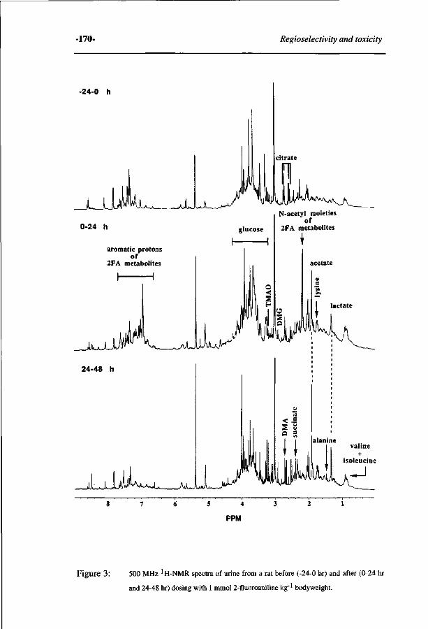
-170- Regioselectivity and toxicity
-24-0 h
JiJjF^Lv-*___jjJ-J j~» y H
citrate
0-24 h
aromatic protons of
2FA metabolites
N-acetyl moieties of
2FA metabolites
acetate
__JA_AJJ
24-48 h
-JLJJJI^ S 4
PPM
Figure 3 : 500 MHz 'H-NMR spectra of urine from a rat before (-24-0 hr) and after (0-24 hr
and 24-48 hr) dosing with 1 mmol 2-fluoroaniline kg"1 body weight.
Chapter 7 -171-
The urine volume (data not shown) was not significantly decreased upon haloaniline
administration, possibly due to the mild nephrotoxic potential of the tested anilines.
Although a mild nephrotoxic effect for the 2-fluoro-4-chloroaniline, 2-fluoro-4-
bromoaniline and 2-fluoro-4-iodoaniline treated animals was observed, the changes
in excretion of endogenous compounds succinate, acetate, lactate, alanine (Table 5)
and the changes of some conventional biochemical parameters (Table 6), were
demonstrated not to be significant. The rats exposed to 2-fluoroaniline and 2,4-
difluoroaniline showed the greatest and significant nephrotoxicity. The excretion of
glucose (0-24 hr), protein (0-24 hr & 24-48 hr), lactate (0-24 hr), succinate (24-48 hr)
and alanine (24-48 hr) in the 2,4-difluoroaniline treated group was significant lower
than observed for the 2-fluoroaniline treated group. Based on these results it was
concluded that the order of decreasing nephrotoxicity was 2-fluoroaniline > 2,4-
difluoroaniline > 2-fluoro-4-chloro-, 2-fluoro-4-bromo- and 2-fluoro-4-iodoaniline
for which no statistically significant nephrotoxic effect was observed.
DISCUSSION.
In the present study the relationship between biotransformation and toxicity
of a series of C4-substituted 2-fluoroanilines was investigated. Although it has been
suggested that aniline-derivatives require bioactivation to either N-hydroxy- or
aminophenol-derivatives to exert their toxic effects [Eyer, 1983; Rankin et ai, 1994;
Rietjens et ai, 1990; Sabbioni, 1992], a combined study on a full metabolite profile
and the actual toxic endpoint of a series of aniline derivatives has not been reported
so far.
The results of the present study support, as expected, that the main
metabolites involved in formation of methemoglobinemia are the N-hydroxyaniline
metabolites. It was even demonstrated that the in vitro rate of N-hydroxylation of the
series of C4-substituted 2-fluoroanilines correlates quantitatively (r=0.96) with the
extent of methemoglobinemia observed upon in vivo exposure of rats.
Besides methemoglobinemia, the series of C4-substituted 2-fluoroanilines
exerted nephrotoxicity as another toxic endpoint. Conventional biochemical
parameters in combination with !H-NMR urinalysis as a sensitive method to detect
kidney damage were used to investigate the nephrotoxicity in rats exposed to these
aniline-derivatives. The results from the conventional parameters and 'H-NMR
urinalysis were most consistent with damage at the tubular site as judged from the
increase in protein, succinate, acetate, lactate and alanine and fluctuation of citrate
[Gartland et ai, 1989a, Kuesel et ai, 1991]. These findings are in agreement with
studies on the nephrotoxicity of various aniline-derivatives reported previously by
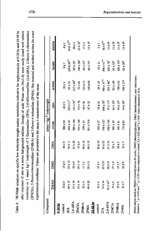
-172- Regioselectivity and toxicity
U ja oo t • s t C S
-a
I M
C S
Ó Is -&1
'5 'x o o u J3 O, ! U c u
cu
"3
CA
u *o
CS
u cu = 6
SP '53
U _cs 3 o "o S
o o
G 'u
1 )
CA
c o CA
' C A
>, 13 e 'C 3
X - >ö _a> S ca H
13
cu >
J 3
'•? •a eu CA O
T 3
> s
cd U i o CU U i <U
T t 1
II
CA
'S
5 « £ <0
13 S
1 - 1
o CA
O , 3
2 O
CA
cu
'S CS
T 3 CU
-t—» CS
e eu 6 0
13 J 3
CA <D
* u (D CA
CS
O
• ^
C A • 4 — 1
cS U i
<4_I
o
sa 3 CA O a. X CU
u C+M cS
CO
'5 CS
o u
2 CJ
4 S o 3
G C S
/—\
g •3 • *
r£ co
'S CS
2 o 3
•3 •4 c i ' \
C S
'S CS
2 o 3
c C S
<4-
o 2
'53 :* s-, o
- O
^ O
S S
N — |
u. O
'S*
1 o o
• 4 — *
ccS X
co Ui
,o C+M
C
O
CU CA
o
'S E
T 3
e es
13 *C CU
'S S CU CU
C/D
^ <
PP.
CU
_c 'S
CS O
T 3
Ó u O 3
G C S
T 3 a C S
< t - i
ffl
CU
1 O
E o u X )
4 o U i O 3
e C S
< 0 PP C S
s CU
E co
Xj <4-i O U i O
fc CU
-o CS
- O
e CS
CA
+1 S CU
E CU
X I
T 3 OJ -4—* C
2
2 a CA
CU
3
13 >
CA
S
e o ü
13 e CU
E u CU
X CU
O
o o.
o U
— +1
en
+1
Tf-
+1 o
o +1
CN
+1 <N +1
_ +1
+1 o
^ ra ra
CO CN
+1 CM
O OO
+1 p -
+1 CN
O +1
+1 o
! 1
+1 ON CN
m +1 • *
-er CM ^o
ON S gj ON S ^ +l +1 +1 + l +1 +1 2 NO CM f ; o oo •* m NO m ^ m
S o ; +l +1 +1
,-! "
+l +l ND co
<N „ +1 +1
+1 CN
' ^ Ô O en •* +1 +1 +1 +1 +1 ^ en cN -^ en
— — oo
+1 O N
CN
+1 O
CN
en +1 m O N
m +1 g
CN
+1 OO NO
e n
+1 NO NO
-* +1 ON
"3-
+1 O N C N
[ ^
+1 CN
CN
+1 ON
+1 O N
+1
cN ._;
NO
+1 NO
+1 O N
— ND
en ^ "O es -H +1 ON Q
— en ^"^
en +1 TI
NO
O
+1 00 "O
00 •«t
+1 Ti
en es +1 ON
CS
ON
+1 CS
i<n
+1 o •*
CS
+1
r»
o en
+1 VN
ON
+1 CS
en
• »
+1 en
O +1 O
CS CN
+1 en
es es +1 —•
en +1 en
en +1 o
< p-
'•B i
P. PU £ O CS CS CS CS CS
o ti c o o
+1 CN
+1 P~
+1 CN CN
+1
FA
,4-d
iFA
< Ö •s i -Un
< J-H
T *
PH
< • v t
u. CS CS CS CS <N
o < 3 S S es
•a -a
<i a o ,_• V g es,
• = 4 2
U O
E v S Q-
- "S 0 ^ s g ; ° 01 ° -o o
£ -s (U ^
•o o S Ö 3 v S a. o* •a .. cd «
J0
<
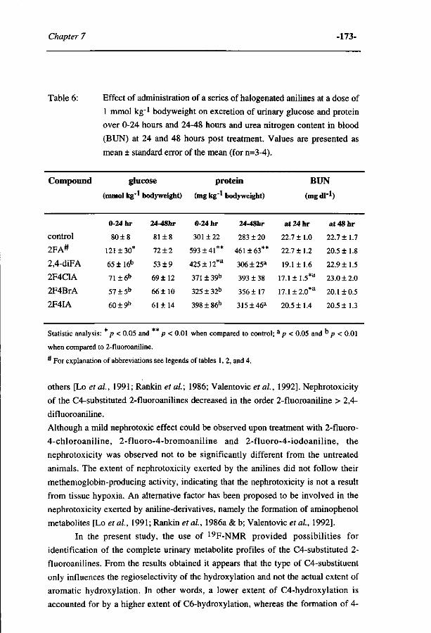
Chapter 7 -173-
Table 6: Effect of administration of a series of halogenated anilines at a dose of
1 mmol kg 1 bodyweight on excretion of urinary glucose and protein
over 0-24 hours and 24-48 hours and urea nitrogen content in blood
(BUN) at 24 and 48 hours post treatment. Values are presented as
mean ± standard error of the mean (for n=3-4).
Compound
control
2FA#
2,4-diFA
2F4C1A
2F4BrA
2F4IA
glucose
(mmol kg'!
0-24 hr
80±8
121 ±30*
65 ± 16b
71 ± 6 b
5 7 ± 5 b
6 0 ± 9 b
bodyweight)
24-48hr
81 + 8
72 + 2
53 ± 9
69 ±12
66 ±10
61 ±14
protein
(mg kg - 1
0-24 hr
301 ± 22
593 + 41**
425 + 12*a
371±39b
325 ± 32b
398 ± 86b
>odyweight)
24-48hr
283 ± 20
461+63**
306±25 a
393 ± 38
356 ±17
315±46 a
BUN
(mgdl-1)
at24hr
22.7 ± 1.0
22.7+1.2
19.1 ±1.6
17.1 ± 1.5*a
17.1±2.0*a
20.5 ±1.4
at48hr
22.7+1.7
20.5 ±1.8
22.9 ±1.5
23.0 ± 2.0
20.1 ±0.5
20.5+1.3
Statistic analysis: p < 0.05 and p < 0.01 when compared to control; a p < 0.05 and D p < 0.01
when compared to 2-fluoroaniline.
* For explanation of abbreviations see legends of tables 1, 2, and 4.
others [Lo et al., 1991; Rankin et al.; 1986; Valentovicef al., 1992]. Nephrotoxicity
of the C4-substituted 2-fluoroanilines decreased in the order 2-fluoroaniline > 2,4-
difluoroaniline.
Although a mild nephrotoxic effect could be observed upon treatment with 2-fluoro-
4-chloroaniline, 2-fluoro-4-bromoaniline and 2-fluoro-4-iodoaniline, the
nephrotoxicity was observed not to be significantly different from the untreated
animals. The extent of nephrotoxicity exerted by the anilines did not follow their
methemoglobin-producing activity, indicating that the nephrotoxicity is not a result
from tissue hypoxia. An alternative factor has been proposed to be involved in the
nephrotoxicity exerted by aniline-derivatives, namely the formation of aminophenol
metabolites [Lo etal, 1991; Rankin et al, 1986a & b; Valentovic et al, 1992].
In the present study, the use of 19F-NMR provided possibilities for
identification of the complete urinary metabolite profiles of the C4-substituted 2-
fluoroanilines. From the results obtained it appears that the type of C4-substituent
only influences the regioselectivity of the hydroxylation and not the actual extent of
aromatic hydroxylation. In other words, a lower extent of C4-hydroxylation is
accounted for by a higher extent of C6-hydroxylation, whereas the formation of 4-
-174- Regioselectivity and toxicity
plus 6-aminophenol derived metabolites is similar for the different aniline-
derivatives tested. The presence of a C4-substituent hampers cytochrome P450-
catalyzed oxidation at this position and increases possibilities for C6-hydroxylation
and also for N-hydroxylation. This observation is in accordance with results
previously obtained for series of C4-fluorinated aniline-derivatives [Cnubben et al.;
1995]. Comparison of the extent of nephrotoxicity to the extent of formation of
respectively the 4-aminophenol and/or 6-aminophenol metabolites, indicates a
predominant role for the C4-hydroxylation route, not the C6-hydroxylation route, in
the induction of nephrotoxic effects. These findings are in agreement with the
observation that derivatives of 6-aminophenol are generally less nephrotoxic than 4-
aminophenol derived isomers [Calder etal., 1975].
It is especially of interest to notice that the 4-aminophenol pathway leading to
nephrotoxicity may proceed through oxidation of the 4-aminophenol to the 4-
benzoquinoneimine. Glutathione conjugation of this reactive electrophilic
quinoneimine and transport of the conjugate to the kidney, ultimately leads to
degradation of the conjugate to a toxic intermediate as is also seen with other
nephrotoxic quinone glutathione conjugates [Monks et al, 1985; Monks et al, 1991;
Monks and Lau, 1992]. Previous results for C4-halogenated anilines even
demonstrated that the reactive quinoneimine metabolite can be formed as the primary
metabolite upon the cytochrome P450-catalyzed oxidative dehalogenation of a C4-
halogenated aniline such as the C4-halogenated 2-fluoroanilines of the present study.
The pathway is schematically presented in Figure 4 [Cnubben et al, 1995; Rietjens
et al, 1990; Rietjens et al 1991]. Previous in vitro results reported the preferential
removal of a fluorine over a chlorine, bromine or iodine substituent in such a
cytochrome P450-catalyzed oxidative dehalogenation [Cnubben etal, 1995], and
this is clearly in line with the in vivo results of the present study indicating a larger
extent of C4 oxidation, and nephrotoxicity, for the C4-fluorinated 2-fluoroaniline
than for the C4-chlorinated, -brominated or -iodinated derivative.
Additional results of the present study demonstrate that 2,4-difluoroaniline,
but also 2-fluoroaniline induces marked nephrotoxicity, whereas the amount of 4-
aminophenol metabolites formed from 2,4-difluoroaniline is several-fold lower than
the amount of 4-aminophenol derived metabolites observed in the metabolite pattern
of 2-fluoroaniline [Table 3]. Presumably, this discrepancy can be understood on the
basis of a direct formation of the fluorinated 1,4-benzoquinoneimine from 2,4-
difluoroaniline by the alternative and effective pathway depicted in Figure 4.
Biotransformation of 2,4-difluoroaniline results in an amount of fluoride anions that
is always significantly higher than the amount that could be accounted for by the
formation of 4-aminophenol-derived metabolites. Previously, this phenomenon was
also observed for other 4-fluorinated aniline derivatives [Rietjens and Vervoort,
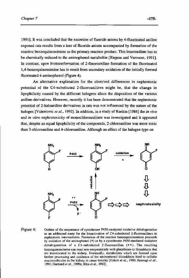
Chapter 7 -175-
1991]. It was concluded that the excretion of fluoride anions by 4-fluorinated aniline
exposed rats results from a loss of fluoride anions accompanied by formation of the
reactive benzoquinoneimine as the primary reaction product. This intermediate has to
be chemically reduced to the aminophenol-metabolite [Riejens and Vervoort, 1991].
In contrast, upon biotransformation of 2-fluoroaniline formation of the fluorinated
1,4-benzoquinoneimine has to result from secondary oxidation of the initially formed
fluorinated 4-aminophenol (Figure 4).
An alternative explanation for the observed differences in nephrotoxic
potential of the C4-substituted 2-fluoroanilines might be, that the change in
lipophilicity caused by the different halogens alters the disposition of the various
aniline-derivatives. However, recently it has been demonstrated that the nephrotoxic
potential of 2-haloaniline derivatives in rats was not influenced by the nature of the
halogen [Valentovic et al, 1992]. In addition, in a study of Rankin [1986] the in vivo
and in vitro nephrotoxicity of monochloroanilines was investigated and it appeared
that, despite an equal lipophilicity of the compounds, 2-chloroaniline was more toxic
than 3-chloroaniline and 4-chloroaniline. Although an effect of the halogen type on
P450
P4S0 -r
oxidation
4
ft T [ Z ^ C ^ C ^ nephrotoxicity
Figure 4 : Outline of the importance of cytochrome P450-catalyzed oxidative dehalogenation as an additional route for the bioactivation of C4-substituted 2-fluoroanilines to nephrotoxic intermediates. Formation of the reactive benzoquinoneimine proceeds by oxidation of the aminophenol (*) or by a cytochrome P450-mediated oxidative dehalogenation of a C4-substituted 2-fluoroaniline (**) . The resulting benzoquinoneimine can react non-enzymatically with glutathione to thioadducts that are translocated to the kidney. Eventually, metabolites which are formed upon further processing and oxidation of the aminophenol thioadducts bind to cellular macromolecules in the kidney to cause toxicity [Eckert et al, 1990; Eyanagi et al, 1991; Gartland étal, 1990a; Klos étal, 1992].
-176- Regioselectivity and toxicity
the disposition cannot be excluded in our study, results of these other studies indicate
that not the lipophilicity, but other factors, such as biotransformation must contribute
to the nephrotoxic potential of these compounds. The results of the present study
provide support for the importance of the cytochrome P450-catalyzed C4-
hydroxylation and oxidative dehalogenation of C4-substituted 2-fluoroanilines as a
bioactivation pathway to nephrotoxic metabolites. Clearly, this provides an
additional perspective to the present views on bioactivation pathways for aniline
derivatives.
Morphological studies indicating the exact location of the renal lesion upon aniline-
exposure may further elucidate whether the lesion is at the corticomedullary junction
as this would further strengthen that 4-aminophenol metabolites contribute to the
nephrotoxicity mechanism. Such morphological studies were beyond the scope of the
present study but serve as an interesting topic for future research.
Altogether, it is concluded that reduced possibilities for C4-hydroxylation in the
series of aniline-derivatives, results in increased possibilities for C6- and N-
hydroxylation and, consequently in a shift in the type of toxic end-point observed,
i.e. from nephrotoxicity to methemoglobinemia.
Acknowledgements. This study was supported by the Netherlands Organization for the
Advancement of Scientific Research (N.W.O.). The authors thank Ms. M.A.W.
Peters for cannulation of the animals, Dr. J. Vervoort for help with !H-NMR
measurements and Mr. P.W.J. Heinen and Mr. J. Bogaards for statistical analysis of
the data.
REFERENCES.
Anthony M.L., Sweatman B.C., Beddell CR., Lindon J.C. and Nicholson J.K. (1994). Pattern
recognition classification of the site of nephrotoxicity based on metabolic data derived from
proton nuclear magnetic resonance spectra of urine, Molec. Pharmacol. 46, 199-211.
Bales J.R., Higham D.P., Howe I., Nicholson J.K. and Sadler P.J. (1984). Use of high-resolution
proton nuclear magnetic resonance spectroscopy for multi-component analysis of urine, Clin.
Chem. 30, 426-432.
Blaauboer B.J., Van Holsteijn C.W.M. and Wit J.G. (1976). Ferrihemoglobin formation by
monohydroxy aniline derivatives in erythrocytes of some avian species in comparison with
mammals. Naunyn-Schmiedeberg's Arch. Pharmacol. 292, 255-258.
Boyland E. and Sims P. (1954). The oxidation of some aromatic amines with persulphate, J. Chem.
Soc, 980-985.
Chapter 7 -177-
Calder I.C., Williams P.J., Woods R.A., Funder C.C, Green CR., Ham K.N. and Tange J.D. (1975).
Nephrotoxicity and molecular structure, Xenobiotica 5, 303-307.
Calder I.C., Yong A.C., Woods R.A., Crowe CA., Ham K.N. and Tange J.D. (1979). The
nephrotoxicity of p-aminophenol. II The effect of metabolic inhibitors and inducers, Chem.-
Biol. Interact. 27, 245-254.
Cnubben N.H.P., Blaauboer B., Juyn S., Vervoort J. and Rietjens I.M.C.M. (1994). A
spectrophotometric assay for the detection of 2-aminophenols in biological samples,
Analytical Biochemistry 220, 165-171.
Cnubben N.H.P., Vervoort J., Boersma M.G. and Rietjens M.C.M. (1995). The effect of varying
halogen substituent patterns on the cytochrome P450 catalysed dehalogenation of 4-
halogenated anilines to 4-aminophenol metabolites, Biochemical Pharmacology 49, 1235-
1248.
Cnubben N.H.P., Vervoort J., Veeger C. and Rietjens M.C.M. (1992). Study on the regioselectivity
and mechanism of the aromatic hydroxylation of monofluoroanilines, Chem.-Biol. Interact.
85, 151-172.
Coleman G.H., McCloskey Ch.M. and Stuart F.A. (1943). Nitrosobenzene, Organic Syntheses 25, 81-
83.
Eadsforth C.V., Logan C.J., Morrison B.J. and Warburton P.A. (1984). 2,4-difluoroaniline and 4-
fluoroaniline exposure: monitoring by methaemoglobin and urine analyses, Int. Arch.
Occup. Environ. Health 54, 223-232.
Eckert K.G. and Eyer P. (1983). Differences in the reactions of isomeric ortho- and para-
aminophenols with hemoglobin, Biochemical Pharmacology 32, 1019-1027.
Eyanagi R., Hisanari Y. and Shigematsu H. (1991). Studies of paracetamol/phenacetin toxicity:
isolation and characterization of p-aminophenol-glutathione conjugate, Xenobiotica 21, 793-
803.
Eyer P. (1988). Detoxication of N-oxygenated arylamines in erythrocytes. An overview, Xenobiotica
18, 1327-1333.
Eyer P. (1983). The red cell as a sensitive target for activated toxic arylamines. Arch. Toxicol, Suppl.
6,3-12.
Fowler L.M., Foster J.R. and Lock E.A. (1993). Effect of ascorbic acid, acivicin and probenecid on
the nephrotoxicity of 4-aminophenol in the Fischer 344 rat, Arch. Toxicol. 67, 613-621.
Fowler L.M., Foster J.R. and Lock E.A. (1994). Nephrotoxicity of 4-amino-3-S-glutathionylphenol
and its modulation by metabolism or transport inhibitors, Arch. Toxicol. 68, 15-23.
Fowler L.M., Moore R.B., Foster J.R. and Lock E.A. (1991). Nephrotoxicity of 4-aminophenol
glutathione conjugate, Human & Experimental Toxicology 10, 451-459.
Gartland K.P.R., Bonner F.W. and Nicholson J.K. (1989a). Investigations into the biochemical effect
of region-specific nephrotoxins, Molec. Pharmacol. 35, 242-250.
Gartland K.P.R., Bonner F.W., Timbrell J.A. and Nicholson J.K. (1989b). Biochemical
characterisation of para-aminophenol-induced nephrotoxic lesions in the F344 rat, Arch.
-178- Regie/selectivity and toxicity
Toxicol. 63,97-106.
Gartland K.P.R., Eason C.T., Bonner F.W. and Nicholson J.K. (1990a). Effects of biliary cannulation
and buthionine sulphoximine pretreatment on the nephrotoxicity of para-aminophenol in
the Fischer 344 rat, Arch. Toxicol. 64, 14-25.
Gartland K.P.R., Beddell CR., Lindon J.C. and Nicholson J.K. (1990b) A pattern recognition
approach to the comparison of PMR and clinical chemical data for classification of
nephrotoxicity, J. Pharmaceutical & Biomedical Analysis 8, 963-968.
Harrison J.H. and Jollow D.J. (1987) Contribution of aniline metabolites to aniline-induced
methemoglobinemia, Molec. Pharmacol. 32, 423-431.
Iles R.A., Hind A.J. and Chalmers R.A. (1985). Use of proton nuclear magnetic resonance
spectroscopy in detection and study of organic acidurias, Clin. Chem. 31, 1795-1801.
Josephy P.D., Eling T.E. and Mason R.P. (1983). Oxidation of p-aminophenol catalysed by
horseradish peroxidase and prostaglandin synthase, Molec. Pharmacol. 23, 461-466.
Kennedy R.J. and Stock A.M. (1960). The oxidation of organic substances by potassium
peroxymonosulphate, J. Org. Chem. 25, 1901-1906.
Kiese M. (1974). Methemoglobinemia: A comprehensive treatise. Chapter 7, CRC Press, Cleveland,
Ohio.
Klos C, Koob M., Kramer C. and Dekant W. (1992). p-Aminophenol nephrotoxicity: Biosynthesis of
toxic glutathione conjugates, Toxicol. Appl. Pharmacol. 115, 98-106.
Kuesel A.C., Kroft T. and Smith I.C.P. (1991). Nuclear magnetic resonance spectroscopy, Clin.
Chem. 63, 237-246
Lenk W. and Riedl M. (1989). N-hydroxy-N-arylacetamides. V. Differences in the mechanism of
haemoglobin oxidation in vitro by N-hydroxy-4-chlroacetaniline and N-hydroxy-4-
chloroaniline. Xenobiotica 19, 453-475.
Lo H.H., Brown P.I., Rankin G.O. (1990). Acute nephrotoxicity induced by isomeric dichloroanilines
in Fischer 344 rats, Toxicology 63, 215-231.
Lo H.H., Brown P.I. and Rankin G.O. (1991). Trichloroaniline effects on renal function in vivo and in
vitro, Toxicology Letters 57, 319-328.
Lock E.A., Cross T.J. and Schnellmann R.G. (1993). Studies on the mechanism of 4-aminophenol-
induced toxicity to renal proximal tubules, Human & Experimental Toxicology 12, 383-388.
Lowry O.H., Rosebrough N.J. Farr A.L. and Randall A.J. (1951). Protein measurements with Folin
phenol reagent. J. Biol. Chem. 193, 265-275.
Maples K.R., Eyer P. and Mason R.P. (1989). Aniline-, phenylhydroxylamine-, nitrosobenzene-, and
nitrobenzene-induced hemoglobin thiyl free radical formation in vivo and in vitro, Molec.
Pharmacol.il, 311-318.
McLean S., Starmer G.A. and Thomas J. (1969). Methaemoglobin formation by aromatic amines, J.
Pharm. Pharmac. 2, 441-450.
Monks T.J., Jones T.W., Hill B.A. and Lau S.S. (1991). Nephrotoxicity of 2-bromo-
(cysteinyl)hydroquinone and 2-bromo-(N-acetyl-L-cysteinyl) hydroquinone thioethers,
Chapter 7 -179-
Toxicol. Appl. Pharmacol. I l l , 279-298.
Monks T.J. and Lau S.S. (1992). Toxicology of quinone-thioethers, Crit. Rev. Toxicol. 22, 243-270.
Monks T.J., Lau S.S., Highet R.J. and Gillette J.R. (1985). Glutathione conjugates of 2-
bromohydroquinone are nephrotoxic, Drug Metab. Dispos. 13, 553-559.
Omura T. and Sato R. (1964). The carbon monoxide-binding pigment of liver microsomes. I.
Evidence for its hemoprotein nature. J. Biol. Chem. 239, 2370-2378.
Rankin G.O., Valentovic M.A., Nicoll D.W., Ball J.G., Anestis D.K., Wang R.-T., Brown P.I. (1994),
In vivo and in vitro 4-amino-2,6-dichlorophenol nephrotoxicity and hepatotoxicity in the
Fischer 344 rat. Toxicology 90, 115-128.
Rankin G.O., Yang D.J., Cressey-Veneziano K., Casto S., Wang R.T. and Brown P.I. (1986a). In vivo
and in vitro nephrotoxicity of aniline and its monochlorophenylderivatives in the Fischer 344
rat, Toxicology 38, 269-283.
Rankin CO. , Yang D.J., Teets V.J., Lo H.H. and Brown P.I. (1986b). 3,5-Dichloroaniline-induced
nephrotoxicity in the Sprague-Dawley rat, Toxicology Letters 30, 173-179.
Remie R., Van Dongen J.J. and Rensema J.W. (1990). Manual of microsurgery on the laboratory rat,
Part I. (J.J. Van Dongen, R. Remie, J.W. Rensema and G.H.J. Van Wunnik, Eds.) pp. 159-
169, Elsevier, Amsterdam.
Rietjens I.M.C.M., Tyrakowska B., Veeger C. and Vervoort J. (1990). Reaction pathways for
biodehalogenation of fluorinated anilines, Eur. J. Biochem. 194, 945-954.
Rietjens I .M.CM. and Vervoort J. (1991). Bioactivation of 4-fluorinated anilines to
benzoquinoneimines as primary reaction products, Chem.-Biol. Interactions 77, 263-281.
Rietjens I.M.C.M. and Vervoort J. (1989). Microsomal metabolism of fluoroanilines, Xenobiotica. 19,
1297-1305.
Sabbioni G. (1992). Hemoglobin binding of monocyclic aromatic amines: molecular dosimetry and
quantitative structure activity relationships for the N-oxidation, Chem.-Biol. Interactions 81,
91-117.
Tietz N.W. (1987). Fundamentals of clinical chemistry, 3 r d ed. W.B. Saunders Co. Philadelphia.
Traina V.M. and Sather B.T. (1973). Effect of starvation on renal function. Lancet 2, 620.
Valentovic M.A., Ball J.G., Anestis D.K., Beers K.W., Madan E„ Hubbard J.L. and Rankin G. (1992).
Acute renal and hepatic toxicity of 2-haloanilines in Fischer 344 rats, Toxicology 75, 121-
131.
Vervoort J., De Jager P.A., Steenbergen J. and Rietjens I.M.C.M. (1990). Development of a 1°F-
n.m.r. method for studies on the in vivo and in vitro metabolism of 2-fluoroaniline,
Xenobiotica 20, 657-670.
Vogel A.J. (1978). Vogel's textbook of practical organic chemistry. (B.S. Furniss, A.J. Hannaford,
P.W.C Smith and A.R. Fatchell, Eds.) p. 684, Longman Scientific & Technical, London.
Vogel A.J. (1989). Vogel's textbook of practical organic chemistry. (B.S. Furniss, A.J. Hannaford,
P.W.C Smith and A.R. Fatchell, Eds.) John Wiley & Sons, New York.
Yoshida M., Yoshikawa H., Goto H. and Hara I., Evaluation of the nephrotoxicity of aromatic nitro-
-180- Regioselectivity and toxicity
amino compounds by urinary enzyme activities, The Journal of Toxicological Sciences
(1989) 14, 257-268.
CHAPTER 8
Different metabolic pathways of 2,5-difluoronitrobenzene and 2,5-
difluoroaminobenzene compared to molecular orbital substrate
characteristics.
Ivonne M.C.M. Rietjens, Nicole H.P. Cnubben, Marjon van Haandel, Bozena
Tyrakowska, Ans E.M.F. Soffers and Jacques Vervoort.
Chemico-Biological Interactions 94, 49-72, 1995.
SUMMARY
The in vivo metabolite patterns of 2,5-difluoroaminobenzene and of its
nitrobenzene analogue, 2,5-difluoronitrobenzene, were determined using ^F-NMR
analysis of urine samples. Results obtained demonstrate significant differences
between the biotransformation patterns of these two analogues. For the
aminobenzene, cytochrome P450-catalyzed aromatic hydroxylation presents the main
metabolic pathway. 2,5-Difluoronitrobenzene was predominantly metabolized
through glutathione conjugation leading to excretion of 5-fluoro-2-(N-
acetylcysteinyl)-nitrobenzene and fluoride anions, and, to a minor extent, through
cytochrome P450-catalyzed hydroxylation and nitroreduction. Pretreatment of the
rats with various inducers of cytochrome P450 enzymes, known to influence
glutathione S-transferase enzyme patterns as well, followed by exposure to the 2,5-
difluoroamino- or 2,5-difluoronitrobenzene, generally resulted in metabolite patterns
that varied only to a small (< 12%) extent. Based on this results it was concluded
that the biotransformation enzyme pattern is not the predominant factor in
determining the metabolic route of these two model compounds. Additional in vitro
-182- Different metabolic pathways
microsomal and cytosolic incubations with 2,5-difluoroaminobenzene and 2,5-
difluoronitrobenzene qualitatively confirmed the in vivo results. NADPH/oxygen-
supported microsomal cytochrome P450-catalyzed hydroxylation was observed only
for 2,5-difluoroaminobenzene, whereas cytosolic GSH conjugation occurred only in
incubations with 2,5-difluoronitrobenzene as the substrate. Outcomes from molecular
orbital calculations provided a working hypothesis that can explain the difference in
metabolic pathways of the nitro- and amino benzene derivative on the basis of their
chemical characteristics. This hypothesis states that the chances for a nitro- or
aminobenzene derivative to enter either a cytochrome P450 or a glutathione
conjugation pathway are determined by the relative energy levels of the frontier
orbitals of the compounds. The aminobenzene derivative has relatively high energy
molecular orbitals leading to an efficient reaction of its highest occupied molecular
orbital (HOMO) with the singly occupied molecular orbital of the cytochrome P450
(FeO)3+ intermediate, but a low reactivity of its lowest unoccupied molecular orbital
(LUMO) with the HOMO of glutathione. The nitrobenzene, on the other hand, has
molecular orbitals of relatively low energy, explaining the efficient interaction, and,
thus, reaction between its LUMO and the HOMO electrons of glutathione, but
resulting in low reactivity with the SOMO electron of the cytochrome P450 (FeO)3+
reaction intermediate.
INTRODUCTION.
Aromatic xenobiotics like substituted benzenes, are generally converted to
more polar metabolites before they can be excreted from the mammalian body.
Reactions available for this conversion are so-called phase I or modification reactions
and phase II or conjugation reactions. The present paper focusses on the marked
difference in biotransformation pathways known to occur upon exposure to
nitrobenzenes on one hand and to their chemically reduced analogues aminobenzenes
(anilines). Previous papers reported that the biotransformation of aminobenzene
derivatives proceeds predominantly through cytochrome P450-catalyzed aromatic
hydroxylation, accompanied by N-acetylation, glucuronidation and/or sulphation (1-
6). In contrast, however, the metabolic pathways of nitrobenzenes have been reported
to proceed only to a minor extent through cytochrome P450-catalyzed aromatic ring
hydroxylation. For nitrobenzenes, nitroreduction leading to aminobenzene
derivatives and their glutathione conjugation leading to mercapturic acids have been
reported to be important metabolic pathways (7-13).
The objective of the present study was to obtain more insight in these
differences in metabolic pathways for nitro- and aminobenzene derivatives.
Chapter 8 -183-
Model compounds used were 2,5-difluoronitrobenzene and 2,5-
difluoroaminobenzene. The in vivo and in vitro metabolite patterns of these
analogues were investigated to quantify the differences between the metabolic
pathways of the two benzene derivatives. Molecular orbital calculations were
performed to find a rational for possible differences observed.
MATERIALS AND METHODS.
Chemicals
2,5-Difluoroaminobenzene and 2,5-difluoronitrobenzene were purchased
from Fluorochem (Derbyshire, UK). 5-Fluoro-2-(N-acetylcysteinyl)-nitrobenzene, 5-
fluoro-2-cysteinyl-nitrobenzene and 5-fluoro-2-glutathionyl-nitrobenzene were
synthesized from 2,5-difluoronitrobenzene as follows: to 0.3 g (1.9 mmol) 2,5-
difluoronitrobenzene dissolved in 2.5 ml methanol, 2.5 ml of a solution containing
0.1 g (1.9 mmol) sodium methanolate and 0.5 mmol N-acety ley steine, cysteine or
reduced glutathione dissolved in methanol, were added over a period of 30 minutes.
The mixture was stirred at room temperature overnight. Upon cooling to 4°C and
centrifugation (10 minutes 13000 rpm in an Eppendorf centrifuge), the supernatant
was mixed with one volume of demineralized water and one volume of 0.2 M
potassium phosphate pH 7.6. Following a second centrifugation step, resulting in a
2,5-difluoronitrobenzene containing pellet, the supernatant was washed three times
with diethylether to remove the final 2,5-difluoronitrobenzene. The water phase was
evaporated under vacuum and the residue, containing the 5-fluoro-2-(N-
acetylcysteinyl)-nitrobenzene, the 5-fluoro-2-cysteinyl-nitrobenzene or the 5-fluoro-
2-glutathionyl-nitrobenzene, was dissolved in demineralized water, and analyzed by 19F-NMR in 0.1 M potassium phosphate pH 7.6, or by !H-NMR, upon lyophilisation
and dissolving in 2H2Û. Spectral characteristics of the compounds were as follows;
5-fluoro-2-(N-acetylcysteinyl)-nitrobenzene: 19F-NMR (0.1 M potassium phosphate
pH 7.6, relative to CFCl3)(ppm): -119.1; •H-NMR (2H20, relative to sodium 3-
trimethylsilyl-(2,2,3,3-2H4)-propionate)(ppm): 8.01 (H6)(dd, 37H-F = 8.6 Hz, 4/H-H =
2.9 Hz), 7.78 (H3) (dd, 47H-F = 5.2 Hz, 3/H-H = 9.2 Hz), 7.57 (H4)(m, 3JH-F = 7.7
Hz, 3/H-H = 9.2 Hz, 4 / H -H = 2-9 H z ) . 4 - 4 4 (CH)(dd, 37H-H = 9.0 Hz, 37H-H = 4.1 Hz),
3.69(lHofCH2)(dd, 27H-H= 14.1 Hz, 3/H-H = 4.1 Hz), 3.32 (1H of CH2)(dd, 2/H-
H= 14.1 Hz, 37H-H= 9.0 Hz), 1.95 (CH3)(s); 5-Fluoro-2-cysteinyl-nitrobenzene: 19F-
NMR (0.1 M potassium phosphate pH 7.6, relative to CFCl3)(ppm): -118.5; !H-
NMR (2H2Û, relative to sodium 3-trimethylsilyl-(2,2,3,3-2H4)-propionate)(ppm):
8.07 (H6)(dd, 3/H-F = 8.7 Hz, 47H-H = 2.9 Hz), 7.74 (H3) (dd, 4/H-F = 5.1 Hz, 37H-H
= 9.1 Hz), 7.57 (H4)(m, 3/H-F = 7.7 Hz, 37H-H = 9.1 Hz, 4/H-H = 2.9 Hz), 3.61
-184- Different metabolic pathways
(CH)(dd, 3yH.H = 7.5 Hz, 3/H-H = 4.9 Hz), 3.49 (1H of CH2)(dd, 2/H-H = 13.3 Hz, 3^H-H = 4.9 Hz), 3.28 (1H of CH2)(dd, 2JH-H = 13.3 Hz, 3/H-H = 7.5 Hz); 5-fluoro-2-
glutathionyl-nitrobenzene: 19F-NMR (0.1 M potassium phosphate pH 7.6, relative to
CFCl3)(ppm): -118.5; !H-NMR (2H20, relative to sodium 3-trimethylsilyl-(2,2,3,3-2H4)-propionate)(ppm): 8.02 (H6)(dd, 3 7 H -F = 8.6 Hz, 4-/H-H = 2-8 Hz)> 7 - 8 0 (H3)
(dd, 47H-F = 5.0 Hz, VH-H = 9.1 Hz), 7.58 (H4)(m, 3/H-F = 7.6 Hz, 37H-H = 9.1 Hz,
4/H-H = 2.8 Hz), 4.65 (CH)(dd, 37H-H= 9.3 Hz, 37H-H= 4.6 Hz), 3.72 (1H of
CH2)(dd, 27H-H = 14.5 Hz, 37H-H = 4.6 Hz), 3.37 (1H of CH2)(dd, 2/H-H = 14.5 Hz, 3JR-U = 9.3 Hz), 3.72 (1 H of CH2)(br.s), 3.74 (1H of CH2)(br.s), 3.61 (CH)(tr, 3/H_
H = 6.4 Hz), 2.04 (m), 2.44 (CH2)(tr, 37H-H= 7.8 Hz).
2,5-Difluoro-N-hydroxyaminobenzene was synthesized by the procedure
described by Coleman et al. (14) for the synthesis of phenylhydroxylamine from
nitrobenzene. In short, a mixture of 0.9 g of NH4CI and 2.33 g (14.6 mmol) of 2,5-
difluoronitrobenzene in 30 ml of water was stirred vigorously and 2.23 g (30.9
mmol) of zinc powder (Merck, Darmstadt, FRG) was added in small portions over a
period of 5 minutes. Following the addition of the zinc, the temperature of the
reaction mixture increased spontaneously to 50-55°C after approximately 5 minutes.
The mixture was maintained at 50-55°C for 15 minutes, adding ice as necessary. The
solution was then filtered and the zinc oxide residue washed with 30 ml of hot water.
The filtrate was cooled immediately to 0°C, to avoid decomposition of the
hydroxylamine. A sample of the reaction mixture was analyzed by 19F-NMR in 0.1
M potassium phosphate pH 7.6 to identify the 19F-NMR resonances of the 2,5-
difluoro-N-hydroxyaminobenzene. 19F-NMR (0.1 M potassium phosphate pH 7.6,
relative to CFCl3)(ppm): -122.0 (F5), -142.0 (F2).
2,5-Difluoronitrosobenzene was synthesized by oxidation of 2,5-
difluoroaminobenzene with potassium peroxymonosulphate in a water-acetic acid
mixture, as described by Kennedy and Stock (15) for the synthesis of 1,4-
dinitrosobenzene from 1,4-diaminobenzene. 2,5-Difluoronitrosobenzene was purified
by preparative column chromatography on Silicagel 60 (0.015-0.040 mm)(Merck,
Darmstadt, FRG) with 5 % (v/v) ether in petroleum benzine as the eluent. The green
2,5-difluoronitrosobenzene containing fractions were collected and concentrated by
evaporation and a sample of this fraction was analyzed by 19F-NMR in 0.1 M
potassium phosphate pH 7.6 to identify the 19F-NMR resonances of the compound. 19F-NMR (0.1 M potassium phosphate pH 7.6, relative to CFCl3)(ppm): -122.4 (F5),
-138.0 (F2).
In vivo exposure of rats
Male Wistar rats (400 g) were exposed to 20 mg of the desired fluorinated
benzene, administered in olive oil by oral injection. After dosing, 24 h urine samples
Chapter 8 -185-
were collected. The 24 h urinary recovery was over 65 % of the dose administered
for the 2,5-difluoronitrobenzene and over 95 % for the 2,5-difluoroaminobenzene.
Pretreatment of the rats with inducers of cytochrome P450 enzymes before
administration of the 2,5-difluoroaminobenzene or the 2,5-difluoronitrobenzene was
carried out as previously described (16). Inducers used were phénobarbital, ß-
naftoflavone (used instead of the carcinogenic 3-methylcholanthrene, administered in
a similar way), isosafrole (l,2-(methylenedioxy)-4-propenylbenzene) and acetone.
Some of these inducers of cytochrome P450 enzymes influence the enzyme pattern
of the glutathione S-transferases as well (17).
Analysis of urine samples
Urine samples were analyzed by 19F-NMR after 1:1 dilution in 0.2 M
potassium phosphate pH 7.6. Enzyme hydrolysis of urine samples was carried out as
previously described (5) using either ß-glucuronidase from Escherichia coli K12
(Boehringer, Mannheim, Germany) or arylsulphatase/ß-glucuronidase from Helix
pomatia (Boehringer, Mannheim, Germany). Samples were routinely made oxygen
free by four cycles of evacuation and filling with argon.
For determination of !H-NMR and 19F-NMR splitting patterns of the
metabolite with its 19F-NMR resonances at -121.7 and -145.1 ppm, 4 ml of an
arylsulphatase/ß-glucuronidase treated urine sample was diluted with 2 ml of 0.2 M
potassium phosphate pH 7.6 and extracted three times with 2 ml of ethyl acetate. The
collected ethyl acetate fractions were evaporated to dryness and dissolved in 0.5 ml
CDCI3, followed by evaporation to dryness and dissolving in 0.5 ml CDCI3. This
sample contained the metabolite to be identified and was analyzed by iH-NMR and 19F-NMR as described below.
19F-NMR measurements. 19F-NMR measurements were performed on a Bruker AMX 300
spectrometer as described previously (5;16-18). Between 1500 and 50000 scans were
recorded, depending on the concentrations of the fluorine containing compounds and
the signal to noise ratio required. The detection limit of these measurements was 1
uM for an overnight run (50000 scans). The sample volume was 1.71 ml containing
100 |il 2H2Û for locking the magnetic field and 10 (il of a 8.4 mM 4-fluorobenzoic
acid solution, added as an internal standard. The ethyl acetate extract from the
tert.butylhydroperoxide supported microsomal incubation was analyzed using a
coaxial insert in the 10 mm NMR tube. The sample consisted of 1.6 ml of the ethyl
acetate extract. The coaxial insert contained 2H20 for locking the magnetic field and
a known amount of the internal standard (4-fluorobenzoic acid) for quantification of
the measurement. Concentrations of the various metabolites could be calculated by
-186- Different metabolic pathways
comparison of the integrals of the 19F-NMR resonances of the metabolites to the
integral of the 19F-NMR resonance of 4-fluorobenzoic acid.
The splitting pattern of the 19F-NMR resonances of the metabolite identified
as 2,5-difluoro-4-hydroxynitrobenzene was measured on a Bruker AMX 500 MHz
using a lH probehead tuned to the 19F frequency (470.5 MHz). The sample volume
was 0.5 ml and the solvent CDCI3 was used for locking the magnetic field. Because
of a smaller and thus more homogeneous sample as well as the higher magnetic field,
resolution in these 19F-NMR measurements on the Bruker AMX 500 MHz were
improved over that obtained at the AMX 300 spectrometer, resulting in well resolved
splitting patterns and 7-values that could be compared to those of the JH-NMR
signals of this metabolite.
JH-NMR measurements.
!H NMR measurements were performed on a Bruker AMX 500 spectrometer.
Upon freeze drying, the synthesized compounds were dissolved in 2H2Û to give a
final sample volume of 0.5 ml. Spectra were recorded using 60° pulses (6 us), a 10
kHz spectral width, a repetition time of 1.9 s, quadrature phase detection and
quadrature phase cycling (CYCLOPS). About 150 scans were recorded. lH chemical
shift values are presented relative to sodium 3-trimethylsilyl-(2,2,3,3-2H4)-
propionate. The splitting pattern of the ÏH-NMR resonances of the metabolite
identified as 2,5-difluoro-4-hydroxynitrobenzene was also measured on a Bruker
AMX 500 MHz. The sample was dissolved in CDCI3 and this solvent was used for
locking the magnetic field.
Preparation of microsomes and cytosol.
Microsomes were prepared from the perfused livers of male Wistar rats (400
g) either untreated or treated with isosafrole as described before (16). Cytosol was
obtained as the supernatant from the first 105000 g ultracentrifugation step
performed during preparation of microsomes from control rats. Cytochrome P450
content of the microsomes was measured as described by Omura and Sato (20).
Protein content was determined by the method of Lowry et al. (21) using bovine
serum albumin as the standard.
Microsomal incubations.
Cytochrome P450 dependent conversion was studied in vitro in microsomal
incubations containing (final concentrations) 0.1 M potassium phosphate pH 7.6, 2
uM microsomal cytochrome P450, 5 mM of the 2,5-difluoroamino- or 2,5-
difluoronitrobenzene added as 1% (v/v) of a 0.5 M stock solution in dimethyl
sulphoxide and 1.0 mM NADPH. The reaction was started by the addition of the
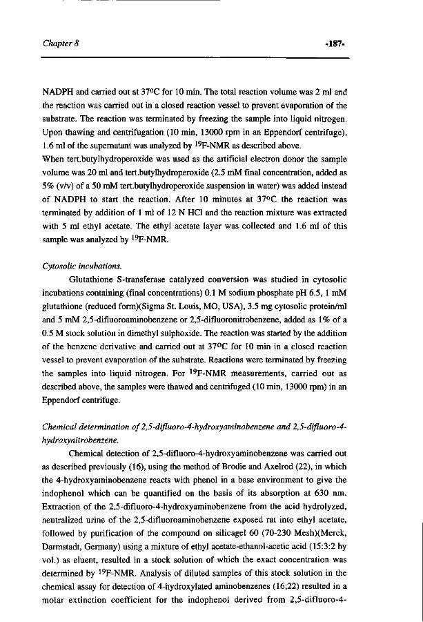
Chapter 8 -187-
NADPH and carried out at 37°C for 10 min. The total reaction volume was 2 ml and
the reaction was carried out in a closed reaction vessel to prevent evaporation of the
substrate. The reaction was terminated by freezing the sample into liquid nitrogen.
Upon thawing and centrifugation (10 min, 13000 rpm in an Eppendorf centrifuge),
1.6 ml of the supernatant was analyzed by 19F-NMR as described above.
When tert.butylhydroperoxide was used as the artificial electron donor the sample
volume was 20 ml and tert.butylhydroperoxide (2.5 mM final concentration, added as
5% (v/v) of a 50 mM tert.butylhydroperoxide suspension in water) was added instead
of NADPH to start the reaction. After 10 minutes at 37°C the reaction was
terminated by addition of 1 ml of 12 N HCl and the reaction mixture was extracted
with 5 ml ethyl acetate. The ethyl acetate layer was collected and 1.6 ml of this
sample was analyzed by 19F-NMR.
Cytosolic incubations.
Glutathione S-transferase catalyzed conversion was studied in cytosolic
incubations containing (final concentrations) 0.1 M sodium phosphate pH 6.5, 1 mM
glutathione (reduced form)(Sigma St. Louis, MO, USA), 3.5 mg cytosolic protein/ml
and 5 mM 2,5-difluoroaminobenzene or 2,5-difluoronitrobenzene, added as 1% of a
0.5 M stock solution in dimethyl sulphoxide. The reaction was started by the addition
of the benzene derivative and carried out at 37°C for 10 min in a closed reaction
vessel to prevent evaporation of the substrate. Reactions were terminated by freezing
the samples into liquid nitrogen. For 19F-NMR measurements, carried out as
described above, the samples were thawed and centrifuged (10 min, 13000 rpm) in an
Eppendorf centrifuge.
Chemical determination of2,5-difluoro-4-hydroxyaminobenzene and 2,5-difluoro-4-
hydroxynitrobenzene.
Chemical detection of 2,5-difluoro-4-hydroxyaminobenzene was carried out
as described previously (16), using the method of Brodie and Axelrod (22), in which
the 4-hydroxyaminobenzene reacts with phenol in a base environment to give the
indophenol which can be quantified on the basis of its absorption at 630 nm.
Extraction of the 2,5-difluoro-4-hydroxyaminobenzene from the acid hydrolyzed,
neutralized urine of the 2,5-difluoroaminobenzene exposed rat into ethyl acetate,
followed by purification of the compound on silicagel 60 (70-230 Mesh)(Merck,
Darmstadt, Germany) using a mixture of ethyl acetate-ethanol-acetic acid (15:3:2 by
vol.) as eluent, resulted in a stock solution of which the exact concentration was
determined by 19F-NMR. Analysis of diluted samples of this stock solution in the
chemical assay for detection of 4-hydroxylated aminobenzenes (16;22) resulted in a
molar extinction coefficient for the indophenol derived from 2,5-difluoro-4-

-188- Different metabolic pathways
hydroxyaminobenzene of 20 mM^cnr1.
In the urine of a 2,5-difluoronitrobenzene exposed rat the presence of 2,5-
difluoro-4-hydroxynitrobenzene was detected essentially as described by Piotrowski
(9). In short, 0.5 ml of urine was diluted with 0.5 ml of 0.1 M sodium acetate pH 5.2
and treated with arylsulphatase/ß-glucuronidase as described previously (5). Half (0.5
ml) of this sample was extracted 2 times with 0.5 ml ethyl acetate. The collected
ethyl acetate extracts were combined and diluted 1:10 in 4 N HCl. This diluted
sample was divided in two parts. To 1.5 ml of one of these samples 0.25 g of zinc
powder (Merck, Darmstadt, FRG) was added to reduce the 2,5-difluoro-4-
hydroxynitrobenzene to 2,5-difluoro-4-hydroxaminobenzene. The 1.5 ml of the other
sample served as a control sample which was analyzed without the zinc reduction
step, leading to quantification of the excreted 2,5-difluoro-4-hydroxyaminobenzene.
The ultimate difference between the two samples quantifies the actual amount of 2,5-
difluoro-4-hydroxynitrobenzene in the urine sample. After 5 minutes of vigorous
shaking the samples were centrifuged (5 min, 2500 rpm) and to 1.2 ml of each
supernatant 0.8 ml of demineralised water was added. To 1.0 ml of this sample 63 ul
of phenol reagent (5% w/v, phenol in water) and 650 ul of cone. NH3 (25%) were
added. After 60 minutes at room temperature the absorbance at 630 nm was
determined, against a blank consisting of the urine of a non-exposed rat treated in a
similar way. On the basis of the A630 nm values thus obtained, concentrations of the
hydroxylated metabolites in the urine were calculated using the extinction coefficient
determined for the indophenol derived from 2,5-difluoro-4-hydroxyaminobenzene to
be 20 mM"1 cm"1.
Molecular orbital calculations.
Molecular orbital calculations were carried out on a Silicon Graphics Iris
4D/85 using Quanta/Charmm (Molecular Simulations, UK). The semi-empirical
molecular orbital method was used, applying the AMI Hamiltonian from the
AMP AC program. All calculations were carried out with PRECISE criteria. For all
calculations the self-consistent field was achieved. Geometries were optimized for all
bond lengths, bond angles and torsion angles using the Fletcher-Powell criteria.
Frontier electron densities were calculated from HOMO (highest occupied
molecular orbital) and HOMO-1 characteristics and from LUMO (lowest unoccupied
molecular orbital) and LUMO+1 characteristics using the equation given by Fukui et
al. (23). In this study, the outcomes of the semi-empirical calculations on molecules
in vacuum are related to the electronic characteristics of the substrates in the active
site of the cytochromes P450. Due to solvation effects and a different dielectric
constant, the intrinsic properties of the compounds might be influenced upon binding
to this active site. However, it is assumed that this phenomenon will not influence the
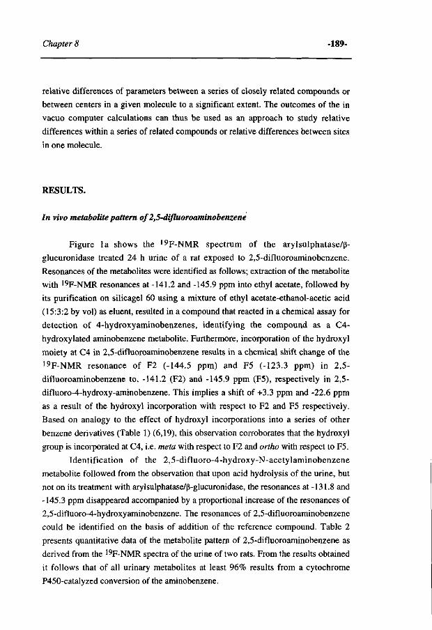
Chapter 8 -189-
relative differences of parameters between a series of closely related compounds or
between centers in a given molecule to a significant extent. The outcomes of the in
vacuo computer calculations can thus be used as an approach to study relative
differences within a series of related compounds or relative differences between sites
in one molecule.
RESULTS.
In vivo metabolite pattern of 2,5-difluoroaminobenzene
Figure la shows the 19F-NMR spectrum of the arylsulphatase/ß-
glucuronidase treated 24 h urine of a rat exposed to 2,5-difluoroaminobenzene.
Resonances of the metabolites were identified as follows; extraction of the metabolite
with 19F-NMR resonances at -141.2 and -145.9 ppm into ethyl acetate, followed by
its purification on silicagel 60 using a mixture of ethyl acetate-ethanol-acetic acid
(15:3:2 by vol) as eluent, resulted in a compound that reacted in a chemical assay for
detection of 4-hydroxyaminobenzenes, identifying the compound as a C4-
hydroxylated aminobenzene metabolite. Furthermore, incorporation of the hydroxyl
moiety at C4 in 2,5-difluoroaminobenzene results in a chemical shift change of the 19F-NMR resonance of F2 (-144.5 ppm) and F5 (-123.3 ppm) in 2,5-
difluoroaminobenzene to, -141.2 (F2) and -145.9 ppm (F5), respectively in 2,5-
difluoro-4-hydroxy-aminobenzene. This implies a shift of +3.3 ppm and -22.6 ppm
as a result of the hydroxyl incorporation with respect to F2 and F5 respectively.
Based on analogy to the effect of hydroxyl incorporations into a series of other
benzene derivatives (Table 1) (6,19), this observation corroborates that the hydroxyl
group is incorporated at C4, i.e. meta with respect to F2 and ortho with respect to F5.
Identification of the 2,5-difluoro-4-hydroxy-N-acetylaminobenzene
metabolite followed from the observation that upon acid hydrolysis of the urine, but
not on its treatment with arylsulphatase/ß-glucuronidase, the resonances at -131.8 and
-145.3 ppm disappeared accompanied by a proportional increase of the resonances of
2,5-difluoro-4-hydroxyaminobenzene. The resonances of 2,5-difluoroaminobenzene
could be identified on the basis of addition of the reference compound. Table 2
presents quantitative data of the metabolite pattern of 2,5-difluoroaminobenzene as
derived from the 19F-NMR spectra of the urine of two rats. From the results obtained
it follows that of all urinary metabolites at least 96% results from a cytochrome
P450-catalyzed conversion of the aminobenzene.
-190- Different metabolic pathways
o o
N H C - C H , NHj NHC-C
F-
IS
I i 1 Ul NO:
&
IS
I . I. . I M ,1.1
N H j
NO,
" T 1 1 1 1 1
-110 -130 -150 PPM
Figure 1: ^F-NMR spectra of the arylsulphatase/ß-glucuronidase treated 24 h urine of a Wistar rat exposed to (a) 2,5-difluoroaminobenzene and (b) 2,5-difluoronitrobenzene. Resonances were identified as described in the text. The resonance marked IS is from the internal standard 4-fluorobenzoic acid. The fluoride anions observed in (a) originate from the food, as a similar amount of fluoride anions was observed in the urine of a non exposed control rat. The SR adduct presents 5-fluoro-2-(N-acetylcysteinyl)-nitrobenzene.
In vivo metabolite pattern of 2,5-difluoronitrobenzene.
The 19F-NMR spectrum of the arylsulphatase/ß-glucuronidase treated 24 h
urine of a rat exposed to 2,5-difluoronitrobenzene is presented in Figure lb.
Metabolites identical to those observed in Figure la, and, thus, representing
aminobenzene derived metabolites are observed.
The main metabolite observed in the urine of the 2,5-difluoronitrobenzene
exposed rat, however, could be identified as 5-fluoro-2-(N-acetylcysteinyl)-
nitrobenzene because addition of the synthesized reference compound resulted in an
increase of this resonance. The glutathione and cysteine adduct of the above-
mentioned compound have resonances at slightly, but markedly, different positions,
again showing that the resonance at -119.1 ppm belongs to 5-fluoro-2-(N-
acetylcysteinyl)-nitrobenzene. In addition to this mercapturic acid metabolite, an
almost equal amount of fluoride anion is observed in the urine (Figure lb) resulting
from the defluorination which accompanies the glutathione addition at C2.
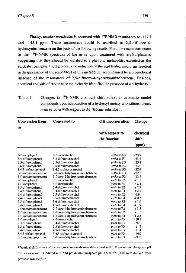
Chapter 8 -191-
Finally, another metabolite is observed with 19F-NMR resonances at -121.7
and -145.1 ppm. These resonances could be ascribed to 2,5-difluoro-4-
hydroxynitrobenzene on the basis of the following results. First, the resonances occur
in the 19F-NMR spectrum of the urine upon treatment with arylsulphatase,
suggesting that they should be ascribed to a phenolic metabolite, excreted as the
sulphate conjugate. Furthermore, zinc reduction of the acid hydrolysed urine resulted
in disappearance of the resonances of this metabolite, accompanied by a proportional
increase of the resonances of 2,5-difluoro-4-hydroxyaminobenzene. Besides,
chemical analysis of the urine sample clearly identified the presence of a 4-hydroxy-
Table 1: Changes in 19F-NMR chemical shift values in aromatic model
compounds upon introduction of a hydroxyl moiety at positions, ortho,
meta or para with respect to the fluorine substituent.
Conversion from Converted to OH incorporation Change
3-fluorophenol 3,4-difluorophenol 3,5-difluorophenol 3,6-difluorophenol 3,4,5-trifluorophenol 3-fluoroaminobenzene 3-fluoroaminobenzene 2-fluorophenol 4-fluorophenol 2,3-difluorophenol 3,4-difluorophenol 2,4-difluorophenol 2,4-difluorophenol 2,5-difluorophenol 3,4-difluorophenol 2-fluoroaminobenzene 2-fluoroaminobenzene 4-fluoroaminobenzene 3-fluorophenol 2,3-difluorophenol 3,5-difluorophenol 3,4-difluorophenol 3,4,5-trifluorophenol 3-fluoroaminobenzene
3-fluorocatechol 3,4-difluorocatechol 3,5-difluorocatechol 3,6-difluorocatechol 3,4,5-trifluorocatechol 3-fluoro-4-hydroxyaminobenzene 3-fluoro-2-hydroxyaminobenzene 3-fluorocatechol 4-fluorocatechol 3,4-difluorocatechol 3,4-difluorocatechol 3,5-difluorocatechol 3,5-difluorocatechol 3,6-difluorocatechol 4,5-difluorocatechol 2-fluoro-4-hydroxyaminobenzene 2-fluoro-6-hydroxyaminobenzene 4-fluoro-2-hydroxyaminobenzene 4-fluorocatechol 3,4-difluorocatechol 3,5-difluorocatechol 4,5-difluorocatechol 3,4,5-trifluorocatechol 3-fluoro-6-hydroxyaminobenzene
with respect to the fluorine
ortho to F3 ortho to F3 ortho to F3 ortho to F3 ortho to F3 ortho to F3 ortho to F3 meta to F2 meta to F4 meta to F2 meta to F4 meta to F2 meta to F4 meta to F2 meta to F4 meta to F2 meta to F2 meta to F4 para to F3 para to F3 para to F3 para to F3 para to F3 para to F3
chemical shift
(ppm)
-23.9 -23.1 -23.4 -23.0 -22.4 -22.5 -23.7 + 1.5 + 2.4 + 2.9 + 2.1 -0.6 + 0.3 + 1.9 + 1.9 + 3.5 + 1.5 + 2.5 -10.2 -9.3 -11.2 -11.4 -14.0 -9.9
Chemical shift values of the various compounds were determined in 0.1 M potassium phosphate pH
7.6, or in urine 1:1 diluted in 0.2 M potassium phosphate pH 7.6 at 7°C, and were derived from
previous results (6,18).
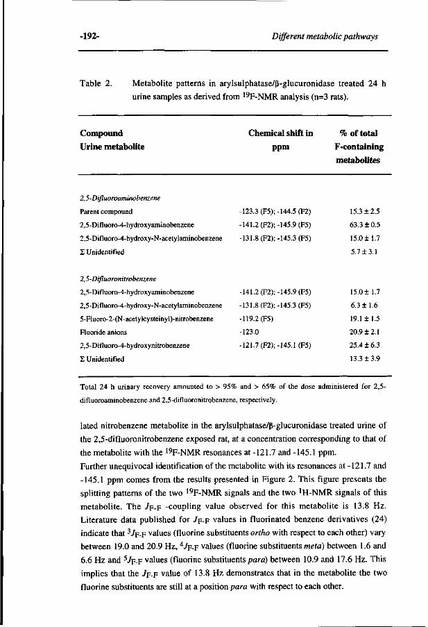
-192- Different metabolic pathways
Table 2. Metabolite patterns in arylsulphatase/ß-glucuronidase treated 24 h
urine samples as derived from 19F-NMR analysis (n=3 rats).
Compound Urine metabolite
Chemical shift in
ppm
% of total
F-containing
metabolites
2,5-Difluoroaminobenzene
Parent compound
2,5-Difluoro-4-hydroxyaminobenzene
2,5-Difluoro-4-hydroxy-N-acetylaminobenzene
£ Unidentified
-123.3 (F5);-144.5 (F2)
-141.2 (Fl); -145.9 (F5)
-131.8 (F2);-145.3 (F5)
15.3 + 2.5
63.3 ± 0.5
15.0 ±1.7
5.7 ±3.1
2,5-Difluoronitrobenzene
2,5-Difluoro-4-hydroxyaminobenzene
2,5-Difluoro-4-hydroxy-N-acetylaminobenzene
5-Fluoro-2-(N-acetylcysteinyl)-nitrobenzene
Fluoride anions
2,5-Difluoro-4-hydroxynitrobenzene
Z Unidentified
141.2 (F2);
131.8 (F2);
119.2(F5)
123.0
121.7 (F2);
-145.9 (F5)
-145.3 (F5)
-145.1 (F5)
15.0+ 1.7
6.3 ±1.6
19.1 ±1.5
20.9 ±2.1
25.4 ±6.3
13.3 ±3.9
Total 24 h urinary recovery amounted to > 95% and > 65% of the dose administered for 2,5-
difluoroaminobenzene and 2,5-difluoronitrobenzene, respectively.
lated nitrobenzene metabolite in the arylsulphatase/ß-glucuronidase treated urine of
the 2,5-difluoronitrobenzene exposed rat, at a concentration corresponding to that of
the metabolite with the 19F-NMR resonances at -121.7 and -145.1 ppm.
Further unequivocal identification of the metabolite with its resonances at -121.7 and
-145.1 ppm comes from the results presented in Figure 2. This figure presents the
splitting patterns of the two 19F-NMR signals and the two JH-NMR signals of this
metabolite. The / F -F -coupling value observed for this metabolite is 13.8 Hz.
Literature data published for 7p-F values in fluorinated benzene derivatives (24)
indicate that 37F-F values (fluorine substituents ortho with respect to each other) vary
between 19.0 and 20.9 Hz, 47F-F values (fluorine substituents meta) between 1.6 and
6.6 Hz and 5 /F-F values (fluorine substituents para) between 10.9 and 17.6 Hz. This
implies that the 7F-F value of 13.8 Hz demonstrates that in the metabolite the two
fluorine substituents are still at a position para with respect to each other.
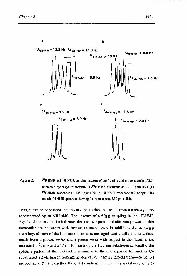
Chapter 8 -193-
^F(2)-F(5) = 13-8 HZ 3«/H(3)-F(2) = 11.6 Hz
• H<6)-F(2) = 6 .9 HZ ^H(3)-F(5) = 7 .0 HZ
3JH(6)-F(6) = 9.9 Hz
^H(6)-F(2) = 6 .9 Hz
•4<(3>-F(2) = 1 1.6 Hz
•4i(3)-F(S) = 7 .0 Hz
Figure 2: ^F-NMR and ^H-NMR splitting patterns of the fluorine and proton signals of 2,5-
difluoro-4-hydroxynitrobenzene. (a)l"F-NMR resonance at -121.7 ppm (F2), (b)
19F-NMR resonance at -145.1 ppm (F5), (c) 'H-NMR resonance at 7.92 ppm (H6)
and (d) ^H NMR spectrum showing the resonance at 6.90 ppm (H3).
Thus, it can be concluded that the metabolite does not result from a hydroxylation
accompanied by an NIH shift. The absence of a 47H-H coupling in the !H-NMR
signals of the metabolite indicates that the two proton substituents present in this
metabolite are not meta with respect to each other. In addition, the two 7H-F
couplings of each of the fluorine substituents are significantly different, and, thus,
result from a proton ortho and a proton meta with respect to the fluorine, i.e.
represent a 3 / H - F and a 47H-F f° r e a c n of t n e fluorine substituents. Finally, the
splitting pattern of this metabolite is similar to the one reported for another C4
substituted 2,5-difluoronitrobenzene derivative, namely 2,5-difluoro-4-S-methyl
nitrobenzene (25). Together these data indicate that, in this metabolite of 2,5-
-194- Different metabolic pathways
difluoronitrobenzene the modification is at C4, and demonstrates that this phenolic
metabolite with its resonances at -121.7 and -145.1 ppm must be 2,5-difluoro-4-
hydroxy nitrobenzene.
The metabolite pattern derived from the 19F-NMR spectra of urine samples of
two 2,5-difluoronitrobenzene exposed rats is summarized in Table 2. From these data
it follows that glutathione conjugation, represented by the sum of the mercapturic
acid and fluoride anion elimination (together being 40.0 ± 3.6 %), is the major
pathway for 2,5-difluoronitrobenzene metabolism.
Metabolite patterns in rats pre treated with inducers of biotransformation enzymes.
To investigate the extent to which the metabolite patterns observed in rats
exposed to 2,5-difluoroaniline and 2,5-difluoronitrobenzene would vary upon
changes in the pattern of biotransformation enzymes, rats were pretreated with
various inducers of the cytochrome P450 system. Some of these inducers are known
to influence the glutathione S-transferase enzyme pattern also (17). Upon
pretreatment, the rats were exposed to either 2,5-difluoroaminobenzene or 2,5-
difluoronitrobenzene. The collected 24 h urines were treated with arylsulphatase/ß-
glucuronidase and analyzed by 19F-NMR. The results obtained are presented in
Table 3. The rats pretreated with phénobarbital, ß-naftoflavone or acetone did not
show marked changes in the metabolite profiles of 2,5-difluoroaminobenzene or 2,5-
difluoronitrobenzene. Generally, the variations observed were only a few (< 12 %)
percent. The amount of metabolites that resulted from the glutathione conjugation
pathway (5-fluoro-2-(N-acetylcysteinyl)-nitrobenzene plus fluoride anions) is > 40 %
of all the fluorinated metabolites. For 2,5-difluoronitrobenzene, the amount of
metabolites resulting from glutathione conjugation is higher than the amount of 2,5-
difluoro-4-hydroxybenzene resulting from the cytochrome P450-catalyzed
hydroxylation of 2,5-difluoronitrobenzene, and also higher than the amount of 2,5-
difluoro-4-hydroxyaminobenzene plus 2,5-difluoro-4-hydroxy-N-acetylamino-
benzene resulting from nitroreduction followed by N-acetylation and/or cytochrome
P450-catalyzed hydroxylation. Together the results presented in Table 3 demonstrate
that the marked difference in the metabolic profile of the nitro- and the amino
analogue, observed in vivo in non-pretreated rats (table 2) is still observed when rats
are pretreated with inducers of the biotransformation enzymes. This implies that the
marked difference in the contribution of the various pathways to the overall
metabolic profile of these two model compounds cannot be ascribed to the pattern of
the biotransformation enzymes, but is more likely due to differences in chemical
characteristics of the amino and the nitro analogue. The results presented in Table 3
also demonstrate that, surprisingly, in the rats pretreated with isosafrole excretion of
the hydroxylated metabolites of 2,5-difluoroaminobenzene is no longer observed. In
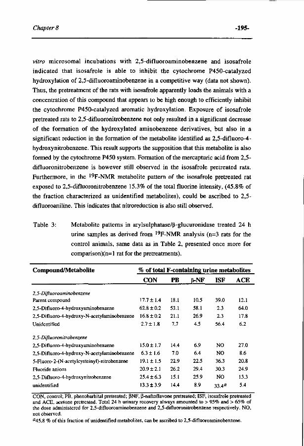
Chapter 8 -195-
vitro microsomal incubations with 2,5-difluoroaminobenzene and isosafrole
indicated that isosafrole is able to inhibit the cytochrome P450-catalyzed
hydroxylation of 2,5-difluoroaminobenzene in a competitive way (data not shown).
Thus, the pretreatment of the rats with isosafrole apparently loads the animals with a
concentration of this compound that appears to be high enough to efficiently inhibit
the cytochrome P450-catalyzed aromatic hydroxylation. Exposure of isosafrole
pretreated rats to 2,5-difluoronitrobenzene not only resulted in a significant decrease
of the formation of the hydroxylated aminobenzene derivatives, but also in a
significant reduction in the formation of the metabolite identified as 2,5-difluoro-4-
hydroxynitrobenzene. This result supports the supposition that this metabolite is also
formed by the cytochrome P450 system. Formation of the mercapturic acid from 2,5-
difluoronitrobenzene is however still observed in the isosafrole pretreated rats.
Furthermore, in the 19F-NMR metabolite pattern of the isosafrole pretreated rat
exposed to 2,5-difluoronitrobenzene 15.3% of the total fluorine intensity, (45.8% of
the fraction characterized as unidentified metabolites), could be ascribed to 2,5-
difluoroaniline. This indicates that nitroreduction is also still observed.
Table 3: Metabolite patterns in arylsulphatase/ß-glucuronidase treated 24 h
urine samples as derived from 19F-NMR analysis (n=3 rats for the
control animals, same data as in Table 2, presented once more for
comparison)(n=l rat for the pretreatments).
Compound/Metabolite
2,5-Difluoroaminobenzene Parent compound
2,5-Difluoro-4-hydroxyaminobenzene
2,5-Difluoro-4-hydroxy-N-acetylaminobenzene
Unidentified
2,5-Difluoronitrobenzene
2,5-Difluoro-4-hydroxyaminobenzene
2,5-Difluoro-4-hydroxy-N-acetylaminobenzene
5-Fluoro-2-(N-acetylcysteinyl)-nitrobenzene
Fluoride anions
2,5-Difluoro-4-hydroxynitrobenzene
unidentified
% of total F-containing urine metabolites
CON
17.7 ±1.4
62.8 ± 0.2
16.8 + 0.2
2.7+1.8
15.0 ±1.7
6.3 ±1.6
19.1 ±1.5
20.9 + 2.1
25.4 ± 6.3
13.3 ±3.9
PB
18.1
53.1
21.1
7.7
14.4
7.0
22.9
26.2
15.1
14.4
ß-NF
10.5
58.1
26.9
4.5
6.9
6.4
22.5
29.4
25.9
8.9
ISF
39.0
2.3
2.3
56.4
NO
NO
36.3
30.3
NO
33.4a
ACE
12.1
64.0
17.8
6.2
27.0
8.6
20.8
24.9
13.3
5.4
CON, control; PB, phénobarbital pretreated; ßNF, ß-naftoflavone pretreated; ISF, isosafrole pretreated and ACE, acetone pretreated. Total 24 h urinary recovery always amounted to > 95% and > 65% of the dose administered for 2,5-difluoroaminobenzene and 2,5-difluoronitrobenzene respectively. NO, not observed. a45.8 % of this fraction of unidentified metabolites, can be ascribed to 2,5-difluoroaminobenzene.
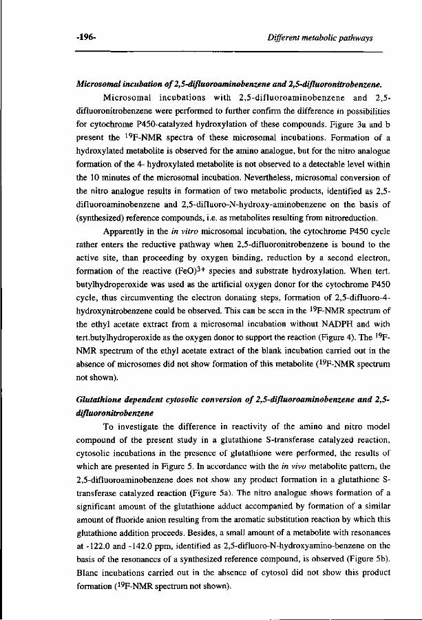
-196- Different metabolic pathways
Microsomal incubation of 2,5-difluoroaminobenzene and 2,5-difluoronitrobenzene.
Microsomal incubations with 2,5-difluoroaminobenzene and 2,5-
difluoronitrobenzene were performed to further confirm the difference in possibilities
for cytochrome P450-catalyzed hydroxylation of these compounds. Figure 3a and b
present the 19F-NMR spectra of these microsomal incubations. Formation of a
hydroxylated metabolite is observed for the amino analogue, but for the nitro analogue
formation of the 4- hydroxylated metabolite is not observed to a detectable level within
the 10 minutes of the microsomal incubation. Nevertheless, microsomal conversion of
the nitro analogue results in formation of two metabolic products, identified as 2,5-
difluoroaminobenzene and 2,5-difluoro-N-hydroxy-aminobenzene on the basis of
(synthesized) reference compounds, i.e. as metabolites resulting from nitroreduction.
Apparently in the in vitro microsomal incubation, the cytochrome P450 cycle
rather enters the reductive pathway when 2,5-difluoronitrobenzene is bound to the
active site, than proceeding by oxygen binding, reduction by a second electron,
formation of the reactive (FeO)3+ species and substrate hydroxylation. When tert.
butylhydroperoxide was used as the artificial oxygen donor for the cytochrome P450
cycle, thus circumventing the electron donating steps, formation of 2,5-difluoro-4-
hydroxynitrobenzene could be observed. This can be seen in the 19F-NMR spectrum of
the ethyl acetate extract from a microsomal incubation without NADPH and with
tert.butylhydroperoxide as the oxygen donor to support the reaction (Figure 4). The 19F-
NMR spectrum of the ethyl acetate extract of the blank incubation carried out in the
absence of microsomes did not show formation of this metabolite (19F-NMR spectrum
not shown).
Glutathione dependent cytosolic conversion of 2,5-difluoroaminobenzene and 2,5-
difluoronitrobenzene
To investigate the difference in reactivity of the amino and nitro model
compound of the present study in a glutathione S-transferase catalyzed reaction,
cytosolic incubations in the presence of glutathione were performed, the results of
which are presented in Figure 5. In accordance with the in vivo metabolite pattern, the
2,5-difluoroaminobenzene does not show any product formation in a glutathione S-
transferase catalyzed reaction (Figure 5a). The nitro analogue shows formation of a
significant amount of the glutathione adduct accompanied by formation of a similar
amount of fluoride anion resulting from the aromatic substitution reaction by which this
glutathione addition proceeds. Besides, a small amount of a metabolite with resonances
at -122.0 and -142.0 ppm, identified as 2,5-difluoro-N-hydroxyamino-benzene on the
basis of the resonances of a synthesized reference compound, is observed (Figure 5b).
Blanc incubations carried out in the absence of cytosol did not show this product
formation (19F-NMR spectrum not shown).
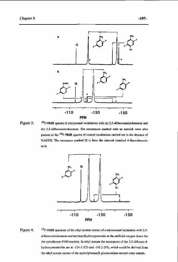
Chapter 8 -197-
IS
IS
to JLJL
-no -130 -150 PPM
Figure 3 : 1 "F -NMR spectra of microsomal incubations with (a) 2,5-difluoroaminobenzene and
(b) 2,5-difluoronitrobenzene. The resonances marked with an asterisk were also
present in the 1°F-NMR spectra of control incubations carried out in the absence of
NADPH. The resonance marked IS is from the internal standard 4-fluorobenzoic
acid.
T
NO2 IS
F
1
-110 -130 PPM
-150
Figure 4: ^F-NMR spectrum of the ethyl acetate extract of a microsomal incubation with 2,5-
difluoronitrobenzene and tert.butylhydroxyperoxide as the artificial oxygen donor for
the cytochrome P450 reaction. In ethyl acetate the resonances of the 2,5-difluoro-4-
hydroxymetabolite are at -124.1 (F2) and -143.2 (F5), which could be derived from
the ethyl acetate extract of the arylsulphatase/ß-glucuronidase treated urine sample.
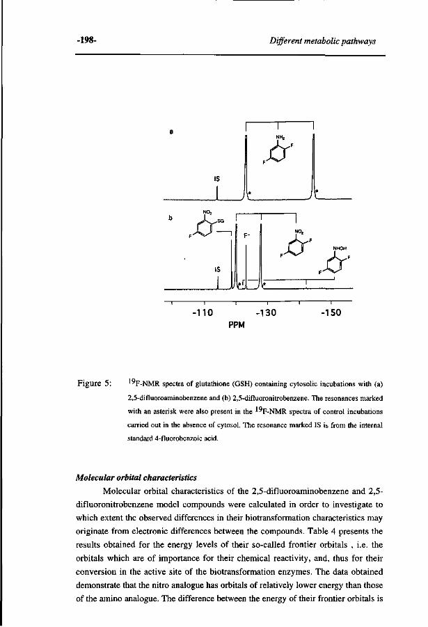
-198- Different metabolic pathways
IS
I r
- n o -130 PPM
-150
Figure 5: ^"F-NMR spectra of glutathione (GSH) containing cytosolic incubations with (a)
2,5-difluoroaminobenzene and (b) 2,5-difluoronitrobenzene. The resonances marked
with an asterisk were also present in the ^F-NMR spectra of control incubations
carried out in the absence of cytosol. The resonance marked IS is from the internal
standard 4-fluorobenzoic acid.
Molecular orbital characteristics
Molecular orbital characteristics of the 2,5-difluoroaminobenzene and 2,5-
difluoronitrobenzene model compounds were calculated in order to investigate to
which extent the observed differences in their biotransformation characteristics may
originate from electronic differences between the compounds. Table 4 presents the
results obtained for the energy levels of their so-called frontier orbitals , i.e. the
orbitals which are of importance for their chemical reactivity, and, thus for their
conversion in the active site of the biotransformation enzymes. The data obtained
demonstrate that the nitro analogue has orbitals of relatively lower energy than those
of the amino analogue. The difference between the energy of their frontier orbitals is
Chapter 8 -199-
1.0 to 1.5 eV, those of the nitro analogue being more negative.
Additional results from the MO calculations are presented in Table 5, namely
the density distribution of the frontier orbitals of importance for nucleophilic and
electrophilic attack on the compounds. Previous results on the cytochrome P450-
catalyzed aromatic hydroxylation of a series of monofluoroaminobenzenes and a
series of fluorinated benzenes demonstrated that the preferential site for the
cytochrome P450-catalyzed hydroxylation could be qualitatively (6) and even
quantitatively (19) predicted on the basis of the frontier density for electrophilic
attack of the substrate. The results presented in Table 5 for 2,5-difluoroaminobenzene
show that for this aminobenzene derivative the preferential site for hydroxylation of a
hydrogen substituted aromatic carbon centre, also corresponds with the position of
the highest frontier orbital density for an electrophilic attack by the cytochrome P450
(FeO)3+ intermediate, C4 being the preferential site for hydroxylation (Figure la).
An attack at C2, substituted with a fluorine substituent and also containing relatively
high frontier density for an electrophilic attack, does not result in product formation
as no significant formation of hydroxylated products resulting from hydroxylation
accompanied by an NIH shift or defluorination is observed (Figure la and 3a).
Furthermore, hydroxylation at C3 resulting from an (FeO)3+ attack at C2, followed
by formation of the 2,3-arene oxide and opening of the epoxide moiety to give the 3-
hydroxylated product is not observed to a significant extent either. Thus, the (FeO)3+
attack at a fluorinated centre is more likely to be reversible than to lead to significant
product formation.
For the nitrobenzene, the situation is different than for the 2,5-
difluoroaminobenzene for which the preferential site for an electrophilic attack is the
site of hydroxylation. The main frontier electron density for an electrophilic attack by
the cytochrome P450 (FeO)3+ is located on C5 and C2 of the 2,5-difluoro-
nitrobenzene, whereas the hydroxylation observed occurs at C4. The frontier density
for a nucleophilic attack, calculated from the LUMO and LUMO+1 is at this C4
centre (Table 5).
In addition to the frontier orbital density distributions of the 2,5-
difluoronitrobenzene and 2,5-difluoroaminobenzene, Table 5 also presents the
frontier orbital density distributions of three other nitrobenzene compounds, namely
nitrobenzene itself, 2-fluoronitrobenzene and 3-fluoronitrobenzene. This is to enable
comparisons between literature data on their aromatic hydroxylation (26-28) and the
calculated chemical reactivity of their aromatic carbon centres (see discussion
section). The results of the calculations demonstrate that, as for 2,5-
difluoronitrobenzene, the preferential site for an electrophilic attack on a hydrogen
substituted carbon centre is at the position(s) meta with respect to the nitro moiety.
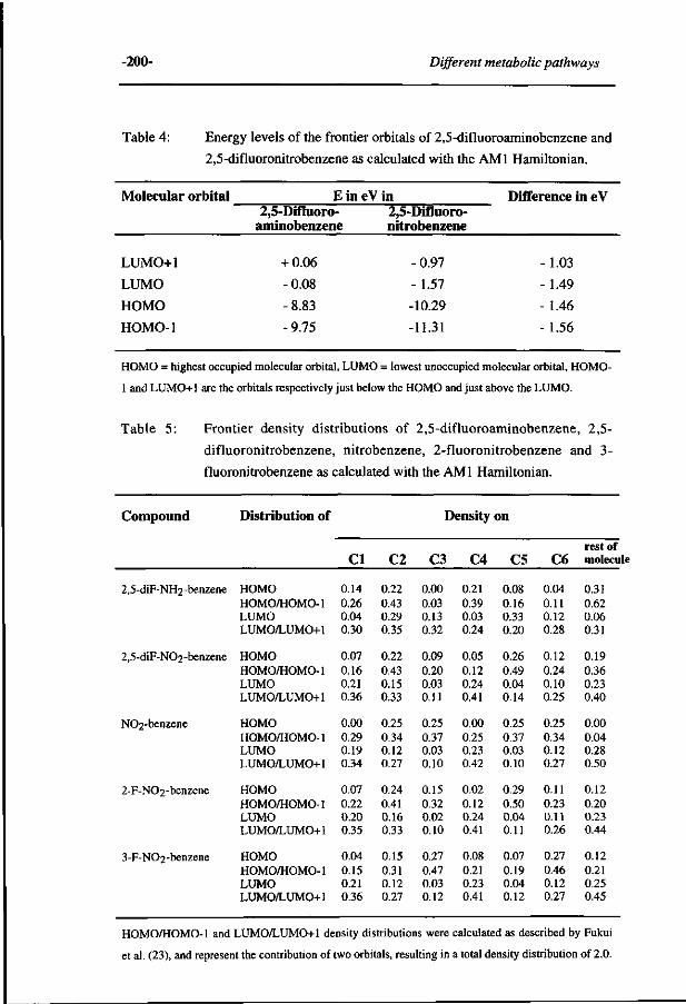
-200- Different metabolic pathways
Table 4: Energy levels of the frontier orbitals of 2,5-difluoroaminobenzene and
2,5-difluoronitrobenzene as calculated with the AMI Hamiltonian.
Molecular orbital
LUMO+1
LUMO
HOMO
HOMO-1
Ein 2,5-Difluoro-
aminobenzene
+ 0.06
-0.08
-8.83
-9.75
eV in 2,5-Difluoro-nitrobenzene
-0.97
-1.57
-10.29
-11.31
Difference in eV
-1.03
-1.49
- 1.46
-1.56
HOMO = highest occupied molecular orbital, LUMO = lowest unoccupied molecular orbital, HOMO-
1 and LUMO+1 are the orbitals respectively just below the HOMO and just above the LUMO.
Table 5: Frontier density distributions of 2,5-difluoroaminobenzene, 2,5-
difluoronitrobenzene, nitrobenzene, 2-fluoronitrobenzene and 3-
fluoronitrobenzene as calculated with the AMI Hamiltonian.
Compound
2,5-diF-NH2-benzene
2,5-diF-N02-benzene
N02-benzene
2-F-N02-benzene
3-F-N02-benzene
Distribution of
HOMO HOMO/HOMO-1 LUMO LUMO/LUMO+1
HOMO HOMO/HOMO-1 LUMO LUMO/LUMO+1
HOMO HOMO/HOMO-1 LUMO LUMO/LUMO+1
HOMO HOMO/HOMO-1 LUMO LUMO/LUMO+1
HOMO HOMO/HOMO-1 LUMO LUMO/LUMO+1
CI
0.14 0.26 0.04 0.30
0.07 0.16 0.21 0.36
0.00 0.29 0.19 0.34
0.07 0.22 0.20 0.35
0.04 0.15 0.21 0.36
C2
0.22 0.43 0.29 0.35
0.22 0.43 0.15 0.33
0.25 0.34 0.12 0.27
0.24 0.41 0.16 0.33
0.15 0.31 0.12 0.27
Density
C3
0.00 0.03 0.13 0.32
0.09 0.20 0.03 0.11
0.25 0.37 0.03 0.10
0.15 0.32 0.02 0.10
0.27 0.47 0.03 0.12
C4
0.21 0.39 0.03 0.24
0.05 0.12 0.24 0.41
0.00 0.25 0.23 0.42
0.02 0.12 0.24 0.41
0.08 0.21 0.23 0.41
on
C5
0.08 0.16 0.33 0.20
0.26 0.49 0.04 0.14
0.25 0.37 0.03 0.10
0.29 0.50 0.04 0.11
0.07 0.19 0.04 0.12
C6
0.04 0.11 0.12 0.28
0.12 0.24 0.10 0.25
0.25 0.34 0.12 0.27
0.11 0.23 0.11 0.26
0.27 0.46 0.12 0.27
rest of molecule
0.31 0.62 0.06 0.31
0.19 0.36 0.23 0.40
0.00 0.04 0.28 0.50
0.12 0.20 0.23 0.44
0.12 0.21 0.25 0.45
HOMO/HOMO-1 and LUMO/LUMO+1 density distributions were calculated as described by Fukui
et al. (23), and represent the contribution of two orbitals, resulting in a total density distribution of 2.0.
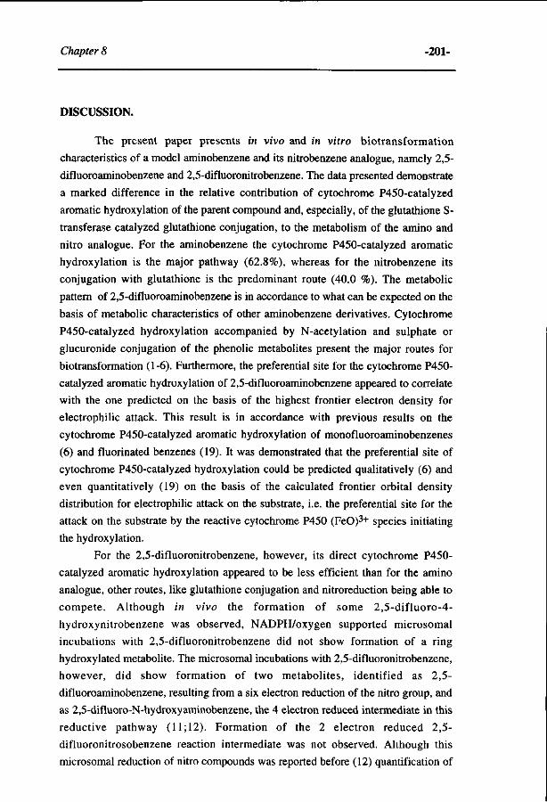
Chapter 8 -201-
DISCUSSION.
The present paper presents in vivo and in vitro biotransformation
characteristics of a model aminobenzene and its nitrobenzene analogue, namely 2,5-
difluoroaminobenzene and 2,5-difluoronitrobenzene. The data presented demonstrate
a marked difference in the relative contribution of cytochrome P450-catalyzed
aromatic hydroxylation of the parent compound and, especially, of the glutathione S-
transferase catalyzed glutathione conjugation, to the metabolism of the amino and
nitro analogue. For the aminobenzene the cytochrome P450-catalyzed aromatic
hydroxylation is the major pathway (62.8%), whereas for the nitrobenzene its
conjugation with glutathione is the predominant route (40.0 %). The metabolic
pattern of 2,5-difluoroaminobenzene is in accordance to what can be expected on the
basis of metabolic characteristics of other aminobenzene derivatives. Cytochrome
P450-catalyzed hydroxylation accompanied by N-acetylation and sulphate or
glucuronide conjugation of the phenolic metabolites present the major routes for
biotransformation (1-6). Furthermore, the preferential site for the cytochrome P450-
catalyzed aromatic hydroxylation of 2,5-difluoroaminobenzene appeared to correlate
with the one predicted on the basis of the highest frontier electron density for
electrophilic attack. This result is in accordance with previous results on the
cytochrome P450-catalyzed aromatic hydroxylation of monofluoroaminobenzenes
(6) and fluorinated benzenes (19). It was demonstrated that the preferential site of
cytochrome P450-catalyzed hydroxylation could be predicted qualitatively (6) and
even quantitatively (19) on the basis of the calculated frontier orbital density
distribution for electrophilic attack on the substrate, i.e. the preferential site for the
attack on the substrate by the reactive cytochrome P450 (FeO)3+ species initiating
the hydroxylation.
For the 2,5-difluoronitrobenzene, however, its direct cytochrome P450-
catalyzed aromatic hydroxylation appeared to be less efficient than for the amino
analogue, other routes, like glutathione conjugation and nitroreduction being able to
compete. Although in vivo the formation of some 2,5-difluoro-4-
hydroxynitrobenzene was observed, NADPH/oxygen supported microsomal
incubations with 2,5-difluoronitrobenzene did not show formation of a ring
hydroxylated metabolite. The microsomal incubations with 2,5-difluoronitrobenzene,
however, did show formation of two metabolites, identified as 2,5-
difluoroaminobenzene, resulting from a six electron reduction of the nitro group, and
as 2,5-difluoro-N-hydroxyaminobenzene, the 4 electron reduced intermediate in this
reductive pathway (11; 12). Formation of the 2 electron reduced 2,5-
difluoronitrosobenzene reaction intermediate was not observed. Although this
microsomal reduction of nitro compounds was reported before (12) quantification of
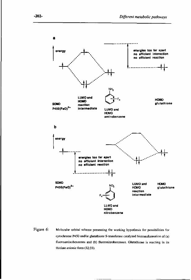
-202- Different metabolic pathways
energy •-K
-K
NK Mf
energies too far apart no efficient interaction no efficient reaction
+f
SOMO
P450(FeO)3
LUMO and HOMO reaction intermediate LUMO and
HOMO aminobenzene
HOMO glutathione
energy
energies too far apart no efficient interaction no efficient reaction
SOMO
P450(FeO)3*
-H-
LL MO and HOMO nitrobenzene
HK >Uc
LUMO and HOMO HOMO glutathione reaction intermediate
Figure 6: Molecular orbital scheme presenting the working hypothesis for possibilities for
cytochrome P450 and/or glutathione S-transferase catalyzed biotransformation of (a)
fluoroaminobenzenes and (b) fluoronitrobenzenes. Glutathione is reacting in its
thiolate anionic form (32;33).
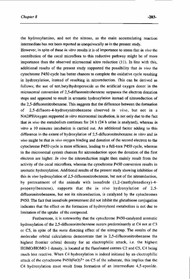
Chapter 8 -203-
the hydroxylamino, and not the nitroso, as the main accumulating reaction
intermediate has not been reported as unequivocally as in the present study.
However, in spite of these in vitro results it is of importance to stress that in vivo the
contribution of the cecal microflora to this reductive pathway might be of more
importance than the observed microsomal nitro reduction (11). In line with this,
additional results of the present study supported the possibility that in vivo the
cytochrome P450 cycle has better chances to complete the oxidative cycle resulting
in hydroxylation, instead of resulting in nitroreduction. This can be derived as
follows; the use of tert.butylhydroperoxide as the artificial oxygen donor in the
microsomal conversion of 2,5-difluoronitrobenzene surpasses the electron donation
steps and appeared to result in aromatic hydroxylation instead of nitroreduction of
the 2,5-difluoronitrobenzene. This suggests that the difference between the formation
of 2,5-difluoro-4-hydroxynitrobenzene observed in vivo, but not in a
NADPH/oxygen supported in vitro microsomal incubation, is not only due to the fact
that in vivo the metabolism continues for 24 h (24 h urine is analyzed), whereas in
vitro a 10 minutes incubation is carried out. An additional factor adding to this
difference in the extent of hydroxylation of 2,5-difluoronitrobenzene in vitro and in
vivo might be that in vivo oxygen binding and donation of the second electron to the
cytochrome P450 cycle is more efficient, leading to a full-turn P450 cycle, whereas
in the microsomal system chances for nitroreduction upon the donation of the first
electron are higher. In vivo the nitroreduction might then mainly result from the
activity of the cecal microflora, whereas the cytochrome P450 conversion results in
aromatic hydroxylation. Additional results of the present study showing inhibition of
this in vivo hydroxylation of 2,5-difluoronitrobenzene, but not of the nitroreduction,
by pretreatment of the animals with isosafrole (l,2-(methylenedioxy)-4-
propenylbenzene), supports that the in vivo hydroxylation of 2,5-
difluoronitrobenzene, but not its nitroreduction, is catalyzed by the cytochromes
P450. The fact that isosafrole pretreatment did not inhibit the glutathione conjugation
indicates that the effect on the formation of hydroxylated metabolites is not due to
limitation of the uptake of the compound.
Furthermore, it is noteworthy that the cytochrome P450-catalyzed aromatic
hydroxylation of the 2,5-difluoronitrobenzene occurs predominantly at C4 not at C3
or C5, in spite of the meta directing effect of the nitrogroup. The results of the
molecular orbital calculations demonstrate that in 2,5-difluoronitrobenzene the
highest frontier orbital density for an electrophilic attack, i.e. the highest
HOMO/HOMO-1 density, is located at the fluorinated centres C2 and C5, C4 being
much less reactive. When C4 hydroxylation is indeed initiated by an electrophilic
attack of the cytochrome P450(FeO)3+ on C5 of the substrate, this implies that the
C4 hydroxylation must result from formation of an intermediate 4,5-epoxide.
-204- Different metabolic pathways
However, in such a case formation of the 5,6-epoxide leading to C6 hydroxylation
can be expected to occur in addition to formation of the 4,5-epoxide and C4
hydroxylation. This is, however, not observed. Another explanation might be that
due to an orienting interaction between the active center of the cytochrome
P450(FeO)3+ and the 2,5-difluoronitrobenzene the substrate is oriented towards the
oxygen of the (FeO)3+ moiety in such a way that the C4 becomes the preferential site
for a reaction in spite of its lower reactivity for an electrophilic attack. The
observation that in the metabolite patterns of the rats pretreated with cytochrome
P450 inducers and exposed to 2,5-difluoronitrobenzene the hydroxylation is always
at C4 suggests that the orienting effect might be the result of an interaction that
occurs in a similar way in the active site of different cytochromes P450. Thus, the
interaction might be the result of an orienting interaction between the porphyrin
(FeO)3+ and the nitrobenzene. Definite prove for such a possible orienting interaction
has to await further investigations on other nitrobenzene derivatives. The preferential
hydroxylation of the 2,5-difluoronitrobenzene at the position para, not meta, with
respect to the nitro moiety is similar to what was reported up to now for nitrobenzene
derivatives. Bray et al. (26) using color formation of TLC spots as means for
identification of urine metabolites of rabbits, reported C5 but also C4 hydroxylation
of 2-chloronitrobenzene, as well as C4-hydroxylation of 3-chloronitrobenzene. In
further studies on 2- and 3-fluoro-, chloro-, bromo- or iodo-nitrobenzenes they
reported that the predominant nitrophenols from all the halogenated nitrobenzenes
were paranitrophenols (27). More recently, Rickert et al. (28) reported hydroxylation
of nitrobenzene at both the positions para and meta with respect to the nitro moiety,
hydroxylation of the C4 position being generally 1.65 ± 0.16 times higher than that
of the C3 position. Comparison of these biotransformation data to the relative
reactivity of the various carbon centres in nitrobenzene and in 2- and 3-
fluoronitrobenzene indicates that, as for 2,5-difluoronitrobenzene, the hydroxylation
is not observed at the carbon centre that is most reactive towards an electrophilic
attack. However, as for the 2,5-difluoronitrobenzene, the preferential site for a
nucleophilic attack is at the C4. This implies that, in addition to the explanations
given above, a third factor might in theory be responsible for the regioselectivity of
the aromatic hydroxylation. A radical attack by the cytochrome P450(FeO)3+ SOMO
(singly occupied molecular orbital) can in theory proceed not only electrophilic, the
SOMO interacting with the HOMO of a substrate, but also nucleophilic, the SOMO
interacting with the LUMO of a substrate (29). Based on this consideration and the
results of the present study it could be proposed that the cytochrome P450-catalyzed
hydroxylaton of the 2,5-difluoronitrobenzene proceeds by a nucleophilic, instead of
by an electrophilic, attack of the cytochrome P450(FeO)3+ SOMO on the substrate.
The fact that the preferential site for hydroxylation of 2,5-difluoronitrobenzene
Chapter 8 -205-
appears to be at C4, combined with the fact that the LUMO/LUMO+1 density is
highest and predicts this C4 site as the preferential site for a nucleophilic attack,
supports this explanation. However, the fact that the cytochrome P450(FeO)3+
species is known to be highly electrophilic seems to argue against this explanation
and leaves the specific stereoselective orientation of the nitrobenzene and/or the
involvement of epoxide intermediates as the best working hypotheses for further
research.
Finally, Figure 6 schematically presents a molecular orbital based model for
the explanation of the observed difference in metabolic pathways for the amino- and
the nitrobenzene derivative, as reported in the present study. From molecular orbital
calculations it followed that the molecular orbitals of the nitro analogue are generally
1 to 1.5 eV lower in energy than those of the amino analogue. As a result, the LUMO
of the 2,5-difluoronitrobenzene might be closer in energy to the HOMO of the
thiolate anion of glutathione than the LUMO of the 2,5-difluoroaminobenzene.
Taking into account the fact that the frontier orbital theory (29) describes that the
reactivity of two reactants is higher when the energy difference between the reacting
orbitals is lower this might result in a more efficient glutathione conjugation of 2,5-
difluoronitrobenzene than of the 2,5-difluoroaminobenzene. In a similar way the
relatively lower energy orbitals of the nitro analogue might explain the lower
reactivity of the HOMO of 2,5-difluoronitrobenzene than of the HOMO of 2,5-
difluoroaminobenzene with the SOMO of the cytochrome P450(FeO)3+ intermediate.
Previous MO-QSAR studies on the aromatic hydroxylation by flavin dependent
monooxygenases have demonstrated an energy difference in the HOMO of the
substrate of 1.0 eV to correspond to a change in the reaction rate of a factor 10 to 50
(30;31), whereas a recently obtained MO-QSAR for the cytochrome P450-catalyzed
4-hydroxylation of a series of aminobenzene derivatives (32) demonstrates that
halogenated anilines with a E(HOMO) value below -9.2 eV appeared to be converted
at relatively low rates by the cytochromes P450. These results imply that the
relatively low energy of the frontier orbitals of the nitrobenzene might indeed explain
its relatively lower reactivity in a cytochrome P450-catalyzed hydroxylation as well
as its relatively higher reactivity in a glutathione S-transferase catalyzed reaction.
Thus, together the results of the present study indicate that, on the basis of the
molecular orbital characteristics of a substrate it must be possible to predict some of
the characteristics of its biotransformation pathways. Further development of this
approach is currently under investigation.
-206- Different metabolic pathways
REFERENCES.
1 J.W. Gorrod and D. Manson, The metabolism of aromatic amines. Xenobiotica 16 (1986)
933-955.
2 J.W. Daly, G. Gurrof, S. Udenfriend and B. Witkop, Hydroxylation of alkyl and halogen
substituted anilines and acetanilides by microsomal hydroxylases. Biochem. Pharmacol. 17
(1968) 31-36.
3 C.V. Eadsforth, CJ . Logan, B.J. Morrison and P.A. Warburton, 2,4-Difluoroaniline and 4-
fluoroaniline exposure: monitoring by methaemoglobin and urine analysis. Int. Arch. Occ.
Env. Health 54 (1984) 223-232.
4 C.V. Eadsforth, P.C. Coveney, D.H. Hutson, C.J. Logan and A.J. Samuel, The metabolism of
o-fluoroaniline by rats, rabbits and marmosets. Xenobiotica, 16 (1986) 555-566.
5 J. Vervoort, P.A. de Jager, J. Steenbergen and I.M.C.M. Rietjens, Development of a " F
NMR method for studies on the in vivo and in vitro metabolism of 2-fluoroaniline.
Xenobiotica, 20 (1990) 657-670.
6 N.H.P. Cnubben, J. Vervoort, C. Veeger and I.M.C.M. Rietjens, Study on the regioselectivity
and mechanism of the aromatic hydroxylation of monofluoroanilines. Chem.-Biol.
Interactions, 85 (1992) 151-172.
7 D.E. Rickert, Metabolism of nitroaromatic compounds, Drug Metabolism Reviews, 18 (1987)
23-53.
8 T. Yoshida, K. Andoh and T. Tabuchi. Identification of urinary metabolites in rats treated
with p-chloronitrobenzene. Arch. Toxicol. 65 (1991) 52-58.
9 T. Yoshida, T. Tabuchi and K. Andoh, Identification of human subjects acutely poisoned by
p-chloronitrobenzene. Xenobiotica 22 (1992) 1459-1470.
10 J. Piotrowski, Further investigations on the evaluation of exposure to nitrobenzene, Brit. J.
Industr. Med., 24 (1967) 60-65.
11 A. A. Levin and J.G. Dent, Comparison of the metabolism of nitrobenzene by hepatic
microsomes and cecal microflora from Fischer-344 rats in vitro and the relative importance of
each in vivo. Drug Metabolism andDipos. 10 (1982) 450-454.
12 N. Harada and T. Omura, Participation of cytochrome P450 in the reduction of nitro
compounds by rat liver microsomes. J. Biochem. 87 (1980) 1539-1554.
13 D. Rickert and S.D. Held, Metabolism of chloronitrobenzenes by isolated rat hepatocytes.
Drug Metabolism and Dispos. 18 (1990) 5-9.
14 G.H. Coleman, Ch. M. McCloskey and F.A. Stuart: Nitrosobenzene. Organic Syntheses 25
(1943)81-83.
15 R.J. Kennedy and A.M. Stock, The oxidation of organic substances by potassium
peroxymonosulphate. J. Org. Chem. 25 (1960) 1901-1906.
16 I.M.C.M. Rietjens and J. Vervoort, Microsomal metabolism of fluoroanilines. Xenobiotica,
19(1989)1297-1305.
Chapter 8 -207-
17 R.M.E. Vos, M.C.Snoek, W.J.H, van Berkel, F. Müller and P.J. van Balderen, Differential
induction of rat hepatic glutathione S-transferase isoenzymes by hexachlorobenzene and
benzyl isothiocyanate: comparison with induction by phénobarbital and 3-
methylcholanthrene. Biochem. Pharmacol. 37 (1988) 1077-1082.
18 I.M.C.M. Rietjens and J. Vervoort, A new hypothesis for the mechanism for cytochrome P-
450 dependent aerobic conversion of hexahalogenated benzenes to pentahalogenated phenols.
Chem. Res. Toxicol. 5 (1992) 10-19.
19 I.M.C.M. Rietjens, A.E.M.F. Soffers, C. Veeger and J. Vervoort, Regioselectivity of
cytochrome P450 catalyzed hydroxylation of fluorobenzenes predicted by calculated frontier
orbital substrate characteristics. Biochemistry, 32 (1993) 4801-4812.
20 T. Omura and R. Sato, The carbon monoxide pigment of liver microsomes, I. Evidence for its
haemoprotein nature. J. Biol. Chem., 239 (1964) 2370-2378.
21 .H. Lowry, N.J. Rosebrough, A.L. Farr and A.J. Randall, Protein measurements with Folin
phenol reagent. J. Biol. Chem., 193 (1951) 265-275.
22 B.B. Brodie and J. Axelrod, The estimation of acetanilide and its metabolic products, aniline,
N-acetyl p-aminophenol and p-aminophenol (free and conjugated) in biological fluids and
tissues. J. Pharmacol. Exp. Ther., 94 (1948) 22-28.
23 K. Fukui, T. Yonezawa, C. Nagata and H. Shingu, Molecular orbital theory of orientation in
aromatic, heteroaromatic and other conjugated molecules. J. Chem. Phys., 22 (1954) 1433-
1442.
24 M.A. Cooper, H.E. Weber and S.L Manatt, The fluorine-19 nuclear magnetic resonance
spectra of some fluoroaromatic compounds. Studies using nise decoupling of protons. J. Am.
Chem. Soc. 93 (1971) 2369-2380.
25 V. Wray, Fluorine nuclei in difluorobenzene derivatives. In : Annual reports on NMR
spectroscopy, vol 14, Academic Press. 1983, (Webb, G.A. ed.) p 273-281.
26 H.G. Bray, S.P. James and W.V. thorpe, The metabolism of monochloronitrobenzenes in the
rabbit. Biochem. J. 64 (1956) 38-44.
27 H.G. Bray, S.P. James and W.V. Thorpe, Metabolism of o-, m- and p-fluoro, -bromo and
-iodo-nitrobenzenes in the rabbit. Biochem. J. 68 (1958) 561-568.
28 D.E. Rickert, J. A. Bond, R.M. Long and J.P. Chism, Metabolism of nitrobenzene by rats and
mice, Toxicol. Applied Pharmacol., 67 (1983) 206-214.
29 I. Fleming (Ed). Frontier orbitals and organic chemical reactions, John Wiley & Sons, New
York, 1976, pp 1-249.
30 J. Vervoort, I.M.C.M. Rietjens, W.J.H, van Berkel and C. Veeger, Frontier orbital study on
the 4-hydroxybenzoate-3-hydroxylase-dependent activity with benzoate derivatives. Eur. J.
Biochem. 206 (1992) 479-484.
31 I.M.C.M. Rietjens, M.G. Boersma, A.E.M.F. Soffers, N.H.P. Cnubben, J. Koerts, S. Peelen,
W.J.H. van Berkel, C. Veeger and J. Vervoort, Use of molecular orbital calculations in
studies on mechanisms of enzyme catalysis. In: Flavins and Flavoproteins 1993. Kunio Yagi
-208- Different metabolic pathways
(ed), Walter de Gruyter & Co, Berlin, (1993)(in press).
32 N.H.P. Cnubben, S. Peelen, J.W. Borst, J. Vervoort, and I.M.C.M. Rietjens, Molecular orbital
based quantitative structure activity relationship for the cytochrome P450-catalyzed 4-
hydroxylation of halogenated anilines, Chem. Res. Tox., (1994) in press.
33 W.J. Chen, G.F. Graminski and R.N. Armstrong, Dissection of the catalytic mechanism of
isozyme 4-4 of glutathione S-transferase with alternative substrates, Biochemistry 27 (1988)
647-654.
34 G.F. Graminski, G.F., Y. Kubo and R.N. Armstrong, Spectroscopic and kinetic evidence for
the thiolate anion of glutathione at the active site of glutathione S-transferase. Biochemistry
28 (1989) 3562-3568.
CHAPTER 9
Influence of the halogen-substituent pattern of fluoronitrobenzenes on their biotransformation and
capacity to induce methemoglobinemia.
Nicole H.P. Cnubben, Ans E. M. F. Soffers, Maria A.W. Peters, Jacques Vervoort
and Ivonne M.C.M. Rietjens.
Accepted for publication in Toxicology and Applied Pharmacology.
SUMMARY.
In the present study both the biotransformation patterns and the capacity to
induce methemoglobinemia of a series of fluoronitrobenzenes were investigated.
This was done to investigate to what extent variation in the number and position of
the halogen substituents influence the metabolic fate of the fluoronitrobenzenes,
thereby influencing their capacity to induce methemoglobinemia. The results
obtained were compared to the effect of the fluorine substituent patterns on the
calculated electronic characteristics, and, thus, on the chemical reactivity of the
fluoronitrobenzenes.
Analysis of the in vivo metabolic profiles demonstrates a dependence of the
extent of nitroreduction, of glutathione conjugation and of aromatic hydroxylation
with the pattern of halogen substitution. With increasing number of fluorine
substituents at electrophilic carbon centres, 24 h urine recovery values decreased and
fluoride anion elimination increased, due to increased reactivity of the
fluoronitrobenzenes with cellular nucleophiles. In vitro studies even demonstrated a
clear correlation between calculated parameters for the electrophilicity of the
fluoronitrobenzenes and the natural logarithm of their rate of reaction with
glutathione or with bovine serum albumin, taken as a model for cellular nucleophiles
(r=0.97 and r=0.98 respectively). Increased possibilities for the conjugation of the
-210- Biotransformation and methemoglobinemia
fluoronitrobenzenes to cellular nucleophiles was accompanied by decreased
contributions of nitroreduction and aromatic hydroxylation to the overall in vivo
metabolite patterns, as well as by a decreased capacity of the fluoronitrobenzenes to
induce the methemoglobinemia. In vitro studies on the rates of nitroreduction of the
various fluoronitrobenzenes by cecal microflora and rat liver microsomes, revealed
that the changes in the capacity of the fluoronitrobenzenes to induce
methemoglobinemia were not due to differences in their intrinsic reactivity in the
pathway of nitroreduction, leading to the methemoglobinemia inducing metabolites.
Thus, the results of the present study clearly demonstrate that the number and
position of fluorine substituents in the fluoronitrobenzenes influence the capacity of
the fluoronitrobenzenes to induce methemoglobinemia, not because their intrinsic
chemical reactivity for entering the nitroreduction pathway is influenced. The
different methemoglobinemic capacity must rather result from differences in the
inherent direct methemoglobinemic capacity and/or reactivity of the various toxic
metabolites, and/or from the fact that the halogen substituent pattern influences the
electrophilic reactivity, thereby changing the possibilities for reactions of the
nitrobenzenes with glutathione and, especially, other cellular nucleophiles. When the
number of fluorine substituents increases, the electrophilicity of the
fluoronitrobenzenes can become so high that glutathione conjugation is no longer
able to compete efficiently with covalent binding of the fluoronitrobenzenes to
cellular macromolecules. As a consequence, it can be suggested that with an
increasing number of fluorine substituents at electrophilic carbon centres in a
nitrobenzene derivative, a toxic endpoint of the nitrobenzene other than formation of
methemoglobinemia can be foreseen.
INTRODUCTION.
Halogenated nitrobenzenes are frequently used as industrial intermediate
reagents in the production of dyes, rubbers, pharmaceuticals or are used as
agrochemicals, like for instance the soil pesticide pentachloronitrobenzene [Rickert,
1987]. Nitrobenzene derivatives are mutagenic in several bacterial strains [Shimizu et
ai, 1983; Debnath et al., 1992], cause DNA damage in vivo [Cesarone et al, 1983],
are hepatotoxic [Beauchamp et al, 1982] and nephrotoxic [Yoshida et al, 1989].
However, the most frequently reported consequence of exposure to nitrobenzenes is
methemoglobinemia and it is generally accepted that the intermediate oxidation
states formed in the process of nitroreduction by the intestinal microflora
(nitrosobenzene, N-hydroxyarylamine or the radical intermediates, all formed in the
six-electron reduction pathway leading to aniline formation) are responsible for
Chapter 9 -211-
inducing methemoglobinemia [for review, see Kiese, 1974]. Thus, some of the toxic
effects require reactive intermediates formed upon the biotransformation of
nitrobenzenes. From several studies it became evident that both the toxicity as well
as metabolism of nitrobenzenes can be dependent on their substituent pattern
[O'Brien et al, 1990; Shimizu et al, 1983; Debnath étal, 1991; Sabbioni, 1992;
Sabbioni, 1994]. The wide variation in capacity of nitrobenzene-derivatives to cause
toxic effects has been partially related to their different susceptibility to
nitroreduction to give their corresponding nitro radical anions, nitrosobenzenes or N-
hydroxyarylamines [Debnath et al, 1991; Lopez de Compadre et al, 1990; O'Brien
et al, 1990; Sabbioni, 1992; Sabbioni, 1994; Tatsumi et al, 1978]. On the other
hand, it has been suggested that differences in occurence of methemoglobinemia or
in the formation of hemoglobin adducts with a change in the substituent pattern, are
related to the effectiveness of biotransformation pathways competing with
nitroreduction [Debnath et al, 1991; Kiese, 1974; Sabbioni, 1994; Rickert and Held,
1990]. A combined study on biotransformation and methemoglobin formation of a
series of nitrobenzenes may provide insight in the relative importance of these two
suggestions, but has not been published yet. Therefore, the objective of the present
study was to investigate both the induction of methemoglobinemia as well as in vivo
metabolic profiles of a series of nitrobenzene derivatives. This was done using a
series of fluoronitrobenzenes. The fluorine label makes it possible to use 19F NMR as
a tool to characterize the complete urine metabolic patterns of the nitrobenzene
derivatives. Besides, the Van der Waals radius of a fluorine substituent is slightly
larger compared to that of a hydrogen substituent, limiting possible steric influences
of the substituents. Finally, the fluorine substituents can be expected to influence the
chemical reactivity characteristics of the substrates that are of importance in
electrophilic reactions like aromatic hydroxylation [Cnubben et al, 1992; Cnubben et
al, 1994; Rietjens et al, 1993], in nucleophilic reactions like glutathione conjugation
[Rietjens et al, 1995 a+b], and in reductions, like nitroreduction [Lopez de
Compadre et al, 1990]. To elucidate the actual influence of the various fluorine
substituents patterns on these electronic characteristics of the nitrobenzene model
compounds, molecular orbital calculations were performed and the results obtained
were compared to the biotransformation characteristics of the nitrobenzenes observed
both in vivo as well as in vitro.
MATERIALS AND METHODS.
Chemicals.
2-Fluoronitrobenzene, 3-fluoronitrobenzene, 4-fluoronitrobenzene and 3,5-
difluoronitrobenzene were obtained from Aldrich (Steinheim, FRG). 2,5-
-212- Biotransformation and methemoglobinemia
Difluoronitrobenzene, 2,4-difluoronitrobenzene, 2,3,4-trifluoronitrobenzene, 2,4,6-
trifluoronitrobenzene and 2,3,4,6-tetrafluoronitrobenzene were purchased from
Fluorochem (Derbyshire, UK). 2-Fluoroaniline, 3-fluoroaniline and 4-fluoroaniline
were from Janssen Chimica (Beerse, Belgium). 2,4-Difluoroaniline, 2,5-
difluoroaniline, 3,5-difluoroaniline, 2,3,4-trifluoroaniline, 2,4,6-trifluoroaniline and
2,3,4,6-tetrafluoroaniline were from Fluorochem. 3-Fluoro-4-nitrophenol and 2-
fluoro-4-nitrophenol were from Fluorochem.
The 19F-NMR resonances of the 2-fluoro-3-nitrophenol, 2-fluoro-5-nitrophenol, 5-
fluoro-3-nitrophenol and 4,6-difluoro-2-aminophenol were identified on the basis of
a characteristic shift of the 19F-NMR resonance known to exist upon the introduction
of a hydroxyl moiety at positions ortho, meta or para with respect to the fluorine
[Rietjens et al, 1995a]. The 19F-NMR resonances of 2,5-difluoro-4-nitrophenol and
2,6-difluoro-4-nitrophenol were identified by reducing the nitro moiety to the amino
moiety with zinc powder yielding respectively 2,5-difluoro-4-nitrophenol and 2,6-
difluoro-4-aminophenol [Rietjens et al, 1995a; Piotrowski, 1967].
3-Fluoro-4-aminophenol, 3-fluoro-2-aminophenol, 4-fluoro-2-aminophenol, 2-
fluoro-4-aminophenol, 5-fluoro-2-aminophenol, 2,5-difluoro-4-aminophenol, 3,5-
difluoro-4-aminophenol and 2,6-difluoro-4-aminophenol were synthetised essentially
as described before [Cnubben et al, 1992; Cnubben et al., 1994]. 3-Fluoro-4-
acetamidophenol, 2-fluoro-4-acetamidophenol, 5-fluoro-2-acetamidophenol, 3-
fluoroacetanilide, 2,6-difluoro-4-acetamidophenol, 3,5-difluoro-4-acetamidophenol
and 2,5-difluoro-4-acetamidophenol were synthetised by acylation of the
corresponding amino-compounds following the procedure described by Vogel
[1978].
All (fluorinated) glutathionyl-nitrobenzene and mercapturic acid derivatives were
synthetised from suitable fluoronitrobenzenes essentially as described before
[Rietjens et al, 1995 a+b]. 19F-NMR and 'H-NMR spectral characteristics of 4-
fluoro-2-glutathionyl-nitrobenzene, 2-fluoro-4-glutathionyl-nitrobenzene, 5-fluoro-2-
glutathionyl-nitrobenzene, 5-fluoro-2-(N-acetylcysteinyl)-nitrobenzene, 2,6-difluoro-
4-glutathionyl-nitrobenzene, 2,4-difluoro-6-glutathionyl-nitrobenzene, 3,4,6-
trifluoro-2-glutathionyl-nitrobenzene and 2,3,6-trifluoro-4-glutathionyl-nitrobenzene
were previously described [Rietjens et al, 1995 a+b]. Spectral characteristics of all
other synthetised compounds, used for identification of the various adducts, are
presented in Table 1. Chemical shift values are in D2O and presented relative to
CFCI3 for 19F-NMR and relative to TSP (sodium 3-trimethylsilyl-(2,2,3,3-2H4)-
propionate) (Isotec, INC) for 'H-NMR.
Fluoronitrosobenzenes were synthetised according to Kennedy and Stock
[I960]. During 3 min 25 mL 15% (w/v) potassiumperoxomonosulphate (minimal 4.5
% active oxygen, Janssen Chimica) were added to a solution of 10 mmol of the
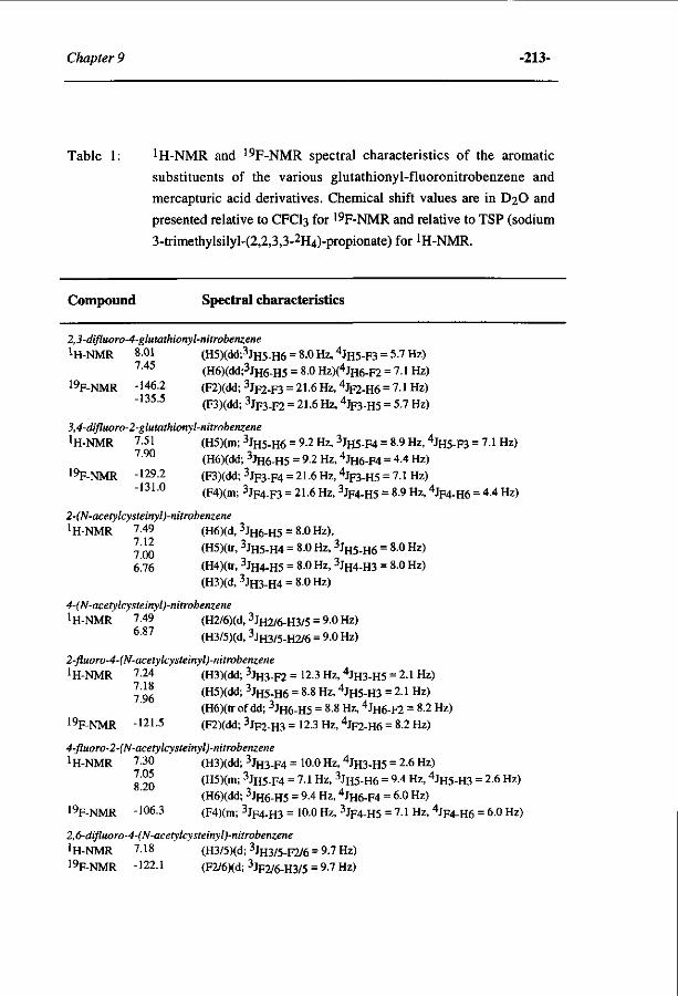
Chapter 9 -213-
Table 1: 1 H-NMR and 19F-NMR spectral characteristics of the aromatic
substituents of the various glutathionyl-fluoronitrobenzene and
mercapturic acid derivatives. Chemical shift values are in D2O and
presented relative to CFCI3 for 19F-NMR and relative to TSP (sodium
3-trimethylsilyl-(2,2,3,3-2H4)-propionate) for !H-NMR.
Compound Spectral characteristics
2,3-difluoro-4-glutathionyl-nitrobenzene ! H - N M R 8.01 (H5)(dd;3JH5-H6 = 80Hz,4JH5-F3 = 5.7Hz)
7 4 5 (H6)(dd;3JH6-H5 = 8.0 Hz)(4JH6-F2 = 7.1 Hz) 19F-NMR -146.2 (F2)(dd; 3 J F 2 - F 3 = 21.6 HZ, 4 J F 2 - H 6 = ?.1 HZ)
-135.5 (F3)(dd; 3JF3-F2 = 21.6 Hz, 4 J F 3 - H 5 = 5.7 Hz)
3,4-difluoro-2-glutathionyl-nitrobenzene JH-NMR 7.51 (H5)(m; 3 J H 5 - H 6 = 9.2 Hz, 3JH5-F4 = 8.9 Hz, 4 J H 5 - F 3 = 7.1 Hz)
130 (H6)(dd; 3JH6-H5 = 9.2 Hz, 4JH6-F4 = 4.4 Hz) 19F-NMR -129.2 (F3)(dd; 3JF3-F4 = 21.6 Hz, 4JF3-H5 = 7.1 Hz)
-131.0 (F4)(m; 3 J F 4 - F 3 = 21.6 HZ, 3JF4-H5 = 8.9 Hz, 4 J F 4 - H 6 = 4.4 Hz)
2-(N-acetylcysteinyl)-nitrobenzene
1 H-NMR 7.49 (H6)(d, 3 J H 6 - H 5 = 8.0 Hz),
J-JJ (H5)(tr, 3JH5-H4 = 8.0Hz, 3JH5-H6 = 80Hz) 6J6 (H4)(tr, 3JH4-H5 = 8.0 Hz, 3JH4-H3 = 8.0 Hz)
(H3)(d,3JH3-H4 = 8.0Hz)
4-(N-acetylcysteinyl)-nitrobenzene 1H-NMR 7.49 (H2/6)(d, 3JH2/6-H3/5 = 9.0 Hz)
6 8 7 (H3/5)(d,3JH3/5-H2/6 = 9.0Hz)
2-fluoro-4-(N-acetylcysteinyl)-nitrobenzene
1 H-NMR 7.24 (H3)(dd; 3JH3-F2 = 12.3 Hz, 4JH3-H5 = 2.1 Hz)
lAJj. (H5)(dd; 3JH5-H6 = 8.8 Hz, 4JH5-H3 = 2.1 Hz) (H6)(tr of dd; 3 J H 6 - H 5 = 8.8 Hz, 4 J H 6 - F 2 = 8.2 Hz)
19F-NMR -121 -5 (F2)(dd; 3JF2-H3 = 12.3 Hz, %2-H6 = 8.2 Hz)
4-fluoro-2-(N-acetylcysteinyl)-nitrobenzene iH-NMR 7.30 (H3)(dd; 3JH3-F4 = 100Hz,4JH3-H5 = 2.6Hz)
™jj (H5)(m; 3JH5-F4 = 7.1 Hz, 3JH5-H6 = 9.4 Hz, 4JH5-H3 = 2.6 Hz) (H6)(dd; 3JH6-H5 = 9.4 Hz, 4JH6-F4 = 6.0 Hz)
19F-NMR -106.3 (F4)(m; 3 J F 4 - H 3 = 100 Hz, 3JF4-H5 = 7.1 Hz, 4JF4.H6 = 6.0 Hz)
2,6-difluoro-4-(N-acetylcysteinyl)-nitrobenzene iH-NMR 7.18 (H3/5)(d; 3JH3/5-F2/6 = 9.7 Hz)
19F-NMR -122.1 (F2/6)(d; 3JF2/6-H3/5 = 9.7 Hz)
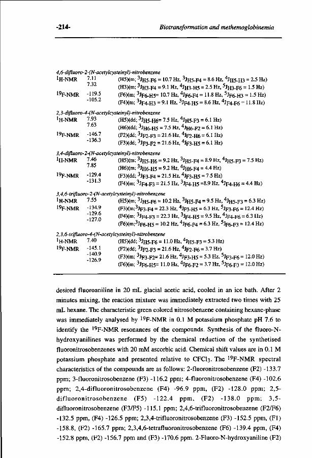
-214- Biotransformation and methemoglobinemia
4,6-difluoro-2-(N-acetylcysteinyl)-nitrobenzene
! H - N M R 7.11 (H5)(m; 3 J H 5 - F 6 = 10.7 Hz, 3JH5-F4 = 8.6 Hz, 4 J H 5 - H 3 = 2.5 Hz) 7 3 2 (H3)(m; 3 J H 3 - F 4 = 9.1 Hz, 4JH3-H5 = 2.5 Hz, 5JH3-F6 = 1.5 Hz)
19F-NMR -119.5 (F6)(m; 3 J F 6 - H 5 = 10.7 Hz, 4JF6-F4 = H-8 Hz, 5 J F 6 - H 3 = 1-5 Hz) -105.2 (F4)(m; 3 J F 4 - H 3 = 9.1 Hz, 3 J F 4 - H 5 = 8.6 Hz, 4 J F 4 - F 6 = 11-8 Hz)
2,3-difluoro-4-(N-acetylcysteinyl)-nitrobenzene
iH-NMR 7.93 (H5)(dd; 3 J H 5 - H 6 = 7.5 Hz, 4 J H 5 - F 3 = 6.1 Hz) 7 6 3 (H6)(dd; 3JH6-H5 = 7.5 Hz, 4JH6-F2 = 6.1 Hz)
19F-NMR -146.7 (F2)(dd; 3 J F 2 - F 3 = 21.6 Hz, 4 J F 2 - H 6 = 6.1 Hz) -136.3 (F3)(dd; 3JF3-F2 = 21.6 Hz, 4 J F 3 - H 5 = 6.1 Hz)
3,4-difluoro-2-(N-acetylcysteinyl)-nitrobenzene iH-NMR 7.46 (H5)(m; 3JH5-H6 = 9.2 Hz, 3JH5-F4 = 8.9 Hz, 4 J H 5 - F 3 = 7.5 Hz)
7 8 5 (H6)(m; 3JH6-H5 = 9.2 Hz, 4JH6-F4 = 4.4 Hz) 19F-NMR -129.4 (F3)(dd; 3Jp3-F4 = 21.5 Hz, 4 J F 3 . H 5 = 7.5 Hz)
-131.3 (F4)(m; 3 J F 4 - F 3 = 21.5 Hz, 3JF4-H5 =8.9 Hz, 4 J F 4 - H 6 = 4-4 Hz)
3,4,6-trifluoro-2-(N-acetylcysteinyl)-nitrobenzene
iH-NMR 7.55 (H5)(m; 3 J H 5 - F 6 = 10.2 Hz, 3JH5-F4 = 9.5 Hz, 4 J H 5 - F 3 = 6.3 Hz) 19F-NMR -134.9 (F3)(m;3JF3-F4 = 22.3 Hz, 4JF3-H5 = 6.3 Hz, 5 J F 3 - F 6 = 12.4 Hz)
-127 0 ( F 4 ) ( m ; 3 j F 4 " F 3 = 2 2 ' 3 H Z ' 3 j F4- H 5 = 9 5 H z ' 4 j F 4 - F 6 = 6 3 Hz~> (F6)(m;3Jp6-H5 = 10.2 Hz, 4JF6-F4 = 6.3 Hz, 5 J F 6 - F 3 = 124 Hz)
2,3,6-trifluoro-4-(N-acetylcysteinyl)-nitrobenzene
iH-NMR 7.40 (H5)(dd; 3 J H 5 - F 6 =110 Hz, 4 J H 5 - F 3 = 5.3 Hz)
19F-NMR -145.1 (F2)(dd; 3 J F 2 - F 3 = 21.6Hz,4Jp2-F6 = 3.7 Hz)
-126 9 ( F 3 ) ( m ; 3 j F 3 - F 2 = 2 L 6 H Z> 4 jF3-H5 = 5.3 Hz, 5 J F 3 . F 6 = 12.0 Hz) (F6)(m; 3JF6-H5= 11 0 Hz, 4JF6-F2 = 3.7 Hz, 5 J F 6 - F 3 = 12.0 Hz)
desired fluoroaniline in 20 mL glacial acetic acid, cooled in an ice bath. After 2
minutes mixing, the reaction mixture was immediately extracted two times with 25
mL hexane. The characteristic green colored nitrosobenzene containing hexane-phase
was immediately analysed by 19F-NMR in 0.1 M potassium phosphate pH 7.6 to
identify the 19F-NMR resonances of the compounds. Synthesis of the fluoro-N-
hydroxyanilines was performed by the chemical reduction of the synthetised
fluoronitrosobenzenes with 20 mM ascorbic acid. Chemical shift values are in 0.1 M
potassium phosphate and presented relative to CFCI3. The 19F-NMR spectral
characteristics of the compounds are as follows: 2-fluoronitrosobenzene (F2) -133.7
ppm; 3-fluoronitrosobenzene (F3) -116.2 ppm; 4-fluoronitrosobenzene (F4) -102.6
ppm; 2,4-difluoronitrosobenzene (F4) -96.9 ppm, (F2) -128.0 ppm; 2,5-
difluoronitrosobenzene (F5) -122.4 ppm, (F2) -138.0 ppm; 3,5-
difluoronitrosobenzene (F3/F5) -115.1 ppm; 2,4,6-trifluoronitrosobenzene (F2/F6)
-132.5 ppm, (F4) -126.5 ppm; 2,3,4-trifluoronitrosobenzene (F3) -152.5 ppm, (Fl)
-158.8, (F2) -165.7 ppm; 2,3,4,6-tetrafluoronitrosobenzene (F6) -139.4 ppm, (F4)
-152.8 ppm, (F2) -156.7 ppm and (F3) -170.6 ppm. 2-Fluoro-N-hydroxyaniline (F2)
Chapter 9 -215-
-135.3 ppm; 3-fluoro-N-hydroxyaniline (F3) -116.8 ppm; 4-fluoro-N-hydroxyaniline
(F4) -125.7 ppm; 2,4-difluoro-N-hydroxyaniline -121.9 ppm (F4), -130.6 ppm (F2);
2,5-difluoro-N-hydroxyaniline -122.0 ppm (F5), -142.0 ppm (F2); 3,5-difluoro-N-
hydroxyaniline (F3/5) -114.0 ppm; 2,4,6-trifluoro-N-hydroxyaniline -114.3 ppm
(F4), -124.8 ppm (F2/6); 2,3,4-trifluoro-N-hydroxyaniline -147.2 ppm (F3), -155.6
ppm (Fl), -165.2 ppm (F2); 2,3,4,6-tetrafluoro-N-hydroxyaniline -131.6 ppm (F6),
-140.1 ppm (F4), -148.1 ppm (F2) and -168.5 ppm (F3).
NMR measurements. 19F-NMR measurements were performed on a Bruker AMX 300 NMR
spectrometer essentially as described before by Vervoort et al. [1990]. The splitting
patterns of the !H-NMR and 19F-NMR resonances of the synthetised compounds
were measured on a Bruker AMX 500 spectrometer. 19F-NMR measurements were
performed using a ^H-probehead tuned to the 19F-frequency (470.5 MHz). Upon
freeze drying the synthetised compounds were dissolved in D2O to give a final
sample volume of 0.5 ml. Spectra were recorded using 60° pulses (6ns), a 10 kHz
spectral width a repetition time of 1.9 s, quadrature phase detection and quadrature
phase cycling (CYCLOPS). Chemical shift values are presented relative to CFCI3 for 19F-NMR and relative to TSP (sodium 3-trimethylsilyl-(2,2,3,3-2H4)-
propionate)(Isotec, INC) for !H-NMR.
In vivo exposure of rats to fluoronitrobenzenes.
Male Wistar rats (body weight 300-325 g) with a permanent cannulation of
the jugular vein in combination with a head attachment apparatus allowing easy
connection of cannulae for blood sampling were used. The animals were placed in
metabolism cages and allowed free access to food and tapwater. Groups of rats (N=3)
were orally dosed with olive oil (controls) or 0.5 mmol of the desired
fluoronitrobenzene per kg body weight in olive oil administered by oral gavage. The
urine was collected for a period of 24 h prior to dosing and at times of 24 and 48 hr
after dosing. The collected urine samples were stored at -20°C prior to analysis.
Blood samples (approximately 250 (xl) of the cannulated rats were collected by
cannules into heparinized tubes 3 days before dosing (t=0) and at 1, 2,4, 8,24 and 48
h after dosing for the determination of methemoglobin content.
Determination of methemoglobinemia.
Methemoglobin content was determined by the method of Harrison and
Jollow [1987]. All samples were immediately analysed after collection in order to
prevent decomposition of the unstable methemoglobin.
•216- Biotransformation and methemoglobinemia
Enzyme hydrolysis of urine samples.
The urine samples of the fluoronitrobenzene exposed rats were subjected to
analysis by 19F-NMR before and after ß-glucuronidase and/or arylsulphatase
treatment essentially as described before [Cnubben et al., 1992].
HPLC analysis of the non-fluorinated N-acetylcysteinyl-nitrobenzene metabolites in
urine.
The presence of N-acetylcysteinyl-nitrobenzene metabolites in the urine of
the 2- and 4-fluoronitrobenzene exposed rats were quantified and detected using a
Kratos 400 HPLC system, with a LiChrosorb C8 column 10 RP (250 x 9.0 mm)
(Chrompack, Middelburg, The Netherlands) equiped with a Waters™ 996
photodiode array detector. This was done because the loss of the fluorine atom
prevents detection of this metabolite by 19F-NMR. After eluting for 1 min with 0.1%
trifluoroacetic acid a linear gradient from 0-100% (v/v) methanol in 60 min was
applied. The synthetised 2- or 4-(N-acetylcysteinyl)-nitrobenzene were used as the
reference compounds for the identification of these metabolites excreted in the urine
of resp. 2- and 4-fluoronitrobenzene exposed rats. The urinary metabolites eluting
from the column at the same retention time as the respective reference compounds
were collected, freeze-dried and resolved in D2O. Their identity was confirmed by
means of !H-NMR analysis as being respectively 2-(N-acetylcysteinyl)-nitrobenzene
as the urinary metabolite of a 2-fluoronitrobenzene exposed rat and 4-(N-
acetylcysteinyl)-nitrobenzene as the metabolite of a 4-fluoronitrobenzene exposed
rat.
Quantitation of urinary fluoronitrobenzene derived metabolites.
Quantitation of fluoro-containing nitrobenzene derived metabolites, using 19F-NMR, was performed by comparison of the integrals of the 19F-NMR
resonances of the metabolites to the integral of the 19F-NMR resonance of 4-
fluorobenzoic acid used as the internal standard.
Quantitation of the urinary non-fluorinated N-acetylcysteinyl-nitrobenzene
metabolites was carried out on a Kratos 400 HPLC system equiped with a
LiChrosorb C8 column (100 x 3.0 mm) (Chrompack, Middelburg, The Netherlands),
eluted isocratically with 37% methanol/0.1% trifluoroacetic acid. Detection was
performed at 370 nm and quantitation was done by integration of the peak areas and
comparison to a calibration curve obtained with the synthetised 2- or 4-(N-
acetylcysteinyl)-nitrobenzene. The concentration of the stock solution of the
synthetised N-acetylcysteinyl-nitrobenzene was determined using !H-NMR by
comparison of the integrals of the !H-NMR resonances to the integral of the lH-
Chapter 9 -217-
NMR resonance of sodium 3-trimethylsilyl-(2,2,3,3-2H4)-propionate (Isotec, INC)
used as the internal standard.
Incubations of fluoronitrobenzenes with GSH and BSA.
The chemical reaction of fluoronitrobenzene derivatives with reduced
glutathione (GSH) or bovine serum albumine (BSA) was studied in incubations
containing 0.1 M potassium phosphate pH 7.6, 3 mM fluoronitrobenzene derivative
and 2 mM GSH (Sigma, St. Louis, MO, USA) or 10 mg BSA/ml (= 0.15
mM)(Boehringer Mannheim, FRG). The reaction was started by the addition of the
fluoronitrobenzene and carried out at 37°C. At different time intervals, 1 ml samples
were taken from the incubation mixture and frozen into liquid nitrogen. Fluoride
anion elimination and/or fluoro-glutathionyl-nitrobenzene formation was monitored
using 19F-NMR. The rate of the chemical reaction between the fluoronitrobenzenes
and GSH or BSA was calculated from the linear increase in time of fluoride anion
and/or adduct content in the incubation mixture. Incubations without GSH or BSA
served as the control. For 2-fluoronitrobenzene the chemical reaction with GSH was
monitored by the decrease of GSH, since the 19F-NMR resonance of the fluoride
anion overlaps with the 19F-NMR resonance of the substrate. In short, 200 ul samples
were taken from the incubation mixture, mixed with 30 ul 33% trichloroacetic acid to
stop the reaction and assayed for GSH content using Ellman's reagent (5,5' dithiobis-
(2-nitrobenzoic acid))(Boehringer Mannheim, FRG). The rate of the chemical
reaction between GSH and the 2-fluoronitrobenzene was calculated from the linear
decrease in time of the GSH content. Incubations without 2-fluoronitrobenzene
served as the control in order to correct for the oxidation of the GSH in time.
Incubations with a suspension of rat intestinal microflora.
A suspension of the intestinal microflora was prepared according to Levin
and Dent [1982]. Anaerobic incubations were carried out at 37°C in 0.1 M potassium
phosphate pH 7.6, containing 2% glucose. The reaction was started by the addition of
fluoronitrobenzene dissolved in dimethylsulphoxide (up to 8 mM final concentration)
and carried out at 37°C in a sealed reaction vessel upon four cycles of evacuating and
filling with argon. At different time intervals, 2 ml samples were taken from the
incubation mixture and immediately frozen into liquid nitrogen. Formation of
reduced metabolites, e.g. corresponding N-hydroxyaniline and/or aniline-derivatives,
was monitored using 19F-NMR. Accumulation of the nitroso-derivative did not occur
to a detectable level. Using increasing substrate concentrations, the Vm a x for
reduction of fluoronitrobenzenes by the intestinal microflora was determined. To
compare apparent Vmax values for nitroreduction for all compounds, sommation of
the amount of the fluorinated N-hydroxyaniline and aniline was performed as such or
-218- Biotransformation and methemoglobinemia
in an electron-weighted manner, by talcing into account that for reduction of
nitrobenzene to nitrosobenzene, N-hydroxyaniline or aniline respectively two, four or
six electrons are required.
Molecular orbital computer calculations.
Molecular orbital computer calculations were performed essentially as
described before using a semi-empirical molecular orbital method, applying the AMI
Hamiltonian from the AMP AC program (Quantum Chemistry Program Exchange,
no. 506, Indiana University, Bloomington, IN, USA) [Cnubben et al, 1992]. The
frontier orbital densities for electrophilic and nucleophilic atttack were calculated
from respectively HOMO/HOMO-1 and LUMO/LUMO+1 characteristics of the
fluoronitrobenzene-derivatives as indicated by Fukui et al. [1954].
RESULTS.
Fluoronitrobenzene induced methemoglobinemia in male Wistar.
Male Wistar rats were orally dosed with different fluoronitrobenzenes and
blood was sampled for determination of methemoglobin content at several time
intervals for 48 hours. Table 2 shows the relative amounts of methemoglobin
observed at various times after exposure to the fluoronitrobenzenes. These results
demonstrate that fluorobenzenes without a fluorine at the C2, C4 or C6 position, like
3-fluoronitrobenzene and 3,5-difluoronitrobenzene, possess the highest capacity for
inducing methemoglobinemia. For the various fluoronitrobenzenes containing a
fluorine at the C2, C4 and/or C6 position, a decrease in the potency to induce
methemoglobinemia was generally observed with an increase in the number of
fluorine substituents.
Rate of nitro-reduction of the nitrobenzenes by cecal microflora and liver
preparations.
Reduction of unsubstituted nitrobenzene is generally accepted to proceed
predominantly by the microflora of the intestine [Reddy et al, 1976]. For a series of
monochloronitrobenzenes, it was demonstrated that hepatocytes of rat liver are also
able to take part in the reduction [Rickert and Held, 1990]. In vitro, the reduction of
the fluoronitrobenzenes was studied using a suspension of rat cecal microflora and
rat liver preparations. This was done in order to elucidate whether the observed
differences in methemoglobin formation with changes in the fluorine substituent
pattern originate from a possible influence of the substituent pattern on chances for
reduction of the nitro moieties to methemoglobin-inducing intermediates.
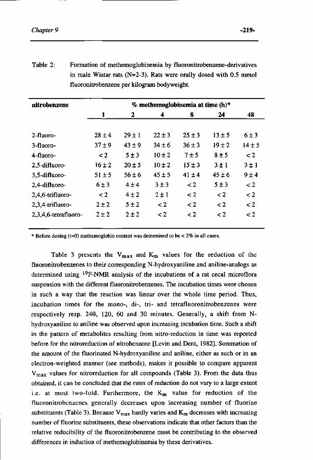
Chapter 9 -219-
Table 2: Formation of methemoglobinemia by fluoronitrobenzene-derivatives
in male Wistar rats (N=2-3). Rats were orally dosed with 0.5 mmol
fluoronitrobenzene per kilogram bodyweight.
nitrobenzene
2-fluoro-
3-fluoro-
4-fluoro-
2,5-difluoro-
3,5-difluoro-
2,4-difluoro-
2,4,6-trifluoro-
2,3,4-trifluoro-
2,3,4,6-tetrafluoro-
1
28+4
37 ±9
<2
16 + 2
51 ±5
6±3
<2
2±2
2±2
% methemoglobinemia at time (h)*
2
29 ±1
43 ±9
5 ± 3
20 ±5
56 ±6
4 ± 4
4 ± 2
5 + 2
2 ± 2
4
22 ±3
34 ±6
10 ±2
10±2
45 + 5
3 ±3
2 ± 1
<2
<2
8
25 ±3
36 ±3
7 ± 5
15 + 3
41+4
<2
<2
<2
<2
24
13±5
19±2
8±5
3±1
45 + 6
5 ± 3
<2
<2
<2
48
6 + 3
14 ±5
<2
3 ± 1
9 ± 4
<2
<2
<2
<2
* Before dosing (t=0) methemoglobin content was determined to be < 2% in all cases.
Table 3 presents the Vm a x and Km values for the reduction of the
fluoronitrobenzenes to their corresponding N-hydroxyaniline and aniline-analogs as
determined using 19F-NMR analysis of the incubations of a rat cecal microflora
suspension with the different fluoronitrobenzenes. The incubation times were chosen
in such a way that the reaction was linear over the whole time period. Thus,
incubation times for the mono-, di-, tri- and tetrafluoronitrobenzenes were
respectively resp. 240, 120, 60 and 30 minutes. Generally, a shift from N-
hydroxyaniline to aniline was observed upon increasing incubation time. Such a shift
in the pattern of metabolites resulting from nitro-reduction in time was reported
before for the nitroreduction of nitrobenzene [Levin and Dent, 1982]. Sommation of
the amount of the fluorinated N-hydroxyaniline and aniline, either as such or in an
electron-weighted manner (see methods), makes it possible to compare apparent
Vm a x values for nitroreduction for all compounds (Table 3). From the data thus
obtained, it can be concluded that the rates of reduction do not vary to a large extent
i.e. at most two-fold. Furthermore, the Km value for reduction of the
fluoronitrobenzenes generally decreases upon increasing number of fluorine
substituents (Table 3). Because Vmax hardly varies and Km decreases with increasing
number of fluorine substituents, these observations indicate that other factors than the
relative reducibility of the fluoronitrobenzene must be contributing to the observed
differences in induction of methemoglobinemia by these derivatives.
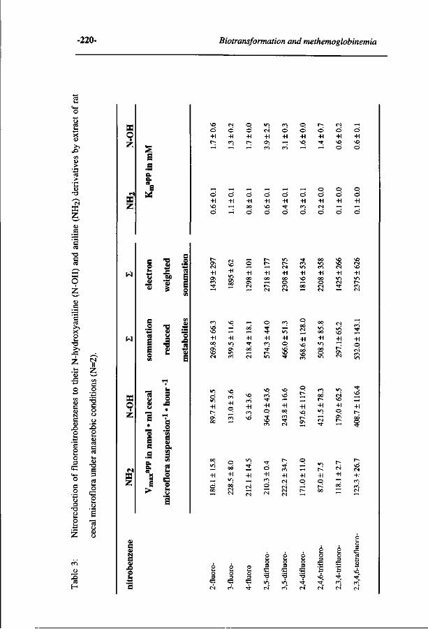
-220- Biotransformation and methemoglobinemia
x ID
- O CO U _> 'S > 'C (D
T 3 f — \
CS K Z u e
O z » c
e es > •
X O Ui
T l P^ 43 Z tl
<l)
43 O co
C O N
e t> o o Ui
o U i O 3
<4-l
O c o
o 3 TJ
o
Z
CS II z e o
T3 C O o o X)
es e CS Ui
T3 e 3 CS u O
G R u
•a CJ
o
CO
3 CS
H
X z
w
w
CS
X Z
e "O O £ J3 «
•8 1 a "öS
•o cu u S
•8
çu J3
a o
i ä .S s o. « " o
« i "
S o
> t
CU
S CU N S V 45 O
*0 O
+1 f-
-
cs o +1 en
~-
o o +1 r-~ H
>r> CS
+1 o\ m
en
o +1 ~ H
en
O O
+1 o - H
t-O
+1 -* ^ H
CS
O +1 ^O
o
~ H
o +1 *o o
o +1 *o o
o +1 r-H
~ H
o +1 00
o
o +1 \o o
o +1 •* o
o +1 en
o
o o +1 CS
o
o o +1 - H
o
o o +1 ~" o
+1 o
—' —i CM
+1 -"
5 1
VO r-H —
m —i
+1
—4 oo ^ |
+1 r ^ ""1 —H
ö
•* • *
+1 Cn
Vi
+1 o
Tt'
+1 — H
CS
CS
Ö +1 O
CS
•*' en +1 CS CS CS CS
+1 o r~
+1 o OO c s ;_; —i
u
3
53 CS
3 53
en
Ui
3
53 -*
H 3
53 T3
>n CS
H 3
53 -O >n en
H 3
53 •o • *
CS
•o •o CS
VO CS
o
o +1
r-l
+1 o
en
•* +1 o t-
\o en
o •— +1 00 en
•* es
O
r~ ^ H
+1 O
OO
r-+1 »o CS Tf
es o
+1 o o\ r-^
•* \o „H
+1 t~
•*
»O CS
+1
3
53
\o
3
53 Ö
• *
H U
VO • *
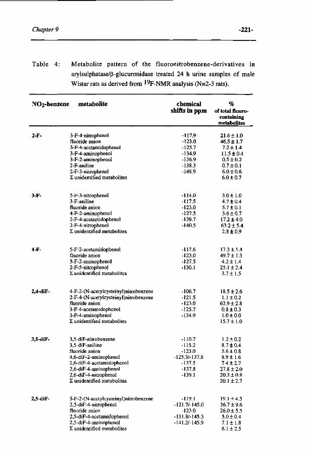
Chapter 9 -221-
Table 4: Metabolite pattern of the fluoronitrobenzene-derivatives in
arylsulphatase/ß-glucuronidase treated 24 h urine samples of male
Wistar rats as derived from ^p-NMR analysis (N=2-3 rats).
NCh-benzene metabolite
2-F-
3-F-
4-F-
2,4-diF-
3,5-diF-
2,5-diF-
3-F-4-nitrophenol fluoride anion 3-F-4-acetamidophenol 3-F-4-aminophenol 3-F-2-aminophenol 2-F-aniline 2-F-3-nitrophenol £ unidentified metabolites
5-F-3-nitrophenol 3-F-aniline fluoride anion 4-F-2-aminophenol 2-F-4-acetamidophenol 2-F-4-nitrophenol £ unidentified metabolites
5-F-2-acetamidophenol fluoride anion 5-F-2-aminophenol 2-F-5-nitrophenol £ unidentified metabolites
4-F-2-(N-acetylcysteinyl)nitrobenzene 2-F-4-(N-acetylcysteinyl)nitrobenzene fluoride anion 3-F-4-acetamidophenol 3-F-4-aminophenol X unidentified metabolites
3,5-diF-nitrobenzene 3,5-diF-aniline fluoride anion 4,6-diF-2-aminophenol 2,6-diF-4-acetamidophenol 2,6-diF-4-aminophenol 2,6-diF-4-nitrophenol X unidentified metabolites
5-F-2-(N-acetylcysteinyl)nitrobenzene 2,5-diF-4-nitrophenol fluoride anion 2,5-diF-4-acetamidophenol 2,5-diF-4-aminophenol X unidentified metabolites
chemical shifts in ppm
-117.9 -123.0 -125.7 -134.9 -136.9 -138.3 -148.9
-114.0 -117.5 -123.0 -127.5 -139.7 -140.5
-117.6 -123.0 -127.5 -130.1
-106.7 -121.5 -123.0 -125.7 -134.9
-110.7 -115.2 -123.0
-125.3/-137.8 -137.5 -137.8 -139.1
-119.1 -121.7/-145.0
-123.0 -131.8/-145.3 -141.2/-145.9
% of total fluoro-
containing metabolites
21.6+ 1.0 46.5 ±1.7 7.2+1.4 11.5 + 0.4 0.5 + 0.2 0.7 + 0.1 6.0 ±0.6 6.0 + 0.7
3.0 ±1.0 4.7 ± 0.4 5.7 ±0.1 3.6 ±0.7
17.2 + 4.0 63.2 ±5.4 2.8 ±0.9
17.3 ±1.4 49.7 ±1.3 4.2 ±1.4
25.1 ±2.4 3.7 ±1.5
18.5 + 2.6 1.1 ±0.2
62.9 ± 2.8 0.8 ± 0.3 1.0 + 0.0
15.7 ±1.0
1.2 + 0.2 8.7 ±0.4 5.6 ±0.8 8.9+1.6 7.4 ±2.7
27.8 ± 2.0 20.3 ±0.8 20.1 ±2.7
19.1+4.5 36.7 ± 9.6 26.0 ±5.5 5.0 ±0.4 7.1 ±1.8 6.1 ±2.5
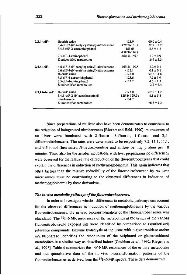
-222- Biotransformation and methemoglobinemia
2,3,4-triF-
2,4,6-triF-
2,3,4,6-tetraF
fluoride anion 3,4-diF-2-(N-acetylcysteinyl)-nitrobenzene 3,4,5-triF-2-acetamidophenol
2,3-diF-4-aminophenol £ unidentified metabolites
4,6-diF-2-(N-acetylcysteinyl)-nitrobenzene 2,6-diF-4-(N-acetylcysteinyl)-nitrobenzene fluoride anion 3,5-diF-4-acetamidophenol 3,5-diF-4-aminophenol Z unidentified metabolites
fluoride anion 3,4,6-triF-2-(N-acetylcysteinyl)-nitrobenzene £ unidentified metabolites
-123.0 -129.2/-131.2
-153.6/ -158.7/-176.9 -160.5/-165.3
-105.5/-119.5 -122.1 -123.0 -123.6 -133.7
-123.0 -126.8/-129.5 /
-134.7
60.0 ±0.4 12.9 ±2.2 8.6 ±4.3
1.9 ±0.9 16.6 + 3.3
2.2 ±0.1 0.1+0.1 72.6 ±4.6 7.9 ±1.9 4.5 ±1.3 12.7 ±2.4
67.6 ±1.2 6.1 + 3.3
26.3 ± 2.2
Since preparations of rat liver also have been demonstrated to contribute to
the reduction of halogenated nitrobenzenes [Rickert and Held, 1990], microsomes of
rat liver were incubated with 2-fluoro-, 3-fluoro-, 4-fluoro- and 2,5-
difluoronitrobenzene. The rates were determined to be respectively 8.3, 11.1, 11.1,
and 9.5 nmol fluorinated N-hydroxyaniline and aniline per mg protein per 10
minutes. Thus, also for the aerobic incubations with liver preparations no differences
were observed for the relative rate of reduction of the fluoronitrobenzenes that could
explain the differences in induction of methemoglobinemia. This again indicates that
other factors than the relative reducibility of the fluoronitrobenzene by rat liver
microsomes must be contributing to the observed differences in induction of
methemoglobinemia by these derivatives.
The in vivo metabolic pathways of the fluoronitrobenzenes.
In order to investigate whether differences in metabolic pathways can account
for the observed differences in induction of methemoglobinemia by the various
fluoronitrobenzenes, the in vivo biotransformation of the fluoronitrobenzenes was
elucidated. The 19F-NMR resonances of the metabolites in the urines of the various
fluoronitrobenzene exposed rats were identified by comparison to (synthetised)
reference compounds. Enzyme hydrolysis of the urine with ß-glucuronidase and/or
arylsulphatase identifies the resonances of the sulphated or glucuronidated
metabolites in a similar way as described before [Cnubben et al, 1992; Rietjens et
al, 1995]. Table 4 summarizes the 19F-NMR resonances of the urinary metabolites
and the quantitative data of the in vivo biotransformation patterns of the
fluoronitrobenzenes as derived from the 19F-NMR spectra. These data demonstrate
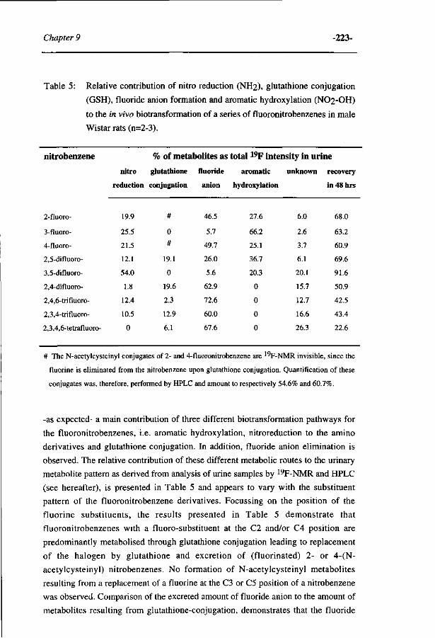
Chapter 9 -223-
Table 5: Relative contribution of nitro reduction (NH2), glutathione conjugation
(GSH), fluoride anion formation and aromatic hydroxylation (NO2-OH)
to the in vivo biotransformation of a series of fluoronitrobenzenes in male
Wistar rats (n=2-3).
nitrobenzene % of metabolites as total 19F intensity in urine
nitro glutathione fluoride aromatic unknown recovery
reduction conjugation anion hydroxylation in 48 hrs
2-fluoro-
3-fluoro-
4-fluoro-
2,5-difluoro-
3,5-difluoro-
2,4-difluoro-
2,4,6- tri fluoro-
2,3,4-trifluoro-
2,3,4,6-tetrafluoro-
19.9
25.5
21.5
12.1
54.0
1.8
12.4
10.5
0
#
0
#
19.1
0
19.6
2.3
12.9
6.1
46.5
5.7
49.7
26.0
5.6
62.9
72.6
60.0
67.6
27.6
66.2
25.1
36.7
20.3
0
0
0
0
6.0
2.6
3.7
6.1
20.1
15.7
12.7
16.6
26.3
68.0
63.2
60.9
69.6
91.6
50.9
42.5
43.4
22.6
# The N-acetylcysteinyl conjugates of 2- and 4-fluoronitrobenzene are 19F-NMR invisible, since the
fluorine is eliminated from the nitrobenzene upon glutathione conjugation. Quantification of these
conjugates was, therefore, performed by HPLC and amount to respectively 54.6% and 60.7%.
-as expected- a main contribution of three different biotransformation pathways for
the fluoronitrobenzenes, i.e. aromatic hydroxylation, nitroreduction to the amino
derivatives and glutathione conjugation. In addition, fluoride anion elimination is
observed. The relative contribution of these different metabolic routes to the urinary
metabolite pattern as derived from analysis of urine samples by 19F-NMR and HPLC
(see hereafter), is presented in Table 5 and appears to vary with the substituent
pattern of the fluoronitrobenzene derivatives. Focussing on the position of the
fluorine substituents, the results presented in Table 5 demonstrate that
fluoronitrobenzenes with a fluoro-substituent at the C2 and/or C4 position are
predominantly metabolised through glutathione conjugation leading to replacement
of the halogen by glutathione and excretion of (fluorinated) 2- or 4-(N-
acetylcysteinyl) nitrobenzenes. No formation of N-acetylcysteinyl metabolites
resulting from a replacement of a fluorine at the C3 or C5 position of a nitrobenzene
was observed. Comparison of the excreted amount of fluoride anion to the amount of
metabolites resulting from glutathione-conjugation, demonstrates that the fluoride
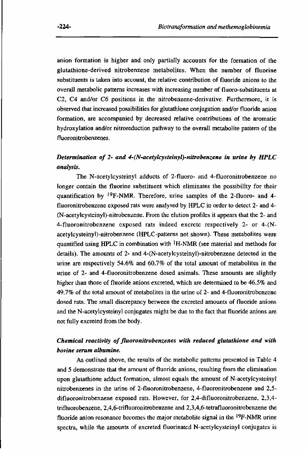
-224- Biotransformation and methemoglobinemia
anion formation is higher and only partially accounts for the formation of the
glutathione-derived nitrobenzene metabolites. When the number of fluorine
substituents is taken into account, the relative contribution of fluoride anions to the
overall metabolic patterns increases with increasing number of fluoro-substituents at
C2, C4 and/or C6 positions in the nitrobenzene-derivative. Furthermore, it is
observed that increased possibilities for glutathione conjugation and/or fluoride anion
formation, are accompanied by decreased relative contributions of the aromatic
hydroxylation and/or nitroreduction pathway to the overall metabolite pattern of the
fluoronitrobenzenes.
Determination of 2- and 4-(N-acetylcysteinyl)-nitrobenzene in urine by HPLC
analysis.
The N-acetylcysteinyl adducts of 2-fluoro- and 4-fluoronitrobenzene no
longer contain the fluorine substituent which eliminates the possibility for their
quantification by 19F-NMR. Therefore, urine samples of the 2-fluoro- and 4-
fluoronitrobenzene exposed rats were analysed by HPLC in order to detect 2- and 4-
(N-acetylcysteinyl)-nitrobenzene. From the elution profiles it appears that the 2- and
4-fluoronitrobenzene exposed rats indeed excrete respectively 2- or 4-(N-
acetylcysteinyl)-nitrobenzene (HPLC-patterns not shown). These metabolites were
quantified using HPLC in combination with ÏH-NMR (see material and methods for
details). The amounts of 2- and 4-(N-acetylcysteinyl)-nitrobenzene detected in the
urine are respectively 54.6% and 60.7% of the total amount of metabolites in the
urine of 2- and 4-fluoronitrobenzene dosed animals. These amounts are slightly
higher than those of fluoride anions excreted, which are determined to be 46.5% and
49.7% of the total amount of metabolites in the urine of 2- and 4-fluoronitrobenzene
dosed rats. The small discrepancy between the excreted amounts of fluoride anions
and the N-acetylcysteinyl conjugates might be due to the fact that fluoride anions are
not fully excreted from the body.
Chemical reactivity of fluoronitrobenzenes with reduced glutathione and with
bovine serum albumine.
As outlined above, the results of the metabolic patterns presented in Table 4
and 5 demonstrate that the amount of fluoride anions, resulting from the elimination
upon glutathione adduct formation, almost equals the amount of N-acetylcysteinyl
nitrobenzenes in the urine of 2-fluoronitrobenzene, 4-fluoronitrobenzene and 2,5-
difluoronitrobenzene exposed rats. However, for 2,4-difluoronitrobenzene, 2,3,4-
trifluorobenzene, 2,4,6-trifluoronitrobenzene and 2,3,4,6-tetrafluoronitrobenzene the
fluoride anion resonance becomes the major metabolite signal in the 19F-NMR urine
spectra, while the amounts of excreted fluorinated N-acetylcysteinyl conjugates is
Chapter 9 -225-
0> CL
CT" ET
CL
CD
?o
« o 3 (
O o\
ft s.
o
O 1/1 33
Ui Ol J^ Ui Ui V H] Éü S3)
V *• "o-, à. éx à. g g g
t3.3. | ? §> 3 ~ " î ? » o o
o o
o o o O O
O O
n n n n n n n i i n
h h S h i i n W W Q u u ^
ö ö *•
o o o o o o o o o
'._. 10 Ö — \b ~ 'Ó ~ gg£°g£g g ö ö ó ö ö ö
o o o o o o o o o
Sgä°8Sg 8
p a p p a p p a p
o " oo • u> o • o
i+i+H-i+l+H-l+i+è M I O U I P P I O P P O N U » O N > O U ) > - , - & . 0 *
< - n 0 0 0 0 ° O ' ° O O 3
1+1+1+1+1+1+1+1+ ' u w w o o o o o * ^ û « l j O - W O O
5. s? o O"
S
o
STB'
3 < E. O *
3 a-
o C/5
Ö5
>
i «
m
s cro
s
2
H
et ON
O CT
1 ff
o ' v> O * * > 3E
et
c o - 1
5'
1 CL S. 3 p S-
o' 3
^
a C O o 3 ^
1 <T CD 3 N ce 3 CD
O ^t EL
3
o ' p
o'
3
1 T3
3 ^ CO 3 C? C L
5' H S3 a ; cô*
r"
C
O 3
^^ 3 " 2. M "
CD P O CJ-
O 3
3
3E
CD C L B O CD CL
<£,
3 * S ' 3 CD
S i
3
2 v-*'
C L
O
< 5' CD
VI CD *-i C
3
CT C
3. 3 CD
^ O
3 era
3
3 -CD
C/3
-^ C o
s
Z
?° H 3 " CD
1 o' 3
CD
3 S3 Vi
3 o 3 3 / O 3 CL
O" *<;
5
S o'
3
O •-h 3 3 O
5.' CD P 3 o' 3 Vi if o
3
3-CD CD 3 .
O CT CD 3 N CD 3 CD i
C L CD 2
S-
et Vi
u o
3
S
3 .
o a* et 3 N CD 3 et Vi
§ CL
r^. B " CD
P O 2". O 3
CD v H> O
et
o o
a. c P a
5' 3 O 3 E
CD
CD
3 .
^ O CT et 3 N CD 3 CD Vi
g. r-l-3 -
o 0 0
a P 3 C L
td 0 0 > P
C L CD CD
5' CD C L
CT *<
_
1
3 " Cl
•-1 et
"S o' Vi CD
et" o
<' 3 , ' •< :
S1
l-i
3 " CD
5'
<_
<' o P 3 CL
3
<
O
/_N Vi
o 3
P 3 CD O C t/5 P 3 C L
CD 3 N •<
3 P
o '
O 0 0
a o o
< 3
c ' tTQ P
o ' 3
O | -h
3 CD
3 c o *n_ 5' P
CD C L
-226- Biotransformation and methemoglobinemia
considerably lower. Furthermore, the total 48 hours recovery of the
fluoronitrobenzenes strongly decreases with increasing number of fluoro-substituents
(Table 5). Together, these observations point at formation of fluoronitrobenzene
adducts, resulting in fluoride anion formation and metabolites which are withheld in
the body.
To investigate the above-mentioned hypothesis, the reactivity of the various
fluoronitrobenzenes in a spontaneous (non-enzymatic) nucleophilic substitution
reaction with GSH and also with a model protein (bovine serum albumin) was
studied. A nucleophilic substitution of a fluorine substituent in a fluoronitrobenzene
by a cellular macromolecule will lead to formation of a covalent macromolecular-
nitrobenzene adduct accompanied by the elimination of a fluoride anion. Table 6
presents the reaction rates for the chemical reaction between the various
fluoronitrobenzene derivatives with GSH or with BSA. The results demonstrate that
C2, C4 and/or C6 fluorinated carbon centres of the fluoronitrobenzenes are sensitive
towards a spontaneous attack by nucleophiles like GSH and BSA. In case of GSH
the molecular weight of the resulting adduct is low enough to detect this product by 19F-NMR and, thus, the regioselectivity of the GSH conjugation with
fluoronitrobenzenes could be determined. A strong preference for glutathione
conjugation at a fluorinated C2 position over a fluorinated C4 position is observed.
The regioselectivity for the spontaneous conjugation of GSH with the various
fluoronitrobenzenes resembles the regioselectivity for the in vivo formation of the
fluorinated N-acetylcysteinyl nitrobenzenes (Table 2). The results in Table 6
additionally demonstrate that the reactivity of the fluoronitrobenzenes with
nucleophilic entities as GSH and BSA increases remarkably with increasing number
of fluoro-substituents.
Molecular orbital calculations.
The electronic substrate characteristics of the nitrobenzene derivatives were
calculated in order to investigate whether the observed differences in
biotransformation pathways, methemoglobin formation as well as the almost similar
rate of their in vitro nitroreduction could be explained by electronic characteristics of
the fluoronitrobenzenes.
Table 7 presents the AMI calculated values for the energy level of the frontier orbital
of importance for a nucleophilic attack on the fluoronitrobenzenes, i.e. the energy of
the lowest unoccupied molecular orbital (E(LUMO)). A decrease in E(LUMO) with
increasing number of electron withdrawing substituents at the aromatic ring is
observed and can be expected to lead to a more efficient interaction with a
nucleophile.
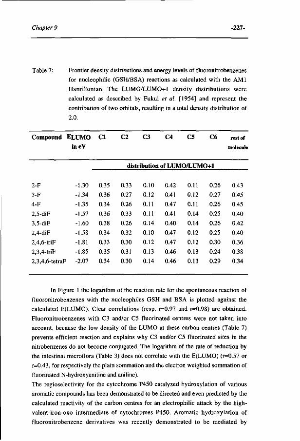
Chapter 9 -227-
Table 7: Frontier density distributions and energy levels of fluoronitrobenzenes
for nucleophilic (GSH/BSA) reactions as calculated with the AMI
Hamiltonian. The LUMO/LUMO+1 density distributions were
calculated as described by Fukui et al. [1954] and represent the
contribution of two orbitals, resulting in a total density distribution of
2.0.
Compound
2-F
3-F
4-F
2,5-diF
3,5-diF
2,4-diF
2,4,6-triF
2,3,4-triF
2,3,4,6-tetraF
ELUMO ineV
-1.30 -1.34
-1.35
-1.57
-1.60
-1.58
-1.81
-1.85
-2.07
CI
0.35
0.36
0.34
0.36
0.38
0.34
0.33
0.35
0.34
C2 C3 C4 C5 C6
distribution of LUMO/LUMO+1
0.33
0.27
0.26
0.33
0.26
0.32
0.30
0.31
0.30
0.10
0.12
0.11
0.11
0.14
0.10
0.12
0.13
0.14
0.42
0.41
0.47
0.41
0.40
0.47
0.47
0.46
0.46
0.11
0.12
0.11
0.14
0.14
0.12
0.12
0.13
0.13
0.26
0.27
0.26
0.25
0.26
0.25
0.30
0.24
0.29
rest of
molecule
0.43
0.45
0.45
0.40
0.42
0.40
0.36
0.38
0.34
In Figure 1 the logarithm of the reaction rate for the spontaneous reaction of
fluoronitrobenzenes with the nucleophiles GSH and BSA is plotted against the
calculated E(LUMO). Clear correlations (resp. r=0.97 and r=0.98) are obtained.
Fluoronitrobenzenes with C3 and/or C5 fluorinated centres were not taken into
account, because the low density of the LUMO at these carbon centres (Table 7)
prevents efficient reaction and explains why C3 and/or C5 fluorinated sites in the
nitrobenzenes do not become conjugated. The logarithm of the rate of reduction by
the intestinal microflora (Table 3) does not correlate with the E(LUMO) (r=0.57 or
r=0.43, for respectively the plain sommation and the electron weighted sommation of
fluorinated N-hydroxyaniline and aniline).
The regioselectivity for the cytochrome P450 catalyzed hydroxylation of various
aromatic compounds has been demonstrated to be directed and even predicted by the
calculated reactivity of the carbon centres for an electrophilic attack by the high-
valent-iron-oxo intermediate of cytochromes P450. Aromatic hydroxylation of
fluoronitrobenzene derivatives was recently demonstrated to be mediated by
-228- Biotransformation and methemoglobinemia
O) o
2 -
0 -
- 1 . 8 - 1 . 6 -1 E(LUMO) in eV
- 1 . 2
Figure 1: Molecular orbital based quantitative structure activity relationship for the
spontaneous conjugation of a series of fluoronitrobenzenes with the nucleophilic
entities glutathione (*) and bovine serum albumine (O). Correlation coefficients
were respectively r = 0.97 and r = 0.98. The chemical reactions are dependent on the
concentrations of both substrates and were carried out at fixed concentrations of
fluoronitrobenzenes (3 mM) and glutathione (2 mM) or BSA (0.15 mM). For both
reactions V is in uM of fluoride anion formed per minute.
cytochromes P450 [Rietjens et al, 1995]. Table 8 presents the regioselectivity for the
cytochrome P450 mediated aromatic hydroxylation of the fluoronitrobenzenes of the
present study compared to calculated reactivity parameters for electrophilic or
nucleophilic attack on those fluoronitrobenzenes. Only nitrobenzenes for which
nitrophenol derived metabolites were actually detected in the urine of the exposed
rats are taken into account. The results show that aromatic hydroxylation
predominantly occurs at the non-fluorinated C4 position, whereas hydroxylation at
the C3 position occurs to a much smaller extent and C2/C6-hydroxylation is not
observed at all. The regioselectivity for aromatic hydroxylation of the
fluoronitrobenzenes does not correspond with the position of the frontier orbital
density for electrophilic attack on the nitrobenzenes e.g. HOMO/HOMO-1, which is
predominantly located on the C2 and C3 carbon centres, whereas the hydroxylation
occurs mainly at C4. Surprisingly, the frontier orbital density for a nucleophilic
attack on the nitrobenzenes, calculated from their LUMO/LUMO+1, is
Chapter 9 -229-
predominantly located at the hydroxylated C4 carbon centres of the fluoronitrobenzene
and to a much smaller extent at C3 (Table 7). The C2 and C6 positions with reasonable
reactivity for either an electrophilic or nucleophilic attack, become never hydroxylated,
apparently due to factors other than their intrinsic reactivity.
Table 8: Regioselectivities for the in vivo aromatic hydroxylation of
fluoronitrobenzene derivatives as determined by l^F-NMR urinalysis
compared to calculated frontier orbital characteristics.
nitro- aromatic hydroxylation
benzene at carbon centres
derivative C2 : C3 : C4 : C5 : C 6
LUMO/LUMO+1 HOMO/HOMO-1
frontier orbital densities at carbon centres
C2 : C3 : C4 : C5 : C6 C2 : C3 : C4 : C5 : C 6
2-F- 0.00:0.22:0.78:0.00:0.00 0.33:0.10:0.42:0.11:0.26 0.41:0.32:0.12:0.51:0.23
3-F- 0.00:0.00:0.95:0.05:0.00 0.27:0.12:0.41:0.12:0.27 0.31:0.47:0.21:0.19:0.46
4-F- 0.00:0.50:0.00:0.50:0.00 0.26:0.11:0.47:0.11:0.26 0.17:0.30:0.39:0.30:0.17
2,5-diF- 0.00:0.00:1.00:0.00:0.00 0.33:0.11:0.41:0.14:0.25 0.43:0.20:0.12:0.49:0.24
3,5-diF- 0.00:0.00:1.00:0.00:0.00 0.26:0.14:0.40:0.14:0.26 0.44:0.31:0.13:0.31:0.44
DISCUSSION.
In this study, possible relationships between various biotransformation
pathways and the methemoglobin inducing capacity of a series of fluoronitrobenzene
were investigated. Induction of methemoglobinemia by fluoronitrobenzenes was
observed to decrease with increasing number of fluorine substituents, especially when
these fluoro-substituents are positioned at the carbon centres C2, C4 and/or C6,
calculated to be most reactive for a nucleophilic attack.
The primary reduction of nitrobenzene-derivatives to nitrosobenzenes and N-
hydroxyarylamines is the crucial step for the methemoglobin inducing activity of this
class of compounds [for review, see Kiese, 1974; Suzuki et al., 1989] as
schematically presented in Figure 2. This reductive metabolism of unsubstituted
nitrobenzene to the reactive methemoglobin inducing species is predominantly
accomplished by the bacterial nitroreductases present in the intestinal environment
[Levin and Dent, 1982]. Besides, for a series of monochloronitrobenzenes, it was
demonstrated that hepatocytes of rat liver are able to reduce these halogenated
nitrobenzenes to their corresponding aniline-derivatives under aerobic conditions
[Rickert and Held, 1990]. In vitro experiments with a suspension of rat cecal
-230- Biotransformation and methemoglobinemia
methemoglobinemia
ADDUCTS + F"
nucleophilic cellular
macromolecule«
F +
excretion excretion excretion
Figure 2: Schematic representation of the metabolic fate of fluorinated nitrobenzenes. GST:
glutathione S-transferase, yGT: yglutamyl transpeptidase, AP: aminopeptidase M
and cysteinylglycine dipeptidase, UDPG: UDP-glucuronyltransferase, ST:
sulphotransferase, NAT: N-acetyltransferase.
microflora or with a preparation of rat liver demonstrate that the rate of reduction of
the various fluoronitrobenzenes varies only slightly, and not in a way that can
account for the differences in extent of methemoglobin formation. Apparently, the
induction of methemoglobinemia by the various fluoronitrobenzenes is not
predominantly determined by their intrinsic capacity to enter the nitro reduction
pathway. The different methemoglobinemic capacity must rather result from
differences in the inherent direct methemoglobinemic capacity and/or reactivity of
the various toxic metabolites, and/or from the fact that the halogen substituent pattern
influences the electrophilic reactivity, thereby changing the possibilities for reactions
with glutathione and, especially, other cellular nucleophiles.
Chapter 9 -231-
In vivo metabolite patterns of the fluoronitrobenzenes, derived from the
urinary 19F-NMR metabolite profiles of the fluoronitrobenzene exposed rats
provided support for the latter explanation. The metabolite patterns indicate that the
differences in the extent of methemoglobin induction with changing fluorine
substituent patterns in the nitrobenzenes is more likely to be related to the presence
of competing reaction pathways. Excretion of mercapturic acids, increased fluoride
anion elimination and reduced recoveries with increasing number of fluorine
substituents were observed, pointing at reactions of the fluoronitrobenzenes with
GSH and/or other cellular nucleophiles (as could be jugded from the yellow colour of
the intestinal tract (unpublished)). A reaction with GSH and/or cellular
macromolecules is a factor that prevents the fluoronitrobenzenes to become reduced
by and/or, as a consequence, reduces their chances to induce methemoglobinemia.
Additional in vitro studies demonstrated QSAR's for these conjugation pathways
relating the natural logarithm of the rates of conjugation of fluoronitrobenzenes with
GSH as well as BSA to a calculated parameter representative for the electrophilicity
of the nitrobenzenes i.e. the energy of their lowest unoccupied molecular orbital
(E(LUMO)). The correlation coefficients obtained were respectively r=0.97 and
r=0.98. Conjugation with GSH at a halogenated C3 or C5 centre is not observed,
which can be ascribed to a low reactivity for a nucleophilic attack at these positions
reflected by the relatively low calculated LUMO/LUMO+1 frontier orbital densities.
Thus, depending on the position and number of fluorine substituents the reactivity of
the fluoronitrobenzenes for nucleophilic attack by either glutathione or cellular
macromolecules is influenced, and this results in more or less efficient alternatives
for the nitro reduction pathway that gives rise to methemoglobin inducing
metabolites.
The exception to this general consideration might be the results obtained for
2- and 4-fluoronitrobenzene. The amounts of reduced metabolites and mercapturic
acids in urine (Table 5), as well as the rate of conjugation with GSH and BSA (Table
6), were almost similar for 2- and 4-fluoronitrobenzene, 2-fluoronitrobenzene
appeared to be a more potent inducer of methemoglobinemia compared to 4-
fluoronitrobenzene (Table 2). An explanation for this observation might be that the
nitroso- and N-hydroxy-metabolites of 4-fluoronitrobenzene are relatively more
efficiently reduced to their aniline-analogs compared to 2-fluoronitrobenzene as
demonstrated in Table 3. Thus, besides the presence of competing reaction pathways
with GSH and/or cellular macromolecules, the extent and rate of complete
nitroreduction also seems to be involved in the in vivo regeneration of
methemoglobinemia inducing metabolites of fluoronitrobenzenes. Furthermore,
additional factors that might contribute to these observed differences might originate
from possible differences in the inherent direct methemoglobinemic capacity of the
-232- Biotransformation and methemoglobinemia
N-hydroxy and nitroso metabolites and/or differences in the stability of the N-
hydroxylamine/nitroso redox pair in the erythrocyt. Conclusive evidence on the
involvement of these factors, however, are not provided yet and these interesting
considerations may serve as a basis for future research.
Finally, the metabolic profiles obtained for the various fluoronitrobenzenes in the
present study demonstrated that the extent of cytochrome P450 mediated aromatic
hydroxylation of the fluoronitrobenzenes did not vary significantly with the
substituent patterns. A recent study on the biotransformation of 2,5-
difluoronitrobenzene clearly demonstrated that cytochrome P450 is the enzyme-
system responsible for the aromatic hydroxylation of this derivative [Rietjens et al,
1995]. This cytochrome P450 mediated aromatic hydroxylation is generally regarded
as a detoxification route, due to the efficient conjugation of nitro- and aminophenols
with sulphate or glucuronyl moieties and their subsequent elimination from the body
(Figure 2). For the aniline metabolites of the present study their (non)preferential site
of hydroxylation can qualitatively be explained on the basis of the calculated
reactivity of the carbon centres for an electrophilic attack by the activated
cytochrome P450 high-valent-iron-oxo intermediate (data not shown), similar to
what was previously reported for other aromatic compounds [Cnubben et al, 1992;
Rietjens et al., 1993]. For the nitrobenzenes, however, the preferential site for
hydroxylation is more related to the frontier orbital density distribution for a
nucleophilic attack. It may be hypothesized that the aromatic hydroxylation of
substrates with a relatively high electrophilicity proceeds not by an electrophilic
attack of the high-valent-iron-oxo species of the cytochromes P450, but by a
nucleophilic attack of another intermediate in the catalytic cytochrome P450 cycle,
for instance the nucleophilic cytochrome Fe3+-0-0~ intermediate. The Fe3+-0-0~
intermediate might react with the substrate instead of decomposing to the usual
(FeO)3+ intermediate, in a way as is proposed for the cleavage of C-C bonds during
oestrogen biosynthesis [Akhtar and Wright, 1991; Ortiz de Montellano 1985]. The
high electrophilic reactivity of the nitrobenzenes may support the preferential
occurence of such a nucleophilic attack by the Fe3+-0-0" intermediate over a
pathway proceeding by an attack of the electrophilic (FeO)3+ intermediate.
REFERENCES.
Akhtar M., and Wright J.N. (1991). A unified mechanistic view of oxidative reactions catalyzed by P-
450 and related Fe-containing enzymes. Nat. Prod. Rep. 8, 527-551.
Beauchamp R.O., Irons R.D., Rickert D.E., Couch D.B., and Hamm T.E. (1982). A critical review of
the literature on nitrobenzene toxicity. CRC Critical Reviews in Toxicology 11, 33-84.
Chapter 9 -233-
Cesarone CF., Bolognesi C , and Santi L. (1983). DNA damage induced in vivo in various tissues by
nitrochlorobenzene derivatives. Mutation Research 116, 239-246.
Cnubben N.H.P., Peelen S., Borst J.W., Vervoort J., and Rietjens I.M.C.M. (1994). Molecular orbital
based quantitative structure activity relationship for the cytochrome P450-catalyzed 4-
hydroxylation of halogenated anilines. Chem. Res. Toxicol. 7, 590-598.
Cnubben N.H.P., Vervoort J., Veeger C , and Rietjens I.M.C.M. (1992). Study on the regioselectivity
and mechanims of the aromatic hydroxylation of monofluoroanilines. Chem.-Biol.
Interactions 85, 151-172.
Debnath A.K., Lopez de Compadre R.L., Debnath G., Shusterman A.J., and Hansch C. (1991).
Structure activity relationship of mutagenic aromatic and heteroaromatic nitrocompounds.
Correlation with molecular orbital energies and hydrophobicity. J. Med. Chem. 34, 785-797.
Debnath A.K., Lopez de Compadre R.L., Shusterman A.J., and Hansch C. (1992). Quantitative
structure activity relationship investigation of the role of hydrophobicity in regulating
mutagenicity in the Ames test: 2. Mutagenicity of aromatic and heteroaromatic
nitrocompounds in Salmonella Typhimurium TA 100. Environmental and Molecular
Mutagenesis 19, 53-70.
Fukui K., Yonezawa T., Nagata C , and Shingu H. (1954). Molecular orbital theory of orientation in
aromatic, heteroaromatic and other conjugated molecules. J. Chem. Phys. 22, 1433-1442.
Harada N., and Omura T. (1980). Participation of cytochrome P-450 in the reduction of nitro
compounds by rat liver microsomes. J. Biochem. 87, 1539-1554.
Harrison J.H., and Jollow D.J. (1987). Contribution of aniline metabolites to aniline-induced
methemoglobinemia. Molec. Pharmac. 32, 423-31.
Kennedy R.J., and Stock A.M. (1960). The oxidation of organic substances by potassium
peroxymonosulphate. J. Org. Chem. 25, 1901-1906.
Kiese M. (1974). Methemoglobinemia: A comprehensive treatise. Chapter 7, CRC Press, Cleveland,
Ohio.
Levin A.A., and Dent J.G. (1982). Comparison of the metabolism of nitrobenzene by hepatic
microsomes and cecal microflora from Fischer-344 rats in vitro and the relative importance of
each in vivo. Drug Metab. Dispos. 10, 450-454.
Lopez de Compadre R.L., Debnath A.K., Shusterman A.J., and Hansch C. (1990). LUMO energies
and hydrophobicity as determinants of mutagenicity by nitroaromatic compounds in
Salmonella typhimurium. Environmental and molecular mutagenesis 15, 44-55.
Maples K.R., Eyer P., and Mason R.P. (1989). Aniline-, phenylhydroxylamine-, nitrosobenzene-, and
nitrobenzene-induced hemoglobin thiyl free radical formation in vivo and in vitro. Molec.
Pharmac. 37,311-318.
O'Brien P.J., Wong W.C., Silva J., and Khan S. (1990). Toxicity of nitrobenzene compounds towards
isolated hepatocytes: Dependence on reduction potential. Xenobiotica 20, 945-955.
Ortiz de Montellano P. (1985). Cytochrome P-450, structure, mechanism and biochemistry, chapter
11, Plenum Press, New York.
-234- Biotransformation and methemoglobinemia
Piotrowski J. (1967). Further investigations on the evaluation of exposure to nitrobenzene. Br. J. Ind.
Med. 24, 60-65.
Reddy B.G., Pohl L.R., and Krishna G. (1976). The requirement of the gut flora in nitrobenzene-
induced methemoglobinemia in rats. Biochem. Pharmac. 25, 1119-1122.
Rickert D.E. (1987). Metabolism of nitro aromatic compounds. Drug Metab. Rev. 18,23 -53.
Rickert D.E., and Held S. (1990). Metabolism of chloronitrobenzenes by isolated rat hepatocytes.
Drug Metab. Disp. 18, 5-9.
Rietjens I.M.C.M., Cnubben N.H.P., van Haandel M., Tyrakowska B., Soffers A.E.M.F., and
Vervoort J. (1995a). Different metabolic pathways of 2,5-difluoronitrobenzene and 2,5-
difluoroaminobenzene compared to molecular orbital substrate characteristics. Chem.-Biol.
Interactions 94,49-72.
Rietjens I.M.C.M., Soffers A.E.M.F., Hooiveld G., Veeger C., and Vervoort J. (1995b). Quantitative
structure activity relationships based on computer calculated parameters for the overall rate of
glutathione S-transferase catalyzed conjugation of a series of fluoronitrobenzenes. Chem. Res.
Toxicol. 8,481-488, 1995.
Rietjens I.M.C.M., Soffers A.E.M.F., Veeger C , and Vervoort J. (1993). Regioselectivity of
cytochrome P450 catalyzed hydroxylation of fluorobenzenes predicted by calculated frontier
orbital substrate characteristics. Biochemistry 32, 4801-4812.
Sabbioni G. (1992). Hemoglobin binding of monocyclic aromatic amines: molecular dosimetry and
quantitative structure activity relationships for the N-oxidation. Chem.-Biol. Interact. 81, 91-
117.
Sabbioni G. (1994). Hemoglobin binding of nitroarenes and quantitative structure-activity
relationships. Chem. Res. Toxicol. 7, 267-274.
Shimizu M., Yasui Y., and Matsumoto N. (1983). Structural specificity of aromatic compounds with
special reference to mutagenic activity in Salmonella typhimurium -a series of chloro- or
fluoro-nitrobenzene derivatives. Mutation Research 116, 217-238.
Suzuki J., Meguro S., Moriat O., Hirayama S., and Suzuki S. (1989). Comparison of in vivo binding
of aromatic nitro and amino compounds to rat hemoglobin. Biochem. Pharmac. 38, 3511-
3519.
Tatsumi K., Kitamura S., Yoshimura H., and Kawazoe Y. (1978). Susceptibility of aromatic nitro
compounds to xanthine oxidase-catalyzed reduction. Chem. Pharm. Bull. 26, 1713-1717.
Vervoort J., de Jager P.A., Steenbergen J., and Rietjens I.M.C.M. (1990). Development of a 1"F
NMR method for studies on the in vivo and in vitro metabolism of 2-fluoroaniline.
Xenobiotica 20, 657-670.
Vogel A.J. (1978). Vogels textbook of practical organic chemistry. (B.S. Furniss, A.J. Hannaford,
P.W.G. Smith and A.R. Tatchell, Eds.) p. 684., Longman Scientific & Technical, London.
Yoshida M., Yoshikawa H., Goto H., and Hara I. (1989). Evaluation of the nephrotoxicity of aromatic
nitro-amino compounds by urinary enzyme activities. The Journal of Toxicological Sciences
14, 257-268.
CHAPTER 10
Comparative MO-QSAR studies in various species including man.
Bozena Tyrakowska, Nicole H.P. Cnubben, Ans E.M.F. Soffers, Th. Wobbes and
Ivonne M.C.M. Rietjens-
In press Chemico-Biological Interactions.
SUMMARY.
In the present study it is demonstrated that MO-QSAR's (quantitative structure-
activity relationships based on calculated molecular orbital substrate characteristics)
of cytochrome P450-catalyzed biotransformation of benzene derivatives obtained in
previous studies for Wistar rats, can be extrapolated to other species, including man.
First, it was demonstrated that the regioselectivity of the in vivo aromatic
hydroxylation of two fluorobenzene derivatives can be quantitatively predicted, on
the basis of the calculated density distribution of the reactive n-electrons in the
aromatic ring of the fluorobenzene derivative, for all experimental animal species
tested. Second, it was investigated whether the preferential site for in vitro aromatic
hydroxylation of 3-fluoroaniline could be predicted on the basis of the same
calculated parameter. This was done because extrapolation to human systems
requires in vitro instead of in vivo experiments. The results obtained indicated that
the variation in the regioselectivity of the aromatic hydroxylation of 3-fluoroaniline
by liver microsomes from different species, including man, was only a few percent,
and was mainly directed by calculated chemical reactivity characteristics of the 3-
fluoroaniline substrate. Finally, possibilities for the extrapolation from rat to other
species, of the MO-QSAR for the rate of in vitro C4 hydroxylation of a series of
aniline derivatives converted in a iodosobenzene-supported microsomal cytochrome
P450 system, were investigated. Experiments with liver microsomes from rats, mice,
-236- Comparative MO-QSAR studies
rabbit and man resulted in clear MO-QSAR's with correlation coefficients for the
relationship between the In kcat and the E(HOMO) of the aniline substrates that were
> 0.97 in all cases.
Thus, the results of the present study clearly demonstrate that MO-QSAR's
previously described for Wistar rats can be extrapolated to mice, rabbit, guinea pig
and even to man. Regioselectivities obtained and QSAR lines for the rate of
conversion plotted against calculated E(HOMO) values of the aniline derivatives are
similar for the various species investigated. Altogether, these results strongly support
the conclusion that the conversion of the relatively small benzene derivatives in the
relatively large and aspecific active sites of the mammalian cytochromes P450, even
when derived from various species, are mainly dependent on chemical reactivity
parameters of the substrates. Therefore, the results of the present study support the
hypothesis that MO-based QSAR's obtained in rat for the cytochrome P450-catalyzed
aromatic hydroxylation of benzene derivatives can provide a basis for prediction of
biotransformation pathways in different species, including man.
INTRODUCTION.
Upon exposure of mammals to benzene derivatives these compounds are
known to be metabolised in phase I and phase II type biotransformation reactions
before they can be excreted from the body, or before they exert their toxic effects (1,
2). Phase I metabolism of substituted benzene derivatives is mediated by the
cytochrome P450 enzyme system, which consists of a series of isoenzymes with
broad, and partially overlapping substrate specificity. Metabolic patterns resulting
from cytochrome P450-catalyzed biotransformation may vary with variation in the
cytochrome P450 enzyme pattern. This enzyme pattern is dependent on both external
and internal factors. Among the internal factors species differences are known to
influence the cytochrome P450 enzyme pattern. Species differences have been
reported for both Phase I and Phase II enzymes and can result in variation in both
routes and rates of metabolite formation ( 3-6). Such species differences, and, as a
result, variations in metabolic profiles, may hamper extrapolation of results obtained
in biotransformation studies with one type of experimental animal to other animal
species, or, ultimately, to man.
Variations in the cytochrome P450 enzyme pattern of the microsomal
preparations, due to pretreatment of animals by specific inducers or due to species
differences, are well-known to be able to affect the outcomes of the
biotransformation. For example, the metabolic profile of chlorobenzenes was
influenced by the type of cytochrome P450 enzymes present in the microsomal
Chapter 10 -237-
preparations (7,8). For bromobenzene it has been reported that 3-methylcholanthrene
induced cytochrome P450 patterns result in preferential hydroxylation at the position
ortho with respect to the bromine substituent, leading to relatively harmless
metabolites, whereas metabolism by a phénobarbital induced cytochrome P450
population results in preferential para hydroxylation of bromobenzene leading to
formation of intermediates that induce liver damage (9,10). In contrast to these
results, previous studies demonstrated that for the cytochrome P450-catalyzed
conversion of relatively small substrates, like fluorinated benzenes or
monofluoroanilines, QSAR's (quantitative structure activity relationships) for their
conversion by cytochromes P450 could be obtained and the influence of variations in
the cytochrome P450 enzyme pattern appeared to be relatively small (11-13). These
QSAR's were based on calculated molecular orbital characteristics, and, thus,
chemical reactivity characteristics of the substrates. It was demonstrated for instance
that the actual regioselectivity of the cytochrome-P450 catalyzed conversion of a
series of fluorinated benzenes could be predicted within 6% accuracy and a
correlation coefficient of 0.96 on the basis of the calculated reactivity of the various
carbon centres in the benzene derivatives for electrophilic attack (11). In a similar
way the preferential site of hydroxylation for a series of monofluoroanilines could be
predicted (12). Moreover, the actual rate of 4-hydroxylation of a series of
halogenated aniline derivatives in a iodosobenzene-supported cytochrome P450-
catalyzed conversion was demonstrated to correlate with the calculated energy of the
reactive ^-electrons in the aniline substrate, providing another MO-QSAR (QSAR
based on calculated molecular orbital characteristics) for a cytochrome P450-
mediated reaction (13).
Thus, for relatively small substrates, chemical reactivity characteristics of the
substrates may dominate over the influence of the specific P450 enzymes in
determining the regioselectivity of the biotransformation both in vitro and in vivo and
also in setting the rate of the reaction in a iodosobenzene supported system in vitro.
This observation would imply that MO-QSAR's obtained up to now for cytochrome
P450 catalysed reactions using the cytochromes P450 from Wistar rats, may be valid
for other species as well. The present study was undertaken to investigate whether
the MO-QSAR's obtained up to now for rat cytochromes P450, are also valid for the
prediction of the metabolism of substituted benzene derivatives by other species than
rat, and, thus, may provide a basis for extrapolation to man. Therefore, in the present
study, the validity of QSAR's previously described for reactions catalysed by
cytochromes P450 from Wistar rats, was investigated for cytochromes P450 obtained
from mice, guinea pig, rabbit and also from man.
-238- Comparative MO-QSAR studies
MATERIALS AND METHODS.
Chemicals.
Aniline, 2-fluoroaniline, 3-fluoroaniline, 2-chloroaniline, 3-chloroaniline and
2,3-difluoroaniline were purchased from Janssen Chimica (Beerse, Belgium). 1,3-
Difluorobenzene was obtained from Fluorochem (Derbyshire, UK). 1,2,4-
Trifluorobenzene was purchased from Aldrich Chemie (Steinheim, FRG). The purity
of all compounds was >98%. Iodosobenzene was synthesized by the base-catalyzed
hydrolysis of diacetoxyiodobenzene (Fluka, Switzerland), essentially as described by
Saltzman and Sharefkin (14), and added to the incubations from a 20 mM sonicated
suspension in 10% DMSO in demineralised water.
In vivo exposure of different species to 1,3-difluoro- and 1,2,4-trifluorobenzene.
Male U inbred, R inbred and Spraque Dawley (SD) rats (400 g) were exposed
to 200 umol (500 umol per kg body weight) of the 1,3-difluoro- or 1,2,4-
trifluorobenzene, administrated in olive oil by oral injection. Male NMRi, C57/BL
mice (40 g) were exposed to 20 umol (500 umol per kg body weight), male New
Zealand White (NZW) rabbit (4000 g) was exposed to 2000 umol (500 umol per kg
body weight) and male guinea pig (800g) was exposed to 400 umol (500 umol per kg
body weight) of the 1,3-difluorobenzene or 1,2,4-trifluorobenzene, administrated in
olive oil by oral injection. After dosing, 0-24 h and 24-48 h urine samples were
collected and stored at -20°C until analysis.
Analysis of urine samples.
Urine samples were analysed by 19F-NMR after 1:1 dilution in 0.2 M
potassium phosphate (pH 7.6). Enzyme hydrolysis of urine samples was carried out
as described previously (15) using either ß-glucuronidase from Escherichia coli Kl2
(Boehringer, Mannheim, Germany) or arylsulphatase/ß-glucuronidase from Helix
pomatia (Boehringer, Mannheim, Germany). Samples were made oxygen free by
four cycles of evacuation and filling with argon.
I9F NMR measurements. 19F NMR measurements were performed on a Bruker AMX 300 spectrometer
as described previously (15-17). Between 1500 and 20000 scans were recorded,
depending on the concentrations of the fluorine-containing compounds and the signal
to noise ratio required. The sample volume was 1.71 ml, containing 100 ul of D2O
for locking the magnetic field and 10 ul of 8.4 mM 4-fluorobenzoic acid solution
added as an internal standard. Chemical shifts are reported relative to CFCI3 (18).
Concentrations of the various metabolites observed in 19F-NMR spectra were
Chapter 10 -239-
calculated by comparison of the integrals of their 19F-NMR resonances to the
integral of the 19F-NMR of 4-fluorobenzoic acid, as an internal standard.
Preparation of microsomes.
Microsomes were prepared from the perfused livers of male rats (400 g), male
mice (40 g) and male NZW rabbit (4000 g), which were treated with isosafrole
(Sigma, St Louis, MO), 150 mg/kg body weight, using a stock solution 100 mg/ml in
olive oil, administrated i.p., daily for 3 days. Following homogenisation of the livers
in Tris-sucrose buffer (50 mM Tris/HCl, 0.25 M sucrose, pH 7.4) and centrifugation
at 10 000 * g (20 min), the supernatants were centrifuged for 75 min at 105 000 * g.
The microsomal pellet was washed once with Tris-sucrose buffer and finally
suspended in 0.1 M potassium phosphate pH 7.25 containing 20% glycerol and 1
mM EDTA. The final preparation was immediately frozen in liquid nitrogen and
stored at -80° C, until use.
Cytochrome P450 contents were determined as described by Omura and Sato (19).
Protein was assayed by the method of Lowry (20) using bovine serum albumin
(Sigma, St Louis, MO) as the standard.
Microsomes from human livers (labelled A, B, C and D) were prepared in a similar
way from specimens obtained from patients who underwent resection of one or two
liver segments for metachronous liver metastases of colorectal carcinoma. In all
patients preoperative liver function tests were normal. Some characteristics of the
patients are given in Table 1. Immediately after resection a representative specimen of
normal liver tissue was excised from the resected segment and cooled. All patients
had epidural anaesthesia and after introduction (fentanylcitrate, thiopental,
vecuroniumbromide) nitrous oxide and isoflurane as anaesthetic agent were
applicated continously during the operation. All patients gave permittance
preoperatively for the use of some liver tissue for scientific purposes.
Microsomal incubations.
Microsomal incubations were carried out at 37° C in 0.1 M potassium
phosphate (pH 7.6) containing 1 uM microsomal cytochrome P450 and 0-20 mM
(halogenated) aniline added as 1 % (v/v) of a stock solution in DMSO. The reaction
was started by the addition of NADPH or iodosobenzene (both at ImM final
concentration) and terminated after 10 min (NADPH) or 1 min (iodosobenzene) by
freezing the reaction mixture in liquid nitrogen for 19F-NMR measurements or by
adding 1.0 ml of the reaction mixture to 0.3 ml 20% trichloroacetic acid (TCA) for
the chemical detection of 4-aminophenol. The cytochrome P450-mediated
microsomal conversion of the aniline derivatives to their 4-aminophenols was
observed to be linear in time for at least 10 min in a NADPH/02-supported reaction
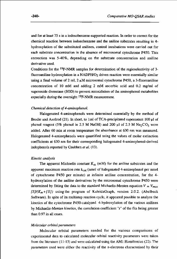
-240- Comparative MO-QSAR studies
and for at least 75 s in a iodosobenzene-supported reaction. In order to correct for the
chemical reaction between iodosobenzene and the aniline substrates resulting in 4-
hydroxylation of the substituted anilines, control incubations were carried out for
each substrate concentration in the absence of microsomal cytochrome P450. This
correction was 5-40%, depending on the substrate concentration and aniline
derivative used.
Conditions for the 19F-NMR samples for determination of the regioselectivity of 3-
fluoroaniline hydroxylation in a NADPH/O2 driven reaction were essentially similar
using a final volume of 2 ml, 2 uM microsomal cytochrome P450, a 3-fluoroaniline
concentration of 10 mM and adding 2 mM ascorbic acid and 0.2 mg/ml of
superoxide dismutase (SOD) to prevent autoxidation of the aminophenol metabolites
especially during the overnight 19F-NMR measurement.
Chemical detection of 4-aminophenol.
Halogenated 4-aminophenols were determined essentially by the method of
Brodie and Axelrod (21). In short, to 1ml of TCA-precipitated supernatant 100 ul of
phenol reagent (5% phenol in 2.5 M NaOH) and 200 ul of 2.5 M Na2C03 were
added. After 60 min at room temperature the absorbance at 630 nm was measured.
Halogenated 4-aminophenols were quantified using the values of molar extinction
coefficients at 630 nm for their corresponding halogenated 4-aminophenol-derived
indophenols reported by Cnubben et al. (13).
Kinetic analysis
The apparent Michaelis constant Km (mM) for the aniline substrates and the
apparent maximum reaction rate kcat (nmol of halogenated 4-aminophenol per nmol
of cytochrome P450 per minute) at infinite aniline concentration, for the 4-
hydroxylation of the aniline derivatives by the microsomal cytochrome P450 were
determined by fitting the data to the standard Michaelis-Menten equation V = Vmax
[S]/(Km + [S]) using the program of KaleidaGraph, version 2.0.2. (Abelbeck
Software). In spite of its multistep reaction cycle, it appeared possible to analyze the
kinetics of the cytochrome P450-catalyzed 4-hydroxylation of the various anilines
by Michaelis-Menten kinetics, the correlation coefficient "r" of the fits being greater
than 0.97 in all cases.
Molecular orbital parameters
Molecular orbital parameters needed for the various comparisons of
experimental data to calculated molecular orbital reactivity parameters were taken
from the literature (11-13) and were calculated using the AMI Hamiltonian (22). The
parameters used were either the reactivity of the rc-electrons characterised by their
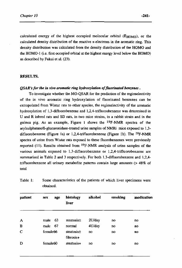
Chapter 10 -241-
calculated energy of the highest occupied molecular orbital (EHOMO). o r the
calculated density distribution of the reactive n-electrons in the aromatic ring. This
density distribution was calculated from the density distribution of the HOMO and
the HOMO-1 (i.e. first occupied orbital at the highest energy level below the HOMO)
as described by Fukui et al. (23).
RESULTS.
QSAR 'sfor the in vivo aromatic ring hydroxylation offluorinated benzenes.
To investigate whether the MO-QSAR for the prediction of the regioselectivity
of the in vivo aromatic ring hydroxylation of fluorinated benzenes can be
extrapolated from Wistar rats to other species, the regioselectivity of the aromatic
hydroxylation of 1,3-difluorobenzene and 1,2,4-trifluorobenzene was determined in
U and R inbred rats and SD rats, in two mice strains, in a rabbit strain and in the
guinea pig. As an example, Figure 1 shows the 19F-NMR spectra of the
arylsulphatase/ß-glucuronidase-treated urine samples of NMRi mice exposed to 1,3-
difluorobenzene (Figure la) or 1,2,4-trifluorobenzene (Figure lb). The 19F-NMR
spectra of urine from Wistar rats exposed to these fluorobenzenes were previously
reported (11). Results obtained from 19F-NMR analysis of urine samples of the
various animals exposed to 1,3-difluorobenzene or 1,2,4-trifluorobenzene are
summarised in Table 2 and 3 respectively. For both 1,3-difluorobenzene and 1,2,4-
trifluorobenzene all urinary metabolite patterns contain large amounts (> 48% of
total
Table 1 : Some characteristics of the patients of which liver specimens were
obtained.
patient sex age histology alkohol smoking medication liver
A
B
C
D
male 63
male 67
female66
female60
steatosis!
normal
steatosis!
fibrosis+
steatosis+
2U/day
4U/day
no
no
no
no
no
no
no
no
no
no
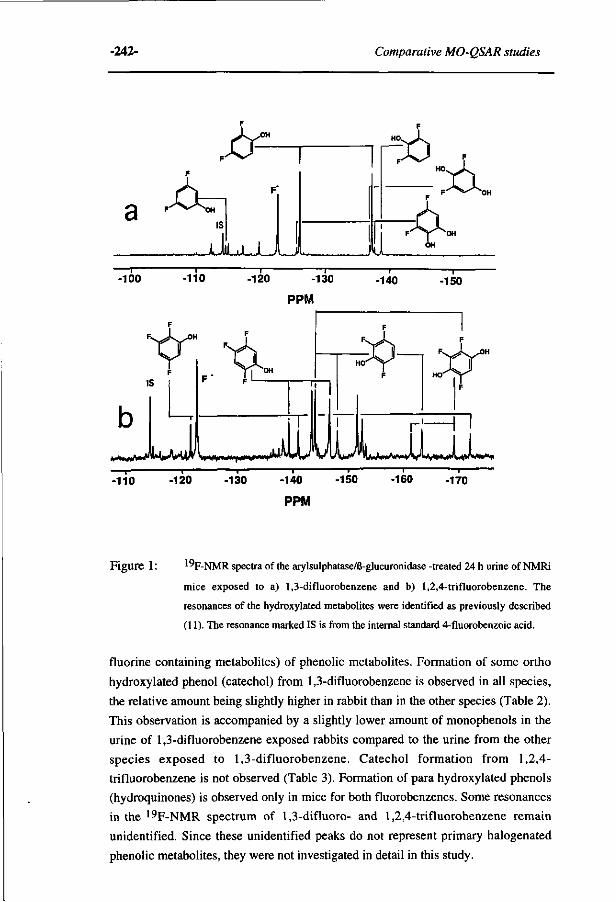
-242- Comparative MO-QSAR studies
.if-F ' ^ ^ O H
l iyLuU^ F-^y^O
-100 -110 — I 1— -130 -140 -120 -150
PPM
tr***if—r L ^ M J U L L L -110 -120 -130 -140
PPM
-150 -160 -170
Figure 1 : ' " F - N M R spectra of the arylsulphatase/ß-glucuronidase -treated 24 h urine of NMRi
mice exposed to a) 1,3-difluorobenzene and b) 1,2,4-trifluorobenzene. The
resonances of the hydroxylated metabolites were identified as previously described
(11). The resonance marked IS is from the internal standard 4-fluorobenzoic acid.
fluorine containing metabolites) of phenolic metabolites. Formation of some ortho
hydroxylated phenol (catechol) from 1,3-difluorobenzene is observed in all species,
the relative amount being slightly higher in rabbit than in the other species (Table 2).
This observation is accompanied by a slightly lower amount of monophenols in the
urine of 1,3-difluorobenzene exposed rabbits compared to the urine from the other
species exposed to 1,3-difluorobenzene. Catechol formation from 1,2,4-
trifluorobenzene is not observed (Table 3). Formation of para hydroxylated phenols
(hydroquinones) is observed only in mice for both fluorobenzenes. Some resonances
in the 19F-NMR spectrum of 1,3-difluoro- and 1,2,4-trifluorobenzene remain
unidentified. Since these unidentified peaks do not represent primary halogenated
phenolic metabolites, they were not investigated in detail in this study.
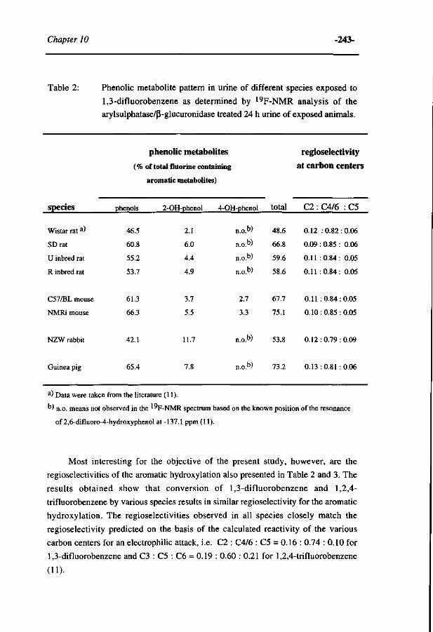
Chapter 10 -243-
Table 2: Phenolic metabolite pattern in urine of different species exposed to
1,3-difluorobenzene as determined by 19F-NMR analysis of the arylsulphatase/ß-glucuronidase treated 24 h urine of exposed animals.
species
Wistar rat a)
SDrat
U inbred rat
R inbred rat
C57/BL mouse
NMRi mouse
NZW rabbit
phenolic metabolites
(% of total fluorine containing
phenols
46.5
60.8
55.2
53.7
61.3
66.3
42.1
aromatic metabolites)
2-OH-phenol 4-OH-phenol
2.1
6.0
4.4
4.9
3.7
5.5
11.7
n.o.b>
n . o »
n . o»
n.o.b>
2.7
3.3
n . o »
total
48.6
66.8
59.6
58.6
67.7
75.1
53.8
regioselectivity at carbon centers
C2:C4/6 :C5
0.12 :0 .82: 0.06
0.09 : 0.85 : 0.06
0.11 :0.84: 0.05
0.11 :0.84: 0.05
0.11 :0 .84: 0.05
0.10 : 0.85 : 0.05
0.12:0.79:0.09
Guinea pig 65.4 n .o» 73.2 0.13 : 0.81 : 0.06
a) Data were taken from the literature (11).
") n.o. means not observed in the 1°F-NMR spectrum based on the known position of the resonance
of 2,6-difluoro-4-hydroxyphenol at -137.1 ppm (11).
Most interesting for the objective of the present study, however, are the
regioselectivities of the aromatic hydroxylation also presented in Table 2 and 3. The
results obtained show that conversion of 1,3-difluorobenzene and 1,2,4-
trifluorobenzene by various species results in similar regioselectivity for the aromatic
hydroxylation. The regioselectivities observed in all species closely match the
regioselectivity predicted on the basis of the calculated reactivity of the various
carbon centers for an electrophilic attack, i.e. C2 : C4/6 : C5 = 0.16 : 0.74 : 0.10 for
1,3-difluorobenzene and C3 : C5 : C6 = 0.19 : 0.60 : 0.21 for 1,2,4-trifluorobenzene
(11).
-244- Comparative MO-QSAR studies
Table 3: Phenolic metabolite pattern in urine of different species exposed to
1,2,4-trifluorobenzene as determined by 19F-NMR analysis of the
arylsulphatase/ß-glucuronidase treated 24 h urine of exposed animals.
species
Wistar ra t a)
U inbred rat
R inbred rat
C57/BL mouse
NMRi mouse
NZW rabbit
phenolic metabolites
( % of total fluorine containing
aromatic metabolites)
phenols
48.6
51.9
53.7
59.6
55.3
69.6
4-OH-phenol
n.o.b>
n . o »
n.o.b>
6.3
7.8
n.o.b)
total
48.6
51.9
53.7
65.9
63.1
69.6
regioselectivity at carbon centers
C3 : C5 : C6
0.25 :0.51 :0.24
0.31 :0.49: 0.20
0.34:0.46: 0.20
0.31 :0.50: 0.19
0.30 : 0.50 : 0.20
0.29 : 0.44 : 0.27
Guinea pig 67.8 n.o.b) 6 7 i i 0.31 : 0.42: 0.27
a) Data were taken from the literature (11).
°' n.o. means not observed in the ^"F-NMR spectrum based on the known position of the resonances
of 2,3,5-trifluoro-4-hydroxyphenol at -144.4, -162.2 and -169.7 ppm (11).
Regioselectivity for the in vitro aromatic hydroxylation of 3-fluoroaniline
In addition to extrapolation to other experimental animals than the Wistar rat, it
is of interest to investigate whether the regioselectivity obtained for the aromatic
hydroxylations catalyzed by cytochromes P450 from Wistar rats can be extrapolated
to man. As, for obvious ethical reasons, the in vivo regioselectivity of the aromatic
hydroxylation can not be investigated in man, in vitro experiments with 3-
fluoroaniline were performed. 3-Fluoroaniline was used for these in vitro studies
instead of the fluorobenzenes because the rate of its conversion is higher and
therefore results in metabolite patterns that can be quantified by 19F-NMR analysis.
Figure 2 presents the 19F-NMR spectrum of an incubation of 3-fluoroaniline with
human liver microsomes.
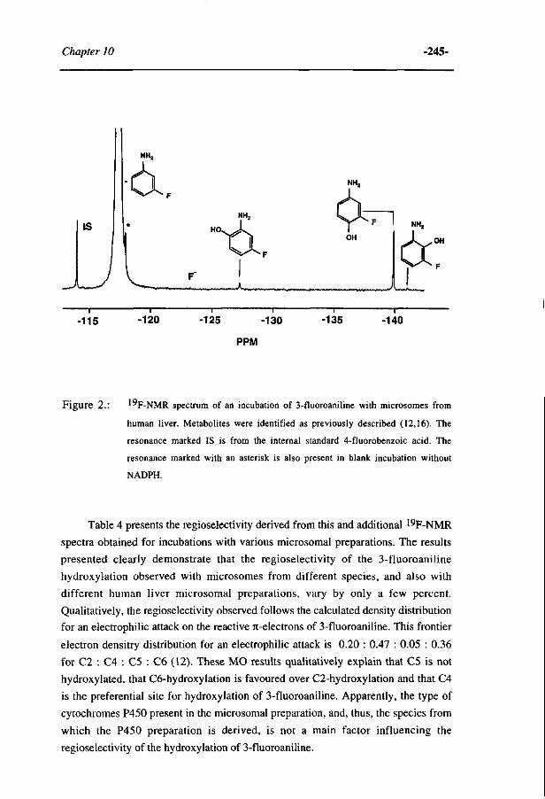
Chapter 10 -245-
— i — -130 -115 -120 -125 -135 -140
PPM
Figure 2.: 1°F-NMR spectrum of an incubation of 3-fluoroaniline with microsomes from
human liver. Metabolites were identified as previously described (12,16). The
resonance marked IS is from the internal standard 4-fluorobenzoic acid. The
resonance marked with an asterisk is also present in blank incubation without
NADPH.
Table 4 presents the regioselectivity derived from this and additional 19F-NMR
spectra obtained for incubations with various microsomal preparations. The results
presented clearly demonstrate that the regioselectivity of the 3-fluoroaniline
hydroxylation observed with microsomes from different species, and also with
different human liver microsomal preparations, vary by only a few percent.
Qualitatively, the regioselectivity observed follows the calculated density distribution
for an electrophilic attack on the reactive 7t-electrons of 3-fluoroaniline. This frontier
electron densitry distribution for an electrophilic attack is 0.20 : 0.47 : 0.05 : 0.36
for C2 : C4 : C5 : C6 (12). These MO results qualitatively explain that C5 is not
hydroxylated, that C6-hydroxylation is favoured over C2-hydroxylation and that C4
is the preferential site for hydroxylation of 3-fluoroaniline. Apparently, the type of
cytochromes P450 present in the microsomal preparation, and, thus, the species from
which the P450 preparation is derived, is not a main factor influencing the
regioselectivity of the hydroxylation of 3-fluoroaniline.
-246- Comparative MO-QSAR studies
Table 4: The regioselectivity for the in vitro aromatic ring hydroxylation of 3-
fluoroaniline in microsomal incubations from different species as
determined by 19F-NMR.
species regioselectivity observed at the
carbon centers
C2 : C4 : C5 : C6
Wistarrat a)
NMRi mouse
NZW rabbit
human batch A batch B batch C batch D
0.05:0.84:0.00:0.11
0.05:0.80:0.00:0.15 0.06:0.77:0.00:0.17
0.06:0.84:0.00:0.10 0.06 : 0.85 : 0.00 : 0.09
0.04 : 0.76 : 0.00 : 0.20 0.06:0.79:0.00:0.15 0.08:0.78:0.00:0.14 0.05 : 0.86 : 0.00 : 0.09
a) data were taken from the literature (12).
QSAR'sfor the rate of in vitro conversion of halogenated aniline derivatives in a
iodosobenzene-supported cytochrome P450-catalysed C4-hydroxylation.
In a final set of experiments it was investigated whether the MO-QSAR
describing a correlation between the rates of conversion of a series of aniline
derivatives in a iodosobenzene-supported cytochrome P450-mediated C4-
hydroxylation and the energy of the reactive 7t-electrons of these aniline derivatives
(13), can be extrapolated to other species than the Wistar rat. To allow comparison of
the data of the present study to those previously reported, isosafrole pretreatment of
the animals was performed before preparing the microsomes for the present in vitro
studies. Previous studies demonstrated aniline hydroxylation in a NADPH/O2 driven
microsomal reaction to be highest with microsomes from isosafrole pretreated rats
(13,16). Table 5 presents the kcat values obtained for the C4-hydroxylation of a
series of (halogenated) anilines by liver microsomes from isosafrole-pretreated rats,
rabbit and mice. It also presents kcat values obtained for a iodosobenzene driven
microsomal system derived from human liver. It appears from the data presented in
Table 5, that kcat values for C4-hydroxylation of aniline derivatives obtained using
microsomes from various species vary at most two-fold. Furthermore, the results
presented in Table 5 show considerable differences in kCat values for the C4-
hydroxylation of the different aniline derivatives.
Chapter 10 -247-
Table 5: Apparent kcat (maximum velocity in nmoles converted per nmol of
cyt P450 per min) for the iodosobenzene-supported microsomal
cytochrome P450-catalyzed C4-hydroxylation of aniline derivatives
with liver microsomes from different species. Values presented are the
mean ± standard error of the mean (n=2-4).
substrate kcat (min -1)
Wistar rats NZW rabbit NMRi mice human (batch A and B)
aniline
2-chloroaniline
2-fluoroaniline
3-chloroaniline
3-fluoroaniline
2,3-difluoroaniline
115.8 + 21.6
104.8 ± 10.5
74.1 ± 10.0
36.5 ±5.2
41.1 ±3.8
15.5+1.0
111.4 ±32.2
95.9 ± 24.4
55.1 ±5.1
28.7 ±1.3
30.8 ±3.8
15.1 ±0.0
124.7 + 22.2
75.6+15.4
58.9 ± 20.2
26.8 ±3.2
29.6 + 2.6
15.7 ±0.0
223.3 ± 77.5
164.1 ±53.6
132.9 + 55.7
69.2 ±13.7
60.4 ±5.1
18.8 ±0.0
In Figure 3 the natural logarithm of the kcat values for the iodosobenzene-
supported C4-hydroxylation of the (halogenated) anilines is plotted against the
E(HOMO) values calculated for the aniline derivatives. These E(HOMO) values of
the aniline derivatives are of importance for their reactivity in the electrophilic attack
by the high-valent-iron-oxo cytochrome P450 intermediate. The results presented in
Figure 3 clearly demonstrate that MO-QSAR's are obtained not only for rat, but also
for mice, rabbit and even human microsomal systems. The correlation coefficients
for the QSAR's describing the relation between the In kCat values and the E(HOMO)
values are r=0.97 for rats, r=0.98 for mice, r=0.97 for rabbit and r= 0.99 for human
microsomes.
Besides, Figure 3 clearly shows that the QSAR's obtained for the various species are
similar. These similar QSAR lines obtained for different species indicate that the
relative change in the rate of conversion with a change in the reactivity of the aniline
substrate is about the same for all species.
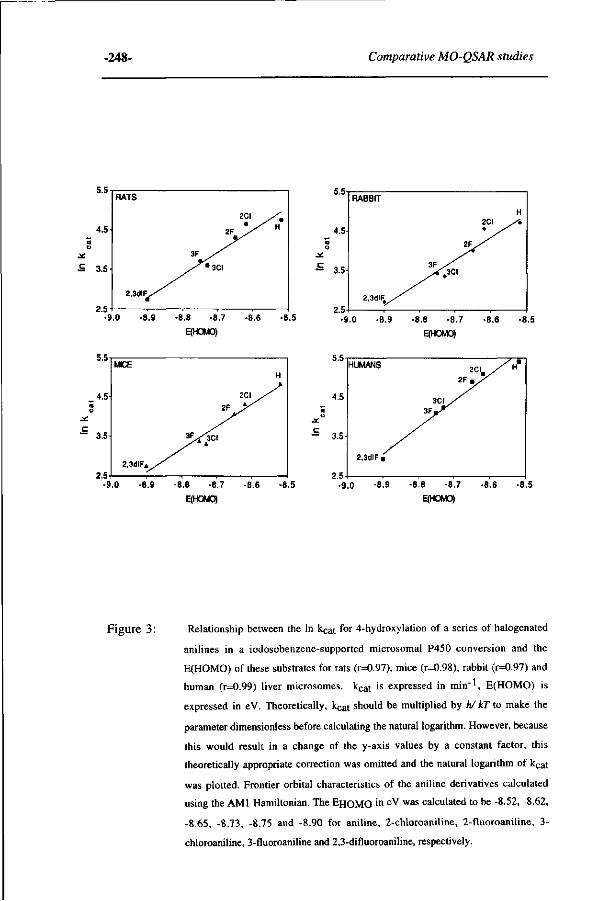
-248- Comparative MO-QSAR studies
5.5
4.5
u
£ 3.5
2.5
RATS
2 , 3 d i F ^ /
2CI
2F JS
3F yS
/*&\
H
-9.0 -8.9 -8.8 -8.7 -8.6 -8.5
E(HOMO)
C
5.5
4.5
S 3.5
2.5
HUMANS
2.3diF ,
3 C I , 3 F » - ^
2 F . /
/ • / H
-9.0 -8.9 -8.8 -8.7 -8.6 -8.5 E(HOMO)
Figure 3 : Relationship between the In k c a t for 4-hydroxylation of a series of halogenated
anilines in a iodosobenzene-supported microsomal P450 conversion and the
E(HOMO) of these substrates for rats (r=0.97), mice (r=0.98), rabbit (r=0.97) and
human (r=0.99) liver microsomes. kcat is expressed in min"1, E(HOMO) is
expressed in eV. Theoretically, kcat should be multiplied by h/ IcT to make the
parameter dimensionless before calculating the natural logarithm. However, because
this would result in a change of the y-axis values by a constant factor, this
theoretically appropriate correction was omitted and the natural logarithm of kca t
was plotted. Frontier orbital characteristics of the aniline derivatives calculated
using the AMI Hamiltonian. The E H O M O i n e V w a s calculated to be -8.52, -8.62,
-8.65, -8.73, -8.75 and -8.90 for aniline, 2-chloroaniline, 2-fluoroaniline, 3-
chloroaniline, 3-fluoroaniline and 2,3-difluoroaniline, respectively.
Chapter 10 -249-
DISCUSSION.
In the present study it was investigated whether MO-QSAR's (quantitative
structure activity relations based on molecular orbital parameters) described
previously for aromatic hydroxylation reactions of halogenated benzene derivatives
catalysed by cytochromes P450 from Wistar rats can be extrapolated to cytochrome
P450 reactions of other species. Because these MO-QSAR's describe relationships
between experimental parameters and a calculated reactivity parameter of the P450
substrate, it might be foreseen that such MO-QSAR's obtained for one species may
be valid for other species including man. Previous results with differently induced
Wistar rat microsomal preparations as well as with different induced rats
metabolising substrates in vivo, already demonstrated that a change in the
cytochrome P450 enzyme pattern did not influence the regioselectivity of the
aromatic hydroxylation of relatively small substrates like fluorinated benzene
derivatives or monofluoroanilines (11,12). These results imply that chemical
characteristics of the substrate instead of the types of cytochromes P450 involved, are
of major importance in determining the outcomes of the catalysis. The present study
was undertaken to provide further support for this hypothesis, and especially to
investigate whether this MO-QSAR approach would indeed form a basis for
extrapolation of biotransformation results from one species to another including man.
First, the regioselectivity of the in vivo hydroxylation of two model fluorinated
benzenes with three discernable sites for aromatic hydroxylation (1,3-
difluorobenzene and 1,2,4-trifluorobenzene), was tested in three additional rat strains,
in two mice strains, in a rabbit strain and in guinea pig. Second, the regioselectivity
of the in vitro 3-fluoroaniline hydroxylation by mice, rabbit and human microsomes
was determined and compared to the results obtained with rat microsomes. Finally, it
was investigated whether the MO-QSAR for the rate of conversion of a series of
aniline derivatives in a iodosobenzene-supported cytochrome P450-catalysed C4-
hydroxylation can be extrapolated to studies with mice, rabbit and human
microsomes.
The results obtained demonstrate that for all three biotransformation QSAR's
the results obtained with mice, guinea pig, rabbit and even human systems were
similar to those obtamed for the rat system. The regioselectivity of the in vivo
aromatic hydroxylation of 1,3-difluorobenzene and 1,2,4-trifluorobenzene could be
predicted on the basis of the calculated electron densitry distribution for an
electrophilic attack by the activated high-valent-iron-oxo species of the cytochromes
P450. Thus, in in vivo studies with mice, guinea pig, other rats strains and rabbit, this
MO-QSAR for the prediction of the regioselectivity of the fluorobenzene
hydroxylation was valid, indicating that the chemical reactivity of the various sites in
-250- Comparative MO-QSAR studies
the fluorinated benzenes, rather than a stereoselective influence of the active site of
the cytochromes P450, determines the regioselectivity of the aromatic hydroxylation.
This observation implies that the regioselectivity of the hydroxylation of these
fluorinated benzenes in man can be expected to be similar. The similar
regioselectivity observed in the present study for the in vitro hydroxylation of 3-
fluoroaniline by microsomes from experimental animals or man, supports this view.
Furthermore, the qualitative correlation between the regioselectivity predicted for 3-
fluoroaniline on the basis of the calculated reactivity of the various carbon centres for
an electrophilic attack and the regioselectivity actually observed, also supports this
conclusion.
Finally, it was demonstrated that the relationship between the natural logarithm
for the kcat for iodosobenzene-supported microsomal C4 hydroxylation of a series of
aniline derivatives and the energy of the reactive n-electrons of these derivatives can
be obtained not only for liver microsomes from Wistar rats as previously described
(13) but also for liver microsomes from mice, from rabbit and even from human
liver. This MO-QSAR indicates that in the iodosobenzene-supported cytochrome
P450-catalysed aniline C4-hydroxylation the actual electrophilic attack of the high-
valent-iron-oxo P450 intermediate on the reactive re-electrons of the aniline substrate
is the rate limiting step in catalysis. The two-fold variation in the various kcat values
observed when comparing results from human liver microsomes with those obtained
for rats, rabbit or mice microsomes, is the only significant species-dependent
difference observed in the present study. Nevertheless, the QSAR lines describing the
relationship between the In kCat for the conversion of the various aniline derivatives
and their E(HOMO) are similar for all species investigated. This implies that the
relative influence of a change in the chemical reactivity of an aniline derivative on its
rate of conversion is about the same for all species.
Altogether, the results of the present study strongly support the conclusion that
the conversion of the relatively small benzene derivatives in the relatively large and
aspecific active sites of the mammalian cytochromes P450, are mainly dependent on
chemical reactivity parameters of the substrates. Therefore, the results of the present
study support the hypothesis that MO-based QSAR's obtained in rat can provide a
basis for prediction of biotransformation pathways in different species, including
man.
Acknowledgements. The authors gratefully acknowledge Prof. dr. C. Veeger for continuous support and
helpfull discussion. Thanks are extended to Mr. J. Haas and Mrs. M.A.W. Peters for
assistance with animal handling.
Chapter 10 -251-
REFERENCES.
1. R.N. Armstrong, Enzyme-catalyzed detoxication reactions: mechanisms and
stereochemistry. CRC Crit. Rev. Biochem., 22 (1987) 39-88.
2. I.G. Sipes and A.J. Gandolfi, Biotransformation of toxicants, in: Casarett and Doull's
Toxicology. The Basic Science of Poisons. M.O. Amdur, J. Doull and CD. Klaassen (Eds.),
4th edn. Pergamon, Oxford, (1991) pp. 64-98.
3. R.T. Williams, Species variations in the pathways of drug metabolism. Environ. Health
Perspect., 22 (1978) 133-138.
4. R. Kato, Characteristics and differences in the hepatic mixed function oxidases of different
species. Pharmac. Ther., 6 (1979) 41-98.
5. J. Caldwell, Problems and opportunities in toxicity testing arising from species differences in
xenobiotic metabolism.Toxicol. Letters, 64/65 (1992) 651-659.
6. E. Dybing and H.S. Huitfeldt, Species differences in carcinogen metabolism and interspecies
extrapolation. IARC Sei. Publ., 116 (1992) 501-522.
7. H.G. Selander, D. Jerina and J.W. Daly, Metabolism of chlorobenzene with hepatic
microsomes and solubilised cytochrome P-450 systems. Arch. Biochem. Biophys., 168
(1975)309-321.
8. J.J.P. Bogaards, B. van Ommen, CR. Wolf and P.J. van Bladeren, Human cytochrome P450
enzyme selectivities in the oxidation of chlorinated benzenes. Toxicol. Appl. Pharmacol. 132
(1995)44-52.
9. S.S. Lau, G.D. Abrams and V.G. Zannoni, Metabolic activation and detoxification of
bromobenzene leading to cytotoxicity. J. Pharmacol. Exp. Ther., 214 (1980) 703-708.
10. T.J. Monks and S.S. Lau, Reactive intermediates and their toxicological significance. Review
paper. Toxicology, 52 (1988) 1-53.
11. I.M.C.M.Rietjens , A.E.M.F. Soffers , C. Veeger and J. Vervoort, Regioselectivity of
cytochrome P-450 catalyzed hydroxylation of fluorobenzenes predicted by calculated
frontier orbital substrate characteristics. Biochemistry, 32 (1993) 4801-4812.
12. N.H.P.Cnubben , J.Vervoort, C.Veeger and I.M.C.M. Rietjens, Study on theregioselectivity
and mechanism of the aromatic hydroxylation of monofluoroanilines. Chem.-Biol. Interact.,
85(1992)151-172.
13. N.H.P. Cnubben , S. Peelen, J.W. Borst, J. Vervoort and I.M.C.M. Rietjens, Molecular
orbital-based quantitative structure activity relationship for the cytochrome P450-catalysed 4-
hydroxylation of halogenated anilines. Chem. Res. Toxicol., 7 (1994) 590-598.
14. H. Saltzman and J.G. Sharefkin, Iodosobenzene. in:Organic Syntheses, Collect. Vol. V, John
Wiley & Sons, New York, (1973) 658-659.
15. I.M.C.M. Rietjens and J. Vervoort, A new hypothesis for the mechanism for cytochrome P-
450 dependent aerobic conversion of hexahalogenated benzenes to pentahalogenated phenols.
Chem. Res. Toxicol., 5 (1992) 10-19.
-252- Comparative MO-QSAR studies
16. I.M.C.M. Rietjens and J. Vervoort, Microsomal metabolism of fluoroanilines. Xenobiotica,
19(1989)1297-1305.
17. J. Vervoort, P.A. De Jager, J. Steenbergen and I.M.C.M. Rietjens, Development of a ' ^F
NMR method for studies on the in vivo and in vitro metabolism of 2-fluoroaniline.
Xenobiotica, 20 (1990) 657-670.
18. V. Wray, Fluorine nuclei in fluorobenzene derivatives, in: G.A. Webb (Ed), Annual Reports
on NMR Spestroscopy, Vol. 14, Academic Press, London, (1983) p. 252.
19. T. Omura and R. Sato, The carbon monoxide pigment of liver microsomes, I. Evidence for its
haemoprotein nature. J. Biol. Chem., 239 (1964) 2370-2378.
20. O.H. Lowry, N.J. Rosenbrough, A.L. Farr and A.J. Randall, Protein measurements with
Folin phenol reagent. J. Biol. Chem. 193 (1951) 2370-2378.
21. B.B. Brodie and J. Axelrod, The estimation of acetanilide and its metabolic products, aniline,
N-acetyl p-aminophenol and p-aminophenol (free and conjugated) in biological fluids and
tissues. J. Pharmacol. Exp. Ther., 94 (1948) 22-28.
22. M.J.S. Dewar, E.G. Zoebisch, E.F. Healy and J.J.P. Stewart, AMI: a new general purpose
quantum mechanical molecular model. J. Am. Chem. Soc, 107 (1985) 3902-3909.
23. K. Fukui, T. Yonezawa, C. Nagata and H. Shingu, Molecular orbital theory of orientation in
aromatic, heteroaromatic and other conjugated molecules. J. Chem. Phys., 22 (1954) 1433-
1442.
CHAPTER 11
Summary and conclusions.
Organisms are frequently exposed to low molecular weight xenobiotic
compounds. An advanced enzymatic machinery modifies these compounds into more
hydrophilic metabolites which are subsequently excreted from the body. This process
of biotransformation aims to detoxify bodyforeign compounds. Ironically, reactive
intermediates may also be formed during the biotransformation process and can
interact with macromolecules or receptors, with possible toxicological consequences
(bioactivation). Toxicological testing of all new and existing compounds is a money -
and time-consuming problem and therefore alternatives are urgently needed. The
main objective of the studies decribed in this thesis, which is outlined in chapter 1, was to describe QSARs (quantitative structure-activity relationships) for the
biotransformation and toxicity of halogenated amino- and nitrobenzene derivatives.
Special attention was focussed on the most important phase 1 biotransformation
enzyme involved, the cytochrome P450 system. In addition, in the course of the
investigations some attention was paid to the glutathione/glutathione S-transferase
dependent phase 2 biotransformation pathway.
Chapter 2 gives an overview of the biotransformation enzymes primarily
involved in the metabolism of halogenated amino- and nitrobenzene derivatives with
respect to their function, regulation, occurrence, molecular/biochemical mechanisms
and role in bioactivation and detoxication of xenobiotics. Biotransformation of
amino- and nitrobenzene derivatives plays a crucial role in the generation of reactive
intermediates assumed to contribute to the toxicity of this class of compounds. Since
biotransformation of nitro- and aminobenzene derivatives is known to include
pathways leading to both detoxication and bioactivation, insight into factors that
direct the rates and regioselectivities of biotransformation processes or interactions
with nucleophiles, like macromolecules or the tripeptide glutathione, will help to gain
insight in factors that direct processes of, and chances on, bioactivation or
detoxication of these compounds. Regioselective enzymatic conversion of relatively
small aromatic substrates in the relatively large and aspecific active sites of
cytochromes P450 is assumed to depend predominantly on chemical characteristics
-254- Summary and conclusions
of the substrates, whereas for relatively large substrates stereoselective positioning
through steric constraints of the protein core in the active site becomes more
important (see also chapter 2). Therefore, the relative differences in reactivity of
various sites in a small aromatic compound can be expected to affect the possibilities
for enzymatic conversion of the molecule. These considerations prompted us to
investigate whether computer calculated molecular orbital characteristics of
halogenated benzene derivatives, in combination with insight into the molecular
mechanisms of their enzymatic conversion, could provide a basis for the prediction of
their metabolic fate.
Chapter 3 presents a study on the regioselectivity and underlying
mechanisms for the cytochrome P450-catalyzed aromatic hydroxylation of
monofluoroanilines. This study provides insight into the molecular mechanism of the
cytochrome P450-catalyzed aromatic hydroxylation of molecules containing a
heteroatom as well as factors that influence the regioselectivity of hydroxylation.
Three mechanisms for aromatic hydroxylation can be proposed; hydrogen abstraction
(I), or electron abstraction followed by proton release (II) both leading to formation
of a NH • radical. Upon rearrangement of the radical and OH-rebound from the
(FeOH)3+ species, the aminophenol product is formed. Aromatic hydroxylation
might also proceed by a direct interaction of the high-valent iron-oxo cytochrome
(FeO)3+ intermediate with the n-electrons of the aromatic ring resulting in a so-called
a-adduct (III), which rearranges to the aminophenol, either directly or through
formation of epoxides and/or ketones as intermediates. First, it was demonstrated that
the regioselectivity of the aromatic hydroxylation was influenced by the position of
fluoro-substituents at the aniline-ring and that the observed regioselectivity for
hydroxylation of these small aromatics was not influenced by the relatively aspecific
and large hydrophobic active sites of the cytochromes P450. As expected, a fluorine-
substituent induces an effect on the electronic characteristics of the aniline-molecule
and this effect was quantified using molecular orbital calculations. Considering the
possible molecular mechanisms for aromatic hydroxylation, it appeared that the
observed in vitro and in vivo regioselectivity correlated best with the frontier orbitals
of importance for a direct interaction of the (FeO)3+ species with the re-electrons of
the aromatic molecule e g. the density distribution of the HOMO/HOMO-1. The spin
density distribution of the NH • radicals -a parameter of importance for the hydrogen
abstraction as well as electron abstraction plus proton release mechanisms- could not
explain the observed regioselectivities, indicating that the regarding mechanisms are
less likely. In a later study it was demonstrated that for a series of fluorobenzenes the
regioselectivity for aromatic hydroxylation could even be predicted on the basis of
the frontier orbital density distribution for electrophilic attack within 6% accuracy
Chapter 1] -255-
(r=0.96) [Rietjens et al, 1993]. For the fluoroanilines the C4 position is hydroxylated
to higher extent, whereas the C2/C6 positions are hydroxylated to an extent lower
than their chemical reactivity predicts. This deviation might result from steric
hindrance of the amino moiety for electrophilic attack, a stereoselective positioning
of the substrate through an interaction of the amino moiety with amino acid residues
in the active site as has been described for P450 debrisoquine 4-hydroxylase, or a
dipole-dipole or electronic interaction between the substrate and the activated
cytochrome P450 (FeO)3+ species. It is stressed here that the juxtaposition of these
small substrates in the active site with respect to the Fe3+ resting state as determined
on the basis of !H-NMR Tl relaxation or cristallography studies, might not represent
the actual orientation with respect to the reactive (FeO)3+ intermediate actually
performing the hydroxylation step [Koerts et ai, 1995].
In chapter 4 the aromatic hydroxylation of anilines was further investigated
with special emphasis on possible relationships between kinetic parameters and both
the physicochemical and electronic substrate characteristics. This was done in order
to provide a basis for molecular orbital based quantitative structure activity
relationships (MO-QSARs) for kinetic characteristics of the cytochrome P450-
mediated aromatic hydroxylation of a homologous series of aniline-derivatives. It
was demonstrated that the kcat for C4-hydroxylation in a series of substituted anilines
strongly correlates with the HOMO energy of the anilines for the iodosobenzene
supported P450 reaction in isosafrole induced microsomes. This observation is in
accordance with a mechanism that proceeds by an initial electrophilic interaction of
the (FeO)3+ intermediate with the frontier n electron of the aniline-substrate. In a
NADPH/oxygen-supported P450 system, however, it was demonstrated that the
interaction of the (FeO)3+ species on the aniline-substrate is no longer rate-limiting,
and therefore cannot be described by this QSAR. However, when the electrophilic
reactivity of the substrates becomes too low, as is the case for 2,3,5,6-
tetrafluoroaniline (EHOMO of -9.24 eV) the initial attack of the cytochrome (FeO)3+
on the substrate might become the rate-limiting step in the overall catalysis. The
relatively low conversion rate observed for aromatic hydroxylation of fluorobenzenes
for example [Rietjens et ai, 1993], might be explained by the relatively low
electrophilic reactivity of these compounds. If the resulting metabolite is less toxic
than its parent compound, a decreased conversion might have implications for the
toxicity and the other way around.
To fully describe the cytochrome P450-mediated biotransformation of these
aniline-derivatives, a sensitive and efficient analytical technique was developed for
the detection and quantification of 2-aminophenols (chapter 5). The principle of the
-256- Summary and conclusions
method was based on a dimerization reaction of 2-aminophenols to an intensively
colored 2-hydroxy-isophenoxazin-3-one in an acidic environment using ferric ions as
the catalyst. It was demonstrated that this method was also applicable for the
detection and quantification of halogenated 2-aminophenol derivatives.
Besides aromatic hydroxylation reactions, also oxidative dehalogenation
reactions mediated by cytochromes P450 were investigated. Halogen substituents are
often introduced into molecules to block positions at the aromatic ring of drugs or
agrochemicals for bioactivation or biodégradation. Fluorine substituents are
frequently used for this purpose, due to the strong C-F bond, and a Van der Waals
radius that almost equals that of a hydrogen. In order to study the molecular
mechanism of oxidative aromatic dehalogenation as well as the consequences of
halogen substitution for regioselective hydroxylation and the formation of reactive
intermediates, the study described in chapter 6 was undertaken. Using halogenated
anilines as the model compound, the effect of a varying halogen substituent patterns
on the cytochrome P450-catalyzed dehalogenation of 4-halogenated anilines to 4-
aminophenols was investigated. In the case of C4-fluorinated aniline derivatives, the
cytochrome P450-mediated metabolic pathway has been unequivocally demonstrated
to result in direct formation of a reactive 1,4-benzoquinoneimine and fluoride anions
the primary reaction products [Rietjens et al, 1993]. These reactive
benzoquinoneimine metabolites may interact with cellular macromolecules and hence
can lead to destruction of molecules essential to living cells.
The study described in chapter 6 clearly demonstrated that upon the
cytochrome P450-mediated oxidative dehalogenation to the primary reactive 1,4-
benzoquinoneimine a fluorine substituent at C4 position of the aromatic aniline-ring
was more easily eliminated than a chloro-, bromo- or iodo-substituent. A similar
decrease in dehalogenation was observed in a NADPH/O2 supported microsomal
P450 system, as well as in a tBuOOH supported microsomal P450 system, or a
system with purified reconstituted P4502B1, or in a system with the heme-based
mini-enzyme microperoxidase 8. This indicates that the decrease in dehalogenation
was not a result of a change in rate-limiting steps in the P450 catalysis or a change in
the contribution of P450 enzymes with a change in the halogen substituent. Structure-
activity relationship principles were applied to investigate the reaction mechanism of
dehalogenation. The results obtained strongly indicate that the possibilities for the
cytochrome P450-mediated dehalogenation of 4-halogenated anilines to 4-
aminophenol metabolites are dependent on i) the characteristics of the halogen that
has to be eliminated, the most electronegative and smallest halogen (fluorine) being
the one most easily eliminated and ii) the electron-withdrawing capacities of the other
Chapter 11 -257-
substituents in the aromatic ring, electron-withdrawing substituents decreasing the
relative rate of the reaction. The conclusion that not only a fluoro-, but also a chloro-,
bromo- and iodo- substituent, is eliminated as a halogen anion serves as the best
explanation for the observations in this study. In addition, it was demonstrated that
blocking the C4-position for aromatic hydroxylation by cytochromes P450 resulted in
a metabolic switch from dehalogenation and 4-aminophenol formation, to formation
of 2-aminophenol- and N-hydroxyaniline-derivatives. Although halogen substitution
at the C4-position of an aniline indeed leads to a decreased metabolism at that site,
the formation of the reactive 1,4-benzoquinoneimine instead of 4-aminophenol as the
primary metabolite, as well as the switch to N-hydroxylation, giving rise to reactive
hydroxylamino- and nitroso-derivatives may have considerable toxicological
implications.
The biological activity of numerous aniline derivatives has been shown to be
closely related to the cytochrome P450 mediated oxidative attack at their nitrogen
center, yielding products with increasing toxic properties, namely N-hydroxyaniline
and nitrosobenzene derivatives. Reactive N-hydroxyanilines cause for example
ferrihemoglobin formation (methemoglobinemia) with concomittant co-oxidation to
nitrosobenzenes. The resulting nitrosobenzenes in turn are able to interact with
cysteine residues of hemoglobin or with the tripeptide glutathione [Eyer, 1988]. On
the other hand, the cytochrome P450 mediated aromatic hydroxylation of anilines
may represent a detoxification pathway due to the efficient conjugation of the
resulting phenolic metabolites and subsequent excretion from the body. However, the
cytochrome P450 mediated formation of aminophenol metabolites has been proposed
to play a role in the nephrotoxicity of halogenated anilines, occuring predominantly
at the proximal tubule and to a lesser extent at the distal tubule [Lo et ai, 1990 &
1991; Rankin et al., 1986a & b; Valentovic et ai, 1992]. Chapter 7 presents a clear
example of a metabolism-toxicity relationship study. Since the cytochrome P450
mediated regioselective hydroxylation seems to direct the toxicity of aniline
compounds, a study was performed on the relationships between the regioselectivity
of the hydroxylation of C4-substituted 2-fluoroaniline derivatives and their toxic
endpoints nephrotoxicity and/or methemoglobinemia. Depending on the derivative,
nephrotoxicity at the tubular site and/or methemoglobinemia was shown to occur.
The extent of nephrotoxicity induced by C4-substituted 2-fluoroanilines was shown
to be related to the extent of C4-hydroxylation, and the extent of methemoglobinemia
was shown to be related to the extent of N-hydroxylation. Consequently, a change in
the regioselective hydroxylation from the cytochrome P450-mediated C4-
hydroxylation to N-hydroxylation resulted in a change of toxic endpoint from
nephrotoxicity to methemoglobinemia.
-258- Summary and conclusions
In chapter 8 a study was directed at understanding the marked differences in
biotransformation pathways of anilines and their chemically oxidized analogues
nitrobenzenes. In vivo and in vitro, 2,5-difluoroaniline was demonstrated to become
predominantly metabolized through a cytochrome P450-mediated pathway, whereas
2,5-difluoronitrobenzene is predominantly converted through glutathione
conjugation, and the a minor extent through nitroreduction and cytochrome P450-
mediated aromatic hydroxylation. On the basis of computer calculations a hypothesis
was presented that might explain the differences in metabolic pathways on the basis
of their molecular orbital substrate characteristics. It was suggested that the HOMO
of the thiolate anion of glutathione will interact more efficiently with the LUMO of a
nitro compound (ELTJMO - -1-57 eV) than with the LUMO of a less electrophilic
amino compound (ELUMO - -0.08 eV). Concerning the aromatic hydroxylation on
the other hand, it was suggested that the SOMO of the cytochrome P450 (FeO)3+
intermediate can interact more efficiently with the amino compound (EHOMO = -8.83
eV) than with the nitro compound (EHOMO = -10.29 eV). These considerations are in
accordance with the observation in chapter 4, that the reaction rate of the cytochrome
P450-mediated C4-hydroxylation decreases with decreasing electrophilic reactivity
of an aniline substrate. Substrates with an EHOMO value below -9.2 eV were
converted at a relatively low rate by the cytochromes P450. The considerations are
also in accordance with the recent observation that the rate of both the chemical and
glutathione S-transferase catalyzed glutathione conjugation of fluoronitrobenzenes
increases with decreasing ELUMO values [Rietjens et al., 1995].
Halogenated nitrobenzenes are known to become metabolized by
nitroreduction, glutathione conjugation and aromatic hydroxylation. For some of the
toxic effects of nitrobenzenes, the formation of reactive metabolites is a prerequisite.
Reduction of nitrobenzenes to the aminobenzene analogues, via the formation of
methemoglobinemia-inducing intermediates (nitrosobenzene, N-hydroxyaniline or
radical intermediates), is a clear example of bioactivation of nitrobenzenes. The
different susceptibility for nitroreduction, and also the presence of competing
biotransformation pathways, like glutathione conjugation and aromatic
hydroxylation, have been proposed to set the chances for methemoglobinemia
exerted by nitrobenzene-derivatives. Applying QSAR approaches, a study was
performed in order to investigate whether a metabolism-toxicity relationship exists
for a series of fluoronitrobenzenes providing insight in the relative importance of
these two suggestions. Chapter 9 describes a combined study concerning the
influence of the substituent pattern of fluoronitrobenzenes on their biotransformation
and their capacity to induce methemoglobinemia. It was demonstrated that increased
possibilities for the conjugation of fluoronitrobenzenes to cellular nucleophiles was
Chapter!! -259-
accompanied by decreased contributions of aromatic hydroxylation and
nitroreduction. A QSAR could be described for the rate of conjugation with the
tripeptide glutathione or with bovine serum albumine (a model for cellular
nucleophiles) and calculated parameters for electrophilicity of the
fluoronitrobenzenes, showing that with increasing number of fluoro-substituents the
conjugation pathway will become more important. In addition, the intrinsic reactivity
of the fluoronitrobenzenes for nitroreduction by cecal microflora and rat liver
preparations was not related to the in vivo methemoglobin-forming capacity. This
observation, in combination with the QSAR for conjugation, led to the conclusion
that the different methemoglobinemic capacity must rather result from differences in
the inherent direct methemoglobinemic reactivity of the various toxic metabolites,
and/or from the difference in reactivity of the fluoronitrobenzenes with glutathione or
other cellular nucleophiles. As a result of an increased electrophilic reactivity of
fluoronitrobenzenes, another toxic endpoint than methemoglobinemia can be
expected.
In the final study described in chapter 10, comparative MO-QSAR studies
clearly demonstrated that outcomes on both the regioselectivity and the rate of the
cytochrome P450-catalyzed aromatic hydroxylation could be described on the basis
of frontier orbital characteristics of the substrates. The MO-QSARs obtained for rats,
were also valid for other species, including man. These results strongly support the
conclusion that the conversion of the relatively small halogenated benzene
derivatives in the relatively large and aspecific active sites of the mammalian
cytochromes P450 -even when derived from various species- are mainly dependent
on chemical reactivity parameters of the substrates. This importance of the substrate
characteristics also dominates over the influence of the specific P450 enzymes in
determining the regioselectivity of biotransformation both in vitro and in vivo.
In conclusion, the studies described in this thesis cleary demonstrate that
molecular orbital calculations in combination with the frontier orbital theory are a
usefull additional tool to study the mechanism of cytochrome P450 enzyme catalysis.
In addition to the theoretical considerations on the molecular reaction mechanisms
and enzyme catalysis, empirical data on the metabolism are essential to construct
valid rules or even quantitative structure activity relationships for the
biotransformation and toxicity of series of related compounds. Insight into the
possibilities for biotransformation have been demonstrated to provide insight into the
formation of toxicity determining reactive intermediates. Defining QSARs for
biotransformation have been shown to offer an approach to explain the type and
extent of the toxic effects exerted by the series of halogenated amino- and
-260- Summary and conclusions
nitrobenzene derivatives.
Refined models, which do not only focus on the chemical reactivity
parameters of the substrates, but also take into account possible stereoselective
positioning or interactions with specific amino acid residues in the active site, will
contribute to a better prediction of metabolic profiles of existing and new drugs,
agrochemicals and other industrially relevant compounds.
Finally, it is challenging to apply the approaches described in this thesis to
other enzymes with more specific active sites than the relatively large aspecific active
sites of the biotransformation enzyme cytochrome P450.
REFERENCES.
Eyer P., Detoxication of N-oxygenated arylamines in erythrocytes. An overview, Xenobiotica 18,
1327-1333, 1988.
Koerts J., Rietjens I.M.C.M., Boersma M.G. and Vervoort J., ^H NMR Ti relaxation rate study on
substrate orientation of fluoromethylanilines in the actives sites of microsomal and purified
cytochrome P450 1A1 and 2B1, FEBSLetters 368, 279-284, 1995.
Lo H.H., Brown P.I., Rankin CO. , Acute nephrotoxicity induced by isomeric dichloroanilines in
Fischer 344 rats, Toxicology 63, 215-231, 1990.
Lo H.H., Brown P.I. and Rankin CO., Trichloroaniline effects on renal function in vivo and in vitro,
Toxicology Letters 57, 319-328, 1991.
Rankin CO. , Yang D.J., Cressey-Veneziano K., Casto S., Wang R.T. and Brown P.I., In vivo and in
vitro nephrotoxicity of aniline and its monochlorophenylderivatives in the Fischer 344 rat,
Toxicology 38, 269-283, 1986a.
Rankin CO . , Yang D.J., Teets V.J., Lo H.H. and Brown P.I., 3,5-Dichloroaniline-induced
nephrotoxicity in the Sprague-Dawley rat, Toxicology Letters 30, 173-179, 1986b.
Rietjens I.M.C.M., Soffers A.E.M.F., Hooiveld C , Veeger C. and Vervoort J., Quantitative structure
activity relationships based on computer calculated parameters for the overall rate of
glutathione S-transferase catalyzed conjugation of a series of fluoronitrobenzenes. Chem. Res.
Toxicol. 8,481-488, 1995.
Rietjens I.M.C.M., Soffers A.E.M.F., Veeger C. and Vervoort J., Regioselectivity of cytchrome P450
catalyzed hydroxylation of fluorobenzenes predicted by calculated frontier orbital substrate
characteristics, Biochemistry 32, 4801-4812, 1993.
Rietjens I.M.C.M. and Vervoort J., Bioctivation of 4-fluorinated anilines to benzoquinoneimines as
primary reaction products, Chem.-Biol. Interact. 22, 263-281, 1991.
Valentovic M.A., Ball J.G., Anestis D.K., Beers K.W., Madan E., Hubbard J.L. and Rankin C , Aute
renal and hepatic toxicity of 2-haloanilines in Fischer 344 rats, Toxicology 75, 121-131, 1992.
Samenvatting voor niet-vakgenoten.
In de huidige moderne samenleving worden organismen veelvuldig
blootgesteld aan lichaamsvreemde verbindingen zoals medicijnen, agrochemicaliên,
oplosmiddelen, milieuverontreinigende stoffen en hun afbraakprodukten met
mogelijk nadelige gevolgen. Het lot van deze stoffen is in het algemeen afhankelijk
van hun fysisch-chemische eigenschappen. Zeer goed vetoplosbare stoffen hebben de
neiging om te accumuleren in vettige weefsels (bijvoorbeeld de PCBs), terwijl goed
wateroplosbare stoffen juist efficient uit het lichaam worden verwijderd via onder
andere de urine (denk maar aan de gekleurde urine na het eten van rode bietjes).
Reactieve stoffen kunnen in een cel schadelijke effecten teweegbrengen door een
interactie aan te gaan met essentiële macromoleculen, zoals eiwitten, enzymen of
DNA. Gelukkig zijn organismen uitgerust met een speciaal enzymatisch
afweersysteem tegen deze verbindingen en dit enzymsysteem is met name
gelocaliseerd in de lever, maar ook bijvoorbeeld in de nieren en longen. Dit complex
systeem heeft tot doel lichaamsvreemde stoffen (xenobiotica) te modificeren tot meer
wateroplosbare verbindingen (metabolieten), die vervolgens efficient via de gal of
urine het lichaam verlaten. Dit proces van enzymatische modificatie van xenobiotica
wordt ook wel biotransformatie genoemd. Biotransformatie dient lichaamsvreemde
verbindingen onschadelijk te maken. Ironisch genoeg kunnen tijdens de
biotransformatie ook ongewenste reactieve produkten ontstaan met mogelijk
toxische (schadelijke) consequenties (bioactivering).
Het toxicologisch testen van alle nieuwe en bestaande verbindingen is een
zeer kostbare en tijdrovende aangelegenheid, en daarom zijn alternatieven hiervoor
zeer gewenst. Het voornaamste doel van het onderzoek dat beschreven is in dit
proefschrift was om regels ofwel "QSARs" (quantitatieve structuur-activiteit relaties)
te formuleren voor de biotransformatie en toxiciteit van gehalogeneerde amino- en
nitrobenzeen derivaten (hoofdstuk 1). Deze verbindingen worden frequent gebruikt
in de agrochemische en pharmaceutische industrie ten behoeve van de produktie van
pesticiden, herbiciden, kleurstoffen, medicijnen en andere industrieel relevante
verbindingen. Het cytochroom P450 systeem en glutathion S-transferase zijn
-262- Samenvatting
belangrijke biotransformatie enzymen, die betrokken zijn bij de detoxificatie
(ontgifting) en bioactivering van deze stoffen. In deze studies is voornamelijk
aandacht besteed aan cytochroom P450, en in mindere mate aan de
glutathion/glutathion S-transferase afhankelijke biotransformatie route.
Hoofdstuk 2 geeft een overzicht van de enzymen die betrokken zijn bij de
biotransformatie van gehalogeneerde amino- en nitrobenzeen derivaten, met
betrekking tot hun functie, regulatie, voorkomen, moleculaire/biochemische
werkingsmechanismen, alswel de rol in bioactivering en detoxificatie van
xenobiotica. Biotransformatie van amino- en nitrobenzeen derivaten is essentieel
voor de vorming van reactieve intermediairen die mogelijk verantwoordelijk zijn
voor de toxische effecten van deze groep verbindingen. Inzicht in factoren die de
biotransformatie van deze verbindingen bepalen, leiden tot een beter begrip van de
processen en van de mogelijkheden tot bioactivering en detoxificatie. De
regioselectieve enzymatische omzetting van relatief kleine aromatische substraten in
het relatief grote aspecifieke katalytisch centrum van cytochroom P450 wordt
verondersteld voornamelijk afhankelijk te zijn van de chemische substraat
karakteristieken. Voor grotere substraten daarentegen, zal ook de stereoselectieve
positionering tengevolge van sterische interacties in het katalytisch centrum de
regioselectieve omzetting bepalen (zie hoofdstuk 2). De mogelijkheden voor de
enzymatische omzetting van een klein aromatisch molecuul worden daarom
waarschijnlijk bepaald door de verschillen in reactiviteit van de diverse plaatsen in
het molecuul. Naar aanleiding van deze overwegingen werd onderzocht of computer
berekende karakteristieken van de amino- en nitrobenzenen, in combinatie met
inzicht in het moleculaire werkingmechanisme van de enzymatische omzetting,
behulpzaam kunnen zijn bij het voorspellen van het metabole lot van deze stoffen.
Hoofdstuk 3 presenteert een studie naar de regioselectiviteit en mechanismen
die ten grondslag liggen aan de cytochroom P450 gekatalyseerde aromatische
hydroxylering van fluor aminobenzenen (= fluor anilines). Deze studie verschaft
zowel inzicht in het mogelijke moleculaire werkingsmechanisme van de cytochroom
P450 gekatalyseerde aromatische hydroxylering, als in de factoren die de
regioselectieve hydroxylering beinvloeden. Op basis van computer-berekende
substraat karateristieken en inzicht in de moleculaire werkingsmechanismen van
cytochroom P450 kon zowel de regioselectiviteit (hoofdstuk 3) als de snelheid
(hoofdstuk 4) van de aromatische hydroxylering van aminobenzenen verklaard
worden. Bovendien kon een QSAR voor de snelheid van de aromatische
hydroxylering worden geformuleerd op basis van de computer berekende substraat
karakteristieken.
-263-
Teneinde de cytochroom P450 afhankelijke biotransformatie van aniline-
derivaten volledig op te helderen, moest een gevoelige en efficiente methode worden
ontwikkeld voor de detectie en quantificering van 2-aminophenol metabolieten,
aangezien deze nog niet voorhanden was (hoofdstuk 5 ).
Naast de aromatische hydroxylerings reacties werd ook de oxidatieve
dehalogenering door cytochroom P450 bestudeerd. Halogenen worden vaak gebruikt
om specifieke plaatsen in medicijnen of agrochemicalien te bezetten, waardoor
bioactivatie of biodegradatie door enzymen wordt verhinderd. Bij voorkeur worden
daarvoor fluor atomen gebruikt, omdat ze bijna net zo klein zijn als waterstof atomen
en vanwege de sterke C-F binding. In hoofdstuk 6 werd aan de hand van SAR
principes het mechanisme van de oxidatieve aromatische dehalogenering bestudeerd.
Tevens werd de invloed van het halogeen substitutie op de regioselectieve
hydroxylering en de vorming van reactieve deeltjes onderzocht. Er werd aangetoond
dat tijdens de biotransformatie de fluoratomen een aminobenzeen gemakkelijker
verlaten dan een chloor-, broom- of joodatoom. Alhoewel het metabolisme op de
gehalogeneerde positie werd geremd, resulteerde deze blokkade tevens in de
(verhoogde) vorming van andere metabolieten (het reactieve 1,4-benzoquinoneimine
en N-hydroxyaniline), met mogelijke toxicologische consequenties.
De toxische effecten van aminobenzenen worden voor een gedeelte bepaald
door de vorming van N-hydroxyaniline metabolieten, die methemoglobinemie
veroorzaken. De aromatische hydroxyleringsmetabolieten (aminophenolen)
daarentegen worden gekoppeld aan goed wateroplosbare groepen en verlaten
dientengevolge efficient het lichaam via de urine. Daarnaast wordt de vorming van
4-aminophenolen verondersteld een rol te spelen bij het onstaan van aniline-
geinduceerde niertoxiciteit. Daar de cytochroom P450 gekatalyseerde regioselectieve
hydroxylering de toxiciteit van anilines lijkt te beinvloeden, werd in hoofdstuk 7 een
studie uitgevoerd naar de relatie tussen de regioselectieve hydroxylering en het
optreden van toxische effecten als methemoglobinemie en niertoxiciteit. De mate van
niertoxiciteit in een serie anilines bleek gerelateerd te zijn aan de mate van
aromatische hydroxylering op de C4-positie. De mate van methemoglobinemie was
gerelateerd aan de mate van N-hydroxylering. Een verandering in regioselectieve
hydroxylering van 4-hydroxylering naar N-hydroxylering leidt dus tot een
verandering in toxisch effect van niertoxiciteit naar methemoglobinemie. Hieruit
blijkt dat in hoofdstuk 7 een duidelijk voorbeeld wordt gepresenteerd van een
verband tussen het metabolisme enerzijds, en de mate en type van toxiciteit
anderzijds.
-264- Samenvatting
Aminobenzenen worden voornamelijk door cytochroom P450 omgezet en
niet door glutathion S-transferase. De chemisch geoxideerde nitrobenzeen analogen
worden voornamelijk door glutathion S-transferase omgezet en in mindere mate door
cytochroom P450. In hoofdstuk 8 wordt een hypothese voorgesteld die verklaart
waarom nitrobenzenen voornamelijk door de glutathion afhankelijke
biotransformatie route worden omgezet, terwijl anilines juist via de P450 route
worden gemodificeerd. De hypothese is gebaseerd op inzicht in de
werkingsmechanismen van de enzymen en met de computer berekende substraat
karakteristieken die van belang zijn voor de twee verschillende biotransformatie
routes. Het actieve deeltje van glutathion S-transferase (thiolaat anion van
glutathion) kan een betere interactie aangaan met het nitrobenzeen substraat dan met
de aniline, terwijl het actieve deeltje van cytochroom P450 een betere interactie kan
aangaan met het aniline substraat dan met de nitrobenzeen.
Gehalogeneerde nitrobenzenen worden via drie biotransformatie routes
gemetaboliseerd, te weten nitro-reductie, glutathion conjugatie en aromatische
hydroxylering. Voor enkele van de toxische effecten van nitrobenzenen is de
vorming van reactieve metabolieten een vereiste. Zo ontstaan bijvoorbeeld tijdens de
nitro-reductie methemoglobinemie vormende deeltjes. De mate van
methemoglobinemie vorming wordt verondersteld afhankelijk te zijn van de
mogelijkheden tot nitroreductie. Factoren die deze nitroreductie kunnen beinvloeden
zijn de intrinsieke gevoeligheid van nitrobenzenen voor nitroreductie, en/of de
aanwezigheid van concurrerende biotransformatie routes als glutathion conjugatie en
aromatische hydroxylering. Met behulp van QSAR benaderingen werd in hoofdstuk 9 een studie uitgevoerd naar een mogelijk verband tussen het metabolisme en de
vorming van methemoglobinemie door een serie fluornitrobenzenen. Hierdoor
ontstond inzicht in het relatieve belang van deze twee veronderstellingen. Er werd
aangetoond dat verhoogde conjugatie mogelijkheden met glutathion of andere
macromoleculen tot een afname in de bijdrage van aromatische hydroxylering en
nitroreductie leiden. Deze conjugatie snelheid van fluornitrobenzenen kon zelfs
voorspeld worden aan de hand van computer berekeningen. De mate van
methemoglobinemie vorming bleek verder niet bepaald te worden door de intrinsieke
gevoeligheid van fluornitrobenzenen voor nitroreductie door darm-microflora en
leverpreparaten. Geconcludeerd werd dat de conjugatie met glutathion en
macromoleculen fungeert als een voor nitroreductie concurrerende biotransformatie
route en daardoor dus de mogelijkheden voor methemoglobinemie beinvloedt.
In hoofdstuk 10 wordt een studie beschreven die aantoont dat de
gedefinieerde QSARs voor de snelheid en regioselectiviteit van de P450
-265-
gekatalyseerde hydroxylering niet alleen gelden voor ratten, maar ook voor diverse
andere species, waaronder de mens. Deze bevindingen ondersteunen de conclusie dat
de omzetting van relatief kleine gehalogeneerde benzeen derivaten in de relatief
grote en aspecifieke actieve centra van de cytochroom P450 enzymen, voornamelijk
worden bepaald door de chemische reactiviteit parameters van de substraten. Het
belang van deze substraat karakteristieken domineert zelfs over de invloed van de
specifieke P450 enzymen op de regioselectiviteit in vitro als in vivo.
Samenvattend kan geconcludeerd worden, dat het onderzoek in dit
proefschrift duidelijk aantoont dat moleculaire orbitaal berekeningen in combinatie
met de frontier orbitaal theorie een bruikbare additionele benadering is om het
mechanisme en de uitkomsten van de cytochroom P450 katalyse te bestuderen. Naast
theoretische overwegingen betreffende het moleculaire reactie mechanisme en de
enzym katalyse, zijn empirische gegevens essentieel om geldige regels of zelfs
QSARs te formuleren voor biotransformatie en toxiciteit. In dit proefschrift wordt
duidelijk geïllustreerd dat inzicht in biotransformatie routes bijdraagt tot inzicht in de
vorming van reactieve metabolieten. Het opstellen van QSARs voor biotransformatie
bleek een zeer bruikbare benadering om zowel het type als de mate van de toxische
effecten, die worden teweeggebracht door deze amino- en nitrobenzenen, te
verklaren.
Meer geavanceerde modellen, die niet alleen rekening houden met de
chemische reactiviteit van de substraten, maar ook met hun stereoselectieve
positionering en interacties met specifieke aminozuurresiduen in het actieve centrum
van cytochromen P450, zullen uiteindelijk leiden tot een betere voorspelling van
metaboliet profielen van bestaande en nieuw te ontwikkelen medicijnen,
agrochemicalien en andere industrieel relevante verbindingen.
Naar aanleiding van bovenstaande resultaten is het een uitdaging, de in dit
proefschrift beschreven benaderingen toe te passen op andere enzymen met meer
specifieke actieve centra dan de relatief grote aspecifieke actieve centra van het
biotransformatie-enzym cytochroom P450.

• 2 6 6 -
-267-
Curriculum Vitae
Nicole Cnubben werd geboren op 13 februari 1967 te Meerssen. Zij
behaalde het VWO diploma (Gymnasium ß) in mei 1985 aan de
Scholengemeenschap Stella Maris te Meerssen. Aansluitend startte zij de studie
Moleculaire Wetenschappen aan de Landbouwuniversiteit te Wageningen.
Tijdens de doctoraalstudie doorliep zij de hoofdvakken Biochemie en
Toxicologie. In 1989 verrichtte zij op de vakgroep Biochemie onder leiding van
professor C. Veeger, Dr. LM.C.M. Rietjens en Dr. W.H.J. van Berkel
onderzoek naar "De rol van FMO en cytochroom P450 in de biotransformatie
van 4-fluoro-N-methylaniline". In 1990 bestudeerde zij op de vakgroep
Toxicologie de invloed van 40H-3,3',4',5-TCB op de plasmakinetiek,
eliminatie en distributie van 125I-TTR en 125I-T4 onder begeleiding van
professor J. H. Koeman en Dr. A. Brouwer. In november 1990 behaalde zij het
doctoraal examen. Vervolgens trad zij als onderzoekster in opleiding in dienst
van de Nederlandse organisatie voor Wetenschappelijk Onderzoek (NWO),
werkgemeenschap farmaca en vergiften, gedetacheerd aan de vakgroep
Biochemie van de Landbouwuniversiteit te Wageningen. De resultaten van dit
onderzoek onder leiding van professor C. Veeger en Dr. I.M.C.M. Rietjens zijn
vastgelegd in dit proefschrift. Tijdens deze periode volgde ze de Postdoctorale
Opleiding Toxicologie ten behoeve van de registratie toxicoloog conform de
SMBWO normen. In februari 1995 was zij werkzaam als toegevoegd
onderzoeker bij de vakgroep Biochemie van de Landbouwuniversiteit en sinds
juni 1995 is zij werkzaam bij TNO Voeding, divisie Toxicologie, te Zeist.

-268-
-269-
List of publications
Cnubben, N.H.P., Vervoort, J., Veeger, C , Rietjens, I.M.C.M. Study on the regioselectivity and the mechanism for the cytochrome P450 dependent aromatic hydroxylation of monofluoroanilines. Chem.-Blol. Interactions 85, 151-172, 1992.
Cnubben, N.H.P., Peelen, S., Borst, J.W., Vervoort, J., Veeger, C , Rietjens, I.M.C.M. Molecular orbital based quantitative structure-activity relationship for the cytochrome P450-catalyzed 4-hydroxylation of halogenated anilines. Chem. Res. Toxicol. 7, 590-598, 1994.
Cnubben, N.H.P., Blaauboer, B., Juyn, S., Vervoort, J., Rietjens, I.M.C.M. A spectrophotometric assay for the detection of 2-aminophenols in biological samples. Anal. Biochem. 220, 165-171, 1994.
Cnubben, N.H.P., Vervoort, J., Boersma, M.G., Rietjens, I.M.C.M. The effect of varying halogen substituents on the cytochrome P450-catalyzed dehalogenation of 4-halogenated anilines to aminophenol metabolites. Biochem. Pharmacol. 49, 1235-1248, 1995.
Cnubben, N.H.P., van den Berg, C.L., Rietjens, I.M.C.M. Relationships between the regioselectivity of the hydroxylation of C4-substituted 2-fluoroaniline derivatives and their toxic endpoints. Submitted for publication in Toxicol. Appl. Pharmacol.
Rietjens, I.M.C.M., Cnubben, N.H.P., van Haandel, M., Tyrakowska, B., Soffers, A.E.M.F., Vervoort, J. Different metabolic pathways of 2,5-difluoronitrobenzene and 2,5-difluoroaminobenzene compared to molecular orbital substrate characteristics Chem.-Biol. Interact. 94 , 49-75., 1995.
Cnubben, N.H.P., Soffers, A.E.M.F., Peters, M., Vervoort, J., Rietjens, I.M.C.M. Influence of the halogen-substituent pattern of fluoronitrobenzenes on their biotransformation and capacity to induce methemoglobinemia. Accepted for publication in Toxicol. Appl. Pharmacol., 1996.
Tyrakowska, B., Cnubben, N.H.P., Soffers, A.E.M.F., Wobbes, Th., Rietjens, I.M.C.M. Comparative MO-QSAR studies in various species including man. Accepted for publication in Chem. Biol.-Interactions, 1996.
Boersma, M.G., Cnubben, N.H.P., van Berkel, W.J.H., Blom, M., Vervoort, J., Rietjens, I.M.C.M. Role of cytochrome P450 and flavin containing monooxyenase in the biotransformation of 4-fluoro-N-methylaniline. Drug Metabolism and Disposition 21, 218-230, 1993.
Rietjens, I.M.C.M., Cnubben, N.H.P., de Jager, P.A., Boersma, M.G., Vervoort, J. Application of NMR in biotransformation studies. In: Developments and ethical considerations in toxicology, Michael I. Weitzner (Ed.), Royal Society of Chemistry, Redwood Press Ltd., Melksham, Wiltshire, 94-109, 1993.
Rietjens, I.M.C.M., Boersma, M.G., Soffers, A.E.M.F, Cnubben, N.H.P., Koerts, J., Peelen, S., van Berkel, W.H.J., Veeger, C , Vervoort, J. Use of molecular orbital calculations in studies on mechanisms of enzyme catalysis. In: Flavins and Flavoproteins 1993, K. Yagi (Ed), Walter de Gruyter & Co., Berlin, 3-12, 1994.
Vervoort, J., Boersma, M.G., Cnubben, N.H.P., van Berkel, W.H.J., Koerts, J., Rietjens, I.M.C.M. Mammalian flavin-containing monooxygenases catalyzed conversion of 4-halo-N-methylanilines. In: Flavins and Flavoproteins 1993, K. Yagi (Ed), Walter de Gruyter & Co. Berlin, 255-258, 1994.
Koerts, J., Soffers, A.E.M.F., Cnubben, N.H.P., de Kraker, J.W., Rietjens, I.M.C.M. The metabolism and bioavailability of teflubenzuron, in preparation.

-270-
-271-
Dankwoord
Met veel plezier denk ik terug aan de periode waarin dit onderzoek is
uitgevoerd en daarbij niet te vergeten aan alle mensen die hebben bijgedragen aan de
totstandkoming van dit proefschrift.
Allereerst wil ik Ivonne, mijn co-promotor, hartelijk bedanken voor haar enorm goede
en enthousiaste ondersteuning. Discussies over experimenten en resultaten waren altijd
zeer verhelderend én gezellig. Voor problemen die niet altijd puur van
wetenschappelijke aard waren kon ik ook altijd bij je terecht. Kortom, ik heb het heel
erg prettig gevonden om "jouw OIO" te zijn en ik weet zeker dat ik mij geen betere co-
promotor had kunnen wensen.
Voorts ben ik mijn promotor, professor Cees Veeger, zeer erkentelijk voor zijn
interesse in mijn onderzoek en voor zijn opbouwende kritieken. Het afgelopen jaar heb
ik mij zeer gesterkt gevoeld door uw medeleven, steun en begrip.
Vervolgens wil ik Jacques bedanken voor zijn hulp bij de NMR metingen. Ondanks
het feit datje het altijd zo druk hebt, maakte je vaak tijd vrij voor een babbel. Wanneer
ik mij weer eens vreselijk zat op te winden over bepaalde zaken, was jouw (soms te)
nuchtere kijk op zaken een verfrissing voor mij.
De medewerkers van het CKP (Jo, Gerrit, etc.) en in het bijzonder Maria Peters, heel
erg bedankt. Maria, jij was mijn grote steun en toeverlaat bij de dierexperimenten.
Jouw professionele aanpak bij de ingewikkelde en arbeidsintensieve operaties, plus je
bereidwilligheid om tijdens de experimenten gedurende 12 uren te assisteren, hebben
het mogelijk gemaakt de studies in hoofdstuk 7 en 9 uit te voeren. Maria, ik en ook de
studenten vonden het altijd heel erg leuk om met je samen te werken.
Fijne kamergenoten zijn essentieel voor een prettige werksfeer. En ik moet zeggen dat
ik een héle prettige werksfeer heb gehad op onze kamer; Walter, Jan-Willem, Marelle,
Janneke, Marion, Petra, Lars en Tatiana, de heerlijke kletspartijen waren een welkome
afwisseling tijdens het nadenken, rommelen en schrijven van publicaties.
Ook Ans, Marelle en Sjef wil ik heel erg bedanken voor al hun professionele hulp,
collegialiteit en gezelligheid.
Dear Bozena, I enjoyed our collaboration very much. I will never forget the thousands
of tubes and "epjes". To me, it was always pleasant to perform these fruitful
experiments together and to discuss the results (and other things ).
Verder wil ik de studenten met wie ik nauw heb samengewerkt, Jan-Willem Borst,
Jeannine, Saskia, Jack, Jan-Willem de Kraker, Elles, Mark, Caroline, Coralie en
Guido heel erg bedanken voor hun inzet en plezierige samenwerking.
-272-
Alle collega's van de vakgroep Biochemie, ontzetttend bedankt voor jullie hulp en
gezelligheid.
Verder wil ik mijn moeder, Theo, Marion, Lenardo, de familie van Peter en alle
vrienden en kennissen heel erg bedanken voor hun interesse, goede raad (Nicole,
rustig aan, er is meer tussen hemel en aarde dan werken) en steun.
Lieve Peter, jouw steun, engelen-geduld, enthousiasme en interesse (Nicole, hoe zit
dat nu met die HOMO's en LUMO's en anilines en P450s en glutathion S-
transferases?) hebben ook bijgedragen tot dit proefschrift. Je staat zelfs een keer
vermeld in de acknowledgements van een publicatie. Misschien had ik dat bij al die
andere ook wel moeten doen? Geamuseerd keek je toe en wijselijk hield je je mond, als
ik het ene moment zat te grienen op de bank (wèèh, en nu wil ik huisvrouw worden en
10 kindertjes krijgen), en het andere moment tot middernacht vlijtig en vrolijk op het
lab met reageerbuizen zat te worstelen. Peter, bedankt voor alles.


