69
r 7 p -A —M I VOLUME 5 3 I 1 / I— I 1— 1 DECEMBER 2001 M ycology 2 CZECH SCIENTIFIC SOCIETY FOR MYCOLOGY PRAHA J \A Y C n r% n I ov ___ M

r 7 p - A — M I VO LUM E 5 3 I1 / I — I 1— 1 DECEM BER 2001
M y c o lo g y
2C ZE C H S C IE N T IF IC S O C IE T Y FOR M Y C O L O G Y PR AH A
J\A Y C nr%n Io v ___
M

^ V Y C rv ISSN «009-0476nN i ,C)O V J-<
Vol. 53, No. 2, December 2001
CZECH MYCOLOGYformerly Česká mykologie
published quarterly by the Czech Scientific Society for Mycology http: / /www. natur.cuni.cz / cvsm/
EDITORIAL BOARD
Editor-in-Chief ZDENĚK POUZAR (Praha)
Managing editor JAROSLAV KLÁN (Praha)
VLADIMÍR ANTONÍN (Brno) LUDMILA MARVANOVÁ (Brno)ROSTISLAV FELLNER (Praha) PETR PIKÁLEK (Praha)ALEŠ LEBEDA (Olomouc) MIRKO SVRČEK (Praha)JIŘÍ KUNERT (Olomouc) PAVEL LIZOŇ (Bratislava)
HANS PETER MOLITORIS (Regensburg)
Czech Mycology is an international scientific journal publishing papers in all aspects of mycology. Publication in the journal is open to members of the Czech Scientific Society
for Mycology and non-members.Contributions to: Czech Mycology, National Museum, Department of Mycology, Václavské
nám. 68, 115 79 Praha 1, Czech Republic. Phone: 02/24497259 or 24964284 SUBSCRIPTION. Annual subscription is Kč 600,- (including postage). The annual subscription for abroad is US $86,- or DM 136,- (including postage). The annual membership fee of the Czech Scientific Society for Mycology (Kč 400,- or US $ 60,- for foreigners) includes the journal without any other additional payment. For subscriptions, address changes, payment and further information please contact The Czech Scientific Society for Mycology, P.O.Box 106, 11121 P raha 1, Czech Republic, http://w w w .natur.cuni.cz/cvsm /
This journal is indexed or abstracted in:Biological Abstracts, Abstracts of Mycology, Chemical Abstracts, Excerpta Medica, Bibliography of Systematic Mycology, Index of Fungi, Review of Plant Pathology, Veterinary
Bulletin, CAB Abstracts, Rewiew of Medical and Veterinary Mycology.
Copyright © The Czech Scientific Society for Mycology, Prague, 2001
No. 1 of the vol. 53 of Czech Mycology appeared in 10. 6. 2001

CZECH M Y C O L O G YPublication of the Czech Scientific Society for Mycology
Volume 53____________________ December 2001____________________Number 2
Notes on the taxonomy and distribution of Aphyllophorales I.
Z d e n ě k P o u z a r
Srbská 2, 160 00 P rah a 6
Pouzar Z. (2001): Notes on the taxonomy and distribution of Aphyllophorales I. - Czech Mycol. 53: 121-131
Tw o new species of corticioid A phyllophorales (Basidiom ycetes) are described. Thanate- phorus brevisporus Pouz. is a species close to T. fusisporus (J . Schrót.) Hauerslev e t P. R oberts, differing however in shorter, m ore rounded spores, known from th e Czech Republic, Slovakia and U kraine, growing on ro tten wood of broad-leaved trees. D endrothele wojewodae Pouz. is close to D. acerina (Pers.: Fr.) P. A. Lemke, b u t is d istinct by its subglobose spores. I t is known from th e Czech R epublic and U kraine, from bark of living trees of A cer pseudoplatanus.
K e y w o rd s : Aphyllophorales, Corticiaceae s.l., new species, Thanatephorus brevisporus Pouz., D endrothele wojewodae Pouz.
Pouzar Z. (2001): Poznámky k taxonomii a rozšíření nelupenatých hub I. - Czech Mycol. 53: 121-131
Popisují se dva nové druhy kornatcovitých hub řádu A phyllophorales (Basidiom ycetes): Thanatephorus brevisporus Pouz., d ruh blízce příbuzný druhu T. fusisporus (J. Schrót.) Hauerslev e t P. R oberts, avšak lišící se kra tším i a kulatějším i výtrusy; je znám z České republiky, Slovenska a U krajiny z hnijícího dřeva listnatých strom ů; Dendrothele wojewodae Pouz. je velmi blízký d ru h u D. acerina (Pers.: Fr.) P. A. Lemke, ale liší se skoro kulovitým i výtrusy; je znám z české republiky a z U krajiny, z kůry živých klenů - A cer pseudoplatanus.
I n t r o d u c t i o n
During the years 1994-2001 a study of the order Aphyllophorales in the South Bohemian mountains of Šumava (mainly the Šumava National Park) has been carried out. Soon it, however, appeared tha t these mountainous or even to some degree also boreal species should be compares with those of southern and more thermophilous nature. Hence a study of these fungi started in forests in the vicinity of Prague where such a mycoflora is richly represented. These studies resulted in the recording of several rare species too, some of which appeared to be undescribed. In this first contribution two new interesting species of Corticiaceae sensu lato are described and discussed.
121

C z e c h m y c o l . 53 (2 ) , 2001
T h an atep h oru s b rev isp o ru s Pouz. spec. nov.
Carposomata effusa, hypochnoidea, cremea usque pallide ochracea. Hyphae absque nodis, glabrae, fere tenuiparietales, basales 7-10 /uri latae, horizontales in angulos rectos ramificatae, mediae 6-7,5 /¿in, verticales, laxe contextae. Basidia 14-18 ¡im longa et 10-12 //rn lata in parte distale, leviter vel late clavata, haud constricta, bisterigmatica, nonnumquam tri-, raro tetrasterigmatica; sterigm ata 8-11 /im longa et basim 2-3.5 /im lata, solum leviter subarcuata vel fere recta. Sporae 9-12.5 x 7-9.5 /xm, subglobosae usque breviter ovoideae, fere tenuiter tunicatae, glabrae, apice cum mamilla distincta, isodiametrica, saepius leviter elongata, seu raro apex sporarum in conum latissimum, brevemque terminatus. Hyphae, basidia, sterigmata sporaeque cyanophilae, sed haud dextrinoideae et haud amyloideae.
Holotypus: Bohemia, Voskov apud Karlštejn, Carpinus betulus - ad truncum iacentem, 7. V. 2001, leg. Z. Pouzar, PRM 895056, in Museo Nationale Pragae asservatur.
Paratypus: Slovakia, silva virginea “Badínsky prales” apud Badín prope Banská Bystrica, ad truncum iacentem Fagi sylvaticae 4. VIII. 1973, leg. Z. Pouzar PRM 895057.
Fruitbodies resupinate, when young forming very small tufts, soon confluent to become hypochnoid or mucedinoid, comparatively widely spread over the substratum , colour cream to pale ochraceous. Basal hyphae rather sparse, those parallel close to the substratum 7-10 fim wide. Median vertical hyphae loosely arranged 6-7.5 fjm broad. Subhymenium of shortly septate hyphae, 7-9 ;im broad. Clamp-connections completely missing. All structures without incrustations. Hyphal ends similar to cystidia, scattered in the hymenium of some few specimens (typically developed in PRM 895057), frequently arising from the hymenium close by basidia, straight 27-50(-90) /mi long, 6-8 /mi broad at the basis and 5-7.5 /¿m broad in the upper half, cylindrical, sometimes in central part slightly broadened, some 1-3 x slightly but abruptly constricted, rounded at the top, 1-2 x (at most 3x) septate, sometimes without septa, filled with strongly cyanophilous content. Basidia 14-18 /tm long and 10-12 /im broad in distal part, narrowly to broadly clavate, not constricted, bisterigmatic, only some tristerigmatic and few tetrasterigmatic. Sterigmata only slightly arcuate to almost straight, 9-11 ¡im long and 2-3.5 /¿m broad at base. Spores 9-12.5 x 7-9.5 ¡im, subglobose to shortly ovoid, a t the apex with a distinct isodiametric or at most slightly elongate mammiform outgrowth, a few spores broadly coniformly terminated; richly producing spores by repetition. All hyphae, basidia and spores with cyanophilous, but indextrinoid and inamyloid walls.
122

PouzAii Z.: N o t e s o n t h e t a x o n o m y a n d d i s t r i b u t i o n o f A p h y l l o p h o r a l e s I.
D is t r i b u t i o n a n d e c o l o g y
Thanatephorus brevisporus is so far known from three localities in the Czech Republic (two in Bohemia, one in Moravia), five in Slovakia and one in Ukraine (Transcarpathian region). This species could occur from the lowlands (elevation 152 m in Ranšpurk Virgin Forest) to mountains up to an elevation of about 1000 m in the Slovak Carpathians (Vrátná dolina in Velká Fatra and Polana near Detva possibily at 1200 m). It is known only from strongly rotten wood of broad-leaved trees, especially Fagus sylvatica, Tilia platyphyllos, Carpinus betulus, Acer pseudoplatanus and also Ulmus sp.
M a t e r ia l s t u d ie d
Czech Republic1. Bohemia, Voškov ap. Karlštejn, (area tuta); Carpinus betulus - ad truncum
iacentem 7. V. 2001, leg. Z. Pouzar, PR 895056 (holotype). Ibid. Tilia platyphyllos - ad truncum iacentem 7. V. 2001, leg. Z. Pouzar, PRM 894928.
2. Bohemia, Karlštejn, loco “Vodopády” (Bubovické vodopády); ad ligna putrida arboris frondosae iacentis, 29. VII. 1962, leg. Z. Pouzar, PRM 793513.
3. Moravia, silva virginea “Ranšpurk” ap. Lanžhot; in trunco putrido Ulmi sp., 28. VII. 1970, leg. Z. Pouzar, PRM 803635.
Slovakia4. Montes Velká Fatra, in valle “Vrátná dolina” (sub Ostredok); in cortice
Fagi sylvaticae (truncus iacens), 4. VII. 1953, leg. F. Kotlaba et Z. Pouzar, PRM 803634.
5. Silva virginea “Badínsky prales” sub monte Laurín prope Badín ap. Banská Bystrica, ca 800 m s.m.; ad truncum iacentem Fagi sylvaticae, 4. VIII. 1973, leg. Z. Pouzar, PRM 895057 (paratype).
6. In silvis virgineis montis PoFana ap. Detva, ca 1200 m s.m.; ad ligna Fagi sylvaticae, 25. VI. 1952, leg. A. Pilát, PRM 189589, 189599.
7. In silvis Jasov ap. Košice; ad codicem Aceris pseudoplatani, 27. VI. 1965, leg.F. Kotlaba, PRM 803633.
8. In monte “Malý Milic” prope Slánská Huta (h.p. Slanec); ad truncum putridumFagi sylvaticae, 17. VII. 1964, leg. F. Kotlaba et Z. Pouzar, PRM 803636.
Ukraine9. Transcarpathian Region (olim Carpatorossia), Jalinka prope Kosovská Po
lana; VII. 1930, leg. A. Pilát, PRM 189598.
When studying rather rich material of Thanatephorus fusisporus (J. Schrot.) Hauerslev et P. Roberts [Uthatobasidium fusisporum (J. Schrot.) Donk], collected in the vicinity of Prague in autum n 2000 and in spring 2001, it appeared
123

th a t the variability of spore size and form in this species is more restricted and narrower than presumed earlier. This experience led to the conclusion that short-spored specimens represent an independent species, which is described here as Thanatephorus brevisporus Pouz. spec. nov. The main body of the spore is in most spores of this species almost isodiametric, with the length nearly equating the breadth. Only the rather strongly developed apiculus and the striking apical projection on the opposite end of the spore breaks the spore isodiametry.
The original idea of a possible distinguishing of three taxa in the Thanatephorus fusisporus complex instead of two, comes from Rogers (1943, p. 107) who, however, came to the opposite solution: “The two, or three, smaller units are easier to define or to insert in a key; but the one species is the natural group.” [he named the species Pellicularia flavescens (Bonord.) D. P. Rogers]. Our experience with this complex leads nevertheless to the distinction of three species, rather than merging all three morphotypes into one species. Besides the rather rare species Thanatephorus ochraceus (Massee) P. Roberts, which is characterised by a rounded spore apex (without a protuberance) there are two taxa in which spores bear an apical outgrowth. One is Thanatephorus fusisporus (J. Schrot.) Hauerslev et P. Roberts, in which the spore apex is gradually attenuating towards the terminal protuberance, the other is Thanatephorus brevisporus Pouz. spec, nov., in which the spore apex is abruptly outgrowing from the top, which is however rounded or abruptly conically formed. There are some individual spores, in almost every specimen, which display the opposite character viz. attenuated spores in T. brevisporus and the apex-rounded (with a terminal protuberance) in T. fusisporus, but if taking a representative majority of spores, the result is quite unambiguous. The second character - the number of sterigm ata - is of slightly lower value, even if in T. brevisporus the basidia have generally two sterigmata, but with a tendency to form three sterigm ata and few basidia even four sterigmata. The situation in T. fusisporus is quite opposite: generally basidia have four sterigmata, mixed however with basidia bearing three, but some basidia are also bisterigmatic. I am distinguishing three species, being well aware tha t there could be some objections, as the mentioned transitional spores and basidia exist. But the rather rich available material can easy be split according the criteria cited, hence its recognition at the level of species seems to be substantiated. Nevertheless Roberts (1999) includes both species with apical outgrowth on spores in one broadly defined Thanatephorus fusisporus (see his figures 41 and 42).
The nomenclature of this group has been explained by Rogers (1935, 1943), Donk (1958), J. Eriksson (1958) and Roberts (1998). The name T. fusisporus could confidently be applied to the species with spores having an attenuated spore apex as in the original diagnosis this character is clearly indicated: “Sporen 11-15 /x
C z e c h m y c o l . 5 3 (2 ) , 200 1
124

■ P o u z a r Z.: N o t e s o n t h e t a x o n o m y a n d d is t r ib u t io n o f A p h y l l o p h o r a l e s I.
■ F ig . 1. Thanatephorus brevisporus F ig . 2 . D endrothele wojewodae Pouz. -sp o re s .I Pouz. - two spores in upper p a rt. -I Thanatephorus fu sisporus (J. Schrot.)■ H auerslev e t P. R oberts - th ree spores■ in lower half.
I (einzeln bis 17) ¡i lang, 7-10 /x breit, an beiden Enden stark verschmälert, . . . ”I (Schroeter 1888, p. 416).
I D e n d ro th e le w ojew odae Pouz. spec. nov.
I Carposoma resupinatum, siccum, firme adhaerens, 15-115 /xrn crassum, mac-I ulas irreguläres formans, albidum seu pallide griseolum, aliquantulum leviterI griseo-rubescens. Hyphae basales 1-3 /im latae, tenuiter tunicatae, haud incrust-I atae, nodoso-septatae. Cystidia 15-25 x 7-10 /im, elongato ovoidea vel lateI ellipsoidea, in carposomate inclusa, in cacumine saepe cum appendice hyphaliI 3-14 [im longo, l-1.8(-2,5) /¿m lato, usque 3 x strangulato, haud incrustato.

Dendrohyphidia 1-1.5 /tin lata, breviter dendroide ramosa, tenuiter tunicata usque solida, leviter cyanophila, incrustatione crystallina tenuiter saepissime tecta. Basidia 27-40 x 7-11 /jm, tetrasterigmatica, late cylindrica, pariete in parte basali leviter incrassata et fortiter dextrinoidea et cyanophila. Sporae 9-12 x 8-11 fim, globosae seu subglobosae, pariete tenui, glabra, cyanophila, haud amyloidea et non dextrinoiden.
Holotypus: Bohemia, montes Šumava, mons “Zdanidla” ap. Prášily; Acer pseudoplatanus - ad corticem trunci vivi 8. X. 2000, leg. Z. Pouzar, PRM 895055, in herbario Musei Nationalis Pragae asservatur.
D e s c r i p t i o n
Fruitbody corticioid, resupinate of firm and of hard consistency, rather thin (15-115 fj,m), forming irregular spots 1-2.5 /tm broad, with determinate and adnate margin, colour of hymenium whitish or pale greyish with a slight greyish-rose tinge. Hyphal system monomitic, generative hyphae inconspicuous,1-3 cm broad, thin-walled, hyaline, with clamps on all septa, inamyloid, in- dextrinoid, but distinctly cyanophilous, partly incrusted with minute crystals. Cystidia embedded (not projecting) 15-25 x 7-10 /¿m, elongately ovoid or broadly ellipsoidal to cylindric-ellipsoidal, with wall slightly thickened, hyaline, top rounded or obtuse-conical, sometimes with a hyphal outgrowth (appendix),3-14 /irn long and l-1.8(-2.5) ¡im broad, thin-walled, hyaline, when longer up to 3 x strangulated, not incrusted, sometimes twice or three times ramified in the middle part. Dendrohyphidia 1-1.5 /im broad, richly branched, th in - to thick-walled or some also solid, slightly cyanophilous, covered mostly with crystalline incrustations. Basidia 27-40 x 7-11 /im, tetrasterigmatic, broadly cylindric, mostly slightly constricted in the middle part, thin-walled, but in the lower half with a slightly thickened wall and here strongly dextrinoid (especially striking in collapsed ones), strongly cyanophilous, inamyloid. Sterigmata ca. 8-11 x 1.5(-2) /¿m, only slightly curved. Spores 9-12 x 8-11 /im, globose or subglobose, with distinct basal apiculus, wall comparatively thin, completely glabrous, not amyloid, not dextrinoid, but distinctly cyanophilous.
The name Dendrothele wojewodae is dedicated to prof. dr. Wladyslaw Woje- woda (Kraków) who contributed substantially to our knowledge of fungi, on the occasion of his forthcoming 70th birthday.
Somewhat similar to our new species is Dendrothele globulispora Boidin et Lanquetin, known from one specimen, collected in the Central African Republic, spores of which are smaller viz. 7-8.2 x 6-7(-7.5) /xm, compared with those
C z e c h m y c o l . 5 3 (2 ) , 200 1
126

of D. wojewodae, which are 10.5-12 x 9.5-11 /an. Spores of D. griseocana (Bres.) Bourd. et Galz. are slightly similar, but in this species clamp-connections on basal hyphae are completely lacking, whereas in D. wojewodae they are invariably present. The main difference is, however, the absence of hymenial hyphal pegs (formed of hyphidia) in D. wojewodae, contrary to their constant presence in D. griseocana. Our new species could be compared with D. incrustans (P. A. Lemke) P. A. Lemke, with which it shares the same form and almost also the same size of spores, but our species differs very strikingly by its cystidia, part of which is provided with an apical digitiform outgrowth (cystidia completely lacking in D. incrustans). Such cystidia are characteristic of Dendrothele acerina (Pers.: Fr.) P. A. Lemke, a species evidently closely related, but distinct in possessing quite different spores. Dendrothele wojewodae and D. acerina share an important character viz. the presence of clamp-connections on hyphae. Even if Lemke (1964, p. 728) indicated the absence of clamps in D. acerina, Boidin et al. (1996) quite correctly treated this species as a clamp-bearing one. To observe clamps in Dendrothele it is necessary to study hyphae on the very bottom of the fruitbody, close to the bark tissue (or even the mixture of fungal tissue and cells of bark).
D i s t r i b u t i o n a n d e c o l o g y
Dendrothele wojewodae is known from four localities: two in the Šumava Mountains in Southern Bohemia and two in the Transcarpathian Region in Ukraine. On all four localities it was collected in virgin forests a t an elevation from 700 to 1200 m s.m.. In all four cases the substrate is the outer side of bark-chips of living trees of Acer pseudoplatanus. It is necessary to look for this fungus in other places to verify our tentative supposition that D. wojewodae is a species of montane forests at high elevations.
M a t e r i a l s t u d i e d
1. Czech Republic, Bohemia, montes Šumava, in monte Zdanidla, declive merid.- -orient. ap. Prášily, ca. 1200 m s.m.; Acer pseudoplatanus - ad corticem trunci vivi, 8. X. 2000, leg. Z. Pouzar, PRM 895055 (holotype).
2. Czech Republic, Bohemia, montes Šumava, loco “Dračí skály” sub Čeňkova Pila,in valle rivi Vydra. ca. 700 m s.m., Acer pseudoplatanus - ad corticem trunci vivi, 29. IX. 2001, leg. Z. Pouzar, PRM 895172
3. Ukraine, Transcarpathian Region, in valle rivi Liščenka prope vicum Trebušany,in silvis virgineis (Abies alba, Picea excelsa, Fagus silvatica), alt. 800T000m s.m.; matrix: Acer pseudoplatanus, VIII. 1936, leg. A. P ilát, PRM 29086.
4. Ukraine, Transcarpathian Region, Záměr prope Kobylecká Polana; ad cortices Aceris pseudoplatani, VIII. 1929, leg. A. Pilát, PRM 650787.
P o u z a r Z .; N o t e s o n t h e t a x o n o m y a n d d i s t r ib u t io n o f A p h y l l o p h o r a l e s I .
127

N o tes on th e v a r ia b i l i ty of sp o re - w all a ra y lo id ity in th e Dendrothele acerina co m p lex
When studying the D. acerina complex the most striking phenomenon appears to be the elsewhere not met variability of spore-wall amyloidity in species of this group. Amyloidity of the spore-wall is a rather constant character in fungi, not displaying a more conspicuous unsteadiness. Here we are, however, confronted with the rare feature of fluctuation of spore-wall amyloidity from almost absent to rather strong. In Dendrothele acerina material was studied, collected one day (13. VII. 2001) on three different trees of Acer campestre in one forest (on the bottom of Radotin Valley near Prague). These specimens were examined the day after collecting. In one collection only collapsed spores were amyloid whereas freshly developed, even fully ripened ones were completely inamyloid. In the collection from another tree amyloidity was very rare, except for a few old, almost nearly collapsed spores (the majority of completely collapsed spores being not amyloid). In the third specimen amyloid spores were very frequent. Not only fully ripened ones, but also those on basidia, and not infrequently also the small, just developing spores were amyloid. Nevertheless, no specimen of D. acerina studied completely missed amyloidity. In the closely related species D. alliacea, amyloidity of the spore-wall is much stronger and more striking. In all specimens studied some degree of amyloidity was observed (none of the spores was completely inamyloid). In some spores, however, the wall is interpenetrated by diffused, minute amyloid granules, hence the spore-wall is dark nebulous greyish, in other specimens the spore-wall is irregularly spotted by an amyloid substance (mostly in the central part of the spore). In D. wojewodae spores are inamyloid, only in one slide four underdeveloped spores were observed on one collapsed basidium with very faintly amyloid walls. A few fully disintegrated, collapsed spores were observed as amyloid.
Amyloidity in the group of Dendrothele acerina could be taken into account in taxonomic considerations, but with restraint, having in mind its high degree of variability.
A key to th e C e n tra l E u ro p e a n sp ec ie s of Dendrothele (recorded or expected possibly to occur here)
la Spores with one or several outgrowths or protuberanceson their top 2
lb Top of spores rounded or amygdaloid-acuminate (with no outgrowth) ....... 42a Spores with three to five outgrowths (on their top) ................ D. tetracomis2b Spores with one outgrowth or protuberance on the top ...................................33a Spores up to 7 /im broad see D. amygdalispora3b Spores more than 7 /im broad D. citrisporella
C z e c h m y c o l . 5 3 (2 ) , 200 1
128

4a Spores strongly allantoid (cylindrical and curved) 28-32 x 10-12 /im............................ D. dryina
4b Spores not curved (ellipsoidal, subglobose, globose, amygdaloid, ovoid) . . . 55a Spores amygdaloid (top part a broad cone) D. amygdalispora5b Spores rounded at their top 66a Basidia mostly with two or three sterigm ata .............................................76b Basidia mostly with four sterigmata 87a Protruding fascicles of dendrohyphidia in the hymenium, spores almost
subglobose .................... D. griseocana7b No protruding fascicles of hyphidia in the hymenium, spores ovoid or short
ellipsoidal ..................... D. commixta8a Spores prolonged ellipsoidal, more than 14 /im lo n g ......................... D. alliacea8b Spores broadly ellipsoidal, ovoid or subglobose to globose, a t most
14 /i m long 99a Cystidia absent, sterigmata 2-3 see D. commixta9b Cystidia present, sterigm ata mostly 4 10
10a Spores broadly ellipsoidal to ovoid D. acerina10b Spores globose to subglobose ................... D. wojewodae
N o tes on sp e c ie s t r e a te d in th e key
Dendrothele acerina (Pers.: Fr.) P. A. Lemke. Illustr.: Eriksson and Ryvarden (1975) p. 352, fig. 144a: Lemke (1964) p. 728, fig. 1; Boidin et al. (1996) p. 91,fig. lAc. Very frequent, especially on bark of living trees of Acer campestre andsome other trees.
Dendrothele alliacea (Quel.) P. A. Lemke. Illustr.: Eriksson and Ryvarden (1975) p. 352, fig. b-f; Boidin et al. (1996) p. 91, fig. 1 Al; K otiranta and Saarenoksa (2000) p. 18, fig. 11 a-f. In Europe widely distributed, common on bark of living trees of oaks ( Quercus spec, div.), but also on Ulmus, Juglans, Tilia, Salix, Robinia, Acer and Alnus; also in North America and recorded from South Africa. A species very closely related and similar to D. acerina, from which it could be distinguished by the slightly narrower and longer spores - the largest spores are mostly longer than 14 /im. The breadth of spores is also different: in D. alliacea mostly 6-7 /im, in D. acerina 7-8 /mi. The smell of fresh fruitbodies is characteristic, reminding of Allium porrum L. - hence the appropriate epithet “alliacea” .
Dendrothele amygdalispora Hjortst. Illustr.: Eriksson and Ryvarden (1975) p. 361, fig. 147 (as Dendrothele sp.); Hjortstam (1987) p. 57, fig. IB; K otiranta and Saarenoksa (2000) p. 19, fig. 19a-d. Known only from Norway (on bark of Prunus padus = Padus avium) and Finland (Salix, Corylus), possibly also in
P o u z a r Z.: N o t e s o n t h e t a x o n o m y a n d d is t r ib u t io n o f A p h y l l o p h o r a l e s I.
129

France (see Boidin et al. 1996, p. 93). Besides the spore form, it is characterised by tetrasterigm atic basidia and absence of clamps.
Dendrothele citrisporella Boid. et Duhem. Illustr.: Boidin et al. (1996) p. 100, fig. 3A. Known only from the western part of France on bark of Salix and Arbutus unedo. Basidia bisterigmatic, clamps and cystidia absent.
Dendrothele commixta (Hohn. et Litsch.) J. Erikss. et Ryv. Illustr.: Eriksson and Ryvarden (1975) p. 356, fig. 145; Boidin et al. (1996) p. 100, fig. 3Co. Known from Sweden, Norway, Poland, Czech Republic and France (also from New Zealand); on bark of oaks ( Quercus spec. div.). Characteristic by absence of cystidia, complete absence of amyloidity and dextrinoidity, presence of clamp-connections and mainly by its basidia having two to three sterigmata.
Dendrothele dryina (Pers.) P. A. Lemke. Illustr.: Boidin et al. (1996) p. 100, fig. 3D. Known with certainty only from Western Europe, on bark of oaks (Quercus spec, div.); the large allantoid spores are diagnostic. The name is probably not definitive, due to uncertainty of the interpretation of the basionym Thelephora dryina Pers. 1822.
Dendrothele griseocana (Bres.) Bourd. et Galz. Illustr.: Boidin et al. (1996) p. 102, fig. 4G; Eriksson and Ryvarden (1975) p. 358, fig. 146. Rather rare species growing on bark of living trees like Salix, Ulmus, Acer and some other trees. Characteristic by pegs of dendrohyphidia scattered on fruitbody, emerging like teeth from the hymenium, also by the absence of clamps, almost globose spores, bisterigmatic basidia and absence of cystidia.
Dendrothele tetracornis Boid. et Duhem. Illustr.: Boidin et al. (1996) p. 102, fig. 4T. Known only from France, growing on bark of living oaks (Quercus spec, div.) and Populus nigra.
A c k n o w l e d g m e n t s
The author would like to thank dr. Jan Holec, National Museum, Prague, for his highly effective help during our common collecting trips in Sumava mountains. The study was financially supported by a Ministry of Culture of the Czech Republic Grant (no. RK 99P030MG002).
R e f e r e n c e s
B o id in J ., L a n q u e t in P . and D u h e m B. (1996): C ontribu tion a la connaissance du genre D endrothele (Basidiom ycotina, Aphylloporales). — Bull. Soc. mycol. France 112: 87—126.
D o n k M. A. (1958): Notes on resup inate H ym enom ycetes V. — Fungus 28: 16-36.EllIKSSON J. (1958): Studies in th e H eterobasidiom ycetes and H om obasidiom ycetes - Aphyllo
phorales of th e M uddus N ational P ark in N orth Sweden. - Sym bolae bo t. Upsalienses 16(1): 1-172.
E r ik s s o n J . and R y v a r d e n L. (1975): T he C orticiaceae of N orth Europe 3: 287-546
C z e c h m y c o l . 5 3 (2 ) , 200 1
130

H jo r t s t a m K. (1987): A check-list to genera and species of corticioid fungi (Hym enom ycetes). - W indahlia 17: 55-85
K o t ir a n t a H . and S a a r e n o k s a R. (2000): Corticioid fungi (Aphyllophorales, Basidiom ycetes) in F inland. - A cta bo t. Fennica 168: 1-55.
L e m k e P . A. (1964): T he genus Aleurodiscus (sensu lato) in N orth Am erica. - C anadian J. Bot. 42: 723-768.
ROBERTS P . (1998): T h anatephorus ochraceus: a saprophytic and orchid endom ycorrhizal species. - Sydowia 50: 252-256.
R o b e r t s P . (1999): R hizoctonia - form ing fungi. - Royal B otanic G ardens, Kew, 239 pp.R o g e r s D. P . (1935): Notes on lower Basidiom ycetes. - Univ. Iowa Studies na t. H ist. 17: 1-43.R o g e r s D. P . (1943): T he genus Pellicularia (Thelephoraceae). - Farlowia 1: 95-118.S c h r o e t e ii J . (1888): Pilze. - In: K ryptogram en-Flora von Schlesien (red. F. C ohn) vol. 3 /1 ,
Bog. 25-32: 385-512.
P o u z a r Z.: N o t e s o n t h e t a x o n o m y a n d d is t r ib u t io n o f A p h y l l o p h o r a l e s I.
131

I C zech mycol . 53 (2 ), 2001
■ Remarks to the taxonomy of Gymnopilus josserandii■ based on records from the Bohemian Forest (Czech Republic)
■ J a n H o l e c
M ycological D epartm ent, N ational M useum ,■ Václavské nám . 68, 115 79 P ra h a 1, Czech Republic
■ Holec J. (2001): Remarks to the taxonomy of Gymnopilus josserandii based on records■ from the Bohemian Forest (Czech Republic) - Czech Mycol. 53: 133-139H Two records of th e ra re species Gym nopilus josserandii (Agaricales, C ortinariaceae) fromH th e B ohem ian Forest a re thoroughly described and discussed. Line drawings of m icrocharacters,H a colour pho tograph of fresh fruitbodies and a d istribu tion m ap of G. josserandii in th e CzechH R epublic are provided. T he species is b e tte r known under th e invalid nam e G. subsphaerosporus.H A detailed com parison of its characters w ith those of the Am erican species G. subbellulus hasH shown th a t th e nam e G. subbellulus represents ano ther species differing above all in th e presenceH of p leurocystidia. G ym nopilus josserandii seems to prefer strongly decayed wood of conifers inH n a tu ra l or sem i-natural forests. A t present, five localities are known in th e Czech Republic.
H K e y w o rd s : basidiom ycetes, Agaricales, C ortinariaceae, Gym nopilus josserandii, Gymno-H pilus subsphaerosporus, taxonom y, Czech Republic
H Holec J. (2001): Poznámky k taxonomii druhu Gymnopilus josserandii založené naI nálezech ze Šumavy (Česká republika) - Czech Mycol. 53: 133-139■ V letech 1997 a 2000 byl n a Šum avě dvakrát nalezen vzácný druh Gym nopilus josserandiiH (Agaricales, C ortinariaceae), k terý byl v Evropě dříve znám pod neplatně publikovaným jm énem■ G ym nopilus subsphaerosporus. V článku jsou podrobně popsány znaky nalezených plodnic■ a zveřejněny kresby m ikroznaků a fotografie čerstvých plodnic, cheilocystid a výtrusů . Krom ěI Šum avy byl G. josserandii sb írán v Beskydech V. A ntonínem a D. Jandou . V české republice je■ dosud znám o 5 lokalit, k teré jsou zakresleny v m apě rozšíření. Gym nopilus josserandii roste na■ silně zetlelém dřevě jehličnanů, hlavně v přirozených nebo polopřirozených lesích. Srovnání znaků■ G. josserandii se severoam erickým druhem G. subbellulus ukázalo, že G. subbellulus p ředstavujeI jiný d ru h lišící se zejm éna přítom ností pleurocystid.
I I n t r o d u c t io n
I The genus Gymnopilus (Agaricales, Cortinariaceae) belongs to the less elab-I orated genera of fungi in Central Europe. No detailed or monographic study hasI been published from this area. In Europe, the genus was thoroughly studied onlyI in France (Kühner & Romagnesi 1953), Norway (H0iland 1990), Great BritainI (Orton in Watling & Gregory 1993) and Switzerland (Breitenbach & KränzlinI 2000). A monograph of Northern American species was published by Hesler (1969).I The present contribution is the first one from a planned series of papers on theI taxonomy, ecology and distribution of Gymnopilus species in Central Europe. It

is based on collections from the Bohemian Forest (Šumava Mts., protected as the Šumava National Park) typical by the presence of well preserved natural or near-natural forests.
M a t e r i a l a n d m e t h o d s
Description of macro- and microcharacters is based exclusively on my own collections mentioned below. Herbarium specimens are kept in the Mycological Department, National Museum, Prague (herbarium PRM). The colour codes are according to Kornerup & Wanscher (1981). Microcharacters were studied in a 5 % KOH solution. The pigmentation of the pileus and stipe cuticle was studied in pure water. Iodine reaction was studied in Melzer’s reagent prepared according to the formula given by Moser (1983), cyanophilous reaction in cotton blue (according to Kotlaba & Pouzar 1964, Singer 1986) after short boiling. For spore size measurements, 20 spores from each collection were randomly selected. Authors’ abbreviations are given according to Brum m itt & Powell (1992).
Abbreviations: E = length/w idth ratio of the spores, Q = mean value of E for all spores studied.
R e s u l t s
G ym n opilu s jo sser a n d ii A n to n in , F u n g i n on d e l in e a t i 11: 13, 2000
Naucoria subsphaerospora Joss., Bull. Soc. Mycol. Fr. 64: 21, 1948 (invalid name: published without Latin diagnosis). - Gymnopilus subsphaerosporus (Joss.) Kühner et Romagn., FI. anal, champ, super.: 323, 1953 (invalid combination: based on invalidly published basionym).
M is id e n tif ic a t io n
Gymnopilus subbellulus Hesler sensu Breitenbach et Kränzlin, Pilze der Schweiz 5: 140, 2000.
S e le c te d ico n es
Moser in Moser & Jülich, Farbatlas der Basidiomyceten, Gymnopilus 4, figure at the top, 1992 (as G. subsphaerosporus). - Antonin in Antonín & Škubla, Fungi non delineati 11: photo no. 5, 2000. - Breitenbach & Kränzlin, Pilze der Schweiz 5: p. 140, photo no. 150, 2000 (as G. subbellulus).
134
C z e c h m y c o l . 53 (2 ) , 2001

H o l e c J.: R e m a r k s t o t h e t a x o n o m y o f G y m n o p il u s jo s s e r a n d ii
S ílit l{i n
F ig . 1 . M icrocharacters of G ym nopilus josserandii. 1, 5: cheilocystidia, 2: spores, 3: caulo- cystid ia, 4: hypha of th e stipe surface w ith outgrow th. 1—4: n a tu re reserve “Pod kanálem ” (PR M 897842), 5: M t. “Spáleniště” (PR M 891945). Scale ba r = 5 /im .
C o lle c t io n s s tu d ie dCzech Republic, Southern Bohemia, Bohemian Forest (Šumava Mts.), P ra
chatice district, 2.2 km NW of the village of Jelení Vrchy near Nová Pec, nature reserve “Pod kanálem” (strictly protected zone of the Šumava National Park), alt. 860 m, mixed natural montane forest dominated by Fagus sylvatica, with adm ixture of Picea abies, Acer pseudoplatanus; on decayed stump of Picea abies covered with mosses, 30 Sept. 2000, leg. et det. J. Holec, JH 173/2000 (PRM 897842). - Czech Republic, Southern Bohemia, Bohemian Forest (Šumava Mts.), Prachatice district, Lenora, Mt. “Spáleniště” near the village of České Zleby (strictly protected zone of the Šumava National Park), alt. 900 m, mixed natural montane forest (Fagus, Abies, Picea) reminding of a virgin forest, on decayed stump of Abies alba, 13 Oct. 1997, leg. et det. J. Holec, JH 755/1997 (PRM 891945).
D e s c r ip t io nFruitbodies growing in small groups, not fasciculate. Pileus 0.5-1.2 cm, nearly
hemisphaerical to conical-convex with involute margin when young, convex to
135

Poland
/ Czech Republic
Germany \ &
Austria Slovakia
F ig . 2 . D istribu tion of Gym nopilus josserandii in th e Czech R epublic (for details on individual collections see chap ters “Collections stud ied” and “D istribution in the Czech Republic” ). 1: n a tu re reserve “Pod kanálem ” , Bohem ian Forest (PR M 8 9 7 8 4 2 ), 2: M t. “Spáleniště” , Bohem ian Forest (PR M 8 9 1 9 4 5 ), 3: “S taré H am ry-Jam ník” , Beskydy M ts., 2 Aug. 1 9 9 9 , coll. no. 9 9 .3 6 (A ntonín & Škubla 2000 : 16 , BRNM 6 4 8 4 8 1 ). 4: “S taré H am ry-Jam ník” , Beskydy M ts., 2 3 Ju ly 2 0 0 1 , coll. no. 0 1 .1 4 8 (BRNM ); th is locality is ab o u t 1 -1 .5 km far from th e previous one. 5: secluded place “ču d áck a” near Bílá, Beskydy M ts., coll. no. 0 1 .1 5 7 (BRNM ).
plano-convex at maturity, in some fruitbodies slightly depressed when old, not hygrophanous, not translucently striate. Surface dry, m at, finely tomentose, rusty-ochre to brown (5D6-7). Lamellae rather sparse, L=30-40 ,1=1-3, ventricose, adnexed, rusty-ochre to rusty-brown when young, dark rusty-brown (6E8) at m aturity, edge concolorous. Stipe 1.5-3 x 0.1-0.2 cm, cylindrical, rusty-ochre, lower part rusty-brown, with white tomentum at base, longitudinally yellow-ochre fibrillose. Taste indistinct (not bitter), smell indistinct.
Spores (4.0-)4.5-6.0(-6.4) x (3.2-)3.5-4.8 /im, E = 1.1-1.4(-1.5), Q = 1.3, rather variable in shape, subglobose, broadly ellipsoid to obovoid in side view, without suprahilar depression but with plane surface near the hilar appendix, in front view subglobose, broadly ellipsoid to broadly lacrymoid; sometimes with a slightly polygonate outline, rusty-ochre in KOH, wall rusty-brown, medium thick, distinctly verruculose, normal spores acyanophilous, those with broken wall cyanophilous, without any reaction in Melzer’s reagent or slightly dextrinoid (with reddish-brown hue on m ature spores and spores with a broken wall). Basidia 20-30 x 5-6 /xm, nar-
136
C z e c h m y c o l . 5 3 (2 ) , 2001

rowly cylindrical to narrowly clavate, often with a median constriction, 4(2-)spored, sterigm ata long, thin, 5-6 /an. Cheilocystidia 30-40 x 4-8 /im, forming a sterile band on the edge, tibiiform with a narrowly lageniform basal part, long narrow neck (1.0-1.5 /im) and distinct globose head (3.5-5 /im) with a slightly thickened wall. Pleurocystidia absent. Lamellar tram a regular, hyphae 4-10(-14) /im broad, near the subhymenium 2-4 /im only, cells cylindrical to slightly inflated (somewhere almost barrel-shaped), with distinct yellow-brown membranal pigment, subhymenium not gelatinous, of densely arranged interwoven hyphae. Pileus cuticle a cutis, not gelatinised, 30-50 /im thick, formed by densely arranged parallel hyphae 2-6 /im broad, with yellow membranal pigment and coarse rusty-brown incrustations, below this layer a hypodermium of parallel to slightly flexuously interwoven hyphae4-8 /im broad, with the same type of pigmentation, pileocystidia absent. Stipe cuticle 2-layered, lower layer a cutis of parallel, densely arranged, cylindrical hyphae2-6 /im broad, with yellow-rusty membranal pigment and rusty-brown incrustations, from this layer emerge loosely arranged and interwoven hyphae 2-6 /im broad, cylindrical but with lageniform-fusiform outgrowths or terminal elements and with numerous caulocystidia resembling cheilocystidia in shape but narrower and longer (up to 45x5 /im).
E co lo g y
Gymnopilus josserandii was found in natural montane forests in the 1st (strictly protected) zone of the Šumava National Park, in both cases on strongly decayed stumps of conifers (Picea, Abies). The records from the Beskydy Mts. (Czech Republic, Moravia) by Antonin (see Antonín & Škubla 2000: 13-16 and unpublished finds in paragraph “Distribution in the Czech Republic”) are both from forests almost untached by man as well as from man-made stands. I agree with Antonin th a t the species prefers natural or semi-natural forests and grows only on strongly decayed wood of conifers, possibly also broadleaved trees (Josserand 1948: Fagus with a question mark).
D is t r ib u t io n in E u ro p e
Gymnopilus josserandii is well documented from France (Josserand 1948, as Naucoria subsphaerospora), Switzerland (Breitenbach & Kränzlin 2000: 140, asG. subbellulus), the Netherlands (Arnolds et al. 1995, as G. subsphaerosporus), Germany (e.g. Luschka 1993:197, as G. subsphaerosporus) and the Czech Republic (Antonín & Škubla 2000: 13-16). A colour photograph, probably from Austria (herbarium specimen IB 67/114), was published by Moser (in Moser & Jülich, Farbatlas der Basidiomyceten, as G. subsphaerosporus). The species is not reported from Great Britain (Orton in Watling & Gregory 1993) and Norway (H0iland 1990).
H o l e c J . : R e m a r k s t o t h e t a x o n o m y o f G y m n o p il u s j o s s e r a n d ii
137

C z e c h m y c o l . 53 ( 2 ) , 2001
D is t r ib u t io n in th e C zech R e p u b lic
Bohemia: Bohemian Forest, 2 localities, see Collections studied. Moravia: 3 localities: “Staré Hamry-Jamník” , near a hunter’s cottage, Beskydy Mts., on decaying stump of Picea abies, 2 Aug. 1999, leg. D. Janda and V. Antonin, coll. no. 99.36 (Antonín & Škubla 2000: 16, BRNM 648481). - “Staré Hamry-Jamník” , Beskydy Mts., semi-natural spruce forest, strongly decayed stump of Picea abies, 23 July 2001, leg. V. Antonin and D. Janda, coll. no. 01.148 (BRNM); this locality is about 1-1.5 km far from the previous one. - Secluded place “Cudácka” near Bílá, Beskydy Mts., man-made spruce forest surrounded by rests of semi-natural stand, on strongly decayed stump of Picea abies, 25 July 2001, leg. V. Antonin and D. Janda, coll. no. 01.157 (BRNM).
D iscu ss io n
This rare species is known as Gymnopilus subsphaerosporus (Joss.) Kühner et Romagn. in European literature. Unfortunately, the name is invalid because of a lacking Latin diagnosis. Consequently, Antonin (2000) described the species and named it validly Gymnopilus josserandii in honour of Marcel Josserand who recognised it for the first time and published a perfect description with exact line drawings.
The fruitbodies described here are typical by the following characters: very small fruitbodies, up to 1.2 cm broad pileus with finely tomentose surface, brown with rusty tinge, dark rusty-brown lamellae when mature, indistinct (not bitter!) taste, small, subglobose, broadly ellipsoid to broadly lacrymoid, not distinctly dextrinoid spores, cheilocystidia of a typical shape - tibiiform with a narrowly lageniform basal part, long narrow neck and globose head, numerous caulocystidia of a similar shape, no pleuro- and pileocystidia.
My records perfectly agree with the original description by Josserand (1948: 21-23) and the later description and colour photograph by Antonin (2000: 13-16). However, I did not observe so much types of caulocystidia as Antonin did, but only those resembling the cheilocystidia. The fruitbodies found by Josserand and Antonin were larger with pilei up to 2.4 cm broad and stipe reaching up to 5x0.3 cm.
The record photographed by Breitenbach & Kränzlin (2000: 140) and identified as Gymnopilus subbellulus Hesler certainly represents Gymnopilus josserandii. Almost all characters well agree with the descriptions mentioned above. The only exception represents the bitterish taste given by Breitenbach and Kränzlin. The authors obviously knew the invalid status of the name G. subsphaerosporus (which is cited by them as a synonym of G. subbellulus) and decided to use the valid name by Hesler. The correctness of this conclusion is discussed below.
138

• H iM ia
E^SMKj&RL ^ v >.'
Fig. 3 . Gymnopilus josserandii, fresh fru itbodies, Czech Republic, S outhern Bohem ia, Bohem ian F o rest, n a tu re reserve "Pod kan álem ", on decayed stu m p of Picea covered w ith m osses, 30 Sept. 2000, leg. e t det. J. Holec, JH 173/2000 (PRM 897842).
Fig. 4 . Gymnopilus josserandii, photographs of m icrocharacters of th e fruitbodies from Fig. 3; cheilocystid ia (left), spores (righ t). Scale ba r = 5 /im .

Gynmopilus subbellulus Hesler, North American species of Gymnopilus: 46, 1969 (in Mycologia Memoir no. 3) was described as a species of Gymnopilus sect. Microspori. It is distinguished by the following characters: non-dextrinoid, ellipsoid, ovoid or subglobose spores reaching 3.5-5.0 x 2.4-3.8 /¿in, pleuro- and cheilocystidia both present, furfuraceous pileus, mild taste, stipe 0.3-0.4 cm thick etc. Many characters are really very close to G. josserandii (= G. subsphaerosporus) but the presence of pleurocystidia is in conflict with all published descriptions of G. josserandii as well as with the finds presented here where no pleurocystidia were observed in spite of long and careful search. Moreover, the spores of G. subbellulus seem to be more prolonged and slightly smaller (see e.g. line drawing by Hesler 1969: p. 90, fig. 18) than those of G. josserandii. For these reasons I consider Gymnopilus subbellulus Hesler a species different from Gymnopilus josserandii Antonin.
A c k n o w l e d g e m e n t s
I thank Dr. Vladimír Antonin (Moravian Museum, Brno, Czech Republic) for information on his unpublished finds of G. josserandii and the Grant Agency of the Czech Republic for financial support of my work on Gymnopilus (project no. 206/01/P050).
R e f e r e n c e s
A n t o n in V. and Š k u b l a P . (2000): In teresting m acrom ycetes found in th e Czech and Slovak Republics. - In: Fungi non delineati, vol. 11, 46 p. Alassio.
A r n o l d s E., K u y p e r T . W . and N o o r d e l o o s M. E. (1995): Overzicht van de paddestoelen in Nederland. — 871 p. W ijster.
B r e it e n b a c h J. and K r ä n z l in F. (2000): Pilze der Schweiz. Vol. 5: B lätterpilze, 3rd p a rt, C ortinariaceae. - 340 p. Luzern.
B r u m m it t R . K. and POWELL C. E. (1992): A uthors of p lant names. — 732 p. Kew.H e s l e r L. R. (1969): N orth A m erican Species of Gym nopilus. — In: Mycologia M emoir, no. 3,
117 p. New York.H0ILAND K. (1990): T he genus G ym nopilus in Norway. - M ycotaxon 39: 257-279.J o s s e r a n d M. (1948): Notes critiques sur quelques cham pignons de la region Lyonnaise. - Bull.
Soc. Mycol. Fr. 64: 5-32.K o r n e r u p and W a n s c h e r (1981): Taschenlexikon der Farben. Ed. 3. - M uster-Schm idt Verlag,
Zürich (G erm an edition of th e M ethuen handbook of colours).K o t l a b a F. and P o u z a r Z. (1964): P relim inary results of the stain ing of spores and o ther
s tru c tu res of H om obasidiom ycetes in co tton blue and its im portance for taxonom y. - Feddes R eperto rium 69: 131-142.
L u s c h k a N. (1993): Die Pilze des N ationalparks Bayerischer W ald. — Hoppea 53: 5—363.M OSER M . (1983): Die Röhrlinge und B lätterpilze. - In: Kleine K ryptogam enflora, ed. 5,
vol. 2 b /2 : 1-533, S tu ttg a rt.S in g e r R. (1986): T he Agaricales in m odern taxonom y. Ed. 4. — 981 p. Koenigstein.W a t l in g R. and G r e g o r y N. M. (1993): C ortinariaceae p.p. - In: B ritish fungus flora. Agarics
and Boleti, vol. 6, 131 p. Edinburgh.
H o l e c J . : R e m a r k s t o t h e t a x o n o m y o f G y m n o p il u s j o s s e r a n d ii
139

Remarks on the distribution of Hymenochaete carpatica in Central and Eastern Europe
M i c h a l T o m š o v s k Ý
D epartm ent of Botany, Faculty of Science, B enátská 2,C harles University, 128 01 P ra h a 2, Czech Republic
Tomšovský M. (2001): Remarks on the distribution of Hymenochaete carpatica in Central and Eastern Europe - Czech Mycol. 53: 141-148
H ym enochaete carpatica P ilá t is an inconspicuous species th a t was alm ost com pletely overlooked un til 1988 (Baici and Léger 1988) since its description in 1930. T he ecology and d istribu tion of the species in C entra l and E astern Europe is described. H ym enochaete carpatica grows only on bark chips of old living trunks of A cer pseudoplatanus and has not been found on any o th er host. T h is species is known from A ustria , th e Czech Republic, France, G erm any and Slovakia. Recently it was also found in R om ania and the Ukraine for the first tim e. Two m aps d em o n stra te th e d istribu tion of H ym enochaete carpatica in the Czech R epublic and E urope to date .
K e y w o rd s : H ym enochaete carpatica, ecology, d istribu tion , Europe
Tomšovský M. (2001): Poznámky k rozšíření druhu Hymenochaete carpatica ve střední a východní Evropě - Czech Mycol. 53: 141-148
Kožovka klenová, H ym enochaete carpatica P ilá t, je nenápadný druh , k terý byl popsán v roce 1930, ale až do roku 1988 byl přehlížen (Baici and Léger 1988). D ruh roste pouze na odlupujících se kusech borky starých stojících km enů javorů klenů (A cer pseudoplatanus). N a jiném hostiteli nebyl druh dosud zaznam enán. Kožovka klenová byla doposud znám a z české republiky, Francie, Německa, Rakouska, Slovenska a Švýcarska. Nyní byla poprvé nalezena v R um unsku a na U krajině. T ext je doplněn m apam i rozšíření druhu v české republice a v Evropě.
I n t r o d u c t i o n
Hymenochaete carpatica was described by Albert Pilát in “Monographie der europäischen Stereaceen” (Pilát 1930). The species description is based on the material collected in the Malé Karpaty Mts. in the former Czechoslovakia, presently Slovakia (PRM 686734). This type specimen dated April 1926 was at first confused with Hymenochaete subfuliginosa Bourdot & Galzin (Pilát 1927). Lizoň & Jančovičová (2000) state, tha t the species was described already in 1927, but this is not correct. Since the time of its description only two specimens found by A. Hilitzer and later identified by A. Pilát (Pilát 1933) and one specimen collected by M. Svrček were known from the former Czechoslovakia. This fungus was then completely overlooked until Baici & Léger (1988) collected Hymenochaete carpatica in Switzerland. Rücker & Forstinger (1991) described its distribution
C z e c h m y c o l . 53 (2), 2001
141
in Austria. Krieglsteiner (1993) summarised records published in literature and supplemented them with his own data from Germany, eastern France and western Bohemia. The search of Hymenochaete carpatica in the Czech Republic was initiated by Z. Pouzar who knew the species from literature (Baici & Léger 1988).
D e s c r i p t i o n
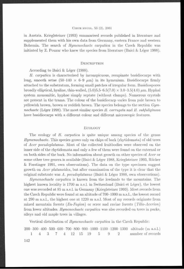
According to Baici & Léger (1988).H. carpatica is characterised by inconspicuous, resupinate basidiocarps with
long, smooth setae (50-140 x 6-9 /¿m) in its hymenium. Basidiocarps firmly attached to the substratum, forming small patches of irregular form. Basidiospores broadly elliptical, hyaline, thin-walled, (5.0)5.5-6.5(7.0) x 3.0-3.5(4.0) ¡im. Hyphal system monomitic, hyphae simply septate (without clamps). Numerous crystals are present in the trama. The colour of the basidiocarp varies from pale brown to yellowish brown, brown or reddish brown. The species belongs to the section Gym -
nochaete (Léger 1998). The most similar species II. corrugata and II. subfuliginosa have basidiocarps with a different colour and different microscopic features.
E c o l o g y
The ecology of II. carpatica is quite unique among species of the genus Hymenochaete. This species grows only on chips of bark (rhytidomata) of old trees of Acer pseudoplatanus. Most of the collected fruitbodies were observed on the inner side of the rhytidom ata and only a few of them were found on the external or on both sides of the bark. No information about growth on other species of Acer or
|H some other tree genera is available (Baici & Léger 1988, Krieglsteiner 1993, RiickerH & Forstinger 1991, own observations). The data on the type specimen suggest
growth on Acer platanoides, but after examination of the type it is clear that the■ original substrate was A. pseudoplatanus (Baici & Léger 1988, own observations).
Hymenochaete carpatica is known from the lowlands to the mountains. The B l highest known locality is 1700 m a.s.l. in Switzerland (Baici et Léger), the lowestB | one was recorded at 85 m a.s.l. in Germany (Krieglsteiner 1993). Most records from
the Czech Republic were found at an altitude of 700-1000 m a.s.l., the lowest record j | | a t 290 m a.s.l., the highest one at 1220 in a.s.l. Most of my records originate fromH mixed mountain forests (Eu-Fagion) or scree and ravine forests ( Tilio-Acerion)
from lower altitudes. Hymenochaete carpatica was also recorded on trees in parks, alleys and old maple trees in villages.
■ Vertical distribution of Hymenochaete carpatica in the Czech Republic:
I 200-300-400- 500-600-700-800-900-1000-1100-1200-1300 altitude (m a.s.l.)H 1 4 3 7 4 12 15 19 5 9 2 number of records
C z e c h m y c o l . 53 (2 ) , 2001
142

T o m š o v s k ý M .: R e m a r k s on t h e d is t r ib u t io n ok H y m e n o c h a e t e c a r p a t ic a
F ig . 1. D istribu tion of H ym enochaete carpatica in Europe.
- t + - i - L f - .......
k « - M ^ 5 i ± f e í e í í i f c - ř
: : s ť ^ : : b í t o si t r i ± ž í p i : u C _ -Z fď V -L .i L ¿ E _ ♦ L X M \i_ LIZ T I _ iL - r - ^ F - - 1 - á h # - * — .....
:rz ± = ^ & E T Í;5 :S íífe k ||i= = x d :iís f^« ¿ i J ..2 b / 4 5 *...r r \ t : * * _j* lí , Ml jl b ě K =l_ x ^ «gi x . Htfrraďmy'
F ig . 2. D istribu tion of H ym enochaete carpatica in the Czech Republic. T he crosses dem onstra te localities published by Kriegelsteiner (1993).
143

D i s t r i b u t i o n
Hymenochaete carpatica is known only from Europe (Austria, Germany, former Czechoslovakia, eastern France, Switzerland). I have completed the information with unpublished records from the Czech Republic, Slovakia, Romania and the Ukraine. Most records are documented by specimens deposited in the herbarium of National Museum, Prague (PRM) or Moravian Museum, Brno (BRNM). The collections marked JH (J. Holec) are kept in PRM, but at present they do not yet have the PRM number. All records were collected on Acer pseudoplatanus, therefore the substrate is not mentioned at the specimens. The abbreviation M. T. refers to the author.
Czech Republic
W estern Bohem ia
Lázně K ynžvart, park by the castle, leg. e t det. M. Svrček, X. 1998, herbarium M. Svrček.
Jizerské hory M ts.
Hejnice, bank of Velký Štolpich brook, 540 m a.s.l., leg. e t det. M. T ., 14. IX. 2000, PR M 894251.
Šum ava M ts.
česk é Žleby, K apraď M t., 980 m a.s.l., leg. e t det. M. T ., 22. IX. 1998, PR M 893833. - České Žleby, K ostelní cesta pathway, 940 m a.s.l., leg. e t det. J. Holec, 3. IX. 1999, JII 139/99. - České Žleby, Radvanovický h řbe t M t., 890 m a.s.l., leg. e t det. M. T ., 8. X. 1998, PR M 893825; ibid., 900 m a.s.l., PR M 893820; ibid., 900 m a.s.l., leg. e t det. J. Holec, 17. X. 1997, JH 866/97; ibid., 930 m a.s.l., JH 877/97. - České Žleby, Spáleniště M t., 930 m a.s.l., leg. e t det. Z. Pouzar,22. IX. 1998, PR M 893816; ibid., 880 m a.s.l., leg. e t det. J. Holec, 13. X. 1997, JII 750/97. - České Žleby, Žlebský kopec hill, 1000 m a.s.l., leg. e t det. M. T ., 13. IX. 1999, PR M 894010. - Horská Kvilda, Pěnivý potok brook, 950 m a.s.l., leg. e t det. J. Holec, 1. VII. 1999, JH 74/99. - Horská Kvilda, Zhůří, 1140 m a.s.l., leg. e t de t. M. T ., 19. IX. 1999, PR M 894022. - Kvilda, Orel, 1140 m a.s.l., leg. e t det. J. Holec, 6. X. 1998, PR M 897494. - M odrává, Vchynicko-Tetovský kanál, 920 m a.s.l., leg. e t det. M. T ., 30. VI. 1999, PRM 893817. - Nová Pec, Chornice, 960 m a.s.l., leg. e t det. M. T ., 22. VI. 1999, PR M 894182. - Nová Pec, Rakouská cesta pathw ay - border stone No. 1/10, 1040 m a.s.l., leg. e t det. M. T ., 22. VI. 1999, PR M 894183.- Nová Pec, Sm rčina M t., 1140 m a.s.l., leg. e t det. J. Holec, 25. IX. 1997, PR M 891333. - Nová Pec, Sm rčina M t., 1180 m a.s.l., leg. e t de t. M. T ., 4. VI. 1998, PR M 893830; ibid., 1200 m a.s.l., PR M 893823; ibid., 1010 m a.s.l., PR M 893819. — Nová Pec, village, 795 m a.s.l., leg. e t det. M. T ., 22. VI. 1999, PR M 893822. - Prášily, Form berg, 950 m a.s.l., leg. e t det. M. T ., 10. VI. 1999, PR M 894013. Prášily, village, 880 m a.s.l., leg. e t det. M. T ., 26. V III. 1998, PR M 893832. - Prášily, village, 950 m a.s.l., leg. et det. F. K otlaba, 5. V III. 1998, PRM 8922710. - Prášily, Stodůlky, 850 m a.s.l., leg. e t det. M. T .,12. VI. 1999, PR M 894015. - Prášily, Ždanidla M t., 1220 m a.s.l., leg. e t det. M. T ., 9. VII. 1998, PR M 894055; ibid., 1190 m a.s.l., PR M 894011; ibid., 1140 m a.s.l., PR M 894017. - Srní, Povydří, 680 m a.s.l., leg. e t det. J. Holec, 8. X. 1997, JH 633/97. - Srní, Povydří, 920 m a.s.l., leg. e t de t. M. T ., 28. VI. 1999, PR M 893818; ibid., 750 m a.s.l., 24. IX. 1998, PR M 893826. - Srní, Povydří, Horní Hrádky, 900 m a.s.l., leg. e t det. M. T ., 30. VI. 1999, PR M 893827. - Srní, Povydří, Hrádecký po tok brook, 800 m a.s.l., leg. e t det. M. T ., 30. VI. 1999, PR M 893828. - Srní, Zadní P aště , 810 m a.s.l., leg. e t det. M. T ., 14. VI. 1999, PR M 893823. - Stožec, Stožecká
C z e c h m y c o l . 53 (2 ) , 2001
144

skála rock, 976 m a.s.l., leg. et det. M. T ., 23. VI. 1999, PR M 893821. - Stožec, Světlá brook, 860 m a.s.l., leg. e t det. M. T ., 23. VI. 1999, PR M 893831. - Strážný, village, 840 rn a.s.l., leg. e t det. M. T ., 23. IX. 1999, PR M 894019. - Zátoň, Boubín-Pažení, 1100 in a.s.l., leg. e t det. J . Holec, 10. V II. 1998, PR M 896998. - Zátoň, Jilm ová skála hill, 960 m a.s.l., leg. e t de t. M. T .,13. X. 1998, PR M 894023; ibid., 1000 m a.s.l., leg. e t det. J . Holec e t Z. Pouzar, 16. X. 1996, PR M 889509. - Železná R uda, Černé jezero lake, V III. 1926, leg. A. H ilitzer, det. A. P ilá t, PR M 686735; ibid., 1926, PR M 686736. - Železná R uda, M edvědí jám y, 850 m a.s.l., leg. e t det. J. Holec, 16. VI. 1997, JH 16/97. - Železná R uda, D ebrník, avenue, 800 m a.s.l., leg. e t det. M. T .,24. V III. 1998, PR M 894021; ibid., 7. VII. 1998, PR M 894020. - Železná R uda, D ebrník, m ixed forest, 800 m a.s.l., leg. e t det. J. Holec, 15. X. 1997, JH 811/97. - Železná R uda, Ferdinandovo údolí valley, 740 m a.s.l., leg. e t det. M. T ., 8. VIII. 1998, PR M 894014; ibid., 850 m a.s.l., leg. e t de t. J . Holec, 16. VI. 1997, PR M 890902. - Železná R uda, by th e road to Špičák, 760 m a.s.l., leg. e t det. M. T ., 25. V III. 1998, PR M 894018. - Železná R uda, P ancíř M t., 1110 m a.s.l., leg. e t det. M. T ., 27. V III. 1999, PRM 894016. - Želnava, Černý les forest, 900 m a.s.l., leg. e t det. M. T ., 24. VI. 1999, PR M 893824.
C en tra l B ohem ia
č esk ý K ras, Srbsko, Bubovické vodopády waterfalls, 290 m a.s.l., leg. e t det. M. T ., 31. V III. 1999, PR M 894465. — Železné hory, Třem ošnice, Lovětínská rokle, 750 m a.s.l., leg. e t det. M. T ., 10. IX. 2000, PR M 894461. — Branov near Křivoklát, valley “V luhu” , leg. M. Svrček,25. VI. 1961, PR M 616218. - Brdy m ountains, avenue by Padrťské rybníky pounds, leg. e t det. J. Holec, 13. XI. 1997, PR M 891657.
Southern Bohem ia
D istrict P rachatice , Křišťanovice, 800 m a.s.l., leg. e t det. M. T ., 14. IV. 2000, PR M 894249. - Nové Hrady, Terčino údolí valley, 500 m a.s.l., leg. e t det. M. T ., 4. V III. 1999, PR M 894240. - B lanský les M ts., Chrášťanský vrch, 750m a.s.l., leg. e t det. D. Dvořák, 23. 5. 2001, herbarium D. Dvořák. - Blanský les M ts., Kleť M t., 950 m a.s.l., leg. e t det. D. Dvořák, 23. 5. 2001, herbarium D. Dvořák. — D istrict česk ý Krum lov, Žofínský prales, 760 m a.s.l., leg. e t det. M. T ., 21. VI. 2001, PRM .
Czech-M oravian H ighlands
C hotěboř, valley of the Doubravka river, 460 m a.s.l., leg. et det. M. T ., 31. V III. 2000, PR M 894238. — Jihlava, Vysoký kámen hill, 650 m a.s.l., leg. e t det. M. T ., 6. IX. 2000, PR M 894247. — Pelhřim ov, top of Křem ešník hill, 760 m a.s.l., leg. e t det. M. T ., 1. IX. 2000, PR M 894244. - S taré Ransko, avenue in the village, leg. e t det. M. T ., 11. V III. 2000, PR M 894235. — Třešť, Velký Špičák M ts., 730 m a.s.l., leg. e t det. M. T ., 7. IX. 1999, PR M 894245. - Žďár nad Sázavou, castle, 560 m a.s.l., leg. e t det. M. T ., 21. V III. 1999, PR M 894243.
E astern B ohem ia
D istric t Ú stí n. Orlicí, Litice, 430 m a.s.l., leg. e t det. M. T ., 23. V III. 2000, PR M 894248. — D istric t Rychnov nad Kněžnou, Po tšte jn , Modlivý důl, 350 m a.s.l., leg. e t det. M. T .,23. V III. 2000, PR M 894242. — D istrict Rychnov nad Kněžnou, Po tšte jn , Vochtánka valley, 320 m a .s.l., leg. e t det. M. T ., 23. V III. 2000, PR M 894246. - Orlické hory M ts., Sněžné, next to th e road to Sedloňov, 590 m a.s.l., leg. e t det. M. T ., 27. XII. 1999, PRM 894239. - D istrict Ústí n. Orlicí, Velká M orava, Sviní hora M t., 790 m a.s.l., leg. e t det. D. Dvořák, 18. XI. 2000, PR M 894366. - D istric t Ú stí n. Orlicí, Horní M orava, Klepý M t., 660 m a.s.l., leg. e t det. D. Dvořák, 14. II. 2001, herbarium D. Dvořák; ibid., 865 m a.s.l., 16. II. 2001.
T o m š o v s k ý M .: R e m a r k s o n t h e d is t r ib u t io n o f H y m e n o c h a e t e c a r p a t ic a
145
C entra l M oravia
Adam ov, Jelení skok, leg. A. Vágner, 5. VI. 1999, BRNM 642784. - Černovice u K un štá tu , Hrádky, leg. A. Vágner, 28. VII. 1999, BRNM 648418. - Černovice u K un štá tu , Káčiny, leg. V. A ntonín, 27. IV. 2000, BRNM 652783. - Ochoz u B rna, valley of Ř íčka brook, 25. V. 2000, leg. V. A ntonín, BRNM . - Osiky, Horní Židovka, leg. A. Vágner, 28. VII. 1999, BRNM 648414. - Letovice, park a t th e castle, leg. A. Vágner, 29. VII. 1999, BRNM 648438. — Úsobrno, D urana, leg. V. A ntonin, 10. V. 2000, BRNM .
Jeseníky Mts.
B ran n á near Šum perk, valley of Vrbenský potok, leg. A. Vágner, 7. IX. 1999, BRNM 648832.
Oderské vrchy hills
Hranice, Jezernice valley, leg. e t det. Z. Pouzar, 13. VI. 2001, PRM .
Beskydy M ts.
D istrict Frýdek-M ístek, Radhošťské Beskydy M ts., Bílá, bank of Salajský po tok brook, c. 750 m a.s.l., leg. M. Vašutová, 12. VII. 1999, herbarium M. Vašutová. - D istrict Frýdek-M ístek, Slezské Beskydy M ts., Nýdek, village, 420 m a.s.l., leg. e t det. M. T ., 30. IX. 1999, PR M 894460.
Bílé K arpaty M ts.
S trán í, Velká Javořina, A cer pseudoplatanus , leg. V. A ntonín e t A. Vágner, 18. V III. 2000, BRNM 652866.
SlovakiaM alé K arp a ty M ts. (Kleine K arpathen), Sklená h u ta (G lasshiitten), leg. J. Hrubý, det.
A. P ilá t, IV. 1925, PR M 686734 (typus). — M uránska p lanina p lateau , betw een Čadová jam a and H avranská šopa, leg. Z. Palice, det. M. T ., 11. V. 1999, PR M 894462. - Oravské Beskydy M ts, B abia hora M t., 1000 m a.s.l., leg. e t det. M. T ., 29. IX. 2000, PR M 894463. - Slovenský ra j, Velký Sokol valley, 640 m a.s.l., leg. e t det. M. T ., 11. VI. 1998, PR M 894464. - Slovenský ra j, Pily, bank of th e brook, 600 m a.s.l., leg. e t det. M. T ., 9. VI. 1998, PR M 894466.
RomaniaBS D istrict Suceava, Gur a Hum orului, in front of the ga te of the Hum or m onastery, leg. M. T . and
M. K olařík, det. M. T ., 30. VI. 2000, PR M 894434. - D istrict Suceava, R aráu M ts., by th e road from C ám pulung Moldovenesc to th e top of R aráu M t., next to th e m onastery, leg. e t det. M. T ., 1. VII. 2000, PR M 894433. - D istrict Suceava, R aráu M ts., to p of R aráu M t., next to m ountain hotel, leg. e t det. M. T ., 1. VII. 2000, PR M 894431. - D istrict Suceava, R aráu M ts., S látioara, prim eval forest, leg. e t det. M. T ., 1. VII. 2000, PR M 894432. - D istrict Suceava, C alim an M ts., village G u ra Haitii, bank of th e brook, 1100 m a s . m., leg. e t det. M. T ., 30. VI. 2000, PR M 894435.
UkrainejHI E astern C arpath ians, D istrict Rachov, village Kvasy, M entchul M t., 48 '1 0 ’52"N, 24 ”18’24” E,
900 m a.s.l., leg. e t det. J. Holec, 14. VII. 1999, PR M 892898; ibid., 1300 m a.s.l., 13. VII. 1999, PR M 892895. - E astern C arpath ians, D istrict Rachov, village Kvasy, M entchul M t., 48 "10’34” N, 24 "18’10” E, 750 m a.s.l., leg. e t det. M. T ., 11. VII. 1999, PR M 894467; ibid., 4 8 ’10’51” N, 24 "17’52” E, 660 m a s . 1., PR M 894468.
C z e c h m y c o l . 53 (2 ) , 2001
146
T o m š o v s k ý M .: R e m a r k s o n t h e d is t r ib u t io n o f H y m e n o c h a e t e c a r p a t ic a
Austria
Voralberg, M ühlviertel, N ordtirol, Salzburg, S teierm ark (K rieglsteiner 1993, Rücker & Forstinger 1991).
France
Vosges (K rieglsteiner 1993).
Germany
B avaria (Bayerischer W ald, Bayerische Alpenvorland, Oberpfälzer W ald), B aden-W ürttem berg, Harz M ts., Schwarzwald M ts. etc. (K rieglsteiner 1993).
Switzerland
K antons Fribourg, G larus, Schwyz, St. Gallen, Zürich (Baicin & Léger 1988).
D i s c u s s i o n
The distribution of Hymenochaete carpatica is not sufficiently known, despite of the old datum of its description. In past the species was overlooked due to its specific ecology. Many Aphyllophorales specialists do not even collect Hymenochaete carpatica. According to my correspondence with A. Bernicchia and M. Tortic the species is not known from Italy and countries of the former Yugoslavia, but I think the species can occur there. Hymenochaete carpatica has neither been reported from Poland. The fungus may be widely distributed in Europe, where it follows the distribution of the only host - Acer pseudoplatanus.
A c k n o w l e d g e m e n t s
I would like to thank dr. Z. Pouzar (National Museum, Prague), whom I consulted several times in this m atter, and dr. J. Holec (National Museum, Prague) for helpful comments on the manuscript. I also wish to thank A. Vágner (Moravian Museum, Brno), dr. M. Svrček (National Museum, Prague), D. Dvořák, Z. Palice and M. Vašutová for valuable information on their records of Hymenochaete carpatica.
R e f e r e n c e s
B a ic i A. and LÉGER .1. (!. (1988): H ym enochaete carpa tica P ilát collected in Sw itzerland. - Mycol. H elvetica 3 (1): 89-98.
K r ie g e l s t e in e r G. .1. ( 1993): H ym enochaete carpa tica P ilá t 1930, die Bergahorn B orstenscheibe, in M ittte leuropa. - Beiträge zur K enntnis der Pilze M itteleuropas 9: 79-96.
147

L e g e r J . C. (1998): Le genre H ym enochaete Léveillé. - In: B ibliotheca mycologica, 319 p.L izo Ň P . and JANČOVIČOVÁ S. (2000): Non-Iichenized fungal tax a described from Slovakia. P a rt
1.— M ycotaxon 55: 479-500.PILÁT A. (1927): Příspěvek ku poznání Aphyllophoraceí západního Slovenska (E in B eitrag zur
K enntnis der A phyllophoraceen der westlichen Slowakei). - Mykologia, 4: 72—74.PlLAT A. (1931): M onographie der europäischen Stereaceen. - Hedwigia, 70: 10-132.P ilÁt A. (1933): D ruhé naleziště zajím avé houby H ym enochaete carpa tica P ilá t [Second locality
of an in teresting fungus H ym enochaete carpa tica Pilát]. - Věda přírodní, P rah a , 14: 219.RÜCKEll T . and F orsting er H. (1991): H ym enochaete carp a tica P ilá t, ein weit verbreite ter,
häufig übersehener Borstenscheibling. — Linzer Biologische Beiträge, 23 (1): 417-424.
C z e c h m y c o l . 53 (2 ) , 2001
148

Lipid, sterol and ergosterol accumulation in isolates of dematiaceous Hyphomycetes
M . E m a n M o s t a f a , A. A. Z o h r i and R a g a a S. K o t b y
B otany D epartm ent, Faculty of Science, A ssiut University,Assiut, Egypt
Mostafa M. E., Zohri A. A. and Kotby R. S. (2001): Lipid, sterol and ergosterol accumulation in isolates of dematiaceous hyphomycetes - Czech Mycol. 53: 149-159
M ycelial d ry weight, lipid and sterol contents of fungi tested varied w ith fungal genus, species and even w ith isolate of one species. T heir d ry m ass fluctuated between l l l . 6 i l 0 . 7 - 4 5 7 . 0 i Í 4 1 .5 m g/50 ml m edium . Lipids, sterols and ergosterol accum ulated by the isolates tested ranged from 4 .5 2 Í 0 .5 -2 9 .0 4 i2 .7 6 % , 1 .2 3 i0 .1 6 -1 0 .6 3 il.2 4 % and 0 .4 3 Í0 .0 5 7 -7 .1 3 Í 0.695% of their d ry m ass, respectively. Cochliobolus sp icifer isolate No. 35 was the highest lipid-producer while Ulocladium a trum No. 90 proved to be superior in the production of sterols and ergosterol. T L C technique and chem ical analysis of lipid classes produced by U. atrium No. 90 revealed th a t th e lipid fractions are composed of free sterols, free fa tty acids, sterol esters, glycolipids, phospholipids and squalene.
K e y w o rd s : Lipid, sterol, ergosterol, dem atiaceous hyphom ycetes
Mostafa M. E., Zohri A. A. a Kotby R. S. (2001): Akumulace tuků, sterolu a ergosterolu v myceliu některých kmenů hyfomycetů (Dematiaceae) - Czech Mycol. 53: 149-159
Sušina mycelia, obsah lipidů a sterolů kolísá v rám ci různých rodů hub, mezi druhy i mezi izoláty jednoho druhu. Cochliobolus sp icifer isolát č. 35 byl nej vyšším producentem lipidů a druh Ulocladium a trum isolát č. 90 produkoval nejvíce steroly a ergosterol.
Many fungi have a high capacity for lipid production. The lipid content of vegetative fungal hyphae varies between 1% and 50% of their dry mass (Weete 1974). Sterol is a group of lipids tha t has extensively attracted the attention of biochemists during the past decades. The utilization of sterols, as precursors for certain vitamins (Ellis 1945) and steroid sex hormones (Kieslich 1985), lead to a thorough investigation of their occurrence in nearly all forms of life (animals, plants and microorganisms). Sterols are also used as taxonomical and phylogenetical secondary metabolites (Weete 1974).
Pharmaceutical research has used ergosterol as a target for such antimycotic drugs as amphotericin-B (Rippon 1982). Ergosterol has been also recorded for antirachitic activity (El-Refai and El-Kady 1969). It is a common fungal sterol known to occur in a broad taxonomic range of fungi (Gordon and Webster 1984) and acts as a precursor of vitamins (Ellis 1945).
The first attem pt to isolate sterols from fungi was accomplished by Naegeli and Loew (1878). A considerable amount of data has been collected during the
149
C z e c h m y c o l . 53 (2), 2001
I ........... .

past, decades on sterols of fungi. In general, different species of filamentous fungi, especially Aspergilli and Penicilli, have been studied for sterol production (Preuss et al. 1931, El-Refai 1964, Weete 1974, El-Kady et al. 1995). All these studies reported that ergosterol was the principle fungal sterol in the fungi.
There are only a few studies on the lipid and sterol contents of dematiaceous fungi. Therefore, the present study investigated the potential of 100 isolates of36 species and one variety belonging to 18 genera of dematiaceous hyphomycetes for the production of lipids, sterols and ergosterol. Quantitative analyses of the different lipid classes produced by selected isolates grown on Gzapek’s medium using thin layer chromatography and chemical analysis were also performed.
M a t e r i a l s a n d m e t h o d s
F u n g a l is o la te sOne hundred different fungal isolates of dematiaceous hyphomycetes belonging
to 18 genera, 36 species and one species variety were collected and used in this study. Sixty- three of these isolates were isolated from medicinal and herbal plants (Youssef 1995, Ragaa Kotby 1996) in our laboratory. The remanining37 isolates were obtained from Assiut University Culture Collection (AUCC), Botany Department, Faculty of Science, Assiut University, Assiut, Egypt. The numbers and names of these isolates are given in Table 1. Stock cultures were maintained on slants with Czapek’s Dox Agar (Smith and Onions 1983). Inocula were prepared from 7 days old cultures as spore suspensions in 0.2% (v/v) aqueous Tween 80.
C u l t i v a t i o nEach individual isolate was cultivated on Czapek’s modified liquid glucose
medium of the following composition (g/L of distilled water): sodium nitrate 2.0; potassium dihydrogen phosphate 1.0; magnesium sulphate 0.5; potassium chloride 0.5; ferrous sulphate 0.01; glucose 10.0 and supplemented with yeast extract 1.0 and peptone 10.0. Erlenmeyer flasks (three flasks for each isolate) of 250 ml capacity were used. Each flask contained 50 ml of the medium. The flasks were sterilized at 121 °C for 20 min. and inoculated after cooling with a 2 ml spore suspension (about 10° spores/ml) of the prepared inocula of each organism. The cultures were incubated at 28 °C as stationary cultures for 10 days.
G r o w t h d e t e r m i n a t i o nGrowth was measured as dry mycelial mass. The mass was filtered on W hatman
No. 1 filter paper (15 cm diameter), washed three times with distilled water and dried for 24 h at 105 °C.
C z e c h m y c o l . 53 (2 ) , 200 1
150

M o s t a f a M . Ii., Z o h r i A. A. a n d K o t b y It. S.: L i p i d , s t e r o l a n d e r u o s t e r o l a c c u m u l a t io n
A n a l y t i c a l m e t h o d s
Total lipids were extracted and determined according to Fanelli and Fab- bri (1980). Total sterols were estimated by the Liebermann-Burchared colour reaction method (Cook 1958). Ergosterol content was estimated using UV spec- trophotornetric analysis at 282 nm (Maguigan and Walker 1940). Total fatty acids were determined by the phosphovanillin method of Zollner and Kirsch (1962). Glycolipids were determined according to Brown and Dupont (1989). Phospholipids were separated by the method described by Galanos and Kapoulas (1962) and estimated by the method of Houser et al. (1970).
F r a c t i o n a t i o n of t h e l ip id c l asses by TL C
Samples of the lipid m atter dissolved in chloroform were used for qualitative fractionation analysis applying thin layer chromatographic techniques (El-Kady 1967, Ragaa Kotby 1996).
R e s u l t s a n d d i s c u s s io n
The mycelial dry weight, lipid and sterol contents of the fungi tested varied with fungal genus, species and even with isolate of the same species (Table 1). These results are similar to those previously recorded by several authors (El-Refai 1964, Mumma et al. 1970, Weete 1974).
Dry mass of the fungal isolates varied between 111.6±10.7 and 457.0±41.5 m g/ /50 ml medium. The highest amount of dry mycelium was produced by Alternaría tenuissima isolate No. 9 followed by Cladosporium cladosporioides No. 15, Ulocladium tuberculatum No. 99, Ernbellisia didymospora No. 52 and Pyrenophora avenae No. 69, while the lowest dry mass was produced by Stachybotrys chartarum No. 82 (Table 1). Similar values were previously recorded by El-Refai (1964) who determined the dry mass of three dematiaceous fungal isolates on six types of synthetic media and found that the dry mass of Stemphylium consortiale, Alternarla tenuis Nees and A. tenuis acut. ranged from 107 2118, 191 2890 and 87.8-1023 mg/100ml depending on the medium, respectively.
The total lipids accumulated by the different isolates of dematiaceous hyphomycetes tested ranged from 45.2±5.02 to 290.40±27.63 m g/g mycelial dry mass (= 4.52±0.5-29.04±2.76% of their dry weight) as shown in Table 1. These results are in close agreement with the findings of many workers. In early studies, Preuss et al. (1934) cultivated 24 moulds for lipid and sterol production on two types of media and found that the total lipid content of the mycelium varied from 1.1-19.9% and 1.5-24.4% of their dry mass on glucose-inorganic salts and glucose-malt-sprouts media, respectively. Prill et al. (1935) recorded that the
151

1 ^ — M M M I ■
total lipid contents of Aspergillus fisheri reached 37% of the dry weight. Mumma et al. (1970) found tha t the total lipids of Chaetomium globosum, Pénicillium chrysogenum, Sporotrichum thermophile, Malbranchea pulchella and M. pulchella var. sulfurea were 54.1%, 9.8%, 15.5%, 26.5% and 24.8% of their dry weight, respectively. Weete (1974) reported that the lipid contents of different fungi varied between 1 and 50% of their dry weight depending on the species, stage of growth development, and culture conditions. Weete et al. (1985) recorded th a t the lipid content comprised 3.4% of the cellular dry m atter of Mucor rouxii cells. Rawia Saad (1992) found th a t the total lipids of Aspergillus amstelodami and A. repens were 51.1% and 18.2% of their dry masses, respectively. Leobardo et al. (1992) determined the total lipid accumulation of two filamentous fungi ( Trichoderma harzianum and T. viride) and found tha t lipid accumulation was about 17% and 32% (w/w) of dry weight for the two species, respectively. Recently, El-Kady et al. (1995) recorded tha t the total lipids of 57 fungal isolates (capable to grow on whey) ranged from 6 ±1 to 38 ±8% of their dry mass.
The highest lipid-producer isolates (Table 1) were Cochliobolus spicifer No. 35 (produced 29.04±2.76% lipid of dry weight), Stachybotrys elegans No. 84 (28.48±2.85%), Monodictys castaneae No. 61 (24.58±2.29%), Ernbellisia didymospora No. 52 (23.94±2.45%), Stachybotrys chartarum No. 81 (23.40±2.27%), Cla- dosporium cladosporioides No. 14 (21.50±2.38%) and Setosphaeria rostrata No. 72 (21.12±1.96%). El-Refai (1964) found that the total lipids of Stemphylium consortiale and Altemaria tenuis reached 18.18% and 23.27% of their dry weight, respectively. The lipid contents of the different species of dematiaceous hyphomycetes tested in the present study are similar to those recorded by different fungal groups such as Pénicillium species (Ward et al. 1935, Ghanem et al. 1990); Phycomycetes, Ascomycetes, Basidiomycetes (Weete 1974, Weete et al. 1985);Eurotium species (Rawia Saad 1992, El-Kady et al. 1995); Trichoderma species (Leobardo et al. 1992), as well as different species of Aspergillus, Emericella and Fennellia (El-Kady et al. 1995).
The average values of total sterols produced by the different isolates tested fluctuated between 12.31±1.60 and 106.3±12.35 mg/gdry weight (1.23±0.16% and 10.63±1.24% of their dry mass; 9.08% and 80.71% of their total lipids). The most sterol-producing isolate was Ulocladium atrum No. 90 (Table 1). These results are in accordance with those previously recorded by several workers (Preuss et al. 1934,El-Refai 1964, Ghanem et al. 1990, El-Kady et al. 1995). Fungi appear to differ quantitatively in their sterol contents (Ratcliffe 1937). El-Refai (1964) found that the total sterols of Stemphylium consortiale and Altemaria tenuis Auct. grown on Bills medium were 1.66 and 1.40% of their dry weight, respectively. Also, he recorded tha t the level of sterol reached 4.68% of Aspergillus fumigatus dry weight in presence of 1.0 /xg/ml pantothenic acid in the basal medium. Ghanem et al. (1990) found that the total sterol yields of Pénicillium crustosum grown
C z e c h m y c o l . 5 3 (2 ) , 2Ü01
152
on a beet medium containing molass reached 8.4% of dry mass at pH 7.0 after 8 days of incubation. El-Kady et al. (1995) investigated the total sterol contents of 57 isolates belonging to Aspergillus (10 species), Emericella (two species varieties), Eurotium (four species and one variety) and Fennellia (two species), which were capable of growing on cheese whey. They noted th a t their total sterol contents ranged between 2±0.6% and 20±4% of their dry mass.
The mean values of ergosterol produced by the different isolates of dematiaceous hyphomycetes tested ranged from 4.31±0.57 to 71.3±6.95 m g/g dry weight (= 0.43±0.057% -7.13±0.695% of their dry weight, 3.35% -42.44% of their lipids and 4.71% -80.64% of their total sterols). Ulocladium atrum No. 90 proved to be the best ergosterol producer (Table 1). Few investigations have determined the production of ergosterol as a metabolite of the different genera and species of dematiaceous hyphomycetes such as Stemphylium consortiate (El-Refai 1964) in addition to Drechslera graminea and D. teres (Gordon and Webster 1986).
Ergosterol was found to be the principle sterol in the isolates screened (Table 1). These results are in accordance with the results previously reported by several workers (El-Refai 1964, Weete 1974, Weete et al. 1985, Fernando and Bean 1986, Ellis et al. 1991). Moreover, Fiore (1948) reported that ergosterol was the only sterol produced by Fusarium lini, F. lyeopersici and F. solani. El-Refai (1964) found th a t ergosterol formed about 92% of the total sterols of A. fumigatus when the organism was cultivated on molasses at a concentration equivalent to 10% sugars and corn steep liquor at a level of 2% solids. Recently, El-Kady et al. (1995) recorded tha t ergosterol production by 57 fungal isolates belonging to 10 species of Aspergillus, two species varieties of Emericella, four species and one variety of Eurotium in addition to two species of Fennellia which could be grown on whey, ranged from 15% to 65% of their total sterols.
An attem pt has also been made to fractionate and determine the different classes of lipids produced by Ulocladium atrum No. 90 which proved to be superior in the production of sterols and ergosterol. Thin layer chromatographic techniques and chemical analysis of the lipid classes produced by U. atrum No. 90 cultivated on Czapek’s medium revealed the following lipid fractions: glycolipids, phospholipids, free sterols, free fatty acids, sterol esters and squalene (Table 2). These lipid fractions have been reported from different fungi (El-Refai and El-Kady 1969, Fanelli and Fabbri 1980, Weete et al. 1985, Naim and Saad 1986, Leobardo et al. 1992). El-Kady (1967) found that the fractionation of yeast lipids by the thin layer chromatographic technique resulted in: phospholipids, free sterols, fatty acids, fatty acid esters, triglycerides, sterol esters and squalene. The presence of squalene was expected as it was elucidated by many workers (Corwn et al. 1956, El-Kady 1967).
M o s t a f a M . E . , Z o h r i A . A . a n d K o t b y R . S .: L i p i d , s t e r o l a n d e r g o s t e r o l a c c u m u l a t io n
153

T a b le 1 . Isolates of dem atiaceous hyphom ycetes screened for to ta l lip id , to ta l sterols and ergosterol production ( the values are the average ¿ s ta n d a rd deviation based on th ree replicates).
No. Species Dry wt. Total lipid Total sterol Ergosterol
(mg/50ml) (mg/g dry wt.) (mg/g dry wt.) (mg/g dry wt.)
Altemaria
1 A. alternata (Fr.) Keissler 2 3 4 .5 ± 2 1 .6 123 .80±11 .72 7 4 .7 8 ± 8 .4 3 1 9 .0 0 ± 2 .0 0
2 A. alternata (Fr.) Keissler 2 8 1 .0 ± 2 6 .2 135 .00±12.51 7 0 .7 2 ± 7 .6 5 3 5 .7 0 ± 3 .6 2
3 A. alternata (Fr.) Keissler 2 0 0 .9 ± 2 0 .5 14 0 .0 0 ± 1 3.71 8 8 .0 0 ± 8 .6 0 2 6 .2 0 ± 2 .2 3
4 A. chlamydospora Mouchacca 1 73 .9± 16 .3 127 .50±10.63 5 5 .5 0 ± 6 .4 8 9 .8 0 ± 1 .0 5
5* A. chlamydospora Mouchacca 269 .3± 24 .1 120 .60±12 .46 7 8 .0 6 ± 8 .6 2 16 .0 0 ± 1 .5 2
6* A. chlamydospora Mouchacca 170 .2 ± 1 5 .6 103 .60±11 .20 6 1 .8 0 ± 5 .4 7 10 .7 0 ± 1 .3 5
7* A. chlamydospora Mouchacca 182 .4 ± 1 6 .8 9 3 .8 0 ± 8 .9 7 68.11 ± 7 .4 1 17 .0 0 ± 1 .4 7
8 A. macrospora Zimm. 184 .7 ± 1 6 .7 6 6 .0 0 ± 7 .4 8 2 4 .6 0 ± 3 .2 7 7 .5 5 ± 0 .8 5
9 A. tenuissima (Kunze) W iltshire 4 5 7 .0 ± 4 1 .5 199 .60±20 .85 3 0 .1 2 ± 2 .8 0 6 .6 9 ± 0 .6 4
10 A. tenuissima (Kunze) W iltshire 189 .4 ± 1 7 .4 9 6 .4 0 ± 7 .8 2 5 9 .9 9 ± 6 .0 0 14 .2 7 ± 1 .2 3
Bipolaris
11* f t neergaardii (Danquah) Alcorn 1 87 .8± 16 .9 4 5 .2 0 ± 5 .0 2 4 4 .5 3 ± 3 .5 4 6 .4 8 ± 0 .7 0
Bispora
12 f t betulina (Corda) Hughes 248 .3± 22 .1 6 3 .2 0 ± 5 .6 3 2 7 .6 4 ± 3 .4 2 6 .0 9 ± 0 .5 8
Cladosporlum
13 C. cladosporioides (Fres.) de Vries 2 6 1 .7 ± 2 3 .9 134 .20±14 .25 3 9 .2 9 ± 3 .5 8 1 0 .38± 9 .5 4
14 C. cladosporioides (Fres.) de Vries 1 5 1 .7 ± 1 4 .3 215 .00± 23 .82 1 9 .5 2 ± 2 .6 6 8 .2 7 ± 1 .0 2
15 C. cladosporioides (Fres.) de Vries 3 9 9 .5 ± 3 6 .5 105 .20±10 .64 3 9 .9 5 ± 4 .4 4 11.79±1 .31
16 C. herbarum (Pers.) Link 1 97 .0± 17 .5 133 .20±14 .06 6 0 .6 4 ± 5 .3 5 9.71 ± 0 .8 7
17 C. oxysporum Berk, et Curt. 2 1 8 .0 ± 1 9 7 163 .80±15 .90 2 4 .3 8 ± 2 .6 4 5 .9 9 ± 0 .6 4
18 C. oxysporum Berk, et Curt. 2 2 6 .3 ± 2 0 .4 153 .00±16 .18 5 3 .0 0 ± 6 .0 0 3 5 .3 0 ± 2 .7 6
19 C. sphaerospermum Penzig 183 .6 ± 1 5 .9 146.60±15.51 6 4 .3 0 ± 6 .5 2 18 .12±2 .01
20 C. sphaerospermum Penzig 238 .7±22 .1 185 .00±19 .04 1 9 .5 2 ± 2 .0 7 9 .5 9 ± 1 .0 5
Cochliobolus
21 C. australiensis (Tsuda et Ueyama) Alcorn 253 .0 ± 2 3 .5 110 .0 0 ± 1 0.63 8 1 .9 9 ± 7 .9 8 11,3 0 ± 1 .21
22 C. australiensis (Tsuda et Ueyama) Alcorn 16 5 .7 ± 1 5.1 6 9 .0 0 ± 7 .1 2 2 8 .2 9 ± 3 .0 5 13.14 ± 1 .40
23 (7. australiensis (Tsuda et Ueyama) Alcorn 2 2 5 .4 ± 2 1 .0 16 5 .6 0 ± 1 6.82 5 0 .8 2 ± 4 .1 5 9 .5 5 ± 1 .00
24 C. australiensis (Tsuda et Ueyama) Alcorn 2 1 3 .6 ± 1 9 .6 120 .20±13 .00 51,4 7 ± 6 .0 2 6.41 ± 0 .7 1
25 C. lunatus Nelson et Haasis 2 8 7 .5 ± 2 6 .0 15 9 .8 0 ± 1 6.67 27 7 7 ± 2 .5 3 6 .7 5 ± 0 .6 9
26 C. lunatus Nelson et Haasis 3 3 2 .8 ± 2 9 .8 17 6 .8 0 ± 1 6.93 44.01 ± 4 .0 9 21.13 ± 1 .95
27 C. lunatus Nelson et Haasis 3 0 2 .0 ± 2 6 .4 148 .00±13 .92 27.11 ± 3 .0 0 1 3 .8 7 ± 1 .6 2
28 C. lunatus Nelson et Haasis 2 5 0 .0 ± 2 3 .2 160 .20±17 .00 3 1 .7 0 ± 3 .4 2 1 0 .6 9 ± 1 .0 2
29 C. lunatus Nelson et Haasis 2 7 3 .8 ± 2 5 .3 176 .23±17 .23 3 3 .2 8 ± 3 .5 5 1 3 .5 3 ± 1 .6 0
30 C. lunatus Nelson et Haasis 2 2 5 .5 ± 2 1 .6 154.41 ± 1 5 .6 0 4 2 .9 6 ± 3 .8 6 9.41 ± 1 .0 5
31 * C. pallescens (Tsuda et Ueyama) Sivan.__________2 3 8 .3 ± 2 3 .0 7 4 .4 0 ± 6 .9 8 4 9 .9 0 ± 5 .1 0 1 2 .9 7 ± 1 .2 2
CZECH MYCOL. 53 (2 ) , 20 0 1
154
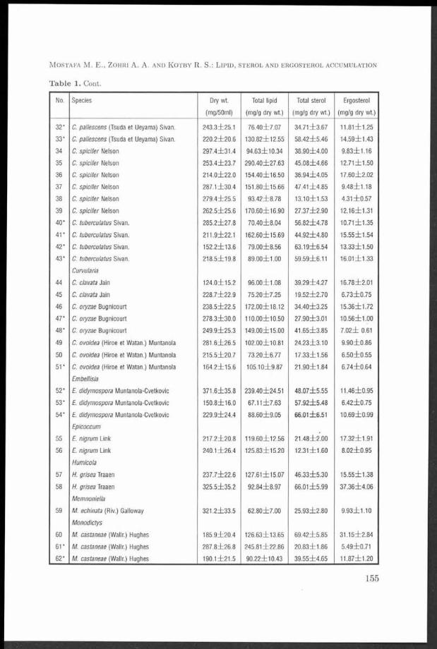
M o s t a f a M . E . , Z o h iii A . A . a n d K o t b y R . S .: L i p i d , s t e r o l a n d e r g o s t e r o l a c c u m u l a t io n
T a b le 1. Corit.
No. Species Dry wt. Total lipid Total sterol Ergosterol
(mg/50ml) (mg/g dry wt.) (mg/g dry wt.) (mg/g dry wt.)
32* C. pallescens (Tsuda et Ueyama) Slvan. 243 .3±25 .1 7 6 .4 0 ± 7 .0 7 3 4 .7 1 ± 3 .6 7 11.81 ± 1 .2 5
33* C. pallescens (Tsuda et Ueyama) Sivan. 2 2 0 .2 ± 2 0 .6 130 .82±12 .55 5 8 .4 2 ± 5 .4 6 1 4 .5 9 ± 1 .43
34 C. spic ifer Nelson 2 9 7 .4 ± 3 1 .4 9 4 .63± 10 .34 3 8 .9 0 ± 4 .0 0 9 .8 3 ± 1 .1 6
35 C. sp ic ife r Nelson 2 5 3 .4 ± 2 3 .7 290 .40± 27 .63 4 5 .0 8 ± 4 .6 6 12 .7 1 ± 1 .5 0
36 C. spicifer Nelson 2 1 4 .0 ± 2 2 .0 154 .40±16 .50 3 6 .9 4 ± 4 .0 5 17 .6 0 ± 2 .0 2
37 C. spic ifer Nelson 287.1 ± 3 0 .4 151 .80±15 .66 4 7 .4 1 ± 4 .8 5 9 .4 8 ± 1 .1 8
38 C. sp ic ife r Nelson 2 7 9 .4 ± 2 5 .5 9 3 .4 2 ± 8 .7 8 13 .1 0 ± 1 .5 3 4 .3 1 ± 0 .5 7
39 C. spic ifer Nelson 2 6 2 .5 ± 2 5 .6 17 0 .6 0 ± 1 6.90 2 7 .3 7 ± 2 .9 0 12.16±1 .31
40* C. tubercuiatus Sivan. 2 8 5 .2 ± 2 7 .8 7 0 .4 0 ± 8 .0 4 5 6 .8 2 ± 4 .7 8 1 0 .71± 1 .3 5
41* C. tubercuiatus Sivan. 211 .9±22 .1 162 .60±15 .69 4 4 .9 2 ± 4 .8 0 1 5 .55± 1 .5 4
42* C. tubercuiatus Sivan. 152 .2 ± 1 3 .6 7 9 .0 0 ± 8 .5 6 6 3 .1 9 ± 6 .5 4 1 3 .33± 1 .5 0
43* C. tubercuiatus Sivan. 2 1 8 .5 ± 1 9 .8 8 9 .0 0 ± 1 .0 0 59 .59±6 .11 16 .0 1 ± 1 .3 3
Curvuiaria
44 C. ciavata Jain 124 .0 ± 1 5 .2 9 6 .0 0 ± 1 .0 8 3 9 .2 9 ± 4 .2 7 16 7 8 ± 2 .0 1
45 C. ciavata Jain 2 2 8 .7 ± 2 2 .9 7 5 .2 0 ± 7 .2 5 19 .5 2 ± 2 .7 0 6 .7 3 ± 0 .7 5
46 C.oryzae Bugnicourt 2 3 8 .5 ± 2 2 .5 172 .00±18 .12 3 4 .4 0 ± 3 .2 5 15 .3 6 ± 1 .7 2
47* C.oryzae Bugnicourt 2 7 8 .3 ± 3 0 .0 110 .00±10 .50 27 .90±3 .01 10 .5 6 ± 1 .0 0
48* C. oryzae Bugnicourt 2 4 9 .9 ± 2 5 .3 149 .00±15 .00 4 1 .6 5 ± 3 .8 5 7 .0 2 ± 0.61
49 C. ovoidea (Hiroe et Watan.) Muntanola 281 .6 ± 2 6 .5 102 .00±10.81 2 4 .2 3 ± 3 .1 0 9 .9 0 ± 0 .8 6
50 C. ovoidea (Hiroe et Watan.) Muntanola 2 1 5 .5 ± 2 0 .7 7 3 .2 0 ± 6 .7 7 17 .3 3 ± 1 .5 6 6 .5 0 ± 0 .5 5
51* C. ovoidea (Hiroe et Watan.) Muntanola 164 .2 ± 1 5 .6 105 .10± 9 .87 2 1 .9 0 ± 1 .8 4 6 .7 4 ± 0 .6 4
Embellisia
52* f . didymospora Muntanola-Cvetkovic 371,6 ± 3 5 .8 239 .40±24.51 4 8 .0 7 ± 5 .5 5 11 .4 6 ± 0 .9 5
53* £ didymospora Muntanola-Cvetkovic 150 .8 ± 1 6 .0 6 7 .1 1 ± 7 .6 3 5 7 .9 2 ± 5 .4 8 6 .4 2 ± 0 .7 5
54* £ didymospora Muntanola-Cvetkovic 2 2 9 .9 ± 2 4 .4 8 8 .6 0 ± 9 .0 5 66 .01± 6 .5 1 10 .6 9 ± 0 .9 9
Epicoccum
55 £ nigrum Link 2 1 7 .2 ± 2 0 .8 119 .60±12 .56 2 1 .4 8 ± 2 .0 0 17 .32±1 .91
56 E. nigrum Link 240.1 ± 2 6 .4 125 .83±15 .20 12 .3 1 ± 1 .6 0 8 .0 2 ± 0 .9 5
Humicola
57 H. grísea Traaen 2 3 7 .7 ± 2 2 .6 127 .61±15 .07 4 6 .3 3 ± 5 .3 0 15 .5 5 ± 1 .3 8
58 H. grísea Traaen 3 2 5 .5 ± 3 5 .2 9 2 .8 4 ± 8 .9 7 66.01 ± 5 .9 9 3 7 .3 6 ± 4 .0 6
Memnoniella
59 M. echinata (Riv.) Galloway 3 2 1 .2 ± 3 3 .5 6 2 .8 0 ± 7 .0 0 2 5 .9 3 ± 2 .8 0 9 .9 3 ± 1 .1 0
Monodictys
60 M. castaneae (Wallr.) Hughes 185 .9 ± 2 0 .4 126 .63±13 .65 6 9 .4 2 ± 5 .8 5 31 .1 5 ± 2 .8 4
61* M. castaneae (Wallr.) Hughes 2 8 7 .8 ± 2 6 .8 245.81 ± 2 2 .8 6 2 0 .8 3 ± 1 .86 5 .49±0 .71
62* M. castaneae (Wallr.) Hughes____________________190 .1 ± 2 1 .5 9 0 .22± 10 .43 3 9 .5 5 ± 4 .6 5 11 .8 7 ± 1 .2 0
155

T a b le 1. Cont.
No. Species Dry wt. Total lipid Total sterol Ergosterol
(mg/50ml) (mg/g dry wt.) (mg/g dry wt.) (mg/g dry wt.)
Mycosphaerella
63* M. tassiana (de Not.) Johans. 2 7 6 .3 ± 2 9 .3 131 .66±13 .50 4 9 .9 4 ± 5 .4 5 1 1 .6 7 ± 0 .9 6
64* M. tassiana (de Not.) Johans. 3 1 5 .3 ± 2 9 .6 131 .66±11 .95 5 4 .4 8 ± 5 .5 6 1 2 .7 4 ± 1 .3 6
Pleospora
65 P. herbarum (Fres.) Rabenh. ex Ces. et de Not. 2 5 4 .0 ± 2 6 .5 137 .00± 12 .09 6 2 .2 3 ± 5 .8 0 25.11 ± 2 .2 5
66 P. herbarum (Fres.) Rabenh. ex Ces. et de Not. 2 4 8 .7 ± 2 5 .8 106 .60±11 .54 7 9 .1 7 ± 8 .6 6 2 6 .7 3 ± 3 .0 2
Pyrenophora
67 P.avenae Ito et Kuribayashi 164 .5 ± 1 8 .2 3 2 .0 0 ± 2 .8 6 2 7 .6 4 ± 2 .5 7 5 .9 7 ± 0 .6 0
68 P. avenae Ito et Kuribayashi 2 6 0 .3 ± 2 4 .8 7 2 .8 0 ± 8 .0 0 3 8 .7 7 ± 4 .0 5 7.61 ± 0 .8 5
69* P. avenae Ito et Kuribayashi 349.1 ± 3 5 .6 93.41 ± 8 .8 0 3 9 .2 9 ± 4 .3 0 6 .8 3 ± 0 .7 6
Setosphaeria
70 S. rostrata Leonard 3 3 6 .9 ± 3 5 .6 146.60±14.51 1 8 .7 3 ± 1 .5 6 9 .4 7 ± 1 .1 1
71 S. rostrata Leonard 3 0 4 .6 ± 3 1 .2 137 .40±14 .30 4 0 .7 3 ± 4 .1 0 1 2 .0 0 ± 1 .3 0
72 S. rostrata Leonard 2 8 5 .9 ± 3 0 .9 211 .20± 19 .64 4 9 .5 1 ± 4 .7 8 1 2 .0 6 ± 1 .2 5
73 S. rostrata Leonard 2 9 0 .4 ± 3 0 .5 7 5 .2 2 ± 6 .9 9 2 8 .2 9 ± 3 .0 8 12 .9 3 ± 1 .5 2
74 S. rostrata Leonard 2 8 1 .1 ± 2 6 .7 4 9 .6 1 ± 5 .5 6 1 8 .0 7 ± 1 .6 8 7 .8 9 ± 0 .6 9
75 S. rostrata Leonard 2 8 7 .6 ± 3 0 .4 6 4 .4 3 ± 8 .0 0 1 3 .6 2 ± 1 .6 5 7 .8 7 ± 0 .7 5
76 S. rostrata Leonard 2 3 0 .8 ± 2 2 .6 138 .48± 14 .57 3 3 .6 3 ± 3 .5 3 8 .7 3 ± 0 .9 5
77 S. rostrata Leonard 1 4 9 .1 ± 1 3 .8 1 1 0 .25± 9 .88 1 6 .4 5 ± 2 .0 0 4 .6 9 ± 0 .5 0
78 S. rostrata Leonard 2 3 7 .9 ± 2 5 .5 126 .20±15.31 4 0 .0 8 ± 3 .8 3 4 .3 1 ± 0 .4 7
Stachybotrys
79* S. atra var. microspora Mathur et Sankhla 2 5 0 .3 ± 2 7 .6 6 2 .2 2 ± 5 .8 9 38.91 ± 3 .6 5 6 .9 7 ± 0 .5 8
80* S. atra var. microspora Mathur et Sankhla 185 .3 ± 2 0 .5 118 .0 0 ± 1 2.60 7 2 .9 5 ± 6 .5 0 1 7 .3 0 ± 2 .0 0
81 S. chartarum (Ehrenb.) Hughes 2 3 1 .4 ± 2 1 .8 234 .00± 22 .67 3 8 .7 7 ± 4 .0 4 1 6 .1 4 ± 1 .7 0
82 S. chartarum (Ehrenb.) Hughes 111 .6 ± 1 0 .7 160 .37±14 .75 5 0 .3 3 ± 5 .4 8 3 8 .7 1 ± 3 .5 5
83* S. eiegans (Pidopl.) Gams 2 0 9 .9 ± 1 8 .8 122 .68±12 .55 4 5 .9 0 ± 5 .0 4 1 4 .1 3 ± 1 .3 8
84* S. eiegans (Pidopl.) Gams 2 3 6 .8 ± 2 5 .6 284 .81± 28 .52 7 8 .5 9 ± 8 .0 0 3 6 .6 5 ± 3 .8 0
Torula
85 T. gram inis Desm. 2 1 3 .7 ± 2 0 .9 120 .60±14 .00 7 9 .2 9 ± 7 .6 0 2 9 .5 1 ± 3 .0 9
86* T. gram inis Desm. 177 .2±20 .1 8 4 .0 0 ± 9 .5 6 4 2 .8 3 ± 3 .4 5 6 .9 5 ± 0 .6 5
87* T. gram inis Desm. 2 5 9 .8 ± 2 7 .3 193 .80± 18 .90 77.81 ± 8 .4 0 1 3 .7 4 ± 1 .2 7
Ulocladium
88* U. alternariae (Cooke) Simmons 185 .0 ± 1 6 .9 189 .00±20 .03 5 5 .5 0 ± 4 .6 5 1 6 .8 1 ± 1 .5 0
89 U. atrum Preuss 2 9 9 .6 ± 3 1 .5 169 .81±20 .00 7 5 .0 5 ± 8 .6 0 14 .0 8 ± 1 .6 4
90 U. atrum Preuss 2 1 9 .0 ± 2 1 .4 168 .00±16 .54 106 .30± 12 .35 7 1 .3 0 ± 6 .9 5
91 U. botrytis Preuss 2 2 1 .0 ± 2 1 .6 128 .42±11 .95 5 7 .6 3 ± 6 .3 8 7 .2 5 ± 0 .8 8
92 U. botrytis Preuss______________________________ 2 5 5 .8 ± 2 8 .5 105 .00±11 .55 6 3 .1 3 ± 7 .0 0 1 2 .3 5 ± 1 .5 2
C z e c h m y c o l . 53 (2 ) , 2001
156
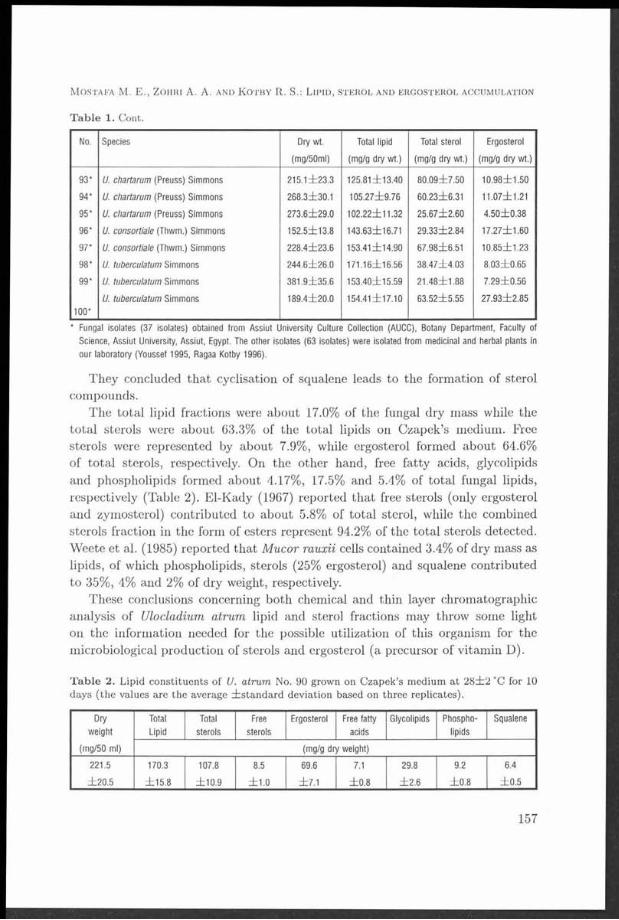
M o s t a f a M . E ., Z o h r i A. A. a n d K o t b y R. S.: L i p i d , s t e r o l a n d e r g o s t e r o l a c c u m u l a t io n
T a b le 1. Cont.
No. Species Dry wt. Total lipid Total sterol Ergosterol
(mg/50ml) (mg/g dry wt.) (mg/g dry wt.) (mg/g dry wt.)
93* U. chartarum (Preuss) Simmons 2 1 5 .1 ± 2 3 .3 125 .81±13 .40 8 0 .0 9 ± 7 .5 0 10 .9 8 ± 1 .5 0
94* U. chartarum (Preuss) Simmons 268 .3±30 .1 105 .27± 9 .76 60 .23±6 .31 11.07±1.21
95* U. chartarum (Preuss) Simmons 2 7 3 .6 ± 2 9 .0 102 .22±11 .32 2 5 .6 7 ± 2 .6 0 4 .5 0 ± 0 .3 8
96* U. consortiale (Thwm.) Simmons 152 .5 ± 1 3 .8 143 .63±16.71 2 9 .3 3 ± 2 .8 4 17 .2 7 ± 1 .6 0
97* U. consortiale (Thwm.) Simmons 2 2 8 .4 ± 2 3 .6 153.41 ± 1 4 .9 0 67 .98± 6 .5 1 1 0 .85± 1 .2 3
98* U. tuberculatum Simmons 2 4 4 .6 ± 2 6 .0 171 .16±16 .56 3 8 .4 7 ± 4 .0 3 8 .0 3 ± 0 .6 5
99* U. tuberculatum Simmons 3 8 1 .9 ± 3 5 .6 153 .40±15 .59 2 1 .4 8 ± 1 .8 8 7 .2 9 ± 0 .5 6
U. tuberculatum Simmons 189 .4 ± 2 0 .0 154.41 ± 1 7 .1 0 6 3 .5 2 ± 5 .5 5 27 .9 3 ± 2 .8 5
1 0 0 *_______________________________________________________________________________________________________________________________________________________
* Fungal isolates (37 isolates) obtained from Assiut University Culture Collection (AUCC), Botany Department, Faculty of
Science, Assiut University, Assiut, Egypt. The other isolates (63 Isolates) were isolated from medicinal and herbal plants in
our laboratory (Youssef 1995, Ragaa Kotby 1996).
They concluded tha t cyclisation of squalene leads to the formation of sterol compounds.
The total lipid fractions were about 17.0% of the fungal dry mass while the to tal sterols were about 63.3% of the total lipids on Czapek’s medium. Free sterols were represented by about 7.9%, while ergosterol formed about 64.6% of to tal sterols, respectively. On the other hand, free fatty acids, glycolipids and phospholipids formed about 4.17%, 17.5% and 5.4% of total fungal lipids, respectively (Table 2). El-Kady (1967) reported tha t free sterols (only ergosterol and zymosterol) contributed to about 5.8% of total sterol, while the combined sterols fraction in the form of esters represent 94.2% of the total sterols detected. Weete et al. (1985) reported th a t Mucor rauxii cells contained 3.4% of dry mass as lipids, of which phospholipids, sterols (25% ergosterol) and squalene contributed to 35%, 4% and 2% of dry weight, respectively.
These conclusions concerning both chemical and thin layer chromatographic analysis of Ulocladium atrum lipid and sterol fractions may throw some light on the information needed for the possible utilization of this organism for the microbiological production of sterols and ergosterol (a precursor of vitamin D).
T a b le 2. Lipid constituen ts of U. atrum No. 90 grown on C zapek’s m edium a t 2 8 ± 2 "C for 10 days (the values are th e average ¿ s ta n d a rd deviation based on th ree replicates).
Dry Total Total Free Ergosterol Free fatty Glycolipids Phospho- Squalene
weight Lipid sterols sterols acids lipids
(mg/50 ml) (mg/g dry weight)
221.5 170.3 107.8 8.5 69.6 7.1 29.8 9.2 6.4
± 2 0 .5 ± 1 5 .8 ± 1 0 .9 ± 1 .0 ± 7 .1 ± 0 .8 ± 2 .6 ± 0 .8 ± 0 .5
157
R e f e r e n c e s
B i io w n D . J. and D u p o n t F. M. (1989): Lipid com position of p lasm a m em branes and endom em branes prepared from roots of barley (H ordeum vulgare L.). — P lan t Physiol. 90: 955-961.
C o o k R. P . (1958): Cholesterol (Chem istry, Biochemistry, and Pathology). - Academ ic Press, New York.
C o r w n L . M., S c h r o e d e r L . J . a n d M e C u l l o u g h W . G . (1 9 5 6 ) : T h e m e ta -b o l is m o f s q u a le n e b y ce ll f ree y e a s t e x t r a c t s . - J . A m . C h e m . S o c . 78: 1 3 7 2 -1 3 7 5 .
E l- K a d y I. A. (1967): Sterol production by fungi. — M Sc Thesis, N ational Research C enter, Cairo, Egypt.
E l - K a d y I. A., E m a n M. M. and Z o h ri A. A. (1995): U tilization of cheese whey for lipid sterol p roduction by some isolates of xerophilous fungi. - Folia Microbiol. 40 (2): 209-212.
E l l is W . J. (1945): A note on the sterol production by A. orzae w ith special references to its an tirach itic potency. - J. Council Sci. Ind. Research. 18: 314-317.
E l l is S. W ., R o s e M. E . and G r in d l e M. (1991): Identification of sterol m u tan t of N eurospora crassa deficient 14,15-reductase activity. - J . Gen. Microbiol. 137: 2627-2630.
E l- R e f a i A . H . (1 9 6 4 ) : Physiological and biochem ical studies on th e m etabolism of fa ts and sterol in fungi. - P h D Thesis, N ational Research C enter , Fac. Sci., C airo Univ., Egypt.
E l - R e f a i A. H. and E l- K a d y I. A. (1969): U tilization of some industria l by-products for the m icrobiological production of sterols from Saccharomyces ferm entati. - J. Bot. V. A. R . 12(1): 45-54.
F a n e l l i C. and F a b b r i A. A. (1980): G row th requirem ents and lipid m etabolism of A spergillus flavus. — Trans. Br. Mycol. Soc. 75 (3): 371-375.
F e r n a n d o T . and B e a n G. (1986): Effects of tricyclazole on grow th, release of aflatoxin, and sterol & fa tty acid content, by Aspergillus isolates. -T ran s. B r. Mycol. Soc. 87(3): 445-449.
F i o r e J. V. (1948): On th e m echanism of enzym e action. XXXII. Fat and stero l in Fusarium lini Bolley, F . lycopersici and F . solani. Purple. - Arch. Biochem. 16: 161-168.
G a l a n o s D. S. and KAPOULAS V. M. (1962): T hin layer chrom atography of phospholipids and glycolipids. - J. Lipid Res. 3: 143.
G h a n e m K . M., N e v in e B . G . and E l - R e f a i A . H . (1 9 9 0 ) : Effect of some n u trien t on .stero ls p roduction by Pénicillium crustosum Thom . - R ev . L atinoam . Microbiol. 3 2 (2 ) : 1 3 3 -1 3 6 .
G o r d o n T . R. and W e b s t e r R. K. (1984): An evaluation of ergosterol as an ind icator of D rechslera gram inea infestation of barley seed. - Phytopathology 74: 1125—1127.
GORDON T . R. and W e b s t e r R. K. (1986): Identification of ergosterol as am etabo lite of D rechslera gram inea and D. teres. - Can. J. Microbiol. 32: 69-71.
K ie s l ic h K . (1985): M icrobial side-chain degradation of sterols. - J. Basic Microbiol. 25 (7): 461-474.
L e o b a r d o S. C ., B e n s o u s s a n Y. H. M. E . and B e l in J. M. (1992): Lipid accum ulation in T richoderm a spp. — Microbiol. L ett. 93(2): 181—188.
MAGUIGAN W . H. and WALKER E. (1940): Sterol m etabolism of m icroorganism (yeast). - Biochem. J. 34: 804-813.
M u m m a R. O., F e r g u s C . L. and S e k u r a R. D. (1970): T he lipids of therm ophilic fungi: Lipid com position com parisons between therm ophilic and m esophilic fungi. - Lipids 5: 100-103.
N a e g e l i and L o e w (1978): - Ann. 193: 322. In El-Refai A. H.(1964): Physiological and biochem ical studies on the m etabolism of fats and sterols in fungi. - P h D Thesis. N ational Research C enter, Fac. Sci., Cairo Univ., Egypt.
N a im N . and SAAD R . R. (1986): P roduction of lipids and sterols by Fusarium oxysporum . - Rev. Latinoam . Microbiol. 28(4): 317—324.
P r e u s s L. M., P e t e r s o n W . H., S t e e n b o c k H. and F r e d E. B. (1931): Sterol con ten t and an tirach itic activity of mold mycelial. - J. Biol. Chem. 90: 369-384.
P r e u s s L. M ., E ic h in g e r E . C. and P e t e r s o n W . H. (1934): T he chem istry of m old tissue. I l l - C om position of certa in m olds w ith special reference to th e lipid content. — Zentr. B akt. Parasitenk. A bt. 11: 89-370.
C z e c h m y c o l . 53 (2 ) , 2001
158
P r il l E. A ., W e n c k P . R. and P e t e r s o n W . H. (1935): Factors influencing th e am ount and n a tu re of the fat produced by A. fischeri. - Biochem. J. 21: 875-891.
R a g a a K o t b y S. M . (1 9 9 6 ) : Lipids and sterols production by dem atiaceous fungi isolated from som e Herbs. - M Sc Thesis, B ot. D ept., Fac. Sci., A ssiut Univ., A ssiut, Egypt.
R atC L IF F E A. (1937): T he sterol and carbohydrates in fungi. - I. Boletus edulis. - Biochem. J. 31: 240-243.
R a w ia S a a d R . (1992): Effect of w ater activ ity on grow th and lipids of xerophilic fungi, A spergillus repens and A. am stelodam i. - Zbl. Mikrobiol. 147: 61-64.
RlPPON J . W . (1982): M edical mycology, the pathogenic fungi and the pathogenic actinom y- cetes. - W . B. Saunders, Philadelphia.
R o u s e ii G., F l e is c h e r S. and Y a m a m a A. (1970): In W ork T . and W ork E. (eds.) (1972): Techniques of lipodology. - p. 44, - N orth Holland Co. (Publ.) A m sterdam .
S m it h D. and O n io n s A. H. S . (1983): T he preservation and m aintenance of living fungi. — Kew, U. K., CAB, In terna tional Mycological In stitu te .
W a r d G. E . , L o c k w o o d L . B., M ay O. E . a n d H e r r ic k H . T . (1 9 3 5 ) : - In d . E n g . C h e m . 2 7 : 3 1 8 . In W e e te J . D . (1 9 7 4 ) : F u n g a l l ip id b io c h e m is tr y ( D is t r ib u t io n a n d m e ta b o l is m ) . — P le n u m P r e s s , N ew Y o rk -L o n d o n .
W e e t e J. D. (1974): Fungal lipid biochem istry (D istribution and m etabolism ). — Plenum Press, New York-London.
W e e t e J. D ., F u r t e r R ., H a n s e l e r E. and R a s t D. M. (1985): Cellular and chitosom al lipids of A garicus bisporus and M ucor rouxii. - Can. J. Microbiol. 31: 1120-1126.
YOUSSEF M. S. M. (1995): Mycoflora and m ycotoxins of some m edicinal plants and their an tim icrobial activ ities in Egypt. - PhD Thesis, Bot. D ept., Fac. Sci., South Valley Univ., Sohag, E gypt.
Z ö l l n e r N . and K ir s c h K . (1 9 6 2 ) : Ü ber die quan tita tive B estim m ung von Lipoiden (m icrom ethods) m itte ls der vielen natürlichen Lipoiden (allen bekannten P lasm alipoiden) gemeinsam en sulpho-phosphovanillin - Reaktion. - Z. Gesam te Exp. Med. 135: 5 4 5 -5 6 1 .
M o s t a f a M . E., Z o h r i A. A. a n d K o t b y R. S.: L ip id , s t e r o l a n d e r g o s t e r o l a c c u m u l a t i o n
159

The role of some saprophytic micromycetes and the fungus Micromucor ramannianus var. ramannianus in forest soils
J o s e f H ý s e k a n d J a n a B r o ž o v á
D epartm ent of Mycology, Div. P lan t Medicine,Research In s titu te of Crop Production
Drnovská 507, 161 06 P rah a 6 — Ruzyně Czech Republic
Hýsek J. and Brožoová J. (2001): The role of some saprophytic micromycetes and the fungus Micromucor ram annianus var. ram annianus in forest soils - Czech Mycol. 53: 161-171
Different saprophytic m icrom ycetes were isolated from the humic horizon (H-A 02) of different types of forest soils (barren land of reforested w aste dum ps, cam bisol of spruce, birch, E uropean m ountain ash, and blue spruce forests) in several areas (a t M ost in th e Krušné hory (O re M ts.), Jizerské hory (Izera M ts.)). Besides the spectrum of comm on species of soil m icrom ycetes (P enicillium spp., H um icola spp., Trichoderma spp., Paecilom yces spp., Scopulariopsis spp., Aureobasidium spp., M ucor spp., Absidia spp.), the fungus M icrom ucor ram annianus (Möller) A rx var. ram annianus (M ortierella ram anniana (Möller) L innem an, M ucor ram annianus Möller) was regularly isolated from all types of soils, except barren soils of w aste dum ps. T he biological quality of forest soils in connection w ith o ther biological characteristics was evaluated in relation to the presence and quantity of th is fungus in forest soils. Basic biological processes (basal and po tential respiration , am m onification, nitrification) show an increased intensity in forest soils in which the proportion of M icrom ucor ram annianus v. ram annianus was no t present in the soil of the worst biological quality (lower values of biological soil param eters), e.g. in of w aste dum ps. It is a topic for discussion w hether th is fungus can also be an ind icator of environm ental pollution.
K e y w o rd s : hum ic horizon, soil fungi, saprophytic m icrom ycetes, M icrom ucor ram annianus var. ra m ann ianus , biological soil functions, respiration , am m onification, nitrification
Hýsek J. a Brožooová J. (2001): Role některých saprofytických mikromycetů a houby Micromucor ram annianus var. ram annianus v lesních půdách - Czech Mycol. 53: 161-171
Z humusového horizontu (H-A02) různých lesních typů (neplodná p ů d a výsypek, kambisol různých ty p ů - sm rk, bříza, jeřáb , stříb rný sm rk) v několika m ístech (výsypky v M ostu, půdy v K rušných a Jizerských horách) byly izolovány různé saprofytické mikromycety. Vedle sp ek tra běžných půdních m ikrom ycetů (P enicillium spp., Hum icola spp., Trichoderm a spp., P aecilom yces spp., Scopulariopsis spp., Aureobasidium spp., M ucor spp., Absidia spp.) byla pravidelně izolována ze všech typů lesních půd houba M icrom ucor ram annianus (Möller) Arx var. ram annianus (M ortierella ram anniana (Möller) Linnem an) krom ě půd výsypek. Biologická kvalita lesních půd ve spojení s jiným i biologickými charakteristikam i byla stanovena společně s p řítom ností a kvantitou té to houby v lesních půdách. Základní biologické procesy (bazální a potenciáln í respirace, amonifikace a nitrifikace) ukazují zvýšenou intenzitu v lesních půdách, ve kterých celkový počet nálezů houby M icrom ucor ram annianus v. ram annianus přesahuje 50 % všech přítom ných m ikrom ycetů. Na druhé straně houba M icrom ucor ram annianus v. ram ann ianus nebyla p říto m n a v půdách horší biologické kvality (nižší hodnoty půdních biologických charak teristik ) např. v půdách výsypek. Je otázkou diskuse, zda ta to houba může bý t také indikátorem znečištění nebo čisto ty životního prostředí.
C z e c h m y c o l . 53 (2), 2001
161

I n t r o d u c t i o n
Decomposition of organic m atter into humus, gases and inorganic salts is a continuous process in forest soils. Soil micromycetes are extraordinary im portant participants in this process. Forest soils are habitats of mainly soil-borne saprophytic and some pathogenic fungi. The genus Pénicillium (Dyr 1940) and its teleomorphs, e.g. Eupenicillium (Takada and Udagawa 1983), are the most frequently isolated genera from forest soils. Several new species of Pénicillium from forest soils have been described. E.g. the new species Pénicillium kananaskense (Seifert et al. 1994) was isolated from forest soil under Pinus contorta v. latifolia. New species of the perfect stage of Eupenicillium were isolated e.g. from Nepalese forest soils - E. angustiporcatum (imperfect stage: Pénicillium, angustiporcatum) and E. nepalense (imperfect stage: Pénicillium nepalense) (Takada and Udagawa 1983). Talarornyces unicus and its anamorph were isolated from Taiwanese soil. The anamorph is characterised by mono- or biverticillate penicilli with long-necked lanceolate phialides (Tzean et al. 1992). The genus Talarornyces is also dominant in soil in which heat-resistant fungi are present (Jesenská, Piecková and Bernât 1992).
Differently broad spectra of micromycetes are usually isolated from forest soils. Grunda and Marvanová (1982) identified the following genera of micromycetes from soil of the forest type group Ulmi-Fraxinetum carpineum with Quercus robur predominating in the stand: 1) (at a depth of 1-2 cm - horizon F) Absidia, Altemaria, Cladosporium, Cylindrocarpon, Fusarium, Humicola, Mucor, Paecilomyces abd Pénicillium, 2) the same genera as in horizon F were isolated at a depth of 2-7 cm (horizon Am), but besides them Sporothrix and Trichoderma were identified. Cephalosporium, Mortierella, Phialophora, Stachybotrys and Trichocladium were isolated from a depth of 15-25 cm (horizon Btg). Besides these the following genera were determinated at a depth of 35-45 cm (horizon Btg): Bispora, Phoma and Rhizopus. It is interesting that the fungus Micromucor ramannianus v. ramannianus (Micromucor ramannianus) was not detected in th a t soil, although it is present in almost all types of forest soils (Dyr 1940). Grunda (1981) investigated methods of research on soil microflora when he studied the floodplain forest soil in Moravia. The following genera of micromycetes were identified in floodplain forest soil: Bispora, C'alcarisporium, Chrysosporiurn, Cladosporium, Cylindrocarpon, Fusarium, Humicola, Mortierella, Mucor, Paecilomyces, Phialophora, Phoma, Pénicillium, Rhizopus, Sporothrix, Stachybotrys, Trichocladium and Trichoderma. Not even in this case the fungus Micromucor ramannianus v. ramannianus (Mucor ramannianus Möller) was detected, even if it is regularly present in forest soils (Dyr 1940).
Saprophytic micromycetes in forest soils were studied by Vláčilíková (1978), who described the presentation of genera of micromycetes in forest soils. Av
C z e c h m y c o l . 53 (2 ) , 2001
162

erage proportions (in percent) of fungal genera in four types of forest soil were as follows: Absidia (7.5), Circinella (0.35), Mucor (16), Rhizopus (0.1), Zygorhynchus (0.6), Cunninghamella (0.1), Mortierella (9.0), Gymnoascus (2.7), Thielavia (2.3), Chaetophoma (0.1), Truncatella (0.2), Aspergillus (2.0), Ceph- alosporium (4.0), Gliocladium (2.0), Monocillium (0.5), Paecilomyces (0.9), Peni- cillium (51.5), Spicaria ((2.1), Sporotrichum (0.9), Trichoderma (6.4), Verticil- lium (0.4), Botryotrichum (0.1), Cladosporium (1.9), Hormiscium (0.4), Humicola (0.9), Masoniella (0,4), Pullularia (0.1), Stachybotrys (1.9), Cylindrocarpon (0.6), Fusarium (0.3), Volutella (0.3), sterile mycelium (0.6), Myrothecium (0.3). Mortierella ramanniana (Möller) Linneman ranked second among all species of the genus Mortierella in forest soils (0, 1.5 %, 13.7 %, 5.1 %). Penicillium nigricans (Bainier) Thom was the species with the highest representation in the different soils (%): 2.5, 49, 18.1, 1.1.
Micromucor (Mortierella) is the best known genus of forest soil zygomycetes. A relationship of Endogone to Mortierella (Mortierellaceae) has been suggested since in some species, especially M. nigrescens and M. renispora, the zygospores are surrounded by hyphae, much like the hyphae developed by zygospores in sporocarps of Endogone (Benny 1982).
Micromucor ramannianus is a fungus with one of the highest frequencies in forest soils. The genus is classified in the genus Mucor by Dyr (1942) as Mucor ramannianus A. Möller 1903 (Zeitschr. F. Forst, und Jagdw. 35, p. 330). It differs from other species by the colour of its colonies beeing deep red to soil pink. The cultures reached a height 1.6-2.5 mm only, fungus growth is very dense and velvety. Sporangiophores are usually not ramified. Sporangia are small, 18-35 /tm and not ramified, they are deep red in colour and have a slightly melting membrane. The columella is round or disk-shaped, colourless, 10.8-12.7 /¿m in height and 11-14 ß m in width. Sporangiospores are shortly eliptic to round (1.2 x 2.3, 2.7 x 3.3 x 3 /im). The species was isolated in Germany (Johann 1932), France (Ling-Young 1930), Austria and Yugoslavia (Pispek 1929), Russia (Raillo 1929), Northern America (Povah 1917), Australia (Dale 1914) and Norway (Hagen 1908).
This species is generally present in forest soils, its frequency being the highest of all species of micromycetes and it could probably be isolated at every forest site. The population density of this species is regularly enormous amounting to more 50 % of micromycetes at the site. The ratio of Mucor ramannianus to other micromycetes was 25:1 in forest soils of the Jevany district. Sites with the highest population density of the fungus had the highest concentration of sporangiospores in the soil. The fungus population density was low at two forest sites only - in the environs of Dobřichovice (right bank of Berounka river) and in Brdy forest. Different types of forest showed different rates of the fungus (out of the total number of micromycetes). It was 64.2 % in beech forest soils, 39.8 % in spruce forest soils, 37.4 % in pine forest soils, 26 % in oak forest soils, 20 % in alder forest
H ý s e k J . a n d B r o ž o v á J.: T h e r o l e o f s o m e s a p r o p h y t i c m i c r o m y c e t e s
163

soils, 32.5 % in birch grove soils. This species had modest environmental demands with respect to the extreme soil factors in broad-leaved and coniferous forest stands. Fungus growth in nutrient media in Petri dishes makes up a continuous coating of a typical colour. Fungus growth is very fast. The species is usually at a soil depth of up to 20 cm, in the upper humus layer under it. Kubátová et al. (1998) reporting on the biodiversity of soil microfungi of the Šumava Mts., Czech republic, confirmed that among the most frequent species Micromucor ramannianus v. ramannianus was. They determined soil micromycetes from 12 localities, including peat-bogs, Norway spruce forest, beech forests, mixed forests in glacial cirques in the period 1993-1996 (121 soil samples). The most frequent species were: Trichoderma viride (in 57 % of all samples), Penicillium spinulosum (55.4 %), Micromucor ramannianus v. ramannianus (33.1 %) and Mucor hiemalis f. hiemalis (24.8 %).
Micromucor ramannianus has not been described as a typical fungus of forest soils in many papers, or its population density is characterised as very low. E.g. Grunda and Vorel (1996) reported a high density of micromycete species with some differences in various types of soils (H-horizons). The following micromycetes were present in the H-horizon of humic acidic Cambisol: Penicillium sp., Aspergillus sp., Gliocladium sp., Trichoderma sp., Cladosporium sp., Sporotrichum sp., Altemaria sp., Humicola fuscoatra and Humicola grisea. Acidic Cambisol contained also these fungi: Botrytis sp., Mortierella nana, Chaetomium sp. The fungi were determined in deep acidic Cambisol in comparison with acidic Cambisol: Mortierella ramanniana (Micromucor ramannianus at present) and Verticillium albo-atrum. In addition, Absidia glauca was found in gley acidic Cambisol.
Many of the soil micromycetes showed intensive metabolic activity, e.g. lign- inolytic activity in Penicillium ehrysogenum (Rodriquez et al. 1994). This m etabolic activity can last very long. Marfenina (1991) studied the morphological development of microscopic fungi in soil. The life cycle (mycelium and conidium formation) is usually very long.
Many species of Mortierella produce a white oily substance in large drops among the aerial hyphae. One group of species, characterised by velvety odourless colonies, is considered by some mycologists to represent a separate genus, Micromucor (Benny 1982). The objective of this study was to determine the importance of Micromucor ramannianus v. ramannianus in soil humic horizons of Czech forests.
M a t e r i a l a n d m e t h o d s
Forest soil samples were taken from the humic horizon (H-horizon) after the F-horizon (fermentation horizon) was disclosed. Soil was sampled into platic bags under sterile conditions, and was processed as soon as it was brought into
C z e c h m y c o l . 53 (2 ) , 2001
164
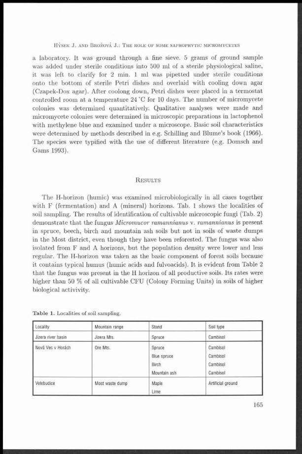
a laboratory. It was ground through a fine sieve. 5 grams of ground sample was added under sterile conditions into 500 ml of a sterile physiological saline, it was left to clarify for 2 min. 1 ml was pipetted under sterile conditions onto the bottom of sterile Petri dishes and overlaid with cooling down agar (Czapek-Dox agar). After coolong down, Petri dishes were placed in a term ostat controlled room at a tem perature 24 °C for 10 days. The number of micromycete colonies was determined quantitatively. Qualitative analyses were made and micromycete colonies were determined in microscopic preparations in lactophenol with methylene blue and examined under a microscope. Basic soil characteristics were determined by methods described in e.g. Schilling and Blume’s book (1966). The species were typified with the use of different literature (e.g. Domsch and Gams 1993).
R e s u l t s
The H-horizon (humic) was examined microbiologically in all cases together with F (fermentation) and A (mineral) horizons. Tab. 1 shows the localities of soil sampling. The results of identification of cultivable microscopic fungi (Tab. 2) demonstrate that the fungus Micromucor ramannianus v. ramannianus is present in spruce, beech, birch and mountain ash soils but not in soils of waste dumps in the Most district, even though they have been reforested. The fungus was also isolated from F and A horizons, but the population density were lower and less regular. The H-horizon was taken as the basic component of forest soils because it contains typical humus (humic acids and fulvoacids). It is evident from Table 2 tha t the fungus was present in the Ii horizon of all productive soils. Its rates were higher than 50 % of all cultivable CFU (Colony Forming Units) in soils of higher biological activivity.
T a b le 1. Localities of soil sam pling.
Locality Mountain range Stand Soil type
Jizera river basin Jizera Mts. Spruce Cambisol
Nová Ves v Horách Ore Mts. Spruce Cambisol
Blue spruce Cambisol
Birch Cambisol
Mountain ash Cambisol
Velebudice Most waste dump Maple Artificial ground
____________________________ Lime ___________________________
H ý s e k J . a n d B r o ž o v á J.: T h e r o l e o f s o m e s a p r o p h y t i c m i c r o m y c e t e s
165

C z e c h m y c o l . 53 (2 ) , 2001
T a b le 2. M icromycetes isolated from H-horizons of cambisol types of forest soils
Locality/Stand Micromycetes Proportion / frequency
Jizera river basin/Spruce M icrom ucor ramannianus var. ram. 55%
Trichoderma viride 20%
Penicillium expansum 10%
Paecilomyces farinosus 5%
Paecilomyces niveus 5%
Scopulariopsis brevicaulis 2,5%
sterile mycelium 2,5%
Nová Ves v Horách/Spruce M icrom ucor ramannianus var. ram. 52%
Trichoderma viride 15%
Penicillium itailcum 15%
Paecilomyces varioti 7%
Paecilomyces fulvus 3%
Scopulariopsis brevicaulis 5%
sterile mycelium 3%
Nová Ves v Horách/Blue spruce M icrom ucor ramannianus var. ram. 58%
Scopulariopsis brevicaulis 12%
ňbsidia corymbifera 10%
Paecilomyces fulvus 8%
Trichoderma viride 5%
sterile mycelium 7%
Nová Ves v Horách/Birch M icrom ucor ramannianus var. ram. 65%
Paecilomyces farinosus 8%
Penicillium expansum 6%
Scopulariopsis brevicaulis 6%
Gilmaniella humicola 5%
Trichoderma viride 2%
sterile mycelium 8%
Nová Ves v Horách/Mountain ash M icrom ucor ramannianus var. ram. 59%
Aureobasidium pullulans 10%
Cladosporium resinae 8%
Paecilomyces fulvus 6%
Trichoderma viride 1%
sterile mycelium 16%
Velebudice/Most waste dump Mucor hiemalis 65%
M ucor piriform is 16%
Humicola fuscoatra 15%
________________________________________ sterile mycelium 4%
166

<r ■■ r— p—p------- ■---------------
F ig . 1 M icrom ucor ram annianus var. ram annianus, mycelium with oil droplets (X 500).
The growth of Micromucor ramannianus var. ramannianus was satisfactory in artificial media (Czapek-Dox agar) at a tem perature of 24 °C. Most colonies produced a red pigment. There exist differences in isolate colour, some isolates are cream-coloured, others are from white to pink, many are brown-red in colour. Sporangia reddish in colour and containing several sporangiospores were exceptionally discovered in the preparats. A major part of the cultures produced a system of sacs containing high amount of oily droplets (Fig. 1).
Table 3 shows the the values of biological soil characteristics from different H-liorizons. It is evident from this figure that soils with higher biological activities have higher counts of Micromucor ramannianus var. ramannianus germs.
D i s c u s s io n
This paper has many features identical with the papers by Dyr (1942), Grunda and Marvanová (1982), Grunda and Vorel (1996), Kubátová et al. (1998). These authors determined Micromucor ramannianus var. ramannianus as the basic microfungus of forest soils. The basic paper by Dyr (1942) contains a relatively successful description of its morphology, although the fungus illustration is not
167
H ý s e k J. a n d B r o ž o v á J.: T h e r o l e o r s o m e s a p r o p h y t i c m i c r o m y c e t e s

Relationship between the occurence of Micromucor and soil biological activity
° 0 0,1 0,2 0,3 0,4 fošl 0,6 0,7 0,8 0,9 10 s
weak biological strong biologicalactivity soils activity soils
F ig . 2 Incidence of M icrom ucor ram annianus var. ram annianus in percent (all m icrofungi) in relation to soil fertility (scale from 0, 0.1.................0.9, to 1.0).
very satisfactory. Sporangia were discovered rarely during our studies. Dyr (1942): “Of all micromycete species, the frequency of this species is highest in forest soils, and it is likely to be isolated from every forest site.” We observed during our studies tha t the fungus was not present in soil of reforested waste dumps and that its population density varied in different types of soils. Micromucor ramannianus var. ramannianus is dominant in “physiological” ( “helthy”) forest soils while soil germ counts of this fungus decrease in soils exposed to degradation processes. A worldwide distribution is typical of this fungus (Johann 1932, Ling-Young 1930, Pispek 1929, Reillo 1929, Povah 1917, Dale 1914, Hagen 1908). Dyr (1942) reported different frequencies in forest soils (beech 64.2 %, spruce 39.8 %, pine 37.4 %, oak 26 %, alder 20 %, birch 32.5 %), but he did not take into into account environmental and soil pollution that was substantially lower sixty years ago than (recently or) at present. Air pollutants (mainly sulphur dioxide) had negative impacts on the environment and forest soils while bacterial counts, especially of the genus Thiobacillus, increased (Lettl 1984) and total counts of micromycetes in forest soils decreased. The population densities of Micromucor ramannianus v. ramannianus are also likely to fluctuate snd decrease, although it has not been
■ verified experimentally in the last sixty years. Since this fungus is not presentin less nutritive soils, it is to deduce th a t it needfs forest soil humus for its life. Some researchers did not discover this fungus in forest (Grunda 1981). Due to its high frequency in forest soils, it is possible to anticipate the fungus’ high
C z e c h m y c o l . 53 (2 ) , 2001

H ý s e k J. a n d B r o ž o v á J.: T h e r o l e o f s o m e s a p r o p h y t ic m ic r o m y c e t e s
T a b le 3 . T ypes of forest soil according to biological characteristics.
High fertile soils Moderately Weakly fertile Non-fertlle
fertile soils soils soils
pH (H20 ) 6 -7 5 -6 4 -5 4 -3
pH (KCI) 5 -6 4 -5 3 -4 3 -2
% humus 20 15-19 10-14 5 -9
% C 20-15 15-10 10-5 5 -0
% N 2-1,5 1-1,5 1-0,5 0,5-0
C: N 20 19-15 10-15 5-10
20-25 25-50 30^10
Basal respiration > 1000 > 500 > 250 < 250
Potential respiration 4 X 3 X 2 X < 1 X
Ammonification > 5 0 > 25 > 1 0 < 1 0
Nitrification 100-80 80-50 25-50 25-0
Number of aer. bad. > 5 mil. > 0,5 mil. > 10 000 > 1000
Number of am. bad. > 1 mil. > 0,5 mil. > 10 000 > 1000
% M icrom ucor from CFU 80-100 50-80 < 5 0
Values: Basal respiration - in mg C02 released after 24 h at 21C
Potential respiration - how many times is higher than basal respiration
Ammonification - in mg ammonia nitrogen after 14 days at 21C
Nitrification - in mg nitrate nitrogen after 14 days at 21C
metabolic activity like e.g. in Penicillium chrysogenum (Rodriquez et al. 1994). The genus Penicillium was isolated from many types of forest soils and there dominates (Tzean et al. 1992). Even new species of Penicillium and their perfect stages were isolated (Takade and Udagawa 1983) A very broad spectrum was isolated by Vláčilíková (1978), although Mortierella ramanniana ranked second among all isolated species in her study (Penicillium was a dominant genus), the rate of the fungus appeared to be low. Many authors did not indicate the horizons where the fungus was detected. The spectra of soil micromycetes were determined at different depths (1-2 cm, 2-7 cm, 7-15 cm, 15-25 cm) (Grunda and Marvanová 1982). The genus Mortierella was isolated from a depth of 15-25 cm, although it was not explicitly described as Micromucor ramannianus. The spectrum of micromycetes detected in our study was not so broad as reported in above paper. M utual interactions of many of the reported genera are possible in vitro as well as in soil. It would also be interesting to study their antagonistic effects (Veselý 1997). Soils can undergo development during the year, and micromycetes along them, as was investigated by Marfenina (1991). They can have fungistatic effects on soil micromycetes. Kubátová, Váňová and Prášil (1998) determined the soil micromycetes from Šumava Mts. (Bohemian Forest). Amongst 139 fungal
169

species Micromucor ramannianus v. ramannianus was on the third place from most frequent species (33.1 %) and this species occurred on all localities.
This paper was exclusively focused on the species spectrum of soil micromycetes tha t were associated with the presence of a fungus typical of forest humic horizons - Micromucor ramannianus v. ramannianus. The fungus appears to be a basic micromycete of forest soils underlying biological functions of soil (respiration, ammonification, nitrification).
A c k n o w l e d g e m e n t
We thank Mrs. L. Závadová from Forestry and Game Management Research Institute in Jíloviště - Strnady for providing the photograph.
R e f e r e n c e s
B e n n y G. L. (1982): Zygomycetes. — In: M cGraw Hill Synopsis and classification of living organism s.
D a l e E . (1 9 1 4 ) : O n t h e fungi of t h e soil. - Ann. Mycol. 12: 33 —62.DOMSCH K. H. and G a m s W . (1993): C om pendium of soil fungi. Volume I and II, IHW -Verlag,
Volume I 1-859, Volume II 1-391.D y r J. (1941): Zygomyceten im W aldboden der Böhm ischer Länder. - S tud ia B otan ica Čechica
Vol. 4, fasc. 3—4: 73-168.GRUNDA B. (1981): Saprotrofní m ikroorganism y v lesním ekosystému. (Saprotrophic m icroor
ganism s in the forest ecosystem ). - Lesnictví 27 (LIV) 4: 303-318.G r u n d a B. and M a r v a n o v Á L. (1982): M icromycetes in soil of a floodplain forest. - S crip ta Fac.
Sei. N at. Univ. Purk . B run. 12, 8: 359-370.GRUNDA B. and V o r e l J. (1996): P ů d a a půdní m ikroflóra N árodní přírodní rezervace Velký
Špičák (Soil and soil m icroflora of th e N ational N ature Reserve Velký Špičák) — Les- n ic tv i/F orestry 42, 4: 174-183.
H a g e m O. (1908): Untersuchungen über norwegische M ucorineen. - I. C hrist. Vidensk. Selsk.Skrift. I. M ath. — naturw . Kl., 7: 1-50.
J e s e n s k á Z., P ie c k o v Á E. and B e r n Át D. (1992): H eat-resistan t fungi in the soil. - In t. J. Food Microbiol. 16: 208-214.
K u b Át OVÁ A., VÁŇOVÁ M. and P r ÁŠil K . (1998): C ontribu tion to th e biodiversity of soil microfungi of th e Šum ava M ts., Czech Republic. - Silva G ab re ta 4: 23-34.
L e t t l A. (1984): T he effect of a tm ospheric SO 2 pollution on th e m icroflora of forest soils. - Folia microbiol. 29: 509—516.
L in g Y o u n g M. (1930): E tu d e biologique des phénom ènes de la sexualité chez les M ucorinées. - Rev. gen. Bot. Paris, 42: 30-43.
M a r f e n in a O. Y ., P o p o v a L. V. and Z v y a g in t s e v D. G. (1991): C haracteristics of th e developm ent cycle of m icroscopic fungi in soils. — Pochvovedeniye 8: 80—87.
P i s p e k P . A. (1929): Edafske m ucorineje Jugoslavije. - A c ta Bot. Inst. Univ. Zagreb 4: 1-36. R a il l o A. (1929): Beiträge zur K enntnis der Bodenpilze. - Z trlbl. F . B akt. II 78: 515-524. R o d r iq u e z A., C a r n ic e r o A., P e r e s t e l o F . , D e l a F u e n t e G., M il s t e in O. and F a l c o n
M. A. (1994): Effect of Penicillium chrysogenum on lignin transform ation . - A ppl. Environm . Microbiology, 60 (8): 2971-2976.
S c h l ic h t t n g E. and B l u m e H. P . (1966): B odenkundliches P rak tikum . H am burg, Berlin.
C z e c h m y c o l . 53 ( 2 ) , 2001
170

S e if e r t K. A., F r is v a l d J. C. and M c L a e n M . A. (1994): Penicillium kananaskense, a new species from A lb erta soil. — Can. J. Bot. 72: 20-24.
T a k a d a M. and U d a g a w a S. (1983): Two new species of Eupenicillium from Nepalese soil. — Trans, mycol. Soc. Jap an 24: 143-150.
T z e a n S. S., C h e n J . L. and Shiu S. H . (1992): Talarornyces unicus sp. nov. from Taiwan. — M ycologia 84 (5): 739-749.
VESELÝ D. (1997): Biologická regulace výskytu fytopatogenních hub v lesních ekosystémech (Biological control of phytopathogenic fungi in forest ecosystem s) - L esnictvi/Forestry, 43, 10, 464-471.
VLACILIKOVA A. (1 9 7 8 ) : M icromycetes in some forest soils of Slovakia. - A cta Fac. Rer. N atur. Univ. Com en. - M icrobiologia 6: 1 0 9 -1 2 3 .
H ý s e k J . a n d B r o ž o v á J . : T h e r o l e o f s o m e s a p r o p h y t ic m ic r o m y c e t e s
171

Supplement to the Checklist of non-vascular and vascular plants of Slovakia.
The species of microscopic fungi of the order Eurotiales
A l e x a n d r a Š i m o n o v ič o v Á
D epartm ent of Soil Science, Com enius University,M lýnská dolina B2, 842 15 B ratislava, Slovakia
Simonovičová A. (2001): Supplement to the Checklist of non-vascular and vascular plants of Slovakia. The species of microscopic fungi of the order Eurotiales. - Czech Mycol. 53: 173-188
S ubm ited are 50 species of m icroscopic fungi of th e order E urotiales, which are not listed in th e C hecklist of non-vascular and vascular plants of Slovakia, p a rt Fungi (Lizoň and Bacigálová 1998). T he first group is presented by those m icroscopic fungi which were isolated only once so far. So we consider them to be scarce or rare. From am ong 30 species of m icroscopic fungi m ore th an a half belongs to th e genus P enicillium (16 species) or genus Aspergillus (6 species). T he genera E upenicillium , E uro tium and Paecilom yces have two new species, Em ericella and M erim bla only one species. T he second group is presented by more frequently isolated species of m icroscopic fungi. From am ong 20 species the genus P enicillium dom inates w ith 8 species, followed by th e genus Aspergillus w ith 4 species. O ther genera (B yssochlam ys , D ichotom om yces , E u p en ic illiu m , E u ro tiu m , Fennellia, Paecilomyces and Talarornyces) are presented w ith one or two species. From th e to ta l num ber of 50 species of microscopic fungi the prevailing p a r t was isolated from different soils (73.3-75.0 %), from different foodstuffs (3.3-30.0 %) and from o ther sources (23.3-45.0 %), including drinking w ater, dwellings and different m aterials in depositories and archives.
K e y w o rd s : new records of microscopic fungi (E urotiales), Checklist of non-vascular and vascular p lan ts of Slovakia.
Simonovičová A. (2001): Doplnok k Zoznamu nižších a vyšších rastlín Slovenska. Druhy mikroskopických húb radu Eurotiales. - Czech Mycol. 53: 173-188
Uvádzam e 50 druhov m ikroskopických húb radu Eurotiales, ktoré nie sú uvedené v Zozna- me nižších a vyšších rastlín Slovenska, časť Huby (Lizoň a Bacigálová 1998). P rv ú skupinu predstavu jú m ikroskopické huby, ktoré boli doteraz na Slovensku izolované iba jedenkrát, a p reto ich považujem e za vzácné, resp. zriedkavé. Z 30 druhov viac ako polovicu tvoria druhy rodu P en icillium (16), potom nasledujú druhy rodu Aspergillus (6), Eupenicillium , E uro tium a P aecilom yces (2), E m ericella a M erimbla (1). D ruhů skupinu tvoria častejšie izolované druhy. Z celkového počtu 20 opáť dom inuje rod P enicillium zastúpený 8 druhm i a Aspergillus zastúpený 4 d ruhm i. O s ta tn é druhy {B yssochlam ys, D ichotom om yces , E upenicillium , E u ro tiu m , Fennellia, P aecilom yces a Talarornyces) sú zastúpené 1-2 X. Z celkového počtu 50 druhov m ikroskopických húb bola převážná časť izolovaná z róznej pódy (73.3-75.0 %), z róznych po trav in (3.3-30.0% ) a z iného zd ro ja (23.3-45.0 %), ktorý představuje napr. p itn á voda, vnútorné steny bytov, archivovaný m ateriá l rózneho póvodu a pod. Druhy, doplněné o synonym á a bibliografické údaje, sú uvedené v abecednom poradí. N om enklatúra je uvedená podia P it t e t al. (2000). A utorské sk ra tk y sú upravené pod ia B rum m itt a Powell (1992).
Czech mycol. 53 (2), 2001
173

The order Eurotiales (Ascomycetes - Ascomycota) includes 52 genera of microscopic fungi (Hawksworth et al. 1995), from which in Slovakia have been isolated only 13 genera. Among them, the most frequent were species of the genus Aspergillus and the genus Penicillium isolated from different soils (Bernát 1954, 1958, 1965, Braunová 1981, Pavlíčková 1994, Simonovičová 1980, 1992, 1993, Vláčiliková 1978), from the phyllosphere of agricultural plants and from corn (Bernát et al. 1983, Bernát et al. 1984, Dubovská 1981, 1984, Dubovská et al. 1982, 1986), but from atypical biotopes and ecotopes, too (Franková and Simonovičová 1999a, b, Franková et al. 1999, Simonovičová and Franková 1998, Simonovičová et al. 2000). For humans a very im portant and dangerous source of species of microscopic fungi of the order Eurotiales are contaminated foodstuffs, cereals and meal, cotton and flax (Jesenská and Poláková 1978, Jesenská and Sepitková 1984a, b, Jesenská et al. 1988, Jesenská and Piecková 1990b, Piecková et al. 1994, Piecková et al. 1992, 1996).
We submit a number of species of microscopic fungi of the order Eurotiales which are not listed in the Checklist of non-vascular and vascular plants of Slovakia, part Fungi (Lizoň and Bacigálová, 1998).
The species are arranged alphabetically, and synonyms and bibliography are included. Nomenclature is according to P itt et al. (2000). The abbreviations of authors’ names follow Brummitt and Powell (1992).
O r d e r E u r o t i a l e s
A sp erg illu s Fr.:Fr.
Aspergillus chevalieři (L. Mangin) Thom et Church —> Eurotium chevalieři L. Mangin.
Aspergillus clavatus Desm.Isolated from garden soil in Bratislava and in Poprad, from compost, maize
corn, peeled wheat, from maize flour, from foodstuff and dust from a mill (Piecková and Jesenská 1999b), from dried milk products for sucklings and baby foods (Jesenská and Poláková 1978), from meal, semolina and bread-crumbs (Jesenská et al. 1984, Jesenská and Sajbidorová 1984), from the inner mycoflora of malt, barley m alt and from the surface of barley malt (Sepitková and Jesenská 1985a,b,c, 1986, 1988, Sepitková et al., 1987a, Šepitková et al. 1988, 1990, Šepitková and Jesenská 1990, 1991), from the surface of corn (Sajbidorová et al. 1988), from samples of substrates and air from different types of factories (Jesenská 1988), from cereals of domestic provenience (wheat corn and different sorts of flour) (Jesenská et al. 1988), from samples of roasted coffee-beans (Jesenská et al. 1989).
C z e c h m y c o l . 5 3 (2 ) , 2001
174

H Aspergillus duricaulis Raper et FennellH Isolated from different material (tonsils, stools, sputum, nose, vagina, dissecting■ material) from immunosuppressive patients with oncological diseases (Trupl et al.
■ Aspergillus fischeri Wehmer —> Neosartorya fischeri (Wehiner) M allochet Cain
■ Aspergillus flavus Link var. columnaris Raper et Fennell■ Isolated from dried milk products for sucklings and baby foods (Jesenská andI Poláková 1978).
I Aspergillus glaucoaffinis Samson et W. Gams —> Eurotium pseudoglaucumI (Blochwitz) Malloch et Cain
■ Aspergillus janus Raper et Thom■ Isolated from the inner mycoflora of malt (Sepitková and Jesenská 1985a), from■ malt-barley and malt (Sepitková and Jesenská 1988).
■ Aspergillus melleus YukawaI Isolated from 45 cotton samples of different origin (Uzbekistan, U. S. A., China,I Egypt, Russia) and from 24 domestic flax samples (Piecková and Jesenská 1996b).
I Aspergillus niger var. cinnamomeus (Schiemann) Thom et RaperI Isolated from agricultural soils (Braunová 1981).
■ Aspergillus parasiticus SpeareI Isolated from different sorts of foodstuff (Jesenská et al. 1980), from importedI foodstuffs, such as roasted and non-roasted peanuts in chocolate, salt, sugar, orI as a part of wafers, grated coconut and rice (Jesenská et al. 1988), from drinkingI water (Franková and Simonovičová 1999b).
I Aspergillus parvulus G. Sm.I Isolated from forest soil in the neighbourhood of Bratislava (Krakovská et al.
I Aspergillus penicillioides Speg.I Isolated from wooden sculptures, picture canvases from depositories and fromI indoor air of the Slovak National Museum (Franková et al. 1999, SimonovičováI and Franková 1998).
I Aspergillus reptans Samson et W. Gams —> Eurotium repens de Bary
I Aspergillus rugulosus Thom et Raper —> Emericella rugulosa (Thom et Raper)
I Aspergillus violaceo-fuscus GasperiniI Isolated from Albic Luvisols (Bernát 1965).
H S im o n o v ič o v á A .: S u p p l e m e n t t o t h e C h e c k l is t o f n o n - v a s c u l a r

B ysso c h la m ys Westling
Byssochlamys fulva Olivier et G. Sm.Isolated from garden, forest and agricultural soils in different parts of Bratislava
and from a flower bed in a private garden (Jesenská and Piecková 1994a), from non-specified soil (Jesenská and Piecková 1994b, Jesenská et al. 1994), from conserved fruit and fruit-juice (Jesenská et al. 1983).
Byssochlamys nivea WestlingIsolated from non-specified soil from a depth of 0-5 cm in the neighbourhood
of Bratislava and Poprad (Jesenská and Piecková 1990a, 1991), from non-specified soils of Slovakia (Jesenská et al. 1992b, Jesenská et al. 1994, Jesenská and Piecková 1994b, 1995a, Piecková et al. 1994), from garden, forest and agricultural soils in different parts of Bratislava (Jesenská et al. 1992c), from soil of a private garden which had been fertilised with organic fertilisers (Jesenská et al. 1993), from a flower bed in a private garden (Jesenská and Piecková 1993, 1994a), from non-specified soils in different parts of Slovakia (Jesenská et al. 1992a, Jesenská and Piecková 1995a, Piecková and Jesenská 1997b), from soil samples after heating at 70 °C for 60 min. in a physiological solution (Piecková and Jesenská 1996a, 1997b, Piecková et al. 1996), from conserved fruit and fruit-juice (Jesenská et al. 1983), from stewed fruit and juice (Jesenská and Petříková 1985), from juice and the surface of stewed fruit, which had some signs of sensorical depreciation (Petříková et al. 1985), from equipment used for conserving fruit and vegetables and final products (Sepitková et al. 1987b, 1989), from mouldy stewed fruit (Jesenská and Piecková 1990a), from apricots (Kubátová et al. 1996).
D ich o to m o m yce s D. B. Scott
Dichotomomyces cejpii (Milko) D. B. ScottIsolated from non-specified soil from a depth of 0-5 cm in the neighbourhood of
Bratislava and Poprad (Jesenská and Piecková 1990a, b, 1991), from garden, forest and agricultural soils in different parts of Bratislava (Jesenská et al. 1992c), from soil of different beds of a private garden which had been fertilised with organic fertilisers (Jesenská et al. 1993), from a flower bed in a private garden (Jesenská and Piecková 1993), from non-specified soils in different parts of Slovakia (Jesenská et al. 1992a,b; Jesenská and Piecková 1994b, 1995a, Piecková et- al. 1992, Piecková et al. 1994), from garden, forest and agricultural soils in Bratislava and in Poprad (Piecková and Jesenská 1997a), from soil samples after heating at 70 °C for 60 min. in a physiological solution (Piecková and Jesenská 1997b).
E m m e rice lla Berk.
Emmericella rugulosa (Thom et Raper) C. R. Benj
C z e c h m y c o l . 53 (2 ) , 2001
176

Anam.: Aspergillus rugulovalvus Samson et W. Gams Syn.: Aspergillus rugulosus Thom et RaperIsolated from Albic Luvisols (Bernát 1965) as Aspergillus rugulosus.
E u p e n ic illiu m F. Ludw.
Eupenicillium javanicum (J. F. H. Beyma) Stolk et D. B. ScottAnam.: Penicillium indonesiae P ittSyn.: Penicillium javanicum J. F. H. BeymaIsolated from Albic Luvisols (Bernát 1965), from floodplain forest soils near
Gabčíkovo (Bůčková and Bacigálová 1999) as Penicillium javanicum.
Eupenicillium pinetorum StolkIsolated from meadow soils in the locality Kaltwasser-1Turček (Marvanová
1998).
Eupenicillium shearii Stolk et D. B. Scott Anam: Penicillium asperum (Shear) Raper et Thom Syn: Penicillium glaucum Link after Bref.Isolated from meadow soils in the locality Kaltwasser-Turček (Marvanová
1998).
E u ro tiu m Link: Fr.
Eurotium chevalieři L. ManginAnam.: Aspergillus chevalieři (L. Mangin) Thom et Church Isolated from Albic Luvisols (Bernát 1965) as Aspergillus chevalieři.
Eurotium pseudoglaucum (Blochwitz) Malloch et Cain Anam.: Aspergillus glaucoaffinis Samson et W. Gams Syn.: Aspergillus pseudoglaucus BlochwitzIsolated during the storage of wheat corn (Dubovská et al. 1986) as Aspergillus
pseudoglaucus.
Eurotium repens de BaryAnam.: Aspergillus reptans Samson et W. Gams Syn.: Aspergillus repens (Corda) de BaryIsolated from forest soils in Tichá, Krížná, Koprová and Furkotská valleys in the
Vysoké Tatry Mts. (Simonovičová 1992, 1993), from agricultural soils (Braunová1981), during the storage of wheat corn (Bernát et al. 1983, Dubovská et al. 1986), from the phyllosphere of maize leaves and other agricultural plants (Dubovská1984) as Aspergillus repens.
S im o n o v ič o v á A .: S u p p l e m e n t t o t h e C h e c k l is t o f n o n - v a s c u l a r
177

C z e c h m y c o l . 53 (2 ) , 2001
F enne llia B. J. Wiley et E. G. Simmons
Fennellia flavipes B. J. Wiley et E. G. Simmons Anam.: Aspergillus flavipes (Bainier et Sartory) Thom et Church Isolated from agricultural soils (Bernát et al. 1984, Braunová 1981), from
forest soils in the neighbourhood of Bratislava (Krakovská et al. 2000) and from non-specified soil (Piecková and Jesenská 1999a) as Aspergillus flavipes.
H a m ig era Stolk et Samson
Hamigera avellanea (Thom et Turesson) Stolk et Samson Anam.: Merimbla ingelheimensis (J. F. II. Beyma) P ittSyn.: Talarornyces avellaneus (Thom et Turesson) C. R. Benj., Penicillium
avellaneum Thom et TuressonIsolated from an altar canvas in a gothic church in Okoličné (Gódyová 2000)
as Merimbla ingelheimensis.
M e rim b la P itt
Merimbla ingelheimensis (J. F. H. Beyma) P itt —> Hamigera avellanea (Thom et Turesson) Stolk et Samsoni
N eo sa r to rya Malloch et Cain
Neosartorya fischeri (Wehmer) Malloch et Cain Anam.: Aspergillus fischerianus WehmerIsolated from conserved fruit and fruit-juice (Jesenská et al. 1983), from stewed
fruit and juice (Jesenská and Petříková 1985), from juice and the surface of stewed fruit which had some signs of sensorical depreciation (Petříková et al.1985), from equipment used for conserving and final products (Sepitková et al. 1987b), from wooden sculptures and picture canvases from depositories and from indoor air of the Slovak National Museum (Franková et al. 1999, Simonovičová and Franková 1998), from prefabricated dwellings and wooden substrates (Franková and Simonovičová 1999a), from drinking water, dwelling and from different materials in depositories and archives (Franková and Simonovičová 1999b) as Aspergillus fischeri.
P a ec ilo m yces Bainier
Paecilomyces carneus (Duché et R. Heim) Brown et Smith Isolated from soil in Strážovské vrchy Mts. (Kubátová et al. 1996).
Paecilomyces farinosus (Holmsk.) Brown et SmithIsolated from wood in Pieniny (Kubátová et al. 1996), from mouldy wall in
a gothic church in Okoličné (Simonovičová et al. 2000).
178

Paecilomyces marquandii (Massee) S. HughesIsolated from soil, Magurka hill, Oravská Magura Mts. (Kubátová et al. 1996).
P e n ic illiu m Link: Fr.
Penicillium arenicola Chalab.Isolated from sepulchral monuments of stone in the Crypt of Chatam Sófer in
Bratislava (Gódyová 2000, Simonovičová et al. 2000).
Penicillium atramentosum ThomIsolated from forest soil in Nízké Tatry Mts. (Kubátová 1990a).
Penicillium brasilianum Bat.Isolated from spruce forest soil in Nízké Tatry Mts., from soil under alder trees
in Oravská Magura Mts. (Kubátová et al. 1996).
Penicillium clavigerum DemeliusIsolated from agricultural soils (Braunová 1981b), from culture contaminant of
an antibiotics factory in Slovenská Lupča (Kubátová et al. 1996).
Penicillium coprobium FrisvadIsolated from soil in Strážovské vrchy Mts. (Kubátová 1993-1994, Kubátová et
al. 1996) as Penicillium coprophilum.
Penicillium cyaneum (Bainier et Sartory) BiourgeIsolated from the phyllosphere of maize corn and maize leaves (Dubovská et al.
1982).
Penicillium cyclopium WestlingSyn: Penicillium verrucosum Dierckx var. cyclopium (Westling) Samson et al.Isolated from floodplain forest soils in Gabčíkovo (Bůčková and Bacigálová
1999) as Penicillium verrucosum var. cyclopium.Isolated from the phyllosphere of maize corn and maize leaves (Dubovská,
1981; Dubovská et al., 1982), from Cambizem Stagnogleyic (Pavlíčková 1994), from different cereals (flour, fine groats, rye, oat and barley corn (Jesenská and Sepitková 1984a, b), from the surface of corn (Sajbidorová et al. 1988), from collections of historical costumes and from the air in depositories of the Slovak National Museum (Simonovičová and Franková 1998), from wooden sculptures and picture canvases in depositories and from indoor air of the Slovak National Museum (Franková et al. 1999, Simonovičová and Franková 1998), from drinking water, dwelling and from different objects and materials in depositories and in archives (Franková and Simonovičová, 1999b) as Penicillium cyclopium.
S im o n o v ič o v á A .: S u p p l e m e n t t o t h e C h e c k l is t o f n o n - v a s c u l a r
179

Penicillium jlavovirens Cooke et Massee [according to P itt 1979 an indeterminate name]
Isolated from agricultural soils (Ondrišová and Gašpieriková 1982).
Penicillium fellutanum BiourgeSyn: Penicillium charlesii G. Sm.Isolated from Albic Luvisols (Bernát 1965) as Penicillium charlesii.Isolated from meadow soils in the locality Kaltwasser-Turček (Marvanová 1998)
as Penicillium fellutanum.
Penicillium geophilum Oudem. apud Oudem. et Koning [according to P itt 1979 not a Penicillium]
Isolated from spruce forest soils (Bernát 1958).
Penicillium griseofulvum DierckxIsolated from sepulchral monuments of stone in the Crypt of Chatam Sófer in
Bratislava (Simonovičová a kol. 2000).
Penicillium herquei Bainier et SartoryIsolated from Albic Luvisols (Bernát 1965).
Penicillium implicatum BiourgeIsolated from Albic Luvisols (Bernát 1965).
Penicillium lanosum WestlingIsolated from soil in Nízké Tatry Mts. (Kubátová et al. 1996).
Penicillium luteum Sopp [according to P itt 1979 an indeterminate name]Isolated from forest soils (Vláčiliková 1978), from agricultural soils (Braunová
1981a, Simonovičová 1980), from the phyllosphere of maize (Dubovská et al. 1986), from Mollic Fluvisol (Ondrišová 1976).
Penicillium megasporum Orpurt et FennellIsolated from forest soils (Vláčiliková 1978).
Penicillium melinii ThomIsolated from soil in Nízké Tatry Mts. (Kubátová et al. 1996), from meadow
soils in the locality Kaltwasser-Turček (Marvanová 1998).
Penicillium montanense M. Chr. et BackusIsolated from humus of peat-bog in the Horná Orava region, from soil under
Pinus mugo in Nízké Tatry Mts. (Kubátová et al. 1996).
Penicillium nalgiovense LaxaIsolated from picture canvases (Franková and Simonovičová 1999a), from differ
ent objects and materials in depositories and archives (Franková and Simonovičová 1999b).
C z e c h m y c o l . 53 (2 ) , 2001
180
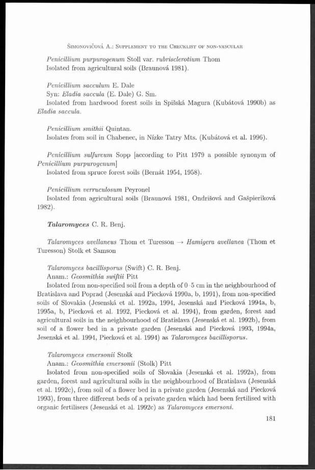
Penicillium purpurogenum Stoll var. rubrisclerotium ThomIsolated from agricultural soils (Braunová 1981).
Penicillium sacculum E. DaleSyn: Eladia saccula (E. Dale) G. Sm.Isolated from hardwood forest soils in Spišská Magura (Kubátová 1990b) as
Eladia saccula.
Penicillium smithii Quintan.Isolates from soil in Chabenec, in Nízké Tatry Mts. (Kubátová et al. 1996).
Penicillium sulfureum Sopp [according to P itt 1979 a possible synonym of Penicillium purpurogenum]
Isolated from spruce forest soils (Bernát 1954, 1958).
Penicillium verruculosum PeyronelIsolated from agricultural soils (Braunová 1981, Ondrišová and Gašpieriková
1982).
Talarornyces C. R. Benj.
Talarornyces avellaneus Thom et Turesson —► Hamigera avellanea (Thom et Turesson) Stolk et Samson
Talarornyces bacillisporus (Swift) C. R. Benj.Anam.: Geosmithia swiftii P ittIsolated from non-specified soil from a depth of 0-5 cm in the neighbourhood of
Bratislava and Poprad (Jesenská and Piecková 1990a, b, 1991), from non-specified soils of Slovakia (Jesenská et al. 1992a, 1994, Jesenská and Piecková 1994a, b, 1995a, b, Piecková et al. 1992, Piecková et al. 1994), from garden, forest and agricultural soils in the neighbourhood of Bratislava (Jesenská et al. 1992b), from soil of a flower bed in a private garden (Jesenská and Piecková 1993, 1994a, Jesenská et al. 1994, Piecková et al. 1994) as Talarornyces bacillisporus.
Talarornyces emersonii StolkAnam.: Geosmithia emersonii (Stolk) P ittIsolated from non-specified soils of Slovakia (Jesenská et al. 1992a), from
garden, forest and agricultural soils in the neighbourhood of Bratislava (Jesenská et al. 1992c), from soil of a flower bed in a private garden (Jesenská and Piecková 1993), from three different beds of a private garden which had been fertilised with organic fertilisers (Jesenská et al. 1992c) as Talarornyces emersoni.
S im o n o v ič o v á A .: S u p p l e m e n t t o t h e C h e c k l is t o f n o n - v a s c u l a r
181

C z e c h m y c o l . 53 (2 ) , 2001
T a b le 1. Species of microscopic fungi w ith a scarce or rare occurrence.
Fungi Occurrence of species
Soil Food-stuffs Other sources
Aspergillus duricaulis Raper et Fennell
A. flavus Link var. cotumnaris
A. melleus Yukawa
A. niger var. cinnamomeus (Schieman) Thom et Raper
A. parvulus G. Sm.
A. violaceo-fuscus Gasperini
Emericella rugulosa ( Thom et Raper) C. R. Benj.
Eupenicillium pinetorum Stolk *
E. sheari Stolk et D. B. Scott
Eurotium chevalieři L. Mangin
Eurotium pseudoglaucum (Blochwitz) Malloch et Cain
Merimbla ingelheimensis (J. F. H. Beyma) Pitt
Paecilomyces carneus (Duché et Helm) Brown et Smith
P. marquandii (Massee) S. Hughes
Penicillium arenicola Chalab.
P. atramentosum Thom
P. brasilianum Bat.
P. coprobium Frisvad
P. cyaneum (Bainier et Sartory) Biourge
P. flavovirens Cooke et Massee
P. geophilum Oudem. apud Oudem. et Konlng
P. griseofulvum Dlerckx *
P. herquei Bainier et Sartory
P. implicatum Biourge
P. lanosum Westling
P. megasporum Orpurt et Fennell
P. montanense M. Chr. et Backus
P. purpurogenum Stoll var. rubrisclerotium Thom
P. sacculum E. Dale
P. sm ith ii Quintan.
£ 30 22 1 7
_____________________________________________ 73.3 % 3.3 %__________23.7 %
Other sources: patients with oncological diceases, cotton, stored wheat, stone monuments, phyllosphere of agricultural plants.
182

S im o n o v ič o v á A .: S u p p l e m e n t t o t h e C h e c k l is t o f n o n - v a s c u l a r
T a b le 2. Species of m icroscopic fungi not listed in th e Checklist of non-vascular and vascular p lants of Slovakia, p a rt Fungi (Lizoň, Bacigálová, 1998).
Fungi Occurrence of species
Soil Food-stuffs Other sources
Aspergillus clavatus Desm. * *
A. ¡anus Raper et Thom * *
A. parasiticus Speare *
A. penicillioides Speg.
Byssochlamys fulva Olivier et Smith *
B. nivea Westling
Dichotomomyces cejpii (M ilko) D. B. Scott
Eupenicillium javanicum (J. F. H. Beyma) Stolk et D. B. Scott *
Fennellia flavipes B. J. W illey et E. G. Simmons
Paecilomyces farinosus (Holmsk.) Brown et Smith
Penicillium clavigerum Demellus
P. cyclopium Westling * * *
P. fellutanum Blourge *
P. luteum Sopp
P. m elin ii Thom
P. nalgiovense Laxa
P. sulphureum Sopp
P. verruculosum Peyronel
Talarornyces bacillisporus (Swift) C. R. Ben).
T. emersonii Stolk
E 20 15 6 9
_________________________________________________ 75.0 %_________30.0 %_________45.0 %
Other sources: air in factory, different material In depositories and archives (wooden sculpture, picture canvases, frames),
drinking water, dwelling, phyllosphere of agricultural plants, stored wheat, culture contaminant from an antibiotics factory.
D i s c u s s io n
Due to its ecological features (suitable amount of organic m atter, moisture, tem perature, protection from sunshine) soil is a natural biotope of microscopic fungi and is therefore the richest in them, too.
The to tal number of 50 species of microscopic fungi which are not listed in the Checklist of non-vascular and vascular plants of Slovakia (Lizoň and Bacigálová 1998) has been divided into two groups.
The first group (Tab. 1) represents those microscopic fungi, which have been isolated in Slovakia only once. So we can consider them to be scarce
183
I " ~ ~ ------------------ -----------

or rare. From the 30 species 22 were 22 isolated from different soils (forest, agricultural and meadow), 1 species from foodstuffs and 7 species from other sources (such as material from patients with oncological diseases, cotton, stored wheat, stone monuments, phyllosphere of different agricultural plants). More than half of them (16 species) belongs to the genus Penicillium (P. aren- icola, P. atramentosum, P. brasilianum, P. coprobium, P. cyaneum, P. fla- vovirens, P. geophilum, P. griseofulvum, P. herquei, P. implicatum, P. lanosum, P. megasporum, P. montanense, P. purpurogenum var. rubrisclerotium, P. sac- culum and P. smithii). Scarce or rare species of the genus Aspergillus can be considered A. duricaulis, A. flavus var. columnaris, A. melleus, A. niger var. cinnamorneus, A. parvulus, and A. violaceo-fuscus. The list is completed with the species Emericella rugulosa, Eupenicillium pinetorum, E. sheari, Eurotium chevalieři, E. pseudoglaucum, Merimbla ingelheimensis, Paecilomyces carneus and P. marquandii.
The second group (Tab. 2) represents species of microscopic fungi which are isolated more often than in the first case. Among the 20 species of microscopic fungi again the genus Penicillium dominates with 9 species (P . clavigerum, P. cyclopium, P. fellutanum, P. luteum, P. melinii, P. nalgiovense, P. sulphureum and P. verruculosum) and the genus Aspergillus with 4 species (A . clavatus, A. janus, A. parasiticus, and A. penicillioides). Species of the genera Byssochlamys, Dichotomomyces, Eupenicillium, Eurotium, Paecilomyes and Talarornyces are represented once or twice. Of the 20 species 15 were isolated from soil, 6 species from foodstuffs and 9 species from other sources (such as air in a factory, different material in depositories and archives, drinking water, dwelling, phyllosphere of different agricultural plants and corn, stored wheat, culture contaminant in an antibiotics factory). Out of all species of microscopic fungi (Tab. 1 and 2) only the species Aspergillus clavatus and Penicillium cyclopium were isolated from soil, foodstuffs as well as other sources.
R e f e r e n c e s
B e r n á t J . (1954): Mykoflóra lesných pod. — Preslia 26: 277-284.B e r n á t J . (1958): M ikroflóra sm rekových porastov. - A c ta Fac. Rer. N atur. Univ. Com en. B ot.,
97-9: 343-353.B e RNAT J . (1965): Mikrobiologická charakteristika mačinovo-podzolových pod (H abilitačná
práca). - K a ted ra fyziologie rastlín P R IF UK, B ratislava, 130 pp.B e r n Át J . , D u b o v s k Á A. and B r a u n o v Á O . (1 9 8 4 ) : M icrom ycetes in agricu ltu ral soils o f
Slovakia. - A cta Fac. Rer. N atur. Univ. Comen. Microbiol., 13: 3 -2 1 .B e r n á t J ., D u b o v s k á A. and F a r k a ŠOVÁ, E . (1 9 8 3 ) : M ikrom ycéty pri uskladňovaní pšenice. -
A c ta Fac. Rer. N atur. Univ. Comen. Form atio P ro t. N atur., 8: 45-52.BRAUNOVÁ O. (1981): M ikrom ycéty v podach experim entálnych báz. I. Druhové zloženie a častost’
výskytu. — Vedecké práce Výskum ného ústavu pčdoznalectva a výživy rastlín 10: 195—205. B r u m m it t R. K. and P o w e l l C. E. (1992): A uthors of p lant nam es, 732 pp. Kew.
C z e c h m y c o l . 53 (2 ) , 2001
184

B u Čk o v Á E. and B a c ig Ál o v Á K. (1999): Mikroskopické huby v podach lužného lesa slovenského úseku D unaja. In: Dugová O. and Vizárová G .(eds.), Zborník z m edzinárodného sem inára Život v pode II., 19.1.1999, p. 94-96 Bratislava.
D u b o v s k á A. (19.81): V ýskyt toxických m ikrom ycét vo fylosfére v závislosti od ekologických podm ienok. — A cta Fac. Rer. N atur. Univ. Comen. Form atio P ro t. n a tu r. 7: 2 4 3 -2 5 1 .
DUBOVSKÁ A. (1 9 8 4 ) : T he phyllospheric mycoflora of agricultural p lan ts - A cta Fac. Rer. N atur. Univ. Com en. Microbiol. 12: 53-60.
D u b o v s k a A., B e r n a t J . and G r e ČO V. (1986): M icromycetes in stored w heat - A cta Fac. Rer. N atu r. Univ. Com en. Microbiol. 14: 65-76.
D u b o v s k á A., B e r n Át , J . and H o r s k Á N. (1982): T he phyllospheric m icroflora of m aize - A cta Fac. Rer. N atur. Univ. Com en. Microbiol. 10: 31-39.
F r a n k o v a E. and Š im o n o v iČo vÁ A. (1999a): Význam mycetických deštruentov v procese biodegradácie kom unálnych odpadov. - In: Fečko P. (ed.), Recyklace odpadů III. 26.11.1999, p. 9-13, VŠB-TU Ostrava.
F r a n k o v á E . and Š im o n o v iČovÁ A. (1999b): Výskyt m ikrom ycétov v životnom prostredí so zretelom na zdravotně riziká. - In: M ikula I., M atějů J., Pilipčinec E . and Tkáčiková E. (eds.), A ktuálně problém y mikrobiologie a imunologie. 13.-15. október 1999 Košice, Bulletin ČSSM. - 40: 189-190.
F r a n k o v á E ., Š im o n o v iČo vÁ A. and B a c ig á l o v á K. (1999): Mikroskopické huby izolované z depozitov Slovenského národného m úzea v M artine - Bull. Slov. Bot. Spol. 21: 39-42.
G Ó d y o v Á M. (2000): M erim bla ingelheimensis (T . H. Beyma) P it t and Penicillium arenicola C halab . (D euterom ycota) - nové druhy pódnych m ikrom ycét pre Slovensko. - Bull. Slov. B ot. Spol. 22: 29-35.
HAWKSWORTH D. L., K ir k P . M., S u t t o n B. C. and P e Gl e r D. N. (1995): D ictionary of the fungi. 616 p. Oxon.
JESENSKÁ Z. (1988): V láknité m ikrom ycéty v pracovnom prostredí. - A cta Hyg. Epid. et Microbiol. 18: 44-46.
JESENSKÁ Z. and P e t ř ík o v á D. (1985): Mikroskopické huby ako agens kazenia ovocných konzerv. - Čs. Ilyg. 30: 175-177.
J e s e n s k á Z. and P o l á k o v á O. (1978): Potentiá ln i producenti m ykotoxínov v sušenej mléčnej výživě. - A c ta Hyg. Epid. e t Microbiol., Suppl. 12: 39-42.
J e s e n s k á Z. and S a jb id o r o v á I. (1984): Mykoflóra múky, krupice, s trúhanky a skadovaných obilných zrn - výsledky výskum nej úlohy. - A cta Hyg. Epid. e t Microbiol. 14, 4: 1-16.
JESENSKÁ Z. and ŠEPITKOVÁ J. (1984a): S tudium výskytu penicílií v cereáliách. - H ydinársky priem ysel 26: 100-112.
J e s e n s k á Z. and Š e p it k o v á J . (1984b): Š túdium výskytu penicílií v cereáliách. — B ulletin Po tr. výživy (B ratislava) 23 (3), 3: 197-207.
J e s e n s k á Z., P o l s t e ii M ., M a t y á š o v á J . and P o l á k o v á O. (1980): C ollaborative s tu d y of the effectiveness of the screening tes t on APA m edium for aflatoxin producing aspergilli. — Zbl. B akt. Hyg., I. A bt. Orig. B 171, 4-5: 408-415.
J e s e n s k á Z . , H a v r a n e k o v Á D. and Š a jb id o r o v Á I. (1983): Tzv. term orezistentné huby - ich prob lem atika a diagnostika. — A cta Hyg. Epid. e t Microbiol. 13: 15-18.
Jesenská Z., Havraneková D. and Šajbidorová I. (1984): Mykoflóra múky, krupice a strúhanky. - Bull. P o tr. výživy (B ratislava) 23 (3), 1: 41-51.
J e s e n s k á Z., Š a jb id o r o v á I. and G r a t z l o v Á .1. (1988): Mikromycéty, aflatoxin B | a ochratoxín A vo vybraných vzorkách poživatin v Slovenskej socialistickej republike. - In: M uzikář V., B a rtl V., Špelina V. (eds.) Zkušenosti s novými laboratorním i přístupy v mikrobiologii po trav in . Z borník přednášek Liblice 3.-5.5.1988, Kom ise potravinářské m ikrobiologie, ČSSM při ČSAV, p. 96-107.
J e s e n s k á Z., Š a jb id o r o v á I. and A d e n s a m L. (1989): M arginálie potravinárskej mikrobiologie: Káva, m ak (mykoflóra, m ykotoxíny). In: M uzikář, V., Špelina, V., B artl, V. (eds.) M ikrobiologické problém y v biotechnologiích a nové poznatky z potravinářské mikrobiologie. Zborník přednášek, Liblice 1989, Komise potravinářské mikrobiologie, ČSSM při ČSAV, p. 86-95.
S im o n o v ič o v á A .: S u p p l e m e n t t o t h e C h e c k l is t o f n o n - v a s c u l a r
185
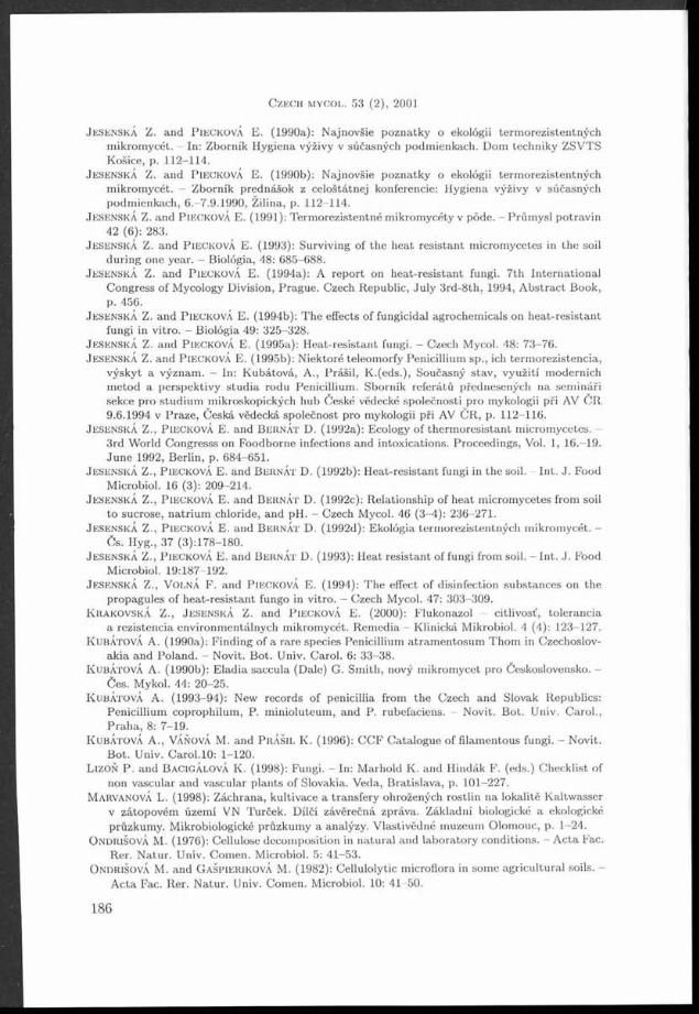
J e s e n s k á Z. and P ie c k o v á E . (1990a): Najnovšie poznatky o ekologii term orezistentných m ikrom ycét. - In: Zborník Hygiena výživy v súčasných podm ienkach. Dom techniky ZSVTS Košice, p. 112-114.
J e s e n s k á Z. and P ie c k o v á E. (1990b): Najnovšie poznatky o ekologii term orezistentných m ikrom ycét. — Zborník prednášok z celoštátnej konferencie: H ygiena výživy v súčasných podm ienkach, 6.-7.9.1990, Žilina, p. 112-114.
JESENSKÁ Z. and P ie c k o v á E. (1991): T erm orezistentné m ikrom ycéty v pode. - Prům ysl po trav in 42 (6): 283.
JESENSKÁ Z . a n d P ie c k o v á E . (1 9 9 3 ) : S u rv iv in g o f t h e h e a t r e s i s t a n t m ic ro m y c e te s in t h e so il d u r in g o n e y e a r . - B io ló g ia , 48: 685-688.
JESENSKÁ Z. and P ie c k o v á E. (1994a): A report on heat-resis tan t fungi. 7 th In terna tional C ongress of Mycology Division, Prague. Czech Republic, Ju ly 3rd-8th , 1994, A b strac t Book,
JESENSKÁ Z. and PIECKOVÁ E. (1994b): T he effects of fungicidal agrochem icals on heat-resis tan t fungi in vitro . — Biológia 49: 325—328.
J e s e n s k á Z. and P ie c k o v á E. (1995a): H eat-resistant fungi. - Czech Mycol. 48: 73-76.JESENSKÁ Z. and P ie c k o v á E. (1995b): N iektoré teleom orfy Penicillium sp., ich term orezistencia,
výskyt a význam . — In: K ubátová, A., P rášil, K .(eds.), Současný stav , využití m oderních m etod a perspektivy s tu d ia rodu Penicillium . Sborník referá tů přednesených n a sem ináři sekce pro stud ium m ikroskopických hub České vědecké společnosti pro mykologii při AV Č R 9.6.1994 v Praze, česk á vědecká společnost pro mykologii při AV ČR, p. 112 -116.
J e s e n s k á Z., PIECKOVÁ E. and B e r n Át D. (1992a): Ecology of therm oresis tan t m icrom ycetes. - 3rd W orld Congresss on Foodborne infections and intoxications. Proceedings, Vol. 1, 16.-19.June 1992, Berlin, p. 684-651.
JESENSKÁ Z., P ie c k o v á E. and B e r n á t D. (1992b): H eat-resistan t fungi in th e soil. - In t. J . Food Microbiol. 16 (3): 209-214.
J e s e n s k á Z., P ie c k o v Á E. and B e iin Át D. (1992c): R elationship of heat m icrom ycetes from soil to sucrose, n a triu m chloride, and pH. - Czech Mycol. 46 (3-4): 236-271.
J e s e n s k á Z., P ie c k o v á E. and B e r n Át D. (1992d): Ekológia term orezistentných m ikrom ycét. - Čs. Hyg., 37 (3): 178-180.
JESENSKÁ Z., PIECKOVÁ E. and B e iin Át D. (1993): H eat resistan t of fungi from soil. - In t. J. Food Microbiol. 19:187-192.
J e s e n s k á Z ., V o l n á F . and P ie c k o v á E . (1 9 9 4 ) : T he effect of disinfection substances on the propagules of heat-resis tan t fungo in vitro. - Czech Mycol. 47: 3 0 3 -3 0 9 .
K r a k o v s k á Z., J e s e n s k á Z. and P ie c k o v á E. (2000): Flukonazol - citlivost’, to lerancia a rezistencia environm entálnych m ikrom ycét. R em edia - Klinická M ikrobiol. 4 (4): 123-127.
KUBÁTOVÁ A. (1990a): F inding of a rare species Penicillium a tram entosum T hom in Czechoslovakia and Poland. — Novit. Bot. Univ. Carol. 6: 33—38.
KUBÁTOVÁ A. (1990b): E ladia saccula (Dale) G. Sm ith, nový m ikrom ycét pro Československo. - Čes. Mykol. 44: 20-25.
KUBÁTOVÁ A . (1 9 9 3 -9 4 ) : New records of penicillia from th e Czech and Slovak Republics:Penicillium coprophilum , P. m inioluteum , and P. rubefaciens. - Novit. B ot. Univ. C arol.,P rah a , 8: 7-19.
KUBÁTOVÁ A., VÁŇOVÁ M. and P r á š il K . (1996): C C F C atalogue of filam entous fungi. - Novit.Bot. Univ. C aro l.10: 1-120.
L iz o n P . and BACIGÁLOVÁ K. (1998): Fungi. - In: M arhold K. and H indák F. (eds.) C hecklist of non vascular and vascular p lan ts of Slovakia. Veda, B ratislava, p. 101-227.
M a r v a n o v Á L. (1998): Záchrana, kultivace a transfery ohrožených rostlin na lokalitě K altw asser v zátopovém území VN Turček. Dílčí závěrečná zpráva. Základní biologické a ekologické průzkum y. Mikrobiologické průzkum y a analýzy. V lastivědné m uzeum Olom ouc, p. 1-24.
ONDRIŠOVÁ M. (1976): Cellulose decom position in n a tu ra l and labora to ry conditions. - A c ta Fac.R er. N atu r. Univ. Com en. Microbiol. 5: 41—53.
O n d r iš o v á M. and G a Šp ie r ik o v Á M. (1982): Cellulolytic m icroflora in some agricultura l soils. - A cta Fac. Rer. N atu r. Univ. Com en. Microbiol. 10: 41-50.
1
C z e c h m y c o l . 53 (2 ) , 2001

P a v l íč k o v á K. (1994): Species com position of m icrom ycetes in cam bizem stagnogleyic. Biológia. - B ratislava, 49: 835-839.
P e t ř ík o v á D ., J e s e n s k á Z . a n d J a n á k o v á V . (1 9 8 5 ): Z n e h o d n o te n ie k o n z e rv á re n s k ý c h v ý ro b - k o v m ik r o s k o p ic k ý m i h u b a m i. - B u lle t in P o t r . v ý ž iv y (B ra t i s la v a ) 24 (4 ) , 2 -3 : 1 3 7 -1 4 1 .
P ie c k o v Á E ., B e r n Át D. and J e s e s n s k Á Z. (1992): H odnoty therm al death tim e (T D T ) pre nové term orezisten tné druhy m ikrom ycét. - In: 19. zjazd ČSSM při ČSAV, Zborn. referátov,Košice 14.-17.9.1992, p. 39.
P ie c k o v á E . , B e ii n Át D . and J e s e n s k á Z . (1 9 9 4 ) : Heat resistan t fungi isolated from soil. - Int.J. Food Microbiol. 22: 297-299.
PlECKOVA E. and J e s e n s k á Z. (1996a): Toxinogenicity of heat-resis tan t m icrom ycetes detected by a biosensor. - In: In terna tiona l m eeting on W aste collection and recycling. Bioaerosol exposure and health problem s. A bstracts, 13-14 Septem ber 1996, Koge, Denm ark.
P ie c k o v á E. and J e s e s n s k Á Z. (1996b): F ilam entous microfungi in raw flax and co tton for textile industry and their ciliostatic activ ity on tracheal organ cultures in vitro. - M ycopathol. 134:91-96.
PIECKOVÁ E. and JESENSKÁ Z. (1997a): Dichotom om yces cejpii - some characteristics of stra ins isolated from soil in th e Slovak Republic. - Czech Mycol. 49 (3-4): 229-237.
PiECKOvÁ E. and JESENSKÁ Z. (1997b): Toxigenicity of heat-resis tan t fungi detected by a bio-assay. - In t. J. Food Microbiol. 36: 227-229.
PIECKOVÁ E. and JESENSKÁ Z. (1999a): Soil fungi to le ran t to fluconazole. - In: IX In terna tional C ongress of Mycology, Sydney 16-20 august 1999, A bstracts, p. 214.
PlECKOVA E. and JESENSKÁ Z. (1999b): O ccurrence of itraconazole-tolerant m icrom ycetes in the soil and food products. - Folia Microbiol. 44: 677-682.
PlECKOVA E . , LAKATOŠOVÁ A . and JESENSKÁ Z . (1 9 9 6 ) : Toxinogenita term orezistentných m ikrom ycét stanovená biosenzorom. - In: M uzikář, V., B artl, V. (eds.), M oderní trendy v m ikrobiologii po trav in . Sborník sem inára, Třešť 3 0 .5 .1 9 9 6 , ČSSM, Komise potravinářské m ikrobiologie, p. 2 7 -3 4 .
P ITT J. I., S a m s o n R. A. and F iu s v a d J. C. (2000): List of accepted species and their synonym s in th e fam ily Trichocom aceae. - In: Sam son R. A. and P it t J. I., In tegration of m odern taxonom ic m ethods for Penicillium and Aspergillus classification, p. 9-72.
Š a j b id o r o v á I., J e s e n s k á Z. and CORNER V. (1988): P rodukcia T-2 toxínu km eňom Fusarium poae CCM -F-584 v přítom nosti skladiskových m ikrom ycét. In: M uzikář V., B artl V. and Špelina V. (eds.) Zkušenosti s novými laboratorním i p řístupy v m ikrobiologii potravin.Sborník přednášek, Liblice 3.—5.5.1988, Komise potravinářské mikrobiologie, ČSSM při ČSAV p., 124-135.
Š e p it k o v á K. and J e s e n s k á Z. (1985a): Analýza mykoflóry sladovnického jačm eňa a sladu. I.Polné m ikrom ycéty. - Bulletin Po tr. výživy (B ratislava) 24 (4), 2-3: 143-151.
Š e p it k o v á K . and J e s e n s k á Z. (1 9 8 5 b ) : Aspergillus flavus v sladovníckom jařm eni. In: P roblem atika m ykotoxinu z hlediska potravinářské a veterinární praxe. Sborník přednášek, Brno,97. Cs. vědeckotechnická společnost, D ům techniky, Brno, p. 46-50.
Š e p it k o v á K. and J e s e n s k á Z. (1985c): K problem atike mikroskopických vláknitých húb v sladovníckom jačm eni a v slade. - In: B artl V. and M uzikář V .(eds.), M ikroflora m asných a drůbežářských výrobku a noviny v taxonom ii. Sborník přednášek sem ináře Liblice 6.-8.5.1985, Komise potravinářské mikrobiologie, ČSSM při ČSAV, p. 67-71.
Š e p it k o v á K. and J e s e n s k á Z. (1986): Výskyt Aspergillus clavatus v potravinářských surovinách, In: B artl V. and M uzikář V. (eds.), Vzájom ný vztah m ikroorganizm u a potravin a m etody sledování. - Sborník přednášek, Liblice 5.-7.5.1986, Komise potravinářské mikrobiologie,ČSSM při ČSAV, p. 68-70.
Š e p it k o v á J . and J e s e n s k á Z. (1988): K problem atike vláknitých m ikrom ycét v sladovníckom jačm eni a v slade. - Čs. Hyg. 33 (10): 671-622.
ŠEPITKOVÁ J . and JESENSKÁ Z . (1 9 9 0 ): Pohlad na akosť vybraných potravinářských surovin z mykologického hladiska. - In: M uzikář, V., Špelina V. and B artl V. (eds.), Mikrobiologické rizikové faktory v poživatinách. ČSSM při ČSAV, Komise potravinářské m ikrobiologie, Liblice 1 3 .-1 5 .3 .1 9 9 0 .
187
■ ” ~
Š im o n o v iČo v Á A .: S u p p l e m e n t t o t h e C h e c k l is t o f n o n -v a s c u l a ii

ŠEPITKOVÁ J . and J e s e n s k á Z. (1991): Pivovarské suroviny z hládiska mykologického. - Kvasný prům ysl 37 (2): 33-37.
Š e p it k o v á J . , H a r g a Š V. and J e s e n s k á Z. (1987b): Mykologická depistáž v konzervárenskej technologii. - In: M uzikář V., B artl V. and Špelina V. (eds.), Nové labora to rn í p řístupy v m ikrobiologii potravin. Sborník přednášek sem ináře Liblice 5.-7.5.1987, Komise po trav inářské m ikrobiologie, CSSM při ČSAV, p. 80-85.
Š e p it k o v á J ., H a iic a Š V. and J e s e n s k á Z. (1989): Mykologické problém y v konzervárenskej technologii. — B ulletin Po tr. výživy (B ratislava) 28 (8), 1—2: 149—158.
Š e p it k o v á J . , M a r k o A. and J e s e n s k á Z. (1987a): K problem atike výskytu m ikrom ycét a aflatoxínu Bi v sladovníckom jačm eni a v slade zo žatvy r. 1985. - B ulletin P o tr. výživy (B ratislava) 26 (2):147-157.
Š e p it k o v Á J . , M a r k o A . a n d J e s e n s k á Z . (1 9 8 8 ) : P o h la d n a v ý s k y t m ik r o m y c é t a n ie k to rý c h m y k o to x ín o v v p o tr a v in á ř s k ý c h s u ro v in á c h . - In : M u z ik á ř V., B a r t l V. a n d Š p e l in a V. (e d s .) , Z k u š e n o s t i s n o v ý m i la b o r a to r n ím i p ř ís tu p y v m ik ro b io lo g ii p o t r a v in . S b o r n ík p ře d n á š e k , L ib lic e 3 . -5 .5 .1 9 8 8 , K o m is e p o t r a v in á ř s k é m ik ro b io lo g ie , C S S M p ř i ČSAV, p . 1 0 8 -1 2 3 .
Š e p it k o v á J . , M a r k o A. and JESENSKÁ Z. (1990): Pohlad na akosť vybraných potravinářských surovin z mykologického hiadiska. — B ulletin PV (B ratislava) 29 (9), 1: 39-54.
ŠIMONOVIČOVÁ A. (1980): Mikroskopické huby vo vybraných pódach Slovenska. - Biológia 35: 213-220.
Š im o n o v iČovÁ A. (1992): Š tru k tú ra spoločenstva mikroskopických húb v lesných pódach severného Slovenska. II. Podobnost’ mykocenóz (K rížna, K oprová a Furkotská dolina). - č e s . Mykol. 46: 106-113.
ŠIMONOVIČOVÁ A. (1993): Microbiological relations in the soils of forest com m unities of Skalnatá dolina valley in High T atras M ountains. - Biológia 48: 639-700.
Š im o n o v iČo vÁ A. and F r a n k o v á E. (1998): Mikroskopické huby ako invázne organizmy. — In: A ntonická B. and H usár M. (eds.), O dpady ’98, 12.-13. november 1998 Spišská Nová Ves, p. 152-156.
Š im o n o v iČo vÁ A., F r a n k o v á E . and G Ó d y o v Á M . (2 0 0 0 ) : Biokorózia historickej arch itek túry ako dóležitý fak tor jej chátran ia . - Život. P rostr. 34 (6 ): 3 1 6 -3 2 0 .
T r u p l J . , B l a h o v á M ., Š p Án ik S., K r Čm Éry V. m l . and J e s e n s k á Z. (1992): M ykotické infekcie u im unodeficitných pacientov. - Lék. O bzor 41 (8): 464-468.
VLÁČILIKOVÁ A. (1978): M icromycetes in some forest soils of Slovakia. - A c ta Fac. Rer. N atur. Univ. Com en. Microbiol. 6: 109-123.
Czech Mycology, published by th e Czech Scientific Society for Mycology. G raphic design byB. B ednář, PISCES. T ypeset by rr|?X. P rin ted by č ih á k Press, P ra h a 10. D istribu ted by theCzech Scientific Society for Mycology, P.O .Box 106, 11121 P ra h a 1, and K ubon & Sagner,P .O .Box 340108, 80328 Miinchen, Germ any. Annual subscription: Vol. 53, 2001 (4 issues),US $ 8 6 ,-, DM 136,-
C z e c h m y c o l . 53 (2 ) , 200 1

INSTRUCTIONS TO AUTHORSPreparation of manuscripts. M anuscripts are to be subm itted in English, German or French. The text of the m anuscript should be w ritten on one side of white paper (A4, 210 x 297mm) with broad margins (maximum 30 lines per page). Each m anuscript must include an abstract (in English) not exceeding 100 words and a maximum of five key words. The paper will be followed by an abstract in Czech (or Slovak). The journal is responsible, however, for the translation of abstracts into Czech for foreign authors. Please send two copies of the typescript. The authors are asked to subm it diskettes with the accepted manuscripts prepared on personal computers. The files should be in ASCII format, graphs in Excel. Both IID and D D/S.ö" and 5.25” diskettes are acceptable.Illustrations and tables. All tables, black and white photographs and figures (in black indian ink on a separate sheet) combined with the legends should be self-explanatory. Legends to the figures must be typed on a separate sheet. Colour photographs can be accepted but the authors will be responsible for the costs. All drawings or a photographs of m icrostructures should be provided with a scale. All illustration should be subm itted as the original drawing and one clear copy. O utput from computer graphics programmes produced on plotters or laser printers is quite acceptable. The dimension of any figure should not exceed 180 x 260 mm in size. References to illustrative m atter in the text should normally in parentheses, e.g. . . . spore sizes (Table 1) and . . . as shown in Fig. 2 . . . Figs. 1-5 . . . Map 1 . . .Nomenclature. Latin names should conform to the International code of botanical nomenclature. New taxa must be substantiated by a Latin diagnosis including a reference to the public herbarium where the type specimen is deposited. The authors are asked to use only the acronyms listed in the Index Herbariorum.References. References are to be listed in alphabetical order according to the surnames of the first authors. The bibliography should be w ritten as follows:Moravec J. (1984): Two new species of Coprobia and taxonomic remarks on the genera
Cheilymenia and Coprobia (Discomycetes, Pezizales). - Ces. Mykol. 38: 146-155.(journal article)
Ryvarden L. (1978): The Polyporaceae of N orth Europe, Vol. 2. Inonotus-Tyromyces. - 507 p. Oslo. (book)
Tom m erup I. C., Kuek C. and M alajczuk N. (1987): Ectomycorrhizal inoculum production and utilization in Australia. - In: Sylvia D. M., Hung L. L., and G raham J. H. (eds.), Proceedings of the 7th North American Conference on Mycorrhizae, p. 93-295, Gainesville.
The references in text should be Moravec (1984), or (Moravec 1984); or Kühner & Romagnesi (1974); W hen there are three or more authors use the form Tommerup et al. (1987).M anuscript evaluation. All manuscripts will be reviewed by two reviewers, and the authors informed about their acceptance, rejection or necessary revisions within two months. If a m anuscript is returned for revision, the authors should submit a revised version within three months.A uthors should preferably have their English language texts approved by a native - English speaker.P roof corrections. Proofs of the paper will be sent to authors together with the original manuscript. If not returned within three weeks, the proof correction will be carried out by the editor. The principal author will receive 30 reprints free of charge. Correspondence. All correspondence concerning the journal should be sent to the following address: Czech Mycology / Česká mykologie, National Museum, Departm ent of Mycology, Václavské nám ěstí 68, 115 79 P raha 1, Czech Republic. Phone: 02/24497259 or 24964284
h ttp ://w w w .natu r.cuni.cz/cvsm /
Czech Mycology, published by th e Czech Scientific Society for Mycology. G raphic design byB. B ednář, PISC ES. T ypeset by T eX. P rin ted by Č ihák Press, P ra h a 10. D istribu ted by the Czech Scientific Society for Mycology, P.O.Box 106, 11121 P ra h a 1, and K ubon & Sagner, P .O .B ox 340108, 80328 M ünchen, Germ any. Annual subscription: Vol. 52, 1999-2000 (4 issues), US $ 8 6 ,- , DM 136,-
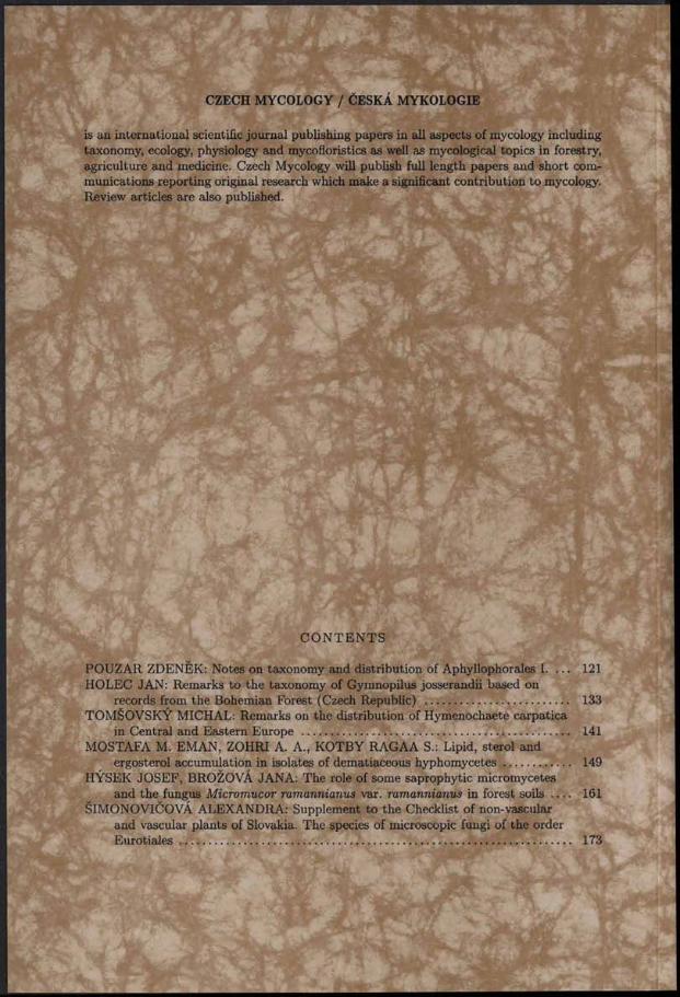
CZECH MYCOLOGY / ČESKÁ MYKOLOGIE
is an international scientific journal publishing papers in all aspects of mycology including taxonomy, ecology, physiology and mycofloristics as well as mycological topics in forestry, agriculture and medicine. Czech Mycology will publish full length papers and short communications reporting original research which make a significant contribution to mycology. Review articles are also published.
C O N T E N T S
POUZAR ZDENĚK: Notes on taxonomy and distribution of Aphyllophorales I. . . . 121 HOLEC JAN: Remarks to the taxonomy of Gymnopilus josserandii based on
records from the Bohemian Forest (Czech Republic) ............................................... 133TOMŠOVSKÝ MICHAL: Remarks on the distribution of Hymenochaete carpatica
in Central and Eastern Europe ....................................................................................... 141MOSTAFA M. EMAN, ZOHRI A. A., KOTBY RAGAA S.: Lipid, sterol and
ergosterol accumulation in isolates of dematiaceous hyphom ycetes...................... 149HÝSEK JO SEF, BROŽOVÁ JANA: The role of some saprophytic micromycetes
and the fungus Micromucor ramannianus var. ramannianus in forest s o i l s 161ŠIMONOVIČOVÁ ALEXANDRA: Supplement to the Checklist of non-vascular
and vascular plants of Slovakia. The species of microscopic fungi of the order E u ro tia le s ................................................................................................................................ 173


