:.' ANALYTICAL BIOCHEMISTRY 107,424-431 (1980) Optimization of Conditions for the Colorimetric Determination of Citrulline, Using Diacetyl Monoxime T. R. C. BOYDE AND MOHAMMED RAHMATULLAH Department of Biochemistry, University of Hong Kong, Hong Kong Received February 1, 1980 A method is described for colorimetric determination of citrulline following deproteiniza- tion, depending upon the reaction with diacetyl monoxime in the presence of sulfu~ic.and phosphoric acids. Ferric chloride is included to sensitize the reaction and thiosemicarbazide to improve light stability of the chromogen. The method offers diminished heating time compared with existing procedures, combined with good sensitivity and 'photostability of the chromophore. The colorimetric estimation of citrulline is most often required in the assay of orni- thine transcarbamylase (OTC,l EC 2.1.3.3), which catalyzes the second step of urea syn- thesis in mammals (1,2), and is also by rea- son of its tissue-specific location a valuable indicator of liver damage (3-5). Depressed hepatic levels of OTC are found in genetic disorders causing hyperammonemia (6-10), in Reye's syndrome (11-13), cirrhosis (14), and acute fatty liver of pregnancy (15). Several recent reports (16-18) describe methods for assay of serum OTC levels in which deproteinization is avoided. But such cannot be applied blindly to tissue samples or homogenates because of widely varying levels and because of the intramitochondrial location of the enzyme (19). There may be circumstances in which a method involving deproteinization should be used for serum samples also since a greater sensitivity is then obtainable. We have accordingly sought to optimize conditions for citrulline assay after removal of proteins and present a method which should prove widely applicable. Determination of citrulline by the method of Archibald (20) is based on the Fearon 1 Abbreviations used: OTC, ornithine transcarbamyl- ase; TCA, trichloroacetic acid; DAMO, diacetyl mon- oxime; TSC, thiosemicarbazide. reaction (21), in which carbamido com- pounds such as urea and citrulline form a color complex with diacetyl monoxime or diacetyl in acid solution. The original method (20) suffers from several disa.dvantages, namely, low sensitivity, nonlinearity of the calibration curve, and instability of the chromogen formed in the presence of light. Removal of protein prior to assay of citrul- line has been found necessary in general (22), although several deproteini'zing agents interfere in color development by producing opalescence (23). Various modifications (24,25) of Archi- bald's method have proved to be tedious and unsatisfactory. The most widely used procedure (that of Guthohrlein and Knappe (26)), though much more sensitive than Archibald' s, requires a prolonged boiling time of 20 min for color development. More- over, the standard curve for citrulline is non- linear at low citrulline concentrations and the color complex although stable in the dark begins to fade after only 30 min in diffuse daylight. Our method for citrulline is based on that of Favreau and Coulombe (27) for colori- metric determination of urea. The boiling time for color development has been cut to 5 min and addition of thiosemicarbazide has 0003- 2697/80/140424-08$02.00/0 Copyright to 1980 by Academic Press, Inc. All rights of reproduction in any form reserved. 424
Transcript
:.'
ANALYTICAL BIOCHEMISTRY 107,424-431 (1980)
Optimization of Conditions for the Colorimetric Determination of
Citrulline, Using Diacetyl Monoxime
T. R. C. BOYDE AND MOHAMMED RAHMATULLAH
Department of Biochemistry, University of Hong Kong, Hong Kong
Received February 1, 1980
A method is described for colorimetric determination of citrulline following deproteiniza
tion, depending upon the reaction with diacetyl monoxime in the presence of sulfu~ic.andphosphoric acids. Ferric chloride is included to sensitize the reaction and thiosemicarbazide
to improve light stability of the chromogen. The method offers diminished heating time
compared with existing procedures, combined with good sensitivity and 'photostability of
the chromophore.
The colorimetric estimation of citrullineis most often required in the assay of ornithine transcarbamylase (OTC,l EC 2.1.3.3),which catalyzes the second step of urea synthesis in mammals (1,2), and is also by reason of its tissue-specific location a valuableindicator of liver damage (3-5). Depressedhepatic levels of OTC are found in geneticdisorders causing hyperammonemia (6-10),in Reye's syndrome (11-13), cirrhosis (14),
and acute fatty liver of pregnancy (15).Several recent reports (16-18) describe
methods for assay of serum OTC levels inwhich deproteinization is avoided. But suchcannot be applied blindly to tissue samplesor homogenates because of widely varyinglevels and because of the intramitochondriallocation of the enzyme (19). There may becircumstances in which a method involvingdeproteinization should be used for serumsamples also since a greater sensitivity isthen obtainable. We have accordingly soughtto optimize conditions for citrulline assayafter removal of proteins and present a methodwhich should prove widely applicable.
Determination of citrulline by the methodof Archibald (20) is based on the Fearon
reaction (21), in which carbamido compounds such as urea and citrulline form acolor complex with diacetyl monoxime ordiacetyl in acid solution. The original method(20) suffers from several disa.dvantages,namely, low sensitivity, nonlinearity of thecalibration curve, and instability of thechromogen formed in the presence of light.Removal of protein prior to assay of citrulline has been found necessary in general(22), although several deproteini'zing agentsinterfere in color development by producingopalescence (23).
Various modifications (24,25) of Archibald's method have proved to be tediousand unsatisfactory. The most widely usedprocedure (that of Guthohrlein and Knappe(26)), though much more sensitive thanArchibald' s, requires a prolonged boilingtime of 20 min for color development. Moreover, the standard curve for citrulline is nonlinear at low citrulline concentrations andthe color complex although stable in the darkbegins to fade after only 30 min in diffusedaylight.
Our method for citrulline is based on thatof Favreau and Coulombe (27) for colorimetric determination of urea. The boilingtime for color development has been cut to5 min and addition of thiosemicarbazide has
0003- 2697/80/140424-08$02.00/0Copyright to 1980 by Academic Press, Inc.
All rights of reproduction in any form reserved.
424
COLORIMETRIC DETERMINATION OF CITRULLINE 425
led to stabilization of the color complex inlight. The standard curve for citrulline islinear for quantities up to at least 0.1 /-tmol;and protein-precipitating agents like trichloroacetic acid (TCA) cause no inhibitionin the development of color. The sensitivityof citrulline estimation is much greater thanthat in Guthohrlein's procedure, whichshould enable OTC activity to be measuredaccurately in tissue homogenates containingdepressed levels of the enzyme.
MATERIALS AND METHODS
Reagents
1. Acid -ferric solution. To 550 ml distilledwater, add 250 ml concentrated sulfuric acid(95-98%, E. Merck, Darmstadt, W. Germany) and 200 ml concentrated phosphoricacid (85%, Mallinckrodt Chemical Co., St.Louis, Mo.). Cool to room temperature, anddissolve FeCI3 (250 mg/liter) in the abovesolution. The solution when kept at roomtemperature is stable for more than 2 months.For the purposes of this paper, the totalvolume of this and similar solutions is takento be 1 liter.
2. Diacetyl monoxime solution. To 100ml of distilled water add 500 mg diacetylmonoxime (DAMO, 2,3-butanedione monoxime, Sigma Chemical Co., St. Louis, Mo.).Stored in a brown bottle, the solution isstable for more than a month.
3. Chromogenic reagent. Just before useadd 5 mg thiosemicarbazide (TSC, SigmaChemical Co.) to 50 ml of Reagent 2, followed by 100 ml of Reagent 1. This solutionshould be used no later than 1 h afterpreparation.
4. Citrulline standard solution (1 mmol/liter). Dissolve 17.52 mg DL-citrulline (SigmaChemical Co.) in 100ml distilled water. Keptfrozen at - 20°C, this can be stored for prolonged periods.
Procedure For Citrulline Estimation InEnzymatic Samples
Typically the sample is the reaction mixture following appropriate incubation for
OTC assay and since urea also forms a colorcomplex with diacetyl monoxime, ureaseshould be included in the incubation mixture. (We generally used urease Type VII,Sigma Chemical Co.) The mixture may bedeproteinized, and the enzyme reactionstopped, by adding TCA solution to a finalconcentration of at least 5%, then centrifuging and collecting the supernatant. (We occasionally experienced slight turbidity whenusing 50% TCA solution. This was overcome by using a larger volume of a moredilute solution and raising the final concen-tration to 7.5%.) .
To 0.1 ml of supernatant add 3 ml of chromogenic solution (Reagent 3). Mix vigorouslyin a Vortex mixer (Whirlimixer, Fison' sScientific Apparatus, England), and boil at100°C for 5 min. A covered water bath ispreferable to minimize evaporation. Following boiling, cool the tubes to room temperature and measure absorbance at 530 nm, forexample, in a Varian Series 634 doublebeam spectrophotometer. Run a blank and acitrulline standard simultaneously withsamples. Nanomoles of citrulline in the aliquot of supernatant can be read off from astandard curve, though the color development is so reproducible that we h~~e habitually run only one confirmatory standardwith each routine assay batch.
We used Pyrex glass tubes 12.5 cm longby 1.5 cm i.d., but have no evidence thattube size affects the results. Mixing was for10-15 s so that this process occupied 5-10min for a batch of 20-40 tubes: tubes already mixed but not yet boiled could be lefton the bench in diffuse daylight for at least30 min without affecting color development.Tubes were not stoppered during either mixing or boiling. Measurements were made incuvettes with a I-cm path length.
The sample volume may be increased tocompensate for low citrulline levels (withoutchanging reagent composition) up to a maximum of 1.0 ml. Preferably, a new standardcurve should be constructed using the samevolumes of aqueous citrulline standards asthe selected sample volume, although in
426 BOYDE AND RAHMA TULLAH
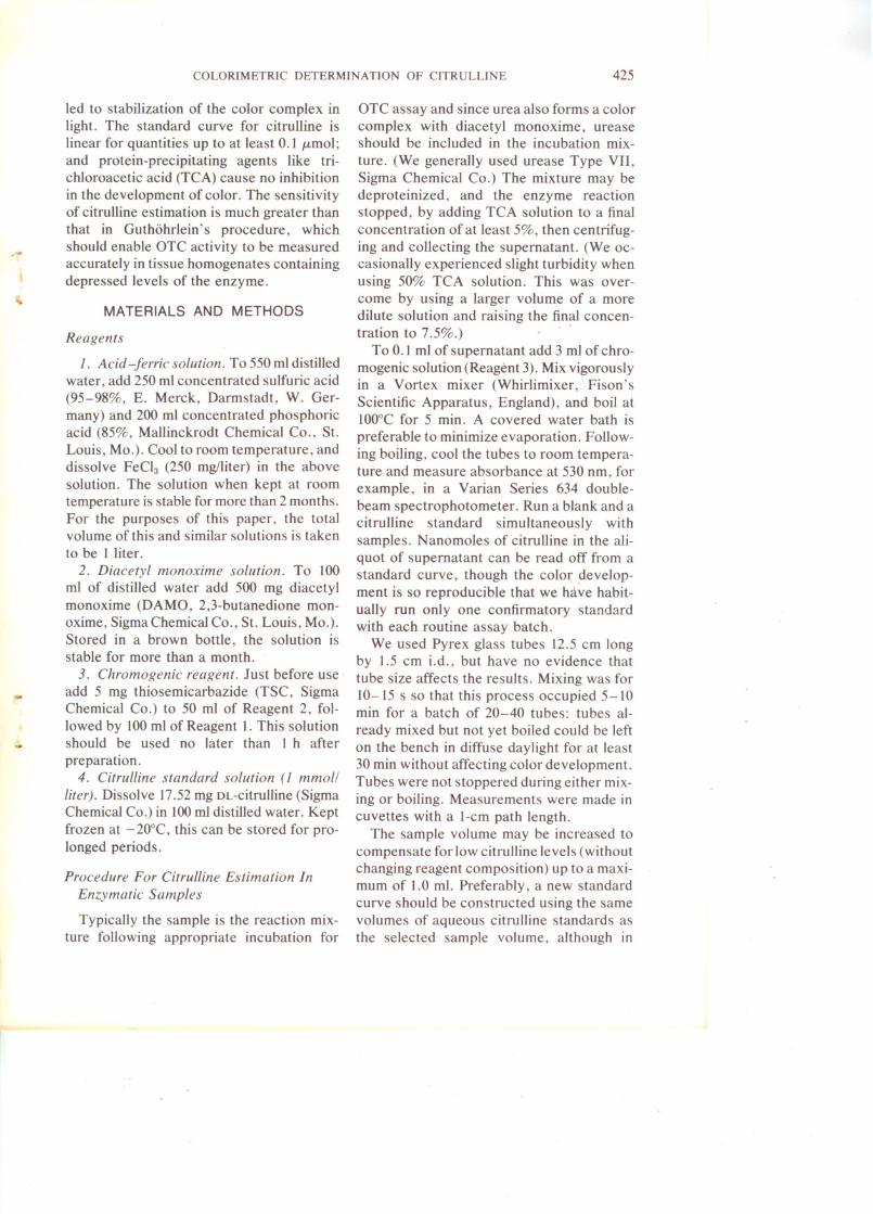
FIG. 2. Studies of sensitivity of"citrulline measure
ment as a function of acid reagent compositions andconcentrations of ferric chloride. Citrulline solution
(0.1 ml, I mmol/liter) was boiled for 5 min in the pres
ence of I ml of DAMO- TSC solution (2 g DAMO and
100 mg TSC per liter) and 2 ml of acid solution having
the following compositions (p~r liter solution): (I) 50 mlH2S04 (0), (11) 450 ml H2S04 (e), (Ill) 50 ml H2S04,
200 ml H3P04 (0), (IV) 250 ml H2S04, 200 ml H3P04/
(.), (V) 450 ml H2S04, 200 ml H3P04 (6), (VI) 450 '"91'H3P04 (.), (VII) 50 ml H2S04, 450 ml H3P04 (*), and(VIII) 250 ml H2S04, 450 ml HaP04 ('<7). Ferric chloride
in the requisite amount was added dissolved in theacid solution.
terms of molar absorbance there is no de
tectable change in sensitivity as water content is increased. (Overall practical sensitivity-absorbance per micromoles-isdiminished.)
EXPERIMENTAL RE8UL T8
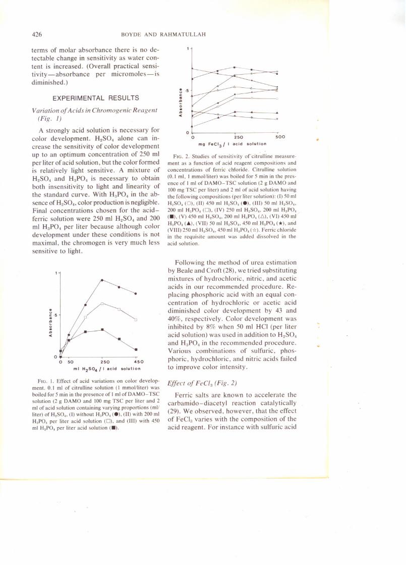
Variation of Acids in Chromogenic Reagent
(Fig. l)
A strongly acid solution is necessary forcolor development. H2S04 alone can increase the sensitivity of color developmentup to an optimum concentration of 250 mlper liter of acid solution, but the color formedis relatively light sensitive. A mixture ofH2S04 and H3P04 is necessary to obtainboth insensitivity to light and linearity ofthe standard curve. With H3P04 in the absence ofH2S04, color production is negligible.Final concentrations chosen for the acid
ferric solution were 250 ml H2S04 and 200ml H3P04 per liter because although colordevelopment under these conditions is notmaximal, the chromogen is very much lesssensitive to light.
•uCtII.ao..aC
oo 250
mg FeCI3/ I acid solution
500
;:~;:/ ~
~5 jl-IJ:~ ~~
,~ , 450250
o 0 50 1' .01••• luU.,m I H2S04
FIG. I. Effect of acid variations on color develop
ment. 0.1 ml of citrulline solution (I mmolJliter) was
boiled for 5 min in the presence of Iml of DAMO- TSCsolution (2 g DAMO and 100 mg TSC per liter and 2
ml of acid solution containing varying proportions (mlJ
liter) of H2S04, (I) without H3P04 (e), (11) with 200 ml
H3P04 per liter acid solution (0), and (III) with 450ml H3P04 per liter acid solution (.).
Following the method of urea estimationby Beale and Croft (28), we tried sl}bstitutingmixtures of hydrochloric, nitric, and aceticacids in our recommended procedure. Replacing phosphoric acid with an equal concentration of hydrochloric or acetic aciddiminished col or development by 43 and40%, respectively. Color development wasinhibited by 8% when 50 ml HCI (per literacid solution) was used in addition to H2S04
and H3P04 in the recommended procedure.Various combinations of sulfuric, phos
phoric, hydrochloric, and nitric acids failedto improve color intensity.
Effect of FeCl3 (Fig. 2)
Ferric salts are known to accelerate the
carbamido-diacetyl reaction catalytically(29). We observed, however, that the effectof FeCl3 varies with the composition of theacid reagent. For instance with sulfuric acid
COLORIMETRIC DETERMINATION OF CITRULLINE 427
only (no phosphoric acid), FeCI3 inhibitedcolor formation. With varying sulfuric acidconcentrations in the presence of phosphoricacid (200 mVliteracid reagent), progressivelyincreasing sensitivities were obtained up toan optimum of 250 mg FeCI3 per liter ofacid solution (0.5 mg FeCI3 per assay tube).With higher concentrations of phosphoricacid (450 ml/liter acid reagent), the sensitivity was found to increase up to a concentration of 500 mg FeCI3 per liter of acid solution (1 mg FeCI3 per assay tube).
The proportionate extent of improvementobtained by the use of FeCI3 also varied withacid reagent composition. Not too surprisingly there was little improvement whenusing the optimum concentrations ofH2S04,H3P04, DAMO, and TSC recommendedabove (+ 12%). Representing acid reagentcompositions as milliliters per liter of H2S04
and H3P04 (H2SOJH3P04), we may citesome other results as follows (250 mg FeCl3per liter): -50/200, + 119%; 50/450, + 121%;250/450, + 113%.
Effect of Salts and Buffers
The influence of a number of cations uponthe reaction rate of a carbamido compound(urea) was reported as previously byCeriotti and Spandrio (29) and in ourexperiments we noticed a decrease of colorif FeCI3 is totally replaced by manganoussalts. It was therefore of interest to study theeffect of various salts on color formation.NaCI, KCI, KH2P04, MnCI2, MnS04,CoS04, ZnS04, and EDT A were added to theacid reagent in concentrations ranging from5 to 100mmoIlliter. There was absolutely noeffect on color formation with aqueouscitrulline standards.
Many buffers have been advocated forassay of OTC (16,17,30), so it seemed worthchecking whether there was any adverse effect. No alteration in color developmentcould be detected when citrulline standardswere made up in the following buffers instead of water: phosphate, 0.1-0.5 moll liter
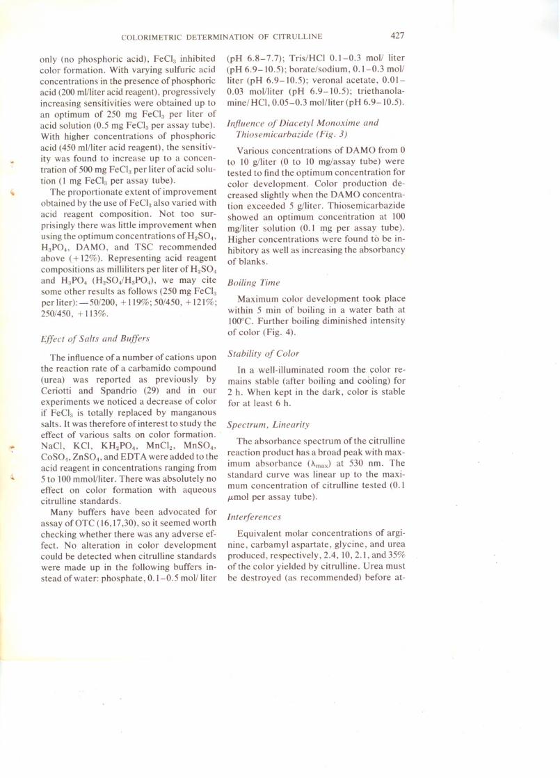
Influence of Diacetyl Monoxime andThiosemicarbazide (Fig. 3)
Various concentrations of DAMO from 0to 10 g/Iiter (0 to 10 mg/assay tube) weretested to find the optimum concentration forcolor development. Color production decreased slightly when the DAMO concentration exceeded 5 g/liter. Thioserp.icarbazideshowed an optimum concentration at 100mg/liter solution (0.1 rpg per assay tube).Higher concentrations were found to be inhibitory as well as increasing the absorbancyof blanks.
Boiling Time
Maximum color development took placewithin 5 min of boiling in a water bath at100°C. Further boiling diminished intensityof color (Fig. 4).
Stability of Color
In a well-illuminated room the. ~olor remains stable (after boiling and cooling) for2 h. When kept in the dark, color is stablefor at least 6 h.
Spectrum, Linearity
The absorbance spectrum of the citrullinereaction product has a broad peak with maximum absorbance (AmaJ at 530 nm. Thestandard curve was linear up to the maximum concentration of citrulline tested (0.1/Lmol per assay tube).
Interferences
Equivalent molar concentrations of arginine, carbamyl aspartate, glycine, and ureaproduced, respectively, 2.4, 10, 2.1, and 35%of the color yielded by citrulline. Urea mustbe destroyed (as recommended) before at-
428 BOYDE AND RAHMA TULLAH
-5
III
0I:IDJ:Il-DIIIJ:I
01TsC 9 /I
Jet ·3·6·91·2
0
510o A MO
9 /I
1
FIG. 3. Studies of sensitivity of citrulline measurement as a function of different concentrations of
diacetyl monoxime and thiosemicarbazide. Citrulline solution (0.1 ml, I mmol/liter) was boiled for 5min with 2 ml of acid-ferric solution (250 ml H2S04, 200 ml H3P04, 250 mg FeCI3 per liter) and I ml
of a solution containing either (a) 100 mg TSC per liter with appropriate amounts of DAMO (e), or (b)
5 g DAMO per liter with appropriate amounts of TSC (D).
tempting the assay of citrulline. The otherinterferences should not often prove serious.
Deproteinization by the Folin- Wu, Nel-
son-Somogyi, and CdS04-Ba(OH)2 procedures did not affect color development. TCAup to 250 g/liter in an aqueous citrulline solu-
1
III
0·5
I: IIIJ:Il-DInJ:Iet 00
5 10 15
Time [mins)
FIG. 4. Effect of boiling time of color development. Citrulline solution (0.1 ml, 1 mmol/liter) was
boiled for various time periods with the recommended (optimum) chromogenic reagent.
COLORIMETRIC DETERMINATION OF CITRULLINE429
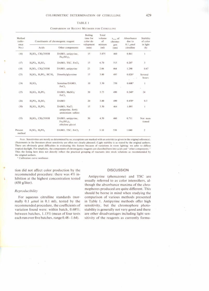
TABLE 1COMPARISON OF RECENT METHODS FOR CITRULLINE
Boiling
TotalMethod
time forvolume"max ofAbsorbanceStability(refer-
Constituents of chromogenic reagentcolor de-ofchromo-due toof colorence
Note. Sensitivities are mostly as determined by us; exceptions are marked with an asterisk (as given in the original reference).(Statements in the literature about sensitivity are often not clearly phrased.) Light stability is as stated by the original authors.There are obviously great difficulties in evaluating this feature because of variations in room lighting: we refer to diffusetropical daylight. For simplicity, the components of chromogenic reagents are classified here into acids and" other components. "Thus the listing here does not directly reflect the practical grouping of reactants into stock solutions as recommended bythe original authors.
" Calibration curve nonlinear.
tion did not affect color production by therecommended procedure: there was 4% inhibition at the highest concentration tested(450 g/liter).
Reproducibility
For aqueous citrulline standards (normally 0.1 M-molin 0.1 ml), tested by therecommended procedure, the coefficients ofvariation found were: within batch, 0.68%;between batches, 1.13% (mean of four tests·each run over five batches, range 0.48-1.64).
DISCUSSION
Antipyrine (phenazone) and TSC areusually referred to as color intensifiers, although the absorbance maxima of the chromophores produced are quite different. Thisshould be borne in mind when studying thecomparison of various methods presentedin Table 1. Antipyrine methods offer highsensitivity, but the chromophore photostability is generally not very good and thereare other disadvantages including light sensitivity of the reagents as currently formu-
430 BOYDE AND RAHMA TULLAH
lated. When comparing sensitivities, itshould also be borne in mind that the recommended procedure above is not the mostsensitive we have studied, being preferredfor other reasons.
The chromogenic reagent for citrullinemeasurement in the present method differsfrom that which we have found optimal fordetermining citrulline in the presence ofserum proteins (manuscript in preparation).The presence of proteins causes the formation of brown pigments, possibly as a resultof acid degradation and consequent liberation of tryptophan. On boiling in acid, carbohydrates present in serum would be converted to furfuraldehyde which might thengive an aldehyde color reaction with tryptophan (Hopkin-Cole reaction) (31). Withhigher concentrations of acids in the reagent,proteins tend to precipitate; also FeCl3 cannot be used because in its presence absorbances due to citrulline and to the protein blank are not additive. In the procedurewithout deproteinization, we therefore deliberately use low concentrations of acidsand high concentrations of DAM0 and TSC,but omit FeCI3• This largely eliminates protein interference, at the cost of halving thesensitivity. A similar nondeproteinizing procedure (with more H2S04) is very sensitivefor the estimation of urea (32), whereas under the present conditions urea yields onlyabout one-third the absorbance of citrulline.
The nature of the complex formed bycitrulline and other carbamido compoundswith diacetyl monoxime or diacetyl is apparently still unknown and a matter of controversy. Beale and Croft (28) postulatedformation of triazines, while other authorshave advocated involvement of glycolurilsin the overall reaction scheme (34).
Besides OTC, citrulline is a product ofarginine deiminase (EC 3.5.3.6) (and theabove method could probably be used in assays of this enzyme) which, however, playsno part in mammalian metabolism. In mammals, apart from liver, OTC is also presentin lesser amounts in other tissues like in-
testine and kidney (35-37), and it is in studyof such tissues, and of conditions where tissue levels are much decreased, that the mostsensitive possible method, free of interferences, may prove valuable. For example,elevated serum levels have been reportedin intestinal disorders (38) and yet there aresharply conflicting reports even on normaltissue levels (35-37).
ACKNOWLEDGMENTS
M.R. thanks the Hong Kong Government for theaward of a Commonwealth Scholarship.
REFERENCES
1. Krebs, H. A., Eggleston, L. V., and Knivett, V. A.(1955) Biochem. J. 59, 185-193.
2. Reichard, H. (1960) J. Lab. Clin. Med. 56,218221.