Single sub-fs soft-X-ray pulses: generation and measurement with the atomic transient recorder R. KIENBERGERy, M. UIBERACKERy, E. GOULIELMAKISy, A. BALTUSKAy, M. DRESCHERz and F. KRAUSZy} yInstitut fu ¨ r Photonik, Technische Universita ¨ t Wien, Gusshausstr. 27, A-1040 Wien, Austria zFakulta ¨ t fu ¨ r Physik, Universita ¨ t Bielefeld, D-33615 Bielefeld, Germany }Max-Planck-Institut fu ¨ r Quantenoptik, Hans-Kopfermann-Straße 1, D-85748 Garching, Germany (Received 5 May 2004; revision received 1 Sepember 2004) Abstract. The change from a zero transition to the maximum amplitude ofthe electric fi eld of visible light lasts shorter than one femtosecond (1fs ¼ 10 À15 s). By precisely controlling the hyperfast electric field oscillations in a short laser pulse we developed a measuring apparatus—the atomic transient recorder—like an ultrafast stopwatch. This apparatus is capable of measuring the duration of atomic processes with an accuracy of less than 100as (1as ¼ 10 À18 s), which is the typical duration of electronic processes (transients) deep inside atoms. A 250as X- ray pulse initiates the atomic process to be measured and the attosecond stopwatch at the same time. For the first time it is now possible with this new measuring method to observe ultrafast processes in the electron shell of atoms. 1. Introducti on Efforts to access ever shorter time scales are motivated by the endea vour to expl ore the micr ocos m in ever sma ller dimensions. Recent ly, femtosecond laser techniques ha ve allowed control and tracing of molecular dynamics and the related motion of atoms on the length scale of internuclear separations without the need for resolving the objects of scrutiny in space [1]. Here we demonstrate that laser light consisting of a few, well-controlled field oscillations [2] extends these capabilities—again without resolvin g particles in space—to the interior of atoms, allowing control and measurement of electronic motion on an atomic scale of time. Meas ur ement of ever shorter intervals of ti me and tracing of dynamics wit hin the se int ervals rel y on re produci ble generation of ever brief er events and on probing techniques of corresponding resol uti on. The brief est events prod uced until rec ent ly have been puls es of visible laser light, with durations of around 5 fs (1 fs ¼ 10 À15 s) [3, 4]. Traditionally, the fastest measurement tech- niques have used the envelope of these laser pulses for sampling [5]. Recently, sub-femtosecond bunc hing of femtosecond (>10 fs) extreme ultrav iolet (XUV) li gh t w as obs erve d in two-col o ur [6, 7] and t wo - pho ton [8] i oni zat io n journal of modern optics, 20 january –15 february 2005 vol. 52, no. 2–3, 261–275 Journal of Modern Optics ISSN 0950–0340 print/ISSN 1362–3044 online # 2005 Taylor & Francis Ltd http://www.tandf.co.uk/journals DOI: 10.1080/09500340412331315114
Transcript
8/3/2019 R. Kienberger et al- Single sub-fs soft-X-ray pulses: generation and measurement with the atomic transient recorder
yInstitut fu ¨ r Photonik, Technische Universita ¨ t Wien,
Gusshausstr. 27, A-1040 Wien, Austria
zFakulta ¨t fu ¨ r Physik, Universita ¨ t Bielefeld,
D-33615 Bielefeld, Germany
}Max-Planck-Institut fu ¨ r Quantenoptik, Hans-Kopfermann-Straße 1,
D-85748 Garching, Germany
(Received 5 May 2004; revision received 1 Sepember 2004 )
Abstract. The change from a zero transition to the maximum amplitude of the electric field of visible light lasts shorter than one femtosecond(1fs ¼ 10À15 s). By precisely controlling the hyperfast electric field oscillationsin a short laser pulse we developed a measuring apparatus—the atomic transientrecorder—like an ultrafast stopwatch. This apparatus is capable of measuringthe duration of atomic processes with an accuracy of less than 100as(1 as ¼ 10À18 s), which is the typical duration of electronic processes (transients)deep inside atoms. A 250 as X-ray pulse initiates the atomic process to bemeasured and the attosecond stopwatch at the same time. For the first time it is
now possible with this new measuring method to observe ultrafast processesin the electron shell of atoms.
1. Introduction
Efforts to access ever shorter time scales are motivated by the endeavour
to explore the microcosm in ever smaller dimensions. Recently, femtosecond
laser techniques have allowed control and tracing of molecular dynamics and
the related motion of atoms on the length scale of internuclear separations without
the need for resolving the objects of scrutiny in space [1]. Here we demonstrate
that laser light consisting of a few, well-controlled field oscillations [2] extends
these capabilities—again without resolving particles in space—to the interior
of atoms, allowing control and measurement of electronic motion on an atomic
scale of time.
Measurement of ever shorter intervals of time and tracing of dynamics
within these intervals rely on reproducible generation of ever briefer events
and on probing techniques of corresponding resolution. The briefest events
produced until recently have been pulses of visible laser light, with durations
of around 5 fs (1 fs ¼ 10À15 s) [3, 4]. Traditionally, the fastest measurement tech-
niques have used the envelope of these laser pulses for sampling [5]. Recently,
sub-femtosecond bunching of femtosecond (>10 fs) extreme ultraviolet (XUV)light was observed in two-colour [6, 7] and two-photon [8] ionization
journal of modern optics, 20 january –15 february 2005
vol. 52, no. 2–3, 261–275
J ournal of Modern Optics ISSN 0950–0340 print/ISSN 1362–3044 online # 2005 Taylor & Francis Ltdhttp://www.tandf.co.uk/journals
DOI: 10.1080/09500340412331315114
8/3/2019 R. Kienberger et al- Single sub-fs soft-X-ray pulses: generation and measurement with the atomic transient recorder
experiments and evidence of sub-femtosecond confinement of XUV emission from
few-cycle-driven (ionizing) atoms was also obtained [9]. However, time-domain
technique has hitherto not been capable of resolving the time structure
of sub-femtosecond transients.
Here we demonstrate an apparatus that allows reconstruction of atomic
processes with a resolution within the Bohr orbit time, which is around 150 as.An accurately controlled few-cycle wave of visible light takes ‘tomographic
images’ of the time–momentum distribution of electrons ejected from atoms
following sudden excitation. From these images the temporal evolution of both
the emission intensity and initial momentum of freed electrons can be retrieved on
a sub-femtosecond time scale. Probing primary (photo-excited or collisionally
excited) and secondary (Auger) electrons yield insight into, respectively, excitation
and subsequent relaxation processes. The transients can be triggered by an
isolated attosecond electron or photon burst synchronized to the probing light
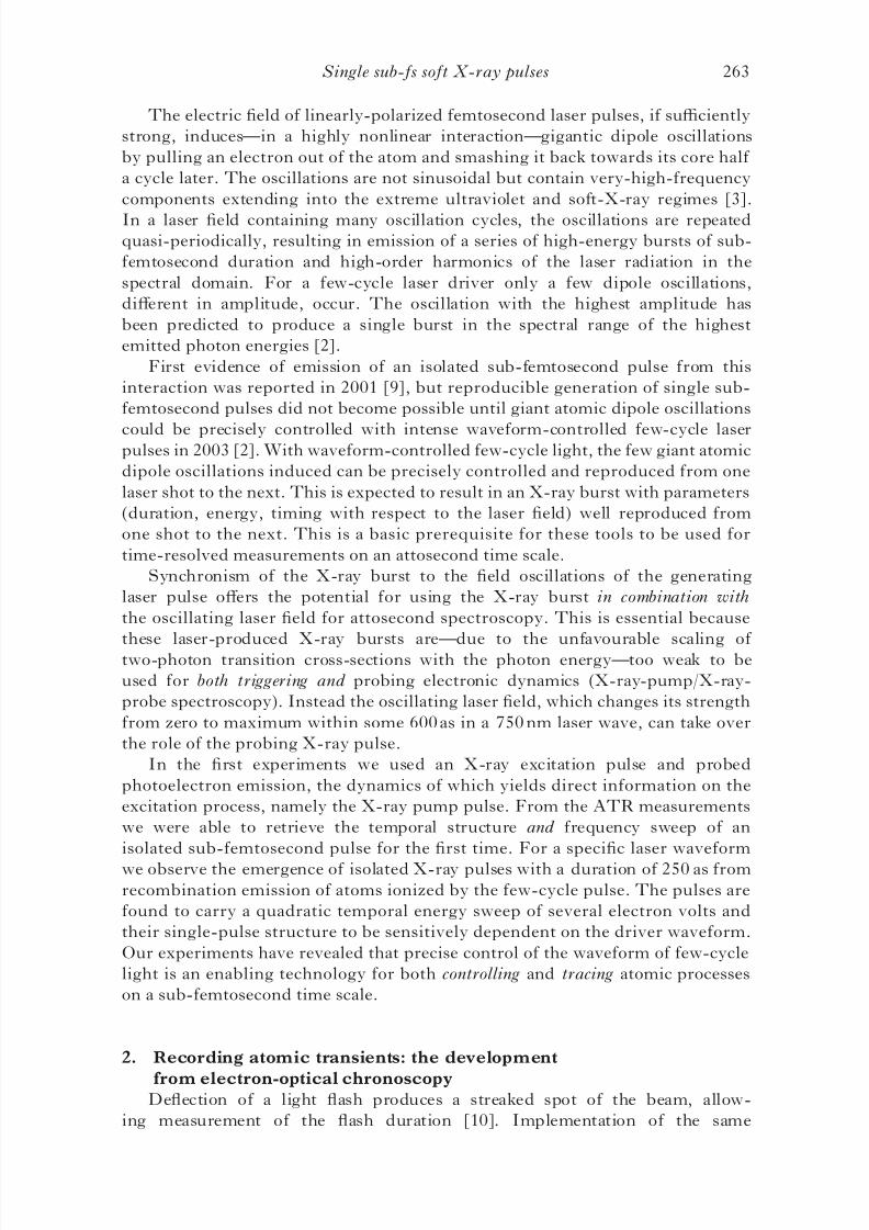
field oscillations. The technique draws on the basic operation principle of a
streak camera [10–14] (figure 1), where a light pulse generates an electron bunchhaving exactly the same temporal structure. Deflection of the electrons in an
electric field allows reconstruction of the duration of the electron bunch.
By measuring the temporal evolution of the emission intensity and momentum
distribution of positive-energy electrons, the atomic transient recorder (ATR) [15]
provides direct temporal insight into the rearrangement of the electronic shell
of excited atoms on a sub-femtosecond scale.
Figure 1. Principle of a conventional streak camera. In its lifetime a light flash ejectselectrons from a metal plate which are then accelerated with a static electric field to afluorescent screen. Before they hit the screen, they are deflected aside with anotherfield increasing linearly with time. The temporally varying deflection ‘streaks’ thepoint of impact of the electrons on the screen. The spatial width of this ‘streak image’(Áx) is directly proportional to the duration of the electron emission, i.e. the durationof the light flash (Át). The faster the deflecting field varies, the shorter are the pulsesthat can be recorded. The most modern streak cameras attain a resolution in theregion of 100 fs.
262 R. Kienberger et al.
8/3/2019 R. Kienberger et al- Single sub-fs soft-X-ray pulses: generation and measurement with the atomic transient recorder
The electric field of linearly-polarized femtosecond laser pulses, if sufficiently
strong, induces—in a highly nonlinear interaction—gigantic dipole oscillations
by pulling an electron out of the atom and smashing it back towards its core half
a cycle later. The oscillations are not sinusoidal but contain very-high-frequency
components extending into the extreme ultraviolet and soft-X-ray regimes [3].
In a laser field containing many oscillation cycles, the oscillations are repeatedquasi-periodically, resulting in emission of a series of high-energy bursts of sub-
femtosecond duration and high-order harmonics of the laser radiation in the
spectral domain. For a few-cycle laser driver only a few dipole oscillations,
different in amplitude, occur. The oscillation with the highest amplitude has
been predicted to produce a single burst in the spectral range of the highest
emitted photon energies [2].
First evidence of emission of an isolated sub-femtosecond pulse from this
interaction was reported in 2001 [9], but reproducible generation of single sub-
femtosecond pulses did not become possible until giant atomic dipole oscillations
could be precisely controlled with intense waveform-controlled few-cycle laserpulses in 2003 [2]. With waveform-controlled few-cycle light, the few giant atomic
dipole oscillations induced can be precisely controlled and reproduced from one
laser shot to the next. This is expected to result in an X-ray burst with parameters
(duration, energy, timing with respect to the laser field) well reproduced from
one shot to the next. This is a basic prerequisite for these tools to be used for
time-resolved measurements on an attosecond time scale.
Synchronism of the X-ray burst to the field oscillations of the generating
laser pulse offers the potential for using the X-ray burst in combination with
the oscillating laser field for attosecond spectroscopy. This is essential because
these laser-produced X-ray bursts are—due to the unfavourable scaling of two-photon transition cross-sections with the photon energy—too weak to be
used for both triggering and probing electronic dynamics (X-ray-pump/X-ray-
probe spectroscopy). Instead the oscillating laser field, which changes its strength
from zero to maximum within some 600 as in a 750 nm laser wave, can take over
the role of the probing X-ray pulse.
In the first experiments we used an X-ray excitation pulse and probed
photoelectron emission, the dynamics of which yields direct information on the
excitation process, namely the X-ray pump pulse. From the ATR measurements
we were able to retrieve the temporal structure and frequency sweep of an
isolated sub-femtosecond pulse for the first time. For a specific laser waveform
we observe the emergence of isolated X-ray pulses with a duration of 250 as from
recombination emission of atoms ionized by the few-cycle pulse. The pulses are
found to carry a quadratic temporal energy sweep of several electron volts and
their single-pulse structure to be sensitively dependent on the driver waveform.
Our experiments have revealed that precise control of the waveform of few-cycle
light is an enabling technology for both controlling and tracing atomic processes
on a sub-femtosecond time scale.
2. Recording atomic transients: the development
from electron-optical chronoscopyDeflection of a light flash produces a streaked spot of the beam, allow-
ing measurement of the flash duration [10]. Implementation of the same
Single sub-fs soft X-ray pulses 263
8/3/2019 R. Kienberger et al- Single sub-fs soft-X-ray pulses: generation and measurement with the atomic transient recorder
concept—mapping the temporal distribution of a bunch of rapid particles to a
spatial distribution by affecting their motion in a time-dependent manner—with
photoelectrons allowed electron-optical chronoscopy by image-tube streak
cameras [11, 12]. From the streaked image of the photoelectron bunch it
is possible to infer with sub-picosecond resolution the temporal structure of
the light pulse triggering the photoemission. The time resolution of thesedevices is ultimately limited by the spread of the electron transit time due to a
spread of their initial momentum. In this work we report temporal characterization
of sub-femtosecond electron emission from atoms by drawing on the same basic
concept.
The three orders of magnitude improvement in time resolution results from
several essential modifications of image-tube streak cameras: (i) the electric field of
light is used for affecting the electrons’ motion, the field (ii) is virtually jitter-free,
(iii) applied along the direction of electron motion—implying their acceleration
or deceleration instead of their deflection—and (iv) directly at the location
and instant of emission. Whereas (i) implies ‘only’ a dramatically-enhancedstreaking speed, the consequences of (ii)–(iv) are much more profound: (ii) allows
the timing of the probing field to be systematically varied with an accuracy
within the electron bunch length and, owing to (iii), this capability results in
‘projecting’ the initial time–momentum distribution of the electron emission into a
series of different final (measurable) momentum distributions, while (iv) prevents
the initial momentum spread from introducing any measurement error. As a result
of (ii)–(iv), not only does the spread of initial electron momenta stop imposing
a limitation on the time resolution, but also its possible temporal variation during
the emission can be captured just as that of the emission intensity.
Inspired by the physics of the first sub-femtosecond experiment [9], Corkumand co-workers [13] put forward the basic concept for ATR metrology and
analysed with a comprehensive quantum theory by Brabec and co-workers [14].
Let us consider electron emission from atoms exposed to a sub-fs X-ray
burst in the presence of an intense, linearly-polarized, few-cycle laser field
E LðtÞ ¼ E 0ðtÞ cos ð!Lt þ ’Þ. The momentum of the freed electrons is changed
by Á p ¼ eALðtÞ along the laser field vector (figure 2). Here ALðtrÞ ¼Ð 1
trE LðtÞdt
is the vector potential of the laser field, e and m stand for the charge and rest
mass of the electron, respectively, and tr is the release time of the electron. This
momentum transfer (grey arrows in figure 3) maps the temporal emission profile
into a similar distribution of final momenta pf ¼ pi þÁ p within a time window of
T 0=2 ¼ =!L, if the electrons’ initial momentum pi is constant in time and their
emission terminates within T 0=2. Under these conditions the temporal evolution
of the electron emission can be unambiguously retrieved from a single ‘streaked’
momentum distribution. However, any sweep of the electrons’ initial momentum
revokes the unique correspondence between the electron’s final momentum and
release time, preventing retrieval of accurate temporal information from single
streak records (violet and orange profiles in figure 3).
In general, the initial momentum spectrum of electrons detached from
atoms by impulsive excitation varies in time during emission. A representative
time–momentum distribution neð p, tÞ of the electron emission rate is depicted in
figure 4. The final electron momentum spectrum (observed after the laser pulse hasleft the interaction region), ð pÞ ¼
Ð 1À1
neð p, tÞdt, can be viewed as the projection
of the time-dependent momentum distribution on the momentum space along
264 R. Kienberger et al.
8/3/2019 R. Kienberger et al- Single sub-fs soft-X-ray pulses: generation and measurement with the atomic transient recorder
the lines of constant p. In the classical description of the freed electrons’ motion in
the strong laser field, the final spectra obtained in the presence of a strong laserfield are generalized projections along the lines where pf ¼ pi þ eALðtÞ is constant
(see figure 4). By delaying the laser field with respect to the the excitation that
triggers electron emission, we obtain a set of tomographic records (briefly: streaked
spectra),
Að pÞ ¼
ð 1À1
neð p À eALðtÞ, tÞdt , ð1Þ
from which, with a suitable set of ALðt ÀÁtnÞ, the complete distribution neð p, tÞ
can be reconstructed. The maximum energetic shift of the photoelectron spectrareveals not only the position of the maxima (and other extreme values) of the
vector potential AL but also their magnitudes.
The method is closely related to frequency-resolved optical gating [15–19] with
the oscillating field as gate and other concepts of tomography for ultrashort pulse
measurements [20–22]. In the simplest cases two streaked spectra (in addition
to the field-free spectrum) may be sufficient. In the absence of a nonlinear
momentum sweep the streak records obtained near the zero transitions of ALðtÞ
with opposite slopes (violet profiles in figure 3) together with the field-free
spectrum (black profile) allow determination of all relevant characteristics: the
temporal profile, duration and momentum chirp of emission. Note that a linearmomentum sweep leads to asymmetric broadening of the streaked spectra at
these delays and it is this asymmetry that makes the measurement highly sensitive
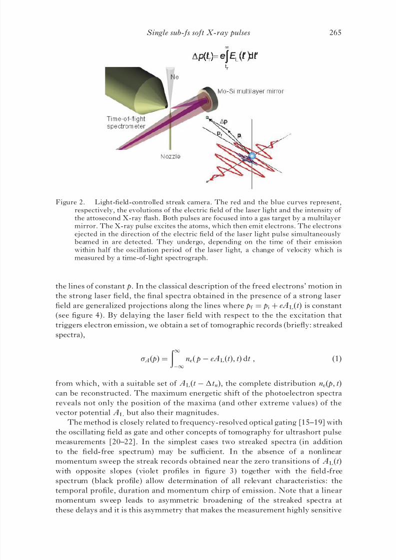
Figure 2. Light-field-controlled streak camera. The red and the blue curves represent,respectively, the evolutions of the electric field of the laser light and the intensity of the attosecond X-ray flash. Both pulses are focused into a gas target by a multilayermirror. The X-ray pulse excites the atoms, which then emit electrons. The electronsejected in the direction of the electric field of the laser light pulse simultaneouslybeamed in are detected. They undergo, depending on the time of their emissionwithin half the oscillation period of the laser light, a change of velocity which ismeasured by a time-of-light spectrograph.
Single sub-fs soft X-ray pulses 265
8/3/2019 R. Kienberger et al- Single sub-fs soft-X-ray pulses: generation and measurement with the atomic transient recorder
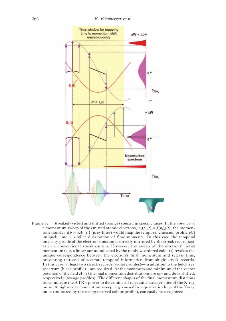
Figure 3. Streaked (violet) and shifted (orange) spectra in specific cases. In the absence of a momentum sweep of the emitted atomic electrons, neð pi, tÞ ¼ f ð piÞ g ðtÞ, the momen-tum transfer Á p ¼ eALðtrÞ (grey lines) would map the temporal emission profile g(t)uniquely into a similar distribution of final momenta. In this case the temporalintensity profile of the electron emission is directly mirrored by the streak record justas in a conventional streak camera. However, any sweep of the electrons’ initialmomentum (e.g. a linear one as indicated by the rainbow-ordered colours) revokes theunique correspondence between the electron’s final momentum and release time,preventing retrieval of accurate temporal information from single streak records.In this case, at least two streak records (violet profiles)—in addition to the field-freespectrum (black profile)—are required. At the maximum and minimum of the vectorpotential of the field ALðtÞ the final momentum distributions are up- and downshifted,respectively (orange profiles). The different shapes of the final momentum distribu-
tions indicate the ATR’s power to determine all relevant characteristics of the X-raypulse. A high-order momentum sweep, e.g. caused by a quadratic chirp of the X-raypulse (indicated by the red–green–red colour profile), can easily be recognized.
266 R. Kienberger et al.
8/3/2019 R. Kienberger et al- Single sub-fs soft-X-ray pulses: generation and measurement with the atomic transient recorder
to the momentum sweep, i.e. highly sensitive to deviations of the pulse duration
from the Fourier limit.
The ATR’s potential for determining a high-order chirp of the X-ray pulse,
too, is indicated by the different shapes of the orange momentum distributions
in figure 3.
3. Experimental set-up
Using the experimental set-up described in [9] and recently employed for
probing Auger electrons on a few-femtosecond time scale [23], we sample electron
emission from atoms. The essential innovation here is that waveform-controlled
few-cycle light now provides a reproducible excitation burst for accurate triggering
and a reproducible streaking field for capturing sub-fs electron emission fromatoms. For excitation, X-ray bursts are produced from Ne atoms ionized by
intense, few-cycle waveform-controlled light pulses [2] in a process giving rise
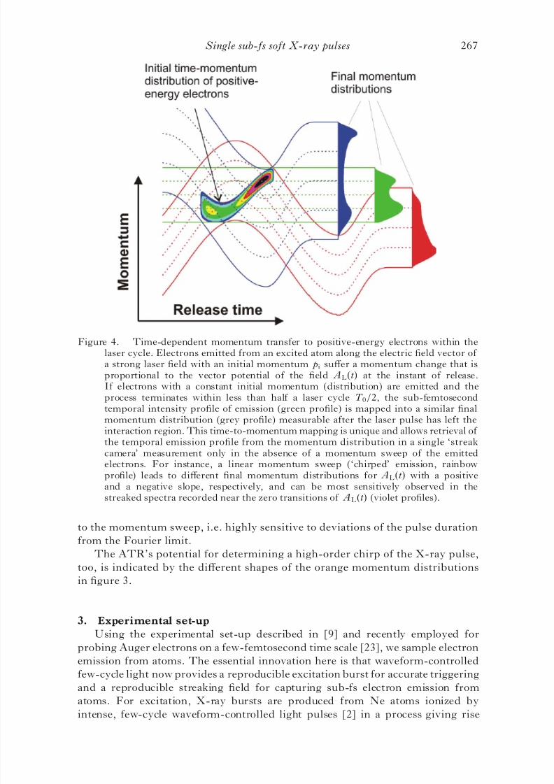
Figure 4. Time-dependent momentum transfer to positive-energy electrons within thelaser cycle. Electrons emitted from an excited atom along the electric field vector of a strong laser field with an initial momentum pi suffer a momentum change that isproportional to the vector potential of the field ALðtÞ at the instant of release.If electrons with a constant initial momentum (distribution) are emitted and theprocess terminates within less than half a laser cycle T 0=2, the sub-femtosecond
temporal intensity profile of emission (green profile) is mapped into a similar finalmomentum distribution (grey profile) measurable after the laser pulse has left theinteraction region. This time-to-momentum mapping is unique and allows retrieval of the temporal emission profile from the momentum distribution in a single ‘streakcamera’ measurement only in the absence of a momentum sweep of the emittedelectrons. For instance, a linear momentum sweep (‘chirped’ emission, rainbowprofile) leads to different final momentum distributions for ALðtÞ with a positiveand a negative slope, respectively, and can be most sensitively observed in thestreaked spectra recorded near the zero transitions of ALðtÞ (violet profiles).
Single sub-fs soft X-ray pulses 267
8/3/2019 R. Kienberger et al- Single sub-fs soft-X-ray pulses: generation and measurement with the atomic transient recorder
to high-order harmonics of the incident light for periodic (multi-cycle) pumping
[24, 25]. The X-ray pulses exciting the neon harmonic source co-propagate with
the laser pulses down the beam delivery tube (figure 5). After 150 cm they hit
a 200 nm thick zirconium foil with an aperture of 2 mm mounted on a nitrocellu-
lose membrane 5m thick. This virtually dispersionless filter produces an annular
laser beam with the X-ray beam in the centre. The energy in the laser beam
is adjusted by a motorized iris and measured by a photodiode. The collinear X-ray
and laser beams are focused into a neon gas jet and delayed with respect to
each other for the ATR measurements with a two-component Mo/Si broadband
multilayer mirror (radius of curvature ¼ À70 mm) placed 2.5 m downstream
from the source. This mirror is mounted on a motorized stage so that it can be
removed from the beam line. In this way the harmonic beam can be detected by an
X-ray charge-coupled device (CCD) camera for optimizing the radial intensity
profile of the X-ray beam (by fine adjustment of the position and pressure of
the neon gas target and of the intensity of the laser pulses). Further on, a
transmission grating having 10 000 lines mmÀ1 can be inserted before the X-ray
CCD camera to observe the spectrum of the harmonic radiation. Fine tuningof the cut-off of the X-rays can be carried out by fine tuning of the energy of
the fundamental laser beam. The reflectivity band of the multilayer extends from
Figure 5. The schematic of the experiment. The focused 5 fs laser beam interacts withneon atoms to produce high-harmonic radiation. The laser and the highly collimatedX-ray beam co-propagate collinearly through a 2 m beam line towards the measure-ment. Differential pumping stages reduce the pressure from $ 4 Â 10À2 mbar in thesource chamber to the range of $ 10À5 mbar in the experimental chamber.An ionization detector in the higher-pressure part of the beam line serves formonitoring the (spectrally integrated) high harmonic flux. In the beam line the laserand harmonic beams pass through a 200 nm thick, 3 mm diameter zirconium foilplaced on a 5mm thick nitrocellulose pellicle to cover a hole 2 mm in diameter.The energy transported by the resulting annular beam can be adjusted with amotorized iris between a fraction of a microjoule and a few tens of microjoules.The Mo/Si multilayer consists of an annular part having an outer diameter of 10 mm
with a concentric hole of 3 mm diameter hosting a miniature mirror of slightly smallerdiameter. Both parts originate from the same substrate, ensuring identical radii of curvature (R ¼ 70 mm). The miniature central mirror is mounted on a quadrant piezostage, allowing alignment and translation with respect to the external part.
268 R. Kienberger et al.
8/3/2019 R. Kienberger et al- Single sub-fs soft-X-ray pulses: generation and measurement with the atomic transient recorder
85 to 100 eV with a peak reflectance of $30% and a full width at half maximum
(FWHM) of $9eV.
Two types of experiments based on the ATR concept were implemented.
First, we used the internal part of the Mo/Si mirror to focus both the X-ray and
the laser beam to eliminate any external source of fluctuations in the timing
between the excitation and probing pulses. In the second type of studies, theX-ray and the laser beam were reflected by different sections of the focusing
mirror: the central piece sits on a piezo stage adjustable in the transverse
and longitudinal directions. In this manner the two pulses can be overlapped
spatially and temporally in the focal plane, exactly where the tip of a nozzle
emitting the atoms to be photoionized is placed. Electrons ejected following the
X-ray excitation are collected within a narrow cone (<4) aligned parallel to
the laser and X-ray polarization and analysed with a time-of-flight spectrometer.
In these investigations the probing laser field could be delayed with respect to
the X-ray excitation pulse by translation of the internal part of the mirror on a
nanometre scale.The profile of the laser focus is imaged by a lens on a CCD camera for
monitoring and course pre-adjustment of the spatial overlap between the beams
reflected by the two components of the Mo/Si mirror.
4. Generation of an isolated single sub-fs X-ray excitation pulse
Figure 6 summarizes representative results of a series of measurements of
streaked X-ray photoelectron spectra recorded with the X-ray and laser pulse
impinging with a fixed relative timing set in the X-ray generation process.
According to an intuitive one-active-electron model of Schafer et al . [26, 27],Corkum [28] and Lewenstein et al . [29] ionization of atoms by a linearly polarized
laser field is accompanied by emission of extreme ultraviolet radiation due to
recombination of the detached electron into its original ground state upon
recollision with the parent ion. These theories along with a number of numerical
simulations [29–34] predict emission of the highest-energy (cut-off) photons to
occur near the zero transition(s) of the electric field following the most intense
half cycle(s) at the pulse peak in a few-cycle driver.
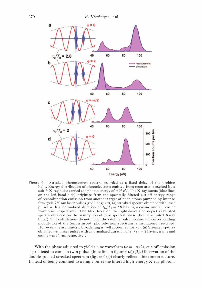
In a cosine waveform (’ ¼ 0) cut-off emission is expected to be confined to
a single bunch emitted at the zero transition of E LðtÞ following the pulse peak.
The photoelectrons ejected in the direction of the peak electric field at this
instant suffer maximum increase of their momentum and hence of their energy.
The streaked spectrum in figure 6 (d ), obtained with 5 fs, 750 nm cosine pulses
used for both X-ray generation and subsequent electron streaking, corroborates
this prediction. The photoelectron spectrum peaking at hh!x-rayÀW b % 72 eV
(where hh!x-ray% 93:5 eV is the centre of the X-ray spectrum selected by the Mo/
Si mirror and W b ¼ 21:5 eV is the binding energy of the most weakly bound
valence electrons in Ne) in the absence of the laser field is upshifted by several
10 eV with only a few electrons scattered outside the shifted band. The clear
upshift is consistent with the X-ray burst coinciding with the zero transition of the
laser electric field (see figure 6 (d )). Possible satellites would appear at the adjacent
zero transitions of E LðtÞ and suffer an energy downshift. The absence of a downshifted spectral peak of substantial intensity indicates clean single sub-fs
pulse generation.
Single sub-fs soft X-ray pulses 269
8/3/2019 R. Kienberger et al- Single sub-fs soft-X-ray pulses: generation and measurement with the atomic transient recorder
With the phase adjusted to yield a sine waveform (’ ¼ À=2), cut-off emission
is predicted to come in twin pulses (blue line in figure 6 (c)) [2]. Observation of thedouble-peaked streaked spectrum (figure 6 (c)) clearly reflects this time structure.
Instead of being confined to a single burst the filtered high-energy X-ray photons
Figure 6. Streaked photoelectron spectra recorded at a fixed delay of the probinglight. Energy distribution of photoelectrons emitted from neon atoms excited by asub-fs X-ray pulse carried at a photon energy of %93 eV. The X-ray bursts (blue lineson the left-hand side) originate from the spectrally filtered cut-off energy rangeof recombination emission from another target of neon atoms pumped by intensefew-cycle 750 nm laser pulses (red lines); (a), (b) streaked spectra obtained with laserpulses with a normalized duration of L=T 0 ¼ 2:8 having a cosine and a Àcosinewaveform, respectively. The blue lines on the right-hand side depict calculatedspectra obtained on the assumption of zero spectral phase (Fourier-limited X-rayburst). The calculations do not model the satellite pulse because the correspondingmodulation of the (unperturbed) photoelectron spectrum is insufficiently resolved.However, the asymmetric broadening is well accounted for. (c), (d ) Streaked spectraobtained with laser pulses with a normalized duration of L=T 0 ¼ 2 having a sine andcosine waveform, respectively.
270 R. Kienberger et al.
8/3/2019 R. Kienberger et al- Single sub-fs soft-X-ray pulses: generation and measurement with the atomic transient recorder
are now distributed in two bursts, each of which is less than half as intense
as the isolated burst produced by the cosine waveform (figure 6 (d )). These
measurements demonstrate how control of the waveform of few-cycle laser light
allows one to control the temporal profile of X-ray emission within the half wave
cycle, i.e. on a sub-femtosecond time scale.
For generation of a single sub-fs X-ray burst a crucial role is played by thenormalized pulse duration L=T 0 other than the carrier-envelope phase ’. This is
demonstrated by repeating the measurements with, slightly longer, 7 fs laser
pulses ( L=T 0 ¼ 2:8). The corresponding streaked spectrum obtained for ’ ¼ 0
(figure 6 (a)) exhibits a sizeable downshifted spectral feature in addition to the
upshifted main peak, indicating the appearance of a substantial satellite pulse
(%30% of the principle burst) in the cosine-wave-driven recombination emission.
A reduced difference in intensity of the adjacent half cycles of the few-cycle
wave implies that the highest-energy spectral components of adjacent bursts
reach the energy band selected by our 15%-bandwidth bandpass filter. Shifting
’ by results in the same emission but the streaking is reversed (figure 6 (b)).In these measurements (figure 6 (a) and (b)) we increased the strength of the
streaking laser field by a factor of approximately 2. The increased streaking reveals
pronounced asymmetry of the up- and downshifted spectra, which is consistent
with a near-quadratic temporal energy sweep of the X-ray burst. This results from
the asymmetric spectral intensity profile filtered by the Mo/Si mirror. The (near)
quadratic momentum sweep of the electron emission yields asymmetric streaking
at the adjacent maxima of ALðtÞ in a way similar to that of a linear chirp at
subsequent zero transitions of ALðtÞ.
5. Measurement of the time–momentum distribution
of atomic electron emission
With isolated sub-fs X-ray pulses at our disposal atomic transients can now be
triggered and their subsequent evolution be captured by probing electron emission
with a synchronized wave of laser light. In the first ATR measurements presented
here, the objects of scrutiny were photoelectrons. The streaking field in these
experiments was produced by blocking the internal part of the laser beam with a
zirconium filter (transmitting the X-ray pulse) and focusing the transmitted
annular beam on the target with the external section of the Mo/Si mirror [9],
which can be delayed with respect to the internal section that focuses the X-ray
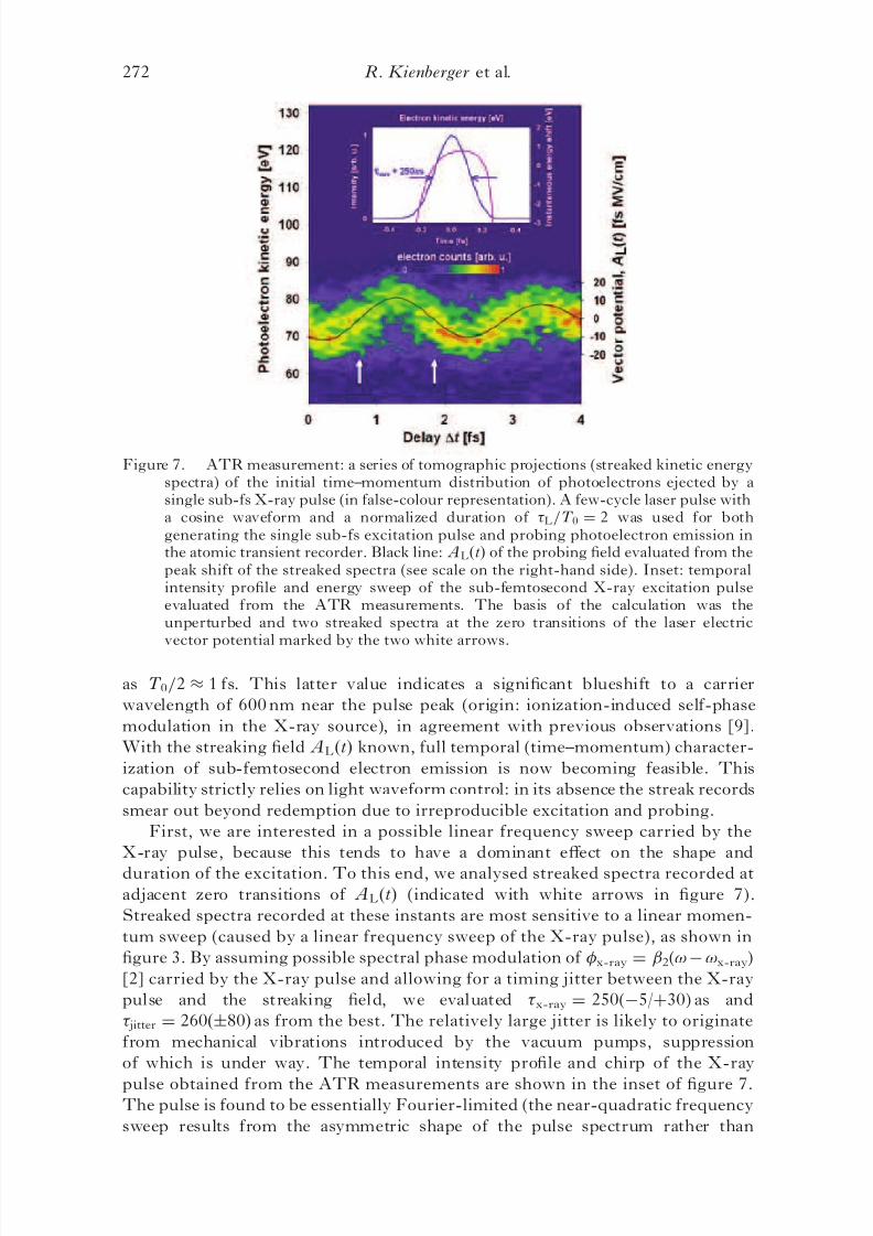
beam. Figure 7 shows a series of streaked spectra of photoelectrons emitted from
neon as a function of Át. The excitation pulse at 93 eV was produced by a cosine
waveform with L=T 0 ¼ 2.
If the electrons are emitted with an initial kinetic energy much larger than their
average quiver energy in the laser field and temporally confined to a fraction of the
half oscillation cycle, theory predicts that their energy shift is linearly proportional
to Á p and hence to the vector potential at the instant of release of the wavepacket,
ÁW ðtrÞ % ð pi=mÞÁ pðtrÞ ¼ ðepi=mÞ AðtrÞ, where pi is the initial momentum of the
electron. As a consequence, ALðtÞ and hence E LðtÞ can be accurately determined
from the peak shifts of the spectra—without having to analyse their detailed
structure. The result is shown by the black line in figure 7, constituting the firstdirect (time-resolved) measurement of a visible light field. From this measurement
we can also evaluate the half oscillation period of the electromagnetic field
Single sub-fs soft X-ray pulses 271
8/3/2019 R. Kienberger et al- Single sub-fs soft-X-ray pulses: generation and measurement with the atomic transient recorder
as T 0=2 % 1 fs. This latter value indicates a significant blueshift to a carrier
wavelength of 600 nm near the pulse peak (origin: ionization-induced self-phase
modulation in the X-ray source), in agreement with previous observations [9].
With the streaking field ALðtÞ known, full temporal (time–momentum) character-
ization of sub-femtosecond electron emission is now becoming feasible. This
capability strictly relies on light waveform control: in its absence the streak records
smear out beyond redemption due to irreproducible excitation and probing.
First, we are interested in a possible linear frequency sweep carried by the
X-ray pulse, because this tends to have a dominant effect on the shape and
duration of the excitation. To this end, we analysed streaked spectra recorded at
adjacent zero transitions of ALðtÞ (indicated with white arrows in figure 7).
Streaked spectra recorded at these instants are most sensitive to a linear momen-
tum sweep (caused by a linear frequency sweep of the X-ray pulse), as shown in
figure 3. By assuming possible spectral phase modulation of x-ray ¼ 2(!À!x-ray)
[2] carried by the X-ray pulse and allowing for a timing jitter between the X-ray
pulse and the streaking field, we evaluated x-ray ¼ 250(À5/þ30) as and
jitter ¼ 260ðÆ80Þ as from the best. The relatively large jitter is likely to originate
from mechanical vibrations introduced by the vacuum pumps, suppression
of which is under way. The temporal intensity profile and chirp of the X-ray
pulse obtained from the ATR measurements are shown in the inset of figure 7.The pulse is found to be essentially Fourier-limited (the near-quadratic frequency
sweep results from the asymmetric shape of the pulse spectrum rather than
Figure 7. ATR measurement: a series of tomographic projections (streaked kinetic energyspectra) of the initial time–momentum distribution of photoelectrons ejected by asingle sub-fs X-ray pulse (in false-colour representation). A few-cycle laser pulse witha cosine waveform and a normalized duration of L=T 0 ¼ 2 was used for bothgenerating the single sub-fs excitation pulse and probing photoelectron emission inthe atomic transient recorder. Black line: ALðtÞ of the probing field evaluated from thepeak shift of the streaked spectra (see scale on the right-hand side). Inset: temporalintensity profile and energy sweep of the sub-femtosecond X-ray excitation pulseevaluated from the ATR measurements. The basis of the calculation was theunperturbed and two streaked spectra at the zero transitions of the laser electricvector potential marked by the two white arrows.
272 R. Kienberger et al.
8/3/2019 R. Kienberger et al- Single sub-fs soft-X-ray pulses: generation and measurement with the atomic transient recorder
from a spectral phase). The remarkable accuracy of x-ray relies on using several
(in this case 3) tomographic projections of the time–momentum distribution of
photoelectrons for the X-ray pulse retrieval, which is the essence of the ATR
concept. Restricting our analysis to only one of the two streaked spectra—in the
spirit of streak camera measurements—would only allow setting an upper limit of
500 as on the X-ray pulse duration. Extension of this analysis to a series of spectrameasured within a delay range of T 0 revealed the absence of higher-order
contributions to x-ray as well. These measurements provide evidence of the
emergence of isolated, bandwidth-limited X-ray bursts over a relative spectral
band as broad as 15% (15 eV at 100 eV) from recombination emission driven by
a cosine waveform with L=T 0 ¼ 2. This has important implications for scaling
the technology.
6. Resolving atomic transients within the Bohr orbit time
Quantum mechanical uncertainty, which dictates that any short time structurecomes with a broad energy spectrum, limits the briefest time interval within which
two different atomic events can be recognized as different by our apparatus. To be
able to resolve the spectral ‘images’ of two events separated by t in time they must
be shifted with respect to each other in the streak record by at least as much as
their own spectral width W % hh=t. From this requirement we obtain the ATR
resolving power as
t ¼T 0
2
hh!L
ÁW max
1=2
, ð2Þ
where ÁW max stands for the energy shift suffered by the electron ejected at the
peak of ALðtÞ. Under our current experimental conditions ÁW max can exceed
20eV (see figures 6 (a) a n d (b)) before the onset of laser-induced ionization,
yielding (for T 0 ¼ 2fs) t % 100 as. The atomic transient recorder driven by a
600 nm wavelength light field and probing electrons with a kinetic energy near
100 eV is able to distinguish two ultrafast atomic events following each other
within 100 as, constituting the shortest interval of time directly measurable to date.
Our experiments indicate that there is room for further improvement.
The absence of spectral phase modulation in the cut-off energy range of few-cycle
driven recombination emission over an energy band as broad as 15% and the
confinement of this emission to a single burst afford promise of the generation of
single 50 as pulses from the same source upscaled to photon energies of 500 eV
[35]. At these excitation energies ÁW max can be enhanced by at least an order of
magnitude, yielding t % 30 as. Extension of the experiments presented to explor-
ing of the sub-femtosecond temporal variation of the emission intensity and
momentum distribution of primary (photo) and secondary (Auger) electron emis-
sion simultaneously will provide unprecedented insight into the excitation and
relaxation dynamics of the electronic shell of atoms and molecules. The currently
used time window of T 0=2 % 1 fs can be extended to several 10 fs by difference
frequency generation with the few-cycle laser pulse whilst keeping the resolution
of the ATR in the sub-fs regime. At near-keV excitation energies, the atomictransient recorder presented here will allow time-domain metrology of atomic
dynamics with a resolution approaching the atomic unit of time (24 as).
Single sub-fs soft X-ray pulses 273
8/3/2019 R. Kienberger et al- Single sub-fs soft-X-ray pulses: generation and measurement with the atomic transient recorder
We gratefully acknowledge the invaluable contributions of V. Yakovlev,
F. Bammer, A. Scrinzi, Th. Westerwalbesloh, U. Kleineberg and U. Heinzmann
to the experiments reviewed in this paper. Sponsored by Fonds zur Fo ¨ rderung der
wissenschaftlichen Forschung (Austria, grants Z63 and F016), Deutsche
Forschungsgemeinschaft (Germany, grants SPP1053, HE1049/9, and KL1077/1)and the European Union’s Human Potential Programme under contract HPRN-
2000-00133 (Atto). R. Kienberger is funded by an APART fellowship of the
Austrian Academy of Sciences.
References[1] ZEWAIL, A., 2000, J . phys. Chem. A, 104, 5660.[2] BALTUSKA, A., UDEM, Th., UIBERACKER, M., HENTSCHEL, M., GOULIELMAKIS, E.,
GOHLE, CH., HOLZWARTH, R., YAKOVLEV, V. S., SCRINZI, A., HAENSCH, T. W.,
and KRAUSZ
, F., 2003,Nature, 421,
611.[3] BRABEC, T., and KRAUSZ, F., 2000, Rev. mod. Phys., 72, 545.[4] KELLER, U., 2003, Nature, 424, 831.[5] DRESCHER, M., HENTSCHEL, M., KIENBERGER, R., TEMPEA, G., SPIELMANN, CH.,
REIDER, G. A., CORKUM, P. B., and KRAUSZ, F., 2001, Science, 291, 1923(published online 15 February 2001; 10.1126/science.1058561).
[6] PAUL, P. M., TOMA, E. S., BREGER, P., MULLOT, G., AUGE ´ , F., BALCOU, PH.,MULLER, H. G., and AGOSTINI, P., 2001, Science, 292, 1689.
[7] MAIRESSE, Y., DE BOHAN, A., FRANINSKI, L. J., MERDJI, H., DINU, L. C.,MONCHICOURT, P., BREGER, P., KOVACEV, M., TAI ¨ EB, R., CARRE ´ , B., MULLER,H. G., AGOSTINI, P., and SALIE ` RES, P., 2003, Science, 302, 1540.
[8] TZALLAS, P., CHARALAMBIDIS, D., PAPADOGIANNIS, N. A., WITTE, K., andTSAKIRIS, G. D., 2003, Nature, 426, 267
[9] HENTSCHEL, M., KIENBERGER, R., SPIELMANN, CH., REIDER, G. A., MILOSEVIC, N.,BRABEC, T., CORKUM, P., HEINZMANN, U., DRESCHER, M., and KARUSZ, F., 2001,Nature, 414, 509.
[10] WHEATSTONE, C., 1835, Phil. Mag., 6, 61.[11] BRADLEY, D. J., LIDDY, B., and SLEAT, W. E., 1971, Opt. Commun., 2, 391.[12] SCHELEV, M. YA, RICHARDSON, M. C., and ALCOCK, A. J., 1971, Appl. Phys. Lett.,
18, 354.[13] ITATANI, J., QUE ´ RE ´ , F., YUDIN, G. L., IVANOV, M . YU, KLAUSZ, F., and
CORKUM, P. B., 2002, Phys. Rev. Lett., 88, 173903.[14] KITZLER, M., MILOSEVIC, N., SCRINZI, A., KRAUSZ, F., and BRABEC, T., 2002, Phys.
BAMMER, F., SCRINZI, A., WESTERWALBESLOH, TH., KLEINEBERG, U.,HEINZMANN, U., DRESCHER, M., and KRAUSZ, F., 2004, Nature, 427, 817.
[16] KANE, D. J., and TREBINO, R., 1993, IEEE J . quantum Electron., 29, 571.[17] SEKIKAWA, T., KATSURA, T., MIURA, S., and WATANABE, S., 2002, Phys. Rev. Lett.,
88, 193902.[18] VAMPOUILLE, M., BARTHLMY, A., COLOMBEAU, B., and FROEHLY, C., 1984, J . Opt.
(Paris), 15, 385.[19] KAUFMAN, M. T., BANYAI, W. C., GODIL, A. A., and BLOOM, D. M., 1994, Appl.
Phys. Lett., 64, 270.[20] BECK, M., RAYMER, M. G., WALMSLEY, I. A., and WONG, V., 1993, Opt. Lett., 18,
2041.[21] WALMSLEY, I., and WONG, V. J., 1996, J . opt. Soc. Am. B, 13, 2453.[22] DORRER, CH., and KANG, I., 2003, Opt. Lett., 28, 1481.[23] DRESCHER, M., HENTSCHEL, M., KIENBERGER, R., UIBERACKER, M., YAKOVLEV, V.,
[24] L’HUILLIER, A., and BALCOU, P., 1993, Phys. Rev. Lett., 70, 774.[25] MACKLIN, J. J., KMETEC, J. D., and GORDON III, C. L., 1993, Phys. Rev. Lett.,
70, 766.[26] SCHAFER, K. J., KRAUSE, J. L., and KULANDER, K. C., 1992, Int. J . nonlinear opt.
Phys., 1, 245.[27] SCHAFER, K. J., YANG, B., DIMAURO, L. F., and KULANDER, K. C., 1993, Phys.
Rev. Lett., 70, 1599.[28] CORKUM, P. B., 1993, Phys. Rev. Lett., 71, 1994.[29] LEWENSTEIN, M., BALCOU, PH., IVANOV, M., YU, L’HUILLIER, A., and CORKUM,
P. B., 1994, Phys. Rev. A, 49, 2117.[30] SALIE ` RES, P., CARRE ´ , B., LE DE ´ ROFF, L., GRASBON, F., PAULUS, G. G., WALTHER,
H., KOPOLD, R., BECKER, W., MILOSEVIC, D. B., SANPERA, A., and LEWENSTEIN,M., 2001, Science, 292, 902.
[31] CHRISTOV, I. P., MURNANE, M. M., and KAPTEYN, H. C., 1997, Phys. Rev. Lett.,
78, 1251.[32] KAN, C., BURNETT, N. H., CAPJACK, C. E., and RANKIN, R., 1997, Phys. Rev. Lett.,
79, 2971.[33] DE BOHAN, A., ANTOINE, P., MILOSEVIC, D. B., and PIRAUX, B., 1998, Phys. Rev.
Lett., 81, 1837.[34] TEMPEA, G., GEISSLER, M., and BRABEC, T., 1999, J . opt. Soc. Am. B, 16, 669.[35] SERES, E., SERES, J., KRAUSZ, F., and SPIELMANN, CH., 2004, Phys. Rev. Lett., 92,