To establish infection, pathogenic microorganisms have evolved many strategies to circumvent host defences and exploit the host cellular machinery. Specific virulence fac-tors disable or subvert vesicular trafficking pathways to and from the host cell surface, which promotes pathogen entry, replication or escape.
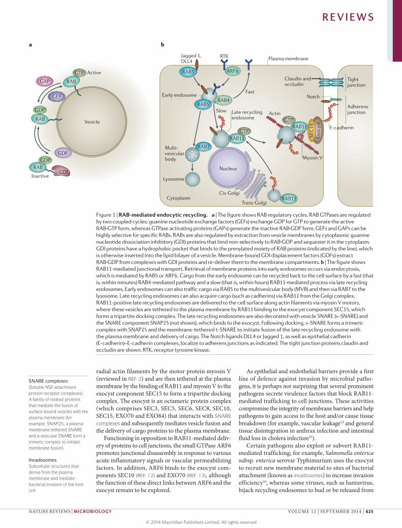
RAB GTPases are key regulators of various mem-brane trafficking events (reviewed in REF. 1) (FIG. 1); they belong to the large protein family of small GTPases and comprise 30–70 members in different organisms. Other small GTPase subfamilies include RAS, RHO–RAC, ARF and RAN, as well as the more recently character-ized RHEB, RAD and RIT subfamilies. Binding of GTP to small GTPases stimulates their effector functions, whereas association with GDP inactivates them (FIG. 1a). Guanine nucleotide exchange factors (GEFs) stimu-late GDP release and are positive regulators, whereas GTPase-activating proteins (GAPs) inhibit GTPase activity by promoting the hydrolysis of GTP to GDP. In addition, guanine nucleotide dissociation inhibitory (GDI) proteins bind to GDP-bound RAB proteins to sequester them in the cytoplasm, and the opposing GDI displacement factors (GDFs) re-deliver GDP-bound RAB proteins to the membrane (FIG. 1a).
RAB11 mediates several cellular processes that involve intracellular vesicle trafficking, including the delivery of plasma membrane proteins to specialized sites (for example, bud sites in yeast or cell–cell junctions in mammalian cells), secretion of various factors (such as growth factors and other peptides, including inter-ferons (IFNs), cytokines, bone morphogenetic protein (BMP) and transferrin receptor), establishment of cell
polarity and targeting of proteins to organelles and cell compartments, which leads to the formation of edges of migrating cells and the midbody during cytokinesis (reviewed in REFS 2,3). Of note, RAB11-mediated target-ing of proteins to organelles such as cilia4,5 or tubular structures6 also requires the activation of the down-stream RAB GTPase RAB8, which may reflect the ances-tral pathway, as this organization is also found in yeast7. Furthermore, RAB11-mediated trafficking events have a role in several aspects of innate immunity, including promoting epithelial and endothelial barrier integrity, targeting of proteins such as Toll-like receptor 4 (TLR4) to phagosomes for receptor-mediated phagocytosis of pathogens8 and expulsion of pore-forming toxins (PFTs) from the cell surface9. Also, the exocyst, which is an eight-protein complex (see below) that mediates many functions of RAB11, has a key role in initiating IFN sig-nalling in response to double-stranded DNA (dsDNA)10 and regulating autophagy versus IFN signalling11 (BOX 1).
One prominent trafficking pathway that patho-gens modulate or exploit by various mechanisms is the final step of endocytic recycling, during which cargo-containing vesicles dock at the cell surface. Endocytic recycling of internalized extracellular molecules or membrane proteins is initiated by RAB5, which directs plasma membrane-derived vesicles to RAB4-positive early endosomes (FIG. 1b). Some cargo proteins can then recycle back to the plasma membrane from the early endosome via two pathways: a fast, direct route that depends on RAB4, or a slow route via RAB11-positive late recycling endosomes. RAB11-positive late recycling endosomes are delivered to the apical cell surface along
Recycling endosomesLate vesicular compartments that are involved in recycling membrane proteins and de novo synthesized cargo from the Golgi complex to the cell surface.
RAB11‑mediated trafficking in host–pathogen interactionsAnnabel Guichard1, Victor Nizet2,3 and Ethan Bier1
Abstract | Many bacterial and viral pathogens block or subvert host cellular processes to promote successful infection. One host protein that is targeted by invading pathogens is the small GTPase RAB11, which functions in vesicular trafficking. RAB11 functions in conjunction with a protein complex known as the exocyst to mediate terminal steps in cargo transport via the recycling endosome to cell–cell junctions, phagosomes and cellular protrusions. These processes contribute to host innate immunity by promoting epithelial and endothelial barrier integrity, sensing and immobilizing pathogens and repairing pathogen-induced cellular damage. In this Review, we discuss the various mechanisms that pathogens have evolved to disrupt or subvert RAB11‑dependent pathways as part of their infection strategy.
1Section of Cell and Developmental Biology, University of California. 2Department of Pediatrics, University of California.3Skaggs School of Pharmacy and Pharmaceutical Sciences, University of California, San Diego, 9500 Gilman Drive, La Jolla, California 92093, USA.Correspondence to V.N., E.B. e‑mails: [email protected]; [email protected]:10.1038/nrmicro3325
R E V I E W S
624 | SEPTEMBER 2014 | VOLUME 12 www.nature.com/reviews/micro
SNARE complexes(Soluble NSF attachment protein receptor complexes). A family of related proteins that mediate the fusion of surface-bound vesicles with the plasma membrane (for example, SNAP25, a plasma membrane tethered SNARE and a vesicular SNARE form a trimeric complex to initiate membrane fusion).
InvadosomesSubcellular structures that derive from the plasma membrane and mediate bacterial invasion of the host cell.
radial actin filaments by the motor protein myosin V (reviewed in REF. 2) and are then tethered at the plasma membrane by the binding of RAB11 and myosin V to the exocyst component SEC15 to form a tripartite docking complex. The exocyst is an octameric protein complex (which comprises SEC3, SEC5, SEC6, SEC8, SEC10, SEC15, EXO70 and EXO84) that interacts with SNARE complexes and subsequently mediates vesicle fusion and the delivery of cargo proteins to the plasma membrane.
Functioning in opposition to RAB11-mediated deliv-ery of proteins to cell junctions, the small GTPase ARF6 promotes junctional disassembly in response to various acute inflammatory signals or vascular permeabilizing factors. In addition, ARF6 binds to the exocyst com-ponents SEC10 (REF. 12) and EXO70 (REF. 13), although the function of these direct links between ARF6 and the exocyst remain to be explored.
As epithelial and endothelial barriers provide a first line of defence against invasion by microbial patho-gens, it is perhaps not surprising that several prominent pathogens secrete virulence factors that block RAB11-mediated trafficking to cell junctions. These activities compromise the integrity of membrane barriers and help pathogens to gain access to the host and/or cause tissue breakdown (for example, vascular leakage14 and general tissue disintegration in anthrax infection and intestinal fluid loss in cholera infection15).
Certain pathogens also exploit or subvert RAB11-mediated trafficking; for example, Salmonella enterica subsp. enterica serovar Typhimurium uses the exocyst to recruit new membrane material to sites of bacterial attachment (known as invadosomes) to increase invasion efficiency16, whereas some viruses, such as hantavirus, hijack recycling endosomes to bud or be released from
Nature Reviews | Microbiology
Early endosome
Trans-Golgi
Cis-Golgi
Nucleus
Actin
Exoc
yst
RAB11
SEC
15
RAB5
RAB7
RAB5
Late recycling endosome
Multi-vesicularbody
Lysosome
Cytoplasm
RAB4
RAB11GTP
Slow
Fast
Tightjunction
Adherensjunction
Notch
E-cadherin
Claudin andoccludin
Plasma membraneRTKJagged 1,DLL4
b
GTPRAB11
ARF6
Myosin V
Vesicle
GEF
Active
Inactive
GDF
GDI
GAP
RABGDP
RAB
GDP
RAB
GTP
a
Figure 1 | RAB-mediated endocytic recycling. a | The figure shows RAB regulatory cycles. RAB GTPases are regulated by two coupled cycles: guanine nucleotide exchange factors (GEFs) exchange GDP for GTP to generate the active RAB·GTP form, whereas GTPase activating proteins (GAPs) generate the inactive RAB·GDP form. GEFs and GAPs can be highly selective for specific RABs. RABs are also regulated by extraction from vesicle membranes by cytoplasmic guanine nucleotide dissociation inhibitory (GDI) proteins that bind non-selectively to RAB·GDP and sequester it in the cytoplasm. GDI proteins have a hydrophobic pocket that binds to the prenylated moiety of RAB proteins (indicated by the line), which is otherwise inserted into the lipid bilayer of a vesicle. Membrane-bound GDI displacement factors (GDFs) extract RAB·GDP from complexes with GDI proteins and re-deliver them to the membrane compartments. b | The figure shows RAB11-mediated junctional transport. Retrieval of membrane proteins into early endosomes occurs via endocytosis, which is mediated by RAB5 or ARF6. Cargo from the early endosome can be recycled back to the cell surface by a fast (that is, within minutes) RAB4-mediated pathway and a slow (that is, within hours) RAB11‑mediated process via late recycling endosomes. Early endosomes can also traffic cargo via RAB5 to the multivesicular body (MVB) and then via RAB7 to the lysosome. Late recycling endosomes can also acquire cargo (such as cadherins) via RAB11 from the Golgi complex. RAB11-positive late recycling endosomes are delivered to the cell surface along actin filaments via myosin V motors, where these vesicles are tethered to the plasma membrane by RAB11 binding to the exocyst component SEC15, which forms a tripartite docking complex. The late recycling endosomes are also decorated with vesicle SNARE (v-SNARE) and the SNARE component SNAP25 (not shown), which binds to the exocyst. Following docking, v-SNARE forms a trimeric complex with SNAP25 and the membrane-tethered t-SNARE to initiate fusion of the late recycling endosome with the plasma membrane and delivery of cargo. The Notch ligands DLL4 or Jagged 1, as well as epithelial cadherin (E-cadherin)–E-cadherin complexes, localize to adherens junctions as indicated. The tight junction proteins claudin and occludin are shown. RTK, receptor tyrosine kinase.
cells17,18. Other pathogens, including Shigella flexneri, disrupt the trafficking of cadherins and potentially of secreted factors such as antimicrobial peptides from the Golgi complex to recycling endosomes19, whereas Chlamydia spp. exploit Golgi-derived membranes to
create a specialized compartment that is suitable for their development20.
In this Review, we first discuss the insights that have been gained into the pathogenic roles of anthrax toxin and cholera toxin in the inhibition of RAB11-dependent
Box 1 | Role of RAB11 and the exocyst in innate immunity
The small GTPase RAB11 and the exocyst complex are involved in several aspects of innate immunity (see the figure). For example, the transmembrane protein STING (stimulator of interferon genes) senses foreign non-CpG double-stranded DNA (dsDNA), which is inadvertently liberated by pathogens, such as lysed bacteria or viruses60 (see the figure, part a). In response to exogenous DNA, STING colocalizes with the exocyst component SEC5 and TANK-binding kinase-1 (TBK1)10, which leads to the induction of interferon-β (IFNβ) gene expression61. The exocyst functions as a hub for the switch-like small GTPase RALB, which, depending on its ubiquitylation state, alternatively engages distinct exocyst components to trigger innate immune signalling (binding of SEC5 when ubiquitylated at lysine 47) or autophagy (when deubiquitylated by USP33, which triggers assembly of a RALB–EXO84–beclin 1 complex)11 (see the figure, part b). The label ‘exocyst’ in parts a and b refers to exocyst components that are not specifically labelled. Similarly, in addition to contributing to the general process of phagocytic engulfment (reviewed in REF. 62), RAB11 is required for trafficking Toll-like receptor 4 (TLR4) from late recycling endosomes to phagosomes to initiate interferon signalling8 (see the figure, part c) (note that a fraction of TLR4 in phagosomes derives from the plasma membrane in a RAB11-independent manner). Following binding to lipopolysaccharide on the surface of Gram-negative bacteria, TLR4 colocalizes with RAB11 in late recycling endosomes and translocates together with its transducer TRAM to the phagosome that is engulfing the pathogen, which leads to the induction of IFNβ expression. Last, RAB11‑dependent trafficking has an important role in clearing pore-forming toxins (PFTs) from the cell surface and repair of the plasma membrane (see the figure, part d). For example, in Caenorhabditis elegans, RAB11 is essential against infection by bacteria that secrete PFTs, such as nemocidal Bacillus thuringiensis (which secretes Cry5 toxin) or Vibrio cholerae (which secretes V. cholerae cytolysin VCC). Endocytic recycling clears PFTs from the cell surface both by internalization — presumably followed by degradation in the lysosome by a RAB5- and RAB7-dependent process — and by RAB11-mediated expulsion of apical microvilli9.
EXO84SEC5
TBK1
STING
SEC5
TBK1RALB
RALB
Beclin 1
Ub
RAB11
RAB11
TLR4TRAM
Trans-GolgiCis-Golgi
Nucleus
IFNβ
GTP
GTP
RAB11GTPPhagosome
IFNβ
IFNβ
Exocyst
Nature Reviews | Microbiology
Late recyclingendosome
Gram-negative bacteria
dsDNALysed bacteria
Cytoplasm
a
c
Exocyst
Autophagy
USP33
Nutrientrestriction
dsDNAVirus
b
d Microvilli
RAB5
PFT
Multi-vesicularbody
Lysosome
Late endosome
Earlyendosome
RAB5
RAB7
Late recyclingendosome
R E V I E W S
626 | SEPTEMBER 2014 | VOLUME 12 www.nature.com/reviews/micro
junctional trafficking. Next, we consider how several additional pathogens exploit RAB11-mediated trafficking to promote bacterial invasion and development. Last, we examine the subversion of exocyst-mediated trafficking as a viral exit strategy.
Inhibition of junctional transportBacillus anthracis and Vibrio cholerae have impor-tant places in the history of infectious disease. Robert Koch showed that B. anthraci s is the causative agent of anthrax in 1876 (REF. 21), and he identified cholera toxin as a ‘poisonous factor’ that is produced by V. cholera e in 1884 (REF. 22). Louis Pasteur elucidated many aspects of anthrax pathogenesis and transmission23 and ultimately developed a crude but effective vaccine24 that ushered in the modern era of medical microbiology. Both of these pathogens secrete exotoxins that lead to the overproduc-tion of intracellular cyclic AMP and thereby provoke the pathognomonic features of anthrax and cholera.
Anthrax toxins disrupt RAB11‑mediated trafficking. Anthrax toxins have a bipartite structure in which a shared B subunit, known as protective antigen (PA), mediates the entry of the catalytic subunits, oedema factor (EF) and lethal factor (LF), into host cells (FIG. 2a). EF is a highly active calmodulin (CAM)-dependent ade-nylyl cyclase, and LF is a metalloproteinase that cleaves and inactivates host MEKs (reviewed in REFS 25,26). EF and LF are essential for the pathogenesis of B. anthraci s, as highlighted in classic experiments by Smith and Keppie, which demonstrated that guinea pigs that were inoculated with B. anthraci s were refractory to antibiot-ics after a crucial point during infection81. Importantly, they also showed that this lethality could be attributed to the accumulation of anthrax toxins, as immunization before infection with the anthrax toxin B subunit was protective, and this has also been shown to be effective in humans82.
Anthrax toxins have several important roles during B. anthraci s infection (reviewed in REF. 25). During the early, nearly asymptomatic prodromal stages of B. anthraci s infection, toxins are essential for ‘disarm-ing’ immune cells and promoting the transport of spores from the lungs to the lymph nodes. A recent study reported that mice that lack the key anthrax toxin receptor CMG2 in myeloid cells are resistant to infec-tion by the attenuated non-capsulated Sterne strain of B. anthracis2 7. However, these mice remain fully sensi-tive to the lethality that is caused by the injection of oedema toxin (comprising EF and PA) or lethal toxin (comprising LF and PA), which indicates that the toxins exert their lethal effects on other cell types (see below). During the fulminant stages of B. anthraci s infection, flu-like symptoms develop and the disease progresses rapidly, as the microbial toxins facilitate bacterial dis-semination throughout the body and disrupt the func-tion of almost every organ system. EF has a central role in causing oedema, hypotension and the loss of vascular endothelial barrier integrity, which can lead to shock-like death and which accounts for a substan-tial fraction of anthrax mortality in humans (reviewed
in REF. 25). Anthrax toxins function cooperatively to induce lethality and oedema during infection of mice with B. anthracis28. Cell type-specific expression or deletion of the CMG2 receptor in mice showed that the cardiovascular system (that is, heart and smooth muscle cells) is the primary target of LF, whereas the primary target of EF is the liver29. The exact mechanisms that lead to the diverse effects of anthrax toxins that have been observed in different cell types remain elusive, and future studies are required to determine whether the inhibition of host endocytic recycling contributes to these various effects.
Although the structures and biochemical activities of anthrax toxins are well known, their cellular effects have been more difficult to elucidate. Expression of the enzy-matic subunits of these toxins in the developing Dros-ophila melanogaster wing using the GAL4–UAS system30, which bypasses the need for receptor-mediated toxin entry, yielded the expected effects of anthrax toxins (for example, growth inhibition by LF and cAMP-dependent activation of protein kinase A (PKA) by EF)31. In addi-tion, these experiments revealed a novel cooperative activity of the two toxins in blocking Notch signalling14: both toxins reduce the cell surface expression of the Notch ligand Delta by inhibiting its endocytic recycling to adherens junctions14, which is a process that is neces-sary for Notch receptor activation on adjacent cells32. In EF-expressing cells, the levels of RAB11 are decreased and endocytic recycling is inhibited, which suggests that EF targets RAB11 to block endocytic recycling (FIG. 2a). Consistent with this finding, a screen of dominant-negative forms of all 31 D. melanogaste r RAB proteins showed that only expression of dominant- negative RAB11 mimics the effect of EF. Conversely, in flies and in human cells, overexpression of RAB11 rescues the junction-disrupting effects of EF14. Although LF does not reduce levels of RAB11, it does reduce the apical levels and function of the RAB11 mediator and binding part-ner SEC15 (REF. 14), which is an effect that cannot be res-cued by overexpression of RAB11. EF also reduces apical SEC15 levels; however, in this case, the effect seems to be mediated indirectly by its reduction of RAB11 activ-ity, as expression of dominant-negative RAB11 causes a similar reduction in SEC15 levels. Thus, EF functions by downregulating RAB11 levels and blocking vesicular transport (thereby also reducing apical SEC15 levels), whereas LF reduces apical levels of SEC15 in a RAB11-independent manner. Inhibition of endocytic recycling by either toxin also results in reduced levels of the cell–cell adhesion protein epithelial cadherin (E-cadherin) at adherens junctions. Similar effects of both EF and LF have been observed in human brain microvascular endothelial cells, in which the toxins block RAB11- and exocyst-mediated trafficking, reduce cadherin levels at junctions, inhibit Notch signalling and compromise barrier integrity (in transwell assays and in vivo in mice), with EF having a predominant role. Future experiments are required to determine the intersecting mechanisms by which EF, via increased cAMP, and LF, via inhibition of MEKs, disrupt RAB11- and exocyst-mediated junctional trafficking.
Notch signallingA signalling pathway that controls a range of cell fate and growth decisions. It is activated at adherens junctions by cell surface-tethered ligands (for example, Delta) on one cell, which stimulate Notch receptors on adjacent cells.
Adherens junctionsSubapically localized cell–cell junctions that consist of transmembrane epithelial cadherin adhesion molecules, which interact with the cytoskeleton via α-catenins and β-catenins and link epithelial and endothelial cells, enabling them to form contiguous sheets.
Cholera toxin disrupts intestinal barrier integrity. Chol-era toxin causes the severe watery diarrhoea that is asso-ciated with cholera33, and its mechanisms of action have been elucidated. Following translocation into the cyto-plasm (which is mediated by the cholera toxin B subunit (CtxB)), the enzymatic moiety CtxA (which is an ADP ribosylase), binds to the host cofactor ARF6·GTP (which interestingly also functions in opposition to RAB11 to promote junction disassembly (FIG. 2b)). CtxA transfers ADP-ribose from NAD to the α-subunit of the stimulat-ing G protein (Gsα), which causes a pathological increase in cellular cAMP levels via the induction of host ade-nylyl cyclases at the plasma membrane (reviewed in REFS 33,34). Increased cAMP levels function via PKA to induce the ion channel cystic fibrosis transmembrane receptor (CFTR), which leads to Cl− ion secretion into the intestinal lumen (reviewed in REF. 35). This trans-cellular secretion of Cl− ions is accompanied by the paracellular efflux of Na+ ions and water to preserve electroneutrality and osmotic balance, which results in a large volume of fluid loss (up to 10–20 litres per day). This model of CtxA action is supported by a range of experimental evidence, a definitive example being the failure of purified cholera toxin to induce fluid secretion in the small intestines of CFTR-null mice36. However, until recently, the mechanisms by which the paracellular efflux of Na+ ions and water occurs had remained unclear.
As both CtxA and EF induce pathological levels of cAMP, the question arose whether CtxA might also dis-rupt cell junctions by inhibiting RAB11-mediated traf-ficking. Such an effect would facilitate the paracellular efflux of Na+ ions and water that accompanies trans-cellular Cl− secretion to generate the profuse diarrhoea that is caused by cholera infection. In agreement with this hypothesis, junctional levels of RAB11, SEC15 and E-cadherin are greatly reduced by treatment of human intestinal epithelial cell lines with CtxA15 (FIG. 2b). CtxA also alters tight junctions, which disrupts their normal strict alignment with the more basal adherens junc-tions, and inhibits Notch signalling. Similar effects are also observed in vivo in mouse ligated ileal loop prepara-tions, where CtxA induces fluid secretion, downregulates RAB11 and SEC15 levels and creates apical gaps between cells (FIG. 2c). In the fly gut, CtxA also causes dye leakage and large lacunal gaps between cells, which are visible at the ultrastructural level (FIG. 2c), as well as weight loss and lethality, all of which can be rescued by the overexpres-sion of RAB11. Conversely, expression of a dominant-negative form of RAB11 mimics CtxA effects and causes the breakdown of intestinal barrier integrity, as judged by the ability of ingested dye to leak into the body cav-ity15. Junctional defects that are caused by CtxA in the fly gut and in human cells are remarkably similar to those that were previously reported in small‑intestine biopsies from human patients with cholera37 (FIG. 2c). As with puri-fied toxin, the CtxA-dependent lethality and junctional defects that were observed following V. cholera e infection of adult flies can be mitigated by overexpressing RAB11 in the midgut (which is the fly equivalent of the small intestine). In flies and in human cells, evidence suggests that the effects of CtxA are mediated by a combination of
the two primary known cAMP effectors, PKA and EPAC (a GEF for the small GTPase RAP1, which has several known links to the exocyst). Activation of either effec-tor pathway by genetic means in flies or using pathway- specific cAMP analogues in mammalian cells also dis-rupts cell junctions, whereas chemical inhibition of either PKA or EPAC reduces the effects of CtxA15. These obser-vations indicate that RAB11-dependent endocytic recy-cling helps to maintain normal intestinal barrier integrity and that boosting this process provides protection against the junctional disruptive effects of CtxA and infection with V. cholera e.
How pathological levels of cAMP, via PKA and EPAC, lead to the downregulation and inhibition of RAB11 in
Tight junctionsThe most apically localized cell–cell junctions; they consist of adhesive claudins and occludin transmembrane proteins, which function as a diffusion barrier to ions, water and other small molecules.
the vascular endothelium (that is, for EF) or gut (that is, for CtxA) is a central question for future research. It is conceivable that the observed decrease in RAB11 protein levels is caused by increased protein turnover (for example, proteasome-dependent degradation of RAB11), alterations in membrane compartment identi-ties (for example, elimination of the late recycling endo-some compartment), reduced production of proteins (for example, RAB11 mRNA instability or inhibition of RAB11 mRNA transcription) or a combination of these factors. Other pathogens liberating toxins that increase intracellular cAMP levels (cAMP toxins) (reviewed in REF. 38) may also possibly inhibit RAB11 activity to disrupt host cell barriers. Prominent examples of such pathogens include: toxigenic Escherichia coli (via heat-labile toxin (Ltx), which is highly related to CtxA, and heat-stable toxin (STa), which stimulates host guanylyl cyclases to indirectly increase cAMP levels and activates CFTR by cGMP-dependent protein kinase II; reviewed in REFS 39,40,48); Bordetella pertussis (via CyaA, which is a CAM-dependent adenylyl cyclase that is related to EF, and pertussis toxin (Ptx), which inhibits endogenous Gi subunits and thereby increases the activity of adenylyl cyclases); and Pseudomonas aeruginosa (via ExoY, which is an adenylyl cyclase). Finally, there are surprising parallels between the junction-disrupting actions of cAMP toxins and microbial PFTs that require further scrutiny (BOX 2).
Pathogens that exploit RAB11 traffickingVarious intracellular pathogens evade destruction following phagocytosis by subverting RAB-mediated trafficking events and reprogramming membrane com-partment identities to create environments that support survival and replication within host cells (reviewed in REFS 41,42) (FIG. 3). In addition, several pathogens exploit or inhibit the endocytic recycling pathway to invade or escape host cells.
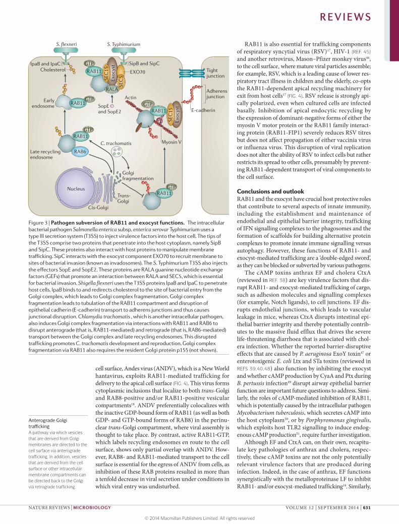
Commandeering the exocyst during bacterial invasion. S. Typhimurium is an intracellular pathogen that uses a type III secretion system (T3SS) to inject effector pro-teins into host cells. The tip of this injection complex comprises two proteins that penetrate the host cyto-plasm. These proteins (SipB and SipC) not only partici-pate in transferring virulence factors into the host cell but also interact with host factors, including the exocyst, to redirect resources to sites of bacterial invasion. SipC interacts with the plasma membrane-associated com-ponent EXO70 to recruit the exocyst complex to sites of S. Typhimurium attachment (FIG. 3). This might lead to the increased delivery of membrane proteins, which promotes membrane expansion and facilitates bacterial invasion16. At the same time, SopE and SopE2, which are two injected S. Typhimurium effectors, function as GEFs to activate the small GTPase RALA, which can bind in a mutually exclusive manner to either of the exocyst components SEC5 or EXO84. Recruitment of the exocyst and activated RALA is essential for bacterial entry, as RNAi of the exocyst components EXO70 or SEC5 or knockdown of RALA leads to reduced bacterial
Nature Reviews | Microbiology
Tightjunction
Adherensjunction
Claudin andoccludin
NotchJagged 1or DLL4
Cytoplasm
Plasma membrane
Late recycling endosome
Late endosome
Late endosome
Earlyendosome
LF
LF
PA
CMG2 orTEM8
a
RAP1
PKA
MEKs MAPKs
cAMP
EPAC
VascularleakageEF
EF
CFTR
Earlyendosome
b
RAP1
PKAcAMP
EPAC
CtxA
GPCR
PAdenylylcyclase
Cl–
αγ
Claudinsand occludin
Na+
H2O
β
CtxACtxB
GM1
Nucleus
Nucleus
E-cadherin
Actin
Exoc
yst
SEC
15
Myosin V
GTPRAB11
Exoc
yst
SEC
15
GTPRAB11
ARF6
Microvilli
c Human cholera biopsies Human CACO2 cells D. melanogaster intestine
ToxisomeA vesicle that contains toxic factors and that is expelled from the cell surface (for example, shed microvilli).
uptake16. Association of RALA with the exocyst is important in this process, as the effect of RNAi-mediated depletion of SEC5 can be reversed by overexpressing wild-type SEC5 but not using a SEC5 point mutant that is defective in binding to RALA. SipC also binds to actin and remodels the apical cytoskeleton43, which again provides a link to the exocyst, as this complex affects the architecture of the apical cytoskeletal. Thus,
S. Typhimuriu m uses a coordinated set of effectors that induce localized membrane expansion and cytoskeleton remodelling to promote its engulfment by host cells.
S. flexneri also uses the tip proteins (IpaB and IpaC) of a T3SS to penetrate into the host cytoplasm. One of these proteins (that is, IpaB) regulates lipid trafficking. IpaB redirects cholesterol to the site of bacterial entry19 (FIG. 3) by altering the architecture and function of the recy-cling endosome, which causes tubulation of the RAB11-positive membrane compartment, although RAB11 is not the direct target of IpaB. Anterograde trafficking of E- cadherin via the Golgi complex and recycling endosome to cell junctions is greatly reduced by S. flexneri infection. This major disruption of the RAB11-positive compart-ment suggests that the trafficking of various secreted factors (for example, cytokines and antimicrobial pep-tides) and cell surface receptors that are required for defence may also be impeded during S. flexneri infection. IpaB binds to cholesterol and thereby depletes this lipid from the Golgi complex, which causes its fragmentation — an effect that can be reversed by co-treatment of cells with IpaB protein and exogenous cholesterol.
The intracellular pathogen Chlamydia trachomatis, which forms inclusion bodies in host cells, also leads to RAB11‑dependent fragmentation of the Golgi com-plex, which is required for bacterial development and reproduction20. RNAi screening for host factors that are required for C. trachomati s replication identified RAB11 and RAB6 as essential elements, which is consistent with a previous study that reported the association of both GTPases with this bacterium during infection44. RAB11, which promotes anterograde trafficking, and RAB6, which promotes retrograde trafficking, are also essen-tial for Golgi complex fragmentation during infection20, as RNAi knockdown of either of these GTPases restores compact Golgi complex morphology and inhibits bac-terial replication (FIG. 3). The importance of inducing Golgi complex fragmentation for C. trachomati s repli-cation is further substantiated by the observation that RNAi of the resident Golgi protein p155, which alone causes fragmentation of this organelle, can overcome the rescuing effect of RNAi-mediated knockdown of RAB11 and restore the development of C. trachomati s. Interestingly, RNAi of another Golgi protein, golgin 84, which can also induce Golgi complex fragmentation, is unable to reverse either the restoration of Golgi com-plex structure or bacterial reproduction that is induced by RNAi-mediated knockdown of RAB11, which again links RAB11-mediated Golgi complex fragmentation and bacterial replication. These genetic epistasis experi-ments further suggest that RAB11 functions at a step fol-lowing that of golgin 84 and before p155 in anterograde Golgi trafficking, which defines a pathway that needs to be investigated in further studies.
Subversion of vesicular trafficking by viruses. An impor-tant step in the viral life cycle is exit from the host cell. Some viruses assemble complete particles at the cell surface, whereas in other cases, particles are assembled intracellularly and then trafficked intact to the cell sur-face. As an example of a virus that traffics intact to the
Box 2 | Similarities between pore-forming toxins and cAMP toxins
Many microbial pathogens can breach epithelial barriers during infection, and various virulence factors, including proteases and actin-modifying enzymes that aid in this process have been identified63. One particularly intriguing example is Staphylococcus aureus α-haemolysin (also known as α‑toxin), which is a pore-forming toxin (PFT) that binds to the ADAM10 metalloproteinase as its host receptor64 (see the figure). Binding of α‑toxin activates the membrane-associated host metalloproteinase ADAM10 (REF. 64), which cleaves epithelial cadherin (E-cadherin) to disrupt junctional integrity64–68. ADAM10 also cleaves Notch receptors in mammals69–72 (or its orthologue Kuzbanian in flies and nematode worms73,74) and thus we speculate that ADAM10-mediated Notch cleavage might have a role in the barrier‑disruptive effects of α‑toxin.
There are potentially important parallels between PFTs and toxins that increase intracellular cyclic AMP levels. For example, in the case of cholera, cholera toxin catalytic A subunit (CtxA) increases intracellular cAMP levels (cAMP toxins), which leads to Cl– ion secretion via protein kinase A (PKA)-mediated activation of the cystic fibrosis transmembrane receptor (CFTR) ion transporter, whereas the PFT Vibrio cholerae cytolysin (VCC) is itself a Cl– ion-conducting pore75 that is processed and activated by the ADAM10-related protease ADAM17 (REF. 76) (see the figure). Also, as RAB11‑dependent endocytic trafficking has a key role in eliminating PFTs from the cell surface (by endocytosis and expulsion of microvilli)9, inhibition of this process by cAMP toxins may augment the effect of the PFTs. This strategy of expelling PFT-containing membrane has also been observed for S. aureus α-toxin77, and the larger group A Streptococcus PFT streptolysin O78 can similarly be shed via toxisome vesicles from several infected cell types.
Determining whether ADAM17, like ADAM10, cleaves E-cadherins or Notch receptors and exploring potential links between α‑toxin, Notch cleavage and endocytic recycling will be interesting areas for further investigation. It may also be informative to study other potential parallels between α‑toxin and cAMP toxins; for example, it is likely that both types of toxins reduce the cellular ATP/AMP ratio, as α‑toxin results in ATP efflux through the pore, which is an effect that contributes to its toxicity79, and high levels of cAMP production also deplete ATP reserves. α-toxin-mediated efflux of ATP (probably evoking host cellular responses via a reduction in the ATP/AMP ratio) is possibly mediated by effectors such as AMP-activated protein kinase (AMPK). Intriguingly, the anthrax toxin lethal factor (LF) also induces the depletion of ATP from macrophages (via a
Cl–
VCC
ATP efflux
pro-VCC
Processing andactivation of VCC
Barrierdisruption?
ADAM17
Adherensjunction
E-cadherin
Tight junction
α-toxin
Notch
Notchcleavage
↓ATP/AMP
Nature Reviews | Microbiology
Jagged 1or DLL4
ADAM10
R E V I E W S
630 | SEPTEMBER 2014 | VOLUME 12 www.nature.com/reviews/micro
Anterograde Golgi trafficking A pathway via which vesicles that are derived from Golgi membranes are directed to the cell surface via anterograde trafficking. In addition, vesicles that are derived from the cell surface or other intracellular membrane compartments can be directed back to the Golgi via retrograde trafficking.
cell surface, Andes virus (ANDV), which is a New World hantavirus, exploits RAB11-mediated trafficking for delivery to the apical cell surface (FIG. 4). This virus forms cytoplasmic inclusions that localize to both trans-Golgi and RAB8-positive and/or RAB11-positive vesicular compartments18. ANDV preferentially colocalizes with the inactive GDP-bound form of RAB11 (as well as both GDP- and GTP-bound forms of RAB8) in the perinu-clear trans-Golgi compartment, where viral assembly is thought to take place. By contrast, active RAB11·GTP, which labels recycling endosomes en route to the cell surface, shows only partial overlap with ANDV. How-ever, RAB8- and RAB11-mediated transport to the cell surface is essential for the egress of ANDV from cells, as inhibition of these RAB proteins resulted in more than a tenfold decrease in viral secretion under conditions in which viral entry was undisturbed.
RAB11 is also essential for trafficking components of respiratory syncytial virus (RSV)17, HIV-1 (REF. 45) and another retrovirus, Mason–Pfizer monkey virus46, to the cell surface, where mature viral particles assemble; for example, RSV, which is a leading cause of lower res-piratory tract illness in children and the elderly, co-opts the RAB11-dependent apical recycling machinery for exit from host cells17 (FIG. 4). RSV release is strongly api-cally polarized, even when cultured cells are infected basally. Inhibition of apical endocytic recycling by the expression of dominant-negative forms of either the myosin V motor protein or the RAB11 family interact-ing protein (RAB11-FIP1) severely reduces RSV titres but does not affect propagation of either vaccinia virus or influenza virus. This disruption of viral replication does not alter the ability of RSV to infect cells but rather restricts its spread to other cells, presumably by prevent-ing RAB11-dependent transport of viral components to the cell surface.
Conclusions and outlookRAB11 and the exocyst have crucial host protective roles that contribute to several aspects of innate immunity, including the establishment and maintenance of endothelial and epithelial barrier integrity, trafficking of IFN signalling complexes to the phagosomes and the formation of scaffolds for building alternative protein complexes to promote innate immune signalling versus autophagy. However, these functions of RAB11- and exocyst-mediated trafficking are a ‘double-edged sword’, as they can be blocked or subverted by various pathogens.
The cAMP toxins anthrax EF and cholera CtxA (reviewed in REF. 38) are key virulence factors that dis-rupt RAB11- and exocyst-mediated trafficking of cargo, such as adhesion molecules and signalling complexes (for example, Notch ligands), to cell junctions. EF dis-rupts endothelial junctions, which leads to vascular leakage in mice, whereas CtxA disrupts intestinal epi-thelial barrier integrity and thereby potentially contrib-utes to the massive fluid efflux that drives the severe life- threatening diarrhoea that is associated with chol-era infection. Whether the reported barrier-disruptive effects that are caused by P. aeruginosa ExoY toxin47 or enterotoxigenic E. col i Ltx and STa toxins (reviewed in REFS 39,40,48) also function by inhibiting the exocyst and whether cAMP production by CyaA and Ptx during B. pertussis infection49 disrupt airway epithelial barrier function are important future questions to address. Simi-larly, the roles of cAMP-mediated inhibition of RAB11, which is potentially caused by the intracellular pathogen Mycobacterium tuberculosis, which secretes cAMP into the host cytoplasm50, or by Porphyromonas gingivalis, which exploits host TLR2 signalling to induce endog-enous cAMP production51, require further investigation.
Although EF and CtxA can, on their own, recapitu-late key pathologies of anthrax and cholera, respec-tively, these cAMP toxins are not the only potentially relevant virulence factors that are produced during infection. Indeed, in the case of anthrax, EF functions synergistically with the metalloproteinase LF to inhibit RAB11- and/or exocyst-mediated trafficking14. Similarly,
Nature Reviews | Microbiology
S. flexneri
Cholesterol
S. Typhimurium
Exoc
yst
SEC
15
SEC
5
RAB11
IpaB and IpaC GTP
RAB11GTP
SopEand SopE2
RALA
SipB and SipC
Earlyendosome
Late recyclingendosome
RAB11GTP
RAB11GTP
RAB6
Golgifragmentation
C. trachomatis
Trans-Golgi
Cis-Golgi
Nucleus
EXO70
Adherensjunction
E-cadherin
Actin
Exoc
yst
SEC
15
GTPRAB11
Tightjunction
Myosin V
Figure 3 | Pathogen subversion of RAB11 and exocyst functions. The intracellular bacterial pathogen Salmonella enterica subsp. enterica serovar Typhimurium uses a type III secretion system (T3SS) to inject virulence factors into the host cell. The tips of the T3SS comprise two proteins that penetrate into the host cytoplasm, namely SipB and SipC. These proteins also interact with host proteins to manipulate membrane trafficking. SipC interacts with the exocyst component EXO70 to recruit membrane to sites of bacterial invasion (known as invadosomes). The S. Typhimurium T3SS also injects the effectors SopE and SopE2. These proteins are RALA guanine nucleotide exchange factors (GEFs) that promote an interaction between RALA and SEC5, which is essential for bacterial invasion. Shigella flexneri uses the T3SS proteins IpaB and IpaC to penetrate host cells. IpaB binds to and redirects cholesterol to the site of bacterial entry from the Golgi complex, which leads to Golgi complex fragmentation. Golgi complex fragmentation leads to tubulation of the RAB11 compartment and disruption of epithelial cadherin (E-cadherin) transport to adherens junctions and thus causes junctional disruption. Chlamydia trachomatis , which is another intracellular pathogen, also induces Golgi complex fragmentation via interactions with RAB11 and RAB6 to disrupt anterograde (that is, RAB11-mediated) and retrograde (that is, RAB6-mediated) transport between the Golgi complex and late recycling endosomes. This disrupted trafficking promotes C. trachomatis development and reproduction. Golgi complex fragmentation via RAB11 also requires the resident Golgi protein p155 (not shown).
RALBA small GTPase that can bind in a mutually exclusive manner to either of the exocyst components SEC5 or EXO84 to function as a molecular switch between immune signalling and autophagy.
Yersinia pestis produces the adenylyl cyclase YpAC38,52 and several factors that inhibit MEK signalling (for example, YopJ53). It remains to be determined whether synergy between these toxins contributes to the patho-genesis of plague. The cAMP toxins might also function collaboratively with other toxic factors, such as the PFTs of B. anthraci s (that is, anthrolysin O) and V. cholera e (that is, V. cholerae cytolysin (VCC)), during infection. Finally, in the case of anthrax, as mitogen-activated pro-tein kinase (MAPK) p38 (REF. 54) and TLR4 signalling55 are key host defence responses to PFTs54, their inhibition by LF and EF, respectively, may result in important neu-tralizing effects. Examination of these key nodes should provide fresh insights into cooperative interactions between virulence factors.
Another area for future investigation is whether other known features of disease that are caused by cAMP toxins may result from the inhibition of RAB11- and exocyst-mediated functions; for example, during the early and
nearly asymptomatic phase of infection, anthrax toxins inhibit various aspects of the immune response, which enables the bacteria to proliferate and disseminate into the circulation (reviewed in REFS 56,57). As myeloid cells are crucial targets of anthrax toxins during this phase27, it would be interesting to examine whether inhibition of RAB11 function by anthrax toxins alters cytokine secretion during the establishment of infection. Con-versely, overexpression of RAB11 might mitigate aspects of toxin-mediated lethality. Similarly, as CtxA has been implicated in disrupting innate immune functions dur-ing the establishment of infection58, how inhibition of RAB11- and exocyst-mediated functions contribute to this role (or roles) of CtxA should be studied. EF effects on the liver are also of interest, as a recent report found that hepatocytes are a key target of EF-mediated lethal-ity in mice29. Experiments could probe how inhibition of RAB11- and exocyst-mediated trafficking by EF disrupts cell–cell junctions in bile duct cholangiocytes, whether it prevents hyperpolarization of hepatocytes to form canaliculi or whether this toxin impedes the secre-tion of key hepatic factors. If such effects are observed, a central question will be how they may contribute to the lethal effects of EF and whether selective overexpres-sion of RAB11 in the liver can reverse these effects and restore viability. As liver pathology does not seem to be a notable feature of lethal anthrax cases in humans59, one might ask whether more fine-scale analysis in mice can elucidate specific cellular phenotypes to examine more closely in human samples. Similarly, the cell type (or cell types) that is essential for generating oedema in response to EF (for example, lung oedema or foot-pad swelling) must be identified to examine whether such swelling can be counteracted by restoring endocytic recycling.
Another area for future study is to determine whether pathogens exploit or inhibit additional RAB11- and exocyst-mediated processes. Toxins that increase cAMP levels or other pathogenic factors may contribute to dis-ease pathogenesis by inhibiting the establishment of apical–basal polarity (for example, in the liver), traffick-ing to cilia (for example, in the kidneys or in the regula-tion of vascular flow), cell migration (for example, of macrophages) or cell division (for example, of intestinal crypt cells). Pathogens may also manipulate the switch-like regulatory function of the exocyst in promoting the activation of innate immune signalling (for exam-ple, via ubiquitylated RALB–SEC5–TBK1 complexes) or autophagy (for example, via RALB–EXO84–beclin 1 complexes). Studies that probe these questions should lead to exciting discoveries and define new molecular targets for pharmacological intervention.
Trans-Golgi
Cis-Golgi
Nucleus
Nature Reviews | Microbiology
Earlyendosome
Late recyclingendosome
RAB11GTP
RAB11GTP
RAB11GDP
RSV ANDV
ActinExocystSEC15
GTPRAB11 Myosin V
Figure 4 | Viral exit via the recycling endosome. Andes virus (ANDV), which is a New World hantavirus, forms cytoplasmic inclusions and undergoes assembly in the trans-Golgi (that is, RAB11·GDP-containing) and recycling endosomal (that is, RAB11·GTP-containing) compartments. Mature viral particles are then trafficked via the recycling pathway in a RAB11-dependent manner to the cell surface, where they are released. Respiratory syncytial virus (RSV) enters host cells either apically or basally and viral components are then transported to the apical cell surface via RAB11 and myosin V-dependent trafficking.
1. Stenmark, H. Rab GTPases as coordinators of vesicle traffic. Nature Rev. Mol. Cell Biol. 10, 513–525 (2009).
2. Heider, M. R. & Munson, M. Exorcising the exocyst complex. Traffic 13, 898–907 (2012).
3. Kelly, E. E., Horgan, C. P. & McCaffrey, M. W. Rab11 proteins in health and disease. Biochem. Soc. Trans. 40, 1360–1367 (2012).
4. Lim, Y. S., Chua, C. E. & Tang, B. L. Rabs and other small GTPases in ciliary transport. Biol. Cell 103, 209–221 (2011).
5. Knodler, A. et al. Coordination of Rab8 and Rab11 in primary ciliogenesis. Proc. Natl Acad. Sci. USA 107, 6346–6351 (2010).
6. Apodaca, G. Opening ahead: early steps in lumen formation revealed. Nature Cell Biol. 12, 1026–1028 (2010).
7. Novick, P. et al. Interactions between Rabs, tethers, SNAREs and their regulators in exocytosis. Biochem. Soc. Trans. 34, 683–686 (2006).
8. Husebye, H. et al. The Rab11a GTPase controls Toll‑like receptor 4‑induced activation of interferon
regulatory factor‑3 on phagosomes. Immunity 33, 583–596 (2010).This study identifies RAB11 as an important regulator of TLR4 and TRAM transport to E. coli phagosomes to activate interferon regulatory factor 3 (IRF3) expression.
9. Los, F. C. et al. RAB‑5‑ and RAB‑11‑dependent vesicle‑trafficking pathways are required for plasma membrane repair after attack by bacterial pore‑forming toxin. Cell Host Microbe 9, 147–157 (2011).
R E V I E W S
632 | SEPTEMBER 2014 | VOLUME 12 www.nature.com/reviews/micro
10. Ishikawa, H., Ma, Z. & Barber, G. N. STING regulates intracellular DNA‑mediated, type I interferon‑dependent innate immunity. Nature 461, 788–792 (2009).
11. Simicek, M. et al. The deubiquitylase USP33 discriminates between RALB functions in autophagy and innate immune response. Nature Cell Biol. 15, 1220–1230 (2013).
12. Prigent, M. et al. ARF6 controls post‑endocytic recycling through its downstream exocyst complex effector. J. Cell Biol. 163, 1111–1121 (2003).
13. Fielding, A. B. et al. Rab11‑FIP3 and FIP4 interact with Arf6 and the exocyst to control membrane traffic in cytokinesis. EMBO J. 24, 3389–3399 (2005).
14. Guichard, A. et al. Anthrax toxins cooperatively inhibit endocytic recycling by the Rab11/Sec15 exocyst. Nature 467, 854–858 (2010).This paper shows that RAB11 and the exocyst are targets that are inhibited by the anthrax toxins EF and LF, respectively, thereby causing loss of barrier function.
15. Guichard, A. et al. Cholera toxin disrupts barrier function by inhibiting exocyst‑mediated trafficking of host proteins to intestinal cell junctions. Cell Host Microbe 14, 294–305 (2013).This study shows that, in addition to its well-appreciated role in stimulating Cl– ion secretion, cholera toxin also weakens cell junctions to enable the efflux of Na+ ions and water in the gut lumen.
16. Nichols, C. D. & Casanova, J. E. Salmonella‑directed recruitment of new membrane to invasion foci via the host exocyst complex. Curr. Biol. 20, 1316–1320 (2010).
17. Brock, S. C., Goldenring, J. R. & Crowe, J. E. Jr. Apical recycling systems regulate directional budding of respiratory syncytial virus from polarized epithelial cells. Proc. Natl Acad. Sci. USA 100, 15143–15148 (2003).
18. Rowe, R. K., Suszko, J. W. & Pekosz, A. Roles for the recycling endosome, Rab8, and Rab11 in hantavirus release from epithelial cells. Virology 382, 239–249 (2008).This study reports the discovery that RAB8 and RAB11 colocalize with ANDV proteins during infection and that downregulation of RAB11 and RAB8 proteins reduce viral secretion from host cells, thereby implicating the recycling endosome and these RAB proteins in hantavirus trafficking to the plasma membrane.
19. Mounier, J. et al. Shigella effector IpaB‑induced cholesterol relocation disrupts the Golgi complex and recycling network to inhibit host cell secretion. Cell Host Microbe 12, 381–389 (2012).This paper shows that Shigella spp. recruits cholesterol to sites of bacterial invasion, which leads to fragmentation of the Golgi complex, inhibition of endocytic trafficking and disruption of host epithelial barriers.
20. Rejman Lipinski, A. et al. Rab6 and Rab11 regulate Chlamydia trachomatis development and golgin‑84‑dependent Golgi fragmentation. PLoS Pathog. 5, e1000615 (2009).This study shows that RAB6 and RAB11 are key regulators of Golgi stability and further shows that C. trachomatis functions via these small GTPases to disrupt Golgi complex structure and promote its intracellular development.
21. Koch, R. (ed.) Beitrage zur Biologie der Pflanzen Vol. 2 (J. U. Kern’s Verlag (Max Müller)) (in German) (1876).
22. Koch, R. Sechster Bericht der deutschen Wissenschaftlichen Commission zur Enforschung der Cholera, Geh Regierungsraths Dr Koch. Dtsch. Med. Wochenschr. 10, 191–192 (in German) (1884).
23. Chamberland, C. (ed). Le charbon et la vaccination charbonneuse d’apres les travaux recents de M. Pasteur (ed. Tignol, B.) (Bernard Tignol) (in French) (1883).
24. Pasteur, L., Chamberland, C. & Roux, E. Compte rendu sommaire des experiences faites a Pouilly‑le‑Fort, pres Melun, sur la vaccination charbonneuse. Cr. Acad. Sci. Paris 92, 1393–1398 (in French) (1881).
25. Guichard, A., Nizet, V. & Bier, E. New insights into the biological effects of anthrax toxins: linking cellular to organismal responses. Microbes Infect. 14, 97–118 (2012).
27. Liu, S. et al. Anthrax toxin targeting of myeloid cells through the CMG2 receptor is essential for establishment of Bacillus anthracis infections in mice. Cell Host Microbe 8, 455–462 (2010).
28. Pezard, C., Berche, P. & Mock, M. Contribution of individual toxin components to virulence of Bacillus anthracis. Infect. Immun. 59, 3472–3477 (1991).
29. Liu, S. et al. Key tissue targets responsible for anthrax‑toxin‑induced lethality. Nature 501, 63–68 (2013).
30. Brand, A. H. & Perrimon, N. Targeted gene expression as a means of altering cell fates and generating dominant phenotypes. Development 118, 401–415 (1993).
31. Guichard, A., Park, J. M., Cruz‑Moreno, B., Karin, M. & Bier, E. Anthrax lethal factor and edema factor act on conserved targets in Drosophila. Proc. Natl Acad. Sci. USA 103, 3244–3249 (2006).
32. Jafar‑Nejad, H. et al. Sec15, a component of the exocyst, promotes Notch signaling during the asymmetric division of Drosophila sensory organ precursors. Dev. Cell 9, 351–363 (2005).
33. De Haan, L. & Hirst, T. R. Cholera toxin: a paradigm for multi‑functional engagement of cellular mechanisms. Mol. Membr. Biol. 21, 77–92 (2004).
34. Sack, D. A., Sack, R. B., Nair, G. B. & Siddique, A. K. Cholera. Lancet 363, 223–233 (2004).
35. Barrett, K. E. & Keely, S. J. Chloride secretion by the intestinal epithelium: molecular basis and regulatory aspects. Annu. Rev. Physiol. 62, 535–572 (2000).
36. Gabriel, S. E., Brigman, K. N., Koller, B. H., Boucher, R. C. & Stutts, M. J. Cystic fibrosis heterozygote resistance to cholera toxin in the cystic fibrosis mouse model. Science 266, 107–109 (1994).
37. Mathan, M. M., Chandy, G. & Mathan, V. I. Ultrastructural changes in the upper small intestinal mucosa in patients with cholera. Gastroenterology 109, 422–430 (1995).
38. Ahuja, N., Kumar, P. & Bhatnagar, R. The adenylate cyclase toxins. Crit. Rev. Microbiol. 30, 187–196 (2004).
39. Sears, C. L. & Kaper, J. B. Enteric bacterial toxins: mechanisms of action and linkage to intestinal secretion. Microbiol. Rev. 60, 167–215 (1996).
40. Weiglmeier, P. R., Rosch, P. & Berkner, H. Cure and curse: E. coli heat‑stable enterotoxin and its receptor guanylyl cyclase C. Toxins 2, 2213–2229 (2010).
41. Brumell, J. H. & Scidmore, M. A. Manipulation of Rab GTPase function by intracellular bacterial pathogens. Microbiol. Mol. Biol. Rev. 71, 636–652 (2007).
42. Ray, K., Marteyn, B., Sansonetti, P. J. & Tang, C. M. Life on the inside: the intracellular lifestyle of cytosolic bacteria. Nature Rev. Microbiol. 7, 333–340 (2009).
43. Hayward, R. D. & Koronakis, V. Direct nucleation and bundling of actin by the SipC protein of invasive Salmonella. EMBO J. 18, 4926–4934 (1999).
44. Rzomp, K. A., Scholtes, L. D., Briggs, B. J., Whittaker, G. R. & Scidmore, M. A. Rab GTPases are recruited to chlamydial inclusions in both a species‑dependent and species‑independent manner. Infect. Immun. 71, 5855–5870 (2003).
45. Varthakavi, V. et al. The pericentriolar recycling endosome plays a key role in Vpu‑mediated enhancement of HIV‑1 particle release. Traffic 7, 298–307 (2006).
46. Sfakianos, J. N. & Hunter, E. M‑PMV capsid transport is mediated by Env/Gag interactions at the pericentriolar recycling endosome. Traffic 4, 671–680 (2003).
47. Sayner, S. L. et al. Paradoxical cAMP‑induced lung endothelial hyperpermeability revealed by Pseudomonas aeruginosa ExoY. Circ. Res. 95, 196–203 (2004).
48. Sack, R. B. The discovery of cholera‑like enterotoxins produced by Escherichia coli causing secretory diarrhoea in humans. Indian J. Med. Res. 133, 171–180 (2011).
49. Carbonetti, N. H. Pertussis toxin and adenylate cyclase toxin: key virulence factors of Bordetella pertussis and cell biology tools. Future Microbiol. 5, 455–469 (2010).
50. Bai, G., Knapp, G. S. & McDonough, K. A. Cyclic AMP signalling in mycobacteria: redirecting the conversation with a common currency. Cell. Microbiol. 13, 349–358 (2011).
51. Wang, M. et al. Microbial hijacking of complement–Toll‑like receptor crosstalk. Sci. Signal 3, ra11 (2010).
52. Michankin, B. N., Chevchenko, L. A. & Asseeva, L. E. Adenylate cyclase. A possible factor in the pathogenicity of Yersinia pestis. Bull. Soc. Pathol. Exot. 85, 17–21 (1992).
53. Bliska, J. B. Yersinia inhibits host signaling by acetylating MAPK kinases. ACS Chem. Biol. 1, 349–351 (2006).
54. Huffman, D. L. et al. Mitogen‑activated protein kinase pathways defend against bacterial pore‑forming toxins. Proc. Natl Acad. Sci. USA 101, 10995–11000 (2004).
55. Park, J. M., Ng, V. H., Maeda, S., Rest, R. F. & Karin, M. Anthrolysin O and other Gram‑positive cytolysins are Toll‑like receptor 4 agonists. J. Exp. Med. 200, 1647–1655 (2004).
56. Moayeri, M. & Leppla, S. H. Cellular and systemic effects of anthrax lethal toxin and edema toxin. Mol. Aspects Med. 30, 439–455 (2009).
57. Tournier, J. N., Rossi Paccani, S., Quesnel‑Hellmann, A. & Baldari, C. T. Anthrax toxins: a weapon to systematically dismantle the host immune defenses. Mol. Aspects Med. 30, 456–466 (2009).
58. Queen, J. & Satchell, K. J. Promotion of colonization and virulence by cholera toxin is dependent on neutrophils. Infect. Immun. 81, 3338–3345 (2013).
59. Grinberg, L. M., Abramova, F. A., Yampolskaya, O. V., Walker, D. H. & Smith, J. H. Quantitative pathology of inhalational anthrax I: quantitative microscopic findings. Mod. Pathol. 14, 482–495 (2001).
60. Ishikawa, H. & Barber, G. N. STING is an endoplasmic reticulum adaptor that facilitates innate immune signalling. Nature 455, 674–678 (2008).This paper shows that STING is essential for recognition of viral dsDNA and that it induces innate signalling via interactions with the exocyst complex.
61. Chien, Y. et al. RalB GTPase‑mediated activation of the IκB family kinase TBK1 couples innate immune signaling to tumor cell survival. Cell 127, 157–170 (2006).
62. Cherry, S. Genomic RNAi screening in Drosophila S2 cells: what have we learned about host–pathogen interactions? Curr. Opin. Microbiol. 11, 262–270 (2008).
63. Guttman, J. A. & Finlay, B. B. Tight junctions as targets of infectious agents. Biochim. Biophys. Acta 1788, 832–841 (2009).
64. Wilke, G. A. & Bubeck Wardenburg, J. Role of a disintegrin and metalloprotease 10 in Staphylococcus aureus α‑hemolysin‑mediated cellular injury. Proc. Natl Acad. Sci. USA 107, 13473–13478 (2010).
65. Berube, B. J. & Bubeck Wardenburg, J. Staphylococcus aureus α‑toxin: nearly a century of intrigue. Toxins 5, 1140–1166 (2013).
66. Powers, M. E., Kim, H. K., Wang, Y. & Bubeck Wardenburg, J. ADAM10 mediates vascular injury induced by Staphylococcus aureus α‑hemolysin. J. Infect. Dis. 206, 352–356 (2012).
67. Inoshima, N., Wang, Y. & Bubeck Wardenburg, J. Genetic requirement for ADAM10 in severe Staphylococcus aureus skin infection. J. Invest. Dermatol. 132, 1513–1516 (2012).
68. Inoshima, I. et al. A Staphylococcus aureus pore‑forming toxin subverts the activity of ADAM10 to cause lethal infection in mice. Nature Med. 17, 1310–1314 (2011).This study shows that ADAM10 functions as the host receptor for the S. aureus PFT α-toxin, which leads to cleavage of E-cadherin and weakens epithelial barriers.
69. Weber, S. et al. The disintegrin/metalloproteinase Adam10 is essential for epidermal integrity and Notch‑mediated signaling. Development 138, 495–505 (2011).
70. Gibb, D. R. et al. ADAM10 is essential for Notch2‑dependent marginal zone B cell development and CD23 cleavage in vivo. J. Exp. Med. 207, 623–635 (2010).
71. Tian, L. et al. ADAM10 is essential for proteolytic activation of Notch during thymocyte development. Int. Immunol. 20, 1181–1187 (2008).
72. Hartmann, D. et al. The disintegrin/metalloprotease ADAM 10 is essential for Notch signalling but not for α‑secretase activity in fibroblasts. Hum. Mol. Genet. 11, 2615–2624 (2002).
73. Wen, C., Metzstein, M. M. & Greenwald, I. SUP‑17, a Caenorhabditis elegans ADAM protein related to Drosophila KUZBANIAN, and its role in LIN‑12/NOTCH signalling. Development 124, 4759–4767 (1997).
74. Pan, D. & Rubin, G. M. Kuzbanian controls proteolytic processing of Notch and mediates lateral inhibition during Drosophila and vertebrate neurogenesis. Cell 90, 271–280 (1997).
75. Debellis, L. et al. The Vibrio cholerae cytolysin promotes chloride secretion from intact human intestinal mucosa. PLoS ONE 4, e5074 (2009).
76. Valeva, A. et al. A cellular metalloproteinase activates Vibrio cholerae pro‑cytolysin. J. Biol. Chem. 279, 25143–25148 (2004).
77. Husmann, M. et al. Elimination of a bacterial pore‑forming toxin by sequential endocytosis and exocytosis. FEBS Lett. 583, 337–344 (2009).
78. Husmann, M. et al. Differential role of p38 mitogen activated protein kinase for cellular recovery from attack by pore‑forming S. aureus α‑toxin or streptolysin O. Biochem. Biophys. Res. Commun. 344, 1128–1134 (2006).
79. Lizak, M. & Yarovinsky, T. O. Phospholipid scramblase 1 mediates type I interferon‑induced protection against staphylococcal α‑toxin. Cell Host Microbe 11, 70–80 (2012).
80. Ali, S. R. et al. Anthrax toxin induces macrophage death by p38 MAPK inhibition but leads to inflammasome activation via ATP leakage. Immunity 35, 34–44 (2011).
81. Smith, H. & Keppie, J. Observations on experimental anthrax; demonstration of a specific lethal factor produced in vivo by Bacillus anthracis. Nature 173, 869–870 (1954).
82. Chitlaru, T., Altboum, Z., Reuveny, S. & Shafferman, A. Progress and novel strategies in vaccine development and treatment of anthrax. Immunol. Rev. 239, 221–236 (2011).
AcknowledgementsThe authors thank E. Troemel and members of the Bier and Nizet laboratories for helpful comments and discussions on the manuscript. They also acknowledge funding from the fol‑lowing US National Institutes of Health (NIH) R01 grants: AI070654 (to E.B.), AI057153 (to V.N.) and AI110713 (to E.B. and V.N.), which have supported their studies on topics covered in this Review.
Competing interests statement The authors declare no competing interests.
R E V I E W S
634 | SEPTEMBER 2014 | VOLUME 12 www.nature.com/reviews/micro


![Fuzzy Relational Maps and Neutrosophic Relational Mapsfs.unm.edu/NRM.pdf · Dr. Iustin Priescu, Academia Technica Militaria, Bucharest, Romania. ... [117]. In reality there are situations](https://static.documents.pub/doc/80x56/5c795f4a09d3f2d2178c3e90/fuzzy-relational-maps-and-neutrosophic-relational-dr-iustin-priescu-academia.jpg)


