+ All Categories
Home > Documents > Radial Jaw 4 Single-Use Biopsy Forceps - Boston Scientific · Histopathology revealed a significant...

Radial Jaw 4 Single-Use Biopsy Forceps - Boston Scientific · Histopathology revealed a significant...

Date post: 08-Sep-2018
Category:
Upload: truongmien
View: 226 times
Download: 0 times
Share this document with a friend
13
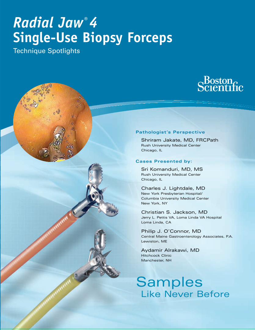
Radial Jaw ® 4 Pathologist’s Perspective Shriram Jakate, MD, FRCPath Rush University Medical Center Chicago, IL Cases Presented by: Sri Komanduri, MD, MS Rush University Medical Center Chicago, IL Charles J. Lightdale, MD New York Presbyterian Hospital/ Columbia University Medical Center New York, NY Christian S. Jackson, MD Jerry L. Pettis VA, Loma Linda VA Hospital Loma Linda, CA Philip J. O’Connor, MD Central Maine Gastroenterology Associates, P.A. Lewiston, ME Aydamir Alrakawi, MD Hitchcock Clinic Manchester, NH Like Never Before Samples
Transcript

Indications, Contraindications, Warnings and Instructions for Use can be found in the product labeling supplied with each device.CAUTION: Federal (USA) law restricts this device to sale by or on the order of a physician.

Boston Scientific CorporationOne Boston Scientific PlaceNatick, MA 01760-1537www.bostonscientific.com

Ordering Information1.800.225.3226

© 2009 Boston Scientific Corporationor its affiliates. All rights reserved.

SME11480 3M June 2009

Radial Jaw®4Single-Use Biopsy ForcepsTechnique Spotlights

Pathologist’s Perspective

Shriram Jakate, MD, FRCPathRush University Medical Center

Chicago, IL

Cases Presented by:

Sri Komanduri, MD, MS Rush University Medical Center

Chicago, IL

Charles J. Lightdale, MDNew York Presbyterian Hospital/

Columbia University Medical Center

New York, NY

Christian S. Jackson, MD Jerry L. Pettis VA, Loma Linda VA Hospital

Loma Linda, CA

Philip J. O’Connor, MDCentral Maine Gastroenterology Associates, P.A.

Lewiston, ME

Aydamir Alrakawi, MDHitchcock Clinic

Manchester, NH

Like Never BeforeSamples

493551.BostonSci.Q6.Cov 6/16/09 8:32 PM Page F

Figure 1Optimally oriented piece from

Barrett’s mucosa showing high grade dysplasia and lack

of surface maturation (H&E stain, magnification x100).

Figure 2ABiopsy of distal esophagus throughstandard forceps showing lack ofgood orientation and difficulty in

diagnosis of mild GERD (H&E, magnification x100).

Figure 2BBiopsy of distal esophagus through

Radial Jaw® 4 (RJ4) Forceps showing good orientation and ease

in diagnosis of mild GERD atexactly the same magnification (H&E, magnification x100).

Advantages for pathologists

1. Ease of optimal orientation of the biopsy specimen by virtue of larger size and

consistent inclusion of muscularis mucosa. This enables embedding the tissue on its

side and creating sections with proper orientation. Optimal orientation can be crucial

in instances such as evaluation of lack of surface maturation in dysplasia (Figure 1),

assessing villous height for gluten sensitive enteropathy in duodenal biopsies,

accurately measuring thickness of collagen band in collagenous colitis and looking for

tall vascular pegs in GERD (Figures 2 A and B).

Advantages for both pathologists and gastroenterologists

1. Ability to sample, confirm and evaluate submucosal masses such as GIST, leiomyoma

(Figure 3), carcinoid, pancreatic rest and lipoma among others.

2. Furnishing wider and deeper surveillance samples in conditions such as Barrett's

(Figure 4 A and B) and ulcerative colitis. Often the specimen size is at least twice

as big, essentially doubling the total sample size for the same number of pinches

compared to the standard forceps.

3. Ability to offer staging information in conjunction with EUS in specific rare superficial

tumors such as localized intramucosal or early gastric carcinoma (Figure 5) with

negative lateral and deep submucosal margins.

Shriram Jakate, MD, FRCPathAssociate Professor of PathologyAdjunct Associate Professor of Gastroenterology and HepatologyRush University Medical Center, Chicago, IL

Pa

th

ol

og

is

t’

s

Pe

rs

pe

ct

iv

e

Figure 3Gastric biopsy showing submucosalspindled cell tumor, immunohisto-chemically confirmed as leiomyoma

(H&E, magnification x200).

Figure 4ABiopsy of Barrett’s mucosa

through standard forceps showing a size of 1740 x 1035 µm

(H&E, magnification x40).

Figure 4BBiopsy of Barrett’s mucosa

through RJ4 Forceps showing a size of 2924 x 1839 µm

(H&E, magnification x40).

Figure 5Biopsy of intramucosal gastric

carcinoma showing uninvolved submucosa and free deep margin

(H&E, magnification x100).

493551 Cover B 6/9/09 4:31 PM Page B

Figure 12cm fundic submucosal mass

Figure 2Routine H&E demonstrating spindle cell

tumor: RJ4 sampling

Figure 3 Figure 4C-Kit (-)

Sri Komanduri, MD, MSAssistant Professor of MedicineDirector, EUSSection of Gastroenterology and NutritionRush University Medical CenterChicago, IL

History

A 43-year-old female underwent a routine upper endoscopy for dyspepsia. An incidental

2 cm submucosal mass was found in the fundus of the stomach. She was referred for

endoscopic ultrasound (EUS). EUS demonstrated a 2 cm mass arising from and limited to

the muscularis propria (Figure 1).

Procedure

At this time, fine needle aspiration (FNA) and jumbo biopsies were obtained using the

Radial Jaw® 4 (RJ4) Jumbo Biopsy Forceps. Cytology from the FNA was suggestive

of a spindle cell neoplasm but tissue was insufficient for immunohistochemistry. The

biopsy specimens from the RJ4 Forceps were also diagnostic for a spindle cell neoplasm

(Figure 2). The RJ4 Forceps also provided enough tissue for immunohistochemistry

(Figure 3). The tumor was determined to be c-kit (-), S100(-) (Figure 4), and SMA (+) (Figure

5). This combination is diagnostic for a leiomyoma. As a result, surgery was avoided.

Post-Procedure

The patient will have a surveillance EGD in 1 year to ensure this mass is not enlarging.

A leiomyoma is a benign muscular tumor with negligible malignant potential and likely

does not account for this patient’s dyspepsia. On the other hand, a gastrointestinal

stromal tumor (GIST) has significant malignant potential and appears no different than

the leiomyoma endoscopically. The differentiation of these two muscle tumors is made

by immunohistochemistry. GISTs are c-kit (+) while leiomyomas are not. Standard

biopsy forceps have been inadequate for diagnosis of submucosal masses. Standard

FNA has also had suboptimal results. The ability of the RJ4 Jumbo Biopsy Forceps

to not only obtain the diagnosis but allow for immunohistochemistry is extremely prom-

ising and should be incorporated into diagnostic algorithms for submucosal masses.

Case presented by:

1

GASTRIC SUBMUCOSAL MASS

Case 1

Figure 5SMA (+)

493551.P01 6/15/09 2:13 PM Page 1

Figure 1Long segment Barrett’s Esophagus

Figure 2Barrett’s Esophagus with Methylene Blue Staining.

Darker stain is suggestive of dysplasia.

Figure 3Routine H&E staining of biopsies

obtained with RJ3 Forceps

Sri Komanduri, MD, MSAssistant Professor of MedicineDirector, EUSSection of Gastroenterology and NutritionRush University Medical CenterChicago, IL

Case presented by:

Figure 5Morphometric analysis comparing RJ3 and RJ4 biopsies

Case 2

2

Figure 4Routine H&E staining of biopsies

obtained with RJ4 Forceps

Foci of LGD

History

A 44-year-old female with no significant medical history underwent upper endoscopy for long

standing Gastroesophageal Reflux (GER). The patient had been on proton pump inhibitor

therapy daily for 10 years with moderate control of her symptoms. Despite this therapy, she

still had significant heartburn. As a result, upper endoscopy was pursued.

Procedure

EGD revealed an irregular Z-line and long segment of salmon colored mucosa suggestive

of Barrett’s esophagus (BE) (Figure 1). Vital Staining with Methylene blue was performed

to further delineate the intestinal metaplasia (Figure 2). Four quadrant sampling of this area

was performed with Radial Jaw® 3 (RJ3) Large Capacity Biopsy Forceps and Radial Jaw® 4

(RJ4) Jumbo Biopsy Forceps.

Post-Procedure

Histopathology revealed a significant difference between the RJ3 and RJ4 (Figures 3 and 4).

The samples from both forceps demonstrated intestinal metaplasia or BE, but only the

samples from the RJ4 forceps demonstrated evidence of low grade dysplasia. The mean RJ4

sample was 2X wider and 2.6X deeper (width 3251 microns or 3.3 mm and depth 2573

microns or 2.6 mm) versus the standard biopsy sample (average width 1591 microns or

1.6 mm and average depth 986 microns or 1 mm) (Figure 5). While the yield in this case is

not necessarily based on depth, the dramatic increase in surface area and completeness of

the biopsy clearly allows for a better assessment for dysplastic change. This is an extremely

exciting finding in the arena of tissue sampling in BE. A prospective investigation is underway

to determine if this can truly be applied universally.

COMPARISON OF RADIAL JAW® 3 LARGE CAPACITY TO RADIAL JAW® 4 JUMBO BIOPSY FORCEPS

493551.P02 6/15/09 2:13 PM Page 2

Figure 2EUS suggestive of lipoma

Figure 3Fat extruding from mass

History

A 52-year-old male was referred for evaluation of a rectosigmoid submucosal mass (Figure 1).

The patient had undergone a routine screening colonoscopy 4 weeks prior. During this

examination, a 3 cm submucosal mass was seen in the rectosigmoid junction. The mass was

smooth without ulceration or umbilication. Initial mucosal biopsies taken with Radial Jaw® 3

(RJ3) Large Capacity Forceps demonstrated only normal overlying mucosa.

Procedure

At this time, we performed endoscopic ultrasound (EUS) of this lesion using a 20mHz through the

scope ultrasound probe (Figure 2). The mass was limited to the submucosa and was hyperechoic,

suggestive of a lipoma. However, given the size of this lesion, a carcinoid tumor had to be

excluded. Fine-needle aspiration (FNA) with a curvilinear echoendoscope is very difficult and not

practical beyond the rectum. We subsequently utilized the Radial Jaw® 4 (RJ4) Jumbo Biopsy

Forceps to perform a “tunnel” biopsy. After 2 bites were taken, a moderate amount of fat was

seen extruding from the mass (Figure 3).

Post-Procedure

Histopathology confirmed a lipoma. The patient was reassured and no further diagnostic workup

was required.

Discussion

Submucosal masses of the gastrointestinal tract can be diagnostic dilemmas. Routine biopsies

with standard forceps rarely provide enough tissue for diagnostic yield. Given the importance of

distinguishing carcinoid tumors from lipomas and gastrointestinal stromal tumors from

leiomyomas, adequate tissue sampling is essential. In the upper GI tract, EUS with FNA is the diag-

nostic test of choice for lesions > 1 cm. However, special consideration for the colon is necessary.

FNA is impractical for lesions beyond the rectum at this time or for any lesion < 1.0 cm.

We recently demonstrated a substantial increase in tissue depth and width with the new RJ4

Jumbo Biopsy Forceps. We investigated 10 patients with Barrett’s Esophagus and found a mean

increase of 0.8 mm in depth (deep submucosa) and 1 mm in width. In addition, we found

33% more dysplasia with the RJ4 Jumbo than with the RJ3 Large Capacity Biopsy Forceps. There

were no complications. The increase in sample size should significantly impact our ability to detect

dysplasia. The increase in depth with these forceps allows us to further address tissue sampling

in submucosal lesions without complications.

We have utilized the RJ4 Jumbo Biopsy Forceps in multiple patients with submucosal lesions of

the GI tract. Specifically, we have had 2 patients with GIST in which the tissue obtained with the

RJ4 was significantly greater than that for EUS-FNA. In these lesions, it is essential

to obtain sufficient tissue for histopathology and immunohistochemistry to allow for proper

diagnosis. In our case, we were able to identify a benign lesion and avoid any further workup and

cost to the patient with the utility of these forceps. The RJ4 Jumbo Biopsy Forceps appear to be

an extremely promising tool when sampling submucosal lesions of the gastrointestinal tract.

Case presented by:

3

Sri Komanduri, MD, MSAssistant Professor of MedicineDirector, EUSSection of Gastroenterology and NutritionRush University Medical CenterChicago, IL

Case 3

Case 3

COLONIC SUBMUCOSAL MASS

Figure 1Rectosigmoid submucosal mass

493551.P03 6/15/09 2:13 PM Page 3

Figure 1This endoscopic image shows the RJ4

forceps emerging from the endoscope in a retroflexed position for biopsy of the

squamo-columnar junction.

Figure 2Photo through the endoscope to show

the fresh biopsy specimens.

Figure 3Low-power histological image shows

Barrett’s metaplasia below squamous mucosa at the esophago-gastric junction.

Figure 3aHigh-power histological image shows

the Barrett’s glands.

Charles J. Lightdale, MDProfessor of Clinical MedicineNew York Presbyterian Hospital/Columbia UniversityMedical CenterNew York, NY

Case presented by:

Case 4

4

History

A 76-year-old man was treated 2 years ago for high-grade dysplasia in long-segment

Barrett’s esophagus with experimental photodynamic therapy using 5-ALA.

Endoscopies have shown possible residual Barrett’s esophagus in short tongues at the

esophago-gastric junction, but no intestinal metaplasia was present on biopsy with

re-usable large capacity forceps.

Procedure

Endoscopy again showed an irregular squamo-columnar junction without ulceration or

nodularity seen at the esophago-gastric junction on either direct or retroflexed view. Using

an Olympus 1T endoscope, biopsies were taken using the single-use Radial Jaw® 4 (RJ4)

Jumbo Biopsy Forceps, in a four-quadrant manner (Figure 1). The RJ4 Forceps operates

easily and well from the retroflexed position. Excellent biopsies can be obtained even

with a tangential approach because of the hinged jaws on the RJ4, which allow

excellent positioning of the forceps on the target. Using the turn-and-suction biopsy

method, large biopsies were obtained (the GI-nurse described them as “huge”), and these

were sent for histological analysis (Figure 2). The larger specimens obtain a greater area

of mucosa allowing better orientation in the pathology laboratory, and the examination

of a greater number of Barrett's glands for the presence of dysplasia.

Post-Procedure

Histological analysis showed a residual 1 mm focus of intestinal metaplasia with some

metaplastic glands under squamous epithelium (Figure 3 and 3a). This image shows

partially “buried” Barrett's metaplasia beneath squamous epithelium.

Based on the biopsy results, ablation of the residual area of Barrett’s epithelium at the

esophago-gastric junction was carried out using radiofrequency energy.

BARRETT’S ESOPHAGUS

493551.P04 6/15/09 2:13 PM Page 4

Case 6

5

Case 5

LARGE GASTRIC ULCER FOUND TO BE A GASTRIC CANCER

Figure 14 cm ulcer base.

Figure 24cm ulcer located along the greater

curvature of the stomach.

Figure 3Gastric ulcer after biopsies with RJ4.

Christian S. Jackson, MDAssistant Professor of MedicineSection of GastroenterologyJerry L. Pettis VA, Loma Linda VA HospitalLoma Linda, CA

History

A 60-year-old male with a known history of Helicobacter pylori gastritis was initially seen

in consultation for evaluation of a microcytic anemia and a drop in hemoglobin. He was

recently diagnosed with a CVA and his neurologists were concerned to place him on

ASA, thus an upper endoscopy was requested. Patient denied prior NSAID use and was

not taking a PPI.

Procedure

His initial endoscopy revealed a 4 cm clean based ulcer with irregular and friable

margins in the mid gastric body (Figure 1 and 2). A total of twelve biopsies with Radial

Jaw® 3 (RJ3) Biopsy Forceps were taken from the margins of the ulcer. Since it was unclear

if he was previously treated for H.pylori, biopsies of the antrum and body were taken.

The initial biopsies from the ulcer were interpreted as highly atypical cells suggestive

of adenocarcinoma. Prior to a surgical oncologic evaluation a diagnosis needed to be

confirmed. It was decided that the patient be scheduled for a repeat upper endoscopy and

Radial Jaw® 4 (RJ4) Jumbo Biopsy Forceps would be used. Eight biopsies of the ulcer mar-

gin were taken (Figure 3). The patient subsequently underwent a CT Scan which revealed a

hypodense lesion in the left lateral segment. A PET Scan was then performed which showed

no evidence for liver or lung metastasis. A biopsy of the lesion revealed that this lesion was

a hemangioma.

Post-Procedure

Once a diagnosis was made, the patient was referred to surgical oncology. He underwent

a distal subtotal gastrectomy with D1 lymph node dissection and Roux-en-Y reconstruction.

The ability of the RJ4 Jumbo Biopsy Forceps not only allowed for a definitive diagnosis to

be made, but helped direct the appropriate intervention for the patient to occur.

Case presented by:

493551.P05 6/15/09 2:13 PM Page 5

History

A 66-year-old woman presented with obstructive jaundice. Imaging studies revealed only

a dilated bile duct and haziness in the head of the pancreas. There was no evidence of a

mass lesion.

Procedure

The patient underwent an ERCP which revealed a discolored extrinsic compression of the

duodenum, superior to the ampula of vator. There was no evidence of breakdown of the

mucosal lining. Biopsies were performed using the Radial Jaw® 4 Jumbo Forceps. The patient

then underwent routine cannulation of her common bile duct and pancreatic duct. The

pancreatic duct appeared to be distorted in the head. There was obvious compression of the

common bile duct, which was treated with a WALLSTENT® Stent System.

Post-Procedure

Biopsies were reviewed the following day and adenocarcinoma was noted at the

deepest edges of the biopsy.

Philip J. O’Connor, MDCentral Maine Gastroenterology Associates, P.A.Lewiston, ME

Case presented by:

Case 6 & 7

6

Gastric Carcinoma

Gastric Adenocarcinoma

History

A 62-year-old male who four years previously had a cystectomy for a poorly differential carci-

noma of his ureterovesico junction presented with the onset of ascites and elevated CEA in

the fluid with malignant cells. He underwent chemotherapy and responded extremely well. He

abruptly presented with nausea and vomiting and a flat plate of his abdomen demonstrated

apparent gastric outlet obstruction.

Procedure

Upper GI endoscopy was performed with the therapeutic gastroscope. The obstruction

was at the pylorus. It was not typical, however, of neoplasis or ulcer disease. With mild to

moderate pressure on the instrument, it easily passed into the second and third portion of the

duodenum. The patient was dilated with a #15 to #18 CRE TTS balloon. He then underwent

biopsies with the Radial Jaw® 4 (RJ4) Jumbo Forceps.

Post-Procedure

The patient improved for only twenty-four hours. During that time the biopsies were read

and remarkably identified malignant cells at the deepest level of the biopsy specimen (i.e.

infiltrating from without). Cells were consistent with anginal pain. The following day a standard

duodenal stent was placed with complete resolution of the patient’s symptoms.

GASTRIC CARCINOMA & GASTRIC ADENOCARCINOMA

493551.P06 6/15/09 2:13 PM Page 6

Figure 1

Figure 2

Figure 3

Aydamir Alrakawi, MDHitchcock ClinicManchester, NH

History

A 53-year-old male with a history of Crohn’s colitis for more than 20 years presented for

a surveillance colonoscopy. He has been doing well and is having 1-2 bowel movements

a day. He is only taking oral mesalamine.
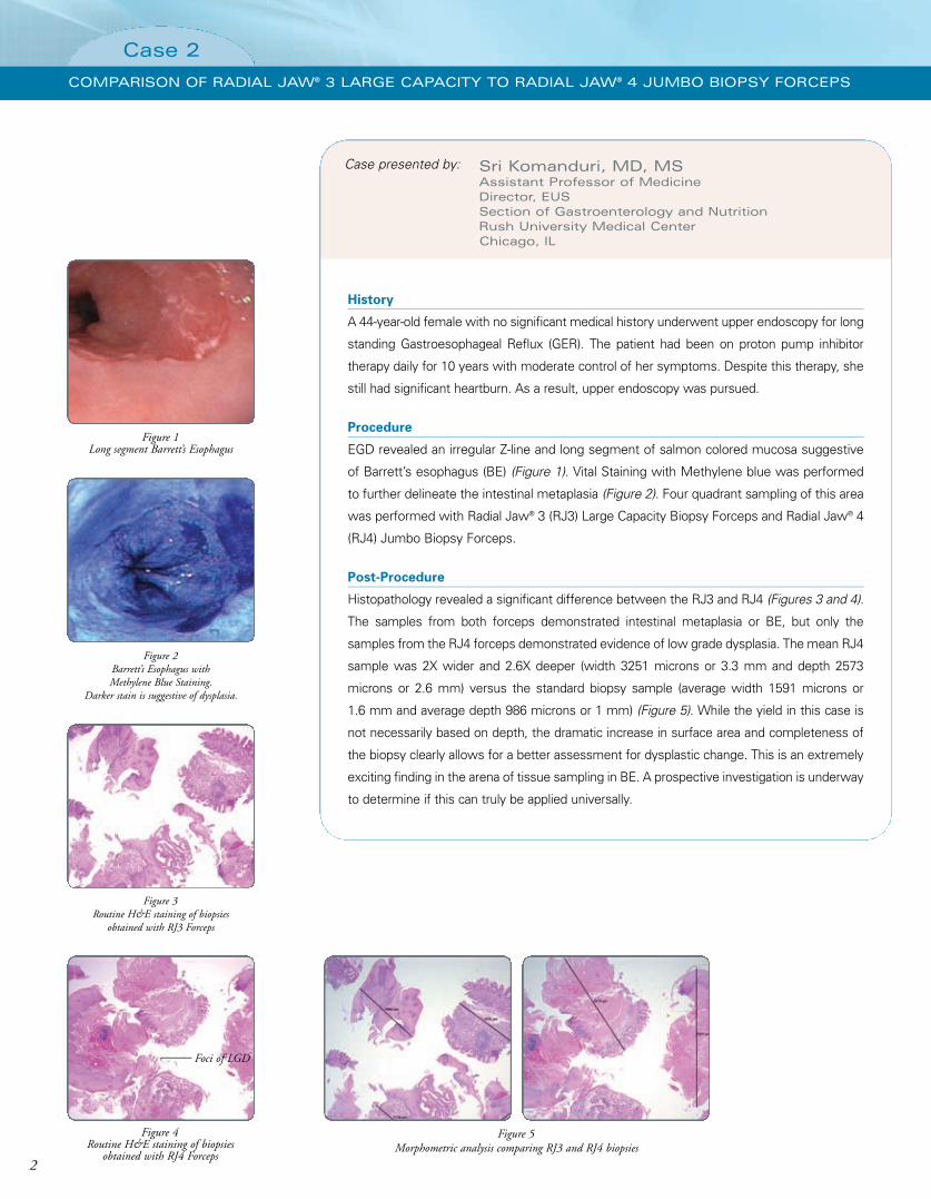
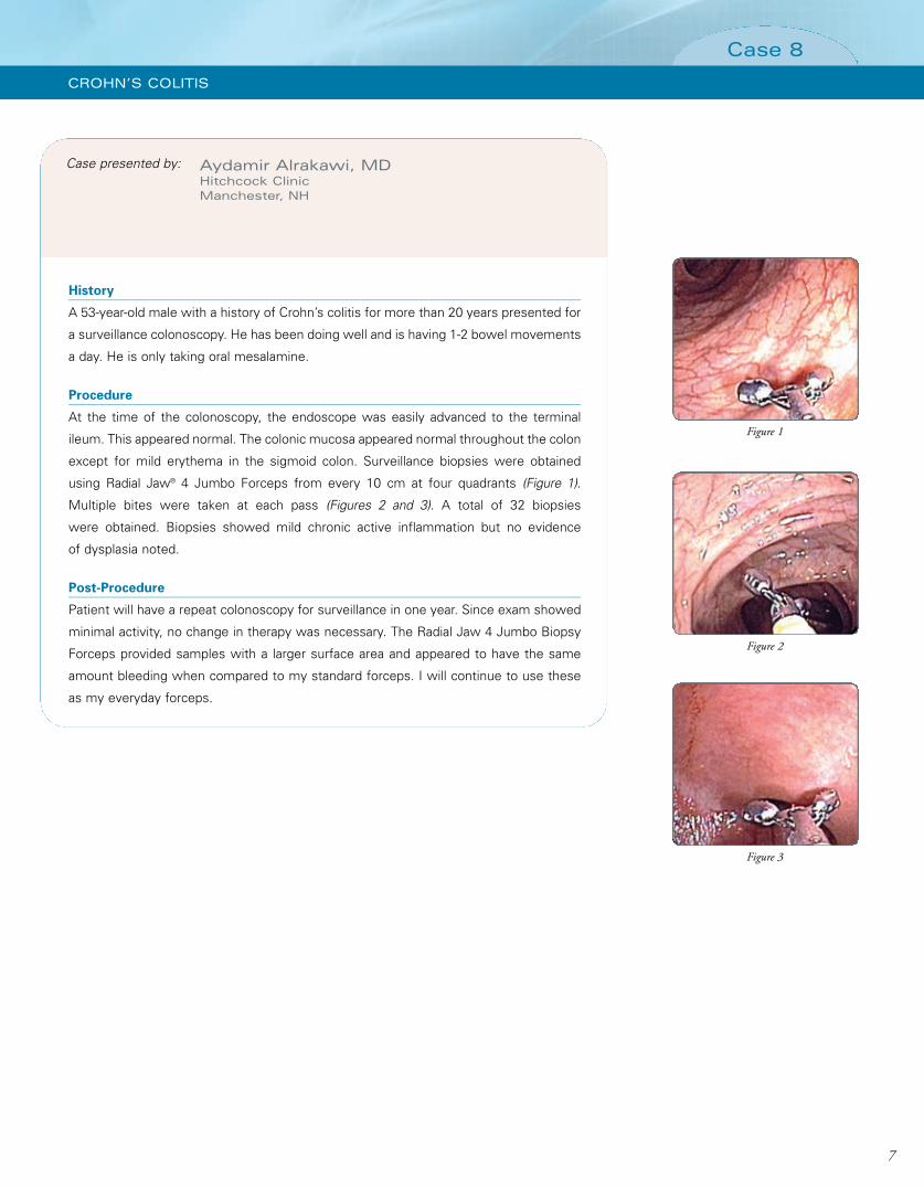
Procedure

At the time of the colonoscopy, the endoscope was easily advanced to the terminal

ileum. This appeared normal. The colonic mucosa appeared normal throughout the colon

except for mild erythema in the sigmoid colon. Surveillance biopsies were obtained

using Radial Jaw® 4 Jumbo Forceps from every 10 cm at four quadrants (Figure 1).

Multiple bites were taken at each pass (Figures 2 and 3). A total of 32 biopsies

were obtained. Biopsies showed mild chronic active inflammation but no evidence

of dysplasia noted.

Post-Procedure

Patient will have a repeat colonoscopy for surveillance in one year. Since exam showed

minimal activity, no change in therapy was necessary. The Radial Jaw 4 Jumbo Biopsy

Forceps provided samples with a larger surface area and appeared to have the same

amount bleeding when compared to my standard forceps. I will continue to use these

as my everyday forceps.

Case presented by:

7

Case 8

CROHN’S COLITIS

493551.P07 6/15/09 2:13 PM Page 7

8

Notes

493551.P08 6/15/09 2:13 PM Page 8

Figure 1Optimally oriented piece from

Barrett’s mucosa showing high grade dysplasia and lack

of surface maturation (H&E stain, magnification x100).

Figure 2ABiopsy of distal esophagus throughstandard forceps showing lack ofgood orientation and difficulty in

diagnosis of mild GERD (H&E, magnification x100).

Figure 2BBiopsy of distal esophagus through

Radial Jaw® 4 (RJ4) Forceps showing good orientation and ease

in diagnosis of mild GERD atexactly the same magnification (H&E, magnification x100).

Advantages for pathologists

1. Ease of optimal orientation of the biopsy specimen by virtue of larger size and

consistent inclusion of muscularis mucosa. This enables embedding the tissue on its

side and creating sections with proper orientation. Optimal orientation can be crucial

in instances such as evaluation of lack of surface maturation in dysplasia (Figure 1),

assessing villous height for gluten sensitive enteropathy in duodenal biopsies,

accurately measuring thickness of collagen band in collagenous colitis and looking for

tall vascular pegs in GERD (Figures 2 A and B).

Advantages for both pathologists and gastroenterologists

1. Ability to sample, confirm and evaluate submucosal masses such as GIST, leiomyoma

(Figure 3), carcinoid, pancreatic rest and lipoma among others.

2. Furnishing wider and deeper surveillance samples in conditions such as Barrett's

(Figure 4 A and B) and ulcerative colitis. Often the specimen size is at least twice

as big, essentially doubling the total sample size for the same number of pinches

compared to the standard forceps.

3. Ability to offer staging information in conjunction with EUS in specific rare superficial

tumors such as localized intramucosal or early gastric carcinoma (Figure 5) with

negative lateral and deep submucosal margins.

Shriram Jakate, MD, FRCPathAssociate Professor of PathologyAdjunct Associate Professor of Gastroenterology and HepatologyRush University Medical Center, Chicago, IL

Pa

th

ol

og

is

t’

s

Pe

rs

pe

ct

iv

e

Figure 3Gastric biopsy showing submucosalspindled cell tumor, immunohisto-chemically confirmed as leiomyoma

(H&E, magnification x200).

Figure 4ABiopsy of Barrett’s mucosa

through standard forceps showing a size of 1740 x 1035 µm

(H&E, magnification x40).

Figure 4BBiopsy of Barrett’s mucosa

through RJ4 Forceps showing a size of 2924 x 1839 µm

(H&E, magnification x40).

Figure 5Biopsy of intramucosal gastric

carcinoma showing uninvolved submucosa and free deep margin

(H&E, magnification x100).

493551 Cover B 6/9/09 4:31 PM Page B

Indications, Contraindications, Warnings and Instructions for Use can be found in the product labeling supplied with each device.CAUTION: Federal (USA) law restricts this device to sale by or on the order of a physician.

Boston Scientific CorporationOne Boston Scientific PlaceNatick, MA 01760-1537www.bostonscientific.com

Ordering Information1.800.225.3226

© 2009 Boston Scientific Corporationor its affiliates. All rights reserved.

SME11480 3M June 2009

Radial Jaw®4Single-Use Biopsy ForcepsTechnique Spotlights

Pathologist’s Perspective

Shriram Jakate, MD, FRCPathRush University Medical Center

Chicago, IL

Cases Presented by:

Sri Komanduri, MD, MS Rush University Medical Center

Chicago, IL

Charles J. Lightdale, MDNew York Presbyterian Hospital/

Columbia University Medical Center

New York, NY

Christian S. Jackson, MD Jerry L. Pettis VA, Loma Linda VA Hospital

Loma Linda, CA

Philip J. O’Connor, MDCentral Maine Gastroenterology Associates, P.A.

Lewiston, ME

Aydamir Alrakawi, MDHitchcock Clinic

Manchester, NH

Like Never BeforeSamples

493551.BostonSci.Q6.Cov 6/16/09 8:32 PM Page F

pacificb
Typewritten Text
pacificb
Typewritten Text
pacificb
Typewritten Text
pacificb
Typewritten Text
pacificb
Typewritten Text
pacificb
Typewritten Text
pacificb
Typewritten Text
ENDO-151004-AA July 2013
pacificb
Typewritten Text
pacificb
Typewritten Text
pacificb
Typewritten Text
pacificb
Typewritten Text
pacificb
Typewritten Text

Recommended