29
Headlines 1
| Date post: | 15-Jul-2015 |
| Category: |
Health & Medicine |
| Upload: | niddhi-bajaj |
| View: | 188 times |
| Download: | 6 times |
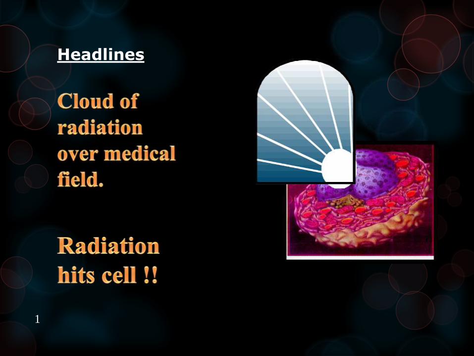
Headlines
1

2

RADIATION PROTECTION
3
Introduction
Radiation protection the main
principal is to those things
which will minimize exposure
of patient and dental personnel
and still provide benefits for
present use of diagnostic
radiography.4
All ionizing radiations are harmful
and produce biological change in
living cells.
It can cause increase risk of
cancers, birth defects ,catracts, etc
CARDINAL PRINCIPAL -
ALARA
AS LOW AS REASONABLE
ACHIEVABLE5

The amount of radiation at
any point depends upon the
distance from the point
source of radiation
Intensity ∞ 1/𝒅𝒊𝒔𝒕𝒂𝒏𝒄𝒆𝟐
6

Sources
Natural
External
• Cosmic
• Terrestrial
Internal
Artificial
Medical
X-ray diagnosis
Nuclear medicine
Consumer
Occupational
Nuclear fuel cycle
Miscellaneous7

Sources in Dental Radiology
–from source
–from irradiated tissues of patients.
– from X-ray tube head
8
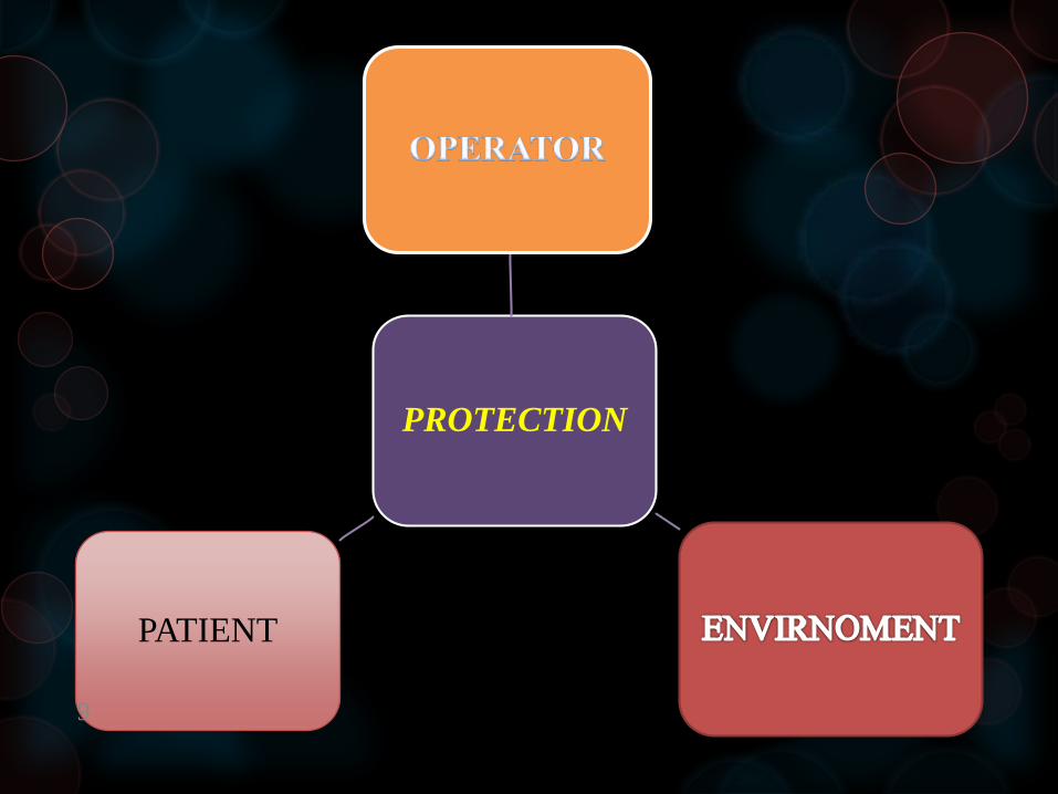
PROTECTION
PATIENT
9

Protection for operator
Primary X-ray beam
Scattered radiation from irradiated tissues of patient
Leakage radiation through tube head housing.
Scattered X-ray from filters, cones.10

1.Protection from primary source
Effort to be made that operator has a suitable room or
take a suitable position behind wall or a barrier.
Position distance rule - operator should stand at least
6 feet away from source or at the angle of 90 to 135.
If no barrier operator should use lead apron.
Film should be holded by film holding devices.
Avoid holding X-ray tube head of machine. The
suspension arms should be adequately maintained to
prevent housing movement and drift.
11

12

Protection against leakage radiation
Neither the tube housing nor
the cone should be hand held
during exposure.
The machine should be
periodically checked for the
leakage.13

Protection from secondary and scattered radiation
Use of high speed films.
Replace short plastic cone with open ended
lead lined cone.
Use of collimator to reduce diameter of beam.
Use of film badge / Pocket dosimeter for
personnel radiation monitoring.
Adequate filtration of primary beam.
14
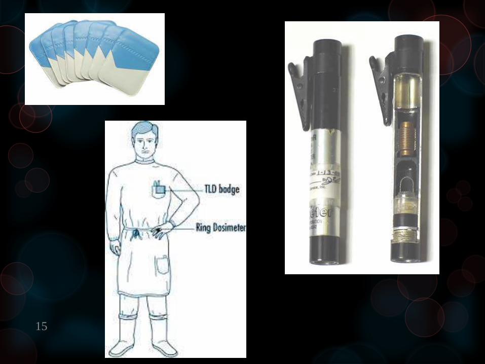
15

Protection for patient
Prior exposure
During exposure
After exposure
16

Prior exposure
Most important is proper prescription
and details of patients to be taken.
Special care to be taken of women
especially during pregnancy period thus
to avoid complication to fetus.
17
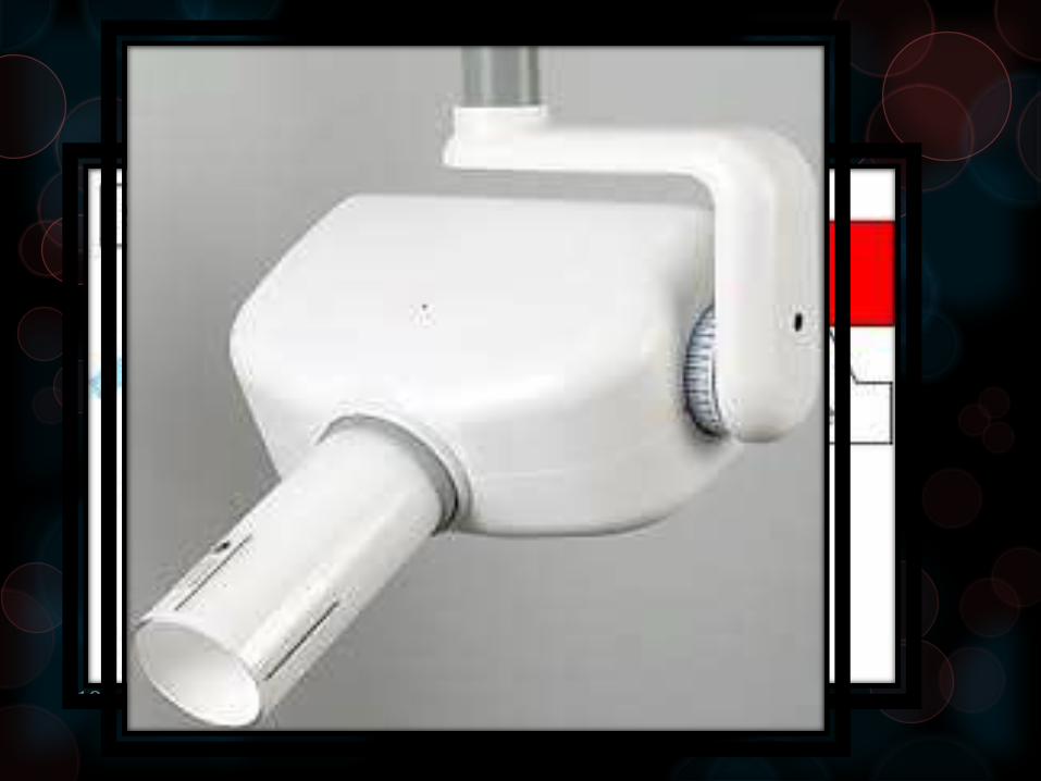
Equipment
1.Filters
2.Collimation of bean
3.Position indicating devices
18

During exposure
Thyroid collars
Lead aprons
Fast films
Film holding devices
Selection of proper exposure factor
Emulsifying screens
Timers
Good technique19
After exposure
Proper film handling.
Proper processing
Proper interpretation.
20

Protection of envirnoment
Beam directed to patient
Lead incorporated in furniture
Patients position
Barium to be used as plaster
(barium plaster)
21

Walls of 3 inch concrete.
Continue education
Radiation room at corner and
caution signs.
22
Various monitoring devices
23
Electrical
Heat
Light
Chemical
Thermoelectric

Electrical
Timble chamber
Ionization chamber
Proportional counter
Griget counter
Heat
Calorimeter
24

Film badge
Mercury and copper
filters in rectangle shape
Blackness is to be noted.
25
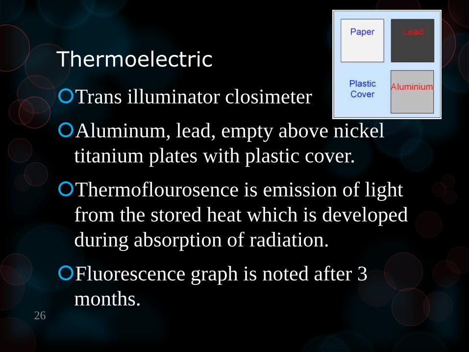
Thermoelectric
Trans illuminator closimeter
Aluminum, lead, empty above nickel
titanium plates with plastic cover.
Thermoflourosence is emission of light
from the stored heat which is developed
during absorption of radiation.
Fluorescence graph is noted after 3
months.26

Every thing comes with a
boon and a curse.
Its just on us how we use it.
27
Conclusion
Radiograph is the most important diagnostic tool in
dentistry.
Thus proper protection should be taken while exposing the patient.
28

THANK YOU


