60
Radiation Safety for Radiographers Bruce Busby Certified Health Physicist
| Date post: | 25-Dec-2015 |
| Category: |
Documents |
| Upload: | mildred-baker |
| View: | 241 times |
| Download: | 0 times |
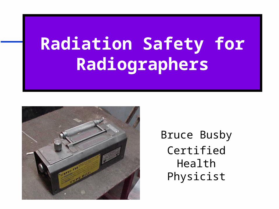
Radiation Safety for Radiographers
Bruce Busby
Certified Health Physicist

Agenda
Why Training?
Radiation and Sources of Radiation
Radiation Protection
Dosimetry
Instruments
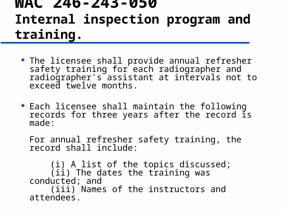
WAC 246-243-050 Internal inspection program and training.
The licensee shall provide annual refresher safety training for each radiographer and radiographer's assistant at intervals not to exceed twelve months.
Each licensee shall maintain the following records for three years after the record is made:
For annual refresher safety training, the record shall include:
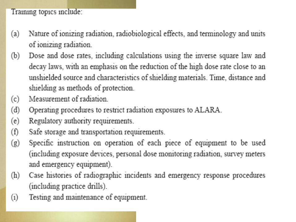
(i) A list of the topics discussed; (ii) The dates the training was conducted; and (iii) Names of the instructors and attendees.


Regulatory Authority Nuclear Regulatory Commission (16)
– Idaho, Alaska, Hawaii, Montana, Wyoming Agreement States (34)
– Washington, Oregon, California, Utah
Washington State WAC 246 220-254– Department of Health, Office of Rad Protection
Oregon ORS 453.605-453.807– Department of Human Services, Public Health
Division, Rad Protection Services

Sources of RadiationSources of Radiation
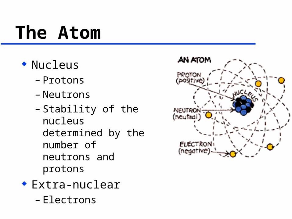
The Atom
Nucleus– Protons– Neutrons– Stability of the
nucleus determined by the number of neutrons and protons
Extra-nuclear– Electrons
Radiation
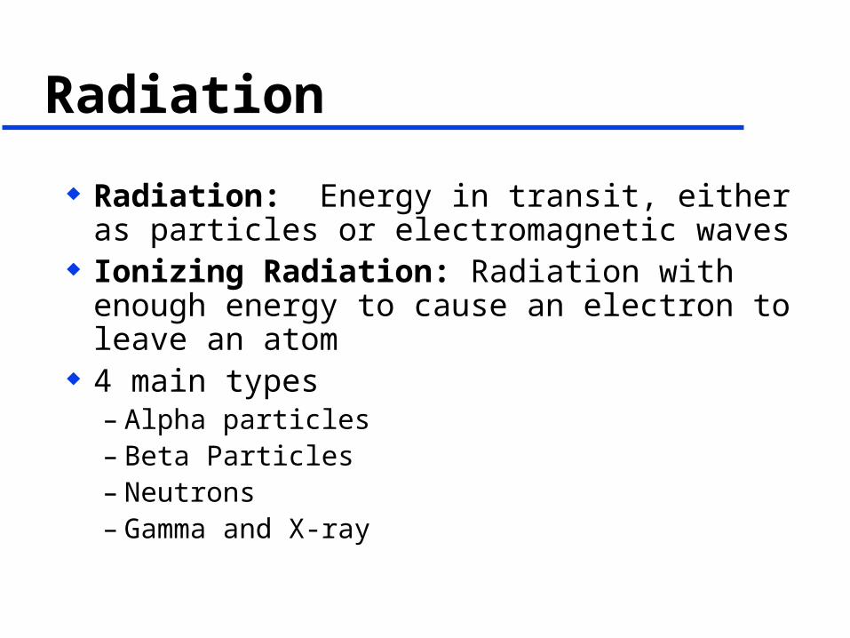
Radiation: Energy in transit, either as particles or electromagnetic waves
Ionizing Radiation: Radiation with enough energy to cause an electron to leave an atom
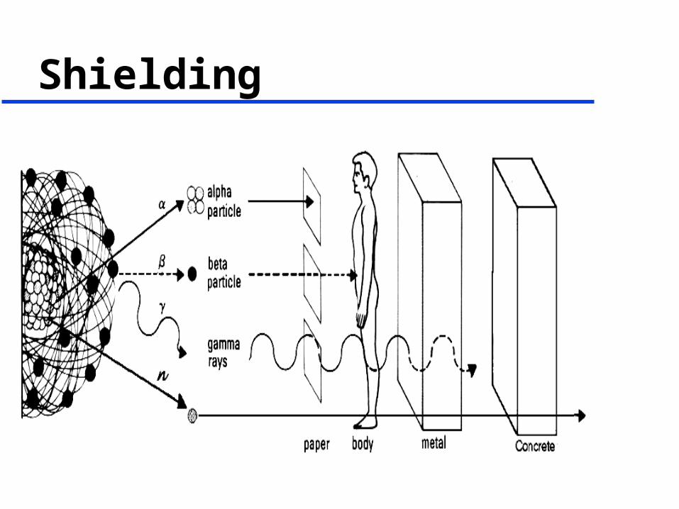
4 main types– Alpha particles– Beta Particles– Neutrons– Gamma and X-ray

Types of Radiation42
00
Alpha
Beta
Gamma and X-rays
Neutron
Paper Plastic Lead Concrete
01
n10
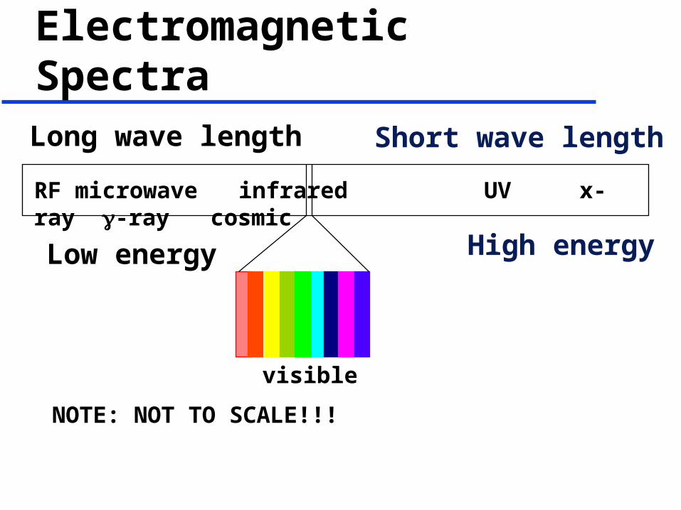
Electromagnetic Spectra
RF microwave infrared UV x-ray -ray cosmic
Low energy High energy
Long wave length Short wave length
NOTE: NOT TO SCALE!!!
visible
Radioactive Material
Radioactive material consists of atoms with unstable nuclei
The atoms spontaneously change (decay) to more stable forms and emit radiation
A person who is contaminated has radioactive material on their skin or inside their body

Example of Radioactive Material
Gamma Rays(317 and 296 keV)
Parent NucleusIr-192
Daughter NucleusPt-192
0~1

Half-Life
0200
400600800
10001200
New 1Half-Life
2Half-Lives
3Half-Lives
4Half-Lives
Activity
Ir-192 – 73.8 days
Co-60 – 5.27 years

X-Ray Production (Bremsstrahlung)
ElectronX-Ray
Target NucleusTungsten
Cathode(-)
Anode (+)
X-Rays
Radiation Interactions with Matter
Radiation with enough energy causes ions to be formed
The amount of ions formed is based on the energy deposited
Use this principle to our advantage for shielding
Causes all effects of radiation – good and bad

Radiation ProtectionRadiation Protection
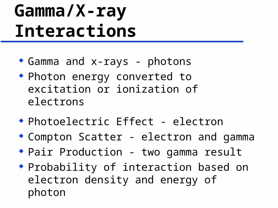
Gamma/X-ray Interactions
Gamma and x-rays - photons Photon energy converted to excitation or
ionization of electrons
Photoelectric Effect - electron Compton Scatter - electron and gamma Pair Production - two gamma result Probability of interaction based on electron
density and energy of photon

Units
Roentgen Rad Rem

Roentgen (R)
Measure of exposure Charge produced in a specific
volume by gamma or x-rays 1 R = 2.58 x 10⁻⁴ C/kg SI unit is C/kg Meters (Ion Chambers and GM
detectors) often read out in mR/hr

RAD
Radiation Absorbed Dose Energy deposited per unit mass 1 rad = 100 erg/gm Does not account for different radiation
damages SI unit is the gray (Gy) 100 rad = 1 Gy
REM
Measure of Biological Damage Effective Dose Equivalent Dose Effective TEDE and CEDE rad x QF = rem SI unit is sievert (Sv) 100 rem = 1 Sv

Good News
For protection – x-ray, beta and gamma radiation
1 R 1 rad 1 rem
For alpha and neutron, have to take into account the quality factor
rad x QF = rem QF for alpha is 20, neutron 2-20, gamma/beta
is 1

Measures of Radioactivity
The quantity of radioactive material present at a given time:
Curie (Ci) : 3.7x10¹⁰ disintegration per second (dps)
or
Becquerel (Bq): 1 dps
Radiological Controls Radiological Controls

ALARA
As Low As Reasonably Achievable
REASONABLE is a key word here
Minimizing the External and Internal radiation exposure
Can you reduce your dose to Zero?????

ALARA
Philosophy of keeping doses low as Reasonable
Used to reduce the risks No dose without benefit Additional controls Administrative – procedures, regulations Engineered - design Still comes down to
– Time, Distance and Shielding

External Methods
Time, Distance and Shielding– Reduce time exposed– Increase distance from source– Use shielding between you and the source
Reduce your waste storage Properly store material Set up lab for work stations away from
sources

Time
Reduce your time in radiation fields Preplan Prep Practice
Know your area, work in low radiation fields as much as possible
Distance
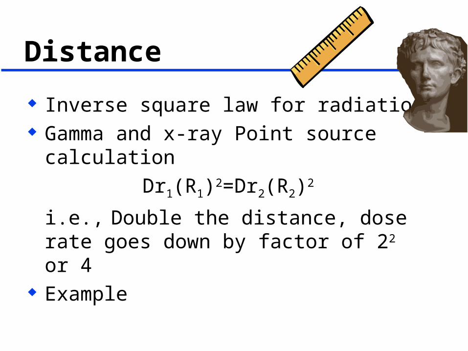
Inverse square law for radiation Gamma and x-ray Point source calculation
Dr1(R1)2=Dr2(R2)2
i.e., Double the distance, dose rate goes down by factor of 22 or 4
Example
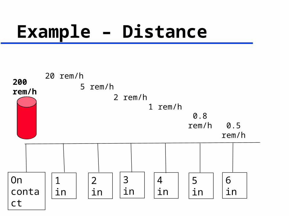
Example – Distance
On contact
200 rem/h
1 in 2 in 3 in 4 in 5 in 6 in
20 rem/h5 rem/h
2 rem/h1 rem/h
0.8 rem/h0.5 rem/h

Shielding
Shielding - use of material to reduce transmitted radiation
A wall or partition may not be a safe shield for persons on the other side.
More dense, the better shield

Shielding - Regulation
The maximum exposure rate limits for storage containers and source changers with the sealed source in the shielded position are:– (a) 2 millisieverts (200 millirem) per hour at
any exterior surface; and– (b) 0.1 millisieverts (10 millirem) per hour at
one meter from any exterior surface.
WAC 246-243-040 (5) Equipment performance requirements
Shielding
Signs and Labels
Caution RAM– Caution Radioactive Materials
Radiation Area High Radiation
All areas in which industrial radiography is being performed shall be conspicuously posted as required
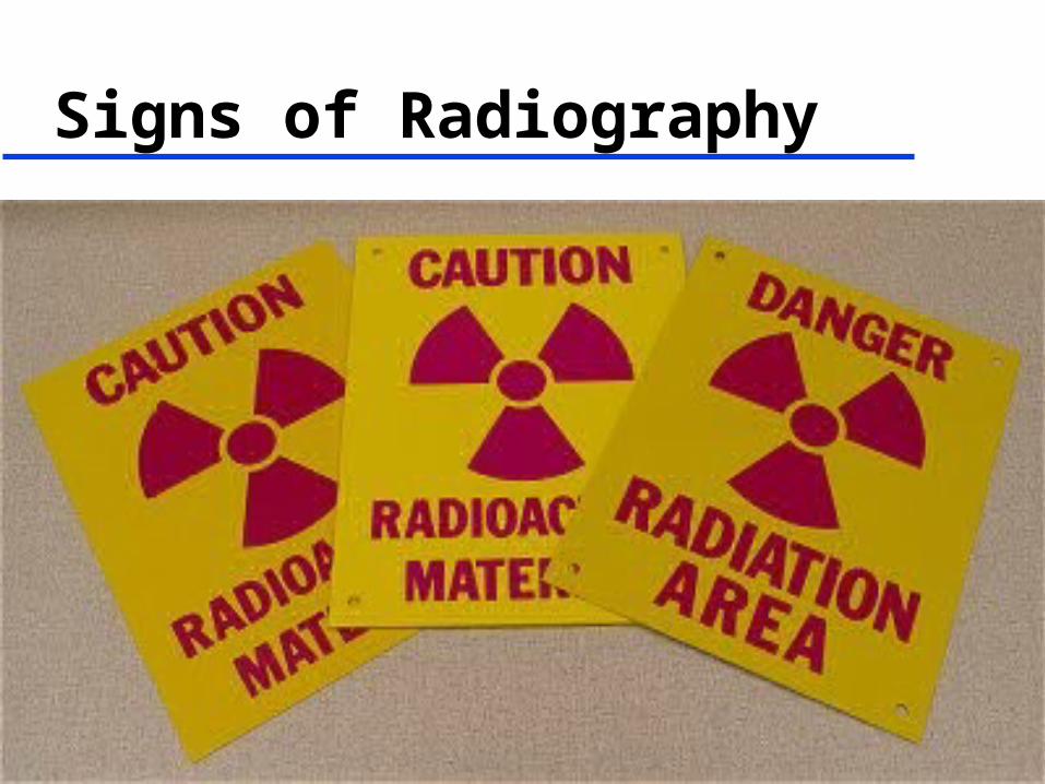
Signs of Radiography
Caution RAM Posting/Labeling
On RAM Room or storage where radioactive
materials can be found
Regulation states minimum amount that requires posting
Public Dose Limits
2 mrem in one hour
100 mrem per year

Radiation Area
Dose rates where a person can receive a whole body dose of
5 to 100 mrem in one hour
30 centimeters from the source Do not loiter

High Radiation Area
Dose rates where a person can receive a whole body dose of
100 mrem in one hour
Requires extra precautions Caution or Danger

Very High Radiation Area
Dose rates where a person can receive a whole body dose of
500 rads in one hour
Grave Danger
Dosimetry Dosimetry

WAC 246-243-150 Says…
A licensee may not permit any individual to act as a radiographer or as a radiographer's assistant unless, at all times during radiographic operations, the individual wears – a direct reading pocket dosimeter,– an alarming rate meter, and – a NAVLAP personnel dosimeter on the trunk of
the body.
Note - In permanent facilities where other appropriate alarming or warning devices are in routine use, the wearing of an alarming rate meter is not required.
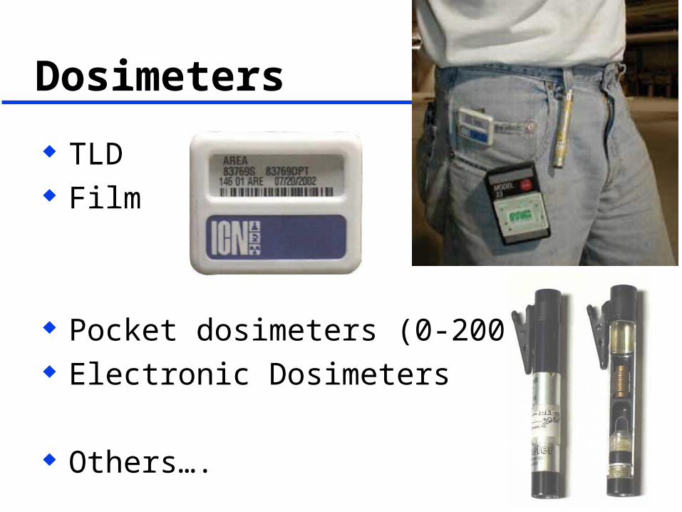
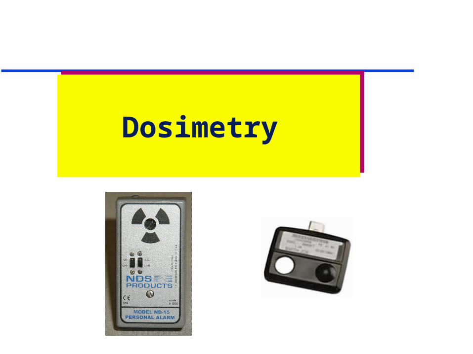
Dosimeters
TLD Film
Pocket dosimeters (0-200 mrem) Electronic Dosimeters
Others….

Pocket Ion Chamber

How to Wear
Whole Body– Must be worn on the front of the body– between waist and neck– facing out

Dose Records
Access– Private– Upon request– Annual report
Legal Permanent Can be requested after you leave only by
yourself

Instruments Instruments
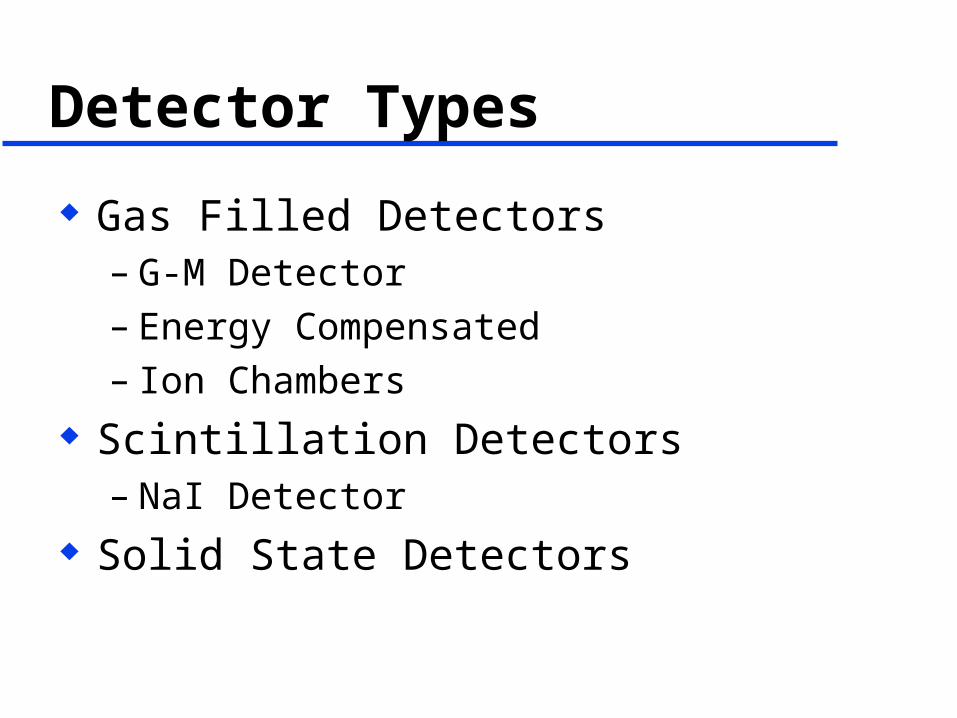
Detector Types
Gas Filled Detectors– G-M Detector– Energy Compensated– Ion Chambers
Scintillation Detectors– NaI Detector
Solid State Detectors
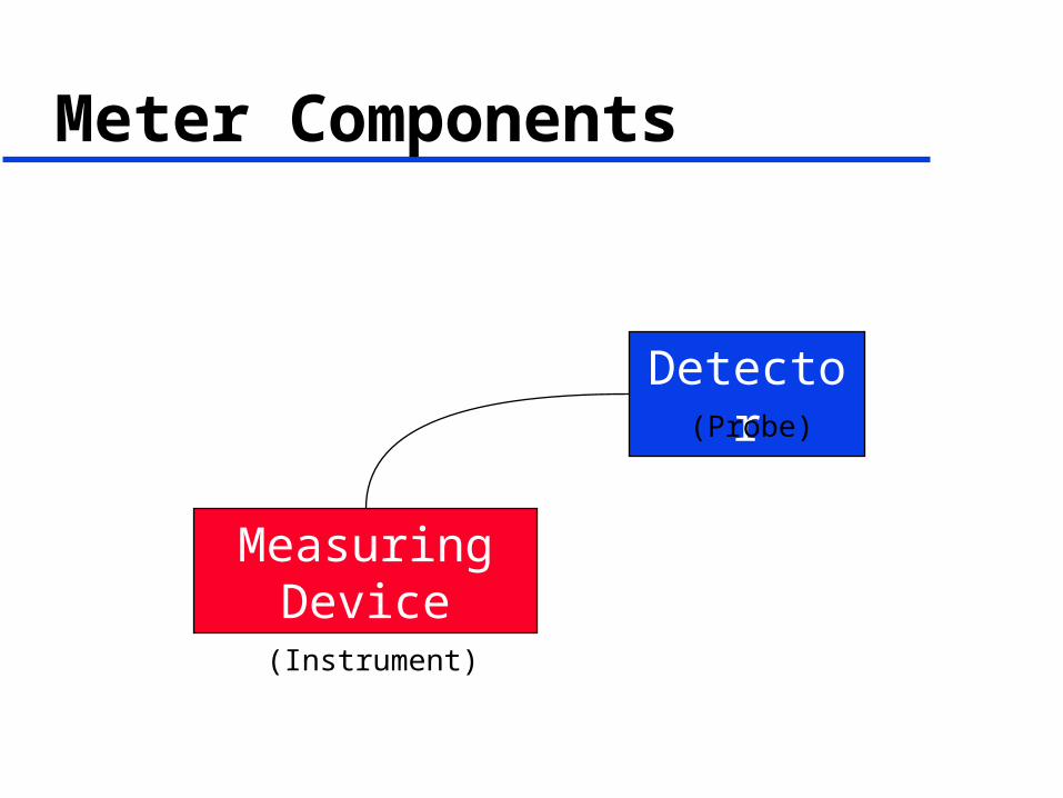
Meter Components
Measuring Device
Detector(Probe)
(Instrument)

Gas Filled Detectors
Air or Other Gas
Incident Ionizing Radiation
ElectricalCurrent
Measuring Device
+
-
Cathode -
Anode +
+ + +
- - -
+ -
Voltage Source
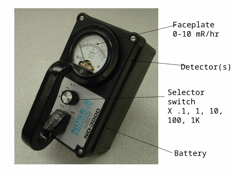
Selector switchX .1, 1, 10, 100, 1K
Faceplate0-10 mR/hr
Battery
Detector(s)

Types of Gas Filled Detectors
Geiger Mueller (GM) Energy compensated GM
– Single of multiple tube Side window, end window GM Proportional Counter Ion Chamber


Instrument Checks Battery Check
Make sure the battery is strong enough to operate the instrument.
Calibration CheckMake sure the instrument has been properly calibrated
Physical CheckCheck the physical condition of the cord, probe, meter face,
etc. Source Check
Check the instrument with a known source of radiation to make sure the meter responds.

Problems with Meters
Must be turned on Must have good battery Must be used correctly Must be with you
GM meters may peg and then read zero

Surveys are Required … of the radiographic exposure device and the guide tube
after each exposure when approaching the device or the guide tube. The survey shall determine that the sealed source has returned to its shielded position before exchanging films, repositioning the exposure head, or dismantling equipment.
any time the source is exchanged and whenever a radiographic exposure device is placed in a storage area to ensure that the sealed source is in its shielded position.
the boundary of the restricted area during radiographic operations not employing shielded room radiography.

Summary
Radiation is energy Gamma and Photons cause ionizations Dose is reduced with time, distance and
shielding Wear your dosimeters Instruments will keep you out of trouble

Questions?


