29
Radiation Safety Technical Guide UCSB Environmental Health & Safety
Radiation Safety Technical Guide UCSB Environmental Health & Safety

7/27/2005 1
General Information Radiation is defined as energy emitted in the form of waves or particles. Ionizing radiation consists of directly or indirectly ionizing particles or a mixture of both. Directly ionizing particles are electrically charged particles having sufficient kinetic energy to produce ionization by collision (over 10 electron Volts). Examples of directly ionizing radiation are alpha, beta particles. Conversely, indirectly ionizing radiation do not possess charge. They penetrate through a medium until, by chance, they make collisions (with electrons, atoms, or nuclei), which result in the liberation of energetic charged particles or initiation of nuclear transformation.
Alpha Particles are energetic helium nuclei (He2+) consisting of two neutrons and two protons. Alpha particles are heavier than an electron by a factor of over 7300 and have double the charge. Sources of alpha radiation are common in nature and include thorium, uranium, and radium; other commonly man-made sources are polonium and americium. Alpha particles are easily stopped by a thin absorber (e.g. sheet of paper or the outer dead layer of skin). Alpha particles are not considered penetrating radiation. However, once taken inside the body (e.g., inhalation, ingestion, etc.), alpha particles are highly damaging. This is because alpha particles deposit a large amount of energy in a short distance (high linear energy transfer), due to their relatively slow velocity (more massive than beta particles) and high charge (2 protons). Beta Particles are high speed electrons which are emitted by the nuclei of atoms during radioactive decay by a process involving the transformation of a neutron into a proton. There are many naturally occurring sources of beta particles as well as those produced artificially by man. At UCSB the most common beta emitting isotopes include 3H, 14C, 35S, and 32P. The ability of a beta particle to penetrate matter is a function of its kinetic energy.
Ranges of Beta Particles Emitted by Various Isotopes
Approximate Maximum Range (inches) Air Water Lucite Glass Aluminum Lead 32P 250.000 .300 .250 .130 .120. .02 35S 13.000 .017 .014 .006 .005 .001 14C 11.000 .014 .009 .005 .004 3H .162 .001 .001 .001 .001 Low energy beta particles, with maximum kinetic energy of less than 0.2 million electron volts (MeV), are easily absorbed in the outer layer of the skin. However, beta particles external to the body are more penetrating than alpha particles. As with alpha particles, beta \particles taken within cells or incorporated into biologically active molecules may give significant doses disabling and killing cells. When a charged particle, such as a beta particle, passes through matter and is decelerated by the atom of the material, secondary radiation is produced. This radiation is called bremsstrahlung and is a type of x-ray. The energy of the bremsstrahlung radiation produced is directly proportional to two parameters: 1) the energy of the beta radiation which is producing the bremsstrahlung, 2) the atomic number of the absorbing materials. High energy beta particles, such as 32P, can produce a bremsstrahlung radiation field which must be shielded. In this case, a material such as Lucite, which has a low atomic number,
7/27/2005 2
is used as the primary shield and is covered by an outer lead shield. Any low energy bremsstrahlung produced in the Lucite would then be absorbed in the lead.
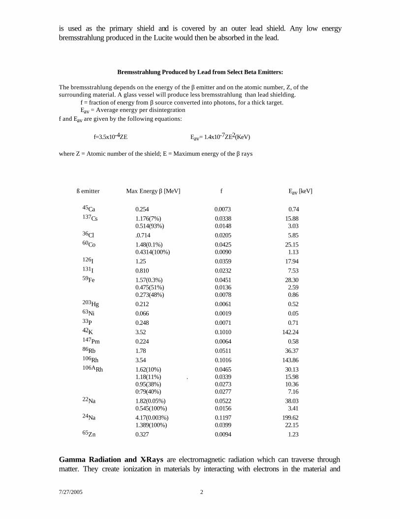
Bremsstrahlung Produced by Lead from Select Beta Emitters: The bremsstrahlung depends on the energy of the β emitter and on the atomic number, Z, of the surrounding material. A glass vessel will produce less bremsstrahlung than lead shielding.
f = fraction of energy from β source converted into photons, for a thick target. Eav = Average energy per disintegration
f and Eav are given by the following equations: f=3.5x10⁻⁴ZE Eav= 1.4x10⁻⁷ZE2(KeV) where Z = Atomic number of the shield; E = Maximum energy of the β rays
ß emitter Max Energy β [MeV] f Eav [keV] 45Ca 0.254 0.0073 0.74 137Cs 1.176(7%) 0.0338 15.88 0.514(93%) 0.0148 3.03 36Cl .0.714 0.0205 5.85 60Co 1.48(0.1%) 0.0425 25.15 0.4314(100%) 0.0090 1.13 126I 1.25 0.0359 17.94 131I 0.810 0.0232 7.53 59Fe 1.57(0.3%) 0.0451 28.30 0.475(51%) 0.0136 2.59 0.273(48%) 0.0078 0.86 203Hg 0.212 0.0061 0.52 63Ni 0.066 0.0019 0.05 33P 0.248 0.0071 0.71 42K 3.52 0.1010 142.24 147Pm 0.224 0.0064 0.58 86Rb 1.78 0.0511 36.37 106Rh 3.54 0.1016 143.86 106ARh 1.62(10%) 0.0465 30.13 1.18(11%) . 0.0339 15.98 0.95(38%) 0.0273 10.36 0:79(40%) 0.0277 7.16 22Na 1.82(0.05%) 0.0522 38.03 0.545(100%) 0.0156 3.41 24Na 4.17(0.003%) 0.1197 199.62 1.389(100%) 0.0399 22.15 65Zn 0.327 0.0094 1.23
Gamma Radiation and X-Rays are electromagnetic radiation which can traverse through matter. They create ionization in materials by interacting with electrons in the material and
7/27/2005 3
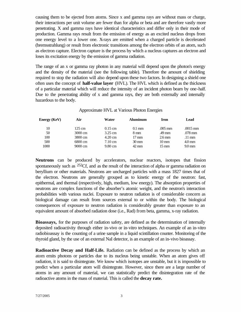
causing them to be ejected from atoms. Since x and gamma rays are without mass or charge, their interactions per unit volume are fewer than for alpha or beta and are therefore vastly more penetrating. X and gamma rays have identical characteristics and differ only in their mode of production. Gamma rays result from the emission of energy as an excited nucleus drops from one energy level to a lower one. X-rays are emitted when a charged particle is decelerated (bremsstrahlung) or result from electronic transitions among the electron orbits of an atom, such as electron capture. Electron capture is the process by which a nucleus captures an electron and loses its excitation energy by the emission of gamma radiation. The range of an x or gamma ray photon in any material will depend upon the photon's energy and the density of the material (see the following table). Therefore the amount of shielding required to stop the radiation will also depend upon these two factors. In designing a shield one often uses the concept of half-value layer (HVL). The HVL which is defined as the thickness of a particular material which will reduce the intensity of an incident photon beam by one-half. Due to the penetrating ability of x and gamma rays, they are both externally and internally hazardous to the body.
Approximate HVL at Various Photon Energies Energy (KeV) Air Water Aluminum Iron Lead
10 125 cm 0.15 cm 0.1 mm .005 mm .0015 mm 50 3000 cm 3.25 cm 8 mm .49 mm .078 mm 100 3800 cm 4.20 cm 17 mm 2.6 mm .11 mm 500 6800 cm 7.10 cm 30 mm 10 mm 4.0 mm 1000 9000 cm 9.80 cm 42 mm 15 mm 9.0 mm Neutrons can be produced by accelerators, nuclear reactors, isotopes that fission spontaneously such as 252Cf, and as the result of the interaction of alpha or gamma radiation on beryllium or other materials. Neutrons are uncharged particles with a mass 1827 times that of the electron. Neutrons are generally grouped as to kinetic energy of the neutron: fast, epithermal, and thermal (respectively, high, medium, low energy). The absorption properties of neutrons are complex functions of the absorber’s atomic weight, and the neutron's interaction probabilities with various nuclei. Exposure to neutron radiation is of considerable concern as biological damage can result from sources external to or within the body. The biological consequences of exposure to neutron radiation is considerably greater than exposure to an equivalent amount of absorbed radiation dose (i.e., Rad) from beta, gamma, x-ray radiation. Bioassays, for the purposes of radiation safety, are defined as the determination of internally deposited radioactivity through either in-vivo or in-vitro techniques. An example of an in-vitro radiobioassay is the counting of a urine sample in a liquid scintillation counter. Monitoring of the thyroid gland, by the use of an external NaI detector, is an example of an in-vivo bioassay. Radioactive Decay and Half-Life. Radiation can be defined as the process by which an atom emits photons or particles due to its nucleus being unstable. When an atom gives off radiation, it is said to disintegrate. We know which isotopes are unstable, but it is impossible to predict when a particular atom will disintegrate. However, since there are a large number of atoms in any amount of material, we can statistically predict the disintegration rate of the radioactive atoms in the mass of material. This is called the decay rate.
7/27/2005 4
The standard unit of activity is the Curie (Ci). The Curie is defined as the activity of that quantity of radioactive material in which the number of disintegrations per minute is 2.22 x 10¹². The Systeme Internationale replaces the Curie by the Bequerel (Bq); 1 Bq = 1 disintegration per second and 1 Ci=3.7x10¹⁰ Bq. The half life (T1/2) of a radioactive nuclide is the time required for the initial number of radioactive atoms to decrease by half. If the half-life of the radionuclide (i.e., physical half life) is known, one can calculate the remaining activity at some point in time by the equation: A = A0 x
e-λτ where λ = 0.693/T1/2 and A0 is the original activity, τ is the elapsed time since the original activity. Once deposited inside of the body, the time for the radioactive nuclide in the body to be diminished by half will depend both on the radioactive decay rate (i.e., physical half-life) and the rate at which that particular substance is eliminated through the body's metabolic process (i.e., biological half-life). Combining the radioactive and biological half-lives gives the effective half-life and is defined by the equation: Teff = (Tr x Tb)/(Tr + Tb) where Tr = radioactive half-life and Tb = biological half life. The biological half-life of a given nuclide will depend upon many factors, such as the particular organ or organs in the body in which the nuclide is concentrated, and the amount of fluids ingested after the uptake. Biological and radioactive half lives of the common radionuclides used at UCSB can be found in the section listing radioisotopes individually. Specific Activity — Specific activity refers to the amount of radioactivity, usually expressed in milliCuries, per amount of material, expressed in millimole or milligram. Radioisotopes which have a short half-life (e.g., 32P) have a high specific activity. Radiation Dosimetry — the units of radiation dosimetry are the roentgen, the rad, and the rem. Roentgen —the special unit of exposure in air is defined as the roentgen (R). This unit is defined only for X-ray or gamma radiation in air and is not applicable to alphas, betas, or neutrons.
R = 2.58 x 10⁻⁴ coulomb/kg or 5.44 x 10⁷ Mev/gm at 0°C and 760 mm pressure
Rad — the rad is the unit of absorbed dose or energy absorbed per gram of material. For practical purposes of radiation protection 1 rad = 1 roentgen, except for alphas and neutrons, which do more biological damage per unit of energy absorbed.
1 rad= 100ergs/gm or 6.25xl07Mev/gm
Rem — the number of rem produced by an absorbed dose in rad is found by multiplying the number of rad absorbed by the quality factor (QF). The equation is expressed as rem = QF x rad. The physical measure for gauging the damage of equal absorbed doses from different types of radiation is the linear energy transfer, LET. The higher the LET of the radiation, the greater the injury produced for a given absorbed dose. The factor expressing the relative effectiveness of a given particle based on its linear energy transfer is the quality factor. Beta, gamma and x-rays
7/27/2005 5
have a quality factor of one; the quality factor for fast neutrons and protons up to 10 MeV is 10 and for alpha particles is 10 to 20. Systeme Internationale (SI) Units— the International Commission on Radiation Units has called for a conversion from the present radiation units to the Systeme Internationale of measurements. These new units are:
- Quantity: 1 Bequerel (Bq) = 1 disintegration per second; replaces the Curie, 1 Ci = 3.7 x 10¹⁰ Bq.
multiples of the Bq: 1 kilobecquerel (kBq) = 103 Bq 1 megabecquerel (MBq) = 106 Bq 1 gigabecquerel (GBq) = 109 Bq 1 terabecquerel (TBq) = 1012 Bq
- Exposure: Coulombs per kilogram will be used instead of the Roentgen - Absorbed Dose: 1 Gray (Gy) = 100 rad; 104 ergs/gram; replaces rad - Dose Equivalent: 1 Sievert (Sv) = 100 rems; replaces the rem
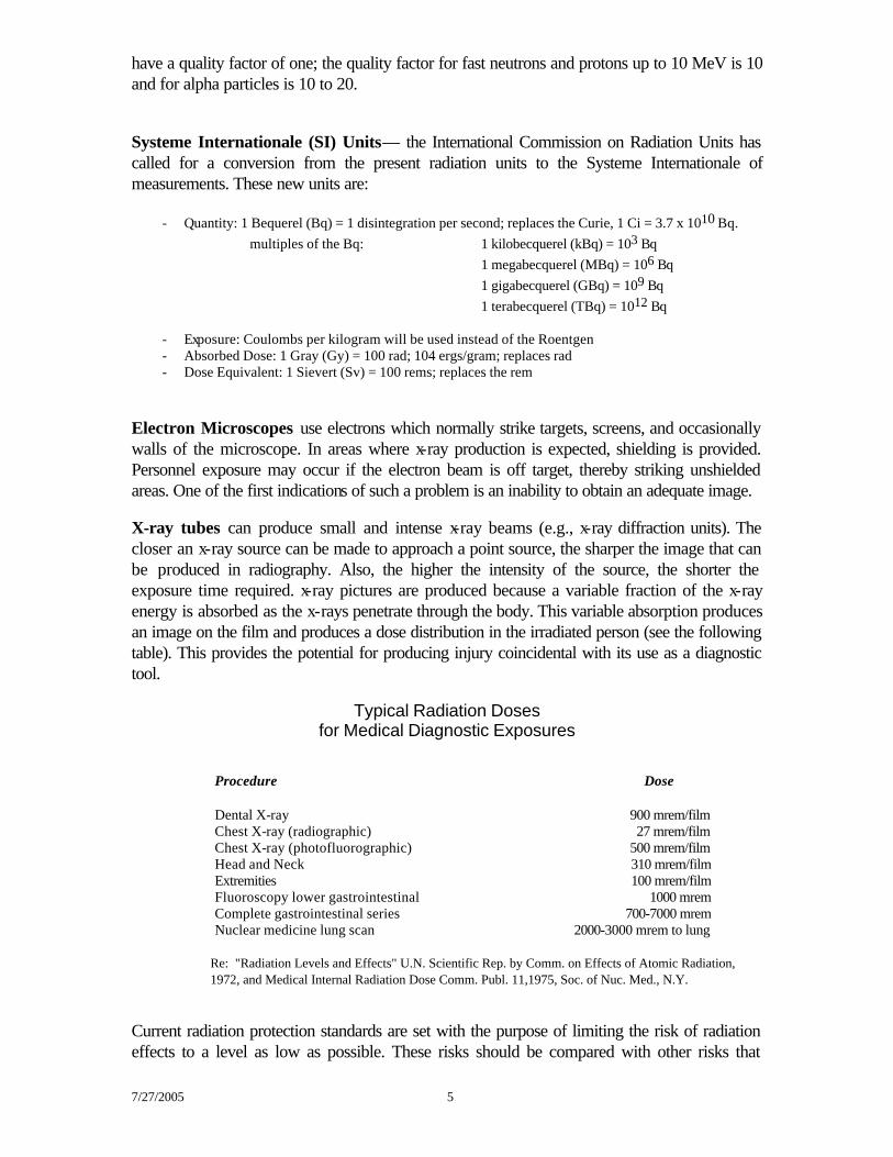
Electron Microscopes use electrons which normally strike targets, screens, and occasionally walls of the microscope. In areas where x-ray production is expected, shielding is provided. Personnel exposure may occur if the electron beam is off target, thereby striking unshielded areas. One of the first indications of such a problem is an inability to obtain an adequate image. X-ray tubes can produce small and intense x-ray beams (e.g., x-ray diffraction units). The closer an x-ray source can be made to approach a point source, the sharper the image that can be produced in radiography. Also, the higher the intensity of the source, the shorter the exposure time required. x-ray pictures are produced because a variable fraction of the x-ray energy is absorbed as the x-rays penetrate through the body. This variable absorption produces an image on the film and produces a dose distribution in the irradiated person (see the following table). This provides the potential for producing injury coincidental with its use as a diagnostic tool.
Typical Radiation Doses
for Medical Diagnostic Exposures
Procedure Dose Dental X-ray 900 mrem/film Chest X-ray (radiographic) 27 mrem/film Chest X-ray (photofluorographic) 500 mrem/film Head and Neck 310 mrem/film Extremities 100 mrem/film Fluoroscopy lower gastrointestinal 1000 mrem Complete gastrointestinal series 700-7000 mrem Nuclear medicine lung scan 2000-3000 mrem to lung Re: "Radiation Levels and Effects" U.N. Scientific Rep. by Comm. on Effects of Atomic Radiation,
1972, and Medical Internal Radiation Dose Comm. Publ. 11,1975, Soc. of Nuc. Med., N.Y.
Current radiation protection standards are set with the purpose of limiting the risk of radiation effects to a level as low as possible. These risks should be compared with other risks that
7/27/2005 6
people assume in their daily lives such as from driving, flying, smoking or drinking. If all the work with radioactive materials or radiation-producing machines stopped, radiation exposures would not be eliminated because we are all continuously exposed to background radiation. Background Radiation. There are three components in background radiation: cosmic radiation (external), arising from outer space; terrestrial radiation (external), resulting from the presence of naturally occurring radionuclides in the soil and earth; and naturally occurring radionuclides (internal) deposited in the human body. The average dose from background radiation varies from one location to another, e.g., at sea level it's about 100 mrem/yr. and in Denver it's about 175 mrem/yr. The atmosphere serves as a shield against cosmic radiation, the thinner this shield the greater the dose rate, thus, it increases with altitude. At sea level the dose is 0.011 mR/hr, at 10,000 ft is 0.02 mR/hr, and at cruising altitude of 33,000 ft is 0.8 mR/hr. The average dose to the U.S. population from cosmic sources (disregarding structural shielding) is estimated to be 40 mrem/yr. When the rate for internal body organs is estimated, this value is generally reduced by 20% to account for structural shielding provided by buildings, and by another 20% to account for shielding provided by outer tissues of the body. Maximum Permissible Doses. The maximum permissible controlled area occupational doses are listed in the California Department of Health Services' Title 17. The permissible dose limits for persons under 18 years of age are 10% of the limits specified for persons 18 years and over. UCSB campus administrative levels and the limits in Title 17 can be found in appendix C of UCSB's Radiation Safety Manual.
Dose from Beta Particles. High energy beta particles loose energy at the rate of about 2 MeV per centimeter in penetrating water or water equivalent thicknesses. A rough calculation of the dose rate from a beam of beta particles incident upon the body is 100 high energy beta particles/cm2/sec = 10 mrem/hr. Low energy beta particles undergo greater attenuation in the dead layer of the skin, and the net result is to increase the dose to the skin while decreasing the dose rate to the basal cells of the epidermis by 70%. The dose rate due to a localized beta source decreases inversely with the square of the distance as a result of increased separation and increased attenuation by the air. The expression for the dose rate at a distance from a source of a given activity is:
dose rate in mrad/hr = 338,000 x activity in mCi/(distance in cm)2.
When beta emitters are taken into the body it is necessary to evaluate the dose rate imparted to organs in which they are incorporated. The average dose rate can be evaluated readily by assuming, because of the short range of the beta particles, that all the beta energy emitted is absorbed in the organ. Thus the calculation of the average absorbed dose per beta particle is done by determining the average beta energy emitted per gram.
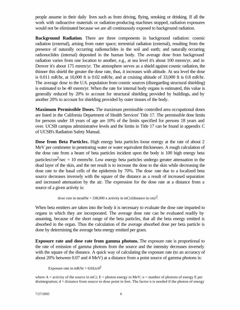
Exposure rate and dose rate from gamma photons. The exposure rate is proportional to the rate of emission of gamma photons from the source and the intensity decreases inversely with the square of the distance. A quick way of calculating the exposure rate (to an accuracy of about 20% between 0.07 and 4 MeV) at a distance from a point source of gamma photons is:
Exposure rate in mR/hr = 6AEn/d2
where A = activity of the source in mCi; E = photon energy in MeV; n = number of photons of energy E per disintegration; d = distance from source to dose point in feet. The factor n is needed if the photon of energy
7/27/2005 7
E is not emitted with each disintegration. If photons of several energies are emitted, the contribution of each must be determined separately and the values added.)
Dose Rates Per mCi for Various Photon Energies
(assuming 1 photon per disintegration) Isotope Photon Energy Dose Rate (MeV) mRem/hr @ 30 cm 0.02 2.1 0.03 0.92
51Cr 0.05 0.42 0.10 0.49 0.20 1.1
125I 0.30 1.8 0.50 3.1
137Cs 0.60 3.7
54Mn 0.80 4.8 1.00 5.8 1.50 7.9 2.00 9.7 3.00 12.8
from The Physics of Radiology by H. Johns and J. Cunningham, Charles Thomas, Publisher, 1974
7/27/2005 8
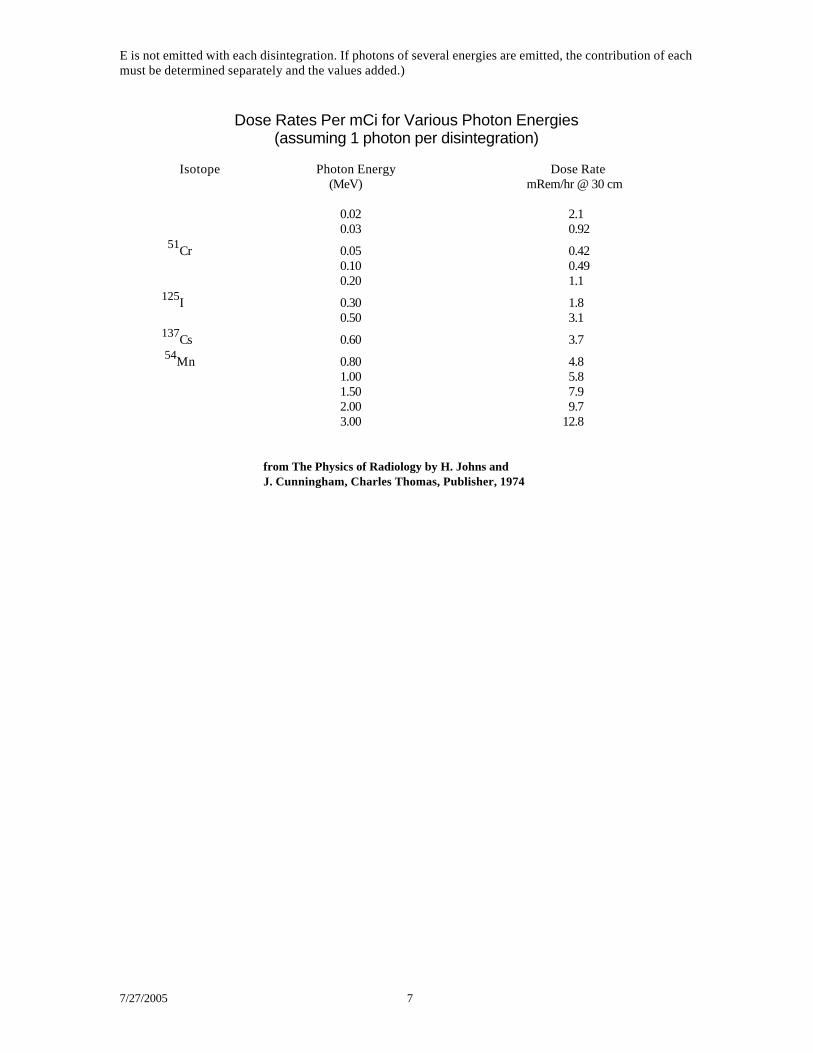
Properties and Precautions for Common Radioisotopes The following section describes some of the typical radioisotopes used in research. More extensive information on particular radioisotopes can be obtained by contacting the Radiation Safety Office. TRITIUM, CARBON-14 and SULFUR-35 Tritium Carbon-14 Sulfur-35 Half-life 12.33 years 5730 years 87.4 days Type of Decay beta beta beta Maximum Energy of Radiation 0.018 MeV 0.158 MeV 0.167 MeV Percentage of Disintegrations 100% 100% 100% Average Radiation Energy/Disintegration 0.005 MeV 0.049 MeV 0.048 MeV Maximum Range in Air ~ 1/16 inch 8.6 inches 9.6 inches Effective Half-Life 12 days 12 days 44-90 days Critical Organ total body total body testis; whole body Dose rate for 6000 dpm/cm2: - at surface - 56 mrem/hr 56 mrem/hr - penetrating dead layer of skin - 6.16 mrem/hr 6.16 mrem/hr Tritium-3 (3H) • Millicurie amounts of tritium do not present an external exposure hazard because the low energy beta rays
emitted do not penetrate the outer dead layer of skin. • Tritium absorbed through the skin or inhaled equilibriates in body fluids within 3-5 hours. In case of an
internal exposure, the elimination rate can be increased by increasing the fluid intake. • Handle potentially volatile compounds in fume hoods.
• For 3H detection use liquid scintillation counters (50-60% efficiency).
• Individuals involved in operations which utilize, at any one time, more than 100 mCi of 3H other than in a sealed source, must contact the Radiation Safety Office for a urinalysis within one week following a single operation and at weekly intervals for continuing operations.
Carbon-14 (14C) • Millicurie amounts of 14C do not present a significant external exposure hazard because the low energy
beta rays emitted barely penetrate the outer layer of the skin. Most 14C labeled compounds are rapidly metabolized and the radionuclides exhaled as 14CO2. Some
compounds and their metabolites are eliminated via the urine. The critical organ for uptake of 14C-labeled carbonates is bone and for some other compounds is fat. Note that 14C-labeled halogenated acids may be incorporated in the skin resulting in high local doses. Biological half-lives vary from a few minutes to 25 days, 12 days being an acceptable value for most compounds.
• Handle potentially volatile compounds in a fume hood. • For 14C detection use end-window G-M detectors (up to 9% efficiency at the surface of the probe) or
liquid scintillation counters (90% efficiency). Sulfur-35 (35S) • Millicurie amounts of 35S do not present a significant external exposure hazard since the low energy beta
rays emitted barely penetrate the outer layer of the skin. • The elimination rate depends on the chemical form. Most 35S labeled compounds are eliminated through
the urine. • Handle potentially volatile compounds in fume hoods. • For 35S detection use end-window G-M detectors (up to 9% efficiency at the surface of the probe) or
liquid scintillation counters (90% efficiency). 35S is difficult to distinguish from 14C because the beta emissions are of similar energy.
7/27/2005 9
PHOSPHORUS-32 (32P) Half-Life 14.3 days Type of Decay beta Maximum Energy of Radiation 1.71 MeV Percentage of Disintegrations 100% Average Radiation Energy/Disintegration 0.694 Maximum Range in Air ~ 18 feet Effective Half-Life 14 days Critical Organ bone Dose Rate for 6000 dpm/cm2: - at surface 11 mrem/hr - penetrating dead layer of skin 10.45 mrem/hr • Although most 32P labeled chemicals, owing to their composition, do not present a direct inhalation
hazard, many which are commonly used are readily absorbed through the skin. In addition, there is always the possibility that removable contamination will be ingested or become airborne and possibly inhaled. About 60% of the 32P that is ingested is excreted within the first 24 hours, but only about 1% per day is excreted thereafter. Urine samples should be submitted to the Radiation Safety Office within 24 hours after suspected internal contamination.
• 32P emits a beta particle sufficiently energetic to penetrate the epidermis and expose the inner layers of tissue. The eye is a critical organ as well, since these beta rays can penetrate to the retina. In addition to the beta hazard itself, bremsstrahlung is frequently created when the beta rays are slowed down in absorbing medium, e.g. glass, steel, insufficient lead thickness of shielding or storage container.
• To work with 32P, shielding is necessary and is best designed by using a relatively low-density primary barrier such as Lucite of sufficient thickness to slow down the high-energy beta particles, followed by lead to absorb the small amount of bremsstrahlung produced, generally 1/2 inch of Lucite and 0.040 inch (1 mm) of lead foil is sufficient.
• Radiation monitoring badges, whole body and ring, must be worn when using 32P. • There are several types of detectors for 32P: the traditional procedure has been to count vials containing
scintillation hours in liquid scintillation counters (80-90% efficiency). This is not the recommended method of counting since it has associated disposal problems. The preferred procedure is to count without scintillation flours in the 3H channel of liquid scintillation counters taking advantage of the Cerenkov radiation (up to 50% efficiency). Survey detectors with a G-M probe to measure the beta rays and with a NaI (gamma) probe to detect the bremsstrahlung, since the characteristics of bremsstrahlung are similar to low-energy X-rays.
• 32P waste should be properly shielded. The bremsstrahlung energy produced by a 32P beta ray slowed down by lead is approximately 35 KeV, with 4.9% of the energy being converted into photons [f = 3.5 x 10⁻⁴ x z x 1.71 = 0.049, where Z = 82 for lead]. The bremsstrahlung produced by a 1 mCi vial of 32P, assuming that all the beta rays were stopped, is 300 mrem/hr x 0.049 = 14.7 mrem/hr at 1 cm. The average energy of bremsstrahlung produced is 0.4 x 82 = 32.8 KeV; this energy is similar to that produced by 125I.
Radiation Dose observed with a 32P combi-vial (NEN)
Distance from Source Average to Hand mrem/hr/mCi At 10 cm from combi-vial 5 At 1 cm from combi-vial 300 Contact on side of combi-vial 439 Drop of 32P in contact with 1 cm2 of skin 2,000,000
7/27/2005 10
IODINE-125 (125I) Half-Life 57.4 days Type of Decay Electron Capture Energy of Radiation gamma 0.035 MeV; x-ray: 0.027 MeV Half Value Layer 0.0037 cm Effective Half-Life 42 days Critical Organ thyroid • Radioiodine is of particular concern because one organ, the thyroid, concentrates approximately 30% of
the free iodine taken into the body. The dose to the thyroid has been calculated at 5 rem/microCi. Since such a large dose per unit activity concentrates in this gland, great care must be taken to avoid exposure.
• Iodine has a very high vapor pressure in solution. In very basic (pH 11) as well as in acidic solutions measurable free iodine is always present in the air above; optimal pH is 8-9. Free iodine can be inhaled or readily absorbed through the intact skin. It has been estimated that up to 15% of the cases of work-related thyroid uptakes of radioiodine are due to absorption through the skin.
• Do not store opened stock bottles or solutions containing any free radioiodine in freezer, refrigerator or cold room. Na125I must be stored and handled in a fume hood, preferably equipped with TEDA charcoal filters. The hood air flow should not be obstructed with pieces of equipment, particularly when not needed for the experiment, and the flow rate should be 100 ft/min with the hood sash closed to the inspection label.
• Before starting any work, prepare a “decontamination/125I trap” solution of 0.1M Na thiosulfate, 0.1M NaI, 0.1M NaOH.
• For the detection of radioiodine use a survey meter with a NaI probe (up to 20% efficiency at the surface of the probe) or a gamma scintillation counter (40-50% efficiency). 129I is commonly used as a mock 125I standard to evaluate counter efficiency because of its long half-life and similar energy of radiation.
• Always work on clean absorbent paper. Cut a piece to fit the job, place it mat side up to absorb any spills. Tape it over anything already in the fume hood but do not cover the metal at the front edge of the hood if present. Discard the paper after use.
• Place stock bottles behind lead bricks as far back as possible in the fume hood. Use another shield for the experiment. Work at least 6" inside the sash of the hood. Use a gamma survey meter (NaI crystal) to monitor the adequacy of the shielding, the presence or absence of 125I on work surfaces and equipment, and to verify the elution of the iodinated material from the column and the retention of the free 125I on the column. [Note that 126I is a frequent contaminant of 125I preparations. See 126I for description of its characteristics.]
• When aliquoting Na125I wear 2 pairs of disposable latex gloves. Once aliquoting is completed, remove the outer pair and replace with a fresh pair. Monitor your hands continually as 125I can permeate thin gloves.
• To separate sample and remove the free iodine from your preparation use exclusion chromatography if possible. Whether using ‘spin-columns’ or chromatographic column s, do not elute the peak of free iodine from the column, but seal and discard columns after elution of the protein peak in the void volume. If the column has to be run cold, prepare the column in a cold-room or refrigerator, chill the eluent before use, and run the column in the fume hood chilling the fractions on ice. Never place 125I in a cold-room since it has no ventilation. If using an ion exchange procedure where the free 125I comes out first, collect this fraction into tubes containing “decontamination/125I trap” solution. If ‘spin-columns’ are used for protein de-salting/Na125I removal, load the column into the centrifuge buckets inside the fume hood, and if possible, seal with greased 0-rings the bucket covers. Re-open the buckets only after transferring them back to the fume hood. Wash out the buckets with “decontamination/125I trap” solution followed by 2% ‘Count-Off’ or similar solution, then rinse with distilled water and dry. If a table-top centrifuge or microfuge is used it should be placed inside the fume hood with the sash completely closed. Before removing the sample, allow several minutes with the sash raised 1-2" for the air to purge. Dialysis is an inefficient and unsafe method to eliminate free125I; it should not be used.
• The fume hood must be left clean after use: All waste must be properly disposed of; and the hood must be checked for contamination with a gamma probe.
• If a spill has occurred or equipment must be decontaminated, wipe the surfaces with a solution of 0.1M NaI, 0.1M NaOH and 0.1M Na Thiosulfate to stabilize the free iodine, then clean-up with a detergent or decontaminant, surveying continuously. In case of personnel contamination with free iodine, remove contaminated clothing to a plastic bag, apply a paste of sodium iodide to contaminated area and let it
7/27/2005 11
react with the free iodine, then wash it with a detergent. Contact the Radiation Safety Office in all cases of skin contamination.
• A person internally contaminated with radioiodine must immediately report this to the Radiation Safety Office. Bioassays (thyroid scan, urinalysis) will be performed to assess the degree of internal deposition. In certain cases, medical treatment may involve administering three hundred milligrams of a saturated solution of potassium iodide (SSKI) to the person to block the thyroid and decrease the incorporation of radioiodine. Administration of SSKI as a ‘protective’ measure is not recommended. Chronic exposures to iodine may have side effects and in some instances may lead to an abnormal thyroid function or allergic reaction. Furthermore, if one follows recommended procedures, uptakes should be minimal and thyroid blocking agents unnecessary.
• The Radiation Safety Office does conduct routine thyroid bioassays of those individuals using 1 millicurie or more in a single vial, or as total experimental activity used per calendar quarter.
• Radiation monitoring badges, both for whole body and ring, must be worn. • Contaminated glassware must be cleaned before returning glassware to use by swabbing or soaking it in
“decontamination/I-125 trap” solution (in fume hood), soaking it in 2% ‘Count-Off’ or similar solution, rinsing it with water, and monitoring it with gamma probe.
CHROMIUM-51 (51Cr) Half-Life 27.8 days Type of Decay Electron Capture Energy of Radiation gamma: V X-rays, 0.320 MeV e-: 0.315 MeV Half Value Layer 1.7 mm of lead Effective Half-Life 27 days Critical Organ Total body • 51Cr should be stored and used behind lead shielding. Use 4 mm of lead for quantities up to 1 mCi and 10
mm for vials containing 10 mCi of 51Cr.
• Radiation monitoring badges, ring and wholebody, must be worn when working with 51Cr. • For 51Cr detection use survey meter with NaI probe or gamma scintillation counter. • The maximum permissible body burden is 800 µCi. CALCIUM-45 (45Ca) Half-life 165 days Type of Decay beta Maximum Energy of Radiation 0.254 MeV Percentage of Disintegrations 100% Average Radiation Energy/Disintegration 0.076 MeV Maximum Range in Air 19 inches Effective Half-Life 162 days Critical Organ bone Dose Rate for 6000 dpm/cm2: - at surface 33 mrem/hr - penetrating dead layer of skin 12 mrem/hr • Millicurie amounts of 45Ca do not present a significant external exposure hazard because the low energy
betas emitted barely penetrate gloves and the outer layer of skin. • The majority of the 45Ca is deposited in the bone and is retained for a long time (1.8 x 10⁴ days biological
half-life). A smaller fraction is rapidly eliminated. • For detection use end-window G-M detectors or liquid scintillation counters. • See bremsstrahling information at the end of this section.
7/27/2005 12
CADMIUM-109 (109Cd) Half-life 453 days Type of Decay Electron Capture Energy of Radiation γ: 0.088 MeV e-: 0.062 MeV, 0.084 MeV Effective Half-life 140 days Critical Organ liver Maximum Permissible Body Burden 20 µCi • Shield with lead. • For detection use survey meter with NaI (gamma) probe or gamma scintillation counter. CALlFORNIUM-252 (252Cf) Half-life 2.646 years Type of Decay α: 96.9%, Spontaneous Fission (SF): 3.1%, β stable Energy of Radiation α: 6.12 MeV (82%), 6.08 MeV (15%) e-: 0.022 MeV, 0.038 MeV γ: Cm L-X-rays SF:fission fragments, neutrons, r rays, electrons,
daughter radiation Effective Half-life 2.2 years Critical Organ bone Maximum Permissible Body Burden 0.01 µCi
CESIUM-137 (137Cs) Half-life 30 years Type of Decay beta Energy of Radiation β: 1.176 MeV max. (7%), 0.514 MeV e-: 0.624 MeV, 0.656 MeV γ Ba X-rays, 0.662 MeV (85%) Average Radiation Energy/Disintegration 0.195 MeV from beta Effective Half-life 70 days Critical Organ total body • Shield with Lucite/lead. • For detection use survey meter with NaI (gamma) probe or gamma scintillation counter. • See bremsstrahlung information at the end of this chapter. CHLORINE-36 (36Cl) Half-life 3.08 x 10⁵years Type of Decay β− 98.1%, Electron Capture 1.9%, β+ 0.0012% Energy of Radiation β−: 0.714 MeV max γ S X-rays, 0.511 MeV (0.003%) Average Radiation Energy/Disintegration 0.252 MeV • Shield with Lucite/lead. • For detection use end-window G-M survey meter or liquid scintillation counter.
7/27/2005 13
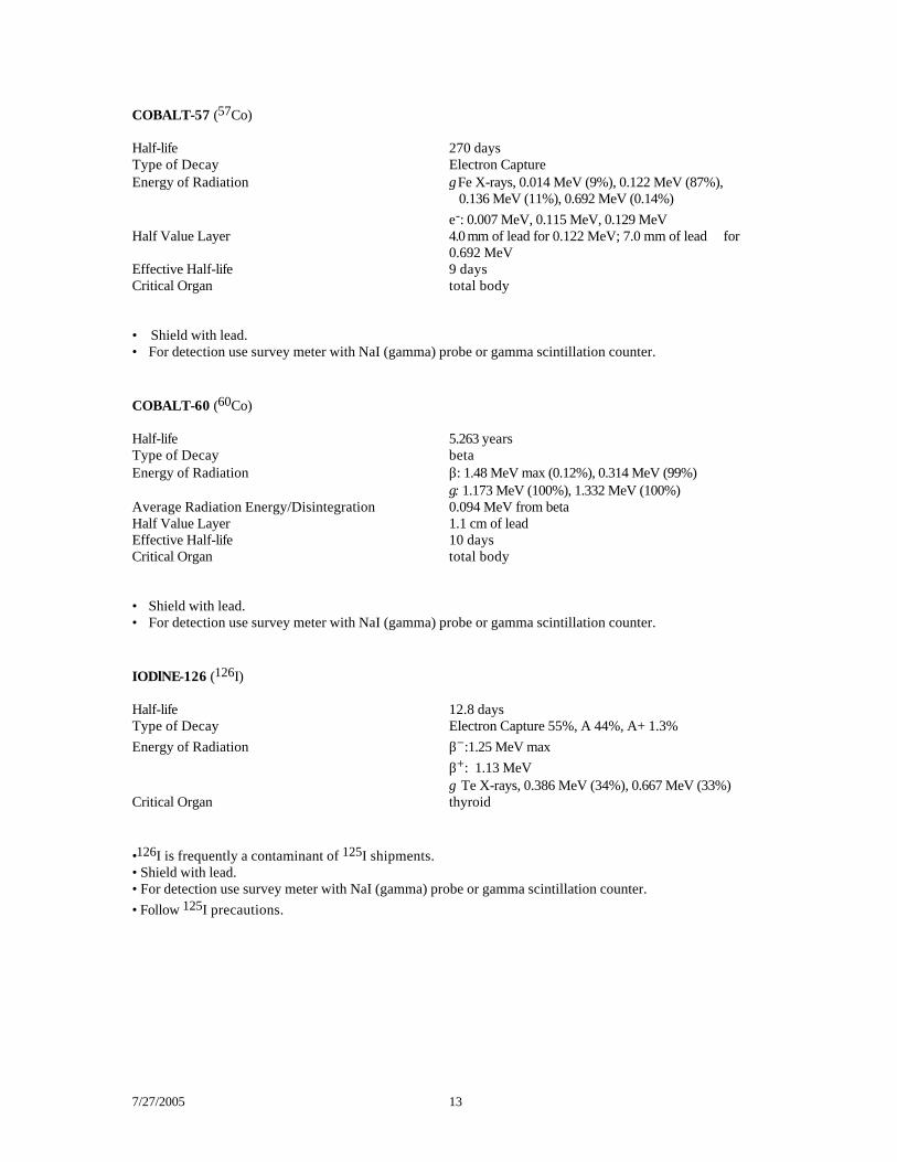
COBALT-57 (57Co) Half-life 270 days Type of Decay Electron Capture Energy of Radiation γ Fe X-rays, 0.014 MeV (9%), 0.122 MeV (87%), 0.136 MeV (11%), 0.692 MeV (0.14%) e-: 0.007 MeV, 0.115 MeV, 0.129 MeV Half Value Layer 4.0 mm of lead for 0.122 MeV; 7.0 mm of lead for
0.692 MeV Effective Half-life 9 days Critical Organ total body • Shield with lead. • For detection use survey meter with NaI (gamma) probe or gamma scintillation counter. COBALT-60 (60Co) Half-life 5.263 years Type of Decay beta Energy of Radiation β: 1.48 MeV max (0.12%), 0.314 MeV (99%) γ: 1.173 MeV (100%), 1.332 MeV (100%) Average Radiation Energy/Disintegration 0.094 MeV from beta Half Value Layer 1.1 cm of lead Effective Half-life 10 days Critical Organ total body • Shield with lead. • For detection use survey meter with NaI (gamma) probe or gamma scintillation counter. IODlNE-126 (126I) Half-life 12.8 days Type of Decay Electron Capture 55%, A 44%, A+ 1.3% Energy of Radiation β−:1.25 MeV max β+: 1.13 MeV γ Te X-rays, 0.386 MeV (34%), 0.667 MeV (33%) Critical Organ thyroid •126I is frequently a contaminant of 125I shipments. • Shield with lead. • For detection use survey meter with NaI (gamma) probe or gamma scintillation counter. • Follow 125I precautions.
7/27/2005 14
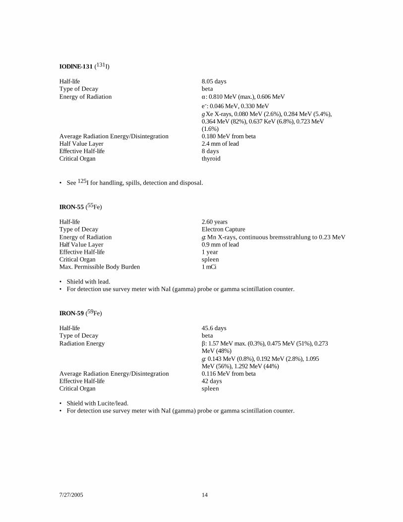
IODlNE-131 (131I) Half-life 8.05 days Type of Decay beta Energy of Radiation α: 0.810 MeV (max.), 0.606 MeV e-: 0.046 MeV, 0.330 MeV γ Xe X-rays, 0.080 MeV (2.6%), 0.284 MeV (5.4%), 0.364 MeV (82%), 0.637 KeV (6.8%), 0.723 MeV (1.6%) Average Radiation Energy/Disintegration 0.180 MeV from beta Half Value Layer 2.4 mm of lead Effective Half-life 8 days Critical Organ thyroid • See 125I for handling, spills, detection and disposal. IRON-55 (55Fe) Half-life 2.60 years Type of Decay Electron Capture Energy of Radiation γ: Mn X-rays, continuous bremsstrahlung to 0.23 MeV Half Value Layer 0.9 mm of lead Effective Half-life 1 year Critical Organ spleen Max. Permissible Body Burden 1 mCi • Shield with lead. • For detection use survey meter with NaI (gamma) probe or gamma scintillation counter. IRON-59 (59Fe) Half-life 45.6 days Type of Decay beta Radiation Energy β: 1.57 MeV max. (0.3%), 0.475 MeV (51%), 0.273 MeV (48%) γ: 0.143 MeV (0.8%), 0.192 MeV (2.8%), 1.095 MeV (56%), 1.292 MeV (44%) Average Radiation Energy/Disintegration 0.116 MeV from beta Effective Half-life 42 days Critical Organ spleen • Shield with Lucite/lead. • For detection use survey meter with NaI (gamma) probe or gamma scintillation counter.
7/27/2005 15
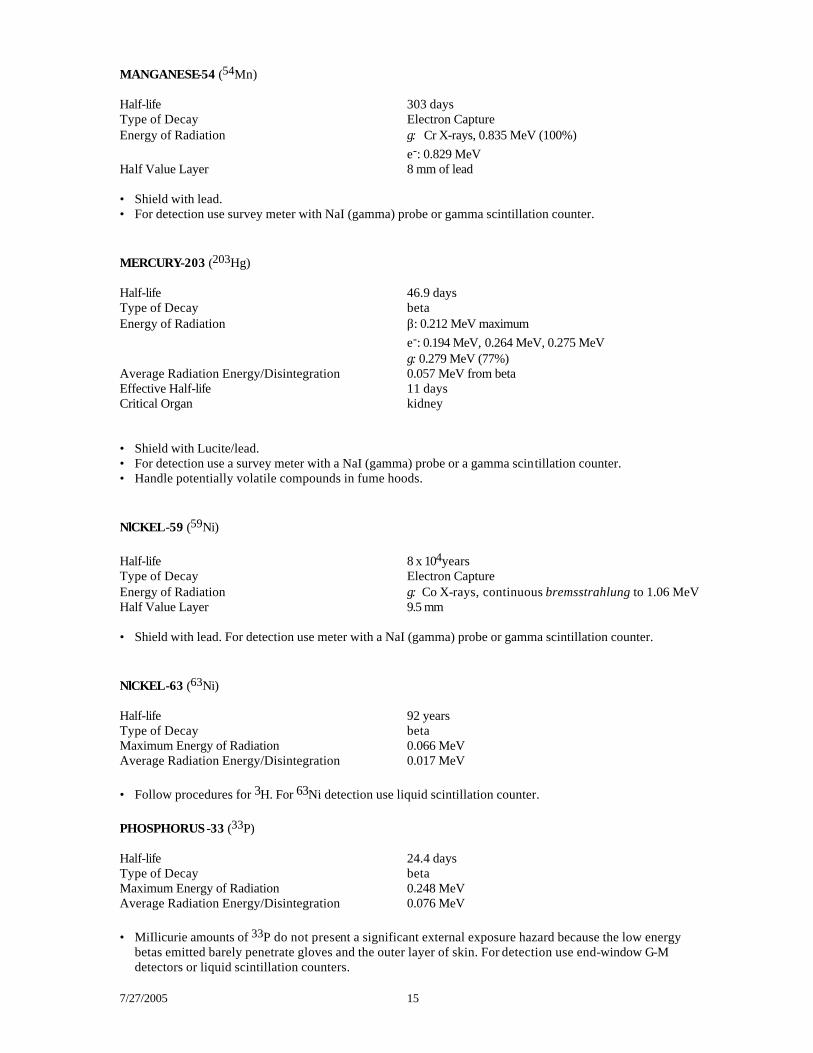
MANGANESE-54 (54Mn) Half-life 303 days Type of Decay Electron Capture Energy of Radiation γ: Cr X-rays, 0.835 MeV (100%) e-: 0.829 MeV Half Value Layer 8 mm of lead • Shield with lead. • For detection use survey meter with NaI (gamma) probe or gamma scintillation counter. MERCURY-203 (203Hg) Half-life 46.9 days Type of Decay beta Energy of Radiation β: 0.212 MeV maximum e-: 0.194 MeV, 0.264 MeV, 0.275 MeV γ: 0.279 MeV (77%) Average Radiation Energy/Disintegration 0.057 MeV from beta Effective Half-life 11 days Critical Organ kidney • Shield with Lucite/lead. • For detection use a survey meter with a NaI (gamma) probe or a gamma scintillation counter. • Handle potentially volatile compounds in fume hoods. NlCKEL-59 (59Ni) Half-life 8 x 10⁴years Type of Decay Electron Capture Energy of Radiation γ: Co X-rays, continuous bremsstrahlung to 1.06 MeV Half Value Layer 9.5 mm • Shield with lead. For detection use meter with a NaI (gamma) probe or gamma scintillation counter. NlCKEL-63 (63Ni) Half-life 92 years Type of Decay beta Maximum Energy of Radiation 0.066 MeV Average Radiation Energy/Disintegration 0.017 MeV • Follow procedures for 3H. For 63Ni detection use liquid scintillation counter. PHOSPHORUS-33 (33P) Half-life 24.4 days Type of Decay beta Maximum Energy of Radiation 0.248 MeV Average Radiation Energy/Disintegration 0.076 MeV • MiIlicurie amounts of 33P do not present a significant external exposure hazard because the low energy
betas emitted barely penetrate gloves and the outer layer of skin. For detection use end-window G-M detectors or liquid scintillation counters.
7/27/2005 16
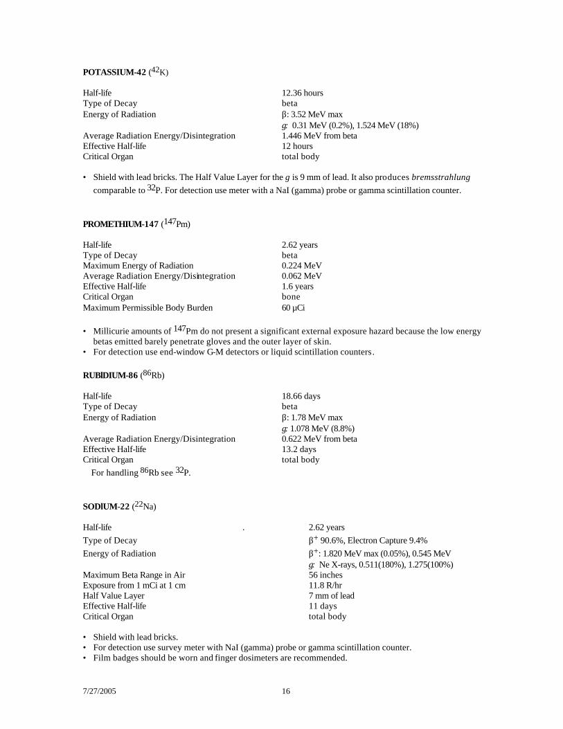
POTASSIUM-42 (42K) Half-life 12.36 hours Type of Decay beta Energy of Radiation β: 3.52 MeV max γ: 0.31 MeV (0.2%), 1.524 MeV (18%) Average Radiation Energy/Disintegration 1.446 MeV from beta Effective Half-life 12 hours Critical Organ total body • Shield with lead bricks. The Half Value Layer for the g is 9 mm of lead. It also produces bremsstrahlung
comparable to 32P. For detection use meter with a NaI (gamma) probe or gamma scintillation counter. PROMETHIUM-147 (147Pm) Half-life 2.62 years Type of Decay beta Maximum Energy of Radiation 0.224 MeV Average Radiation Energy/Disintegration 0.062 MeV Effective Half-life 1.6 years Critical Organ bone Maximum Permissible Body Burden 60 µCi • Millicurie amounts of 147Pm do not present a significant external exposure hazard because the low energy
betas emitted barely penetrate gloves and the outer layer of skin. • For detection use end-window G-M detectors or liquid scintillation counters. RUBlDIUM-86 (86Rb) Half-life 18.66 days Type of Decay beta Energy of Radiation β: 1.78 MeV max γ: 1.078 MeV (8.8%) Average Radiation Energy/Disintegration 0.622 MeV from beta Effective Half-life 13.2 days Critical Organ total body For handling 86Rb see 32P. SODlUM-22 (22Na) Half-life . 2.62 years Type of Decay β+ 90.6%, Electron Capture 9.4% Energy of Radiation β+: 1.820 MeV max (0.05%), 0.545 MeV γ: Ne X-rays, 0.511(180%), 1.275(100%) Maximum Beta Range in Air 56 inches Exposure from 1 mCi at 1 cm 11.8 R/hr Half Value Layer 7 mm of lead Effective Half-life 11 days Critical Organ total body • Shield with lead bricks. • For detection use survey meter with NaI (gamma) probe or gamma scintillation counter. • Film badges should be worn and finger dosimeters are recommended.
7/27/2005 17
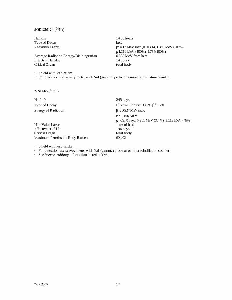
SODlUM-24 (24Na) Half-life 14.96 hours Type of Decay beta Radiation Energy β: 4.17 MeV max (0.003%), 1.389 MeV (100%) γ 1.369 MeV (100%), 2.754(100%) Average Radiation Energy/Disintegration 0.553 MeV from beta Effective Half-life 14 hours Critical Organ total body • Shield with lead bricks. • For detection use survey meter with NaI (gamma) probe or gamma scintillation counter. ZlNC-65 (65Zn) Half-life 245 days Type of Decay Electron Capture 98.3%,β+ 1.7% Energy of Radiation β+: 0.327 MeV max. e-: 1.106 MeV γ: Cu X-rays, 0.511 MeV (3.4%), 1.115 MeV (49%) Half Value Layer 1 cm of lead Effective Half-life 194 days Critical Organ total body Maximum Permissible Body Burden 60 µCi • Shield with lead bricks. • For detection use survey meter with NaI (gamma) probe or gamma scintillation counter. • See bremsstrahlung information listed below.
7/27/2005 18
Biological Effects and Risks
Overview It was recognized early after the discovery of x-rays and radioactivity that exposure to ionizing radiation could have detrimental effects on living things. The detrimental effects against which protection is required are both somatic and genetic. "Somatic” effects are those that become manifest in the exposed individual; the genetic (or hereditary) effects are those that affect the descendants of the exposed individuals. The harmful effects may be classified also as to whether the effects vary in severity as a function of dose (called non-stochastic effects) or vary in probability of occurrence (with similar severity) as a function of dose (called stochastic effects). Non-stochastic effects are those usually associated with high dose, such as radiation burns, cataracts. Non-stochastic effects exhibit a “safe” threshold dose and would not be expected to occur in individuals exposed to levels within occupational limits. Stochastic effects, such as genetic mutation and carcinogenesis exhibit no threshold. This means that all exposure to radiation bears some risk of genetic harm or of increasing the risk of cancer in the exposed individual. Minimizing stochastic effects is now the main concern of radiation protection. The radiation dose limits have been established so essentially no non-stochastic (threshold) effects would occur even if a person were exposed to the limit over his/her entire working life. Limiting the stochastic effects (especially cancer induction) is achieved by keeping all justifiable exposures as low as reasonably achievable (ALARA). Different dose limits have been set for various organs in the body because of varying sensitivity of tissues or organs to stochastic damage. Also, young individuals are more sensitive than adults to damage from ionizing radiation. This is especially true if an embryo or fetus is exposed in utero. For this reason, occupational exposure is more restricted (by a factor of 10) for individuals below the age of 18. Also, it is recommended that pregnant individuals restrict their occupational exposure so that a fetus does not receive more than 1/10 the yearly dose equivalent limit for workers. Radiation dose limits in uncontrolled areas (e.g. outside of radioactive material laboratories), and for members of the general public, are also much lower than those for controlled areas and individuals who work with radioactive materials. On the UCSB campus, the Radiation Safety Committee has implemented an ALARA program. Toward this end, a whole body (e.g. head, trunk of body) exposure guideline of 500 mrem/year (1/10 of the legal limit) was established for radiation workers. Adherence to the guideline will reduce, proportionately, the risk of producing stochastic effects in all exposed individuals. Also, no fetus would then receive a dose of more than 500 mrem (the recommended limit). In practice, the vast majority of researchers using ionizing radiation on campus have received zero or minimal radiation exposure. The average dose to persons living in the Santa Barbara area from natural background radiation (excluding occupational dose) is about 100 mrem per year and the average medical exposure is also about 100 mrem per year.
7/27/2005 19
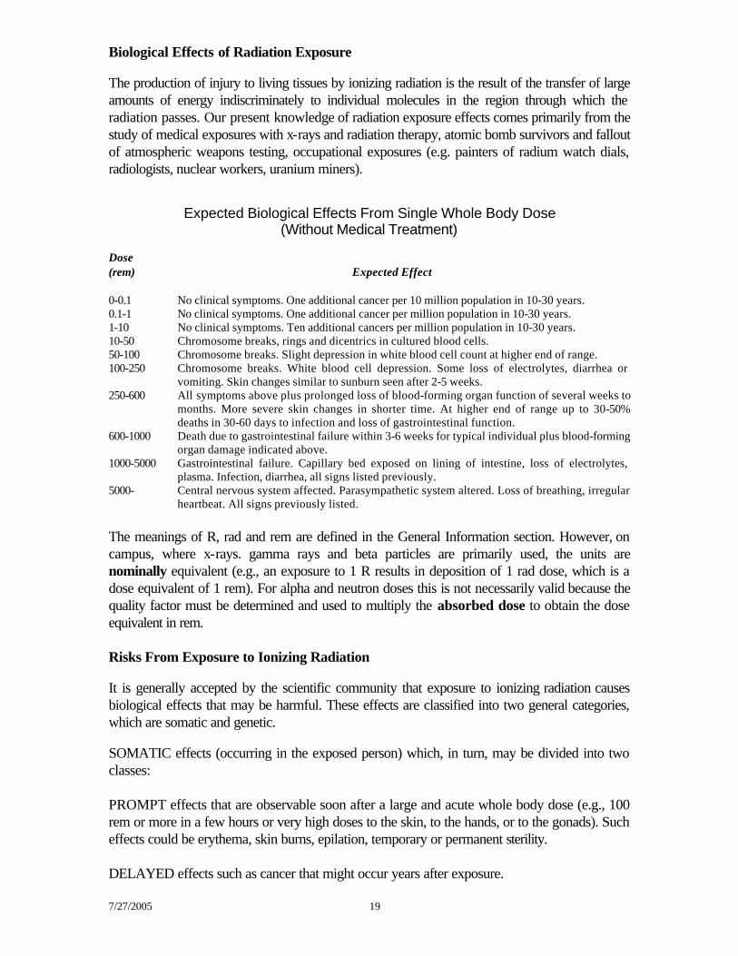
Biological Effects of Radiation Exposure The production of injury to living tissues by ionizing radiation is the result of the transfer of large amounts of energy indiscriminately to individual molecules in the region through which the radiation passes. Our present knowledge of radiation exposure effects comes primarily from the study of medical exposures with x-rays and radiation therapy, atomic bomb survivors and fallout of atmospheric weapons testing, occupational exposures (e.g. painters of radium watch dials, radiologists, nuclear workers, uranium miners).
Expected Biological Effects From Single Whole Body Dose (Without Medical Treatment)
Dose (rem) Expected Effect 0-0.1 No clinical symptoms. One additional cancer per 10 million population in 10-30 years. 0.1-1 No clinical symptoms. One additional cancer per million population in 10-30 years. 1-10 No clinical symptoms. Ten additional cancers per million population in 10-30 years. 10-50 Chromosome breaks, rings and dicentrics in cultured blood cells. 50-100 Chromosome breaks. Slight depression in white blood cell count at higher end of range. 100-250 Chromosome breaks. White blood cell depression. Some loss of electrolytes, diarrhea or
vomiting. Skin changes similar to sunburn seen after 2-5 weeks. 250-600 All symptoms above plus prolonged loss of blood-forming organ function of several weeks to
months. More severe skin changes in shorter time. At higher end of range up to 30-50% deaths in 30-60 days to infection and loss of gastrointestinal function.
600-1000 Death due to gastrointestinal failure within 3-6 weeks for typical individual plus blood-forming organ damage indicated above.
1000-5000 Gastrointestinal failure. Capillary bed exposed on lining of intestine, loss of electrolytes, plasma. Infection, diarrhea, all signs listed previously.
5000- Central nervous system affected. Parasympathetic system altered. Loss of breathing, irregular heartbeat. All signs previously listed.
The meanings of R, rad and rem are defined in the General Information section. However, on campus, where x-rays. gamma rays and beta particles are primarily used, the units are nominally equivalent (e.g., an exposure to 1 R results in deposition of 1 rad dose, which is a dose equivalent of 1 rem). For alpha and neutron doses this is not necessarily valid because the quality factor must be determined and used to multiply the absorbed dose to obtain the dose equivalent in rem. Risks From Exposure to Ionizing Radiation It is generally accepted by the scientific community that exposure to ionizing radiation causes biological effects that may be harmful. These effects are classified into two general categories, which are somatic and genetic. SOMATIC effects (occurring in the exposed person) which, in turn, may be divided into two classes: PROMPT effects that are observable soon after a large and acute whole body dose (e.g., 100 rem or more in a few hours or very high doses to the skin, to the hands, or to the gonads). Such effects could be erythema, skin burns, epilation, temporary or permanent sterility. DELAYED effects such as cancer that might occur years after exposure.
7/27/2005 20
GENETIC effects; abnormalities that might occur in the children of exposed individuals and in subsequent generations. Concerns about these biological effects have resulted in stringent controls of radiation sources and in efforts to control and minimize the doses to the workers. Common Questions About Risks Associated With Radiation Exposure (Reference: NRC Regulatory Guide OH-902-1) What is meant by risk? Risk can be defined in general as the chance (probability) of injury or death resulting from some activity. The intent of this section is to estimate and explain the possible risk of injury, illness or death resulting from occupational radiation exposure. What are the possible health effects of exposure to radiation? Some of the health effects that exposure to radiation may cause are cancer (including leukemia), cataracts, and inherited birth defects in children of exposed parents. These effects (with the exception of genetic effects) have been demonstrated in studies of medical radiologists, radium workers, radiotherapy patients who have received excessive doses, and studies of people exposed to radiation from atomic weapons. In addition, the study of radiation effects in laboratory animals have provided a large body of data. All of the studies mentioned, however, involve levels of radiation exposure that are much higher than those permitted occupationally today. Studies have NOT shown a clear cause and effect relationship between health effects and current levels of occupational radiation exposure. The estimates of risks at low doses are therefore extrapolations from higher doses and dose rates. What is meant by prompt effects and delayed effects? Prompt, non-stochastic effects are observable shortly after a very large dose of radiation in a short period of time. For example, a dose of 450 rem to an average adult will cause vomiting and diarrhea within a few hours; loss of hair, fever and weight loss within a few weeks and about 50 percent chance of death within about 1 month without medical treatment. Delayed effects (both stochastic and non-stochastic), such as cancer and cataracts may occur years after exposure to radiation. (Note: Cataracts are not likely to be caused by doses of x-rays, gamma rays, or beta particles within the occupational limits.) What about potential effects to the children of workers who are exposed to radiation? Genetic effects occur when there is radiation damage to the germ cells carried by the parents, due to radiation exposure to the gonads of either parent. These effects may show up as birth defects or other conditions in the offspring of the exposed parents, and succeeding generations. These effects have been demonstrated in animal experiments, although they have not been shown in human populations. The risk of producing genetic harm is about 1 in 10,000 per rem
7/27/2005 21
of dose to the parent’s gonads. Genetic effects should not be confused with damage to an embryo or fetus due to exposure to radiation while still in utero, which is discussed below. As radiation workers, which effects should concern us most? Immediate or prompt effects are very unlikely since large exposures would normally occur only if there were a serious radiation accident. In fact, such exposures could not occur in the typical radioisotope laboratory because only limited quantities of radionuclides are present. The probability of serious genetic effects in the children of workers is estimated to be about one third of the other delayed effects. The probability of harm to a fetus due to occupational exposure of the pregnant mother is one major area of concern . Probably the greatest concern to the worker for most of his/her working life is the possibility of an increased risk of cancer. The increased risk of cancer is believed to depend on how much radiation a person receives; therefore, every reasonable effort should be made to keep exposures low. Of course, genetic effects are also a concern to those persons who are at, or below, reproductive age. What is the difference between acute and chronic exposure? Acute radiation exposure, which causes delayed effects, refers to a large dose of radiation received in a short time, for example, 450 rem received in a few hours or less. The effects of acute exposures are well known from studies of radiotherapy patients, atomic bomb casualties, and accidents that have occurred. Chronic exposure, which may cause delayed effects but not prompt effects, refers to small doses received over long time periods, for example, 20-100 millirem per week every week for several years. Concern with occupational radiation risks is primarily focused on chronic exposure to low levels of radiation over long time periods. How does radiation cause cancer? Radiation can cause changes in the DNA and alter the genetic codes within cells and cause visible damage to the chromosomes. However, how radiation causes cancer is not well understood. It is impossible to tell whether a given cancer was caused by radiation or by some other of the many apparent causes. However, most diseases are caused by the interaction of several factors. General physical condition, inherited traits, age, sex and exposure to other cancer-causing agents such as cigarette smoke are a few possible interacting factors. One theory is that radiation activates existing viruses in the body that then attack the cells causing them to grow rapidly. Another is that radiation reduces the body’s normal resistance to existing viruses, which can then multiply and attack cells. Since radiation can induce mutations in the genes which provide the information regulating cell growth, the damage could result in the cell’s being directed along abnormal growth patterns. What is know is that in groups of highly exposed people a higher than normal incidence of cancer is observed. An increased risk of cancer has not been shown at radiation dose levels within Federal and State occupational limits. UCSB guideline is 10% of those limits. Studies of groups who receive occupational doses of radiation are still underway; until there is evidence to the contrary, it is prudent to maintain doses at ALARA levels.
7/27/2005 22
If I receive a radiation dose, does it mean I am certain to get cancer? Not at all, everyone receives a radiation dose from cosmic rays and natural radioactivity but most people do not get cancer. Even when doses of radiation far above legal limits are received, most individuals will experience no delayed effects. There is evidence that the human body will repair some of the damage. The danger from radiation is much like the danger from cigarette smoke. Only a fraction of the people who breathe cigarette smoke get lung cancer, but there is good evidence that smoking increases a person’s chances of getting lung cancer. Similarly, there is evidence that large radiation doses increase the chance of getting cancer. Radiation is like most substances that cause cancer in that the effects can only be observed at large doses. Still, it is prudent to assume that smaller doses also have some chance of causing cancer. (For example, you may recall the debate about saccharin.) This assumption is applied to natural cancer-causes such as sunlight and natural radiation as it is for those that are man-made such as smog, cigarette smoke, and man-made radiation. As even very small doses may have some risk, it follows that no dose should be received without a reason. Thus, it is a time-honored principle that radiation doses should be kept as low as reasonably achievable (ALARA). We don’t know exactly what are the chances of getting cancer from a radiation dose, but we have good estimates. The estimates of radiation risks are at least as reliable as estimates for the effects from any other important hazard. Being exposed to typical occupation radiation doses is taking a chance, but that chance is small and reasonably well understood. It is important to understand the probability factors here. A similar question would be: If you select one card from a full deck, will you get the ace of hearts? This question cannot be answered with a simple yes or no. The best answer is that your chances are 1 in 52. However, if 1000 people each select one card from full decks, we can predict that about 20 of them will get the ace of hearts. each person will have 1 chance in 52 of drawing the ace of hearts, but there is no way of predicting which persons will get the right card. The issue is further complicated by the fact that in one drawing only 15 persons may draw the card while in the next drawing 25 persons may get the card. We can say that if you receive a radiation dose, you will have increased your statistical chances of eventually developing cancer or some other radiation-related injury. The more radiation exposure you get, the more you increase your chances of cancer. Clearly, there is no simple answer to the question. The best we can do is to provide estimates for large population groups of the increased chances of cancer or other radiation injury resulting from exposure to radiation. What is the risk of developing cancer from low-level radiation ? The following cancer risk estimate is from the National Research Council's Committee on the Biological Effects of Ionizing Radiation, 1990, Report: "Health Effects of Exposure to Low Levels of Ionizing Radiation." If a population of 10,000 people were each given 1 Rem of radiation to the whole body in a single, brief exposure, we would expect to see 8 additional cancer deaths. This is in addition to the 20,000 cancer deaths which would occur naturally in that population. The normal incidence
7/27/2005 23
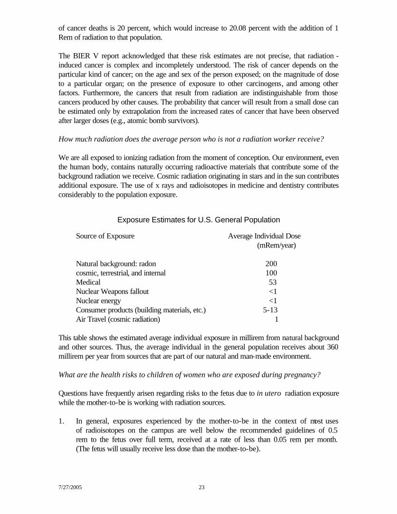
of cancer deaths is 20 percent, which would increase to 20.08 percent with the addition of 1 Rem of radiation to that population. The BIER V report acknowledged that these risk estimates are not precise, that radiation -induced cancer is complex and incompletely understood. The risk of cancer depends on the particular kind of cancer; on the age and sex of the person exposed; on the magnitude of dose to a particular organ; on the presence of exposure to other carcinogens, and among other factors. Furthermore, the cancers that result from radiation are indistinguishable from those cancers produced by other causes. The probability that cancer will result from a small dose can be estimated only by extrapolation from the increased rates of cancer that have been observed after larger doses (e.g., atomic bomb survivors). How much radiation does the average person who is not a radiation worker receive? We are all exposed to ionizing radiation from the moment of conception. Our environment, even the human body, contains naturally occurring radioactive materials that contribute some of the background radiation we receive. Cosmic radiation originating in stars and in the sun contributes additional exposure. The use of x rays and radioisotopes in medicine and dentistry contributes considerably to the population exposure.
Exposure Estimates for U.S. General Population
Source of Exposure Average Individual Dose (mRem/year) Natural background: radon 200 cosmic, terrestrial, and internal 100 Medical 53 Nuclear Weapons fallout <1 Nuclear energy <1 Consumer products (building materials, etc.) 5-13 Air Travel (cosmic radiation) 1 This table shows the estimated average individual exposure in millirem from natural background and other sources. Thus, the average individual in the general population receives about 360 millirem per year from sources that are part of our natural and man-made environment. What are the health risks to children of women who are exposed during pregnancy? Questions have frequently arisen regarding risks to the fetus due to in utero radiation exposure while the mother-to-be is working with radiation sources. 1. In general, exposures experienced by the mother-to-be in the context of most uses of radioisotopes on the campus are well below the recommended guidelines of 0.5 rem to the fetus over full term, received at a rate of less than 0.05 rem per month. (The fetus will usually receive less dose than the mother-to-be).
7/27/2005 24
2. The estimated (calculated) risk associated with the fetus receiving a dose of 1 rem are believed to be less that 1 in 1600 additional risk of childhood cancer. (This is about two times the carcinogenic effect that would be expected for adults receiving such a dose). There may also be risk of about 1 in 250 of mental retardation resulting after a 1 rem dose received acutely during the 8th to the 15th week of gestation. However, the normal risk of this birth defect is approximately 1 in 100 (at least 2 1/2 times greater). Thus, the additional risk to the fetus carried by a radiation worker is small, even when an exposure to one-half rem occurs, which has not happened on the campus to date. It is important that women employees who work around radiation be aware of the possible risk so that they can take steps they think appropriate to protect their offspring. Note: This is one of the reasons that UCSB adopted the Guideline of 0.5 rem per year versus the legal dose limit, which is 5 rem per year. 3. Some additional risk to the fetus could occur if the mother-to-be inhales or ingests radioiodine. This could expose the fetal thyroid gland, which begins to function after the 12th week, to radiation from the iodine deposited therein. Also, radiation doses could also be delivered to a baby's thyroid if a nursing mother inhales or ingests radioiodine, which then is excreted in milk. 4. In some cases it may be possible to take extra precautions to reduce the potential of exposure by wearing lead aprons, using improved shielding, working more efficiently during exposure, wearing double gloves, etc. It may be practical to rotate those duties that have higher exposure potential to other persons. However, this should depend upon the nature of the tasks to be performed in a lab and the availability and skills of the persons involved. This is a management option. there is no guarantee that an alternate assignment will be available. The Nuclear Regulatory Commission makes the following suggestions to women of child bearing age who work around radiation sources: 1. If you are now pregnant or expect to be soon, you could decide not to continue to work in such areas. (Note that it is possible that there may be no alternate jobs in the University in your discipline that involve no risks of exposure.) You should also note that very few radiation workers at UCSB accumulate doses at the level of 0.5 rem in a year (most receive much less that 0.05 rem in a calendar quarter), and that the doses to the fetus are usually considerable less than the doses monitored by the badge, which measures the dose at the surface of the body. 2. You could in some cases take steps to reduce your exposure by decreasing theamount of time spent near radiation sources, by increasing your distance from sources, or by using better shielding. For example, in some cases lead aprons can be used. 3. If you are not pregnant, you could decide to postpone pregnancy until you are no longer working in areas where you may be exposed to radiation.
7/27/2005 25
What ever course you may wish to follow, it is appropriate to reach a decision early. The fetus is most sensitive during the first three months of pregnancy. Any worker exposed to ionizing radiation as part of their job who is pregnant, or planning to become pregnant, is urged to review the exposure potential and associated risks and methods of reducing exposure with their supervisor and with the Radiation Safety Officer (RSO). Can radiation exposure within occupation limits cause sterility? There is no need to concerned about sterility at occupational dose levels. The dose needed to cause sterility is about 100 times larger that the legal limits for occupational exposure. Can radiation cause cataracts? At occupational radiation dose-limit levels (for x-rays, gamma rays, and beta particles) the risks of inducing cataracts is believed to be extremely small. The most recent data from the International Commission on Radiation Protection and Measurements (ICRP) indicate that 40 years of doses of 15 rem per year (to the eyes) would not be expected to cause any lens opacities that could lead to the deterioration of one's vision. (The legal limit is 5 rem per year.) However, it should be noted that high doses of x-rays, an acute dose to the lens of the eye greater than 200 rem, could lead to cataracts.
7/27/2005 26
Radiation Surveys Good laboratory practice dictates that radiation surveys be made during and after experiments to ensure that sources are adequately shielded and that contamination is controlled. At UCSB, as part of a condition of our radioactive materials license, users are required to survey their areas within the month that radioisotopes are used and to document all surveys performed (see the "Survey Documentation" section below). Survey Requirements Users are required to conduct radiation surveys no less frequently than the month in which radioisotopes are used. Technique For 3H users, smear surveys of use areas are the only reliable means of surveying for contamination. Use filter paper (e.g. Whatman) and wipe about 100 square centimeters of surface area and count the smear in a liquid scintillation counter. There are no absolute rules regarding "permissible" levels of contamination. Usually any removable contamination should be promptly removed. If serious contamination is found (e.g. greater that 1000 dpm/100 cm2 on floors, benches, etc.) call Radiation Safety for assistance. It is recommended that surveys for contamination from 14C or 35S be made with smear techniques as described above. This is because a Geiger counter has a low efficiency for the detection of weak beta emitters from these radioisotopes. Surveys for 125I contamination should be made either by smears or with a survey meter equipped with a thin crystal (1" x 1 mm.) NaI probe. (Note: A GM survey meter equipped with a thin-end-window Geiger probe cannot detect levels of 125I at less than about 1 microcurie). For 32P, a thin-window "pancake" Geiger counter can be used for surveying. The energetic beta from 32P is easily detected with this type of survey meter. Also, a thin-crystal NaI probe will also detect 32P due to the "bremsstrahlung" produced by the energetic beta. Survey Documentation A User Survey Log of each required survey must be maintained in the Radiation Safety Records & Resource Binder. A check-list is provided in the binder which lists the compliance items Radiation Safety inspects during their laboratory surveys. Review of these items for compliance with current practice within your laboratory is recommended. The survey information generally includes the following:
7/27/2005 27
1. A sketch of the lab. Sketches of rooms are provided by Radiation Safety during project reviews to serve as a basis for the internal survey program. Survey locations are indicated on the lab sketch. 2. Identification of the meter (or counting system) used (e.g. manufacturer, model, serial number). Note: Survey meters must not be used if out of calibration. 3. Background radiation survey meter reading and/or counting of a"blank" sample during liquid scintillation counting. 4. Name of surveyor (e.g., initials), and date of survey. Calibration of Radiation Survey Meters Notify the Radiation Safety Office when a new survey meter is acquired or repaired. Our license requires that survey meters be calibrated at specified intervals and taken out of service, and not used, when out of calibration. Characteristics of Radiation Survey Meters Three types of portable instruments are commonly used to make direct measurements of radiation and contamination; the air filled ion chamber, the Geiger counter and the scintillation counter. 1. The Geiger counter is more sensitive that the ion chamber. Exposure rates as low as 0.01
mR/h can be detected with a Geiger counter. Geiger instruments are used primarily when surveying for contamination or locating lost sources. Geiger counters have detectors filled with a gas other than air. Therefore, Geiger counters don't measure exposure directly since an exposure unit (e.g., the roentgen) is defined in air only.
Radiation causes electrical discharges in the Geiger detector tube. These discharges are
converted to electronic pulses (counts) per minute (or second) and are read on a meter. The sensitivity varies markedly with energy, so that an instrument calibrated with 137Cs (0.662 MeV gamma), as is common practice, will tend to over respond to lower energy radiation. A Geiger counter used on campus should be equipped with a thin window (e.g., 1.5 mg/cm2) Geiger tube to permit detection of low energy beta radiation (e.g., 14C, 35S). Geiger counters should also be equipped with a speaker or other audible indicator, to allow one to survey without watching the meter. When using a meter to survey for contamination, move the probe slowly (not more than 2" per second) and as close to the surface as possible. Moving the probe too quickly across the monitored surface will not allow sufficient time for the meter to respond to contamination. Most Geiger counters can become paralyzed (e.g., meter indicates zero reading) in radiation fields greater than a few R/h.
Desirable Features of Geiger Counters
a. paralysis protection: sometimes an option at extra cost b. audible output: sometimes an option at extra cost c. coil cord, if detachable probe: sometimes an option (MHV connectors)
7/27/2005 28
d. large diameter probe: extra cost for most instruments e. response data for different types of radiation f. available service and parts g. reliability 2. Scintillation detectors, either solid or liquid, give off light when ionizing radiation is absorbed in the detector. The quantity of light given off in each event is proportional to the amount of the ionizing radiation absorbed in the scintillation detector. This light is converted into an electrical pulse, by ejecting photoelectrons from a photocathode, which in turn is amplified using a photomultiplier tube. The electrical pulses are then counted using a portable count rate meter similar (or sometimes identical ) to the meter used with the Geiger tube. The response (sensitivity) of scintillation counters is very dependent upon the material used as the scintillator. One common scintillation media used is an inorganic crystal, sodium iodide, doped with traces of Thallium. Because the detector is a solid rather than a gas and the atomic number of iodine is much higher than air or tissue, these detectors are more sensitive to low energy photons than ion chambers or Geiger counters. The principal application of NaI detectors on campus is for surveying labs where 125I or 32P (detecting bremsstrahlung radiation) is used. 3. The air-filled ion chamber measures the number of primary ions produced by ionizing radiation in a confined volume of air. Detection is by collecting the primary ions on the anode and cathode and forming an electrical current. This is similar to a Geiger counter with the exception that the ions collected here are not accelerated by an applied voltage to produce secondary ionizations (as in the case of a Geiger counter). Ion- chambers can accurately measure radiation exposure with minimal dependence upon the energy of the photons. They are principally effective for measuring fields of radiation around gamma sources. It is very difficult to detect less than 0.2 mR/h on most of these instruments and they are not routinely used for contamination monitoring. These instruments are usually good for surveying x-ray machines. Proper Use of Radiation Survey Meters When using Geiger or ion chamber instruments, make sure meters are used with a detector window of a proper thickness for the radiation detected. Be sure that all instruments are calibrated annually for ion chambers, and biannually for Geiger counters or after major repairs are made. Also before use, make sure of the characteristics of the meter by reviewing the meter's operating manual and/or checking with the Radiation Safety Office. When using the meter always move the detector slowly over the area monitored. Avoid contaminating the instrument or breaking the thin window of the detector. As indicated previously, the radiation source used for calibration of the meter is a very important consideration in interpreting the results of a "reading". It is not possible to detect 3H with any of the above instruments.


