49
. RADIATION SAFETY TRAINING MANUAL October 2009 VIRGINIA POLYTECHNIC INSTITUTE AND STATE UNIVERSITY ENVIRONMENTAL, HEALTH AND SAFETY SERVICES RADIATION SAFETY OFFICE
.
RADIATION SAFETY TRAINING
MANUAL
October 2009
VIRGINIA POLYTECHNIC INSTITUTE
AND STATE UNIVERSITY
ENVIRONMENTAL, HEALTH AND SAFETY SERVICES
RADIATION SAFETY OFFICE
2
PREFACE
The Radiation Safety Training Manual has been developed by the Virginia Tech Radiation Safety
Office and is supplemented with the Radioactive Material Safety Program (requirements for use of
radioactive material) and three videos relating to contamination control, contamination detection
and decontamination.
The training program is designed to explain the fundamentals of radiation, the safe use of
radioactive materials, and the Federal, State, and University rules and regulations that control their
use. The primary purpose of the training program is to limit unnecessary internal and external
radiation exposures, by ensuring that each individual knows how to work safely with radioactive
material. In order to document that each person has received this training, and understands the
information, a written test must be passed after the training program has been completed.
If there is a question about any of the material in this manual, or for inquiries concerning the use of
ionizing radiation, please contact the Radiation Safety Office at (540)231-5364.

3
TABLE OF CONTENTS
FUNDAMENTALS OF RADIOACTIVITY . . . . . . . . . . . . . . . . . . . . . . . . . . . . . 5
THE ATOM . . . . . . . . . . . . . . . . . . . . . . . . . . . . . . . . . . . . . . . . . . . . . . . . . . . . . . . . .5
THE DECAY PROCESS . . . . . . . . . . . . . . . . . . . . . . . . . . . . . . . . . . . . . . . . . . . . . . .5
RADIOACTIVE BEHAVIOR. . .. . . . . . . . . . . . . . . . . . . . . . . . . . . . . . . . . . . . . . . . .6
UNITS OF ACTIVITY. . . . . . . . . . . . . . . . . . . . . . . . . . . . . . . . . . . . . . . . . . . . . . . . .8
UNITS OF DOSE. . . . . . . . . . . . . . . . . . . . . . . . . . . . . . . . . . . . . . . . . . . . . . . . . . . . .9
NUCLEAR REACTIONS. . . . . . . . . . . . . . . . . . . . . . . . . . . . . . . . . . . . . . . . . . . . . 10
INTERACTIONS OF RADIATION WITH MATTER. . . . . . . . . . . . . .. . . . . . .12
ALPHAS. . . . . . . . . . . . . . . . . . . . . . . . . . . . . . . . . . . . . . . . . . . . . . . . . . . . . . . . . . .12
BETAS. . . . . . . . . . . . . . . . . . . . . . . . . . . . . . . . . . . . . . . . . . . . . . . . . . . . . . . . . . . . 12
NEUTRONS. . . . . . . . . . . . . . . . . . . . . . . . . . . . . . . . . . . . . . . . . . . . . . . . . . . . . . . .13
GAMMAS AND X-RAYS. . . . . . . . . . . . . . . . . . . . . . . . . . . . . . . . . . . . . . . . . . . . .13
RADIATION DETECTION INSTRUMENTATION . . . . . . . . . . . . . . . . . . . . .15
POCKET DOSIMETERS. . . . . . . . . . . . . . . . . . . . . . . . . . . . . . . . . . . . . . . . . . . . . .15
FILM BADGES. . . . . . . . . . . . . . . . . . . . . . . . . . . . . . . . . . . . . . . . . . . . . . . . . . . . .15
THERMOLUMINESCENT DOSIMETERS. . . . . . . . . . . . . . . . . . .. . . . . . . . . . . . 15
OPTICALLY STIMULATED LUMINESCENT DOSIMETERS. . . . . . . . . . . . . . 16
SURVEY INSTRUMENTS – THEORY OF OPERATION. . . . . . . . . . . . . . . . . . .16
SURVEY INSTRUMENTS - PRACTICAL. . . . . . . . . . . . . . . . . . . . . . . . . . . . . . .18
IONIZATION CHAMBERS. . . . . . . . . . . . . . . . . . . . . . . . . . . . . . . . . . . . . . . . . . .18
SCINTILLATION DETECTORS. . . . . . . . . . . . . . . . . . . . . . . . . . . . . . . . . . . . . . .19
NONPORTABLE INSTRUMENTS. . . . . . . . . . . . . . . . . . . . . . . . . . . . . . . . . . . . .19
RADIATION MONITORING TECHNIQUES. . . . . . . . . . . . . . . . . . . . . . . . . . .20
BIOLOGICAL EFFECTS OF RADIATION . . . . . ………. . . . . . . . . . . . . . . .21
SOMATIC EFFECTS. . . . . . . . . . . . . . . ……. . . . . . . . . . . . . . . . . . . . . . . . . . . . . .21
GENETIC EFFECTS. . . . . . . . . . . . . . . . . . . . . . . . . . . . . . . . . . . . . . . . . . . . . . . . .23
TERATOGENIC EFFECTS………………... . . . . . . . . . . . . . . . . . .. . . . . . . . . . . . 23
FEDERAL, STATE, AND UNIVERSITY REGULATIONS. . . . . . . . . . . . . . . .27
FEDERAL REGULATIONS. . . . . . . . . . . . . . . . . . . . . . . . . . . . . . . . . . . . . . . . . . . 27
STATE REGULATIONS . . . . . . . . . . . . . . . . . . . . . . . . . . . . . . . . . . . . . . . . . . . . . 28
UNIVERSITY REGULATIONS . . . . . . . . . . . . . . . . . . . . . . . . . . . . . . . . . . . . . . . .29

4
LABORATORY DESIGN, OPERATIONS AND PROCEDURES . . . . . . . . . . 30
PROPER MARKING OF LABORATORIES, AREAS, AND EQUIPMENT . . . . .30
RECOMMENDED EQUIPMENT AND WORK SURFACES . . . . . . . . . . . . . . . . 31
CONTAMINATION SURVEILLANCE. . . . . . . . . . . . . . . . . . . . . . . . . . . . . . . . . .31
DECONTAMINATION. . . . . . . . . . . . . . . . . . . . . . . . . . . . . . . . . . . . . . . . . . . . . . .32
RADIOACTIVE WASTE DISPOSAL. . . . . . . . . . . ……... . . . . . . . . . . . . . . . . . . 33
PERSONNEL MONITORING . . . . . . . . . . . . . . . . . . . . . ……… . . . . . . . . . . . . . .35
RECORD KEEPING . . . . . . . . . . . . . . . . . . . . . . . . . . . . . . . . . . ………. . . . . . . . .36
INSTRUCTIONS TO CLEANING PERSONNEL. . . . . . . . . . . . . . . . . …….. . . . 36
SECURITY OF AREAS AND RADIOACTIVE MATERIAL . . . . . . . . . . . ……..37
PERSONNEL PROTECTIVE EQUIPMENT………. . . . . . . . . . . . . . . . . . . . . . . . 37
REDUCTION OF EXPOSURE TO THE WORKER. . . . . . . . . . . . . . . . . . . . . . . . 37
APPENDICES …………………………………………………………. . . . . . . . . 41
APPENDIX 1: EXEMPT QUANTITIES.. . . . . . . . . . . . . . . . . . . . . . . . . . . . . . . . .41
APPENDIX 2: TENTH VALUE LAYERS FOR SHIELDING GAMMAS. . . . . . .42
APPENDIX 3: SHIELD THICKNESSES FOR STOPPING BETAS . . . . . . . . . . .43
APPENDIX 4: ISOTOPE CHART………………………………….. . . . . . . . . . . .44
REFERENCES. . . . . . . . . . . . . . . . . . . . . . . . . . . . . . . . . . . . . . . . . . . . . . . . . . . . . 46
GLOSSARY. . . . . . . . . . . . . . . . . . . . . . . . . . . . . . . . . . . . . . . . . . . . . . . . . . . . . . .47

5
FUNDAMENTALS OF RADIOACTIVITY
THE ATOM
An atom is the smallest division of matter that still displays the chemical properties of an element.
Atoms are composed of an extremely small, positively charged nucleus, which is surrounded by a
cloud of negatively charged electrons. In neutral atoms the positive and negative charges are equal.
Most nuclear effects involve only the nucleus, which is made up of protons and neutrons. The
proton has a mass of 1.007897 atomic mass units (AMU) and a single positive unit of charge, while
the neutron has a mass of 1.009268 AMU and has no charge. The electrons circle the nucleus in
distinct orbits, called energy shells. These shells are labeled alphabetically, starting with the letter
K, and going outward.
THE DECAY PROCESS
The simplest nucleus is that of hydrogen, which consists of a single proton. The second simplest
nucleus belongs to another type of hydrogen called deuterium, consisting of a proton and neutron.
Since the charge is what characterizes an element, nuclei with different numbers of neutrons in the
nucleus, but the same number of protons, are called isotopes of that element. For example, there
are three isotopes of hydrogen that have none, one, or two neutrons in the nucleus. The two
lightest isotopes of hydrogen are stable, while the third is unstable. These means that the third
isotope, called tritium, can spontaneously decay and change into another isotope. When this
happens a negative electron, called a beta (β-) particle is emitted and one of the two neutrons
becomes a proton:
13H → β + 2
3He
so that an unstable isotope has decayed into a stable one, an isotope of helium. The beta particle is
similar to ordinary electrons, except that it has kinetic energy to ensure conservation of energy.
Stable isotopes with light nuclei tend to have equal numbers of neutrons and protons. As the
number of neutrons and protons increase, the stable isotopes begin to have more neutrons than
protons. This is because the protons are confined in a very small space and strongly repel each
other due to their like charges. Since neutrons have no charge, more of them can be close together.
However, nuclear forces prevent too many from being in a stable nucleus. The largest stable
nucleus that has equal numbers of protons and neutrons is an isotope of calcium with 20 of each.
There can be both stable and unstable isotopes for a given element. Tin has the most stable
isotopes, 10, while there are no completely stable isotopes for elements with atomic numbers
greater than 83. Unstable isotopes decay until the decay product is stable. This may take more
than one step. For example, in a chain decay one unstable isotope will decay to another unstable
one, which will then decay to a stable one. There are many different ways in which an unstable
isotope decays. The following list depicts the primary decay modes for the radioisotopes used at
the University.
• ALPHA DECAY - this occurs when an isotope emits an alpha particle (α). An alpha particle is
a helium nucleus made up of 2 protons and 2 neutrons, so that it has a mass of approximately 4

6
AMU and a positive charge of 2 units. Many heavy isotopes decay by this means. Alphas are
emitted with discrete energies (monoenergetic), and typically have energies of 4 to 9 million
electron volts (MeV). An example of an alpha emitter is 241Am.
• BETA DECAY - this happens when a nucleus emits a particle similar to an electron (β). This
particle has a unit charge which may be negative or positive. In the latter case they are called
positrons. They are very light, with a mass of approximately 1/1837 AMU. Their maximum
energies range from 0.015 to 3 MeV. They are not monoenergetic, but are emitted with an
energy which can vary up to a maximum value for a given isotope. Beta emitters include 3H, 14C, 32P, 32P, 35S, 36Cl, and 45Ca.
• GAMMA DECAY - these isotopes decay by emitting electromagnetic radiation called gamma
rays (γ) which are like radio, TV, or visible light, but are of very short wave length. They have
no mass or charge. Their energies are monoenergetic and range from a few thousand electron
volts (keV), up to approximately 3 MeV. Isotopes that decay by this process include 51Cr, 57Co, 60Co, 109Cd, 125I, and 131I.
• ELECTRON CAPTURE - sometimes isotopes decay by capturing an electron from the orbital
cloud around the nucleus. In such a case x-rays are emitted with energies comparable to low
energy gamma rays. 125I can decay in this way.
Some isotopes can decay by more than one process, such as 125I listed above, which can decay by
gamma emission and electron capture. Other examples are 134Cs and 137Cs, both of which emit
betas and gammas. The types of decay listed above are the ones of primary concern for the
isotopes in use at the University. Another source of radiation associated with the emission of the
betas is called bremsstrahlung or braking radiation. When a beta particle passes close to a
nucleus, the strong attractive forces cause it to deviate sharply from its original path. This
deviation requires considerable kinetic energy loss. Since energy must be conserved x-rays are
emitted. The intensity of the bremsstrahlung depends on the energy of the emitted particle and the
atomic number of the material it is passing through. A lead container would be a much stronger
source of bremsstrahlung than an aluminum one, due to its much greater density.
RADIOACTIVE BEHAVIOR
A radioisotope decays spontaneously. There is no way to speed up or delay the decay of a given
atom. The radioactive decay process is purely statistical. The likelihood of a given atom decaying
at any time can be determined by the use of a statistical constant. If an atom is very unstable and
likely to decay quickly, this constant is large. If it decays slowly the constant is small. This does
not mean that all unstable isotopes of a given element will decay at a given instant, the constant
simply states the probability of a given atom of that element decaying in a unit of time.
The total number of atoms decaying at a specific time for a specific isotope depends on the decay
constant and the number of atoms present. This can be expressed mathematically as:
A = λΝ

7
where: A = activity
λ = decay constant
Ν = number of atoms present
This equation is not very useful, since the number of atoms there are at any given moment is rarely
known. However, there are instruments which are calibrated to determine activity. As time passes
the activity decreases as the atoms decay. The amount of activity present at any time can be
calculated from the amount that was initially present using the following equation:
At = Aoe-λt
where: At = activity after a period of elapsed time
Ao = original activity
e = base of the natural log, 2.718
λ = decay constant
t = time elapsed
- sign indicates that the number of atoms is decreasing
The decay constant (λ), represents the fraction of the atoms that decay per unit of time, with the
actual value being 0.693/half-life of the isotope. The half-life is the time required for the initial
activity to decrease one half. Since activity is directly related to the number of atoms present, the
following table illustrates the decay process of 1000 radioactive atoms:
TIME (units of half-life) number of radioactive atoms
0 1000
1 500
2 250
3 125
4 62.5
According to the table, after four half-lives there are 62.5 radioactive atoms remaining. This is an
impossibility, but it shows the statistical nature of radioactive decay. There might actually have
been 503 atoms left after one half-life, 245 after two, 126 after three, 64 after four, and so on until
no radioactive atoms are left.
The previously mentioned chain decay, where the daughter product is unstable, is rarely
encountered at the University. In these cases the single decay equation is not correct for the
second unstable isotope. The equation for a two member chain decay is not required knowledge
for this course, but it is shown to help illustrate the effects of a chain decay.
A2(t) = A1(o)[e-λ1t
/(λ2-λ1) +e-λ2t
/(λ1-λ2)]
The subscript 1 refers to the first unstable atom and subscript 2 to the second. If the parent half-life
is shorter than the daughters', the activity A2 will increase for a time, until there are more type 2

8
atoms decaying than are being replaced by decaying type 1 atoms. When the half-life of the first
member of the chain is longer than the second, eventually both isotopes will reach equilibrium and
decay at the same rate.
An example of the longer parent half-life is the medical use of an isotope of technetium. It has a
mass of 99 AMU with a half-life of 6 hours. It is formed by the decay of a molybdenum isotope of
the same mass, with a half-life of 66 hours. The favorable relationship of the half-lives makes it
possible for the parent to be made in a reactor and shipped over substantial distances with low
decay losses. Once at the hospital the short lived daughter can be chemically separated,
administered to the patient, and then allowed to decay away in a short period of time. From a
radiation protection standpoint, it is very desirable to have the isotopes decay and become stable
after they have served their purpose. Short-lived isotopes should be used whenever possible.
A common rule of thumb is: after 10 half-lives have elapsed, all activity is effectively gone. This is
based on the fact that the activity decreases by a factor of 2 as each half-life passes. After 10 half-
lives have elapsed the activity has been diminished by a factor of 1024, or to less than 0.1%.
However, if there was originally a large amount of activity, there may still be considerable activity
remaining even after 10 half-lives. For example, if there were originally 1 Curie of an isotope there
would still be approximately 1 mCi remaining after 10 half-lives.
UNITS OF ACTIVITY
In order to describe a specific amount of activity, a unit called the Curie is used. The Curie is
defined as 3.7 x 10¹⁰
disintegrations per second (dps). It refers to a fairly large amount of activity.
In most cases the amounts of activity used in an experiment would be in the range of a few
microcuries to a few millicuries. Below are some of the derivative units based on the Curie:
UNIT SYMBOL DISINTEGRATIONS PER SECOND DISINTEGRATIONS PER MINUTE
Curie Ci 3.7x 10¹⁰
2.22 x 10¹²
MilliCurie mCi 3.7x 10⁷
2.22 x 10⁹
MicroCurie µCi 3.7x 10⁴
2.22 x 10⁶
NanoCurie nCi 3.7 x 101
2.22 x 10³
PicoCurie pCi 3.7 x 10⁻²
2.22
Another unit of activity is the Becquerel which is used in most countries outside of the United
States. This unit will not normally be used at this University, but a basic understanding is
important because the Becquerel is often the only unit used in research publications. The following
table depicts convenient multiples of the Becquerel:
UNIT SYMBOL DISINTEGRATIONS PER SECOND DISINTEGRATIONS PER MINUTE
Becquerel Bq 1.0 6.0 x 101
Kilo Becquerel kBq 1.0 x 10³
6.0 x 10⁴
Mega Becquerel ΜΒq 1.0 x 10⁶
6.0 x 10⁷
Giga Becquerel GBq 1.0 x 10⁹
6.0 x 10¹⁰
Tera Becquerel TBq 1.0 x 10¹²
6.0 x 10¹³

9
Specific activity (SpA) is an important concept in experimental design and is defined as the
concentration of activity. SpA is expressed in units of Ci/g, mCi/ml, mCi/mm, etc. For example,
for 1 mCi of 125
I with a SpA of 10 mCi/ml then the total volume would be 0.1 ml.
UNITS OF DOSE
Units of activity are intensity units. An activity of a radioisotope in millicuries or microcuries does
not translate easily into exposure effects to the worker. A standard unit of exposure is the roentgen
(R). A roentgen is defined as the amount of x or γγγγ radiation which will cause ionization of one
electrostatic unit of charge in one cubic centimeter of dry air at standard temperature and
pressure.
The roentgen defines a radiation field, but it does not provide a measure of absorbed dose in
ordinary matter or tissue. To take absorption properties of the exposed material into account, a
dose unit called the rad (rad) is used. The rad is defined as an amount of absorbed radiation
dose of 100 ergs per gram of matter.
A method to remember the concept of a rad is, Radiation Absorbed Dose. The rad is not greatly
different from a roentgen. An exposure of one roentgen would yield an absorbed dose of 87.6
ergs/gm of air or 95 ergs/gm of tissue.
In terms of human exposure another factor must be taken into account. Exposures to equal
activities of different types of radiation do not cause equal amounts of damage to humans. In order
to take these varying effects into account, a unit called the rem (rem) is used. The rem stands for
Radiation Equivalent Man, and the dose in rems is equal to the dose in rads times the quality
factor.
The quality factor takes into account the varying effects when assessing doses to tissue. Quality
factors for different types of radiation are given below.
QUALITY FACTOR
TYPE OF RADIATION QUALITY FACTOR
Alphas 20
Betas 1
Gammas 1
X-rays 1
Thermal neutrons 3
Fast neutrons 10
Fission fragments 20

10
NUCLEAR REACTIONS
Many radioisotopes commonly used in research are artificially produced by nuclear reactions. One
of the most common reactions is to cause a neutron to interact with a natural element. This is
shown symbolically as:
n + X → (Y)** → Y * + a
where: n = incident neutron
X = atomic nucleus of target element
Y** = compound nucleus
Y* = reaction product in excited state
a = secondary particle
At the time of the formation of a compound nucleus, several prompt gamma rays are usually
emitted. This compound nucleus is very short-lived and only present for a fraction of a second.
The asterisk (*) on the product nucleus Y* indicates that it is left in an excited state and will decay
by emitting alpha, beta and/or gamma radiation. An example is given below with the compound
nucleus stage omitted.
n + 31
P → 32
P* + γ
The unstable 32
P* decays with a 14.28 day half-life to the stable isotope 32
S by emitting a 1.710
MeV beta. An example of a different reaction is:
n + 14
N → 14
C*'+ ρ
The 14
C decays with a 5730 year half-life to stable 14
N when a 0.156 MeV beta is emitted. Another
type of neutron induced reaction is the fission reaction, shown below when thermal neutrons are
captured by 235
U.
nth + 235
U → X* + Y* + neutrons
X and Y are the fission products with mass numbers of approximately 90 and 140.
Some commonly used radioisotopes can be obtained by reprocessing used nuclear fuel and
separating the useful fission fragments (e.g. 90
Sr, 131
I and 137
Cs).
Not all artificially generated radioisotopes are created by neutron irradiation. An example of a
radioisotope produced by a charged particle reaction is 22
Na, which is produced in a cyclotron.
ρ + 25
Mg → 22
Na* + α

11
The 22
Na decays with a 2.6 year half-life to 22
Ne by emitting a 0.545 MeV β+ and a 1.275 MeV
gamma. Annihilation radiation (0.511 MeV gammas) is associated with 22
Na decay as well due to
the positrons emitted.

12
INTERACTIONS OF RADIATION WITH MATTER
The two different kinds of radiation are particulate (alphas, betas and neutrons) and
electromagnetic radiation (gamma rays, x-rays and bremsstrahlung). Each type of radiation
interacts with matter in a unique way.
Charged particles have an electric field, similar to the orbital electrons of an atom. As a charged
particle passes an atom the influence of its electric field can either remove an electron from the
atom or raise an electron to an excited orbital state. The first process creates an ion pair while the
second leaves the atom intact. Both types require energy which is derived from the kinetic energy
of the incident particle. The kinetic energy of the particle is reduced by the amount of energy
transferred during the interaction. These interactions continue until the particle loses all of its
energy.
ALPHAS
An alpha particle is a relatively large subatomic particle (4 AMU) that has a charge of +2. This
causes the ionization per unit length (linear energy transfer) to be high and the range of the particle
to be very short. An alpha loses about 35 electron volts (eV) for each ion pair it creates in air or
soft tissue. A typical alpha creates more than 100,000 ion pairs before all of its energy is lost. The
alpha particle loses most of its energy near the end of its path. Because these particles are
monoenergetic, they have well defined ranges in matter. To illustrate its penetrability, a 4 MeV
alpha has a range of approximately 2.3 cm in air and 0.003 cm in tissue. This is much less than the
thickness of human skin which is approximately 0.1 cm. The greatest hazard posed by alpha
radiation is from ingestion or inhalation, which allows the radionuclide to be deposited in tissue.
BETAS
Since beta particles are also charged particles they interact with matter in basically the same way as
alpha particles. Due primarily to the much smaller mass of the beta (1/1837 AMU), there are some
differences. For a given energy, their speeds are much greater which causes them to spend less
time in the vicinity of an atom. This results in fewer interactions per unit distance. Since they have
the same mass as the orbital electrons, a larger portion of their energy can be given up to a target
electron. Consequently, they can be scattered through relatively large angles so that their paths are
not as well defined. They can also lose energy by bremsstrahlung as their paths are bent by the
electric fields of the nucleus and orbital electrons.
The absorption of betas also differs from alpha particles because they are not monoenergetic. Betas
are emitted with energies ranging between 0 and a maximum value. The average energy is usually
about 1/3 of the maximum energy. The beta energies vary because a neutrino is emitted along with
the beta and the maximum energy is shared between them. Since interaction between the
uncharged neutrino and matter is so slight, it does not transmit appreciable energy to any material it
passes through.
Although a beta will penetrate much more deeply in matter than an alpha, the range is still not
great. For example, the 1.71 MeV beta of 32
P has a range of about 0.8 cm in tissue (1/3 inch). In

13
air the 32
P beta has a much greater range of 610 cm (20 feet). The advantage of using low energy
beta emitters can be illustrated by comparing 14
C and 32
P. The 14
C 0.156 MeV beta has a range in
tissue of 0.04 cm (1/25 inch) and a range in air of 31 cm (1 foot). Shielding is not necessary for 14
C while considerable shielding is necessary for 32
P.
Bremsstrahlung is another energy loss mechanism for betas in which the beta energy is converted
into X-rays. This occurs when the attractive forces from an atom cause the beta to rapidly
decelerate and change its path. The quantity of bremsstrahlung increases as the shield density
increases. The X-ray energies are determined by the incident beta energy, but their average energy
is 1/3 of the maximum beta energy. The use of low atomic number shields (e.g. plastic) minimizes
the production of bremsstrahlung.
Some isotopes decay by emitting positrons (positive charged betas, β+). These particles have a
very short lifetime because they rapidly combine with electrons in an annihilation process. This
process creates two 0.511 MeV gammas.
In summary, betas are more penetrating than alphas, however the most serious hazards are posed
by ingestion or inhalation.
NEUTRONS
Since neutrons are not charged, they interact differently with matter than charged particles. They
may either be scattered or absorbed by the nucleus of the target atoms. Fast neutrons can disrupt
chemical bonds in scattering due to their mass. Enough recoil energy can be transmitted to the
target nucleus to break the bonds. When neutrons are absorbed in a nuclear reaction, prompt
gammas are emitted and charged particles may be emitted. Additionally the element may be
changed when the residual nucleus decays by either alpha or beta decay along with gammas in
some instances. Since all of these processes can be highly disruptive to the chemical bonds of the
material, neutrons can cause severe radiation damage.
GAMMAS AND X-RAYS
Gammas and x-rays are electromagnetic radiation which is not electrically charged. These photons
interact with matter differently from particles. Gammas and x-rays are identical in nature, but are
different in origin. Gammas are produced in processes that involve the nucleus of an atom, while
x-rays are produced by interactions that take place outside of the nucleus. X-rays are emitted with
discrete energies or with a broad spectrum of energies, while gammas are always released with
discrete energies. There are three processes by which these photons interact with matter: the
photoelectric effect, Compton scattering, and pair production.
In the photoelectric effect a gamma ray interacts with an orbital electron and transfers essentially
all of its energy to it. The reaction involves the entire atom and usually affects the most tightly
bound orbital electrons. After the interaction the gamma ray no longer exists and the electron is
ejected from the atom to interact with the material as a beta particle.

14
In Compton scattering a gamma ray interacts with a free or very loosely bound electron. The
gamma ray cannot give up all of its energy to the electron. This causes the electron to be scattered
in one direction, while a lower energy gamma is scattered in another direction. The electron (β-)
and gamma will then continue to interact with matter. The energy of the scattered beta is the
difference between the energies of the original and scattered gammas.
In pair production the energy of the incident gamma is sufficient to create one negative and one
positive beta. The gamma must have an energy of at least 1.022 MeV. When this process occurs,
the original gamma disappears with its kinetic energy shared between the electron and positron.
These particles will interact as betas.
In all of the mechanisms by which a gamma ray interacts with matter, the original gamma
disappears, but no energy loss occurs until the reaction takes place. This is their primary
distinction from particles. Gammas have no finite range in matter. They diminish in number as
they penetrate material, but theoretically some will exist at any depth. An example using the 0.661
MeV gamma emitted by 137
Cs will illustrate the penetrability of gammas. The thickness of several
materials to reduce the number of gammas transmitted by a factor of ten (tenth value) would be: 2
cm (0.8 inches) of lead, 6.6 cm (2.6 inches) of iron, or 24 cm (9.5 inches) of concrete. Additional
tenth values can be used to further reduce the number of gammas transmitted through matter. This
example also shows that high density materials shield gamma emitters better than low density
materials.

15
RADIATION DETECTION INSTRUMENTATION There are many devices available to detect radiation, several of which are used in laboratories
where either isotopes or x-ray producing equipment is used. They are used for personnel
monitoring or area and equipment monitoring.
Personnel monitoring devices integrate radiation exposure over a period of time, providing a record
of that exposure. Four commonly used devices are: the pocket dosimeter, the film badge, the
thermoluminescent dosimeter (TLD) and the optically stimulated luminescent dosimeter (OSLD).
The first of these is usually employed to provide monitoring over a few hours or a day, while the
other three are used for longer periods such as a month or quarter.
POCKET DOSIMETERS
The pocket dosimeters used at the University are direct reading. This pencil shaped device has a
fine gold coated quartz fiber that is charged to a potential of about 200 volts. The fiber is repelled
from a similarly charged electrode. The unit is discharged by ion pairs created by radiation
interacting with the gas between the fiber and the electrode. The fiber is viewed by the user
through a lens. Superimposed in the field of view is a scale calibrated so that the change in
location of the fiber corresponds to a given exposure. A typical pocket dosimeter detects gammas
and X-rays with an energy of .060 - 2 MeV, and a dose range of 0-200 milliroengten (mR).
FILM BADGES
Film badges rely on the sensitizing of the silver halide in photographic film caused by ionizations
from incident radiation. The film will detect both betas and gammas. Neutrons can be detected
when a special film emulsion is used. The film is not energy dependent except for gamma
radiation from about 0.04-0.2 MeV. Below about 0.04 MeV the cover on the film affects the
sensitivity. Selective filtration of various parts of the film provides information about the type of
radiation. A badge will normally have an open window, and areas with one or more filters of
materials such as aluminum, copper, silver, and lead. Beta doses can be read from the open
window area, and x-rays or different energy gammas can be distinguished by looking at the relative
darkening under the different filters. The energy dependence of the film must be taken into account
when film badges are used to monitor for x-rays. An advantage of these badges is that the film
darkening can be reread if an error in reading is suspected. The film badges at the University are
used to detect and differentiate between primary and scattered x-rays, and are changed on a
monthly basis. These badges are used to determine whole body, lens and skin doses.
THERMOLUMINESCENT DOSIMETERS
The thermoluminescent dosimeters (TLDs) in use at the University have lithium fluoride (LiF)
crystals. The TLD crystals can be used in the form of powder, as small chips, or impregnated in
plastic. The incident radiation creates excited states in the crystals which trap electrons. This
energy is released in the form of light by heating the chip in a carefully controlled heating cycle.

16
The amount of light released is proportional to the integrated radiation exposure. The chips are
used in badges, similar to those for film, with filters to characterize the radiation.
A TLD can be used many times to provide accurate and reliable radiation readings. Unlike film,
the process of reading destroys the information, so a badge can only be read once. There are two
types of TLD badges in use at the University. The first is called a body badge which is used to
determine whole body, lens and skin doses. The second is called a ring badge and is used for
extremities, specifically the hands. These badges are changed on either a monthly or quarterly
basis. They are sent to an outside company for processing to determine personnel doses.
OPTICALLY STIMULATED LUMINESCENT DOSIMETERS
The optically stimulated luminescent dosimeters (OSLDs) in use at the University have aluminum
oxide (Al2O3) crystalline material. Strips impregnated with Al2O3 are stimulated with selected
frequencies of laser light causing them to luminesce in proportion to the amount of radiation
exposure and the intensity of stimulation light. The strips are used in badges, similar to those for
TLDs, with filters to characterize the radiation. These dosimeters can be reanalyzed numerous
times to confirm the accuracy of the measurement. Most of the body badges at Virginia Tech are
OSLDs. The badges are changed on either a monthly or quarterly basis and are sent to an outside
company for processing to determine personnel doses.
SURVEY INSTRUMENTS - THEORY OF OPERATION
Most commonly used area survey instruments are based on the collection of ion pairs in a gas filled
enclosure. Many designs use a cylinder that has a very fine central wire as the positive electrode
(anode) and the wall of the cylinder as the negative electrode (cathode). The negative ions
(electrons) are collected by the anode while the positive ions are collected by the cathode. A
complete detector system must have an external circuit, including a high voltage supply and a high
valued resistor.
At very low voltages some of the ions may recombine before they are collected by the electrodes.
This area is called the recombination region. As the voltage is increased, a point will be reached
when recombination becomes negligible and all of the ions created by the incident radiation are
collected. This is known as the saturation region.
If the voltage continues to be raised, another increase in the number of ions collected is observed.
This occurs when the light and easily accelerated electrons gain enough energy to interact with the
gas near the anode, and cause secondary ionizations. This process is called an avalanche which
results in the collection of more ions per event than were originally created by the incident
radiation. The increase is dependent on the voltage, due to the avalanche spreading along the
anode with increasing voltage.
The voltage will reach a point where the avalanche has spread along the entire anode, and enough
positive ions have been created to reduce the electric field below the point at which multiplication
can take place. All radiation events, regardless of energy, will then result in the same number of

17
ions being collected. This is the Geiger-Mueller (GM) region. Most survey instruments operate in
this region. If the voltage is increased further a continuous discharge between the electrodes can
occur, independent of the presence of incident radiation, and the detector can be damaged.
Time is required to collect the charge and for the interelectrode potential to return to normal
through the external circuit. The anode potential decreases as the charges are collected and begin
to return to normal as the external battery supplies current through the external circuit. The result
is a negative pulse appearing at the output for each event. If the detector is operated in the GM
region, the charge collection time is appreciable and the counter is insensitive during this collection
interval. Until enough positive ions are collected to permit additional avalanches to occur the
detector is dead. For another period of time, smaller pulses than normal result from an interaction.
The time required for the detector to be able to distinguish two separate events is called the
resolving time or dead time. A typical GM counter will have a dead time of 100 microseconds or
more. The fill gas is often a mixture of argon with a quenching gas of either a halogen or a
hydrocarbon. The quenching gas eliminates secondary avalanches. The hydrocarbons are
permanently destroyed, while the halogen molecules can recombine and remain useful.
A typical GM counter can be employed to count either betas or gammas. The betas enter the gas
through a fragile thin window, typically located at the end of the cylinder. The window is as thin
as 1.5 mg/cm2. The counter would be able to detect betas with energies as low as 0.030 MeV and
would even be useful for counting alphas. If the window is covered by a shield to prevent charged
particles from entering, the response of the counter can be limited to gammas. This permits
characterization of the radiation field.
The long resolving time of the GM counter is a serious limitation, since it results in many events
not being detected. At high levels of radiation, a GM counter might even indicate zero. Typically,
GM counters are used to measure dose rates of 200 mR/hr or less.
Higher dose rates can be measured by operating a counter in the ionization region, using a very
high resistance, and measuring the voltage developed across this resistor with an electrometer.
Dose rates of up to 10,000 R/hr can be measured with a counter operated in this manner. However,
ionization counters are sensitive to humidity and temperature due to leakage through circuit
components other than the resistor.
Betas from 3H cannot be adequately monitored by any gas filled devices because of their very low
energies. However, an alternate method is to wipe the area or equipment with a piece of filter
paper and analyze it in a liquid scintillation counter.
Liquid scintillation counters make use of the fluorescent properties of certain materials when
exposed to radiation. Material from the swipes is either dissolved or suspended in a solution, and
almost all of the emitted radiation passes through some portion of the scintillator. Therefore,
counting efficiencies can approach 100%.
The light from the detection of a single event is very weak. In order to obtain a useful signal, the
light is allowed to fall upon a photomultiplier tube which incorporates a light sensitive surface that
emits electrons. The initial electrons are accelerated through a potential of approximately 100 to
18
200 volts and are collected at an anode. At the anode each electron causes several more electrons
to be emitted so that the number of electrons is multiplied. This process is repeated by placing
several anodes in series with each at a successively higher voltage. Amplification factors of a
million or more are achieved. The resulting electrical pulse can be further amplified and counted.
Although liquid scintillators do not offer good energy resolution, they do have a light output related
to the energy of the betas. Pulses of specific energies can be selected so that a liquid scintillation
system can differentiate betas of different energies.
Generally, gammas are not detected well by a liquid scintillation counter. A gamma counter can be
used to detect activity on swipes with high efficiencies. This system is similar to the liquid
scintillation counter except solid scintillators (NaI) are connected to the photomultiplier tubes.
Liquid or dry samples can be put into the gamma counter.
SURVEY INSTRUMENTS - PRACTICAL
The least expensive and simplest type of survey instrument is a Geiger counter. This type of
instrument uses a gas filled detector which operates in the GM region. The detector is used with
the following configurations: a side window, a thin end window or a pancake type probe with a
thin window. The side window detector has a relatively thick window which the radiation must
penetrate. Typically, betas of less than 200 keV would not be energetic enough to be detected, and
no alphas would penetrate the window. This detector would not be satisfactory for 3H,
14C or
35S,
since their beta energies do not exceed 200 keV. The 1.71 MeV betas of 32
P could be detected but
better probe designs are normally used. This type of detector is effective for gammas with energies
greater than 50 keV. Betas and gammas can be differentiated by sliding a built-in metal shield over
the window to completely block out the betas.
The next detector type has a thin end window. The window on this tube permits betas with
energies as low as 40 keV to be detected, still not adequate to allow 3H to be detected. This
detector type can be used to detect the betas from 14
C, 35
S and 32
P with efficiencies ranging from
5% (14
C) to 10% (32
P). Alphas with energies greater than 4 MeV are detectable. Some
beta/gamma discrimination can be achieved by covering the window with a shield which only
gammas can penetrate.
The last type of GM detector has a large pancake shaped probe. This probe will detect alphas,
betas and gammas similar to the thin end window detector. The pancake probe has a greater
sensitivity than the end window type because the probe's active surface area is about 2 times larger
than the end window achieving efficiencies ranging from 10% (14
C) to 25% (32
P).
IONIZATION CHAMBERS
Another type of instrument uses an ionization chamber detector. It has a detector constructed
similar to a GM detector except a typical ion chamber is air filled and vented to the atmosphere.
Another difference is that it operates in a current mode rather than in a pulse counting mode. The
current (flow of electrons) going through the meter is a direct measure of the total number of ion
pairs created by the incident radiation. Since one ion pair is produced per ionization event, the

19
instrument is relatively ineffective for measuring rates less than 1 mR/hr. For this reason ion
chambers are primarily used in areas of high radiation intensity. Because the chamber is vented to
the atmosphere, position and temperature changes can affect the radiation measurement. The large
front window allows for the detection of betas with energies of at least 300 keV. A removable
shield allows the instrument to differentiate between betas and gammas. Typically, gamma and X-
ray energies over 50 keV are detectable.
SCINTILLATION DETECTORS
Scintillation detectors use a crystal that scintillates or releases light when exposed to x-rays or
gamma rays. The crystal is coupled to a photomultiplier tube (PMT) that converts the light flashes
to amplified electrical pulses. The number of pulses is directly proportional to the intensity, and
the size of the pulse is directly proportional to the energy of the incident radiation.
Since scintillation crystals are solid, rather than gaseous, their higher density makes scintillation
detectors very efficient and sensitive instruments for the measurement of x-rays and gamma rays.
Portable scintillation detectors are more sensitive than Geiger counters because of their increased
efficiency. Two of the types of scintillation detectors are: a thin crystal and a thick crystal. They
are used primarily to detect gamma radiation. The thin crystal can detect gamma and x-rays with
an energy range of approximately 10-60 keV, while the thick crystal has a range from about 50 keV
to 1 MeV.
NONPORTABLE INSTRUMENTS
The use of portable survey instruments is normally coupled with contamination surveys analyzed
by more sensitive instrumentation. The two types of nonportable equipment used are: a liquid
scintillation counter and a gamma counter. They are used to analyze filter paper that has been
wiped on surfaces or equipment to determine if removable contamination is present. Liquid
scintillation cocktail is added to each sample vial to allow for appropriate analysis.
The liquid scintillation counter is primarily used for detection of beta contamination. Detection
efficiencies range from approximately 50% (for 3H) to almost 100% (
32P). The instrument can also
detect alpha (up to 100% efficiency) or gamma (approximately 20% efficiency) contamination.
The gamma counter is used for detection of gammas. This instrument has a higher detection
efficiency (up to 75%) than a liquid scintillation counter. The principal advantage to this
instrument is that virtually no sample preparation is necessary. This instrument will count assay
tubes and requires no cocktails.

20
RADIATION MONITORING TECHNIQUES
Two types of instruments are commonly used for monitoring contamination of personnel,
equipment or areas. Portable survey instruments provide direct measurement capabilities. Fixed
instruments such as liquid scintillation counters provide an indirect means to determine
contamination by analyzing paper wipes of test areas. While portable instruments allow for faster
and more thorough assessment, the fixed instruments allow for greater sensitivity.
Before each use of a portable instrument, several quality checks must be made. The calibration
sticker must be checked to ensure that the instrument is not due for recalibration. The batteries
must be checked to ensure the instrument will be powered properly. Finally, the instrument
response must be tested with a check source. The survey instrument would now be ready for use.
Most instruments have a response time selector. This will vary the response from slow (10-15
seconds to reach 70% of true readings) to fast (1-3 seconds). The fast response times will greatly
reduce the survey time. After the proper response time is selected, turn on the instrument to its
most sensitive scale (e.g. x1 or x0.1) and determine the background readings for that scale. Once
the background is determined, the monitoring must be performed slowly at a rate of approximately
1 – 3 inches per second and very close to the surface without touching. If the probe has a window,
this must be directed at the surface being monitored. However, small or pointed objects can
puncture the thin windows if care is not exercised. If a reading above background is indicated, the
probe movement should be stopped to determine the extent over background. Since the clean limit
is 220 DPM, the actual value can be calculated as in the following example:
Gross CPM - Background CPM = Net CPM
Net CPM times the Efficiency (a multiplier specific to the isotope and instrument used) = DPM
500 CPM – 200 CPM = 300 CPM; 300 CPM x 10 = 3000 DPM
The exact determination of DPM values is not usually required for portable survey instrument use.
Consider a CPM measurement that is at least twice the background to be contaminated and to
require decontamination.
The other method of monitoring requires that paper wipes are analyzed in a fixed instrument such
as a liquid scintillation counter (LSC). A piece of dry filter paper is rubbed on the area to be tested
with moderate pressure. An area of 100 cm2 (a little larger than the size of your palm) should be
tested. An effective swipe test is done by randomly wiping the test area instead of wiping a small
square area. To analyze the filter paper, it must be: placed in a LS vial, have LS fluid added, and
be counted by the LSC. The results are calculated in the same manner as with the portable
instrument except counting efficiencies are usually much better.

21
BIOLOGICAL EFFECTS OF RADIATION
Exposure of the human body to ionizing radiation can result in harmful biological effects. The
nature and severity of the effects depends on the dose of radiation absorbed and the rate at which it
is received. The biological effects of ionizing radiation are generally grouped into three categories:
somatic, genetic, and teratogenic effects.
SOMATIC EFFECTS
ACUTE SOMATIC EFFECTS: Observable changes in the exposed individual are called somatic
effects and can be classified as either short or long term. Short term effects occur after exposure to
large doses of radiation in a short period of time, usually greater than 100 Rem to the whole body
in a few hours. However, transient somatic effects can be observed for exposures as low as 25
Rem.
The sequence of events that follow exposure to high levels of radiation is termed the "acute
radiation syndrome". Symptoms can become apparent within a few hours or days depending on the
dose received. The first stage of the acute radiation syndrome is usually characterized by nausea,
vomiting and diarrhea. Following this initial period of sickness the symptoms may subside and the
individual may feel well. This stage can last from hours to weeks and while no symptoms are
present, changes are occurring in the internal organs. Severe illness, which may lead to death,
follows this asymptomatic period. Depending on the dose initially received, hematological,
gastrointestinal and/or neuromuscular symptoms will appear. Hematological symptoms can
include fatigue, progressive anemia, and the inability to resist infection. Gastrointestinal and
neuromuscular symptoms include vomiting, severe diarrhea, dehydration, disorientation,
respiratory and cardiovascular collapse. The radiation dose to the whole body at which 50% of
those exposed will die within 30 days, if untreated, is approximately 400-500 Rem.
Another effect which results after an acute over-exposure to the skin of greater than 100 Rem is
erythema or reddening of the skin. Because the skin is on the surface of the body it can absorb
greater doses of radiation than other tissues. This is especially true for low energy X-rays. Large
exposures may lead to other changes in the skin such as pigmentation changes, blistering, and
ulceration.
CHRONIC SOMATIC EFFECTS: Personnel can be exposed to small doses of radiation over
long periods of time resulting in delayed effects that may become apparent years after the initial
exposure. Delayed effects may include life span shortening, premature aging, and chronic fatigue.
However, the principal somatic delayed effect from chronic exposure to radiation is an increased
incidence of cancer. Radiation is a well known carcinogenic agent in animals and humans and has
been implicated as capable of inducing all types of human cancers. Those types of cancer with the
strongest association with radiation exposure include leukemia, cancer of the lung, bone, female
breast, liver, skin, and thyroid gland.
It is not known how radiation induces cancer. However, several theories have been proposed to
explain the carcinogenic properties of radiation. Cancer is characterized by an over-proliferation of
cells in any tissue. According to one theory, radiation damages the chromosomes in the nucleus of

22
a cell resulting in the abnormal replication of that cell. Another theory postulates that radiation
decreases the overall resistance of the body and allows existing viruses to multiply and damage
cells. A third theory suggests that as a result of irradiation of water molecules in the cell, highly
reactive and damaging agents called "free radicals" are produced which may play a part in cancer
formation.
Evidence that ionizing radiation can induce cancer in humans has been demonstrated among
radiation workers exposed to high doses of radiation, children exposed in-utero to diagnostic X-
rays, patients receiving therapeutic X-rays and internal radiation exposure, individuals exposed to
fallout, and the Japanese A-bomb survivors. Some of these are summarized below:
• Increased incidences of cancer have been noted among several groups of radiation workers.
Among these were the early radiologists, uranium miners and radium watch dial painters.
• Increased incidences of leukemia were demonstrated in children x-rayed in-utero. An increase
in breast cancer was noted among women with tuberculosis who received repeated fluoroscopic
examinations.
• Exposure to therapeutic X-rays has resulted in increased incidences of cancer among patients
treated for ringworm of the scalp, arthritis of the spine, and enlargement of thymus glands.
• Residents of the Marshall Islands were accidentally exposed to fallout from a nuclear bomb test
in 1954. Increased incidences of thyroid carcinoma have been demonstrated in these
individuals.
• The strongest evidence for radiation induced carcinogenesis has come from studies of the
Japanese A-bomb survivors. These data have suggested that radiation may be a general
carcinogenic agent in humans. Increased incidences of leukemia, cancer of the breast,
respiratory organs, digestive organs, and urinary organs have been reported.
Increases in cancer have not been clearly demonstrated at levels below the occupational limit of
5000 mRem/year. However, the cancer risks associated with these levels have been extrapolated
from the observable effects on those populations exposed to large doses of radiation.
The Nuclear Regulatory Commission (NRC) has adopted a linear model for calculating the cancer
risks associated with low level radiation exposure. According to the NRC, this model neither
seriously underestimates nor overestimates the risks involved from radiation exposure. Under the
linear model, the risks decrease proportionally to the dose of radiation. Thus, a worker who
receives 5000 mRem/yr is assumed to have incurred ten times the risk as a worker who receives
500 mRem/yr.
Approximately 25% of all adults between the ages of 20 and 65 will develop cancer from all causes
during their lifetime. It is not known what an individual's chances are of getting cancer from
exposure to ionizing radiation. However, risk estimates can be made based on statistical increases
in the incidence of cancer among large populations. Based on linear extrapolation from high doses,
the best risk estimates available today are that an additional 300 cancer cases would occur among a

23
population of one million individuals exposed to 1000 mRem each of radiation. Therefore, in a
group of 10,000 workers not exposed to radiation on the job, 2500 cancer cases would be expected
to occur. An additional 3 cancer cases would result in a group of 10,000 radiation workers exposed
to 1000 mRem each.
GENETIC EFFECTS
Radiation exposure to the genetic material in the reproductive cells can alter the genetic code and
result in mutations in future generations. Genetic mutations resulting from radiation have been
clearly demonstrated in animals, but genetic mutations have not been observed in human
populations exposed to radiation.
Based on irradiation of animals the following inferences can be made regarding genetic effects in
humans:
• Radiation is a powerful mutagenic agent and any amount of radiation can potentially damage a
reproductive cell.
• The vast majority of genetic mutations are recessive. Both a male and female must possess the
same genetic alteration in their chromosomes in order for the mutation to be expressed.
• Most genetic mutations are harmful. Therefore, genetic mutations tend to decrease the overall
biological fitness of a species.
• Because genetic mutations may decrease the viability of the human species it is desirable that
the level of genetic defects in the population be kept as low as possible. This can be
accomplished by avoiding any unnecessary radiation exposure to the reproductive cells.
TERATOGENIC EFFECTS
Malformations induced in the embryonic or fetal stages of development are termed teratogenic
effects. The sensitivity of cells to radiation damage is directly related to their reproductive activity
and inversely related to their degree of specialization. Thus, a developing embryo or fetus, whose
cells are rapidly dividing and unspecialized, is very sensitive to radiation damage.
There is no time during the development of the unborn child when it can be exposed to radiation
without incurring some risk of biological damage. The human fetus is particularly sensitive to
radiation damage during the first trimester, and especially during the first few weeks when the
organs are forming. It is during this time that a woman may not even be aware that she is pregnant.
Radiation damage to the fetus during the first two weeks results in a high risk of spontaneous
abortion. The second through sixth weeks are the most critical with respect to the development of
visible abnormalities. Exposure during the second and third trimesters has also been associated
with abnormal growth and development of the fetus.
These observations are based on studies performed on experimental animals and from human
epidemiological (population) studies. Visible abnormalities in animals have been produced from

24
exposure of the embryo to doses as low as 25 Rem. Subtle changes in the nerve cells of rats have
been observed from exposures to short term doses in the range of 10 to 20 Rem. Abnormalities in
animals resulting from exposure to doses below 10 Rem have not been conclusively shown.
Chronic exposures of up to one Rem per day over a large part of the period before birth have
shown no radiation induced changes in experimental animals.
Although it is difficult to extrapolate the results from animal experiments to humans, the data
suggest that a human embryo would have to be exposed to at least 25 Rem before visible
malformations would occur. This level is considerably above the whole body occupational limit of
5 Rem/year. Animal studies further suggest that doses of approximately 10 Rem to the human
embryo may produce small alterations in intelligence or behavior.
In humans, epidemiological studies of children who were exposed to radiation while inside the
womb have shown an increased incidence of abnormal growth and development. These data come
primarily from the Japanese A-bomb survivors and women who received diagnostic x-rays during
their pregnancies. Among the children of the Japanese A-bomb survivors, increased risk of mental
retardation, small head size and a generally smaller body size than normal have been observed.
Doses received by these children were above 50 Rem. It has been theorized, although not yet
proven, that less severe effects on intelligence and behavior may have occurred at doses
considerably below 50 Rem.
The primary concern from exposure of the unborn child to ionizing radiation is an increased
incidence of childhood cancers, especially leukemia, during the first ten years of a child's life. An
increased incidence of leukemia and other childhood cancers has been associated with radiation
exposure to the fetus during all stages of development. However, the carcinogenic effect is greatest
for exposure during the first trimester. Recent studies have shown that the risk of leukemia and
other cancers in children increases if the mother was exposed during pregnancy to estimated
radiation doses averaging 2 Rem, with a range of 0.2 to 20 Rem. One study involved the follow-up
of 77,000 children exposed to diagnostic x-rays before birth. Another study followed 1292
children who were exposed before birth during the bombing of Hiroshima and Nagasaki. The
evidence from these studies suggests an association between exposure of the unborn child and an
increased risk of childhood cancer.
Based on these studies the incidence of leukemia among children from birth to 10 years of age in
the U.S. could rise from 3.7 cases per 10,000 children to 5.6 cases per 10,000 children if the
children were exposed to 1 Rem of radiation before birth. An equal number of other types of
cancer could result from this level of radiation. Other studies, however, have suggested a much
smaller effect from exposure of the unborn child to radiation.
The evidence from animal studies and human epidemiological studies indicates that the embryo
and fetus are more sensitive to radiation than adults. The effects produced are strongly related to
the developmental stage during which the radiation was received, with the unborn child becoming
more resistant to radiation as it develops.
Adult radiation workers are permitted to receive 5000 mRem/yr. Since the unborn child is more
sensitive to radiation injury, a pregnant radiation worker may want to limit her exposure to below
25
this amount. To minimize potential biological injury to the unborn child, it is recommended that
the occupational exposure of the expectant mother not exceed 500 mRem during the course of her
pregnancy.
It is the employer's responsibility to take all practical steps to reduce radiation exposure to its
employees. It is the responsibility of the expectant mother to decide if she wishes to continue to
work with radioactive materials or equipment. If a woman decides that she wishes to limit her
exposure to below 500 mRem, she should contact the Radiation Safety Officer to review radiation
levels in the work area. If it is likely she will receive a dose in excess of 500 mRem she may:
• Decide not to continue working in the area.
• Ask for reassignment to areas involving less radiation exposure.
• Attempt to decrease her exposure through the proper application of time, distance, and
shielding.
• Continue to work in the area with the full awareness that she is doing so at some small
increased risk to her unborn child.
The following facts should be noted in making a decision:
• Because the first three months of the pregnancy are the most critical, a decision should not be
delayed.
• The actual dose received by the unborn child will probably be less than the dose recorded for
the mother because some of the dose will be absorbed by the mother's body.
• The actual risk to an unborn child at the present occupational limit of 5000 mRem is small, but
experts disagree on the exact amount of risk.
• Doses received by personnel that work with radiation at Virginia Tech are very low. The
average dose to radiation workers is less than 50 mRem per year, it is very rare to see doses
over 1000 mRem per year, and less than five individuals exceed 500 mRem per year.
Pregnant radiation workers who decide to continue to work with radioactive material or equipment
shall:
• Wear an extra whole body personnel monitoring device worn on the lower abdomen if working
with penetrating beta, x or gamma radiation sources.
• Be informed of her radiation exposure on a quarterly basis.
• Wear a pocket dosimeter if there is a reasonable probability of receiving a dose in excess of 500
mRem.

26
Pregnant radiation workers should:
• Notify the Radiation Safety Officer as soon as her pregnancy is known (confidentiality will be
maintained if requested).
• Consider voluntary declaration of the pregnancy to their supervisor.
• Limit her exposure to less than 500 mRem during the course of the pregnancy.
• Keep her exposure to the very lowest practical level by reducing the amount of time spent in a
radiation area, increasing the distance from a radiation source, and using shielding.

27
FEDERAL, STATE, AND UNIVERSITY REGULATIONS
FEDERAL REGULATIONS
An individual authorized to use radioisotopes or ionizing radiation must comply with all
regulations and procedures established in order to protect both the user and other personnel from
unnecessary exposure to radiation. These rules have been incorporated into the Radioactive
Material Safety Program document. The Nuclear Regulatory Commission (NRC) is the federal
agency that develops the regulations for the use of radioactive material. The NRC has establised
regulations that govern the use of special nuclear material, source material, byproduct material,
naturally occurring material and accelerator produced materials. Special nuclear material (SNM) is
defined as: plutonium, uranium-233, or uranium enriched in the isotope 233 or 235. Source
material is defined as: uranium or thorium in any physical or chemical form or ores that contain by
weight at least 0.05% of uranium or thorium. Source material does not include SNM. Byproduct
material is either fission products from SNM, or materials made radioactive in a reactor that utilizes
SNM. Radium-226 is an example of a naturally occurring material and sodium-22 is an example of
accelerator produced material.
The regulations of the NRC are published in title 10 of the Code of Federal Regulations. This
document is very comprehensive, however only some of the parts concern the University's use of
material. Those parts are:
• Part 19: Notices, Instructions, and Reports to Workers; Inspections
• Part 20: Standards for Protection Against Radiation
• Part 33: Specific Domestic Licenses of Broad Scope for Byproduct Material
• Part 40: Domestic Licensing of Source Material
• Part 61: Licensing Requirements for Land Disposal of Radioactive Waste
• Part 70: Domestic Licensing of Special Nuclear Material
• Part 71: Packaging and Transportation of Radioactive Material
The most applicable sections are in 10 CFR Part 19 and 10 CFR Part 20. The full text of these
parts is available for review at the following web site: www.nrc.gov in the electronic reading
room.
Part 19 is primarily concerned with the rights of employees. Each worker must be informed of the
radiation risks and hazards associated with their working conditions. Any employee may request
an inspection by the NRC for any working conditions that may be unsafe. The employee is
protected from any discriminatory actions by the University. Any worker can request their own
radiation exposure history at any time.

28
Part 20 is the basic regulation that establishes the standards for protection against ionizing
radiation. This regulation addresses the following areas:
• Permissible doses, levels and concentrations - this section sets the exposure limits to authorized
personnel, members of the public, and minors. Limitations on air and water concentrations of
radioactivity are provided as well.
• Precautionary procedures - this section establishes the requirements for radiation surveys,
personnel monitoring, warning signs and labels, and receipt of packages.
• Waste disposal - This section requires the proper disposal of waste and allows for release into
the sanitary system as well as incineration.
• Records, reports, and notification - this section requires that records of radiation surveys,
personnel monitoring, and waste disposal be maintained. Reports on theft or loss of material
and overexposures or excessive levels or concentrations are also required.
STATE REGULATIONS
The Commonwealth of Virginia became an Agreement State on March 31, 2009. This status
means that Virginia has assumed regulatory authority from the NRC for University activities. The
applicable regulations are found in the Virginia Radiation Protection Regulations found in Chapter
12VAC5-481 of the Virginia Administrative Code. The most applicable parts are:
• Part III: Licensing of Radioactive Material
• Part IV: Standards for Protection Against Radiation
• Part V: Notices, Instructions, and Reports to Workers; Inspections
• Part XI: Licensing Requirements for Land Disposal of Radioactive Waste
• Part XIII: Transportation of Radioactive Material
The Commonwealth of Virginia has issued a license to the University which contains a number of
conditions that must be met. The terms of the license state such things as:
• the chemical and physical form of the radioisotopes,
• the limits of possession to include all the radioisotopes in storage, in use, or in waste,
29
• the locations where use may occur such as the main campus or the Equine Medical Center in
Leesburg,
• sealed sources must be tested for leakage every 6 months and if excessive leakage is detected,
the Commonwealth must be notified,
• radioactive waste with no more than 120 day half-lives can be held for decay and then
discarded as ordinary trash if after at least 10 half-lives the waste is surveyed to ensure at
background, and has any radiation labels obliterated, and
• licensed material cannot be used in or on humans.
UNIVERSITY REGULATIONS
Based upon the Federal and State regulations, the Radioactive Material Safety Program document
has been prepared. This document contains the specific rules that must be followed for use of
radioactive material at the University. It is important that all personnel review this document prior
to beginning work with radioisotopes at Virginia Tech.

30
LABORATORY DESIGN, OPERATIONS AND SAFETY PROCEDURES
The design of a laboratory plays an important role in the safe use of radioisotopes. The ideal wet
chemistry laboratory has two exits, remote from each other. This separation should allow for a safe
exit in the event of an emergency. Conversely, emergency response personnel would be able to
gain access to the laboratory without passing through the hazardous area. For emergency treatment
of spills or personnel, a deluge shower and eye wash station should either be in the laboratory or in
the immediate vicinity.
A fume hood rated for radioisotope use should be located in a low traffic area of the laboratory,
away from windows or air intakes for the room. Make up air for the hood should be sufficient to
provide six or more air changes per hour. Since a number of laboratory operations cause aerosol
production, work with radioisotopes should be done in the hood to control aerosol dispersion.
When hood use is not possible, the planned bench work should be evaluated by the Radiation
Safety Office. The bench tops should be constructed of an impervious material such as stainless
steel or Formica. During the actual work the bench top should have extra protection by using a
plastic backed absorbant paper. This paper can be discarded as solid radioactive waste rather than
necessitating the decontamination of bench tops. The bench tops should be designed with smooth
corners and no cracks, to reduce areas in which radioactive materials can be trapped.
The floor should be of seamless construction of an impervious design or covered with vinyl or
other similar material. The edges should not form a sharply defined crack, but should curve
upward for ease of cleaning up any contamination.
If the radioisotopes are used in conjunction with flammable solvents, refrigerators must be rated for
flammable material storage. Large volumes of flammable material must be stored in a flammable
material storage cabinet. Whether radioisotopes are stored in a refrigerator or cabinet, the storage
unit must be lockable, so that all radioactive material can be secured.
A waste storage area must be designated, to keep all radioactive waste. Occasionally the waste
may present hazards in addition to radiation (e.g. flammable). The waste storage must comply with
the appropriate chemical safety rules.
Protective clothing should be available and used when appropriate. Goggles or safety glasses,
gloves, and lab coats may be required. Disposable gloves are the minimum protection required.
PROPER MARKING OF LABORATORIES, AREAS AND EQUIPMENT
The laboratories at the University are involved in many functions. Not all individuals that frequent
these laboratories are trained in Radiation Safety. The appropriate use of warning labels is
necessary to inform all people about the location of radioactive material in the laboratory.
Any laboratory that contains radioisotopes must have the warning label posted on the door. This
immediately informs a visitor of the presence of radioactive material. Storage areas such as
refrigerators and cabinets must also be labeled. Any equipment that may be contaminated must be
31
labeled. Some examples are: centrifuges, vortex units, flasks and traps, a filtering apparatus,
pipetters, forceps, scissors, and tube racks.
The warning label must be put on any container of radioactive material. This label must also state
the isotope, activity and date measured. Containers are not required to be labeled if: the activity is
less than the exempt quantity value in Appendix 1 or the material is used in the constant presence
of the user.
RECOMMENDED EQUIPMENT AND WORK SURFACES
Bench tops should be constructed of an impervious material such as stainless steel or stoneware
(when 32
P is used bench tops should have lower density surfaces such as Formica or working in
cafeteria style trays). The integrity of these surfaces leads to easy decontamination. To avoid
unnecessary decontamination activities, work areas should be covered with plastic backed
absorbent paper.
The house vacuum lines should not be used for radioactive work unless no other alternative is
present. If these lines must be used, traps and filters must be incorporated into the apparatus to
protect the vacuum system. The ideal vacuum system would consist of a vacuum pump exhausting
into a fume hood.
The use of equipment dedicated for isotope work reduces the potential for spread of contamination
and avoids the potential exposure of personnel not working with isotopes. Examples of such
designated equipment are: microfuges, water baths, incubators, pipetters, electrophoresis
equipment, and filtering equipment.
The majority of radioactivity is contained in the isotope stock vials. Adequate storage is critical to
preventing contamination problems. The use of a secondary enclosure for these stocks is an
effective control, such as using Rubbermaid products. Storage areas should be lined with absorbent
paper.
One sink should be designated for decontamination activities. This area must be monitored
frequently to ensure no residual contamination.
CONTAMINATION SURVEILLANCE
The RSO performs a contamination check on the outside of each box received from manufacturers
prior to delivery to laboratories. Each laboratory must check the inside of the outer packaging and
deface any radioactive markings or symbols prior to disposal as clean trash. The intermediate
container for the stock vial should also be checked for contamination to avoid spread in the storage
area. The stock vial must always be treated as contaminated unless shown to be clean.
Periodic laboratory surveys must be performed to show control of contamination. The immediate
work areas must be checked at least once daily when active use of radioisotopes has occurred.
Weekly or monthly comprehensive surveys must be performed to include the work areas,
equipment used, phones, door knobs, handles, floors, etc. Weekly surveys are required when at

32
least 200 µCi is removed from a stock vial at any time while monthly surveys are required when
less than 200 µCi is used. The recommended surveillance technique is to perform the daily checks
with a portable survey instrument (tritium labs must always conduct swipe surveys), while the
weekly/monthly checks would be done with swipe tests analyzed in scintillation counters.
Whenever radioactive contamination is found, it must be cleaned immediately.
Once equipment has been used with radioactive material, no alternate uses are allowed until it is
shown to be clean. The equipment cannot be serviced in the lab, sent back to manufacturers or
repair shops, or sent as surplus until proven to be clean.
DECONTAMINATION
PERSONNEL
Removal of skin contamination (excluding wounds or near body openings) can be accomplished by
using a wide variety of methods. The simplest and safest technique is with ordinary soap and cool
water. It is important to thoroughly monitor affected areas prior to cleaning to determine the
effectiveness of decontamination activities. The soap should be worked into a good lather to wash
the affected areas for several minutes. After rinsing and drying, the area should be monitored to
determine progress. This process should not be repeated more than 3 times, but a soft brush can
also be used. Caution should be exercised to prevent spread to noncontaminated areas and to
prevent defating of the skin. More aggressive methods can be employed while under supervision
of the RSO.
Decontamination of the eyes, ears, nose and mouth or wounds is a more sensitive procedure and
should be done in the presence of medical personnel as well as the RSO. Flushing with water is the
most acceptable means of decontaminating the body openings and wounds.
EQUIPMENT
A number of decontamination techniques can be used to clean equipment. The two basic types of
decontamination methods are corrosive and noncorrosive. Corrosive techniques are less desirable
because surface removal is often caused. This results in a surface that is harder to decontaminate in
the future. The size of equipment, extent and chemical form of contamination and construction of
equipment can dictate the technique used. If short half-life isotopes are used, storage of
contaminated equipment for 7-10 half lives can be an effective decontaminant especially when
radiation levels pose a hazard.
Washing equipment with a special decontaminating solution (e.g. Radiacwash, Count-off, Lift-
away) is recommended. A several hour to 24 hour soak in this solution can often remove more
stubborn contamination. After the equipment has been cleaned, it must be dried before a final
survey can be performed. The use of sprays such as Fantastik or Windex may be an effective
decontaminant.
Organic solvents such as ethanol could be used by wiping the equipment. Harsher methods involve
soaking in dilute acids or bases. One additional method uses abrasives such as steel wool or
33
sandpaper. Abrasives usually remove the surface layer which will increase the difficulty of future
decontamination efforts. When the equipment cannot be cleaned below the limits, disposal as
radioactive waste is necessary.
AREAS
Any areas such as bench tops or floors that become contaminated must be cleaned up promptly.
Initially, a detailed survey must be performed to determine the extent of contamination. The
affected area can be outlined with a wax pencil or magic marker. For very small areas with dry
contamination, masking or duct tape pressed on the area and removed may decontaminate
effectively. For larger areas cleanup is best accomplished by applying a decontamination solution
to the area and working from low activity areas to high activity areas. If scrubbing with towels or
sponges aren't sufficient, a brush should be used. Other methods involve organic solvents, acids,
bases, and abrasives in a similar fashion to equipment cleaning. These later steps must be
performed under RSO supervision only. Widespread contamination or high activities must be
cleaned under RSO supervision. Area contamination must be cleaned to below the limits.
Removal of surfaces such as floor tile may be necessary if contamination cannot be cleaned
adequately.
RADIOACTIVE WASTE DISPOSAL
Radioactive waste is classified according to the half-life of the isotope. For isotopes with half-lives
no more than 120 days, the waste is stored at the University until it decays for 10 half-lives. After
the decay period, the waste is surveyed and discarded as clean trash if found to be at background.
This category is segregated into two groups: less than 30 days – e.g. 32
P, 33
P, 51
Cr and 30 – 120
days - e.g. 125
I, 35
S. Another category includes isotopes with half-lives over 120 days - e.g. 3H,
14C.
This category is shipped from the University for disposal.
The waste must be collected according to its physical form as solid, liquid or biowaste. The solid
waste is primarily composed of absorbent paper, gloves, plastic or glass. No liquid other than trace
amounts can be put into the solid waste. Capped tubes or vials must be emptied into liquid waste
prior to disposal as solid waste. This waste is collected in 5 gallon, 20 gallon or 55 gallon
containers provided by the RSO.
The liquid waste consists of bulk liquids, stock vials and liquid scintillation fluids. 14
C and 3H
liquid waste should be segregated from other isotopes. The bulk liquid must be segregated as
aqueous or non-aqueous. This waste is either shipped off site for disposal or poured into the
sanitary system by the RSO. Bulk liquids must be collected in 1 gallon or 2.5 gallon containers
provided by the RSO. Only liquid waste can be in the containers, solid waste such as pipette tips
must be put in solid waste containers. All original solutions and first rinses must be collected as
waste. Other rinses or equipment decontamination water can be released into the sanitary system
only if several criteria are met. The liquid must be readily soluble or dispersible in water. The
maximum release cannot exceed 1 uCi per day. Any release must be flushed with copious amounts
of water.
34
Liquid scintillation vials are collected in 5 gallon or 55 gallon containers. Safer cocktails (less than
150o C flashpoint) must be segregated from conventional cocktails (toluene, xylene base). The use
of the safer cocktails (e.g. Econosafe, Scintiverse BD, Ecolume, Ecoscint) is recommended due to
greater personnel safety. Specific activities less than 0.05 uCi/ml of cocktail must be kept separate
from others. Additionally, 3H and
14C must be separate from all others. Reuse of vials is
discouraged due to the additional personnel exposure to the chemicals in the fluid. The intact
closed vials should be placed in the waste container. This waste is shipped from the University for
reprocessing at a licensed facility.
Stock vials must be segregated from other liquid waste because of the high concentrations of
activity present. These vials must be collected in 5 gallon containers.
Biowaste (e.g. animal carcasses and excreta) is the last form of waste to discuss. Biowaste must be
collected in plastic bags and maintained frozen until removal by the RSO. It must be segregated by
specific activity with 3H and
14C less than 0.05 uCi/g kept separate from any other isotopes.
Biowaste is shipped from the University for incineration.
Radioactive waste is regularly removed from laboratories every Wednesday. Prior to removal of
any waste, a radioactive waste label must be properly completed for each container. The activity
section is very important and must be determined as accurately as possible. The activity in
biowaste can be determined from the initial dose given to them. Liquid waste activities can be
determined by analysis of aliquots of the waste. By using the calculated dpm/ml and multiplying
by the total volume of waste, the activity can be calculated by determining the total dpm in the
container and dividing by 2.2 x 10⁶ dpm/µCi. Liquid scintillation activities can be determined by
either adding all counts of vials up or estimating by average. Once the counts are converted to
dpm, the value can be converted to microcuries. The solid waste activities are the most difficult to
determine. The simplest method is to infer by subtracting all other waste forms from the total
activity used. All activities reported must be corrected for decay for all isotopes with half-lives less
than 2 years.
Biohazardous radioactive wastes must be autoclaved or chemically inactivated. The preferred
method is by chemical treatment (e.g. bleach or Lysol) specific to the biohazard. Any syringes
used must have the needles removed and capped before disposal to avoid a puncture hazard.
All containers used for radioactive waste must be labeled with either "Caution - Radioactive
Material" or "Caution - Radioactive Waste". The standardized waste containers provided by the
RSO (1 gallon or 2.5 gallon jugs, and 5, 20, 55 gallon drums) must be used at all times. In addition
to labeling of the primary collection containers, all secondary containers such as beakers must also
be labeled.
Before any waste containers will be removed from a laboratory a contamination check must be
performed. If a swipe of the outer container surfaces shows in excess of 220 dpm, the container
must be cleaned and rechecked to ensure it is below 220 dpm. However, if a container cannot be
decontaminated, special arrangements can be made by the RSO. Waste will not be removed if the
contamination check has not been done.

35
PERSONNEL MONITORING
External and internal radiation monitoring is performed on University personnel. OSLD and TLD
badges are the principal types of monitoring devices used. A body badge can be issued to monitor
exposure to the whole body, eyes and the skin. This badge must be worn on the outer clothing
usually on the lab coat pocket or collar.
A finger badge can be issued to monitor hand exposure. This badge should be worn on the hand
expected to receive the highest exposure and worn with the label facing inward. Generally, a right
handed person will receive more exposure to the left hand because more holding is done with the
left hand. The ring badge should always be worn under gloves to avoid contamination.
Badges are normally issued only to users of high energy beta, or medium to high energy x-ray or
gamma emitting isotopes such as 32
P, 51
Cr, 131
I or 137
Cs. Personnel that use 3H,
14C,
35S or
125I are
not issued badges. Individuals that are issued badges must wear them anytime isotopes are
received, handled or otherwise used. The personnel monitoring badges must not be worn during
non-occupational exposures such as dental x-rays.
Any lost or damaged badges should be reported to the RSO promptly. A replacement will be
acquired and a dose assignment will be determined. Normally badges will be exchanged by the
RSO every 3 months (January, April, July and October). The old badges are sent to a commercial
laboratory for determination of the radiation dose.
Periodically, bioassays are performed to determine any uptakes of radioactive material. Generally,
if an uptake is suspected, the individual would provide a urine specimen to the RSO. These
specimens are analyzed by a commercial laboratory to determine if personnel are internally
contaminated. Individuals that use 125
I or 131
I have thyroid scans performed after iodinations of
proteins or other compounds or work with any unbound radioiodine. The scans are done between
24 - 72 hours after the procedure.
36
RECORD KEEPING
The receipt, usage and disposal of all radioactive material must be documented by laboratory
personnel. The "Radioactive Material Usage and Survey Record" is a daily usage record that must
be maintained. This form is used to track the activity remaining in the stock vial, the activity used
on a given day, the division of activity put into waste, and any activity left in experiments. Daily
surveys are documented on this form by initialing the line entry. The proper use of these forms is
very important to demonstrate appropriate use and disposal of all isotopes. In order to simplify
record keeping, no decay corrections should be performed on these records.
Contamination surveys must be documented by laboratory personnel. The “Contamination Survey
Record” is used to document the weekly and monthly swipe surveys and to show success of
decontamination efforts when applicable. It is helpful to use a sketch of the lab numbered at the
survey points so that the actual survey record can have numbers referenced to simplify the record
completion.
INSTRUCTIONS TO CLEANING PERSONNEL
Custodial personnel must not be involved in the clean-up of radioactive material. These people
must be informed by lab personnel of areas to avoid. The following guidelines are provided to
custodians:
• Any room marked with the special symbol may contain radioactive material or radiation from a
machine. Ask the person in charge to show you the possibly dangerous areas and explain any
special safety steps that need to be followed.
• Most radioactive materials used in laboratories are dangerous only if they enter the body
through the mouth, nose or cuts. If you do not handle them, they should not cause any harm.
Do not handle any container marked with the radiation symbol.
• Radioactive materials which could cause harm without entering the body are kept in special
containers or used in protected areas. These containers and areas are always marked with the
radiation symbol.
• All radioactive waste is placed in special containers that are marked with the radiation symbol.
Do not ever remove these containers or their contents, which are in yellow bags, from any area.
• If yellow bags or anything else marked with the radiation symbol are found in normal trash, do
not remove this trash. The person in charge of the lab and the Radiation Safety Officer should
be contacted. These people will take action to correct the situation.
• Spilled radioactive materials must never be cleaned up by custodians. Do not clean up ANY
spills in areas marked with the radiation symbol.
• Do not clean bench tops, hoods, refrigerators or sinks. This is the responsibility of laboratory
personnel.

37
SECURITY OF AREAS AND RADIOACTIVE MATERIAL
Radioactive material must be protected from removal by unauthorized personnel. Visitors must be
protected from exposure to radiation emitted from the radioisotopes used in the laboratory. Storage
areas of stock vials such as refrigerators must be locked when not in use or unattended by
authorized personnel. The lab must be locked whenever an authorized user is not present. This
precaution ensures that radioactive waste or radioactive experiments in progress are protected from
unauthorized access.
PERSONNEL PROTECTIVE EQUIPMENT
The use of protective equipment is always required when isotopes are used in an unsealed form.
The extent of equipment is determined by the potential hazards. The minimum protection required
is gloves. Double gloves would be advisable when higher levels of activity are used. Because
various chemical forms are used, the choice of glove composition should be made according to
chemical resistance. For example, latex gloves provide resistance to acids, bases, salts and ketones.
PVC gloves provide similar protection including aromatics such as toluene or xylene. Polyethylene
gloves provide excellent protection from toluene. Generally, neoprene and nitrile gloves perform
better than other types of disposal gloves. Go to www.showabestglove.com to determine
the glove selection suited for the specific compounds to be used.
A laboratory coat is recommended to provide protection to exposed skin and personal clothing
from spills. Eye protection is required in many situations; this includes safety glasses, goggles or a
full face shield. Generally, the hazards associated with these requirements are: flying glass, liquid
splashing or spattering, and fumes or particles. Contact lenses should not be worn during
chemistry operations due to the risk of eye injury without the ability to remove the lenses.
Respiratory protection may be necessary when the generation of fumes, mists or particles cannot be
controlled by an enclosure. A dust mask can be used for protection from particulates in the air.
Exposure to fumes, mists and fine particulates can only be controlled by wearing a half-face
respirator. The respirator must be equipped with filters capable of removing the specific hazard.
Respirators are issued to individuals by the Environmental, Health and Safety Services Department.
Prior to issuance the individual must pass a pulmonary function test to ensure that the person is
physically capable of using a respirator.
REDUCTION OF EXPOSURE TO THE WORKER
Because any amount of radiation is potentially harmful every effort should be made by personnel to
reduce their doses to a level as low as reasonably achievable. This is known as the ALARA
concept. The University Administration fully supports the use of appropriate controls to limit
radiation exposures to ALARA.
Time, distance, and shielding represent the most practical methods that laboratory personnel can
use to minimize external radiation exposure. The dose of radiation received is directly proportional
to the amount of time spent in a radiation field. Reducing the time spent in a radiation field by half
would also reduce the dose by half. A very effective method to reduce time is job preparation.

38
Before any work with radioactive material is done, the individual must be very familiar with the
procedure to be used. Often, a full scale run of the experiment without radioactive material present
can provide an effective means for familiarization of the procedure. This also ensures that the
person will be completely prepared for the work.
Radiation exposure decreases rapidly as the distance between the worker and the source of the
radiation increases. Maximizing distance represents one of the simplest and most effective
methods for reducing radiation exposures. The exposure from a small source of X-ray or gamma
radiation is inversely proportional to the change in distance. This relationship is called the inverse
square law. For example, if the dose rate at one foot from a source is 20 mR/hr, then the dose rate
at two feet (twice the distance) will be 5 mR/hr (R1 x D12 = R2 x D2
2 where D is the distance from
the source and R is the dose rate). This example illustrates the importance of maximizing distance
from a radioactive source. The use of tongs or long-handled forceps allows a distance separation
when containers or tubes must be manipulated.
In contrast to x or γ radiation, β particles have a finite range in air. Low energy β emitters such as 3H,
14C, or
35S do not pose an external radiation exposure problem when the material is handled in
containers. Higher energy β emitters such as 32
P do pose an external hazard. Since the energy
distribution of betas is from zero to some maximum (dependent upon the isotope), the average
energy is approximately one-third of the maximum. Once the distance from a beta source exceeds
4 inches, dose rate reduction follows the inverse square law as the separation distance increases.
Radiation exposure can also be decreased by placing a shielding material between a worker and the
source of radiation. The shielding used can take many forms ranging from bench top shields to
shielded holders for test tubes, ependorf tubes and waste containers. Shielding attenuates the
quantity of gammas emitted from a source. Materials with high densities are the most effective
shielding choice for gammas. As the energy of the gammas increase, the thickness of shielding
must also increase to provide comparable stopping power. Lead bricks, lead sheets, lead foil and
leaded glass are commonly used, while steel or concrete may be used occasionally as shielding
materials. An example of a shielding device is a bench top shield. The upright portion shields the
whole body while an angled top piece of leaded glass shields the face. This angled top feature
allows for optimum viewing while maintaining exposures low. This type of shield would be used
when stock solutions of gamma emitters such as 65
Zn or 59
Fe are manipulated. When low energy
gamma emitters are used, lead foil can effectively reduce the emissions. For example, a column
used for purification of a freshly made 125
I hormone can be totally shielded with a layer of lead foil.
The tenth value layer (TVL) is useful for developing shielding plans. The TVL reduces the dose by
a factor of 10. Appendix 2 provides tenth value layers for a number of isotopes and various
shielding materials.
The shielding principles applied to gamma radiation are different from the principles for beta
radiation. Since beta particles have a finite range, shields are designed to totally stop all betas from
the isotope in use. While gamma shields rely on high density, beta shielding materials must be low
density. If beta shields are composed of materials with an atomic number higher than aluminum
(13), the incidence of, "bremsstrahlung" increases to unacceptable levels. The bremsstrahlung
phenomenon causes beta energy to be converted into x-rays because of interactions with atoms.
These secondary x-rays can pose a greater hazard than the original betas. Shields are commonly

39
composed of plexiglass, glass or aluminum. Water can also be used to effectively shield betas.
Refer to Appendix 3 for a list of the minimum thicknesses of several materials that would be
required to stop the betas emitted from various isotopes. A number of shielding devices can be
used. Plexiglass bench top shields provide protection to the body and eyes, plexiglass or aluminum
blocks for tube holders protect hands, several thicknesses of tygon tubing provide excellent hand
shielding when a tube must be held, and plexiglass cylinders or PVC pipe shield liquid waste or
other containers of radioactive solutions.
Efforts should also be made to keep internal radiation exposures ALARA. Radioactive material
can be internally deposited if there is: skin contact, inhalation or ingestion. The use of good
cleanliness practices coupled with adequate contamination surveillance can avoid skin
contamination problems and the associated ingestion or skin absorption hazard. Airborne
radioactivity can pose a significant inhalation problem. Procedures that generate aerosols or
produce volatile or gaseous products should be performed in a closed apparatus. For instance,
capped tubes should be vortexed and closed systems should be used in conjunction with filters or
traps when volatile or gaseous products are expected. If absolute containment is not achievable,
the work must be performed in a fume hood.
The isotope selection process is another effective method to reduce potential radiation exposures.
The areas to be considered are: the radioactive half-life, the energy and type of emissions, the
quantity of isotope, and the chemical form of the isotope. The half-life of the isotope selected can
affect waste management. Generally, shorter lived isotopes are preferred over longer lived. Since
the University stores waste with half-lives up to 120 days until decayed to background, this
category of waste causes minimal monetary and environmental impact because it is not buried in a
radioactive waste disposal facility.
The energy and type of emissions from the perspective isotopes must be considered. Selection of
low energy beta or gamma emitters is preferred because radiation hazards are proportionally related
to the energy. Beta emitters are preferred over gamma emitters because betas require less
shielding. The radiation hazard is also proportionally related to the quantity (radioactivity) of the
isotope to be used. The use of small activities is preferred. The chemical form selected for the
experiment can also affect the radiation hazards associated with the work. It is preferred to avoid
the use of compounds that are or produce volatile or gaseous compounds.
Several examples can be used to illustrate the selection process. When considering the use of
phosphorus, two isotopes are feasible. 32
P has a short 14 day half-life but emits high energy betas
(1.710 MeV). 33
P has a longer half-life (25 days) but emits low energy betas (0.248 MeV). 33
P
would be the most desirable isotope to use, however, availability is limited and cost may be an
issue. Another substitute for use in some molecular biology procedures is 35
S, with its 87 day half-
life and low 0.168 MeV beta. The low energy improves resolution of autoradiographs and requires
no shielding or remote handling.
A common use of iodine involves studies with iodinated hormones. Two isotopes of iodine, 125
I
and 131
I, are feasible. The low x and gamma radiation of 125
I makes it more acceptable than 131
I
(high energy beta and gamma emitter). Another decision making level is chemical form. The
iodinated hormone can either be made at the University or purchased premade. The use of
40
available kits is preferable to production within the University. The iodination process begins with
the very volatile form of Na 125
I. Additional precautions are advisable including work within a
charcoal filtered hood. Thus it is much safer to work with bound iodine rather than unbound
iodine.
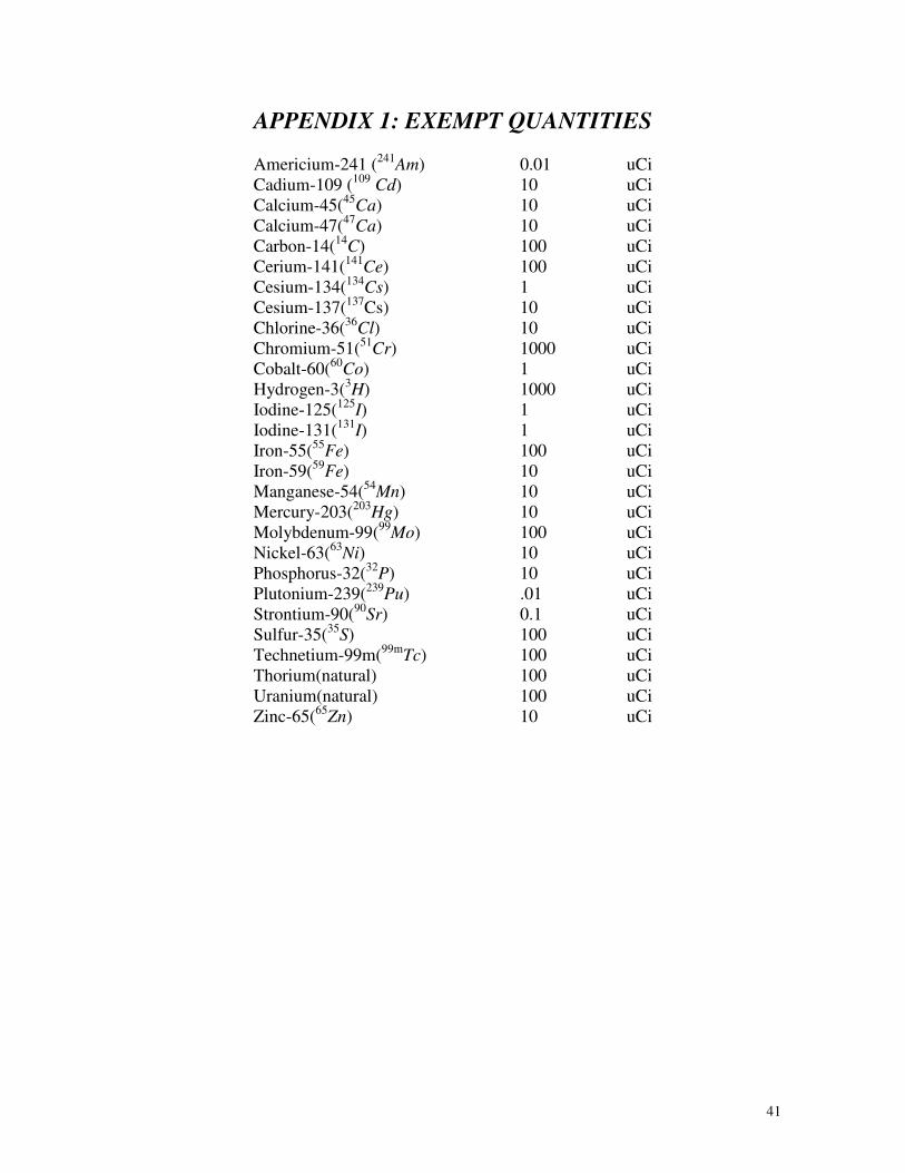
41
APPENDIX 1: EXEMPT QUANTITIES
Americium-241 (241
Am) 0.01 uCi
Cadium-109 (109
Cd) 10 uCi
Calcium-45(45
Ca) 10 uCi
Calcium-47(47
Ca) 10 uCi
Carbon-14(14
C) 100 uCi
Cerium-141(141
Ce) 100 uCi
Cesium-134(134
Cs) 1 uCi
Cesium-137(137
Cs) 10 uCi
Chlorine-36(36
Cl) 10 uCi
Chromium-51(51
Cr) 1000 uCi
Cobalt-60(60
Co) 1 uCi
Hydrogen-3(3H) 1000 uCi
Iodine-125(125
I) 1 uCi
Iodine-131(131
I) 1 uCi
Iron-55(55
Fe) 100 uCi
Iron-59(59
Fe) 10 uCi
Manganese-54(54
Mn) 10 uCi
Mercury-203(203
Hg) 10 uCi
Molybdenum-99(99
Mo) 100 uCi
Nickel-63(63
Ni) 10 uCi
Phosphorus-32(32
P) 10 uCi
Plutonium-239(239
Pu) .01 uCi
Strontium-90(90
Sr) 0.1 uCi
Sulfur-35(35
S) 100 uCi
Technetium-99m(99m
Tc) 100 uCi
Thorium(natural) 100 uCi
Uranium(natural) 100 uCi
Zinc-65(65
Zn) 10 uCi

42
APPENDIX 2: TENTH VALUE LAYERS FOR GAMMA EMITTING ISOTOPES
Isotope Principle Gamma Concrete Steel Lead
keV inches cm inches cm inches cm 51
Cr 320 3.8 9.7 1.1 2.8 0.3 0.8 54
Mn 835 5.6 14.2 1.8 4.6 1 2.5 55
Fe 6 0.0008 0.002 0.0002 0.0005 57
Co 122 2.7 6.9 0.4 1 0.02 0.06 59
Fe 1292 7.3 18.5 1.8 4.6 1.4 3.6 60
Co 1332 8.2 20.8 2.7 6.9 1.6 4 65
Zn 1116 7 17.8 2.1 5.3 1.3 3.3 75
Se 401 4.2 10.7 1.3 3.3 0.4 1 85
Sr 514 4.8 12.2 1.4 3.6 0.6 1.5 86
Rb* 1077 6.8 17.3 2 5.1 1.3 3.3 99
Mo* 739 5.4 13.7 1.7 4.3 0.9 2.3 99m
Tc 141 2.8 7.1 0.5 1.3 0.03 0.08 109
Cd 88 0.2 0.6 0.01 0.03 125
I 35 0.02 0.06 0.003 0.007 131
I 723 5.4 13.7 1.7 4.3 0.9 2.3 134
Cs 1365 8.2 20.8 2.7 6.9 1.6 4 137
Cs 662 6.2 15.7 2.1 5.3 0.8 2.1 141
Ce* 145 2.8 7.1 0.5 1.3 0.03 0.08 144
Ce* 134 2.8 7.1 0.5 1.3 0.03 0.08 203
Hg 279 3.6 9.1 1 2.5 0.2 0.5 226
Ra 1764 9.2 23.4 2.9 7.4 2.2 5.5
* Should consider beta shielding on inside of gamma shielding
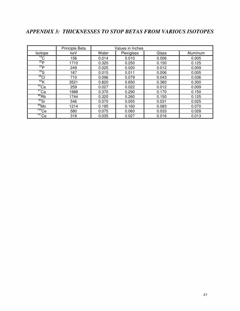
43
APPENDIX 3: THICKNESSES TO STOP BETAS FROM VARIOUS ISOTOPES
Principle Beta Values in Inches
Isotope keV Water Plexiglass Glass Aluminum 14
C 156 0.014 0.010 0.006 0.005 32
P 1710 0.320 0.250 0.150 0.125 33
P 249 0.025 0.020 0.012 0.009 35
S 167 0.015 0.011 0.006 0.005 36
Cl 710 0.096 0.079 0.043 0.036 42
K 3521 0.820 0.650 0.360 0.300 45
Ca 259 0.027 0.022 0.012 0.009 47
Ca 1988 0.370 0.290 0.170 0.150 86
Rb 1744 0.320 0.260 0.150 0.125 90
Sr 546 0.070 0.055 0.031 0.025 99
Mo 1214 0.195 0.160 0.083 0.070 144
Ce 580 0.075 0.060 0.033 0.028 141
Ce 318 0.035 0.027 0.016 0.013
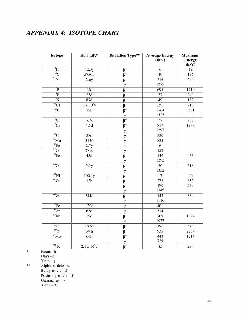
44
APPENDIX 4: ISOTOPE CHART
Isotope Half-Life* Radiation Type** Average Energy
(keV)
Maximum
Energy
(keV) 3H 12.3y β− 6 19
14C 5730y β−
49 156 22
Na 2.6y β+
γ
216
1275
546
32P 14d β−
695 1710 33
P 25d β− 77 249
35S 87d β−
49 167 36
Cl 3 x 10⁵y β−
251 710 42
K 12h β−
γ
1564
1525
3521
45Ca 163d β− 77 257
47Ca 4.5d β−
γ
817
1297
1988
51Cr 28d γ 320
54Mn 313d γ 835
55Fe 2.7y x 6
57Co 271d γ 122
59Fe 45d β−
γ
149
1292
466
60Co 5.3y β−
γ
96
1332
318
63Ni 100.1y β− 17 66
64Cu 13h β+
β−
γ
278
190
1345
653
578
65Zn 244d β+
γ
143
1116
330
75Se 120d γ 401
85Sr 65d γ 514
86Rb 19d β−
γ
709
1077
1774
90Sr 28.6y β− 196 546
90Y
64 h β− 935 2284
99Mo 66h β−
γ
443
739
1214
99Tc 2.1 x 10
5y β− 85 294
* Hours - h Days - d Years - y
** Alpha particle - α
Beta particle - β−
Positron particle - β+
Gamma ray - γ X-ray – x
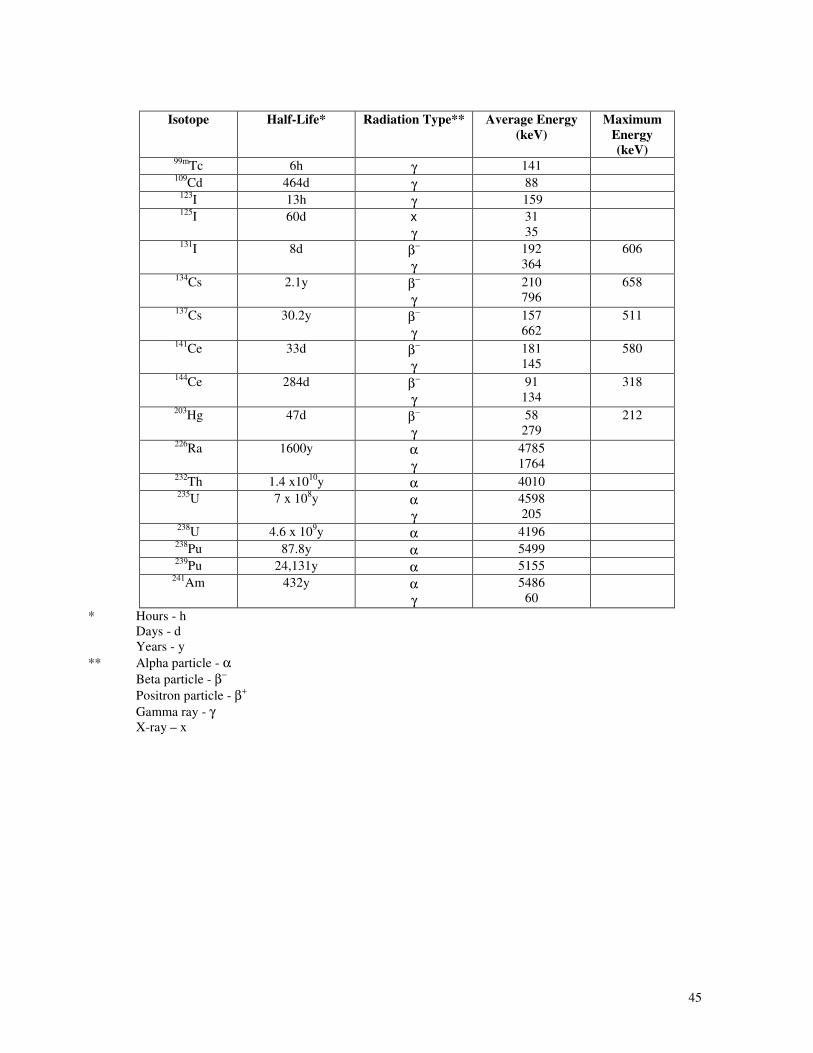
45
Isotope Half-Life* Radiation Type** Average Energy
(keV)
Maximum
Energy
(keV) 99m
Tc 6h γ 141 109
Cd 464d γ 88 123
I 13h γ 159 125
I 60d x
γ
31
35
131I 8d β−
γ
192
364
606
134Cs 2.1y β−
γ
210
796
658
137Cs 30.2y β−
γ
157
662
511
141Ce 33d β−
γ
181
145
580
144Ce 284d β−
γ
91
134
318
203Hg 47d β−
γ
58
279
212
226Ra 1600y α
γ
4785
1764
232Th 1.4 x10
10y α 4010
235U 7 x 10
8y α
γ
4598
205
238U 4.6 x 10
9y α 4196
238Pu 87.8y α 5499
239Pu 24,131y α 5155
241Am 432y α
γ
5486
60
* Hours - h Days - d Years - y
** Alpha particle - α
Beta particle - β−
Positron particle - β+
Gamma ray - γ X-ray – x

46
REFERENCES
Shapiro, J. 1972 RADIATION PROTECTION, Harvard University Press,Cambridge, Mass. 1972
Early, P. J., Razzak, M. A., Sodee, D. B. 1975 TEXTBOOK OF NUCLEAR MEDICINE
TECHNOLOGY, The C.V. Mosby Company, Saint Louis, Mo.
Gollnick, D.A. 1983 BASIC RADIATION PROTECTION TECHNOLOGY, Pacific Radiation
Corporation, Temple City, Ca.
Shleien, B., Terpilak, M.S. 1984 THE HEALTH PHYSICS AND RADIOLOGICAL HEALTH
HANDBOOK, Nucleon Lectern Associates, Olney, Md.
Department of Health, Education, and Welfare, 1970 RADIOLOGICAL HEALTH HANDBOOK,
Consumer Protection and Environmental Health Service, Rockville, Md.
Committee on the Biological Effects of Ionizing Radiation, THE EFFECTS ON POPULATIONS
OF EXPOSURE TO LOW LEVELS OF IONIZING RADIATION: 1980, National Academy
Press, Washington, D.C., 1980.
U.S. Department of Health and Human Services, EFFECTS OF IONIZING RADIATION ON THE
DEVELOPING EMBRYO AND FETUS: A REVIEW, HHS Publication FDA 81-8170,
Bureau of Radiological Health, Rockville, Md., 1981.
U.S. Department of Health and Human Services, PROCEDURES TO MINIMIZE DIAGNOSTIC
X-RAY EXPOSURE OF THE HUMAN EMBRYO AND FETUS, HHS Publication FDA
81-8178, Bureau of Radiological Health, Rockville, Md., 1981.
U.S. Nuclear Regulatory Commission, INSTRUCTIONS CONCERNING PRENATAL
RADIATION EXPOSURE, REGULATORY GUIDE 8.13, U.S. Nuclear Regulatory
Commission, Washington, D.C., 1975.
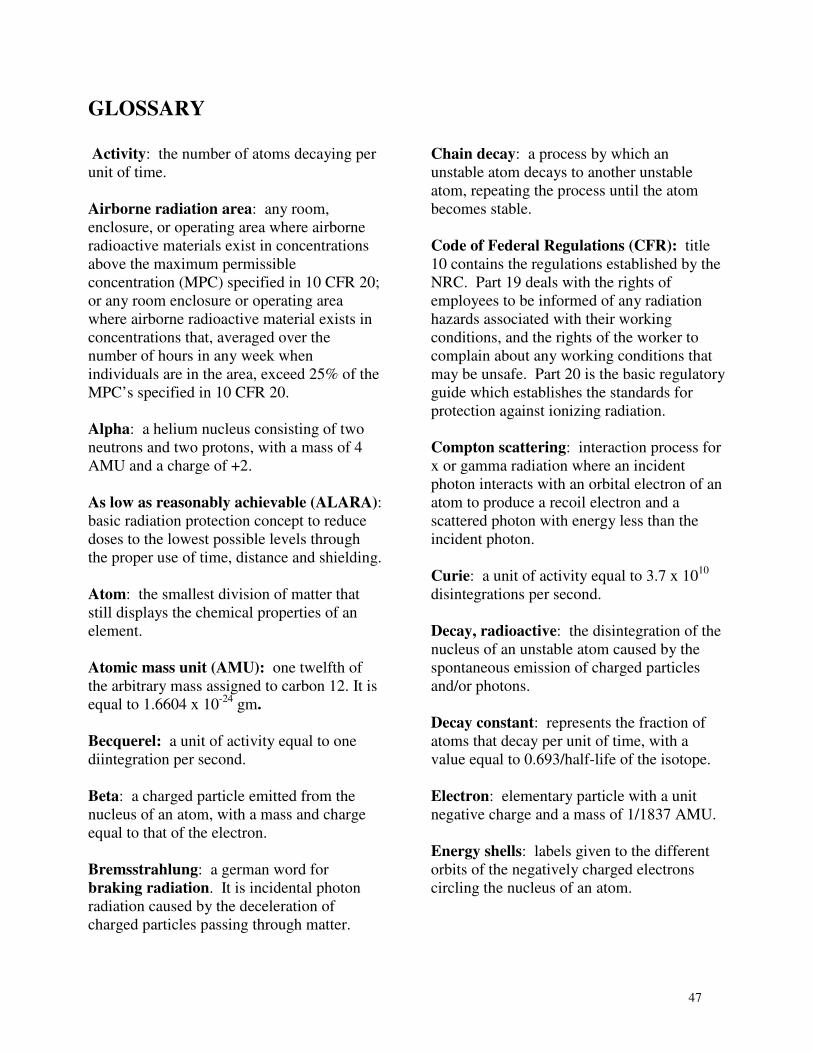
47
GLOSSARY
Activity: the number of atoms decaying per
unit of time.
Airborne radiation area: any room,
enclosure, or operating area where airborne
radioactive materials exist in concentrations
above the maximum permissible
concentration (MPC) specified in 10 CFR 20;
or any room enclosure or operating area
where airborne radioactive material exists in
concentrations that, averaged over the
number of hours in any week when
individuals are in the area, exceed 25% of the
MPC’s specified in 10 CFR 20.
Alpha: a helium nucleus consisting of two
neutrons and two protons, with a mass of 4
AMU and a charge of +2.
As low as reasonably achievable (ALARA):
basic radiation protection concept to reduce
doses to the lowest possible levels through
the proper use of time, distance and shielding.
Atom: the smallest division of matter that
still displays the chemical properties of an
element.
Atomic mass unit (AMU): one twelfth of
the arbitrary mass assigned to carbon 12. It is
equal to 1.6604 x 10⁻²⁴
gm.
Becquerel: a unit of activity equal to one
diintegration per second.
Beta: a charged particle emitted from the
nucleus of an atom, with a mass and charge
equal to that of the electron.
Bremsstrahlung: a german word for
braking radiation. It is incidental photon
radiation caused by the deceleration of
charged particles passing through matter.
Chain decay: a process by which an
unstable atom decays to another unstable
atom, repeating the process until the atom
becomes stable.
Code of Federal Regulations (CFR): title
10 contains the regulations established by the
NRC. Part 19 deals with the rights of
employees to be informed of any radiation
hazards associated with their working
conditions, and the rights of the worker to
complain about any working conditions that
may be unsafe. Part 20 is the basic regulatory
guide which establishes the standards for
protection against ionizing radiation.
Compton scattering: interaction process for
x or gamma radiation where an incident
photon interacts with an orbital electron of an
atom to produce a recoil electron and a
scattered photon with energy less than the
incident photon.
Curie: a unit of activity equal to 3.7 x 10¹⁰
disintegrations per second.
Decay, radioactive: the disintegration of the
nucleus of an unstable atom caused by the
spontaneous emission of charged particles
and/or photons.
Decay constant: represents the fraction of
atoms that decay per unit of time, with a
value equal to 0.693/half-life of the isotope.
Electron: elementary particle with a unit
negative charge and a mass of 1/1837 AMU.
Energy shells: labels given to the different
orbits of the negatively charged electrons
circling the nucleus of an atom.
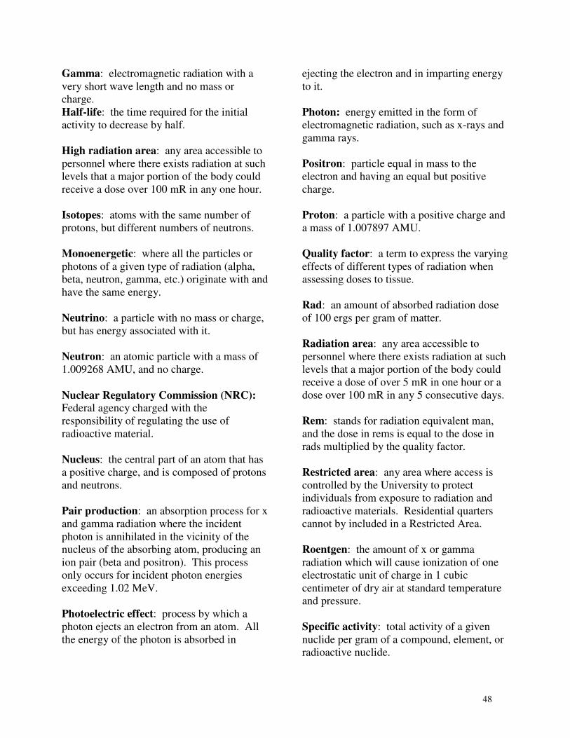
48
Gamma: electromagnetic radiation with a
very short wave length and no mass or
charge.
Half-life: the time required for the initial
activity to decrease by half.
High radiation area: any area accessible to
personnel where there exists radiation at such
levels that a major portion of the body could
receive a dose over 100 mR in any one hour.
Isotopes: atoms with the same number of
protons, but different numbers of neutrons.
Monoenergetic: where all the particles or
photons of a given type of radiation (alpha,
beta, neutron, gamma, etc.) originate with and
have the same energy.
Neutrino: a particle with no mass or charge,
but has energy associated with it.
Neutron: an atomic particle with a mass of
1.009268 AMU, and no charge.
Nuclear Regulatory Commission (NRC):
Federal agency charged with the
responsibility of regulating the use of
radioactive material.
Nucleus: the central part of an atom that has
a positive charge, and is composed of protons
and neutrons.
Pair production: an absorption process for x
and gamma radiation where the incident
photon is annihilated in the vicinity of the
nucleus of the absorbing atom, producing an
ion pair (beta and positron). This process
only occurs for incident photon energies
exceeding 1.02 MeV.
Photoelectric effect: process by which a
photon ejects an electron from an atom. All
the energy of the photon is absorbed in
ejecting the electron and in imparting energy
to it.
Photon: energy emitted in the form of
electromagnetic radiation, such as x-rays and
gamma rays.
Positron: particle equal in mass to the
electron and having an equal but positive
charge.
Proton: a particle with a positive charge and
a mass of 1.007897 AMU.
Quality factor: a term to express the varying
effects of different types of radiation when
assessing doses to tissue.
Rad: an amount of absorbed radiation dose
of 100 ergs per gram of matter.
Radiation area: any area accessible to
personnel where there exists radiation at such
levels that a major portion of the body could
receive a dose of over 5 mR in one hour or a
dose over 100 mR in any 5 consecutive days.
Rem: stands for radiation equivalent man,
and the dose in rems is equal to the dose in
rads multiplied by the quality factor.
Restricted area: any area where access is
controlled by the University to protect
individuals from exposure to radiation and
radioactive materials. Residential quarters
cannot by included in a Restricted Area.
Roentgen: the amount of x or gamma
radiation which will cause ionization of one
electrostatic unit of charge in 1 cubic
centimeter of dry air at standard temperature
and pressure.
Specific activity: total activity of a given
nuclide per gram of a compound, element, or
radioactive nuclide.

49
Tenth value: the thickness of a given
material that will decrease the amount of
radiation to one-tenth of the original value.
X-ray: penetrating electromagnetic radiation
similar to visible light, but having extremely
short wave lengths.


