32
Running Header: Case Study of Leiomyosarcoma 1 Radiation Therapy Clinical Training Case Study on Leiomyosarcoma (LMS) By: Benjamin Rodriguez Argosy University, Twin Cities 2013/14
Running Header: Case Study of Leiomyosarcoma 1
Radiation Therapy Clinical Training
Case Study on Leiomyosarcoma (LMS)
By: Benjamin Rodriguez
Argosy University, Twin Cities
2013/14

Leiomyosarcoma 2
Abstract
Leiomyosarcoma (LMS) is a malignant soft tissue sarcoma tumor of the muscle.
Sarcomas are very vascular, with large internal and external tumor blood vessels
that typically occurs in adults, especially women. (Chu et al., 2003) The majority
of patients diagnosed with LMS are currently treated with surgical excision limb
preserving surgery, radiation therapy or occasionally with chemotherapy or a
combination of both. Almost 45% of all soft tissue sarcomas are located in the
extremities, especially in the lower limb (Gomez ve Morcuende, 2004).
Leiomyosarcoma 3
Introduction
In the previous six months, I had the pleasure of meeting and following a
patient that I will refer to as Mrs. P. She was so generous to allow me to take part
in her fight with LMS. I was involved in Mrs. P’s initial consultation to her final
treatment and her clinic follow-ups. In this case study I will address the
consultation, CT simulation, treatment planning, daily treatment and any side
effects Mrs. P experienced during or after treatment. I will conclude with current
treatment options that are available for this type of sarcoma.
Consultation
Mrs. P is an 83 year old female that was recently diagnosed with LMS of the
right calf. She noticed an enlargement of her right calf with some tenderness while
pressure was applied. The patient has a prior history of LMS of her pelvis, status
post resection with positive margins. Mrs. P was treated with radiation therapy to
the pelvic mass consisting of 5,400cGy in 30 treatment fractions, treatment was
completed on (3/5/2008).
Due to her prior history of LMS the oncologist diagnostic testing done. Mrs.
P was scheduled for a diagnostic Computer Tomography (CT), Magnetic
Resonance Imaging (MRI), biopsy and a Computer Tomography Positron
Emission Tomography (PET/CT) scans of the right lower extremity. On (1/9/2013)
a CT with contrast was administered, results of this scan reviled a large 10cm soft

Leiomyosarcoma 4
tissue mass that expands the soleus muscle. Her MRI scan on (1/15/2013)
presented a 10.2cm x 6.6cm x 7.2cm heterogeneous, necrotic-appearing mass in
the soleus muscle consistent with a tumor. On (1/17/2013) Mrs. P underwent a
biopsy of the right calf mass, the biopsy reviled LMS. Mrs. P also underwent a
PET/CT scan on (1/30/2013), the scan showed uptake of Flurorodeoxyglucose
(FDG) in the mass in her right lower extremity of a SVU of (9.6).
The plan of action taken for Mrs. P was resection the right lower extremity
calf mass, also a posterior tibial vessel resection with post-surgical radiation
therapy.
Detection and Diagnosis
Pathology reviled a high grade LMS tumor measuring 13cm x 12cm x 9cm
involving subcutaneous tissue, with sarcoma present at the posterior soft tissue
edge adjacent to the skin. The anterior tibial artery was also grossly involved with
tumor, the discovery of a secondary 5cm x 2cm x 2cm additional tumor was
focused adjacent to posterior tibial artery with margins negative less than 1mm.
There was no evidence of lymph node involvement. The pathological classification
of this malignancy is a high grade LMS, stage: III (pT2b Nx M)
The oncologist and pathologist discovered upon examination of Mrs. P’s CT
scan and PET/CT from (1/9 and 1/30/2013) there is a small right lung nodule. The

Leiomyosarcoma 5
nodule did appear to present with minimal uptake of FDG. The oncologist elected
to wait for any changes to appear due to an unclear significance.
Simulation
The simulation was performed with a Phillips big bore CT scanner equipped
with LAP laser tumor localization technology. Mrs. P was positioned prone with
one pillows under her abdomen, prone wedge under her hips and a prone pillow for
head placement and comfort. (See Image below)
I constructed the custom Vac-Loc to position the right leg more superiorly to
avoid any chance for the left leg/calf to be in the beams path, also formed the Vac-
Loc around the lateral edges of Mrs. P’s ankle and upper thigh also formed the
sides of the Vac-Loc below the calf. Due to the beams possible angles. I did not
want any part of the vac-loc to attenuate the beams and possibly adding a bolus
effect. For easy reproducibility I abutted the vac-loc to the prone wedge with a
Styrofoam block under her ankle for support. (See Image below)
Rt. Calf Patient Table Set-up for Treatment

Leiomyosarcoma 6
Mrs. P’s scar was marked with (CT-Spot) field outline, and (Y-Spot) or bee
bees where placed on locations marked for tattoos. The superior and inferior
tattoos are used for patient straightness and the center tattoo is the CAX (central
access) with a shift of 2cm shift left from center of the scar. (See Images below)
Rt. Calf Side Photo of Patient Set-up with Vac-Loc
Rt. Calf Scar Markings and Outline

Leiomyosarcoma 7
Treatment Planning
After simulation of Mrs. P, I was able to partake in the treatment planning
process. The oncologist expressed his intention of treatment fields consisting of
two parallel opposed fields. Using the pinnacle treatment planning software in
dosimetry, I constructed the Digital Reconstructed Radiograph (DRR) used for
treatment, and outlined the regions of interest. The outlined regions consisted of
the tumor bed and surgical scare. There wasn’t a need for a Dose Volume
Histogram (DVH), due to the area of interest was located in the lower right calf
and no critical structures where located in the treatment fields. Developing the
treatment fields alongside the dosimetrist, physicist, and oncologist was a great
experience.
The angels I developed for the treatment plan was approved by the
oncologist are as follow. Beam one was a left lateral (Lt. Lat) with a gantry angle
Rt. Calf Patient Alignment Marks, Scar Outline,
and Shift for Isocenter.

Leiomyosarcoma 8
of 900, a collimator angle of 730 with a couch kick of 50 to 1850. The (Lt. Lat)
utilized Multi-leaf Collimation (MLC) blocking with an Electronic Dynamic
Wedge (EDW) of 300. Beam Two was a Right lateral (Rt. Lat) with a gantry angle
of 2700, a collimator angle of 2780 with a couch kick of 50 to 1850. The (Rt. Lat)
also utilized Multi-leaf Collimation (MLC) blocking with an Electronic Dynamic
Wedge (EDW) of 300. The prescription for this treatment plan consisted of using
6MV photons of 200cGy per treatment fraction for 25 fractions totaling a dose of
5,000cGy. Treatment was administered from (6/4/2013-7/12/2013).
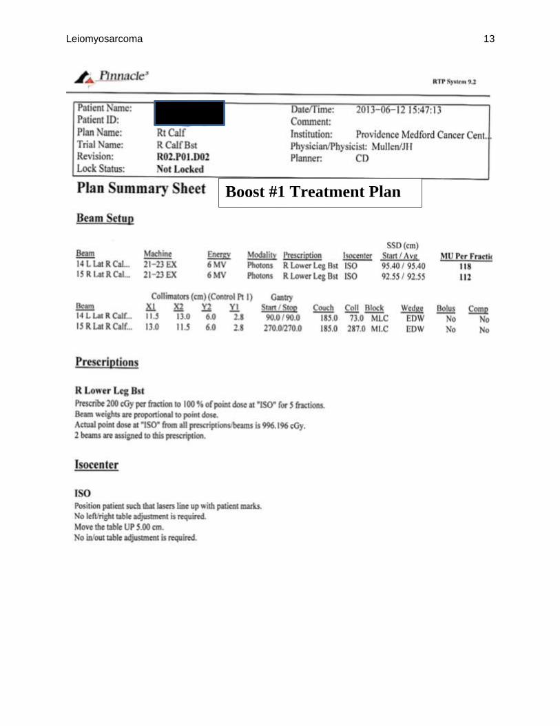
There was also additional plans for two treatment boosts. Boost one was
planned with a cone down consisting of all the same angels as the main treatment
plan with a prescription of 6MV photons of 200cGy per treatment fractions for 5
fractions totaling a dose of 1,000cGy. Treatment was administered for boost one
from (7/15/2013-7/19/2013).
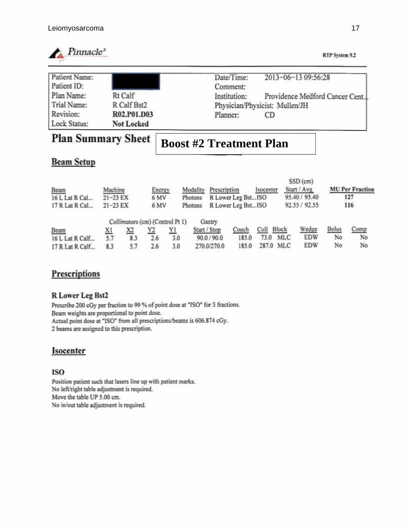
Boost two was planned with an additional cone down from boost one
consisting of all the same angels as the main treatment plan with a prescription of
6MV photons of 200cGy per treatment fractions for 3 fractions totaling a dose of
600cGy. Treatment was administered for boost two from (7/30/2013-8/1/2013).
The total number fractions for all treatments delivered was a total of 33 fractions
with a total dose of 6,600cGy.

Leiomyosarcoma 9
(Below are the treatment plans, boosts 1, and 2 also field block shapes)
Main Treatment Plan

Leiomyosarcoma 10
Main Treatment Plan Field Block Shape for Rt. Lat
Leiomyosarcoma 11
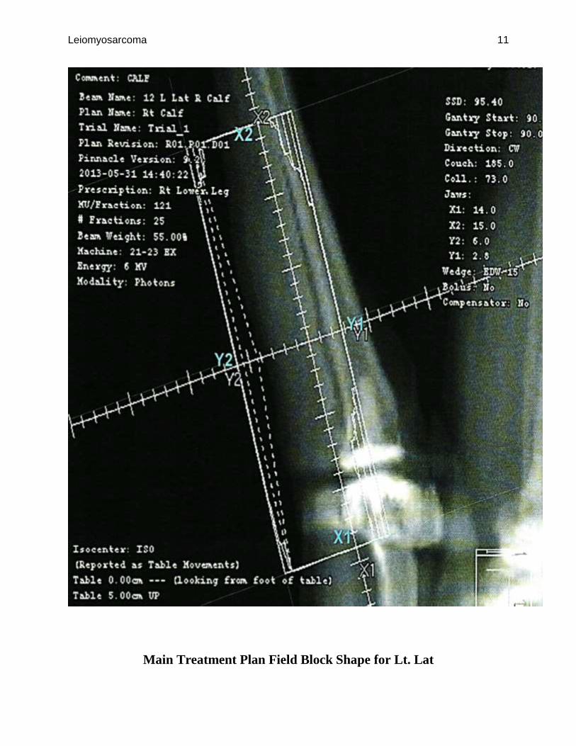
Main Treatment Plan Field Block Shape for Lt. Lat
Leiomyosarcoma 12
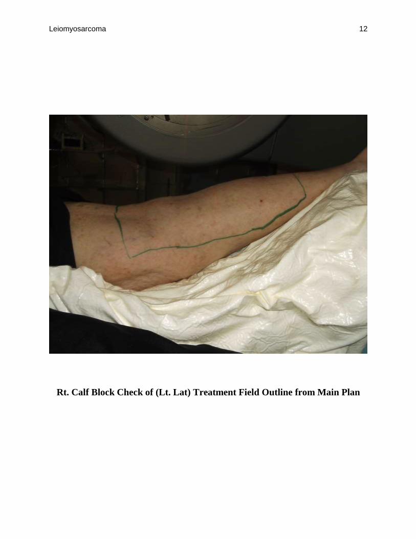
Rt. Calf Block Check of (Lt. Lat) Treatment Field Outline from Main Plan
Leiomyosarcoma 13
Boost #1 Treatment Plan

Leiomyosarcoma 14
Boost #1 Treatment Plan Field Block Shape for Lt. Lat

Leiomyosarcoma 15
Boost #1 Treatment Plan Field Block Shape for Rt. Lat
Leiomyosarcoma 16
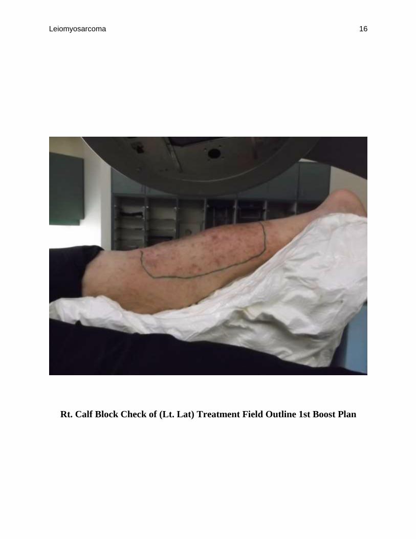
Rt. Calf Block Check of (Lt. Lat) Treatment Field Outline 1st Boost Plan
Leiomyosarcoma 17
Boost #2 Treatment Plan
Leiomyosarcoma 18
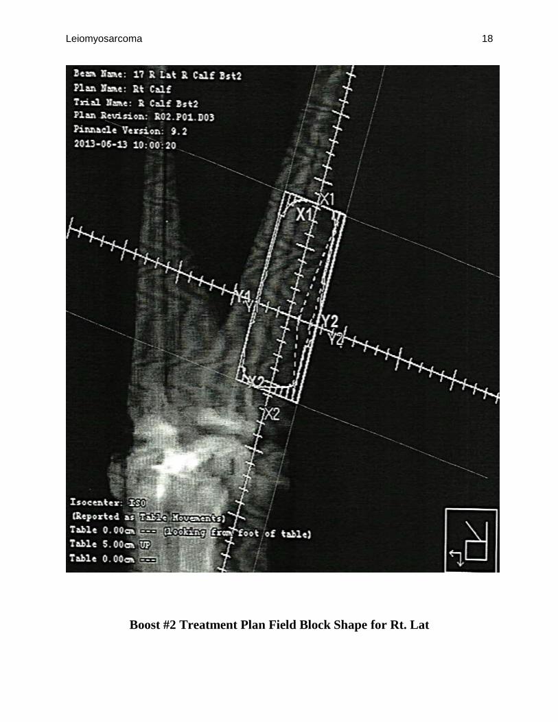
Boost #2 Treatment Plan Field Block Shape for Rt. Lat

Leiomyosarcoma 19
Boost #2 Treatment Plan Field Block Shape Lt. Lat
Leiomyosarcoma 20
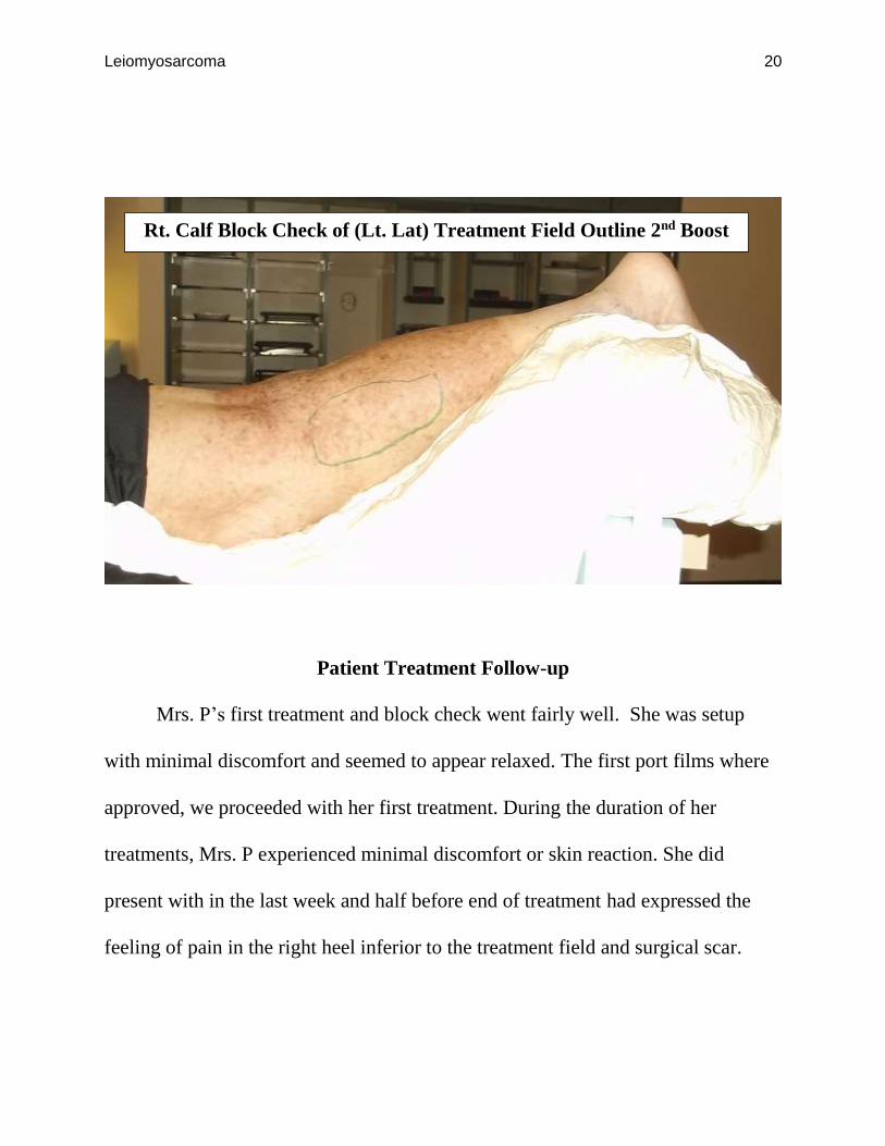
Patient Treatment Follow-up
Mrs. P’s first treatment and block check went fairly well. She was setup
with minimal discomfort and seemed to appear relaxed. The first port films where
approved, we proceeded with her first treatment. During the duration of her
treatments, Mrs. P experienced minimal discomfort or skin reaction. She did
present with in the last week and half before end of treatment had expressed the
feeling of pain in the right heel inferior to the treatment field and surgical scar.
Rt. Calf Block Check of (Lt. Lat) Treatment Field Outline 2nd Boost
Boost Plan

Leiomyosarcoma 21
On August 6th, 2013 one week after her last treatment Mrs. P presented with
mild erythema, pigmentation and a small opening at the inferior aspect of the scar
with a small amount of drainage. Over the course of treatment she handled
treatment better than expected.
On September 4th, 2013 one month after her last treatment Mrs. P still
complained of right heel pain that has been present since surgery. Within the prior
radiation fields the skin is well healed, with some residual increased pigmentation
and the absences of erythema, desquamation, or any evidence of infection. The
inferior portion of the scar still demonstrates a small (4-5mm) opening with
drainage of yellowish fluid without the presents of bleeding or infection. Also she
is experiencing tenderness to palpation of the right calf in the location around the
prior surgical and radiation therapy areas.
On October 23rd, 2013 two and a half months after her last treatment. Mrs. P
continues to report pain in her right heel, approximately with a level of 5 out of 10.
She also underwent an MRI for her calf on (10/14/2013), which showed no
evidence of infection at the surgical site or no obvious fistula at the small wound
opening. Mrs. P on (10/14/213) underwent a CT of her chest that showed an
increase number in size of the bilateral pulmonary nodules that were previously
discovered on a prior scan before Radiation treatment.

Leiomyosarcoma 22
On December 11th, 2013 four months after the completion of adjuvant
radiation therapy of the right calf for resected leiomyosarcoma. The examination of
the right calf showed posteriorly a small (3-4mm) unhealed opening and an
additional tiny (1mm-2mm) opening just superiorly os scar. There was no evidence
of erythema or infection. Mrs. P still has tenderness of the right calf which is
noticeably less than prior exam. The oncologist presented Mrs. P with the findings
from her (10/25/2013) CT guided biopsy of the lung. The results of the pathologic
report showed metastatic LMS. Consultation with Mrs. P about palliative radiation
therapy of her lung mass was addressed with Mrs. P. She expressed she was not
going to proceed with any more radiation treatments or surgeries.
Leiomyosarcoma (LMS)
Leiomyosarcoma (LMS) is classified by their anatomical site of presentation,
retroperitoneum, subcutaneous skin, blood vessels or extremities. (De Vita Jr Vt,
1997) In the case with Mrs. P, the location involved is the lower right extremity
(right calf).
Etiology
Current researchers speculate a contributing role in causing LMS, is genetic
changes that occur spontaneously or may be inherited. (De Vita Jr Vt, 1997)
LMS malignancies may develop due to abnormal changes in the structure and
orientation of certain cells known as oncogenes (tumor suppressor genes).

Leiomyosarcoma 23
Oncogenes control cell growth and cell division and ensure that cells die at the
appropriate time.
The specific cause of the changes is unknown. Research suggests that
abnormalities of in DNA (deoxyribonucleic acid), is the underlying source of
cellular malignant transformation. (Sarcoma, 2012) There aren't many known risk
factors for LMS, but higher risk may be increased due to Age. LMS can occur at
any age, but overall is more common in older adults. Chemical exposure of certain
chemicals, such as vinyl chloride and dioxin, can increase the risk of soft tissue
sarcomas. (Moynihan TJ, 2007) Radiation exposure of previous radiation treatment
for other cancers can increase the risk of soft tissue sarcomas.
Epidemiology
Soft tissue sarcomas account for 1% of all adult cancers in the U.S.
According to one estimate, Leiomyosarcomas account for 7-11% of all cases of
soft tissue sarcomas. (American Cancer Society, 2012) Leiomyosarcoma is
primarily a disease of middle-aged people, presenting between the years 50-60,
especially women. (Gomez ve Morcuende, 2004) Individuals diagnosed with LMS
have an 8% chance of developing local recurrence also a 45% of LMA metastases.
(Washington, Leaver, 2010) The American Cancer Society's estimates about
(12,020) new soft tissue sarcomas will be diagnosed this year (6,550 cases in males
and 5,470 cases in females) and 4,740 in Americans (2,550 males and 2,190

Leiomyosarcoma 24
females) are expected to die of soft tissue sarcomas in the United States for 2014.
(American Cancer Society, 2012)
Diagnostic methods
General symptoms with patients diagnosed with LMS is pain located in the
affected area. Swelling and a mass is commonly detected. Mrs. P was experiencing
pain and numbness in her right lower extremity, and was also noticing the right
calf was significantly larger with a hard mass palpable under her skin. The
oncologist performed a complete physical exam. After completion of the exam he
ordered standard imaging tests.
The tests ordered was a diagnostic X-ray, Computerized Tomography (CT)
scans, Magnetic Resonance Imaging (MRI) and Positron Emission Tomography
(PET), also a CT guided biopsy. The battery of test ordered appeared to be
excusive, but due to Mrs. P’s history with prior Leiomyosarcoma of her pelvis. Out
of all the diagnostic procedures completed, the most important test was the full
body PET/CT scan preformed on (10/30/2013). The scan revealed there was an
increase of Flurorodeoxyglucose (FDG) uptake in the lung nodules and increase in
size, with an additional noticeable uptake in the thyroid. This was conclusive of
metastatic disease.
Leiomyosarcoma 25
Pathology
Leiomyosarcoma is classified as a soft tissue sarcoma. Sarcomas are
malignant tumors that arise from the connective tissue, this connects, supports and
surrounds various structures and organs in the body. (Moynihan TJ, 2007) Most
forms of LMS are aggressive tumors that may spread (metastasize) to other areas
within the body. Because LMS typically spreads via the bloodstream and soft
tissues is found all over the body, LMS can form almost anywhere where there are
blood vessels. This includes the lungs the most prevalent site of metastases, heart,
liver, pancreas, muscle, nerves, tendons the genitourinary and gastrointestinal tract
and the abdominal cavity, uterus, skin. The lungs or liver, potentially causing life-
threatening complications, if not diagnosed early. The 5-year relative survival rates
for Leiomyosarcoma is: Stage I - 60%, Stage II - 35%, Stage III - 28% and Stage
IV - 15%. (Washington, Leaver, 2010)
The pathologist evaluates the scans and determines how far the cancer has
metastasizes. This is called staging. The evaluation of the appearance of the
biopsied tissues under the microscope and judge how fast the cancer seems to be
growing. When examining the biopsy sample accounts for the number of cells
actively dividing and how closely the cancer cells resembles normal tissue. This
determines the cell type and grade and estimates how rapidly the tumor will grow
and spread. (Gomez ve Morcuende, 2004) After evaluation of Mrs. P’s biopsy and

Leiomyosarcoma 26
scans the prior pathological classification of a high grade LMS, stage: III (pT2b Nx
M) was updated to a high grade spindle cell neoplasm LMS, stage: IV (pT2b Nx
M1)
Current Treatment Options
There are currently three treatment modalities available for LMS: surgery,
radiation therapy and chemotherapy. The specific procedures and interventions is
dependent upon several pathological factors. (NCCN, 2011)The factors for a
specific treatment options for Mrs. P was the location of the primary tumor, extent
of the primary tumor (stage), and degree of malignancy (grade). Also taken in to
consideration is whether the tumor has metastasized to distant sites, her age and
overall general health.
Surgery
Surgery is the most common treatment for soft tissue sarcomas, especially if
malignant cells haven't spread to other parts of the body. Surgery for LMC
generally involves removing the tumor and some surrounding healthy tissues, as
with Mrs. P. If the sarcoma has spread, surgical removal of the primary and
secondary tumors may be possible. Surgical amputation was previously a common
treatment for soft tissue sarcomas in the arms or legs. (Washington, Leaver, 2010)
Currently advancements in surgical techniques with the combination of
chemotherapy and radiation therapy before or after surgery has allowed for the
Leiomyosarcoma 27
possibility for limb-sparing surgery. In rare cases amputation is still required if
LMS has invaded underlining nerves, arteries or muscle. (Moynihan TJ, 2007)
This allow for the complete removal of all malignant tissue and tumor cells in the
arms or legs. If initial surgery is not an option due to the specific location or
progression of the malignancy, radiation therapy maybe include alone.
Radiation
Radiation therapy is also used to treat LMC. Oncologists likely will
recommend using radiation therapy before surgery (neoadjuvant radiation therapy)
or after surgery (adjuvant radiation therapy). The postoperative radiation helps
treat known or possible residual disease and preoperative radiation administered
halts the growth of tumor and reduces the size for possible surgical resection
(Sarcoma, 2012).
Radiation and chemotherapy are also used before surgery (neoadjuvant
chemo radiation therapy) or after surgery (adjuvant chemo radiation therapy).
(Moynihan TJ, 2007) Chemotherapy administered alongside with radiation allows
for the radiation to be more effective in destroying cancer cells (Sarcoma, 2012).
Treatment recommendations for patients with a surgical resectable disease,
followed by radiation therapy is recommended for high-grade LMS. The benefits is
a smaller treatment field and potentially less chance for tumor seeding during
resection. The normal prescribed dose for radiation for LMS is 5,000cGy. (NCCN,

Leiomyosarcoma 28
2011) Mrs. P was prescribed the normal radiation dosage for LMS treatment of
200cGy per day for 25 treatment fraction for a total dose of 5,000cGy, but she also
was prescribed two treatment boosts. Boost one was administered 200cGy per day
for 5 treatment fraction for a total dose of 1,000cGy and boost two was
administered 200cGy per day for 3 treatment fraction for a total dose of 600cGy.
The total dose given for her LMS treatment was 6,600cGy. The only negative
aspect of such a high dose is significant wound healing complications. (NCCN,
2011)
Chemotherapy
Chemotherapy uses medications to kill rapidly dividing cells. These cells
include cancer cells, which continuously divide to form more cells, and healthy
cells that divide quickly, such as those in your bone marrow, gastrointestinal tract,
reproductive system and hair follicles. (Sarcoma, 2012) Unlike radiation therapy,
which treats only the part of your body exposed to the radiation, chemotherapy
treats your body as a whole (systemically). Due to LMS spreads throughout the
bloodstream. (Chemotherapy treats cells that passably have spread beyond where
the cancer originated.
In Mrs. P case the oncologist felt that she wouldn’t benefit from
chemotherapy alongside radiation. His choice was for radiation alone was due to
the possible side effects from chemotherapy, her daily regimen of insulin for
Leiomyosarcoma 29
diabetes and pain medications could cause extreme nausea, vomiting, fatigue,
increased risk of infection, weakness and increased bleeding. (Washington, Leaver,
2010)
Conclusion
I have had the privilege to follow Mrs. P’s course of treatment for six
months, from her initial consultation, final treatment until her last follow up in
December of 2013. The treatment Mrs. P received was in accordance with the
standard plan of treating LMS. She did receive two boosts to the initial treatment
area, that the oncologist procived as beneficial for no possible reoccurrence of Mrs.
P’s LMS.
The prognosis for Mrs. P unfortunately isn’t good. As mentioned in the
follow up portion of the case study. Mrs. P on (10/14/2013) underwent a CT of her
chest and a PET/CT full bod scan on (10/30/2013) that showed an increase number
in size and uptake of FDG in the bilateral pulmonary lung nodules as well an
additional noticeable uptake in the thyroid. This is pathologically decisive of
metastatic LMS.
On December 11th, 2013 four months after the completion of adjuvant
radiation therapy of the right calf for resected leiomyosarcoma. The oncologist
presented Mrs. P with the pathological findings from her (10/25/2013) CT guided
biopsy from her lung. The results of the report showed metastatic LMS. After Mrs.
Leiomyosarcoma 30
P was presented with the finding from pathologist, the oncologist consulted with
Mrs. P about her choices of treatment options. He discussed about surgery
although there was a slim chance her lesions where resectable. He also discussed
palliative radiation therapy of her bilateral pulmonary nodules. After some
questions from Mrs. P on the likelihood of curability. She expressed that she
wasn’t going to proceed with any more surgeries or radiation treatments.
Mrs. P indicated that she has lived and has a great life and was tired of all
the surgeries, radiation and just wanted to enjoy the time she has left with her
husband, children and grandchildren. She did express she knew this was coming
after LMS of her pelvis and calf and now lungs. I knew before the biopsy was
preformed what the outcome would be.

Leiomyosarcoma 31
References
Chu, W.C., Howard, R.G., Roebuck, D.J., Chik, K.W., Li, C.K. (2003).
“Periorbital alveolar soft part sarcoma with radiologic features
mimicking hemangioma”, Med Pediatr Oncol., 41:145-146.
De Vita Jr Vt, Hellman S, Rosenburg SA. Eds. Cancer: Principles and
Practice on Oncology. 5th ed. Philadelphia, PA: J.B. Lippincott
Company; 1997:1748-9.
Gomez, P. (2004). “Morcuende J. High-grade sarcomas mimicking
traumatic http://www.mayoclinic.org/diseases-conditions/soft-tissue
sarcoma/basics/treatment/con-20033386 intramuscular hematomas: a
report of three cases”, Iowa Orthop J., 24:106-110.
Moynihan TJ (expert opinion). Mayo Clinic, Rochester, Minn. July 31,
2011.
NCCN Clinical Practice Guidelines in Oncology: Soft Tissue Sarcoma.
Available at
http://www.nccn.org/professionals/physician_gls/pdf/sarcoma.pdf.
Accessed March 28, 2011.
Sarcoma - Adult Soft Tissue Cancer. American Cancer Society.
http://www.cancer.org/Cancer/Sarcoma AdultSoftTissueCancer/index.
Accessed August 29, 2012.
Leiomyosarcoma 32
Washington, C. M., & Leaver, D. (2010). Principles and Practice of
Radiation Therapy. St. Louis: Mosby Elsevier.


