Advances in radiation therapy have brought forth necessary changes in the education of radiation therapists. A national committee representing a variety of program types from across the country developed the curriculum. Input from The American Registry of Radiologic Technologists (ARRT) and the Joint Review Committee on Education in Radiologic Technology (JRCERT) were included in this revision to maintain continuity among the professional curriculum, accreditation standards and the certification examination. This curriculum is divided into specific content areas that represent the essential components of an entry-level radiation therapy program. The content and objectives should be organized to meet the mission, goals and needs of each radiation therapy program. Faculty are encouraged to expand and broaden these fundamental objectives as they incorporate them into their curricula. Specific instructional methods were intentionally omitted to allow for programmatic prerogative as well as creativity in instructional delivery. Advances in radiation therapy and employer expectations demand more independent judgment by radiation therapists. Consequently, critical thinking skills must be fostered, developed and assessed in the educational process. Critical thinking has been incorporated in multiple content areas. These areas include, but are not limited to, clinical practice and the required postsecondary general education. It is expected that the faculty will continue to develop and implement critical thinking throughout the curriculum. New content and objectives have been added to the radiation therapy curriculum. New areas include adaptive technologies, expanded imaging for radiation oncology and information technology. Clinical and didactic competencies have been correlated. Some content areas have been retitled or reorganized and outdated content eliminated. The guidance provided by this curriculum document will span the time period prior to and after the projected Jan. 1, 2015, start date of the ARRT’s minimum associate degree requirement for candidates seeking professional certification. The focus of this document is on the preprofessional core instructional content that will be expanded with institution-specific course content to fulfill metrics for receipt of an academic degree. It is beyond the scope of this document to outline administrative strategies for programs that are unable to award graduates an academic degree to comply with the ARRT’s 2015 degree requirement. The general education content objectives in this curriculum were purposely labeled “global content objectives” to give program officials flexibility in determining specific credit-bearing course work that will satisfy these objectives. Following 2015, it is expected that this component of the professional curriculum will be satisfied with general education courses needed to fulfill institution-specific degree requirements.
In summary, the new radiation therapy curriculum is based on the latest data relevant to the profession and reflects the dynamic health care environment. The curriculum offers a foundation for individual professional development at a minimum level of a baccalaureate degree. It allows for flexibility in the development of a curriculum designed to meet the needs of the local community yet meet the requirements for the JRCERT Standards and the ARRT examination. The radiation therapy professional curriculum is in line with a baccalaureate education. Note: The general education and professional content areas of the curriculum are not courses. To preserve the flexibility for all radiation therapy programs, content within each topic may be integrated into various courses.
Required General Education General education is an integral part of the radiation therapy professional curriculum. This portion of the curriculum gives students opportunities to explore broad areas of commonly held knowledge and to prepare them to contribute to society through personal, social and professional interactions with others. General education provides the intellectual flexibility and knowledge to support continued professional development in a rapidly changing profession. General Education Content College-level coursework provides foundational knowledge on which to build the radiation therapy content. The framework for general education may be delivered prior to or congruent to the professional curriculum.
• Oral and written communication can be expanded to facilitate technical and scientific inquiry, analysis and dissemination of knowledge.
• Knowledge of human anatomy can be increased to include the correlation of topographic landmarks to internal organs, in-depth examination of the lymphatic system and transference of sagittal and coronal anatomical structure identification to cross-sectional views.
• Knowledge of human physiology can be expanded to explore the processes by which tumors originate, grow, metastasize and affect the normal functioning of each of the body systems.
• Computer skill literacy can be demonstrated for application in radiation therapy simulation, treatment delivery, information processing and treatment planning.
• Mathematical analysis can be expanded to include calculation for radiation treatment and protection, radioactivity and radiobiological functions.
• Basic physics principles will facilitate the application of radiation physics. Required General Education Content Note: These are not specific courses, but represent minimal content areas.
• Human Anatomy – This content will include terminology and organization of the human organism at the cellular, tissue and organ levels. Structures of human systems including integumentary, skeletal, muscular, nervous, endocrine, sensory, circulatory, respiratory, digestive, urinary and reproductive will be covered. To facilitate understanding, a laboratory section is recommended.
• Human Physiology – A general introduction of the functional integration of all human body systems will include: integumentary, skeletal, muscular, nervous, endocrine, sensory, circulatory, respiratory, digestive, urinary and reproductive systems. To facilitate understanding of the content material, a laboratory section is recommended.
• Mathematics – This content will include a study of the real number system, algebra of sets, exponents, equations and inequalities, polynomial functions, graphing, radical expressions, operations, inverses of function, equations of lines and systems of linear equations and elementary statistics, logarithmic and trigonometric functions and their applications, and plane analytic geometry.
• Written Communication – This content will include the written expression of thoughts, ideas, perceptions and observations derived from the critical thinking process. Additionally, it may complement the critical thinking process by providing a vehicle for the organization and clarification of thoughts, for the establishment of conceptual relationships, for the analysis of data and for synthesis of conclusions or new ideas.
• Verbal Communication – This content will include the theory and practice of public speaking, development of thought processes necessary to organize speech content for informative and persuasive situations, application of language and delivery skills to specific audiences.
• General Physics – This content will include the application of physical principles, conservation laws, gravitation, electricity, magnetism, wave motion, heat and thermodynamics as it relates to scientific disciplines.
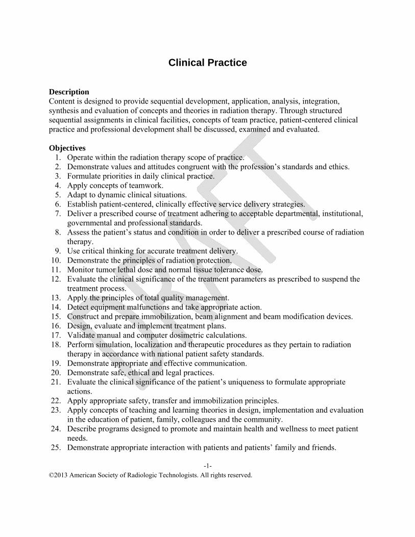
Clinical Practice Description Content is designed to provide sequential development, application, analysis, integration, synthesis and evaluation of concepts and theories in radiation therapy. Through structured sequential assignments in clinical facilities, concepts of team practice, patient-centered clinical practice and professional development shall be discussed, examined and evaluated. Objectives
1. Operate within the radiation therapy scope of practice. 2. Demonstrate values and attitudes congruent with the profession’s standards and ethics. 3. Formulate priorities in daily clinical practice. 4. Apply concepts of teamwork. 5. Adapt to dynamic clinical situations. 6. Establish patient-centered, clinically effective service delivery strategies. 7. Deliver a prescribed course of treatment adhering to acceptable departmental, institutional,
governmental and professional standards. 8. Assess the patient’s status and condition in order to deliver a prescribed course of radiation
therapy. 9. Use critical thinking for accurate treatment delivery.
10. Demonstrate the principles of radiation protection. 11. Monitor tumor lethal dose and normal tissue tolerance dose. 12. Evaluate the clinical significance of the treatment parameters as prescribed to suspend the
treatment process. 13. Apply the principles of total quality management. 14. Detect equipment malfunctions and take appropriate action. 15. Construct and prepare immobilization, beam alignment and beam modification devices. 16. Design, evaluate and implement treatment plans. 17. Validate manual and computer dosimetric calculations. 18. Perform simulation, localization and therapeutic procedures as they pertain to radiation
therapy in accordance with national patient safety standards. 19. Demonstrate appropriate and effective communication. 20. Demonstrate safe, ethical and legal practices. 21. Evaluate the clinical significance of the patient’s uniqueness to formulate appropriate
actions. 22. Apply appropriate safety, transfer and immobilization principles. 23. Apply concepts of teaching and learning theories in design, implementation and evaluation
in the education of patient, family, colleagues and the community. 24. Describe programs designed to promote and maintain health and wellness to meet patient
needs. 25. Demonstrate appropriate interaction with patients and patients’ family and friends.
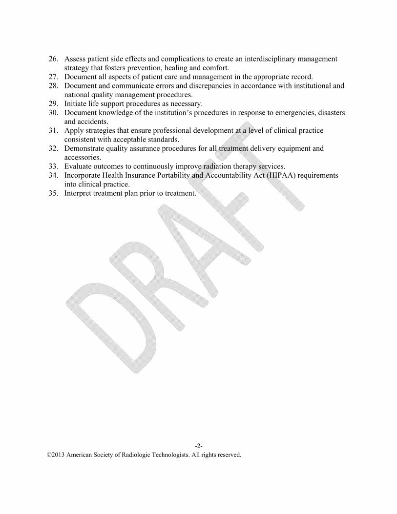
26. Assess patient side effects and complications to create an interdisciplinary management strategy that fosters prevention, healing and comfort.
27. Document all aspects of patient care and management in the appropriate record. 28. Document and communicate errors and discrepancies in accordance with institutional and
national quality management procedures. 29. Initiate life support procedures as necessary. 30. Document knowledge of the institution’s procedures in response to emergencies, disasters
and accidents. 31. Apply strategies that ensure professional development at a level of clinical practice
consistent with acceptable standards. 32. Demonstrate quality assurance procedures for all treatment delivery equipment and
accessories. 33. Evaluate outcomes to continuously improve radiation therapy services. 34. Incorporate Health Insurance Portability and Accountability Act (HIPAA) requirements
into clinical practice. 35. Interpret treatment plan prior to treatment.
C. Care 1. Management of side effects 2. Effects of multidisciplinary treatment on the patient
a. Surgery b. Chemotherapy
3. Infection control 4. Medical emergencies 5. Preprocedural and postprocedural education 6. Nutrition 7. Physical activity considerations 8. Safety and transfer positioning 9. End-of-life services
D. Clinical competencies*
III. Simulation A. Radiation safety and environmental protection practices B. Equipment operation C. Patient and machine monitoring D. Patient positioning and immobilization
I. Image processing, capture and export J. Documentation
K. Patient assessment, care, management and education L. Clinical competencies*
IV. Treatment Planning A. Pertinent patient information B. Collaboration with team members C. Equipment operation D. Procedures
1. Volume definition 2. Critical structures 3. Beam arrangement and modification 4. Implementation and verification
E. Clinical competencies*
V. Treatment Delivery A. Radiation safety and environmental protection practices B. Equipment operation C. Patient identification D. Patient and machine monitoring
E. Treatment verification and prescription F. Monitor dose to critical structures G. Patient and machine setup H. Machine malfunctions and troubleshooting I. Documentation
J. Comparison analysis of images for verification/localization
K. Patient assessment, care, management and education
1. Interpret treatment plan L. Clinical competencies*
VI. Quality Assurance and Quality Management A. Documentation B. General area conditions C. Safety devices
1. Interlocks 2. Power supply disconnection 3. Emergency buttons
D. Accessory and immobilization devices E. Communication devices F. Computerization G. Simulation and treatment units H. Brachytherapy I. Treatment planning
J. Device fabrication equipment
K. Clinical competencies*
*Refer to ARRT minimum core clinical competencies.
Ethics in Radiation Therapy Practice Description Content is designed to provide sequential development, application, analysis, integration and evaluation of ethical concepts and theories as they relate to radiation therapy practice. Objectives
1. Identify theories and principles that guide ethical decision making for practice situations. 2. Define practice situations that carry high potential for dilemmas that require ethical
scrutiny. 3. Discuss basic ethical duties of health care providers. 4. Demonstrate an awareness of and sensitivity to various cultural and ethnic differences
among various client groups. 5. Discuss the concept of patient advocacy in support of patients’ rights. 6. Discuss ethical theories and models. 7. Discuss the radiation therapy scope of practice, code of ethics and practice standards. 8. Examine concepts of personal honesty, integrity, accountability and professional
compassion as ethical imperatives in professional practice. 9. Differentiate between distributive, compensatory and retributive justice.
10. Differentiate between provider and patient relationships. 11. Discuss the duty of the radiation therapist to take responsibility for actions and decisions. 12. Discuss the elements of an informed consent. 13. Discuss standards of disclosure. 14. Analyze issues related to the use and flow of patient information to determine
confidentiality. 15. Explain ethical issues related to different age groups. 16. Identify current ethical issues in health care. 17. Demonstrate application of a system of examination, clarification, determination, the
doctrine of informed consent and other issues related to patient rights. 18. Explain ethical issues related to the profession. 19. Discuss the relationship between ethics and health care policy. 20. Examine ethical issues arising daily in a radiation therapy department.
Imaging and Processing in Radiation Oncology Description Content is designed to establish a knowledge base in factors that govern and influence the production and recording of radiographic images for patient simulation, treatment planning and treatment verification in radiation oncology. Radiation oncology imaging equipment and related devices will be emphasized. Objectives 1. Define terminology associated with digital imaging systems. 2. Describe the various types of digital receptors. 3. Discuss the fundamentals of digital imaging. 4. Discuss image acquisition. 5. Describe the evaluative criteria for digital imaging detectors. 6. Describe the histogram and the process or histogram analysis as it relates to automatic
rescaling and determining an exposure indicator. 7. Identify the exposure indices for digital imaging receptors. 8. Discuss the response of digital imaging systems to background and scatter radiation. 9. Use appropriate means of scatter control.
10. Explain methods to avoid histogram analysis errors. 11. Describe image processing employed for digital images. 12. Associate the impact of image processing parameters to the image appearance. 13. Associate the effects of inappropriate processing on image clarity or conspicuity. 14. Describe and apply the fundamental physical principles of exposure for digital detectors. 15. Describe the selection of technical factors to ensure appropriate receptor exposure levels for
digital detectors. 16. Describe the conditions that cause quantum mottle in a digital image. 17. Explain methods to avoid poor quality images. 18. Examine the potential impact of digital imaging systems on patient exposure and methods of
practicing the as low as reasonably achievable (ALARA) concept with digital systems. 19. Describe picture archiving and communications system (PACS) and its function. 20. Identify components of a PACS system. 21. Describe patient benefits gained through the use of telemedicine. 22. Identify modality types that may be incorporated into a PACS. 23. Define digital imaging and communications in medicine (DICOM). 24. Describe data flow for a DICOM image from an imaging modality to a PACS. 25. Describe HIPAA concerns with electronic information. 26. Identify common problems associated with retrieving/viewing images. 27. Describe the components and the operation of a conventional simulator. 28. Analyze relationships of factors affecting image contrast, density and resolution to
determine optimal image quality. 29. Apply techniques to enhance image details and reduce image distortion.
30. Determine artifact types, cause and preventive measures. 31. Explain the basic principles of image formation for each of the following modalities:
magnetic resonance (MR), ultrasound imaging and nuclear medicine. 32. Describe and explain functions of the components of the computed tomography (CT)
imaging system. 33. Differentiate between conventional and spiral/helical CT scanning. 34. List the CT computer data processing steps. 35. Name the functions of the array processor used for image reconstruction. 36. Explain the difference between reconstructing and reformatting an image 37. Describe the application of the following terms to CT:
• Pixel. • Matrix. • Voxel. • Linear attenuation coefficient. • CT/Hounsfield number. • Partial volume averaging. • Window width (ww) and window level (wl). • Spatial resolution. • Contrast resolution. • Noise. • Annotation. • Region of interest (ROI). • Standard vs. volumetric data acquisition.
38. Identify the types and appearance of artifacts most commonly affecting CT images. 39. Explain how artifacts can be reduced or eliminated. 40. Describe current data storage techniques used in CT. 41. Name the radiation protection devices that can be used to reduce patient dose in CT and
1. Rows and columns read line by line 2. Data transferred to external electronics 3. Digitized by analog-to-digital converter (ADC) 4. Histogram created and analyzed by software 5. Initial image processing
a. Exposure field recognition b. Histogram analysis c. Automatic rescaling – risk of failure
D. Image Formation
1. Image extraction a. TFT, CMOS, CCD b. PSP plate scanned by laser
2. Digitized by analog-to-digital converter (ADC) 3. Exposure field recognition 4. Histogram created and analyzed by software 5. Initial image processing
a. Exposure indicator determination b. Automatic rescaling
6. Initial image processing a. Exposure field recognition b. Histogram analysis c. Automatic rescaling
E. Exposure indicators
1. Dose area product (DAP) 2. Vendor-specific values
a. Relationship to patient exposure b. Optimal value ranges
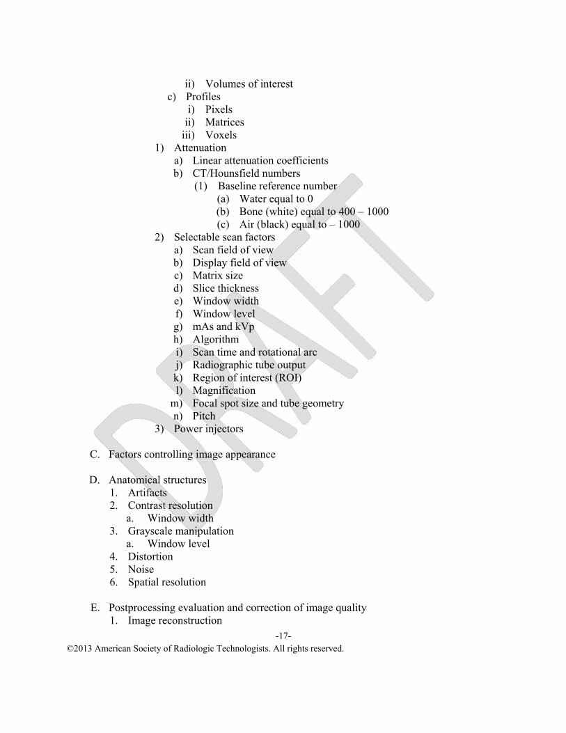
a) Linear attenuation coefficients b) CT/Hounsfield numbers
(1) Baseline reference number (a) Water equal to 0 (b) Bone (white) equal to 400 – 1000 (c) Air (black) equal to – 1000
2) Selectable scan factors a) Scan field of view b) Display field of view c) Matrix size d) Slice thickness e) Window width f) Window level g) mAs and kVp h) Algorithm i) Scan time and rotational arc j) Radiographic tube output k) Region of interest (ROI) l) Magnification
m) Focal spot size and tube geometry n) Pitch
3) Power injectors
C. Factors controlling image appearance
D. Anatomical structures 1. Artifacts 2. Contrast resolution
a. Window width 3. Grayscale manipulation
a. Window level 4. Distortion 5. Noise 6. Spatial resolution
E. Postprocessing evaluation and correction of image quality
1. Methods for reducing radiation dose to the patient a. Technical factor selection b. Technical adjustments for children c. Scatter radiation reduction
V. Image Acquisition Errors
A. Histogram analysis error 1. Incorrect anatomic menu selection 2. Unexpected material in data set (i.e., metal) 3. Large exposure error – plate saturation
B. Scatter control
1. Beam limiting 2. Optimal exposure – overexposure produces more scatter 3. Background is 40 µR/day to 80 µR/day
VI. Software (Default) Image Processing
A. Final image processing 1. Gradient processing
a. Brightness b. Contrast
2. Frequency processing a. Smoothing b. Edge enhancement
3. Equalization B. Recognition of image processing errors that affect image clarity
VII. Fundamental Principles of Exposure A. Selection of exposure factors
1. Maintain consistent specific receptor exposure 2. Control scatter 3. Adjust for differences in:
a. Structure composition b. Source-to-image receptor distance (SID)
1. Picture archiving communication system (PACS) 2. Access to report information 3. Access from multiple locations 4. Image retrieval 5. PACS issues – contingency plans 6. System components and function 7. DICOM 8. HL-7 9. Image management storage
B. Telemedicine C. Radiation therapist responsibilities
a. HIPAA and patient confidentiality 6. Image orientation to patient anatomy and position
X. Imaging Equipment
A. Conventional simulator 1. Components
a. X-ray tube b. Collimators c. Field defining wires d. Table e. Grid f. Film tray or digital receptor g. Fluoroscopic unit h. Video system i. Control console
XI. Other Imaging Modalities
A. Description, basic principles and advantages/disadvantages of each imaging modality 1. Radiography 2. CT 3. MR
Introductory Law in Radiation Therapy Description Content is designed to develop and use problem solving and critical thinking skills in discussion of the sources of law, causes of action and litigation processes related to the professional practice of radiation therapy. The inter-relatedness of standards of care, law, ethical standards and competence will be examined. Objectives 1. Apply concepts related to social, political, economic and historical issues to analyze the
different sources of law. 2. List the steps in a civil legal procedure and identify the potential role of a radiation
therapist. 3. Assess the role of effective communication skills in reducing legal action. 4. Analyze negligence related to clinical practice issues of simulation, treatment delivery,
patient assessment, patient education and quality assurance to determine if negligence is present.
5. Examine the role of the radiation therapist in the informed consent process, patient rights and practice standards.
6. Analyze safety programs to reduce patient injury. 7. Examine the importance of documentation and maintenance of clinical practice records. 8. Formulate a risk management program. 9. Analyze the role of code of ethics, radiation therapy scope of practice and radiation therapy
practice standards as guides to assess the appropriateness of professional actions. 10. Discuss the practice of lifelong learning in maintaining professional competence.
A. Assault and battery B. False imprisonment C. Intentional infliction of emotional distress D. Defamation E. Vicarious liability
F. Communication as a deterrent to legal action
III. Negligence A. Elements of a negligent act B. Comparative negligence C. Contributory negligence D. Medical negligence E. Doctrine of res ipsa loquitur F. Doctrine of respondent superior G. Negligence related to clinical practice issues
C. Health professional as a party D. Health professional as a witness E. Deposition F. Trial
V. Components of Informed Consent, Patient Rights and Standard of Care
A. Design of consent form B. Role of the radiation therapist in the consent process C. Patient’s Bill of Rights D. Standard of care E. HIPAA
VI. Safety Issues
A. Developing a safety program B. Equipment safety
VII. Documentation and Record Maintenance A. Record requirements of The Joint Commission accreditation or equivalent B. Critical documentation C. Correction of documentation D. Ownership of records E. HIPAA
Medical Terminology Description Content is designed to establish a foundation in the standardized language of medical practice, including its abbreviations and symbols. A word-building system will be presented preparatory to reading, understanding, interpreting and applying physician prescriptions to radiation therapy and related services. Objectives 1. Identify primary language sources from which medical terms are derived. 2. Define medical terms according to basic elements. 3. Interpret language, abbreviations and symbols in the medical record.
Operational Issues in Radiation Therapy Description Content is designed to focus on various radiation therapy operational issues. Continuous quality improvement (CQI) project development and evaluation and assessment techniques will be emphasized. Human resource concepts and regulations impacting the radiation therapist will be examined. Accreditation agencies and the radiation therapist’s role in the accreditation process will be emphasized. Billing and reimbursement issues pertinent to the radiation therapy department will be presented. Objectives 1. Identify CQI opportunities. 2. Explain the differences between CQI and QA. 3. Select appropriate CQI tools for specific situations. 4. Apply CQI principles to specific situations. 5. Discuss human resources’ role in the work environment. 6. Discuss the need for organizational and departmental accreditation. 7. Recognize accreditation effects on radiation therapy operations. 8. Use appropriate current procedural terminology (CPT) codes for professional and technical
charges. 9. Summarize the various types of insurance and the mechanisms necessary for approval of
care. 10. Discuss reimbursement for radiation therapy services. 11. Compare the components and methods of developing and managing a departmental budget.
Orientation to Radiation Therapy Description Content is designed to provide student with an overview of the foundations in radiation therapy and the practitioner's role in the health care delivery system. Principles, practices and policies of the educational program, health care organizations, radiation and health safety and professional responsibilities of the radiation therapist will be discussed and examined. Objectives 1. Discuss the policies and procedures of the educational program. 2. Discuss the policies and procedures of clinical education settings. 3. Identify the responsibilities of a radiation therapy student. 4. Use library/Internet resources pertinent to radiation oncology. 5. Discuss maintaining patient and student confidentiality. 6. Analyze the importance of multidisciplinary care of cancer patients. 7. Discuss the philosophy and mission of health care delivery systems and educational
programs. 8. Incorporate key terms used in the principles and practice of radiation therapy. 9. Identify the contents/sections of the patient's records.
10. Explain radiation safety procedures for radiation therapy. 11. Explain health safety procedures for personnel and patients. 12. Differentiate between accreditation, credentialing, certification, registration, licensure and
regulations. 13. Explain the purposes, functions and activities of international, national, state and local
professional organizations. 14. Discuss the importance of professional and community commitment. 15. Discuss the radiation therapist scope of practice, practice standards and professional code of
ethics. 16. Discuss the benefits of continuing education as related to improving the quality of patient
care, professional development and personal enhancement. 17. Discuss career advancement and opportunities for the radiation therapist.
Pathophysiology Description Content is designed to introduce concepts related to the disease process. An emphasis on etiological considerations, neoplasia and associated diseases in the radiation therapy patient should be presented. Objectives 1. Describe the physiological response in inflammation and cell injury due to pathological
insult. 2. Assess the predictive factors, including genetics, lifestyles, age and environment as they
influence the development of cancer and associated diseases. 3. Compare the body’s response to hereditary, lifestyle, age and environmental factors. 4. Given a specific oncologic-related disease, determine probable diagnostic, prognostic,
staging, grading and the rationale for the appropriate therapeutic pathway. 5. Given the histology of a neoplasm, determine the tumor characteristics. 6. Given a common disease, anticipate the effects of the disease on the oncologic patient.
Principles and Practice of Radiation Therapy I Description Content is designed to provide an overview of cancer and the specialty of radiation therapy. The historic and current aspects of cancer treatment will be covered. The roles and responsibilities of the radiation therapist will be discussed. In addition, treatment prescription, techniques and delivery will be covered. Objectives 1. Given diagnostic information about a particular cancer, determine the appropriateness of
using radiation therapy as a primary treatment modality. 2. Determine the medical and patient information necessary to develop a radiation therapy
treatment plan. 3. Determine the appropriate treatment energy for any given tumor type or location. 4. Differentiate between beam modifiers and their uses with a variety of treatment energies. 5. Determine the appropriate treatment setup aid, immobilization technique and beam
modifier for a given treatment technique. 6. Identify inconsistencies between treatment prescription and treatment plan. 7. Develop a conventional simulation plan for a particular tumor to include steps needed prior
to, during and after the procedure. 8. Develop a CT simulation plan for a particular tumor to include steps needed prior to, during
and after the procedure. 9. Critique treatment images in relation to simulation images.
10. Discuss the radiation therapist scope of practice and practice standards.
5. Radiosensitivity of surrounding normal structures 6. Medical status of patient 7. Quality of life 8. Survivorship
IV. Radiation Therapy Equipment
A. Simulators 1. Purpose 2. Equipment
a. 2-D (conventional) b. CT c. Fusion imaging
3. Method of radiation production 4. Auxiliary devices 5. Radiation protection 6. Patient observation and communication 7. Emergency procedures
B. Imaging devices 1. Purpose 2. Methods of radiation production 3. Components 4. Radiation protection 5. Accessories 6. Patient observation and communication 7. Emergency procedures
C. External beam
1. Purpose 2. Megavoltage
a. Linear accelerators 1) Components 2) Methods of radiation production
a) Photons b) Electrons
3) Energy 4) Depth of maximum dose 5) Target-to-skin distance (TSD)/target-to-axis distance (TAD) 6) Auxiliary devices 7) Radiation protection 8) Patient observation and communication 9) Emergency procedures
a) Components b) Methods of radiation production c) Energy deposition/Bragg peak d) Compensation e) Radiation production
7) Tomotherapy 8) Emerging units
c. Kilovoltage units 1) Purpose 2) Components 3) Method of radiation production 4) Energy 5) Auxiliary devices 6) Patient observation and communication 7) Emergency procedures
d. Radioisotope units 1) Teletherapy
a) Purpose b) Methods of radiation production c) Half-life d) Energy e) Components f) Radiation protection g) Auxiliary devices h) Patient observation and communication i) Emergency procedures
a. Medical and diagnostic information b. Physician orders c. Procedure and room preparation
2. Adaption of treatment protocols to patient-specific conditions 3. Simulation
a. Patient assessment and evaluation b. Patient education c. Patient safety d. Construction of immobilization devices e. Patient positioning f. Determination of isocenter
1) Programmable lasers g. Treatment field delineation h. Measurements i. Imaging techniques
Principles and Practice of Radiation Therapy II Description Content is designed to examine and evaluate the management of neoplastic disease using knowledge in arts and sciences, while promoting critical thinking and the basis of ethical clinical decision making. The epidemiology, etiology, detection, diagnosis, patient condition, treatment and prognosis of neoplastic disease will be presented, discussed and evaluated in relation to histology, anatomical site and patterns of spread. The radiation therapist’s responsibility in the management of neoplastic disease will be examined and linked to the skills required to analyze complex issues and make informed decisions while appreciating the scope of the profession. Objectives 1. Distinguishes tumor histology to determine pathways associated with cancer and neoplastic
disease. 2. Examine the role of surgical, radiation and medical oncology to include immunotherapy
(biological therapy) and personalized medicine in the management of neoplastic disease. 3. Discuss multidisciplinary emerging approaches to neoplastic disease management. 4. Discuss the role of radiation therapy in the management of all patient populations with
benign and malignant diseases. 5. Discuss epidemiologic and etiologic information pertinent to each neoplastic site. 6. Discuss the clinical presentation for each anatomic neoplastic site. 7. Discuss preventive methods/screening tools associated with each neoplastic site. 8. Explain detection, diagnosis, grading and staging systems for each neoplastic site. 9. Implement the principles and practice of simulation to prepare a patient for treatment.
10. Apply the parameters of treatment field design and arrangement used to treat neoplastic diseases.
11. Examine the role of radiation therapy in palliative disease management. 12. Identify the treatment regimens and fractionalization schemes used in palliative disease
management. 13. Describe the role of radiation therapy in the management of oncology emergencies.
G. Detection and diagnosis 1. History and physical examination 2. Imaging studies 3. Tumor markers 4. Laboratory studies 5. Surgical and pathology reports
H. Histopathology
1. Disease classification a. Staging b. Grading
I. Multidisciplinary treatment approach
1. Treatment modality combinations
J. Principles of surgical oncology 1. Surgical detection and biopsy for tissue diagnosis 2. Principles of curative surgery 3. Complications associated with surgery as the treatment modality
K. Role and scope of medical oncology 1. Rationale for the use of chemotherapy 2. Chemotherapeutic agents 3. Medical oncology management approaches 4. Chemotherapy toxicities
Quality Management Description Content is designed to focus on the components of quality improvement (QI) programs in radiation oncology. Topics will include developing a culture of safety through quality control and assurance checks for the clinical aspects of patient care, medical records, treatment delivery and localization equipment and treatment planning equipment. The role of the various radiation therapy team members in continuous quality improvement will be discussed as well as the legal and regulatory implications for maintaining appropriate quality care. Objectives 1. Discuss the components of a quality management (QM) program in developing a culture of
safety in radiation oncology. 2. Discuss the purpose, function and member’s role on a quality management team. 3. Explain federal, state and institutional accreditation standards and reporting regulations for
quality management. 4. Examine outcomes of quality management in radiation oncology. 5. Explain the purpose, procedures and frequency for manual and electronic treatment
documentation. 6. Identify errors in treatment documentation. 7. Describe the procedure for assuring accuracy of manual and electronic records. 8. Examine the purpose and function of record and verify systems. 9. Examine the patient chart in terms of medical and legal issues.
10. Discuss the significance of treatment outcomes for patient care, education and research in radiation oncology.
11. Discuss the quality indicators to evaluate patient care areas. 12. Explain the purpose, procedure and frequency for all QA and QM procedures in a radiation
therapy department. 13. Evaluate how the outcomes of QA and QM procedures impact patient care, education and
research. 14. Examine statistical reporting available through quality assurance computerization. 15. Perform quality measures for computerized operation, data collection and reporting. 16. Determine sources of malfunction on the treatment and simulation/localization units. 17. Distinguish between safe and hazardous equipment operation. 18. Comply with acceptable quality limits for treatment operation. 19. Identify the source of error and determine the effect on treatment delivery, education and
research. 20. Differentiate between quality management programs. 21. Discuss the importance of patient education in the quality management process. 22. Discuss the importance of proper patient identification and treatment field documentation. 23. Discuss aspects of clinical evaluation, therapeutic decision-making and informed consent. 24. Identify the key aspects of delivering a precise prescribed treatment dose.
25. Discuss quality control procedures and recommended tolerances for simulation equipment, megavoltage treatment units and treatment planning systems.
26. Discuss quality control procedures and recommended tolerances for the safe handling of brachytherapy sources and remote afterloading equipment.
27. Defend the rational for near miss and error report. 28. Critique the safety in radiation oncology.
7. Reporting and evaluating near-misses and errors 8. Implementing corrective actions related to QM data collection
III. Clinical Aspects QI Checks
A. General conditions of patient care area 1. Purpose, procedure and frequency 2. Corrective measures 3. Material safety data sheet (MSDS) 4. Documentation
B. Communication
1. Purpose, procedure and frequency 2. Corrective measures 3. Documentation
C. Mold/block fabrication area
1. Purpose, procedure and frequency 2. Protective measures 3. Corrective measures 4. Documentation
D. Accessory devices 1. Purpose, procedure and frequency 2. Corrective measures 3. Documentation
E. Treatment chart
1. Required contents 2. Treatment documentation 3. Record and verify 4. Electronic and paper 5. Medical/legal aspects of documentation 6. Corrective measures and documentation 7. Chart review purpose, procedure and frequency
F. Portal/onboard imaging
1. Purpose, procedure and frequency 2. Corrective measures 3. Documentation 4. Reject analysis
VI. Brachytherapy A. Current AAPM Task Group, or equivalent, reports recommendation
B. Purpose, procedure and frequency of checks
C. Sources of malfunction/error
D. Materials and methodology
E. Safety and hazards
F. Corrective measures
G. Guidelines to tolerance values
H. Documentation
VII. Medical Dosimetry and Treatment Planning
A. Purpose, procedure and frequency of checks B. Sources of malfunction/error C. Data acquisition D. Materials and methodology E. Safety and hazards F. Corrective measures G. Documentation
VIII. Quality Assurance and Maintenance Issues A. Initial acceptance testing B. Cassette-based system reader preventive maintenance (PM) C. Plate maintenance
1. Cleaning and inspecting plates 2. Erasing plates
Radiation Biology Description Content is designed to present basic concepts and principles of radiation biology. The interactions of radiation with cells, tissues and the body as a whole and resultant biophysical events will be presented. Discussion of the theories and principles of tolerance dose, time-dose relationships, fractionation schemes and the relationship to the clinical practice of radiation therapy will be discussed, examined and evaluated. Objectives 1. Integrate laws and principles of radiation biology to the clinical practice of radiation
therapy. 2. Identify radiosensitive components of the cell. 3. Distinguish between units of radiation quantities and radiobiologic measures. 4. Differentiate between direct and indirect effects of ionizing radiation. 5. Explain factors affecting relative biological effectiveness (RBE). 6. Discuss the effects of electromagnetic and particulate radiations on cellular interactions. 7. Evaluate factors influencing radiobiologic/biophysical events at the cellular and subcellular
level. 8. Determine biologic damage due to radiation-induced chemical reactions. 9. Discuss radiation effects on the cell cycle.
10. Compare somatic and genetic effects of radiation. 11. Describe factors influencing radiation response of cells and tissues. 12. Discuss the laws of Bergonié and Tribondeau. 13. Interpret cell survival curves to determine radiosensitivity under numerous conditions. 14. Discuss the relationship of radiation quality and dose to systemic responses. 15. Describe radiation syndromes and factors influencing response. 16. Differentiate between linear, nonlinear, and threshold and nonthreshold dose response
curves. 17. Describe the 5 Rs of radiobiology. 18. Describe the clinical significance of TD5/5,TD50/5 and QUANTEC. 19. Discuss the concept of LD50/30. 20. Compare the relationship of time, dose, fractionation, volume, distance and site to radiation
effects. 21. Discuss the use of radiation response modifiers. 22. Describe the influence of chemotherapy and hyperthermia alone and in combination with
6. Time-dose relationships a. Nominal standard dose (NSD) b. Isoeffect curves c. Rad equivalent therapy (RETS) d. Dose rate e. Alpha-beta ratios (α-β ratios) f. Biological effective dose (BED) calculation
7. Volume a. Tumor volume b. Treatment volume c. Volume vs. complications d. Time-dose-volume relationship e. Radiobiological effects from radiation therapy techniques
B. Chemotherapeutic considerations
1. Chemotherapy and radiation therapy a. Concurrent b. Neoadjuvant
2. Radioprotectors and sensitizers a. Strategy b. Action
C. Hyperthermia
1. Cellular response to heat 2. Methods of heating 3. Interactions of heat and radiation
Radiation Physics Description Content is designed to establish a basic knowledge of physics pertinent to developing an understanding of radiations used in the clinical setting. Fundamental physical units, measurements, principles, atomic structure and types of radiation are emphasized. Also presented are the fundamentals of x-ray generating equipment, x-ray production and its interaction with matter. Objectives 1. Define the fundamental units of the English, metric and Système International d’Unites (SI)
systems. 2. Calculate various unit conversions. 3. Demonstrate applications of the general principles that relate to inertia, work, energy and
momentum. 4. Describe Bohr’s theory of atomic structure. 5. Compare the characteristics and functions of a proton, neutron and electron. 6. Discuss the energy levels of the atom. 7. Define the terms relating to atomic nomenclature. 8. Compare covalent bonding and ionic bonding. 9. Describe the process of ionization.
10. Differentiate between the characteristics of a mixture, substance and element. 11. Classify the characteristics of an element using the periodic table. 12. Compare the characteristics of a molecule and compound. 13. Describe the nature of light. 14. Explain the relationship between wavelength, frequency and velocity. 15. Differentiate between the radiations of the electromagnetic (EM) spectrum. 16. Explain the relationship of energy and frequency to Planck’s constant. 17. Distinguish between electrical charge and electrical field. 18. Describe the methods of electrification. 19. Explain the laws of electrostatics and their application. 20. Describe the properties and laws of magnetism. 21. Explain the electronic spin of an element to its potential magnetic properties. 22. Describe the principle of magnetic induction. 23. Define potential difference, current, resistance, circuit and electric power. 24. Compare the characteristics of direct and alternating currents. 25. Compare electrical measuring devices. 26. Discuss electrical protective devices. 27. Discuss the interaction between electric and magnetic fields. 28. Describe the characteristics and functions of a cathode and rotating anode. 29. Describe the construction and function of tube housing. 30. Identify the parts of an x-ray tube.
31. Determine heat units and cooling characteristics of x-ray tube housings. 32. Propose methods to extend tube life. 33. Discuss application and components of automatic exposure devices. 34. State the principles of x-ray production. 35. Compare the production of bremsstrahlung with the production of characteristic radiations. 36. Compare various photon interactions in terms of description of interaction, relation to
atomic number and applications. 37. Discuss relationships of wavelength and frequency to beam characteristics. 38. Define units of radiation measurement and provide an example of its application.
Radiation Protection Description Content is designed to present basic principles of radiation protection and safety for the radiation therapist. Radiation health and safety requirements of federal and state regulatory agencies, accreditation agencies and health care organizations are incorporated. Specific responsibilities of the radiation therapist are discussed, examined, performed and evaluated. Objectives 1. Distinguish between somatic and genetic effects of radiation exposure. 2. Differentiate between stochastic and nonstochastic effects of radiation exposure. 3. Defend the concept of as low as reasonably achievable (ALARA). 4. Discuss the concept of negligible individual risk. 5. Describe the legal and ethical radiation protection responsibilities of radiation workers. 6. Use appropriate terminology and units when discussing radiation protection issues. 7. Select the correct units of radiation for exposure, absorbed dose, dose equivalence and
radioactivity. 8. Discuss the interrelationship between relative biological effectiveness and quality factors. 9. Explain the theory, operation, applications and limitations of radiation detection devices.
10. State the authority, boundaries and regulations of the state and national regulatory agencies. 11. Discuss the requirements and responsibilities of the radiation safety officer. 12. Compare the various methods used for personnel monitoring. 13. State the exposure limits for occupational and nonoccupational individuals. 14. Explain techniques used to reduce unnecessary dose to the patient. 15. Develop an emergency action plan for equipment failure. 16. Discuss the principles of radiation protection room design factors. 17. Describe the elements of a radiation protection survey for an inpatient undergoing
brachytherapy. 18. Calculate exposure doses based on time, distance and type of radioactivity. 19. Describe the procedure for a hot lab room survey. 20. Describe procedures to receive and ship radioactive materials. 21. Evaluate a record keeping system for radioactive sources to ensure inclusion of all required
a. Use (U) controlled/uncontrolled b. Workload (W) c. Occupancy (T) d. Distance (d)
3. Safety ancillary equipment a. Interlocks b. Visual monitors c. Audio monitors d. Emergency controls e. Quality assurance
4. Equipment safety a. Beam defining equipment b. Exposure control devices c. On and off switches d. Performance standards per design specifications e. Calibrations f. Quality assurance g. Emergency switches/breakers
B. Regulation and advisory recommendations
1. NRC 2. National Council on Radiation Protection and Measurements (NCRP) 3. State agency
C. Cardinal principles in protection
D. Emergency procedures
VI. Brachytherapy
A. Storage 1. Inventory systems 2. Containers 3. Room design
Radiation Therapy Patient Care Description Content is designed to provide the student with foundation concepts and competencies in assessment and evaluation of the patient for service delivery. Psychological and physical needs and factors affecting treatment outcome will be presented and examined. Routine and emergency care procedures will be presented. Objectives 1. Differentiate between the roles and responsibilities of health care team members treating
cancer patients. 2. Demonstrate applications of professional self-care. 3. Examine different psychological aspects of dying. 4. Explain the dynamics of communicating with the cancer patient and family. 5. Recognize radiation side effects and complications and select the appropriate medical
intervention. 6. Identify factors that influence a patient’s emotional responses. 7. Formulate content for answers to questions frequently asked by patients. 8. Assess the physical condition of the patient before, during and after treatment delivery. 9. Demonstrate application of the principles of health safety.
10. Discuss the principles of medication administration. 11. Recognize common medications and explain their actions and side effects. 12. Evaluate a patient for an adverse reaction to medication. 13. Describe emergency response procedures. 14. Describe the proper care of patients with tubes. 16. Provide patient education for medical procedures. 18. Assess the patient before, during and after brachytherapy procedures. 19. Demonstrate the application of the principles of radiation protection during brachytherapy
procedures. 20. Assess the nutritional status of the cancer patient to provide nutritional education or
intervention. 21. Demonstrate proper use of the principles of patient safety and transfer. 22. Provide appropriate patient education following patient assessment. 23. Select patient education materials appropriate for patient needs. 24. Compare conventional and integrative medicine.
B. The radiation oncology team II. Communication in Patient Care
A. Health-illness continuum
B. Developing professional attitudes 1. Serve as health role models
a. Avoiding burnout 1) Definition 2) Factors that increase burnout 3) Signs and symptoms 4) Principles of self-care 5) Patient advocacy
2. Empathy 3. Assertiveness
C. Communication
1. Verbal 2. Nonverbal 3. Challenges in patient communication
a. Hearing, vision and speech problems b. Impaired mental function c. Literacy d. Altered states of consciousness e. Pediatric and adolescent patients f. Geriatric patients g. Communicating in stressful circumstances h. Cultural diversity i. Artificial speech
1) Reducing distance 2) Listening 3) Using therapeutic silence 4) Responding to the feeling and the meaning of the patient’s statement 5) Restating the main idea 6) Reflecting the main idea
b. Body language 6. Communicating with families 7. Communicating with other health care professionals
D. Psychological considerations
1. End-of-life issues a. Understand the process b. Aspects of death
1) Emotional 2) Psychological
a) Depression b) Coping c) Quality of life
3) Physical a) Pain b) Suffering c) Disability d) Deterioration
c. Stages of dying 1) Disbelief 2) Denial 3) Anger 4) Bargaining 5) Acceptance
d. Patient support services 1) Family/friends 2) Pastoral care 3) Patient-to-patient support groups 4) Cancer-specific support groups 5) Hospice 6) Palliative care 7) Survivorship 8) Health professionals 9) Community agencies
2. Patient’s emotional responses a. General behavior
VIII. Medications and Their Administration A. Role of the radiation therapist
B. Medication information
1. Generic name 2. Trade name 3. Drug information
a. Physician’s Desk Reference (PDR) b. Product information sheets
4. Appropriate abbreviation usage 5. Pharmacology and administration
a. Adrenergic blocking agents b. Analgesics c. Anesthetics d. Antibacterials e. Anticonvulsants f. Antidepressants g. Antiemetics h. Antineoplastics i. Antifungals j. Antihistamines k. Contrast media l. Hypoglycemics
m. Narcotics 1) Narcotic antagonists
n. Radioactive materials o. Sedatives p. Skeletal muscle relaxants q. Stimulants r. Vasodilators
6. Biological response modifiers a. Monoclonal antibodies b. Immunotherapy c. Other
7. Nutrients, fluids and electrolytes 8. Clinical research 9. Clinical trials
B. Types of malnutrition 1. Primary 2. Secondary (malignancy-related)
C. Dietary considerations
1. General a. Benefits b. Effect on outcome
2. Irradiated site specific 3. Types of diet 4. Dietary supplements 5. Continued assessment 6. Documentation
D. Total parenteral alimentation
1. Nutritional dysfunctions a. Anorexia b. Cachexia
XIII. Physical Activity Considerations
A. Karnofsky scale/performance status B. Activity as appropriate C. Recognizing limitations
XIV. Patient Transfer
A. Body mechanics
B. Movement techniques 1. Assessing the patient’s mobility 2. Rules for safe patient transfer 3. Wheelchair transfer 4. Stretcher transfer 5. Patients with tubes and catheters
Radiation Therapy Physics Description Content is designed to review and expand concepts and theories in the radiation physics course. Detailed analysis of the structure of matter, properties of radiation, nuclear transformations, x-ray production and interactions of ionizing radiation are emphasized. Also presented are treatment units used in external radiation therapy, measurement and quality of ionizing radiation produced, absorbed dose measurement, dose distribution and scatter analysis. Objectives 1. Compare and contrast atomic structure and composition among the elements, including but
not limited to particles (their location, energy level and charge), atomic number and mass number.
2. Compare isotope, isotone, isobar and isomer. 3. Discuss nuclear stability and types of radioactive decay. 4. Categorize the four fundamental forces of nature. 5. Differentiate between electromagnetic (EM) radiation and their characteristics. 6. Describe the processes of ionization and excitation. 7. Calculate radioactivity, decay constant, activity and half-life, average life and attenuation
requirements for commonly used isotopes in radiation therapy. 8. Differentiate between artificially produced and naturally occurring therapeutic nuclides. 9. Identify the radioactive series and the decay schemes for commonly used radiation therapy
nuclides. 10. Explain the various forms of radioactive equilibrium. 11. Identify nuclear reactions by recognizing the projectile and radiation emitted. 12. Define fission and fusion. 13. Discuss the activation of nuclides in terms of yield, probability, activity growth and
saturation activity. 14. Describe methods of artificial production of radionuclides. 15. Describe x-ray production for linear accelerators. 16. Compare and contrast the factors that influence x-ray production and output. 17. Compare and contrast the energy ranges and characteristics of the various radiation therapy
modalities (Grenz-ray through megavoltage). 18. Discuss all components and function in a linear accelerator. 19. Discuss methods of x-ray production in alternate therapy units (e.g., tomotherapy,
stereotactic radiosurgery, etc.) 20. Compare the characteristics of other radiation therapy beams (cyclotron and other
accelerated particles). 21. State the gamma energies and average gamma energy of cobalt 60 (60Co). 22. Describe the basic components of a 60Co unit. 23. Compare the characteristics of an isotope beam and an x-ray beam. 24. Explain linear energy transfer (LET).
25. Compare photon interactions with matter and classify radiations produced by direct and indirect ionization.
26. Explain major influencing factors of photon beam attenuation. 27. Describe the parameters of narrow beam geometry used in the measurement of attenuation. 28. Plot heteroenergetic and monoenergetic beam attenuation data. 29. Calculate half-value layer (HVL). 30. Calculate the homogeneity coefficient. 31. Calculate attenuation requirements for beam modification devices. 32. Discuss activation of clinical accessories and alternate shielding materials due to
photodisentigration. 33. Explain charged particle interactions with matter, describing dose deposition, energy loss
and shielding requirements. 34. Define mass stopping power. 35. Describe a Bragg curve. 36. Discuss the purpose and importance of the National Institute of Standards and Technology
(NIST). 37. Discuss the purpose and importance of the Accredited Dosimetry Calibration Labs
(ADCL). 38. Demonstrate use of the appropriate type of radiation detector for given clinical applications. 39. Calculate correction factors for chamber calibration, temperature, pressure and other factors
used to correct a chamber reading. 40. Discuss protocols used for external beam calibration. 41. Analyze spot check data to make appropriate judgment decisions regarding machine
treatment parameters. Describe the quality of a gamma-ray (γ) beam in terms of HVL, γ energy or mean γ energy/nuclide of origin.
42. Describe beam filtration for the various external beam modalities, including but not limited to purpose, types of filters and their construction, energy considerations, inherent vs. added filtration and effect on HVL.
43. Calculate the approximate mean energy of a megavoltage beam. 44. Compare absorbed dose vs. exposure. 45. Discuss the relationship between kinetic energy released in the medium (KERMA),
exposure and absorbed dose. 46. Calculate air dose to absorbed dose conversions in tissue, including but not limited to,
energy considerations, applicable conversion factors, necessary instrumentation and methods.
47. Discuss the clinical importance of phantom material and size when applying the Bragg-Gray Cavity Theory.
48. Critique how dose distribution measured in a phantom is used to predict dose distribution in a patient.
49. Compare the characteristics and composition of various phantoms. 50. Compare source-skin distance (SSD) and isocentric methods of calibration.
1. Yield 2. Probability 3. Activity growth 4. Saturation activity 5. Methods of production by nuclear reactors and by acceleration 6. Relevant artificial therapeutic nuclides
L. Nuclear reactors M. Charged particle accelerators
III. Review of Production of X-rays
A. The x-ray tube
B. Physics of x-ray production 1. Bremsstrahlung x-rays 2. Characteristic x-rays 3. Percentage relationship with energy
IV. Radiation Therapy Treatment Units (External Teletherapy) A. Historical Equipment B. Equipment in current use
1. Contact, superficial, orthovoltage or deep therapy a. Tube voltage b. Tube current c. Reflection target d. Typical treatment distance e. Typical filtration f. Typical HVL g. Beam characteristics
B. Unit of exposure 1. Roentgen – special unit 2. Coulomb per kilogram (C/kg) 3. Photon fluence and fluence rate
C. Collection of charge instruments
1. Free-air (standard) ionization chamber a. Primary standard
1) National Institute of Standards and Technology (NIST) 2) Accredited Dosimetry Calibration Labs (ADCL)
b. Schematic of free-air chamber 1) Electric field 2) Ion collection plates 3) Current 4) Specified air volume 5) Ionization beyond specified volume 6) Electronic equilibrium 7) Saturation
c. Energy limitations 2. Thimble chambers
a. Function b. Principle of operation
1) Air equivalence 2) Chamber wall
a) Effective atomic number (Zeff) b) Electronic equilibrium and build-up caps
3) Central electrode 4) Air cavity, sensitive volume and sensitivity
c. Chamber calibration d. Desirable chamber characteristics
3. Practical thimble chambers a. Condenser chambers
b. Clinical impact 3. Dose calibration with ion chamber 4. Dose measurement of exposure with ion chamber in a medium
D. Bragg-Gray cavity theory
1. Advantages 2. Components overview
E. Calibration of megavoltage beams overview
1. Current American Association of Physicists in Medicine (AAPM) RTC Task Group report
2. Current International Atomic Energy Agency report
F. Other methods of measurement of absorbed dose 1. Calorimetry 2. Chemical dosimetry 3. Solid state
a. Thermoluminescence dosimetry b. Film dosimetry
G. Monte Carlo Methods
IX. Dose Distribution and Scatter Analysis Overview A. Phantoms
1. Purpose 2. Properties
a. Zeff b. Number of electrons per gram c. Mass density
3. Physical properties of various phantom materials 4. Anthropomorphic phantoms
B. Depth dose distribution
1. Percentage depth dose a. Dependence on beam quality and depth
1) Dose buildup and skin sparing 2) KERMA vs. absorbed dose
b. Effect of field size and shape 1) Geometric field size 2) PDD function of field size and beam quality 3) Square fields vs. rectangular, irregular and circular fields
a) Equivalent square tables b) Sterling’s “Rule of Thumb” equation
Research Methods and Information Literacy Description Research methods and information literacy are important because the health care profession is continually changing, which requires the radiation therapist to possess new knowledge to function competently. The radiation therapist should contribute to the body of knowledge and be able to effectively analyze resources to promote growth in the profession. The attitude of professional development enables the radiation therapist to stay in step with the current health care environment and be prepared to help foster the future and increase awareness of the profession in the global community. This content is geared to increase and disseminate intellectual inquiry, information literacy and the use of scholarly research methods. Objectives 1. Analyze research articles to determine the accuracy and validity of findings. 2. Integrate information literacy concepts into a research project. 3. Critique research projects to determine appropriateness and usefulness to the profession.
A. Assessing appropriateness of article for source material 1. Scholarly (peer-reviewed) publications 2. News magazines, other nonpeer-reviewed material
B. Assessing quality of information
1. Research design 2. Research bias 3. Study validity
C. Assessing value of article
1. Application for future research and recommendations 2. Implications for professional practice
II. Information Literacy Concepts
A. Research quality 1. Technical accuracy 2. Reader comprehension 3. Scholarly 4. Relevance to professional practice 5. Effectiveness of writing style 6. Appropriateness of form and style
B. Systematic literature analysis
1. Determining sources of information 2. Using information search strategies 3. Assessing value and appropriateness of source material
C. Paper organization
1. Appropriate title 2. Title page 3. Abstract 4. Introduction 5. Definition of terms 6. Literature review 7. Research design or methodology 8. Hypothesis or purpose of research 9. Results or analysis
10. Conclusions, discussions and recommendations
III. Types of Research Projects A. Literature review
Sectional Anatomy Description Content will introduce students to medical imaging methods currently used in the field of radiation therapy. Students will identify normal anatomical structures via a variety of imaging formats. Basic anatomical relationships will be compared using topographical and cross-sectional images. Objectives 1. Relate the importance of imaging with computed tomography, magnetic resonance and
PET-CT in radiation therapy. 2. Differentiate between sagittal, coronal and axial planes of the body. 3. Review the principles of imaging for imaging modalities using relevant terminology. 4. Compare the imaging modalities for application to radiation therapy. 5. Identify normal anatomical structures on sectional images. 6. Identify topographic anatomy used to locate underlying internal structures. 7. Describe image formation and orientation for computed tomography, magnetic resonance,
positron emission tomography, ultrasonography and image fusion.
Treatment Planning Description Content is designed to establish factors that influence and govern clinical planning of patient treatment. This encompasses isodose descriptions, patient contouring, radiobiologic considerations, dosimetric calculations, compensation and clinical application of treatment beams. Optimal treatment planning is emphasized along with particle beams. Stereotactic and emerging technologies are presented. Objectives 1. Compare photon isodose curves for clinically relevant photon beams. 2. Describe the general influencing factors that distinguish various isodose curves. 3. Determine internal and external patient factors that influence a beam’s distribution and
apply isodose correction methods. 4. Describe methods of determining a patient’s external contour, definition of internal
structures and volumes of interest used in treatment planning. 5. Identify organs and tissues at risk and their dose limitations using published tolerance dose
tables. 6. Describe how biologic effective dose is influenced by prescription and treatment variables. 7. Compare fractionation schemes. 8. Discuss the integral dose concept. 9. Use appropriate factors for treatment calculations.
10. Describe the interrelationships of the various factors used in treatment calculations. 11. Perform dose calculations for external photon and electron beam treatments for all clinical
variations. 12. Calculate the absorbed dose to off-axis points of interest. 13. Compare absorbed doses within a treatment volume with beam variations. 14. Explain algorithms incorporated into treatment planning computers. 15. Describe the clinical applications for moving beam techniques. 16. Describe the past pointing technique. 17. Calculate equivalent squares using various methods and consider the limitations of each. 18. Describe the effect of asymmetric beam collimation on dose distribution. 19. Describe methods for determining dose distribution at points outside the treatment field. 20. Calculate dose under a block. 21. Evaluate a variety of treatment plans for clinical use. 22. Identify all possible techniques that may be employed to clinically match adjacent fields. 23. Describe the multiple junction shift methods. 24. Examine hot and cold regions that occur with the various matching methods, and describe
the methods used to eliminate them. 25. Describe procedures for permanent record and legal documentation of matching fields. 26. Analyze dose distributions to determine the need for beam modifiers. 27. Compare various methods of tissue compensation and the dosimetric impact.
28. Examine the fabrication of 2-D and 3-D compensators. 29. Construct manual and computerized isodose curves. 30. Differentiate between isodose distributions for all clinical variations. 31. Evaluate possible corrections for treatment errors to correct misadministration of prescribed
dose. 32. Differentiate between the treatment planning terms: maximum, minimum, mean, modal and
median dose. 33. Describe International Commission on Radiological Units (ICRU) recommendations on
dose variance within a target volume and the effect that variances may have on cure rates, local control and tolerance.
34. Analyze dose volume histograms relative to treatment planning. 35. Evaluate patient changes to determine the integrity of a treatment plan. 36. Compare electron beam depth dose characteristics for various energies. 37. Identify clinical factors that would influence beam type and energy selection. 38. Differentiate between standard treatment distance and virtual distance. 39. Discuss why equivalent squares used with photon beams are inappropriate with electron
beams. 40. Describe how inhomogeneities influence electron beam path. 41. Discuss the considerations of matching an electron field to other adjacent photon or
electron fields. 42. Analyze which shielding materials and thickness would be needed to attenuate electron
beams to appropriate levels. 43. Describe how electron shielding materials should be arranged for external vs. internal
shielding. 44. Discuss changes in dose rate and dose distribution with changes in blocking and electron
energy. 45. Compare calculations of shielding thicknesses to measured data for electron beams. 46. Determine why specific isodose lines are prescribed for various clinical situations involving
critical and noncritical structures. 47. Calculate percentage depth dose for 10%, 50%, 80% and 90% lines for various electron
energies. 48. Describe the considerations in the clinical application of special electron treatments,
including total skin irradiation and arc therapy. 49. Compare the general isodose pattern of particle beams. 50. Determine clinical usefulness of various beam types and the clinical implications involved. 51. Describe the various imaging modalities in tumor localization and planning. 52. Discuss planning techniques used to accommodate the treatment volume shape. 53. Discuss isocenter localization for radiosurgery. 54. Identify vital structures considered during treatment planning. 55. Compare single dose delivery to fractionated dose delivery schedules. 56. Discuss the need for specific equipment used to deliver radiation for conformal therapy. 57. Discuss the purpose and contents of the ICRU Report 62 and supplements. 58. Discuss the computer system features necessary for conformal therapy treatment planning.
59. Identify common sites amenable to conformal therapy and the typical doses employed for those sites.
60. Compare configurations of multileaf collimation systems. 61. Discuss considerations for multileaf collimators. 62. Review the differences between static and dynamic multileaf collimation systems. 63. Identify appropriate clinical applications for brachytherapy. 64. Compare and contrast brachytherapy delivery systems. 65. Describe the techniques and applicators used for intracavitary, interstitial and endovascular
brachytherapy procedures. 66. Explain how simulation and CT data is used for source localization. 67. Discuss the objective of treatment planning for brachytherapy procedures. 68. Summarize dose specification and prescription techniques for different types of implants. 69. Describe optimization techniques used in computer aided dose calculations. 70. Discuss record keeping requirements for radioactive material. 71. State radiation safety requirements for brachytherapy procedures. 72. Identify appropriate clinical applications for using intensity modulated radiation therapy
(IMRT). 73. Describe the general flow of the IMRT process from patient immobilization through
B. Photon beams dose distributions and general dose distributions at Dmax, central axis and off-axis 1. Low energy x-ray 2. Gamma 60Co 3. Megavoltage x-ray 4. Influencing Factors
a. Dmax b. Central axis c. Off axis d. Without flattening filter e. With flattening filter f. Flatness and symmetry g. Overflattenting/underflattening
5. Field size definition (50% isodose line) 6. Build up dose region for various energies
C. Influencing external patient factors
1. Oblique incidence of patient/beam defined 2. Isodose correction methods 3. Limitations of various methods
b. Treatment unit settings calculation 1) Time 2) Monitor units
c. Weighted fields
C. Irregular field technique 1. Calculation techniques
a. Clarkson's method 1) Scatter-air ratio (SAR)
a) Definition b) Factors affecting SAR value c) Applicable clinical situations
2) Scatter-maximum ratio (SMR) a) Definition b) Application c) Approximation method – effective field/collimator field
b. SAR, SMR and approximation calculation 1) Algorithms 2) Absorbed dose calculation
a) Entrance dose b) Exit dose c) Entrance and exit dose summation d) Area of interest dose
(1) Target volume dose (2) Critical organ dose (3) Dose to multiple patient points/depths
3) Treatment unit settings calculation a) Time b) Monitor units
4) Weighted fields
D. Moving beam techniques 1. Definition 2. Concepts, basic formulas and equations 3. Dose rate at isocenter (average TAR/TMR) 4. Correction of first and last TAR/TMR ray values 5. Monitor unit per degree (Gantry rotation speed) 6. Rotation/arc calculations
a. Absorbed dose calculation 1) Dose at isocenter 2) Target dose specifications
B. Treatment planning 1. Forward planning 2. Inverse planning
C. Delivery techniques
D. Quality assurance
1. Multileaf collimator a. Design b. Divergence c. Penumbra d. Interleaf leakage e. Intraleaf leakage
2. Small segment dosimetry/treatment verification a. Dose per segment b. Energy stability c. Flatness and symmetry stability d. Beam interruption effects e. Verification of ports
XII. Particle Beams and General Dose Distributions
A. Electron beam 1. Physical characteristics
a. Rapid dose build-up (ratio of surface to Dmax dose) b. Dose fall-off (low vs. high energy) c. Dose distribution
1) Central axis 2) Off axis
d. Constriction of isodose curve at depth (field size) e. Ballooning of isodose curve at depth f. Percentage depth dose data unique to treatment unit, cone and field size g. Field size relationship to central axis PDD
1) Scattering foil(s), scanning magnet, air 2) Brems photon contamination of electron beam 3) Collimator opening effect on dose rate
j. Equivalent area 1) Equivalent squares 2) Square root method 3) Measured data
k. Equivalent path length 2. Beam energy selection 3. Biological considerations in patient treatment 4. Energy decelerators for special treatment 5. Build up bolus 6. Adjacent fields 7. Shielding materials, thicknesses, energy and dose relationship
a. Mass stopping power (low vs. high Z) 1) Density, Z number and electrons per gram 2) Material choices and rationales
b. External shielding c. Internal shielding (tissue interfaces) d. Changes in dose rate and dose distribution e. Thickness (MeV/3 = mm pb)
8. Treatment prescriptions and calculations a. Physician prescription to specific isodose line b. Critical structure c. Noncritical structure d. Determining PDD
9. Applications of electron beam a. Single beam b. Multiple beams
1) Mixed (photon and electron) 2) Abutting
a) Electron fields b) Electron and photon fields
c. Complex 1) Electron arc 2) Total skin irradiation
b. Clinical applications and treatment delivery 1) Immobilization requirements 2) Simulation 3) Treatment planning 4) Treatment verification
3. High LET charged particles (negative ions) a. Nonexponential attenuation b. Proximal and distal dose gradients c. General isodose curve pattern d. Bragg peak/star effect advantage e. Percentage depth dose energy dependence f. Precision immobilization requirements g. Penumbra h. Clinical applications
4. Heavy ions a. Types
1) Carbon 2) Neon 3) Argon 4) Silicon 5) Other
b. Nonexponential attenuation c. Proximal and distal dose gradients d. General isodose curve pattern e. Bragg peak advantage f. Percentage depth dose energy dependence g. Precision immobilization requirements h. Penumbra i. Clinical applications
Resources Textbooks Adler AM. Carlton RR. Introduction to Radiologic Sciences and Patient Care. 5th ed. St. Louis, MO: Elsevier Saunders; 2012. Agur AMR. Grant’s Atlas of Anatomy. 9th ed. Baltimore, MD: Williams & Wilkins; 1991. American Association of Physicists in Medicine. AAPM Reports. www.aapm.org/pubs/reports. American College of Legal Medicine. Legal Medicine. 7th ed. St. Louis, MO: Mosby-Year Book Inc; 2007. American Medical Association. CPT 2013 Standard Edition. Chicago, IL: American Medical Association; 2012. Ang KK. Radiotherapy for Head and Neck Cancers: Indications & Techniques. 4th ed. Philadelphia, PA: Lippincott Williams & Wilkens; 2011. Baird S, Varricchio C, Pierce M, et al. A Cancer Source Book for Nurses. 8th ed. Atlanta, GA: American Cancer Society Inc; 2004. Barakat RR, Markman M, Randall ME. Principles and Practices of Gynecologic Oncology. 5th ed. Philadelphia, PA: Lippincott Williams & Wilkens Publishers; 2009. Beauchamp TL, Childress JF. Principles of Biomedical Ethics. 6th ed. New York, NY: Oxford University Press; 2008. Bentel CG. Radiation Therapy Planning. 2nd ed. New York, NY: McGraw-Hill Inc; 1995. Bentel CG, Nelson CE, Noell KT, et al. Treatment Planning and Dose Calculation in Radiation Oncology. 4th ed. New York, NY: McGraw-Hill; 1989. Bomford C, Kunkler. Walter and Miller’s Textbook of Radiotherapy: Radiation Physics, Therapy and Oncology. 6th ed. New York, NY: Churchill Livingstone Inc; 2002. Bontrager K, Lampignano J. Textbook of Radiographic Positioning and Related Anatomy. 7th ed. St. Louis, MO: Mosby-Year Book Inc; 2010. Bridge P, Tipper DJ. CT Anatomy for Radiotherapy. London, England: M & K Publishing: 2011.
Bushong SC. Radiologic Science for Technologists: Physics, Biology and Protection. 10th ed. St. Louis, MO: Mosby-Year Book Inc; 2012. Carlton RR, Adler AM. Principles of Radiographic Imaging. 5th ed. Albany, NY: Delmar Publishers; 12012. Cassady JR. Radiation Therapy in Pediatric Oncology. New York, NY: Springer-Verlag New York Inc; 2011. Chao KSC. Practical Essentials of Intensity Modulated Radiation Therapy, 2nd ed. Philadelphia, PA: Lippincott Williams & Wilkins Publishers, 2005. Chao KS, Perez CA, Brady LW. Radiation Oncology Management Decisions. 3rd ed. Philadelphia, PA: Lippincott Williams & Wilkins; 2011. Coddington DC, Moore KD, Fisher EA. Strategies for the New Healthcare Marketplace: Managing the Convergence of Consumerism and Technology. San Francisco, CA: Josey-Bass Publishers; 2001. Code of Federal Regulations - 10. Office of the Federal Register, National Archives and Records Administration. Cox JD, Ang KK. Radiation Oncology: Rationale, Techniques, Results. 9th ed. St. Louis, MO: Mosby-Year Book Inc; 2009. Damjanov, I. Pathology for the Health Related Professions. 4th ed. Philadelphia, PA: WB Saunders Co; 2011. Dasher B, Wiggers N, Vann AM, et al. Portal Design in Radiation Therapy. 2nd ed. Columbia, S.C.: R.L. Bryan Co.; 2006. DeLaney TF,Kooy HM, eds. Proton and Charged Particle Radiotherapy.Philadelphia, PA: Lippincott Williams & Wilkins Publishers, 2008. DePoy E, Gitlin LN. Introduction to Research: Understanding and Applying Multiple Strategies. 4th ed. St. Louis, MO: Elsevier Mosby; 2012. DeVita VT, Hellman S, Rosenberg SA, et al. Cancer: Principles and Practice of Oncology. 8th ed. Philadelphia, PA: Lippincott Williams & Wilkins Publishing; 2001. Dobbs J, Roques T. Practical Radiotherapy Planning. 4th ed. Baltimore, MD: Williams & Wilkins; 2009.
Edge R, Groves J. The Ethics of Health Care: A Guide for Clinical Practice. 3rd ed. Albany, NY: Delmar Publishers; 2005. Edge SC, Byrd DR, Compton CC, Fritz AG. AJCC Cancer Staging Manual. 7th ed. New York, NY: Springer; 2009. Ehrlich RA, McClosky ED. Patient Care in Radiography. 8th ed. St. Louis, MO: Mosby-Year Book Inc; 2012. Flight M, Meacham MR. Law, Liability & Ethics for Medical Office Professionals. 5th. Albany, NY: Delmar Publishers; 2010. Hall EJ, Giaccia AJ. Radiobiology for the Radiologist. 7th ed. Philadelphia, PA: Lippincott-Williams & Wilkins; 2011. Gollnick DA. Basic Radiation Protection Technology. 5th ed. Altadena, CA: Pacific Radiation Corp; 2006. Gylys BA, Wedding ME. Medical Terminology, A Systems Approach. 6th ed. Philadelphia, PA: FA Davis Co; 2009. Halperin EC, Conscine LS, Tarbell NJ, Kun LE. Pediatric Radiation Oncology. 5th ed. Philadelphia, PA: Lippincott Williams & Wilkins Publishers; 2010. Hansen EK, Roach III M, eds. Handbook of Evidence-Based Radiation Oncology. 2nd ed. New York, NY: Springer; 2010. Hebda TL, Czar P. Handbook of Informatics for Nurses and Health Professionals. 5th ed. Upper Saddle River, NJ: Prentice Hall; 2012. Hendee WR, Ibbott GS, Hendee EG. Radiation Therapy Physics. 3rd ed. New York, NY: Wiley-Liss; 2004. Hurria A, Balducci L. Geriatric Oncology: Treatment Assessment & Management. New York, NY: Springer Publishers; 2009. ICRU Reports. Bethesda, MD: International Commission on Radiation Units and Measurements. Iverson C, et al. AMA Manual of Style. 10th ed. New York, NY: Oxford Press; 2007. KhanFM. The Physics of Radiation Therapy. 4th ed. Philadelphia, PA: Lippincott Williams & Wilkins Publishers; 2009.
Karzmark CJ, Morton RJ. A Primer of the Theory and Operation of Linear Accelerators in Radiation Therapy. 2nd ed. Madison, WI: Medical Physics Publishing Corp; 1997. Kiefer J. Biological Radiation Effects. New York, NY: Springer-Verlag New York Inc; 1990. Knoll GF. Radiation Detection and Measurement. 4th ed. New York, NY: John Wiley & Sons Inc; 2010. Kübler-Ross E. On Death and Dying. New York, NY: Maxwell MacMillan International; 1997. Langhorne M, Fulton J, Otto SE. Oncology Nursing. 5th ed. St. Louis, MO: Mosby-Year Book Inc; 2007. Levitt J, Seymour H, et al, eds. Technological Basis of Radiation Therapy: Practical Clinical Applications. 5th ed. New York, NY: Springer; 2012. Lewis RJ. Sax’s Dangerous Properties of Industrial Materials. 12th ed., 5 vols. New York, NY: Van Nostrand Reinhold; 2012. Madden ME. Introduction to Sectional Anatomy. 3rd ed. Baltimore, MD: Lippincott Williams & Wilkins; 2013. Madden ME. Introduction to Sectional Anatomy: Workbook and Board Review Guide. 3rd ed. Baltimore, MD: Lippincott Williams & Wilkins; 2013. Marieb E, Hoen K. Human Anatomy and Physiology. 4th ed. Menlo Park, CA: The Benjamin Cummings Publishing Co Inc; 2012. Martin A, Harbison S. An Introduction to Radiation Protection. 6th ed. London, England: Chapman and Hall; 2012. McCance KL, Huether SE. Pathophysiology: The Biologic Basis for Disease in Adults And Children. 6th ed. St. Louis, MO: Mosby-Year Book Inc; 2009. McConnell TH. The Nature of Disease: Pathology for the Health Professions. 2nd ed. Baltimore, MD: Lippincott Williams Wilkins; 2013. Meyer JL. IMRT, IGRT, SBRT: Advances in the Treatment Planning and Delivery of Radiotherapy. 2nd ed. Basel, Switzerland: S. Karger; 2011. Mizer S, Scheller R, Deye J, et al. Radiation Therapy Simulation Workbook. New York, NY: Pergamon Press Inc; 1986.
Mosby’s Medical, Nursing, and Allied Health Dictionary. 9th ed. St. Louis, MO: Mosby-Year Book Inc; 2012. Munson R. Intervention and Reflection: Basic Issues in Medical Ethics. 8th ed. Belmont, CA: Wadsworth Publishing Co; 2012. Murshed MD, Hasan. Clinical Fundamentals for Radiation Oncologists. 2nd ed. Madison, WI: Medical Physics Publishing; 2011. National Council on Radiation Protection and Measurements. Bethesda, MD. Netter FH. Atlas of Human Anatomy. 5th ed. Philadelphia, PA: Saunders; 2010. Noz ME, Maguire GQ. Radiation Protection in the Health Sciences. 2nd ed. Hackensack, NJ: World Scientific; 2007. Occupational Safety & Health Administration Guidelines for Universal Precautions and Environmental Hazards. Washington, DC. Order SE, Donaldson S. Radiation Therapy of Benign Diseases. 2nd ed. New York, NY: Springer-Verlag New York Inc; 2003. Orthner H, Blum BI. Implementing Health Care Information Systems. New York, NY: Springer-Verlag New York Inc; 2011. Perez CA, Brady LW. Principles & Practices of Radiation Oncology. 5th ed. Philadelphia, PA: Lippincott-Raven Publishing; 2007. Porter-O’Grady T, Malloch K. Quanum Leadership: Advancing Information, Transforming Healthcare. 3rd ed. Sudbury, MA: Jones and Bartlett Learning; 2011. Purtilo RB, ed. Ethical Dimensions in the Health Professions. 5th ed. Philadelphia, PA: WB Saunders Publishing Co; 2010. Purtilo RB, Haddad A. Health Professional and Patient Interaction. 7th ed. Philadelphia, PA: Saunders; 2007. Rubin P. Clinical Oncology: A Multidisciplinary Approach for Physicians and Students. 8th ed. Philadelphia, PA: WB Saunders Publishing Co; 2001. Rubin E, Reisner HM. Essentials of Rubin’s Pathology. 5th ed. Philadelphia, PA: Lippincott-Raven Publishers; 2008.
Scherer E, Streffer C, Trott KR, et al. Radiation Exposure and Occupational Risks. New York, NY: Springer-Verlag New York Inc; 2012. Schlegel W, Bortfeld T, Grosu A, et al. New Technologies in Radiation Oncology. New York, NY: Springer; 2006. Selman J. The Basic Physics of Radiation Therapy. 3rd ed. Springfield, IL: CC Thomas Publishing; 1990. Selman J. Elements of Radiobiology. Springfield, IL: CC Thomas Publishing; 1983. Selman J. The Fundamentals of X-ray and Radium Physics. 8th ed. Springfield, IL: CC Thomas Publishing; 1993. Smith MM, Smith TL. Pocket Atlas of Normal CT Anatomy. 2nd ed. Baltimore, MD: Lippincott, Williams, Wilkins; 2002. Smith MM, Smith TL. Pocket Atlas of Normal CT Anatomy of the Head and Brain. 2nd ed. Baltimore, MD: Lippincott, Williams, Wilkins; 2001. Stanton R, Stinson D. Applied Physics for Radiation Oncology. 2nd ed. Madison, WI: Medical Physics Publishing Corp; 2009. Tannock IF, Hill RP, eds. The Basic Science of Oncology. 5th ed. New York, NY: McGraw-Hill Inc; 2013. Torres LS, Norcutt TA, Dutton, AG. Basic Medical Techniques and Patient Care for Radiologic Technologists. 7th ed. Philadelphia, PA: Lippincott-Raven Publishers; 2009. Towsley D, Cunningham E. Biomedical Ethics for Radiographers. Dubuque, IA: Eastwind Publishers; 1994. Travis EL. Primer of Medical Radiobiology. 2nd ed. St. Louis, MO: Mosby-Year Book Inc; 1989. Veatch RM, Hagdad AM, English DC. Case Studies in Allied Health Ethics. Upper Saddle River, NJ: Prentice Hall, 2009. Videtic G, Vassil A. Handbook of Treatment Planning in Radiation Oncology. New York, NY: Demos Medical Publishing; 2010. Washington CM, Leaver DT. Principles and Practice of Radiation Therapy.3rd ed. St. Louis, MO: Mosby Elsevier; 2010.
Williamson JF, Thomadsen B, Nath R, et al. Brachytherapy Physics. Madison, WI: Medical Physics Publishing Corp; 1995. Yeung SJ, Escalante CP, Gagel RF. Medical Care of the Cancer Patient. Shelton, CT: People’s Medical Publishing House; 2009.