Radiography Safety The Academy of Dental Learning and OSHA Training, LLC, designates this activity for 3 continuing education credits (3 CEs). US Department of Health and Human Services Public Health Service Food and Drug Administration Revised: 2012 Originally Edited by: MaryLou Austin, RDH, MS Reviewed and Edited by: Health Science Editor: Megan Wright, RDH, MS Publication Date: October 2012 Reviewed Date: January 2017 Expiration Date: February 2020 The Academy of Dental Learning and OSHA Training, LLC is an ADA CERP Recognized Provider. ADA CERP is a service of the American Dental Association to assist dental professionals in identifying quality providers of continuing dental education. ADA CERP does not approve or endorse individual courses or instructors, nor does it imply acceptance of credit hours by boards of dentistry. Concerns or complaints about a CE provider may be directed to the provider or to the Commission for Continuing Education Provider Recognition at ADA.org/CERP. Conflict of Interest Disclosure: ADL does not accept promotional or commercial funding in association with its courses. In order to promote quality and scientific integrity, ADL's evidence- based course content is developed independent of commercial interests. Refund Policy: If you are dissatisfied with the course for any reason, prior to taking the test and receiving your certificate, return the printed materials within 15 days of purchase and we will refund your full tuition. Shipping charges are nonrefundable. California Registered Provider Number: RP5631
Transcript
Radiography Safety
The Academy of Dental Learning and OSHA Training, LLC, designates this activity for 3 continuing education credits (3 CEs).
US Department of Health and Human Services
Public Health Service
Food and Drug Administration
Revised: 2012
Originally Edited by: MaryLou Austin, RDH, MS
Reviewed and Edited by: Health Science Editor: Megan Wright, RDH, MS
Publication Date: October 2012
Reviewed Date: January 2017
Expiration Date: February 2020
The Academy of Dental Learning and OSHA Training, LLC is an ADA CERP Recognized
Provider. ADA CERP is a service of the American Dental Association to assist dental
professionals in identifying quality providers of continuing dental education. ADA CERP does not
approve or endorse individual courses or instructors, nor does it imply acceptance of credit hours
by boards of dentistry. Concerns or complaints about a CE provider may be directed to the
provider or to the Commission for Continuing Education Provider Recognition at ADA.org/CERP.
Conflict of Interest Disclosure: ADL does not accept promotional or commercial funding in
association with its courses. In order to promote quality and scientific integrity, ADL's evidence-
based course content is developed independent of commercial interests. Refund Policy: If you
are dissatisfied with the course for any reason, prior to taking the test and receiving your
certificate, return the printed materials within 15 days of purchase and we will refund your full
***PLEASE PRINT CLEARLY; ILLEGIBLE ANSWER SHEETS WILL NOT BE
PROCESSED.
Notes:
2
Course Evaluation
Please place an X in the box to rate these
statements:
Poor Fair Good Very
Good
Excellent
The content fulfills the overall purpose of the course.
The content fulfills each of the course objectives.
The course subject matter is accurate.
The material presented is understandable.
The teaching/learning method is effective.
The answers to the test questions are appropriately
covered in the course.
How would you rate this course overall?
Time to complete the entire course and the test? Hours: _________ Minutes: _______
Google
Other Search Engine
Friend/Coworker
Other
Do you have any suggestions about how we can improve this course? If so please note them on a
separate sheet of paper and send it in with your answer sheet.
If you studied the course online, did all the links work? If not please note the page and link on a separate
sheet of paper and send it in with your answer sheet so we can fix it.
3
Instructions
1. Review the Objectives: Objectives provide an overview of the entire course.
2. Read the course material.
3. Complete the test:
a. Return to our website: www.dentallearning.org, click on Take the Exam,
enter your answers, register, if you are new customer (existing customers
login), pay for the course, click Grade Test. Your test will be graded
immediately. If required, complete the course evaluation. Your certificate
will display for you to print.
b. If you would rather, you may return your completed answer sheet and
course evaluation to us via the options listed below.
To successfully complete the course you must score 80% or above on the test. If you
do not score 80% you may retake the test one more time free of charge. If you fail a
second time you must purchase a new course and test.
If you’ve downloaded this coursebook off the Internet you can:
Return to our website (www.dentallearning.org) to take the test online (only if you have not purchased the coursebook separately). You will need to provide credit card information at the time you submit your test online for scoring.
Write your answers on the one-page answer sheet included in this book, complete the credit card payment information, and return the form to the address below, fax, or email address below. Or, you may send a check or money order to the address below with your answer sheet.
Academy of Dental Learning and OSHA Training, LLC (ADL)
If someone else would like to use this material after you are done, he or she may register with us and take advantage of a “sharing discount”. Courses downloaded from the Internet can be shared at the same tuition rate as currently available on our website. Please call us if you need an extra answer sheet or download one from our website. There is no “sharing discount” for online exams. The author and ADL have made every effort to include information in this course that is
factual and conforms to accepted standards of care. This course is not to be used as a
sole reference for treatment decisions. It is your responsibility to understand your legal
obligations and license requirements when treating patients. ADL is not responsible for
the misuse of information presented in this course. The material in this course cannot
be reproduced or transmitted in any way without the written consent of ADL.
5
Table of Contents
Answer Sheet 1
Evaluation 2
Instructions 3
Table of Contents 5
Objectives 6
Background 6
Introduction 7
Patient Selection Criteria 7
Recommendations for Prescribing Dental Radiographs 9
Explanation of Recommendations for Prescribing Dental Radiographs 12
Limiting Radiation Exposure 20
Receptor Selection 21
Receptor Holders 21
Collimation 22
Operating Potential and Exposure Time 22
Patient Shielding and Positioning 23
Operator Protection 23
Hand-Held X-Ray Units 24
Film Exposure and Processing 24
Quality Assurance 25
Technique Chart/Protocols 25
Radiation Risk Communication 26
Training and Education 27
Technology and Safety 28
Conclusion 28
References 28
Course Test 37
6
Objectives
• Understand the FDA’s role in the regulation and promotion of radiation safety
in clinical dentistry.
• Know updated FDA recommendations for patient selection criteria in
prescribing dental radiographs.
• Understand basic radiography technique and rationale for clinical choices in
types of dental radiography.
• Review criteria for radiographic screening with regard to patient medical
history, clinical presentation, and risk assessment.
• Know clinical methods to limit radiation exposure for operator and patient.
• Know the recommendations from the FDA for particular clinical situations.
Background
The dental profession is committed to delivering the highest quality of care to each of its
individual patients and applying advancements in technology and science to continually
improve the oral health status of the U.S. population. These guidelines were developed
to serve as an adjunct to the dentist’s professional judgment of how to best use
diagnostic imaging for each patient. Radiographs can help the dental practitioner
evaluate and definitively diagnose many oral diseases and conditions. However, the
dentist must weigh the benefits of taking dental radiographs against the risk of exposing
a patient to x-rays, the effects of which accumulate from multiple sources over time.
The dentist, knowing the patient’s health history and vulnerability to oral disease, is in
the best position to make this judgment in the interest of each patient. For this reason,
the guidelines are intended to serve as a resource for the practitioner and are not
intended as standards of care, requirements or regulations.
The guidelines are not substitutes for clinical examinations and health histories. The
dentist is advised to conduct a clinical examination, consider the patient’s signs,
symptoms and oral and medical histories, as well as consider the patient’s vulnerability
to environmental factors that may affect oral health. This diagnostic and evaluative
information may determine the type of imaging to be used or the frequency of its use.
Dentists should only order radiographs when they expect that the additional diagnostic
information will affect patient care. The guidelines can be used by the dentist to
optimize patient care, minimize radiation exposure and responsibly allocate health care
resources.
7
This course deals only with standard dental imaging techniques of intraoral and
common extra-oral examinations, excluding cone-beam computed tomography (CBCT).
At this time the indications for CBCT examinations are not well developed and are still
under consideration by the FDA and ADA.
Introduction
The guidelines titled, The Selection of Patients for X-Ray Examination, were first
developed in 1987 by a panel of dental experts convened by the Center for Devices and
Radiological Health of the U.S. Food and Drug Administration (FDA). The development
of the guidelines at that time was spurred by concern about the U.S. population’s total
exposure to radiation from all sources. Thus, the guidelines were developed to promote
the appropriate use of x-rays. In 2002, the American Dental Association, recognizing
that dental technology and science continually advance, recommended to the FDA that
the guidelines be reviewed for possible updating. The FDA welcomed organized
dentistry’s interest in maintaining the guidelines, and so the American Dental
Association, in collaboration with a number of dental specialty organizations and the
FDA, published updated guidelines in 2004. This report updates the 2004 guidelines
and includes recommendations for limiting exposure to radiation. These materials
issued by the FDA are in the public domain and dental professionals are encouraged to
use the information to make better clinical choices for patient and operator safety.
Patient Selection Criteria
Radiographs and other imaging modalities are used to diagnose and monitor oral
diseases, as well as to monitor dento-facial development and the progress or prognosis
of therapy. Radiographic examinations can be performed using digital imaging or
conventional film. The available evidence suggests that either is a suitable diagnostic
method. Digital imaging may offer reduced radiation exposure and the advantage of
image analysis that may enhance sensitivity and reduce error introduced by subjective
analysis.
A study of 490 patients found that basing selection criteria on clinical evaluations for
asymptomatic patients, combined with selected periapical radiographs for symptomatic
patients, can result in a 43 percent reduction in the number of radiographs taken without
a clinically consequential increase in the rate of undiagnosed disease. The development
and progress of many oral conditions are associated with a patient’s age, stage of
dental development, and vulnerability to known risk factors. Therefore, the guidelines in
Table 1 are presented within a matrix of common clinical and patient factors, which may
determine the type (s) of radiographs that is commonly needed. The guidelines assume
that diagnostically adequate radiographs can be obtained. If not, appropriate
management techniques should be used after consideration of the relative risks and
8
benefits for the patient.
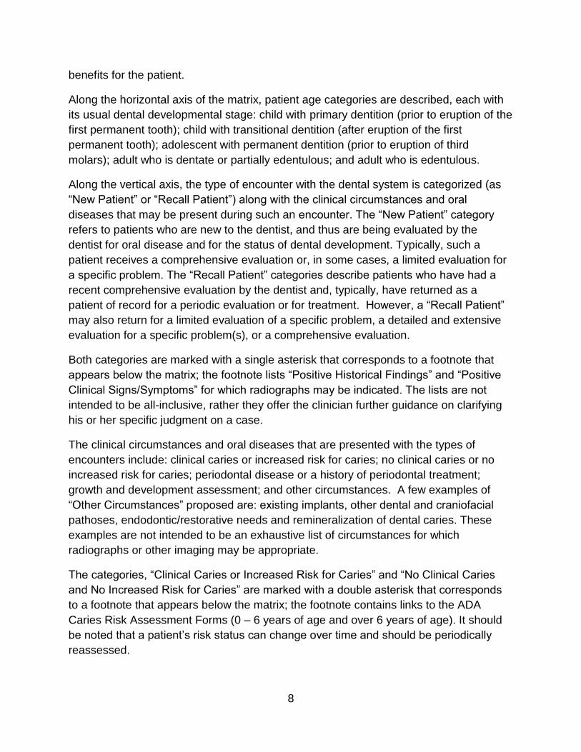
Along the horizontal axis of the matrix, patient age categories are described, each with
its usual dental developmental stage: child with primary dentition (prior to eruption of the
first permanent tooth); child with transitional dentition (after eruption of the first
permanent tooth); adolescent with permanent dentition (prior to eruption of third
molars); adult who is dentate or partially edentulous; and adult who is edentulous.
Along the vertical axis, the type of encounter with the dental system is categorized (as
“New Patient” or “Recall Patient”) along with the clinical circumstances and oral
diseases that may be present during such an encounter. The “New Patient” category
refers to patients who are new to the dentist, and thus are being evaluated by the
dentist for oral disease and for the status of dental development. Typically, such a
patient receives a comprehensive evaluation or, in some cases, a limited evaluation for
a specific problem. The “Recall Patient” categories describe patients who have had a
recent comprehensive evaluation by the dentist and, typically, have returned as a
patient of record for a periodic evaluation or for treatment. However, a “Recall Patient”
may also return for a limited evaluation of a specific problem, a detailed and extensive
evaluation for a specific problem(s), or a comprehensive evaluation.
Both categories are marked with a single asterisk that corresponds to a footnote that
appears below the matrix; the footnote lists “Positive Historical Findings” and “Positive
Clinical Signs/Symptoms” for which radiographs may be indicated. The lists are not
intended to be all-inclusive, rather they offer the clinician further guidance on clarifying
his or her specific judgment on a case.
The clinical circumstances and oral diseases that are presented with the types of
encounters include: clinical caries or increased risk for caries; no clinical caries or no
increased risk for caries; periodontal disease or a history of periodontal treatment;
growth and development assessment; and other circumstances. A few examples of
“Other Circumstances” proposed are: existing implants, other dental and craniofacial
pathoses, endodontic/restorative needs and remineralization of dental caries. These
examples are not intended to be an exhaustive list of circumstances for which
radiographs or other imaging may be appropriate.
The categories, “Clinical Caries or Increased Risk for Caries” and “No Clinical Caries
and No Increased Risk for Caries” are marked with a double asterisk that corresponds
to a footnote that appears below the matrix; the footnote contains links to the ADA
Caries Risk Assessment Forms (0 – 6 years of age and over 6 years of age). It should
be noted that a patient’s risk status can change over time and should be periodically
reassessed.
9
The panel also has made the following recommendations that are applicable to all
categories:
1. Intraoral radiography is useful for the evaluation of dento-alveolar trauma. If
the area of interest extends beyond the dento-alveolar complex, extra-oral
imaging may be indicated.
2. Care should be taken to examine all radiographs for any evidence of caries,
bone loss from periodontal disease, developmental anomalies and occult
disease.
3. Radiographic screening for the purpose of detecting disease before clinical
examination should not be performed. A thorough clinical examination,
consideration of the patient history, review of any prior radiographs, caries
risk assessment and consideration of both the dental and the general health
needs of the patient should precede radiographic examination.
In the practice of dentistry, patients often seek care on a routine basis in part because
oral disease may develop in the absence of clinical symptoms. Since attempts to
identify specific criteria that will accurately predict a high probability of finding
interproximal carious lesions have not been successful for individuals, it was necessary
to recommend time-based schedules for making radiographs intended primarily for the
detection of dental caries. Each schedule provides a range of recommended intervals
that are derived from the results of research into the rates at which interproximal caries
progresses through tooth enamel. The recommendations also are modified by criteria
that place an individual at an increased risk for dental caries. Professional judgment
should be used to determine the optimum time for radiographic examination within the
suggested interval.
Recommendations for Prescribing Dental Radiographs
These recommendations are subject to clinical judgment and may not apply to every
patient. They are to be used by dentists only after reviewing the patient’s health history
and completing a clinical examination. Even though radiation exposure from dental
radiographs is low, once a decision to obtain radiographs is made it is the dentist's
responsibility to follow the ALARA Principle (As Low as Reasonably Achievable) to
minimize the patient's exposure.
10
Table 1
Patient Age and Dental Developmental Stage
Type of Encounter
Child with Primary Dentition (prior to eruption of first permanent tooth)
Child with Transitional Dentition (after eruption of first permanent tooth)
Adolescent with Permanent Dentition (prior to eruption of third molars)
Adult, Dentate or Partially Edentulous
Adult, Edentulous
New Patient* being evaluated for oral diseases
Individualized radiographic exam consisting of selected periapical/occlusal views and/or posterior bitewings if proximal surfaces cannot be visualized or probed. Patients without evidence of disease and with open proximal contacts may not require a radiographic exam at this time.
Individualized radiographic exam consisting of posterior bitewings with panoramic exam or posterior bitewings and selected periapical images.
Individualized radiographic exam consisting of posterior bitewings with panoramic exam or posterior bitewings and selected periapical images. A full mouth intraoral radiographic exam is preferred when the patient has clinical evidence of generalized oral disease or a history of extensive dental treatment.
Individualized radiographic exam, based on clinical signs and symptoms.
Recall Patient* with clinical caries or at increased risk for caries**
Posterior bitewing exam at 6-12 month intervals if proximal surfaces cannot be examined visually or with a probe
Posterior bitewing exam at 6-18 month intervals
Not applicable
Recall Patient* with no clinical caries and not at increased risk for caries**
Posterior bitewing exam at 12-24 month intervals if proximal surfaces cannot be examined visually or with a probe
Posterior bitewing exam at 18-36 month intervals
Posterior bitewing exam at 24-36 month intervals
Not applicable
Recall Patient* with periodontal disease
Clinical judgment as to the need for and type of radiographic images for the evaluation of periodontal disease. Imaging may consist of, but is not limited to, selected bitewing and/or periapical images of areas where periodontal disease (other than nonspecific gingivitis) can be demonstrated clinically.
Not applicable
11
Patient Age and Dental Developmental Stage
Type of Encounter
Child with Primary Dentition (prior to eruption of first permanent tooth)
Child with Transitional Dentition (after eruption of first permanent tooth)
Adolescent with Permanent Dentition (prior to eruption of third molars)
Adult, Dentate or Partially Edentulous
Adult, Edentulous
Patient (New and Recall) for monitoring of dento-facial growth and development, and/or assessment of dental/skeletal relationships
Clinical judgment as to need for and type of radiographic images for evaluation and/or monitoring of dento-facial growth and development or assessment of dental and skeletal relationships
Clinical judgment as to need for and type of radiographic images for evaluation and/or monitoring of dento-facial growth and development, or assessment of dental and skeletal relationships. Panoramic or periapical exam to assess developing third molars
Usually not indicated for monitoring of growth and development. Clinical judgment as to the need for and type of radiographic image for evaluation of dental and skeletal relationships.
Patient with other circumstances including, but not limited to, proposed or existing implants, other dental and craniofacial pathoses, restorative/ endodontic needs, treated periodontal disease and caries remineralization
Clinical judgment as to need for and type of radiographic images for evaluation and/or monitoring of these conditions
12
*Clinical situations for which radiographs may be indicated include, but are not limited to: A. Positive Historical Findings
1. Previous periodontal or endodontic treatment 2. History of pain or trauma 3. Familial history of dental anomalies 4. Postoperative evaluation of healing 5. Remineralization monitoring 6. Presence of implants, previous implant-related pathosis or evaluation for implant placement
B. Positive Clinical Signs/Symptoms 1. Clinical evidence of periodontal disease 2. Large or deep restorations 3. Deep carious lesions 4. Malposed or clinically impacted teeth 5. Swelling 6. Evidence of dental/facial trauma 7. Mobility of teeth 8. Sinus tract (“fistula”) 9. Clinically suspected sinus pathosis 10. Growth abnormalities 11. Oral involvement in known or suspected systemic disease 12. Positive neurologic findings in the head and neck 13. Evidence of foreign objects 14. Pain and/or dysfunction of the temporomandibular joint 15. Facial asymmetry 16. Abutment teeth for fixed or removable partial prosthesis 17. Unexplained bleeding 18. Unexplained sensitivity of teeth 19. Unusual eruption, spacing or migration of teeth 20. Unusual tooth morphology, calcification or color 21. Unexplained absence of teeth 22. Clinical tooth erosion 23. Peri-implantitis
Explanation of Recommendations for Prescribing Dental Radiographs
The explanation below presents the rationale for each recommendation by type of
encounter and patient age and dental developmental stages.
New Patient Being Evaluated for Oral Diseases
Child (Primary Dentition)
Proximal carious lesions may develop after the interproximal spaces between posterior
primary teeth close. Open contacts in the primary dentition will allow a dentist to
visually inspect the proximal posterior surfaces. Closure of proximal contacts requires
radiographic assessment.16-18. However, evidence suggests that many of these
lesions will remain in the enamel for at least 12 months or longer depending on fluoride
exposure, allowing sufficient time for implementation and evaluation of preventive
interventions. A periapical/anterior occlusal examination may be indicated because of
the need to evaluate dental development, dento-alveolar trauma, or suspected
pathoses. Periapical and bitewing radiographs may be required to evaluate pulp
pathosis in primary molars.
13
Therefore, an individualized radiographic examination consisting of selected periapical/occlusal views and/or posterior bitewings if proximal surfaces cannot be examined visually or with a probe is recommended. Patients without evidence of disease and with open proximal contacts may not require radiographic examination at this time.
Child (Transitional Dentition)
Overall dental caries in the primary teeth of children from 2-11 years of age declined
from the early 1970s until the mid 1990s. From the mid 1990s until the 1999-2004
National Health and Nutrition Examination Survey, there was a small but significant
increase in primary decay. This trend reversal was larger for younger children. Tooth
decay affects more than one-fourth of U.S. children aged 2–5 years and half of those
aged 12-15 years; however, its prevalence is not uniformly distributed. About half of all
children and two-thirds of adolescents aged 12–19 years from lower-income families
have had decay.
Children and adolescents of some racial and ethnic groups and those from lower-
income families have more untreated tooth decay. For example, 40 percent of Mexican
American children aged 6–8 years have untreated decay, compared with percent of
non-Hispanic whites It is, therefore, important to consider a child’s risk factors for
caries before taking radiographs.
Although periodontal disease is uncommon in this age group, when clinical evidence
exists (except for nonspecific gingivitis), selected periapical and bitewing radiographs
are indicated to determine the extent of aggressive periodontitis, other forms of
uncontrolled periodontal disease and the extent of osseous destruction related to
metabolic diseases.
A periapical or panoramic examination is useful for evaluating dental development. A
panoramic radiograph also is useful for the evaluation of craniofacial trauma. Intraoral
radiographs are more accurate than panoramic radiographs for the evaluation of dento-
alveolar trauma, root shape, root resorption and pulp pathosis. However, panoramic
examinations may have the advantage of reduced radiation dose, cost and imaging of a
larger area.
Occlusal radiographs may be used separately or in combination with panoramic
radiographs in the following situations:
1. unsatisfactory image in panoramic radiographs due to abnormal incisor
relationship,
2. localizations of tooth position, and
3. when clinical grounds provide a reasonable expectation that pathosis exists.
14
Therefore, an individualized radiographic examination consisting of posterior bitewings with panoramic examination or posterior bitewings and selected periapical images is recommended.
Adolescent (Permanent Dentition)
Caries in permanent teeth declined among adolescents, while the prevalence of dental
sealants increased significantly.35 However, increasing independence and
socialization, changing dietary patterns, and decreasing attention to daily oral hygiene
can characterize this age group. Each of these factors may result in an increased risk of
dental caries. Another consideration, although uncommon, is the increased incidence of
periodontal disease found in this age group compared to children.
Panoramic radiography is effective in dental diagnosis and treatment planning.
Specifically, the status of dental development can be assessed using panoramic
radiography. Occlusal and/or periapical radiographs can be used to detect the position
of an unerupted or supernumerary tooth. Third molars also should be evaluated in this
age group for their presence, position, and stage of development.
Therefore, an individualized radiographic examination consisting of posterior bitewings with panoramic examination or posterior bitewings and selected periapical images is recommended. A full mouth intraoral radiographic examination is preferred when the patient has clinical evidence of generalized oral disease or a history of extensive dental treatment.
Adult (Dentate or Partially Edentulous)
The overall dental caries experience of the adult population has declined from the early
1970s until the most recent (1999-2004) National Health and Nutrition Examination
Survey. However, risk for dental caries exists on a continuum and changes over time as
risk factors change. Therefore, it is important to evaluate proximal surfaces in the new
adult patient for carious lesions. In addition, it is important to examine patients for
recurrent dental caries.
The incidence of root surface caries increases with age. Although bitewing radiographs
can assist in detecting root surface caries in proximal areas, the usual method of
detecting root surface caries is by clinical examination.
The incidence of periodontal disease increases with age. Although new adult patients
may not have symptoms of active periodontal disease, it is important to evaluate
previous experience with periodontal disease and/or treatment. Therefore, a high
percentage of adults may require selected intraoral radiographs to determine the current
status of the disease.
15
Taking posterior bitewing radiographs of new adult patients was found to reduce the
number of radiological findings and the diagnostic yield of panoramic radiography. In
addition, the following clinical indicators for panoramic radiography were identified as
the best predictors for useful diagnostic yield: suspicion of teeth with periapical
pathologic conditions, presence of partially erupted teeth, caries lesions, swelling, and
suspected unerupted teeth.
Therefore, an individualized radiographic examination consisting of posterior bitewings with panoramic examination or posterior bitewings and selected periapical images is recommended. A full mouth intraoral radiographic examination is preferred when the patient has clinical evidence of generalized oral disease or a history of extensive dental treatment.
Adult (Edentulous)
The clinical and radiographic examinations of edentulous patients generally occur
during an assessment of the need for prostheses. The most common pathological
conditions detected are impacted teeth and retained roots with and without associated
disease. Other less common conditions also may be detected: bony spicules along the
alveolar ridge, residual cysts or infections, developmental abnormalities of the jaws,
intraosseous tumors, and systemic conditions affecting bone metabolism.
The original recommendations for this group called for a full-mouth intraoral
radiographic examination or a panoramic examination for the new, edentulous adult
patient. Firstly, this recommendation was made because examinations of edentulous
patients generally occur during an assessment of the need for prostheses. Secondly,
the original recommendation considered edentulous patients to be at increased risk for
oral disease.
Studies have found that from 30 to 50 percent of edentulous patients exhibited
abnormalities in panoramic radiographs. In addition, the radiographic examination
revealed anatomic considerations that could influence prosthetic treatment, such as the
location of the mandibular canal, the position of the mental foramen and maxillary sinus,
and relative thickness of the soft tissue covering the edentulous ridge. However, in
studies that considered treatment outcomes, there was little evidence to support
screening radiography for new edentulous patients. For example, one study reported
that less than 4 percent of such findings resulted in treatment modification before
denture fabrication, and another showed no difference in post- denture delivery
complaints in patients who did not receive screening pretreatment radiographs.
This panel concluded that prescription of radiographs is appropriate as part of the initial
assessment of edentulous areas for possible prosthetic treatment. A full mouth series of
16
periapical radiographs or a combination of panoramic, occlusal or other extra-oral
radiographs may be used to achieve diagnostic and therapeutic goals. Particularly with
the option of dental implant therapy for edentulous patients, radiographs are an
important aid in diagnosis, prognosis, and the determination of treatment complexity.
Therefore an individualized radiographic examination, based on clinical signs, symptoms, and treatment plan is recommended.
Recall Patient with Clinical Caries or Increased Risk for Caries
Child (Primary and Transitional Dentition) and Adolescent (Permanent Dentition)
Clinically detectable dental caries may suggest the presence of proximal carious lesions
that can only be detected with a radiographic examination. In addition, patients who are
at increased risk for developing dental caries because of such factors as poor oral
hygiene, high frequency of exposure to sucrose-containing foods, and deficient fluoride
intake (see caries risk assessment forms, 0 – 6 years of age and over 6 years of age)
are more likely to have proximal carious lesions.
The bitewing examination is the most efficient method for detecting proximal lesions.
The frequency of radiographic recall should be determined on the basis of caries risk
assessment. It should be noted that a patient’s caries risk status may change over time
and that an individual’s radiographic recall interval may need to be changed
accordingly.
Therefore a posterior bitewing examination is recommended at 6 to 12 mounth intervals if proximal surfaces cannot be examined visually or with a probe.
Adult (Dentate and Partially Edentulous)
Adults who exhibit clinical dental caries or who have other increased risk factors should
be monitored carefully for any new or recurrent lesions that are detectable only by
radiographic examination. The frequency of radiographic recall should be determined on
the basis of caries risk assessment. It should be noted that a patient’s risk status can
change over time and that an individual’s radiographic recall interval may need to be
changed accordingly.
Therefore a posterior bitewing examination is recommended at 6 to 12 month intervals.
17
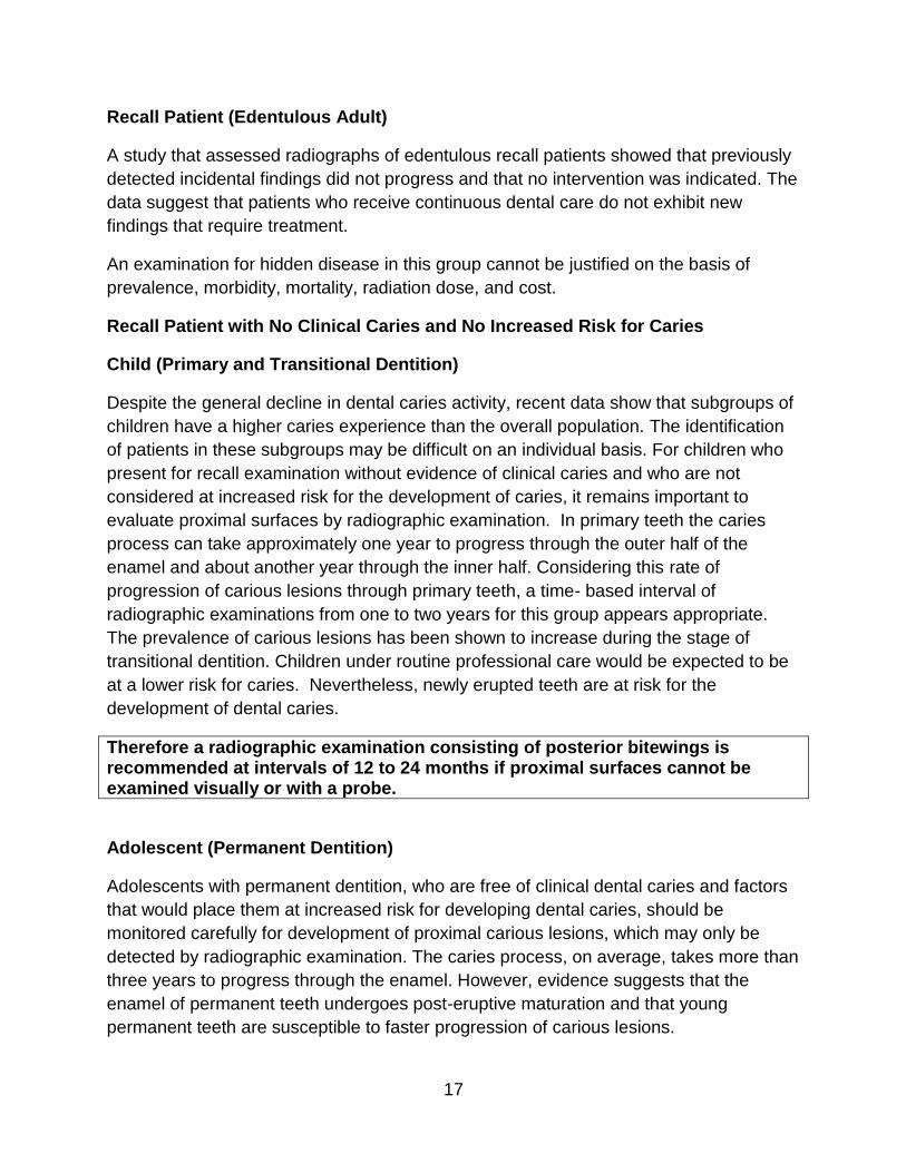
Recall Patient (Edentulous Adult)
A study that assessed radiographs of edentulous recall patients showed that previously
detected incidental findings did not progress and that no intervention was indicated. The
data suggest that patients who receive continuous dental care do not exhibit new
findings that require treatment.
An examination for hidden disease in this group cannot be justified on the basis of
prevalence, morbidity, mortality, radiation dose, and cost.
Recall Patient with No Clinical Caries and No Increased Risk for Caries
Child (Primary and Transitional Dentition)
Despite the general decline in dental caries activity, recent data show that subgroups of
children have a higher caries experience than the overall population. The identification
of patients in these subgroups may be difficult on an individual basis. For children who
present for recall examination without evidence of clinical caries and who are not
considered at increased risk for the development of caries, it remains important to
evaluate proximal surfaces by radiographic examination. In primary teeth the caries
process can take approximately one year to progress through the outer half of the
enamel and about another year through the inner half. Considering this rate of
progression of carious lesions through primary teeth, a time- based interval of
radiographic examinations from one to two years for this group appears appropriate.
The prevalence of carious lesions has been shown to increase during the stage of
transitional dentition. Children under routine professional care would be expected to be
at a lower risk for caries. Nevertheless, newly erupted teeth are at risk for the
development of dental caries.
Therefore a radiographic examination consisting of posterior bitewings is recommended at intervals of 12 to 24 months if proximal surfaces cannot be examined visually or with a probe.
Adolescent (Permanent Dentition)
Adolescents with permanent dentition, who are free of clinical dental caries and factors
that would place them at increased risk for developing dental caries, should be
monitored carefully for development of proximal carious lesions, which may only be
detected by radiographic examination. The caries process, on average, takes more than
three years to progress through the enamel. However, evidence suggests that the
enamel of permanent teeth undergoes post-eruptive maturation and that young
permanent teeth are susceptible to faster progression of carious lesions.
18
Therefore a radiographic examination consisting of posterior bitewings is recommended at intervals of 18 to 36 months.
Adult (Dentate and Partially Edentulous)
Adult dentate patients, who receive regularly scheduled professional care and are free
of signs and symptoms of oral disease, are at a low risk for dental caries. Nevertheless,
consideration should be given to the fact that caries risk can vary over time as risk
factors change.
Advancing age and changes in diet, medical history and periodontal status may
increase the risk for dental caries.
Therefore a radiographic examination consisting of posterior bitewings is recommended at intervals of 24 t0 36 months.
Recall Patient with Periodontal Disease
Child (Primary and Transitional Dentition), Adolescent (Permanent Dentition), and
Adult (Dentate and Partially Edentulous)
The decision to obtain radiographs for patients who have clinical evidence or a history
of periodontal disease/treatment should be determined on the basis of the anticipation
that important diagnostic and prognostic information will result. Structures or conditions
to be assessed should include the level of supporting alveolar bone, condition of the
interproximal bony crest, length and shape of roots, bone loss in furcations, and
calculus deposits. The frequency and type of radiographic examinations for these
patients should be determined on the basis of a clinical examination of the periodontium
and documented signs and symptoms of periodontal disease. The procedure for
prescribing radiographs for the follow-up/recall periodontal patient would be to use
selected intraoral radiographs to verify clinical findings on a patient-by-patient basis.
Therefore it is recommended that clinical judgment be used in determining the need for, and type of, radiographic images necessary for evaluation of periodontal disease. Imaging may consist of, but is not limited to, selected bitewing and/or periapical images of areas where periodontal disease (other then nonspecific gingivitis) can be identified clinically.
19
Patient (New and Recall) for Monitoring of Dento-facial Growth and Development,
and/or Assessment of Dental/Skeletal Relationships
Child (Primary and Transitional Dentition)
For children with primary dentition, before the eruption of the first permanent tooth,
radiographic examination to assess growth and development in the absence of clinical
signs or symptoms is unlikely to yield productive information. Any abnormality of growth
and development suggested by clinical findings should be evaluated radiographically on
an individual basis. After eruption of the first permanent tooth, the child may have a
radiographic examination to assess growth and development. This examination need
not be repeated unless dictated by clinical signs or symptoms. Cephalometric
radiographs may be useful for assessing growth, and/or dental and skeletal
relationships.
Therefore it is recommended that clinical judgment be used in determining the need for, and type of, radiographic images necessary for evaluation and/or monitoring of dento-facial growth and development or assessment of dental and skeletal relationships.
Adolescent (Permanent Dentition)
During adolescence there is often a need to assess the growth status and/or the dental
and skeletal relationships of patients in order to diagnose and treat their malocclusion.
Appropriate radiographic assessment of the malocclusion should be determined on an
individual basis.
An additional concern relating to growth and development for patients in this age group
is to determine the presence, position and development of third molars. This
determination can best be made by the use of selected periapical images or a
panoramic examination, once the patient is in late adolescence (16 to 19 years of age).
Therefore it is recommended that clinical judgment be used in determining the need for, and type of, radiographic images necessary for evaluation and/or monitoring of dento-facial growth and development or assessment of dental and skeletal relationships. Panoramic or periapical examination may be used to assess developing third molars.
20
Adult (Dentate, Partially Edentulous and Edentulous)
In the absence of any clinical signs or symptoms suggesting abnormalities of growth
and development in adults, no radiographic examinations are indicated for this purpose.
Therefore in the absence of clinical sign and symptoms, no radiographic examination is recommended.
Patients with Other Circumstances
(Including, but not limited to, proposed or existing implants, other dental and craniofacial
pathoses, restorative/endodontic needs, treated periodontal disease and caries
remineralization)
All Patient Categories
The use of imaging, as a diagnostic and evaluative tool, has progressed beyond the
longstanding need to diagnose caries and evaluate the status of periodontal disease.
The expanded technology in imaging is now used to diagnose other orofacial clinical
conditions and evaluate treatment options. A few examples of other clinical
circumstances are the use of imaging for dental implant treatment planning, placement,
or evaluation; the monitoring of dental caries and remineralization; the assessment of
restorative and endodontic needs; and the diagnosis of soft and hard tissue pathoses.
Therefore it is recommended that clinical judgment be used in determining the need for, and type of, radiographic images necessary for evaluation and/or monitoring in these circumstances.
Limiting Radiation Exposure
Dental radiographs account for approximately 2.5 percent of the effective dose received
from medical radiographs and fluoroscopies. Even though radiation exposure from
dental radiographs is low, once a decision to obtain radiographs is made it is the
dentist's responsibility to follow the ALARA Principle (As Low as Reasonably
Achievable) to minimize the patient's exposure. Examples of good radiologic practice
include:
• use of the fastest image receptor compatible with the diagnostic task (F-
speed film or digital);
• collimation of the beam to the size of the receptor whenever feasible;
• proper film exposure and processing techniques;
• use of protective aprons and thyroid collars, when appropriate; and
21
• limiting the number of images obtained to the minimum necessary to obtain
essential diagnostic information.
Receptor Selection
Though digital is widely used now, it is important to know the standards for film speeds
and the history of film speeds before, leading up to our digital technology. The American
National Standards Institute and the International Organization for Standardization have
established standards for film speed. Film speeds available for dental radiography are
D-speed, E-speed and F-speed, with D-speed being the slowest and F-speed the
fastest. According to the U.S. Food and Drug Administration, switching from D to E
speed can produce a 30 to 40 percent reduction in radiation exposure. The use of F-
speed film can reduce exposure 20 to 50 percent compared to use of E-speed film,
without compromising image quality.
Exposure of extra-oral films such as panoramic radiographs requires intensifying
screens to minimize radiation exposure to patients. The intensifying screen consists of
layers of phosphor crystals that fluoresce when exposed to radiation. In addition to the
radiation incident on the film, the film is exposed primarily to the light emitted from the
intensifying screen. Previous generations of intensifying screens were composed of
phosphors such as calcium tungstate. However, rare-earth intensifying screens are
recommended because they reduce a patient’s radiation exposure by 50 percent
compared with calcium tungstate-intensifying screens. Rare-earth film systems,
combined with a high-speed film of 400 or greater, can be used for panoramic
radiographs. Older panoramic equipment can be retrofitted to reduce the radiation
exposure to accommodate the use of rare-earth, high-speed systems.
Digital imaging provides an opportunity to further reduce the radiation dose by 40 to 60
percent. In digital radiography, there are three types of receptors that take the place of
semiconductor (CMOS), and photo-stimulable phosphor (PSP) plates. Systems that use
CCD and CMOS- based, solid-state detectors are called “direct.” When these sensors
receive energy from the x- ray beam, the CCD or CMOS chip sends a signal to the
computer and an image appears on the monitor within seconds. Systems that use PSP
plates are called “indirect.” When these plates are irradiated, a latent image is stored on
them. The plate is then scanned and the scanner transmits the image to the computer.
Receptor Holders
Holders that align the receptor precisely with the collimated beam are recommended for
periapical and bitewing radiographs. Heat-sterilizable or disposable intraoral radiograph
receptor-holding devices are recommended for optimal infection control. Dental
professionals should not hold the receptor holder during exposure. Under extraordinary
22
circumstances in which members of the patient’s family (or other caregiver) must
provide restraint or hold a receptor holder in place during exposure, such a person
should wear appropriate shielding.
Collimation
Collimation limits the amount of radiation, both primary and scattered, to which the
patient is exposed. An added benefit of rectangular collimation is an improvement in
contrast as a result of a reduction in fogging caused by secondary and scattered
radiation. The x-ray beam should not exceed the minimum coverage necessary, and
each dimension of the beam should be collimated so that the beam does not exceed the
receptor by more than 2 percent of the source-to-image receptor distance. Since a
rectangular collimator decreases the radiation dose by up to fivefold as compared with a
circular one, radiographic equipment should provide rectangular collimation for
exposure of periapical and bitewing radiographs. Use of a receptor-holding device
minimizes the risk of cone-cutting (non-exposure of part of the image receptor due to
malalignment of the x-ray beam). The position-indicating device should be open ended
and have a metallic lining to restrict the primary beam and reduce the tissue volume
exposed to radiation. Use of long source-to-skin distances of 40 cm, rather than short
distances of 20 cm, decreases exposure by 10 to 25 percent. Distances between 20
and 40 cm are appropriate, but the longer distances are optimal.
Operating Potential and Exposure Time
The operating potential of dental x-ray units affects the radiation dose and backscatter
radiation. Lower voltages produce higher-contrast images and higher entrance skin
doses, and lower deep-tissue doses and levels of backscatter radiation. However,
higher voltages produce lower contrast images that enable better separation of objects
with differing densities. Thus, the diagnostic purposes of the radiograph should be used
to determine the selection of kilovolt setting. A setting above 90 kV(p) will increase the
patient dose and should not be used. The optimal operating potential of dental x-ray
units is between 60 and 70 kVp.
Filmless technology is much more forgiving to overexposure often resulting in
unnecessary radiation exposure. Facilities should strive to set the x-ray unit exposure
timer to the lowest setting providing an image of diagnostic quality. If available, the
operator should always confirm that the dose delivered falls within the manufacturer’s
exposure index. Imaging plates should be evaluated at least monthly and cleaned as
necessary.
23
Patient Shielding and Positioning
The amount of scattered radiation striking the patient’s abdomen during a properly
conducted radiographic examination is negligible The thyroid gland is more susceptible
to radiation exposure during dental radiographic exams given its anatomic position,
particularly in children. Protective thyroid collars and collimation substantially reduce
radiation exposure to the thyroid during dental radiographic procedures. Because every
precaution should be taken to minimize radiation exposure, protective thyroid collars
should be used whenever possible. If all the recommendations for limiting radiation
exposure are put into practice, the gonadal radiation dose will not be significantly
affected by use of abdominal shielding. Therefore, use of abdominal shielding may not
be necessary.
Protective aprons and thyroid shields should be hung or laid flat and never folded, and
manufacturer’s instructions should be followed. All protective shields should be
evaluated for damage (e.g. tears, folds, and cracks) monthly using visual and manual
inspection.
Proper education and training in patient positioning is necessary to ensure that
panoramic radiographs are of diagnostic quality.
Operator Protection
Although dental professionals receive less exposure to ionizing radiation than do other
occupationally exposed health care workers, operator protection measures are essential
to minimize exposure. Operator protection measures include education, the
implementation of a radiation protection program, occupational radiation exposure
limits, recommendations for personal dosimeters and the use of barrier shielding. The
maximum permissible annual dose of ionizing radiation for health care workers is 50
millisieverts (mSv) and the maximum permissible lifetime dose is 10 mSv multiplied by a
person’s age in years. Personal dosimeters should be used by workers who may
receive an annual dose greater than 1 mSv to monitor their exposure levels. Pregnant
dental personnel operating x-ray equipment should use personal dosimeters, regardless
of anticipated exposure levels.
Operators of radiographic equipment should use barrier protection when possible, and
barriers should ideally contain a leaded glass window to enable the operator to view the
patient during exposure. When shielding is not possible, the operator should stand at
least two meters from the tube head and out of the path of the primary beam. The
National Council on Radiation Protection & Measurements report “Radiation Protection
in Dentistry” offers detailed information on shielding and office design. State radiation
control agencies can help assess whether barriers meet minimum standards.
24
Hand Held X-Ray Units
Hand-held, battery-powered x-ray systems are available for intra-oral radiographic
imaging.
The hand-held exposure device is activated by a trigger on the handle of the device.
However, dosimetry studies indicate that these hand-held devices present no greater
radiation risk than standard dental radiographic units to the patient or the operator. No
additional radiation protection precautions are needed when the device is used
according to the manufacturer’s instructions. These include:
1. holding the device at mid-torso height,
2. orienting the shielding ring properly with respect to the operator, and
3. keeping the cone as close to the patient’s face as practical.
If the hand-held device is operated without the ring shield in place, it is recommended
that the operator wear a lead apron.
All operators of hand-held units should be instructed on their proper storage. Due to the
portable nature of these devices, they should be secured properly when not in use to
prevent accidental damage, theft, or operation by an unauthorized user. Hand-held
units should be stored in locked cabinets, locked storage rooms, or locked work areas
when not under the direct supervision of an individual authorized to use them. Units
with user-removable batteries should be stored with the batteries removed. Records
listing the names of approved individuals who are granted access and use privileges
should be prepared and kept current.
Film Exposure and Processing
Though digital radiography is more widely used, it is important to understand film and its
safety concerns, still, both in the effects on the patient, but, also to the practitioner and
unto the environment. All film should be processed following the film and processer
manufacturer recommendations. Once this is achieved, the x-ray operator can adjust
the tube current and time and establish a technique that will provide consistent dental
radiographs of diagnostic quality. Poor processing technique, including sight-
developing, most often results in underdeveloped films, forcing the x-ray operator to
increase the dose to compensate, resulting in patient and personnel being exposed to
unnecessary radiation.
A safelight does not provide completely safe exposure for an indefinite period of time.
Extra-oral film is much more sensitive to fogging. The length of time for which a film can
be exposed to the safelight should be determined for the specific safelight/film
combination in use.
25
Quality Assurance
Quality assurance protocols for the x-ray unit, imaging receptor, film processing, dark
room, and patient shielding should be developed and implemented for each dental
health care setting. All quality assurance procedures, including date, procedure, results,
and corrective action, should be logged for documentation purposes. A qualified expert
should survey all x- ray units on their placement and should resurvey the equipment
every four years or after any changes that may affect the radiation exposure of the
operator and others. Surveys typically are performed by state agencies, and individual
state regulations should be consulted regarding specific survey intervals. The film
processor should be evaluated at its initial installation and on a monthly basis afterward.
The processing chemistry should be evaluated daily, and each type of film should be
evaluated monthly or when a new box or batch of film is opened. Abdominal shielding
and thyroid collars should be inspected visually for creases or clumping that may
indicate voids in their integrity on a monthly basis. Damaged abdominal shielding and
collars should be replaced. Table 2 (below) lists specific methods of quality assurance
procedures, covering not only inspection of the x-ray unit itself but also of the film
processor, the image receptor devices, and the darkroom and abdominal shielding and
collars.
It is imperative that the operator’s manual for all imaging acquisition hardware is readily
available to the user, and that the equipment is operated and maintained following the
manufacturer’s instructions, including any appropriate adjustments for optimizing dose
and image quality.
Technique Charts/Protocols
Size-based technique charts/protocols with suggested parameter settings are important
for ensuring that radiation exposure is optimized for all patients. Technique charts
should be used for all systems with adjustable settings, such as tube potential, tube
current, and time or pulses. The purpose of using the charts is to control the amount of
radiation to the patient and receptor. Technique charts are tables that indicate
appropriate settings on the x-ray unit for a specific anatomical area and will ensure the
least amount of radiation exposure to produce a consistently good-quality radiograph.
Technique charts for intraoral and extra-oral radiography should list the type of exam,
the patient size (small, medium, large) for adults and a pediatric setting. The speed of
film used, or use of a digital receptor, should also be listed on the technique chart. The
chart should be posted near the control panel where the technique is adjusted for each
x-ray unit. A technique chart that is regularly updated should be developed for each x-
ray unit. The charts will also need to be updated when a different film or sensor, new
unit, or new screens are used.
26
Radiation Risk Communication
Dentists should be prepared to discuss benefits and risks of the x-ray exam. To help
answer patient and parent questions about dental radiology radiation safety, the
American Academy of Oral and Maxillofacial Radiology and the Alliance for Radiation
Safety in Pediatric Imaging partnered to create a brochure targeted at parents and
patients.
The following procedures for periodic assessment of the performance of radiographic
equipment, film processing, equipment, image receptor devices, dark room integrity,
and abdominal and thyroid shielding are adapted from the National Council for
Radiation Protection and Measurements report, Radiation Protection in Dentistry.
Please refer to state guidelines for specific regulations.
Table 2
Quality Assurance Procedures for Assessment of Radiographic Equipment
Equipment Frequency Method X-ray Machine • On installation
• At regular intervals as recommended by state regulations
• Whenever there are any changes in installation workload or operating conditions
Inspection by qualified expert (as specified by government regulations and manufacturers recommendations).
Film Processor • On installation • Daily
Method 1: Sensitometry and Densitometry A sensitometer is used to expose a film, followed by standard processing of the film. The processed film will have a defined pattern of optical densities. The densities are measured with a densitometer. The densitometer measurements are compared to the densities of films exposed and processed under ideal conditions. A change in densitometer values indicates a problem with either the development time, temperature or the developer solutions. Advantages Accuracy Speed Disadvantage Expense of additional equipment Method 2: Reference Film A film exposed and processed under ideal conditions is attached to the corner of a view box as a reference film. Subsequent films are compared with the reference film. Advantage: Cost effectiveness Disadvantage: Less sensitive
27
Quality Assurance Procedures for Assessment of Radiographic Equipment
Equipment Frequency Method Image Receptor Devices Intensifying Screen and
• Monthly • With each new batch of film • Every six months
Method 1: Sensitometry and Densitometry (as described above) Method 2: Reference Image (as described above) Visual inspection of cassette integrity Examination of intensifying screen for scratches
Extra-oral Cassettes Development of an unexposed film that has been in the cassette exposed to normal lighting for one hour or more
Darkroom Integrity • On installation • Monthly • After a change in the
lighting filter or lamp
While in a darkroom with the safelight on, place metal object (such as a coin) on unwrapped film for a period that is equivalent to the time required for a typical darkroom procedure Develop film Detection of the object indicates a problem with the safelight or light leaks in the darkroom
Abdominal and Thyroid Shielding
• Monthly (visual and manual inspection)
All protective shields should be evaluated for damage (e.g., tears, folds, and cracks) monthly using visual and manual inspection. If a defect in the attenuating material is suspected, radiographic or fluoroscopic inspection may be performed as an alternative to immediately removing the item from service. Consideration should be given to minimizing the radiation exposure of inspectors by minimizing unnecessary fluoroscopy.
Training and Education
Where permitted by law, auxiliary dental personnel can perform intraoral and extra-oral
imaging.103 Personnel certified to take dental radiographs should receive appropriate
education. Practitioners should remain informed about safety updates and the
availability of new equipment, supplies and techniques that could further improve the
diagnostic quality of radiographs and decrease radiation exposure. Free training
materials are available for limiting radiation exposure in dental imaging through the
International Atomic Energy Agency.
28
Technology and Safety
As stated in the American Dental Association’s website: