+ All Categories
Home > Documents > Radiological findings in Birt–Hogg–Dubé syndrome: a rare differential for pulmonary cysts and...

Radiological findings in Birt–Hogg–Dubé syndrome: a rare differential for pulmonary cysts and...

Date post: 05-Sep-2016
Category:
Upload: pramod-gupta
View: 216 times
Download: 1 times
Share this document with a friend
4
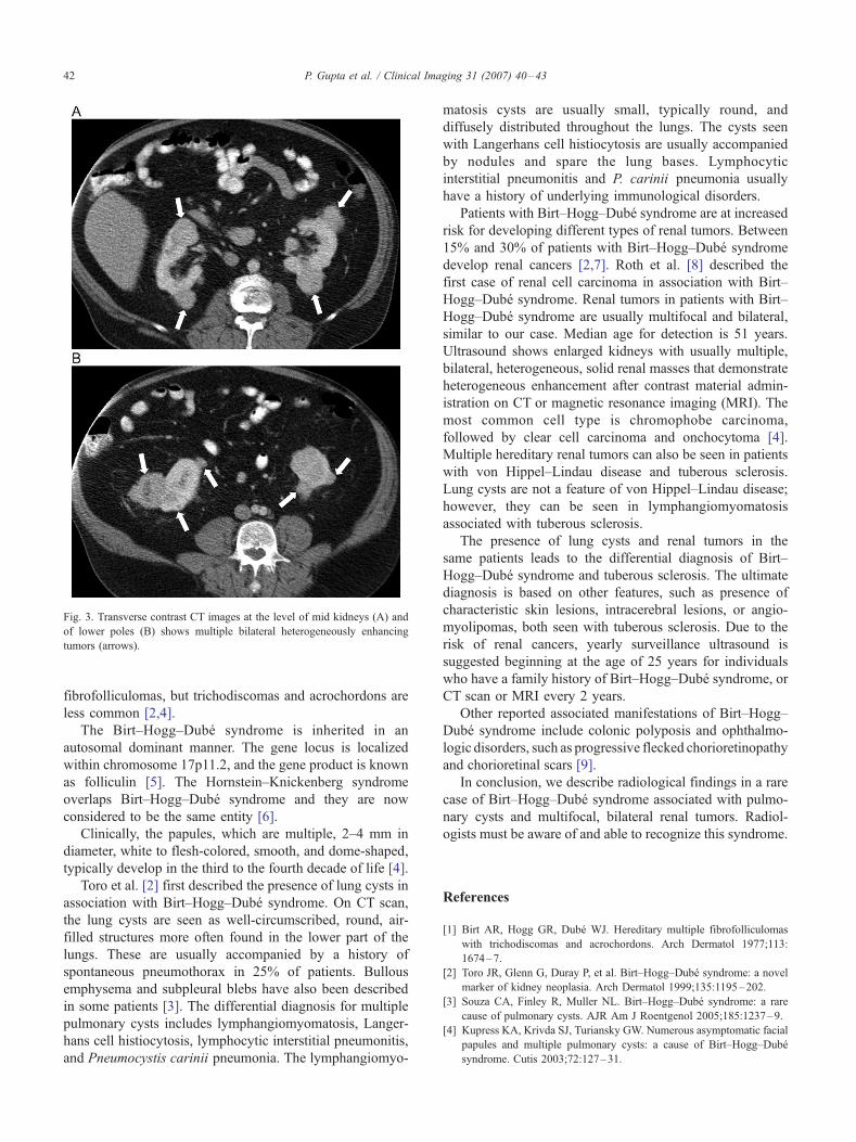
Radiological findings in Birt–Hogg–Dube ´ syndrome: a rare differential for pulmonary cysts and renal tumors Pramod Gupta 4 , Nahid Eshaghi, Thompson T. Kamba, Vidisha Ghole, Francisco Garcia-Morales Radiology Service, Dallas VA Medical Center, VA North Texas Health Care System, 4500, South Lancaster Road, Dallas, TX 75216, USA Received 25 July 2006; accepted 20 September 2006 Abstract Birt–Hogg–Dube ´ syndrome is a rare disorder characterized by cutaneous hair follicle tumors, pulmonary cysts, and renal tumors. We report a case of a 63-year-old male patient with this syndrome. The radiological findings seen with this syndrome are described. Radiologists should be aware of and able to recognize this syndrome. D 2007 Elsevier Inc. All rights reserved. Keywords: Birt–Hogg–Dube ´ syndrome; Autosomal dominant; Pulmonary cyst; Renal tumor 1. Introduction Birt–Hogg–Dube ´ syndrome is a rare, autosomal domi- nant, inherited dermatologic disorder characterized by cutaneous hair follicle tumors (fibrofolliculomas), pulmo- nary cysts, and renal tumors [1–3]. Descriptions of pulmo- nary and renal involvement in the radiology literature are scanty and mainly published in the dermatologic literature. We report imaging findings of Birt–Hogg–Dube ´ syndrome in a patient with pulmonary and renal involvement. 2. Case report A 63-year-old white male presented to our institution’s emergency room with shortness of breath. The patient provided a history of having had spontaneous pneumo- thorax on the left side at the age of 23 years and on the right side at the age of 42 years, both times treated with chest tube thoracostomy. For reasons not known to the patient, a renal sonogram was performed approximately 15 years ago, which demonstrated multiple renal cysts. Approximately 6 years ago, during a routine visit to his primary care physician, he requested for a check on the status of his kidney cysts. At that time, a renal ultrasound and computed tomography (CT) scan done at another institution showed multiple renal masses. The biopsy of one of the left renal masses showed it to be onchocytoma. Considering his skin lesions, renal masses, and history of pneumothorax, a diagnosis of Birt–Hogg–Dube ´ syndrome was made. His physical examination showed multiple scattered 1- to 3-mm papules over his face, neck, and trunk. His family history revealed that his brother also had similar skin findings. Considering his shortness of breath and past history, we obtained a chest radiograph, which showed no pneumo- thorax. CT angiogram of the chest was negative for pulmonary embolism but revealed multiple pulmonary cysts, most numerous in the lower part of the lungs (Fig. 1). The pulmonary cysts were sharply marginated, air-containing lesions with walls of 2 mm or less and measuring 0.5–3.5 cm. The angiogram also showed bilateral pleural thickening likely related to his prior thoracostomy for pneumothorax. His pulmonary function tests revealed a mild obstructive lung disease pattern with mild air trapping. He had a remote smoking history of approximately one to two packs per 0899-7071/07/$ – see front matter D 2007 Elsevier Inc. All rights reserved. doi:10.1016/j.clinimag.2006.09.023 4 Corresponding author. 2704, Oates Drive, Plano, TX 75093, USA. Tel.: +1 214 857 0185; fax: +1 775 855 4624. E-mail address: [email protected] (P. Gupta). Clinical Imaging 31 (2007) 40 – 43
Transcript

Clinical Imaging 31

Radiological findings in Birt–Hogg–Dube syndrome:

a rare differential for pulmonary cysts and renal tumors

Pramod Gupta4, Nahid Eshaghi, Thompson T. Kamba, Vidisha Ghole, Francisco Garcia-Morales

Radiology Service, Dallas VA Medical Center, VA North Texas Health Care System, 4500, South Lancaster Road, Dallas, TX 75216, USA

Received 25 July 2006; accepted 20 September 2006

Abstract

Birt–Hogg–Dube syndrome is a rare disorder characterized by cutaneous hair follicle tumors, pulmonary cysts, and renal tumors. We

report a case of a 63-year-old male patient with this syndrome. The radiological findings seen with this syndrome are described. Radiologists

should be aware of and able to recognize this syndrome.

D 2007 Elsevier Inc. All rights reserved.

Keywords: Birt–Hogg–Dube syndrome; Autosomal dominant; Pulmonary cyst; Renal tumor

1. Introduction

Birt–Hogg–Dube syndrome is a rare, autosomal domi-

nant, inherited dermatologic disorder characterized by

cutaneous hair follicle tumors (fibrofolliculomas), pulmo-

nary cysts, and renal tumors [1–3]. Descriptions of pulmo-

nary and renal involvement in the radiology literature are

scanty and mainly published in the dermatologic literature.

We report imaging findings of Birt–Hogg–Dube syndrome

in a patient with pulmonary and renal involvement.

2. Case report

A 63-year-old white male presented to our institution’s

emergency room with shortness of breath. The patient

provided a history of having had spontaneous pneumo-

thorax on the left side at the age of 23 years and on the right

side at the age of 42 years, both times treated with chest tube

thoracostomy. For reasons not known to the patient, a renal

sonogram was performed approximately 15 years ago,

0899-7071/07/$ – see front matter D 2007 Elsevier Inc. All rights reserved.

doi:10.1016/j.clinimag.2006.09.023

4 Corresponding author. 2704, Oates Drive, Plano, TX 75093, USA.

Tel.: +1 214 857 0185; fax: +1 775 855 4624.

E-mail address: [email protected] (P. Gupta).

which demonstrated multiple renal cysts. Approximately

6 years ago, during a routine visit to his primary care

physician, he requested for a check on the status of his

kidney cysts. At that time, a renal ultrasound and computed

tomography (CT) scan done at another institution showed

multiple renal masses. The biopsy of one of the left renal

masses showed it to be onchocytoma. Considering his skin

lesions, renal masses, and history of pneumothorax, a

diagnosis of Birt–Hogg–Dube syndrome was made. His

physical examination showed multiple scattered 1- to 3-mm

papules over his face, neck, and trunk. His family history

revealed that his brother also had similar skin findings.

Considering his shortness of breath and past history, we

obtained a chest radiograph, which showed no pneumo-

thorax. CT angiogram of the chest was negative for

pulmonary embolism but revealed multiple pulmonary

cysts, most numerous in the lower part of the lungs

(Fig. 1). The pulmonary cysts were sharply marginated,

air-containing lesions with walls of 2 mm or less and

measuring 0.5–3.5 cm. The angiogram also showed bilateral

pleural thickening likely related to his prior thoracostomy

for pneumothorax.

His pulmonary function tests revealed a mild obstructive

lung disease pattern with mild air trapping. He had a remote

smoking history of approximately one to two packs per

(2007) 40–43

P. Gupta et al. / Clinical Imaging 31 (2007) 40–43 41

week for a 6-year period from age 10 to age 16. His renal

ultrasound, done subsequently as an outpatient procedure,

showed multiple bilateral hypo- to isoechoic masses (Fig. 2).

Renal CT scan revealed multiple, heterogeneously enhanc-

ing masses in both kidneys (Fig. 3). Although previous

biopsy showed the left renal mass to be benign, considering

Fig. 1. Transverse chest CT images at the level of the upper lungs (A),

lower lungs (B), and coronal reconstruction image (C) show multiple

pulmonary cysts (arrows), more numerous in the lower lungs. Also note few

subpleural blebs (A, curved arrows). Bilateral pleural thickening and

subpleural scarring is also noted posteriorly in the lower part of the chest

(B, arrowheads), likely related to prior thoracostomy for pneumothorax.

Fig. 2. Longitudinal images of the ultrasound of the right (A) and left (B)

kidney shows multiple hypo- to isoechoic partly exophytic masses in both

kidneys (arrows).

the risk of malignancy associated with this syndrome, he is

currently being evaluated for bilateral nephrectomy.

3. Discussion

Birt–Hogg–Dube syndrome is a rare inherited genoder-

matosis characterized by distinctive cutaneous lesions, an

increased risk for renal neoplasia, and the development of

pulmonary cysts [1–3]. In 1977, Birt, Hogg, and Dube

studied large kindred of 70 members of three generations

with familial occurrence of small popular lesions originating

in hair follicles on the face, neck, and upper trunk. The skin

lesions were fibrofolliculomas, trichodiscomas, and acro-

chordons [1]. This hereditary cancer syndrome became

known as Birt–Hogg–Dube syndrome. Subsequent studies

showed that all patients with this syndrome have skin

Fig. 3. Transverse contrast CT images at the level of mid kidneys (A) and

of lower poles (B) shows multiple bilateral heterogeneously enhancing

tumors (arrows).

P. Gupta et al. / Clinical Imaging 31 (2007) 40–4342

fibrofolliculomas, but trichodiscomas and acrochordons are

less common [2,4].

The Birt–Hogg–Dube syndrome is inherited in an

autosomal dominant manner. The gene locus is localized

within chromosome 17p11.2, and the gene product is known

as folliculin [5]. The Hornstein–Knickenberg syndrome

overlaps Birt–Hogg–Dube syndrome and they are now

considered to be the same entity [6].

Clinically, the papules, which are multiple, 2–4 mm in

diameter, white to flesh-colored, smooth, and dome-shaped,

typically develop in the third to the fourth decade of life [4].

Toro et al. [2] first described the presence of lung cysts in

association with Birt–Hogg–Dube syndrome. On CT scan,

the lung cysts are seen as well-circumscribed, round, air-

filled structures more often found in the lower part of the

lungs. These are usually accompanied by a history of

spontaneous pneumothorax in 25% of patients. Bullous

emphysema and subpleural blebs have also been described

in some patients [3]. The differential diagnosis for multiple

pulmonary cysts includes lymphangiomyomatosis, Langer-

hans cell histiocytosis, lymphocytic interstitial pneumonitis,

and Pneumocystis carinii pneumonia. The lymphangiomyo-

matosis cysts are usually small, typically round, and

diffusely distributed throughout the lungs. The cysts seen

with Langerhans cell histiocytosis are usually accompanied

by nodules and spare the lung bases. Lymphocytic

interstitial pneumonitis and P. carinii pneumonia usually

have a history of underlying immunological disorders.

Patients with Birt–Hogg–Dube syndrome are at increased

risk for developing different types of renal tumors. Between

15% and 30% of patients with Birt–Hogg–Dube syndrome

develop renal cancers [2,7]. Roth et al. [8] described the

first case of renal cell carcinoma in association with Birt–

Hogg–Dube syndrome. Renal tumors in patients with Birt–

Hogg–Dube syndrome are usually multifocal and bilateral,

similar to our case. Median age for detection is 51 years.

Ultrasound shows enlarged kidneys with usually multiple,

bilateral, heterogeneous, solid renal masses that demonstrate

heterogeneous enhancement after contrast material admin-

istration on CT or magnetic resonance imaging (MRI). The

most common cell type is chromophobe carcinoma,

followed by clear cell carcinoma and onchocytoma [4].

Multiple hereditary renal tumors can also be seen in patients

with von Hippel–Lindau disease and tuberous sclerosis.

Lung cysts are not a feature of von Hippel–Lindau disease;

however, they can be seen in lymphangiomyomatosis

associated with tuberous sclerosis.

The presence of lung cysts and renal tumors in the

same patients leads to the differential diagnosis of Birt–

Hogg–Dube syndrome and tuberous sclerosis. The ultimate

diagnosis is based on other features, such as presence of

characteristic skin lesions, intracerebral lesions, or angio-

myolipomas, both seen with tuberous sclerosis. Due to the

risk of renal cancers, yearly surveillance ultrasound is

suggested beginning at the age of 25 years for individuals

who have a family history of Birt–Hogg–Dube syndrome, or

CT scan or MRI every 2 years.

Other reported associated manifestations of Birt–Hogg–

Dube syndrome include colonic polyposis and ophthalmo-

logic disorders, such as progressive flecked chorioretinopathy

and chorioretinal scars [9].

In conclusion, we describe radiological findings in a rare

case of Birt–Hogg–Dube syndrome associated with pulmo-

nary cysts and multifocal, bilateral renal tumors. Radiol-

ogists must be aware of and able to recognize this syndrome.

References

[1] Birt AR, Hogg GR, Dube WJ. Hereditary multiple fibrofolliculomas

with trichodiscomas and acrochordons. Arch Dermatol 1977;113:

1674–7.

[2] Toro JR, Glenn G, Duray P, et al. Birt–Hogg–Dube syndrome: a novel

marker of kidney neoplasia. Arch Dermatol 1999;135:1195–202.

[3] Souza CA, Finley R, Muller NL. Birt–Hogg–Dube syndrome: a rare

cause of pulmonary cysts. AJR Am J Roentgenol 2005;185:1237–9.

[4] Kupress KA, Krivda SJ, Turiansky GW. Numerous asymptomatic facial

papules and multiple pulmonary cysts: a cause of Birt–Hogg–Dube

syndrome. Cutis 2003;72:127–31.

P. Gupta et al. / Clinical Imaging 31 (2007) 40–43 43

[5] Schmidt LS, Warren MB, Nickerson ML, et al. Birt–Hogg–Dube

syndrome, a genodermatosis associated with spontaneous pneumo-

thorax and kidney neoplasia, maps to chromosome 17p112. Am J Hum

Genet 2001;69:876–82.

[6] Schulz T, Hartschuh W. Birt–Hogg–Dube syndrome and Hornstein–

Knickenberg syndrome are the same: different sectioning tech-

nique as the cause of differential histology. J Cutan Pathol 1999;26:

55–61.

[7] Choyke PL, Glenn GM, Walther MM, Zbar B, Linehan WM.

Hereditary renal cancers. Radiology 2003;226:33–46.

[8] Roth JS, Rabinowitz AD, Benson M, Grossman ME. Bilateral renal cell

carcinoma in the Birt–Hogg–Dube syndrome. J Am Acad Dermatol

1993;29:1055–6.

[9] Zbar B, Alvord WG, Glenn G, et al. Risk of renal and colonic

neoplasms and spontaneous pneumothorax in the Birt–Hogg–Dube

syndrome. Cancer Epidemiol Biomarkers Prev 2002;11:393–400.


Recommended