In the last 30 years Raman spectroscopy has become an essential
experimental tool for the analysis of a wide variety of artists' materials,
often in a non-destructive, non-invasive manner. The Raman effect
provides a quick and relatively straightforwardmolecular identification
of amaterial under examination. A Raman spectrumcan be considered
as a fingerprint that could be used for compound identificationwhen a
database of standard spectra is available for comparison purposes.
Its use has become widespread because Raman spectroscopy isa non-contact technique and, provided the correct experimentalconditions are used by an expert operator, the materials underexamination are not damaged in any way. Because of this, thetechnique has now become mainstream in heritage science.There is no single type of Raman instrument that can be usedfor all purposes: users now have a choice of instruments andoperating conditions, depending on the analytical require-ments and the type of art objects to be analysed.
What is Raman good for?Types of materials
When Raman experienced a renaissance in the 1980s, its mainapplication in cultural heritage was for the analysis of tradi-tional artists' materials, mostly pigments of mineral origin. Thisis still one of its main applications, although the technique isalso used for the identication of a few natural organic dyes (forexample indigo and gamboge), many modern synthetic dyes,most gems and semi-precious stones, minerals in general, andcorrosion products. Other cultural heritage materials that have
847
been examined more recently include plastics, parchment,paper, textile bres, resins, bone and bone-like materials andvarnishes. In the investigation of artists' materials the successand speed of the analysis oen depends on what medium ismixed with the pigments (see “Pitfalls and drawbacks – uo-rescence” below).
Types of objects
With conventional bench-top instruments coupled with amicroscope, only relatively small, at items that t under themicroscope can be analysed. For this reason, it is customary touse the technique with small samples or loose fragments ormaterials either individually or aer mounting as a crosssection. Paper- or parchment-based objects, such as smallwhole manuscripts, single illuminated pages and portraitminiatures are also easy to examine. Other items that can beanalysed with relative ease include painted textiles, textile bres(both natural and synthetic), gems still encased in jewellery andsmall, at sculptures or other painted objects.
Types of instrumentsBench-top microscope
This is the most common Raman instrument found in a labo-ratory and is the instrument with the highest performance interms of speed, signal intensity, spatial and spectral resolution,stability and freedom from disruptive vibrations (Fig. 1). A widevariety of lasers can be associated with such an instrument (seebelow). The use of a microscope also ensures that a very smallarea is analysed each time, less than a micrometre across and afew micrometres in depth, depending on the specications ofthe microscope objectives used. This helps in limiting theinterference of surrounding materials.
Probe
This is an easily transportable piece of equipment that, contraryto the better-performing bench-top microscope, can be used on
Fig. 1 A 19th century Chinese watercolour analysed with a bench-topRaman microscope.
AMC Technical Briefs Analytical Methods
Publ
ishe
d on
04
June
201
5. D
ownl
oade
d on
13/
06/2
015
22:5
3:12
. View Article Online
site during an excavation, or on unmoveable objects such aswall paintings, cave paintings, mosaics etc.When compared to abench-top microscope, the drawbacks of a probe includereduced signal intensity, spatial and spectral resolution, alimited choice in terms of lasers, a less-than-ideal ability to viewand evaluate with a proper microscope the sample underexamination, and the presence of vibrations that can hinder theanalysis.
Handheld
This type of instrument is easy to use and is especially suited forthe analysis of inorganic materials, for example during amineral survey in the eld. However it has an even more limitedspatial and spectral resolution compared to the probe. It mayalso be difficult to set the power intensity at the sample to asuitable level, and having no microscope objective at the end ofthe instrument, a handheld Raman probe does not allow theanalyst to inspect and choose the target region carefully.
Types of lasers
Most of the lasers commonly found in a cultural heritagelaboratory are in the visible range: blue, green, red and far red. Alaser in the near infrared can also be used, and lasers in the UVare becoming more common in Raman instruments. Whenchoosing a laser in the visible range, a general rule of thumb isto choose the laser with a colour as similar as possible to that ofthe sample under examination. The Raman effect is based onlight scattering, and absorption of the laser beam by the sampleneeds to be limited as much as possible: the more a laser beamis absorbed by the sample, the less of it is le for any scatteringeffect, and the weaker the Raman bands. Moreover, a signicantabsorption of a laser beam by a pigment or a dye is also likely tocause local overheating, which in turn can degrade or even burnthe microscopic portion of the sample under examination. Thisis a very conned damage, as usually the burnt area is only a fewmicrometres across and is invisible to the naked eye, but it isdamage nonetheless. The laser-degraded area may also give rise
to a Raman spectrum which can be mistaken for a differentmaterial.
The intensity of the Raman signal is inversely proportional tothe excitation wavelength: for example, a sample will give astronger Raman signal if it is analysed with a blue laser ratherthan a red laser.
There are of course exceptions to the following ‘rules’, but ingeneral for a red or orange pigment it would be best to use a He–Ne laser (632 nm) or a krypton ion laser (647 nm) or any othertrue-red solid state laser. It is not normally advisable to use agreen laser. For blue pigments it is best to use a blue (argon ionlaser, 488 nm) or a green laser (argon ion, 514.5 nm; Nd:YAG,532 nm; or any other solid state green laser). White and yellowpigments can usually be analysed with any type of laser. Far red(solid state lasers usually between 780 and 785 nm) and nearinfrared lasers (Nd:YAG, 1064 nm, usually found on FT-Ramanspectrometers) are particularly suitable for dyes and organicmaterials, but tend to burn dark inorganic materials as thelatter absorb rather than scatter such laser radiation.
How to set up a protocol
It is advisable to do a preliminary test on the specimen underthe microscope using a very low laser power (well below 1 mW).The laser power can be progressively increased as needed toobtain a good spectrum, always making sure that the sample isnot being damaged by the laser irradiation. Commercial Ramanmicroscopes usually come equipped with a set of neutraldensity lters; these allow ne-tuning of the laser power at thesample reducing it from 100% to below 1% of the maximumintensity. Please remember that laser-induced damage to thesample can occur even if no obvious change is visible under themicroscope: if the Raman bands change on the screen within asecond or two from the start of the analysis, it is likely that thespecimen is being adversely affected by the laser beam.
Pitfalls and drawbacksFluorescence
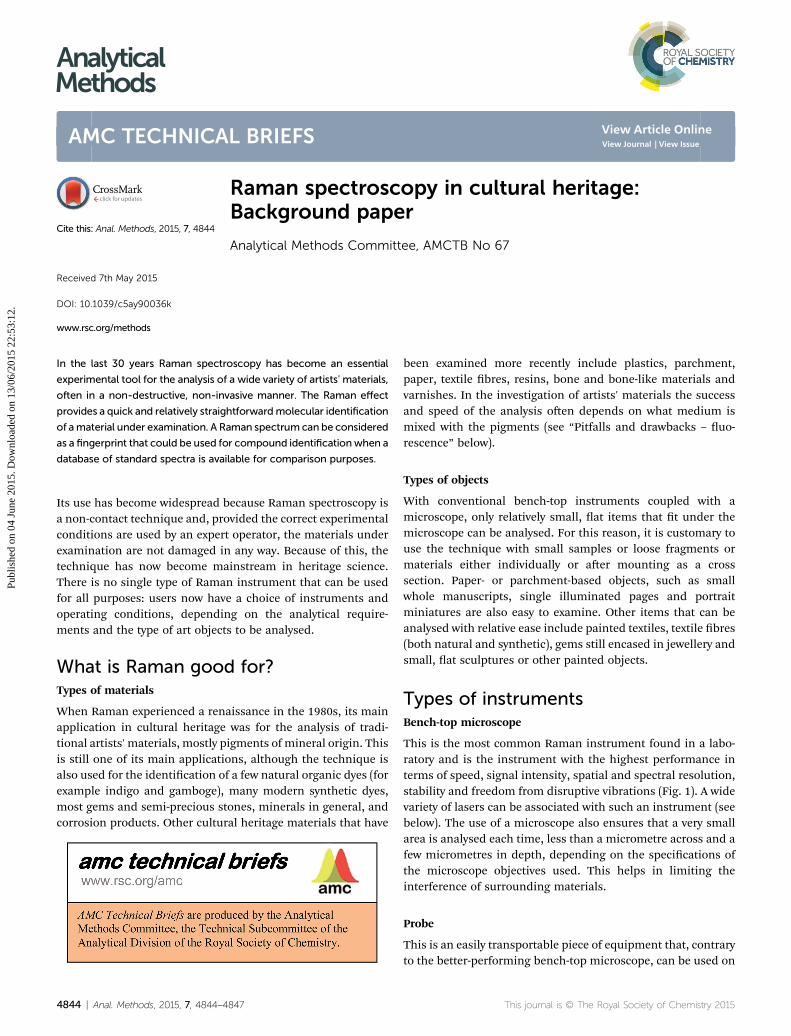
The laser beam can excite electronic transitions that may maskthe Raman signal; if any Raman peaks are visible at all, theyappear as if they are just coming out of a very bumpy or hillybaseline. In some cases, uorescence can be reduced by using alaser beam with a longer wavelength. Many organic dyes giverise to signicant uorescence and are not usually analysed byRaman. An oil-based binding medium oen generates enoughuorescence to hide most of the Raman spectral features of thepigment or dye mixed with it (see Fig. 2).
Stability and focusing issues
One of the most frustrating practical aspects in the Ramananalysis of relatively light objects is a loss of focus during theanalysis, due to small movements of the object under themicroscope. For manuscripts for example, unless the pageunder observation is kept still, any air movement or oorvibration can alter the position of the page under the laser. It is
Fig. 2 (A) Raman spectrum of the red pigment vermilion on plaster,showing no fluorescence (note the flat baseline) and clearly definedbands; (B) Raman spectrum of vermilion in an oil paint, showingsignificant fluorescence (raised and ‘bumpy’ baseline); the main peakof vermilion only just emerges from the fluorescent baseline.
Fig. 3 Example of Raman spectrum produced from a sample of greenpaint (top) compared to the spectra of two referencematerials (middleand bottom). By overlapping the spectra it is possible to observe thecorrespondence of the peaks.
Analytical Methods AMC Technical Briefs
Publ
ishe
d on
04
June
201
5. D
ownl
oade
d on
13/
06/2
015
22:5
3:12
. View Article Online
critical to a successful outcome of the Raman analysis that thesingle particle under observation is kept in focus at all times. Toavoid vibrations, or at least reduce them as much as possible, avariety of aids can be used. These include padded snake weightsand glass weights, which can be positioned on the page underanalysis as close as possible to themicroscope objective in orderto keep the area under observation at and still.
Size restrictions
If only a traditional microscope set up is available for theanalysis, then only relatively small objects can be analysedbecause large ones simply cannot t under themicroscope. Thisproblem is more easily overcome if an open architecture systemis available or if a bre optic probe or hand-held Ramaninstrument can be used, as discussed at the beginning of thisbrief.
Interference
As mentioned above, if an oil binding medium is present in thepigment mixture, it can cause uorescence which may mask theRaman signal of the pigment. It is much easier to analysepigments mixed with a water-based binding medium (gumArabic or animal glue for example).
Miscellaneous limitations
A Raman microscope using visible excitation is not particularlysuitable for the analysis of binding media, whose spectrum isusually very weak and is swamped by that of the pigments ordyes. It is also of limited usefulness with many very darkmaterials that absorb most of the incident light, as they areprone to local overheating and laser-induced degradation.
Analysis of spectra
Identifying compounds on the basis of their Raman bands canbe a complex operation, which requires a detailed knowledge ofgroup theory and involves lengthy calculation. Realistically, this
4846 | Anal. Methods, 2015, 7, 4844–4847
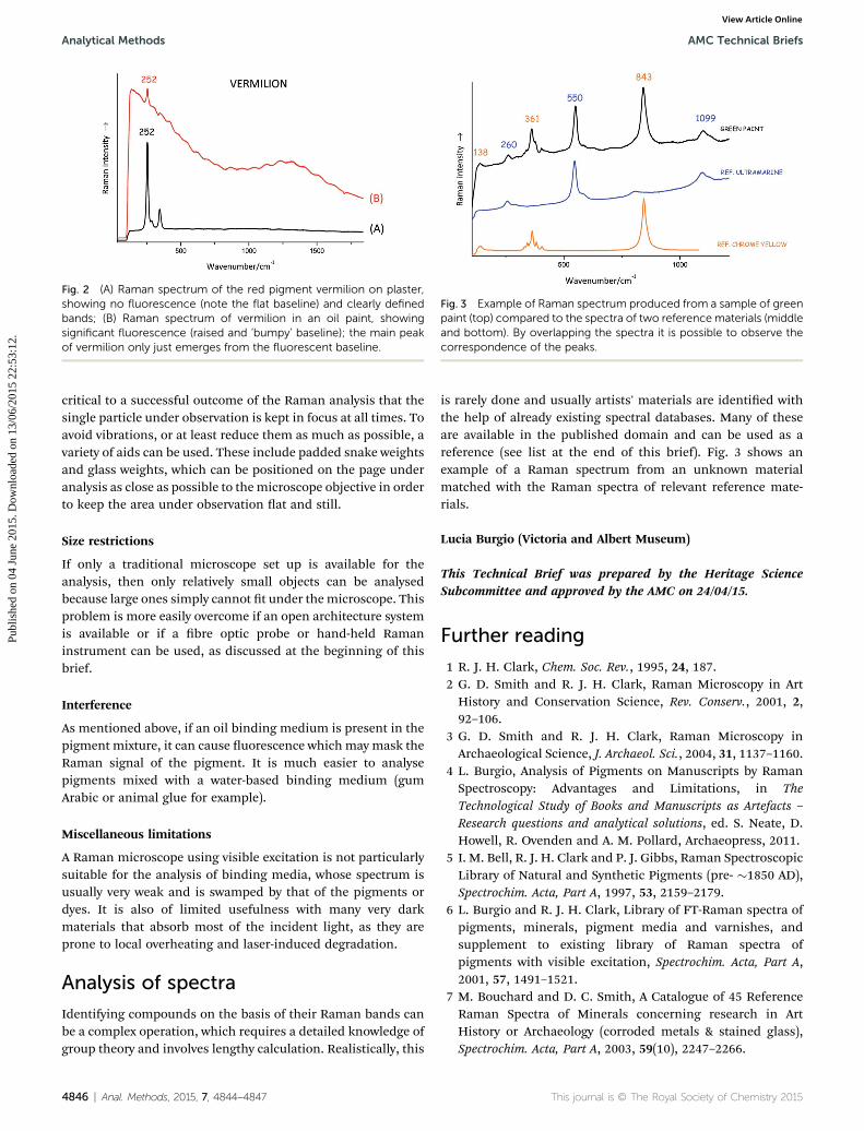
is rarely done and usually artists' materials are identied withthe help of already existing spectral databases. Many of theseare available in the published domain and can be used as areference (see list at the end of this brief). Fig. 3 shows anexample of a Raman spectrum from an unknown materialmatched with the Raman spectra of relevant reference mate-rials.
Lucia Burgio (Victoria and Albert Museum)
This Technical Brief was prepared by the Heritage ScienceSubcommittee and approved by the AMC on 24/04/15.
Further reading
1 R. J. H. Clark, Chem. Soc. Rev., 1995, 24, 187.2 G. D. Smith and R. J. H. Clark, Raman Microscopy in ArtHistory and Conservation Science, Rev. Conserv., 2001, 2,92–106.
3 G. D. Smith and R. J. H. Clark, Raman Microscopy inArchaeological Science, J. Archaeol. Sci., 2004, 31, 1137–1160.
4 L. Burgio, Analysis of Pigments on Manuscripts by RamanSpectroscopy: Advantages and Limitations, in TheTechnological Study of Books and Manuscripts as Artefacts –Research questions and analytical solutions, ed. S. Neate, D.Howell, R. Ovenden and A. M. Pollard, Archaeopress, 2011.
5 I. M. Bell, R. J. H. Clark and P. J. Gibbs, Raman SpectroscopicLibrary of Natural and Synthetic Pigments (pre- �1850 AD),Spectrochim. Acta, Part A, 1997, 53, 2159–2179.
6 L. Burgio and R. J. H. Clark, Library of FT-Raman spectra ofpigments, minerals, pigment media and varnishes, andsupplement to existing library of Raman spectra ofpigments with visible excitation, Spectrochim. Acta, Part A,2001, 57, 1491–1521.
7 M. Bouchard and D. C. Smith, A Catalogue of 45 ReferenceRaman Spectra of Minerals concerning research in ArtHistory or Archaeology (corroded metals & stained glass),Spectrochim. Acta, Part A, 2003, 59(10), 2247–2266.
8 P. Vandenabeele, L. Moens, H. G. M. Edwards and R. Dams,Raman spectroscopic database of azo pigments andapplication to modern art studies, J. Raman Spectrosc.,2000, 31, 507–517.
9 D. Bersani, Raman database of minerals, University ofParma, http://www.s.unipr.it/phevix/ramandb.php,accessed 18 December 2013.
10 P. Vandenabeele, B. Wehling, L. Moens, H. Edwards, M. DeReu and G. Van Hooydonk, Analysis with micro-Ramanspectroscopy of natural organic binding media andvarnishes used in art, Anal. Chim. Acta, 2000, 407, 261–274.
11 N. C. Scherrer, S. Zumbuehl, F. Delavy, A. Fritsch andR. Kuehnen, Synthetic organic pigments of the 20th and21st century relevant to artist's paints: Raman spectrareference collection, Spectrochim. Acta, Part A, 2009, 73(3),403–580.
12 P. Ropret, S. A. Centeno and P. Bukovec, Ramanidentication of yellow synthetic organic pigments in
modern and contemporary paintings: Reference spectraand case studies, Spectrochim. Acta, Part A, 2008, 69, 486–497.
13 E. Huang, Raman spectroscopy study of 15 gem minerals, J.Geol. Soc. China, 1999, 42, 301–308.
14 C. M. Schmidt and K. A. Trentelman, 1064 dispersive Ramanmicro-spectroscopy for the in situ identication of organicred colorants, e-Preserv. Sci., 2009, 6, 10–21.