RAN proteins and RNA foci from antisense transcripts in C9ORF72 ALS and frontotemporal dementia Tao Zu a,b,1 , Yuanjing Liu a,b,1 , Monica Bañez-Coronel a,b,2 , Tammy Reid a,b,2 , Olga Pletnikova c , Jada Lewis d , Timothy M. Miller e , Matthew B. Harms e , Annet E. Falchook f , S. H. Subramony a,f , Lyle W. Ostrow g , Jeffrey D. Rothstein g , Juan C. Troncoso c , and Laura P. W. Ranum a,b,f,h,3 b Department of Molecular Genetics and Microbiology, f Department of Neurology, and d Department of Neuroscience, a Center for NeuroGenetics, h Genetics Institute, College of Medicine, University of Florida, Gainesville, FL 32610; c Department of Pathology and g Department of Neurology, The Johns Hopkins University School of Medicine, Baltimore, MD 21205; and e Department of Neurology, Washington University in St. Louis, St. Louis, MO 63110 Edited* by Don W. Cleveland, University of California, San Diego, La Jolla, CA, and approved October 31, 2013 (received for review August 16, 2013) The finding that a GGGGCC (G 4 C 2 ) hexanucleotide repeat expan- sion in the chromosome 9 ORF 72 (C9ORF72) gene is a common cause of amyotrophic lateral sclerosis (ALS) and frontotemporal dementia (FTD) links ALS/FTD to a large group of unstable micro- satellite diseases. Previously, we showed that microsatellite ex- pansion mutations can be bidirectionally transcribed and that these mutations express unexpected proteins by a unique mecha- nism, repeat-associated non-ATG (RAN) translation. In this study, we show that C9ORF72 antisense transcripts are elevated in the brains of C9ORF72 expansion-positive [C9(+)] patients, and anti- sense GGCCCC (G 2 C 4 ) repeat-expansion RNAs accumulate in nuclear foci in brain. Additionally, sense and antisense foci accumulate in blood and are potential biomarkers of the disease. Furthermore, we show that RAN translation occurs from both sense and antisense expansion transcripts, resulting in the expression of six RAN proteins (antisense: Pro-Arg, Pro-Ala, Gly-Pro; and sense: Gly-Ala, Gly-Arg, Gly-Pro). These proteins accumulate in cytoplasmic aggregates in affected brain regions, including the frontal and motor cortex, hip- pocampus, and spinal cord neurons, with some brain regions show- ing dramatic RAN protein accumulation and clustering. The finding that unique antisense G 2 C 4 RNA foci and three unique antisense RAN proteins accumulate in patient tissues indicates that bidirec- tional transcription of expanded alleles is a fundamental pathologic feature of C9ORF72 ALS/FTD. Additionally, these findings suggest the need to test therapeutic strategies that target both sense and antisense RNAs and RAN proteins in C9ORF72 ALS/FTD, and to more broadly consider the role of antisense expression and RAN trans- lation across microsatellite expansion diseases. cytoplasmic inclusions | clustered aggregates | noncoding RNA T he chromosome 9p21-linked form of amyotrophic lateral sclerosis (ALS) and frontotemporal dementia (FTD), the most common cause of familial FTD and ALS identified to date, is caused by an expanded GGGGCC (G 4 C 2 ) hexanucleotide repeat in intron 1 of chromosome 9 ORF 72 (C9ORF72) (1, 2). The C9ORF72 mutation is found in 40% of familial and 7% of sporadic ALS cases and 21% of familial and 5% of sporadic FTD patients (3). The discovery of the C9ORF72 expansion has gen- erated substantial excitement because it connects ALS and FTD to a large group of disorders caused by microsatellite expansion mutations (4). Traditionally, microsatellite expansion mutations located in predicted coding and noncoding regions were thought to cause disease by protein gain- or loss-of-function or RNA gain-of-function mechanisms (4). Protein loss-of-function has been proposed to underlie C9ORF72-driven ALS/FTD because the expansion muta- tion leads to decreased levels of variant 1 transcripts and potential decreases in C9ORF72 protein expression (1, 5). Additionally, be- cause the C9ORF72 G 4 C 2 expansion mutation is located in an in- tron, several studies have pursued the hypothesis that C9-linked ALS/FTD results from a toxic RNA gain-of-function mecha- nism in which G 4 C 2 expansion RNAs sequester important cellular factors in nuclear RNA foci. Multiple G 4 C 2 RNA binding proteins have been identified, but so far there is no demonstration that any of these candidates directly bind endog- enous expansion transcripts or colocalize with RNA foci observed in patient cells or autopsy tissue (6–9). Repeat-associated non-ATG (RAN) translation, which we initially discovered in spinocerebellar ataxia type 8 (SCA8) and myotonic dystrophy type 1 (DM1) (10), has also been proposed for C9ORF72 ALS/FTD (11, 12) and Fragile X-associated tremor ataxia syndrome (13). In this mechanism, hairpin-forming microsatellite expansion transcripts express proteins in one or more reading frames without an AUG-initiation codon (10). A variety of names have recently been ascribed to these RAN- translated proteins (e.g., homopolymeric, dipeptide, RANT). We propose that all proteins expressed across microsatellite ex- pansion mutations in the absence of an ATG-initiation codon be referred to as RAN proteins to prevent confusion, because additional expansion mutations that undergo RAN translation are identified. In C9ORF72 ALS/FTD, RAN proteins expressed from G 4 C 2 sense transcripts were shown to accumulate as protein aggregates in C9(+) autopsy tissue (11, 12). A strength of these studies, Significance A GGGGCC expansion mutation located in intron 1 of chromo- some 9 ORF 72 (C9ORF72) was recently described as a common cause of familial amyotrophic lateral sclerosis/frontotemporal dementia (ALS/FTD). We show that this single mutation results in the accumulation of sense and antisense RNA foci plus six expansion proteins expressed by repeat-associated non-ATG (RAN) translation. RNAs accumulate in nuclear foci and the RAN proteins form cytoplasmic aggregates in neurons that often cluster in affected brain regions. These results indicate that bi- directional transcription and RAN translation are fundamental pathologic features of C9ORF72 ALS/FTD. Additionally these data have broad implications that change our understanding of how microsatellite expansion mutations are expressed in pa- tient cells and how they cause disease. Author contributions: T.Z., Y.L., M.B.-C., T.R., J.C.T., and L.P.W.R. designed research; T.Z., Y.L., M.B.-C., and T.R. performed research; O.P., J.L., T.M.M., M.B.H., A.E.F., S.H.S., L.W.O., J.D.R., and L.P.W.R. contributed new reagents/analytic tools; T.Z., Y.L., M.B.-C., T.R., J.C.T., and L.P.W.R. analyzed data; and T.Z., Y.L., M.B.-C., J.C.T., and L.P.W.R. wrote the paper. Conflict of interest statement: T.Z. and L.P.W.R. are listed as inventors on pending patents on RAN proteins. *This Direct Submission article had a prearranged editor. Freely available online through the PNAS open access option. 1 T.Z. and Y.L. contributed equally to this work. 2 M.B.-C. and T.R. contributed equally to this work. 3 To whom correspondence should be addressed. E-mail: ranum@ufl.edu. This article contains supporting information online at www.pnas.org/lookup/suppl/doi:10. 1073/pnas.1315438110/-/DCSupplemental. E4968–E4977 | PNAS | Published online November 18, 2013 www.pnas.org/cgi/doi/10.1073/pnas.1315438110
Transcript
RAN proteins and RNA foci from antisense transcriptsin C9ORF72 ALS and frontotemporal dementiaTao Zua,b,1, Yuanjing Liua,b,1, Monica Bañez-Coronela,b,2, Tammy Reida,b,2, Olga Pletnikovac, Jada Lewisd,Timothy M. Millere, Matthew B. Harmse, Annet E. Falchookf, S. H. Subramonya,f, Lyle W. Ostrowg,Jeffrey D. Rothsteing, Juan C. Troncosoc, and Laura P. W. Ranuma,b,f,h,3
bDepartment of Molecular Genetics and Microbiology, fDepartment of Neurology, and dDepartment of Neuroscience, aCenter for NeuroGenetics, hGeneticsInstitute, College of Medicine, University of Florida, Gainesville, FL 32610; cDepartment of Pathology and gDepartment of Neurology, The Johns HopkinsUniversity School of Medicine, Baltimore, MD 21205; and eDepartment of Neurology, Washington University in St. Louis, St. Louis, MO 63110
Edited* by Don W. Cleveland, University of California, San Diego, La Jolla, CA, and approved October 31, 2013 (received for review August 16, 2013)
The finding that a GGGGCC (G4C2) hexanucleotide repeat expan-sion in the chromosome 9 ORF 72 (C9ORF72) gene is a commoncause of amyotrophic lateral sclerosis (ALS) and frontotemporaldementia (FTD) links ALS/FTD to a large group of unstable micro-satellite diseases. Previously, we showed that microsatellite ex-pansion mutations can be bidirectionally transcribed and thatthese mutations express unexpected proteins by a unique mecha-nism, repeat-associated non-ATG (RAN) translation. In this study,we show that C9ORF72 antisense transcripts are elevated in thebrains of C9ORF72 expansion-positive [C9(+)] patients, and anti-sense GGCCCC (G2C4) repeat-expansion RNAs accumulate in nuclearfoci in brain. Additionally, sense and antisense foci accumulate inblood and are potential biomarkers of the disease. Furthermore, weshow that RAN translation occurs from both sense and antisenseexpansion transcripts, resulting in the expression of six RAN proteins(antisense: Pro-Arg, Pro-Ala, Gly-Pro; and sense: Gly-Ala, Gly-Arg,Gly-Pro). These proteins accumulate in cytoplasmic aggregates inaffected brain regions, including the frontal and motor cortex, hip-pocampus, and spinal cord neurons, with some brain regions show-ing dramatic RAN protein accumulation and clustering. The findingthat unique antisense G2C4 RNA foci and three unique antisenseRAN proteins accumulate in patient tissues indicates that bidirec-tional transcription of expanded alleles is a fundamental pathologicfeature of C9ORF72 ALS/FTD. Additionally, these findings suggestthe need to test therapeutic strategies that target both sense andantisense RNAs and RAN proteins in C9ORF72 ALS/FTD, and to morebroadly consider the role of antisense expression and RAN trans-lation across microsatellite expansion diseases.
The chromosome 9p21-linked form of amyotrophic lateralsclerosis (ALS) and frontotemporal dementia (FTD), the
most common cause of familial FTD and ALS identified to date,is caused by an expanded GGGGCC (G4C2) hexanucleotiderepeat in intron 1 of chromosome 9 ORF 72 (C9ORF72) (1, 2).The C9ORF72 mutation is found in 40% of familial and 7% ofsporadic ALS cases and 21% of familial and 5% of sporadic FTDpatients (3). The discovery of the C9ORF72 expansion has gen-erated substantial excitement because it connects ALS and FTDto a large group of disorders caused by microsatellite expansionmutations (4).Traditionally, microsatellite expansion mutations located in
predicted coding and noncoding regions were thought to causedisease by protein gain- or loss-of-function or RNA gain-of-functionmechanisms (4). Protein loss-of-function has been proposed tounderlie C9ORF72-driven ALS/FTD because the expansion muta-tion leads to decreased levels of variant 1 transcripts and potentialdecreases in C9ORF72 protein expression (1, 5). Additionally, be-cause the C9ORF72 G4C2 expansion mutation is located in an in-tron, several studies have pursued the hypothesis that C9-linkedALS/FTD results from a toxic RNA gain-of-function mecha-nism in which G4C2 expansion RNAs sequester important
cellular factors in nuclear RNA foci. Multiple G4C2 RNAbinding proteins have been identified, but so far there is nodemonstration that any of these candidates directly bind endog-enous expansion transcripts or colocalize with RNA foci observedin patient cells or autopsy tissue (6–9).Repeat-associated non-ATG (RAN) translation, which we
initially discovered in spinocerebellar ataxia type 8 (SCA8) andmyotonic dystrophy type 1 (DM1) (10), has also been proposedfor C9ORF72 ALS/FTD (11, 12) and Fragile X-associatedtremor ataxia syndrome (13). In this mechanism, hairpin-formingmicrosatellite expansion transcripts express proteins in one ormore reading frames without an AUG-initiation codon (10). Avariety of names have recently been ascribed to these RAN-translated proteins (e.g., homopolymeric, dipeptide, RANT). Wepropose that all proteins expressed across microsatellite ex-pansion mutations in the absence of an ATG-initiation codonbe referred to as RAN proteins to prevent confusion, becauseadditional expansion mutations that undergo RAN translationare identified.In C9ORF72 ALS/FTD, RAN proteins expressed from G4C2
sense transcripts were shown to accumulate as protein aggregatesin C9(+) autopsy tissue (11, 12). A strength of these studies,
Significance
A GGGGCC expansion mutation located in intron 1 of chromo-some 9 ORF 72 (C9ORF72) was recently described as a commoncause of familial amyotrophic lateral sclerosis/frontotemporaldementia (ALS/FTD). We show that this single mutation resultsin the accumulation of sense and antisense RNA foci plus sixexpansion proteins expressed by repeat-associated non-ATG(RAN) translation. RNAs accumulate in nuclear foci and the RANproteins form cytoplasmic aggregates in neurons that oftencluster in affected brain regions. These results indicate that bi-directional transcription and RAN translation are fundamentalpathologic features of C9ORF72 ALS/FTD. Additionally thesedata have broad implications that change our understanding ofhow microsatellite expansion mutations are expressed in pa-tient cells and how they cause disease.
Author contributions: T.Z., Y.L., M.B.-C., T.R., J.C.T., and L.P.W.R. designed research; T.Z.,Y.L., M.B.-C., and T.R. performed research; O.P., J.L., T.M.M., M.B.H., A.E.F., S.H.S., L.W.O.,J.D.R., and L.P.W.R. contributed new reagents/analytic tools; T.Z., Y.L., M.B.-C., T.R., J.C.T.,and L.P.W.R. analyzed data; and T.Z., Y.L., M.B.-C., J.C.T., and L.P.W.R. wrote the paper.
Conflict of interest statement: T.Z. and L.P.W.R. are listed as inventors on pending patentson RAN proteins.
*This Direct Submission article had a prearranged editor.
Freely available online through the PNAS open access option.1T.Z. and Y.L. contributed equally to this work.2M.B.-C. and T.R. contributed equally to this work.3To whom correspondence should be addressed. E-mail: [email protected].
This article contains supporting information online at www.pnas.org/lookup/suppl/doi:10.1073/pnas.1315438110/-/DCSupplemental.
E4968–E4977 | PNAS | Published online November 18, 2013 www.pnas.org/cgi/doi/10.1073/pnas.1315438110
which used antibodies directed against RAN-predicted GR, GP,and GA repeat motifs, is that they provide a potential link be-tween the mutation and known pathologic features of C9ORF72ALS/FTD. A potential weakness is that the antibodies used weredirected only against the predicted repeat motifs, which are alsofound in unrelated endogenous proteins (14–16).Although it is now clear that a growing number of expansion
mutations are bidirectionally transcribed (17–22), nearly all re-search on these disorders has focused on understanding theimpact of expansion mutations expressed from the sense direction.Our earlier studies on SCA8 and DM1 CAG•CTG expansionmutations showed that sense and antisense expansion transcriptsare expressed and RAN proteins accumulate in patient tissues (10).Mori et al. also presented evidence by RT-PCR that C9ORF72antisense transcripts upstream of the expansion mutation areexpressed in patient brains (12). Here we show that C9ORF72ALS/FTD antisense transcripts containing the GGCCCC (G2C4) ex-pansion accumulate in patient brains as nuclear foci. Additionally,we developed a panel of antibodies directed to both the repeatmotifs and unique C-terminal regions, and demonstrate that bothsense and antisense RAN proteins accumulate in C9ORF72 patientCNS autopsy tissue. The identification of antisense G2C4 RNA fociand three unique antisense RAN proteins in C9ORF72 brainssuggests that bidirectional transcription and RAN translation arefundamental pathologic features of C9ORF72 ALS/FTD.
ResultsAntisense RNA Foci in C9ORF72-Expansion Patients. Based on ouridentification of bidirectional transcription and RAN trans-lation in SCA8 (10, 21), we performed a series of experimentsto test the hypotheses that antisense (AS) C9ORF72 expansiontranscripts form AS-G2C4 RNA foci and express antisense pro-teins by RAN translation or from short antisense open-readingframes (AS-ORFs).First, we confirmed C9ORF72 antisense transcripts are expressed
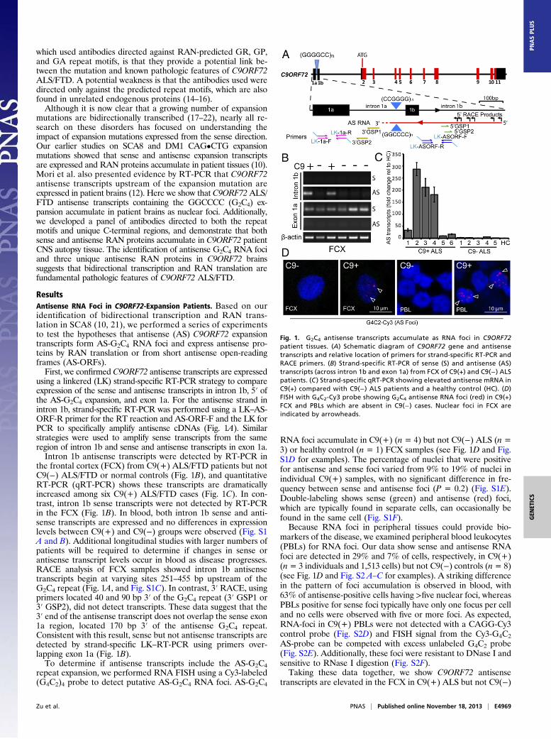
using a linkered (LK) strand-specific RT-PCR strategy to compareexpression of the sense and antisense transcripts in intron 1b, 5′ ofthe AS-G2C4 expansion, and exon 1a. For the antisense strand inintron 1b, strand-specific RT-PCR was performed using a LK–AS-ORF-R primer for the RT reaction and AS-ORF-F and the LK forPCR to specifically amplify antisense cDNAs (Fig. 1A). Similarstrategies were used to amplify sense transcripts from the sameregion of intron 1b and sense and antisense transcripts in exon 1a.Intron 1b antisense transcripts were detected by RT-PCR in
the frontal cortex (FCX) from C9(+) ALS/FTD patients but notC9(−) ALS/FTD or normal controls (Fig. 1B), and quantitativeRT-PCR (qRT-PCR) shows these transcripts are dramaticallyincreased among six C9(+) ALS/FTD cases (Fig. 1C). In con-trast, intron 1b sense transcripts were not detected by RT-PCRin the FCX (Fig. 1B). In blood, both intron 1b sense and anti-sense transcripts are expressed and no differences in expressionlevels between C9(+) and C9(−) groups were observed (Fig. S1A and B). Additional longitudinal studies with larger numbers ofpatients will be required to determine if changes in sense orantisense transcript levels occur in blood as disease progresses.RACE analysis of FCX samples showed intron 1b antisensetranscripts begin at varying sites 251–455 bp upstream of theG2C4 repeat (Fig. 1A, and Fig. S1C). In contrast, 3′ RACE, usingprimers located 40 and 90 bp 3′ of the G2C4 repeat (3′ GSP1 or3′ GSP2), did not detect transcripts. These data suggest that the3′ end of the antisense transcript does not overlap the sense exon1a region, located 170 bp 3′ of the antisense G2C4 repeat.Consistent with this result, sense but not antisense transcripts aredetected by strand-specific LK–RT-PCR using primers over-lapping exon 1a (Fig. 1B).To determine if antisense transcripts include the AS-G2C4
repeat expansion, we performed RNA FISH using a Cy3-labeled(G4C2)4 probe to detect putative AS-G2C4 RNA foci. AS-G2C4
RNA foci accumulate in C9(+) (n = 4) but not C9(−) ALS (n =3) or healthy control (n = 1) FCX samples (see Fig. 1D and Fig.S1D for examples). The percentage of nuclei that were positivefor antisense and sense foci varied from 9% to 19% of nuclei inindividual C9(+) samples, with no significant difference in fre-quency between sense and antisense foci (P = 0.2) (Fig. S1E).Double-labeling shows sense (green) and antisense (red) foci,which are typically found in separate cells, can occasionally befound in the same cell (Fig. S1F).Because RNA foci in peripheral tissues could provide bio-
markers of the disease, we examined peripheral blood leukocytes(PBLs) for RNA foci. Our data show sense and antisense RNAfoci are detected in 29% and 7% of cells, respectively, in C9(+)(n= 3 individuals and 1,513 cells) but not C9(−) controls (n= 8)(see Fig. 1D and Fig. S2 A–C for examples). A striking differencein the pattern of foci accumulation is observed in blood, with63% of antisense-positive cells having >five nuclear foci, whereasPBLs positive for sense foci typically have only one focus per celland no cells were observed with five or more foci. As expected,RNA-foci in C9(+) PBLs were not detected with a CAGG-Cy3control probe (Fig. S2D) and FISH signal from the Cy3-G4C2AS-probe can be competed with excess unlabeled G4C2 probe(Fig. S2E). Additionally, these foci were resistant to DNase I andsensitive to RNase I digestion (Fig. S2F).Taking these data together, we show C9ORF72 antisense
transcripts are elevated in the FCX in C9(+) ALS but not C9(−)
Fig. 1. G2C4 antisense transcripts accumulate as RNA foci in C9ORF72patient tissues. (A) Schematic diagram of C9ORF72 gene and antisensetranscripts and relative location of primers for strand-specific RT-PCR andRACE primers. (B) Strand-specific RT-PCR of sense (S) and antisense (AS)transcripts (across intron 1b and exon 1a) from FCX of C9(+) and C9(−) ALSpatients. (C ) Strand-specific qRT-PCR showing elevated antisense mRNA inC9(+) compared with C9(−) ALS patients and a healthy control (HC). (D)FISH with G4C2-Cy3 probe showing G2C4 antisense RNA foci (red) in C9(+)FCX and PBLs which are absent in C9(−) cases. Nuclear foci in FCX areindicated by arrowheads.
Zu et al. PNAS | Published online November 18, 2013 | E4969
ALS or normal controls. We also demonstrate that unique an-tisense transcripts containing the AS-G2C4 expansion mutationare expressed and accumulate in nuclear RNA foci in the C9(+)FCX. Additionally, we show sense and antisense foci accumulatein blood, providing potential biomarkers of C9ORF72 ALS/FTDin a readily accessible tissue.
Dual Immunological Strategy to Detect RAN Proteins. When we firstdescribed RAN translation in SCA8 and DM1, multiple anti-bodies directed against epitope tags, the repeat motif, and theC-terminal regions of these proteins were used (10) to demon-strate their expression. Although two groups recently reportedevidence that RAN proteins are expressed in C9ORF72 ALS/FTD patients (11, 12), both groups relied on a single antibody foreach of the GP, GR, or GA repeat motifs. Because these repeatmotifs are found in a number of other proteins (14–16), we useda dual immunological strategy and developed antibodies thatrecognize the predicted repeat motifs or their correspondingunique C-terminal regions.A schematic diagram showing eight putative C9ORF72 RAN
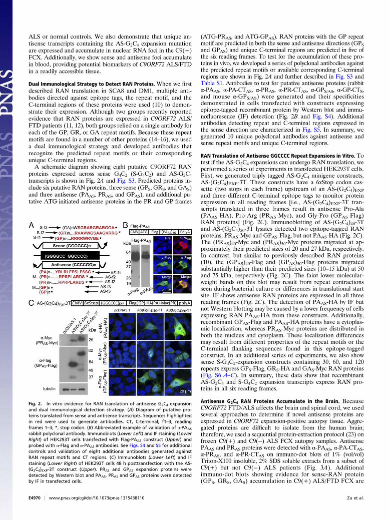
proteins expressed across sense G4C2 (S-G4C2) and AS-G2C4transcripts is shown in Fig. 2A and Fig. S3. Predicted proteins in-clude six putative RAN proteins, three sense (GPS, GRS, and GAS)and three antisense (PAAS, PRAS, and GPAS), and additional pu-tative ATG-initiated antisense proteins in the PR and GP frames
(ATG-PRAS, and ATG-GPAS). RAN proteins with the GP repeatmotif are predicted in both the sense and antisense directions (GPSand GPAS) and unique C-terminal regions are predicted in five ofthe six reading frames. To test for the accumulation of these pro-teins in vivo, we developed a series of polyclonal antibodies againstthe predicted repeat motifs or available corresponding C-terminalregions are shown in Fig. 2A and further described in Fig. S3 andTable S1. Antibodies to test for putative antisense proteins (rabbitα-PAAS, α-PA-CTAS, α-PRAS, α-PR-CTAS, α-GPS/AS, α-GP-CTS,and mouse α-GPS/AS) were generated and their specificitiesdemonstrated in cells transfected with constructs expressingepitope-tagged recombinant protein by Western blot and immu-nofluoresence (IF) detection (Fig. 2B and Fig. S4). Additionalantibodies detecting repeat and C-terminal regions expressed inthe sense direction are characterized in Fig. S5. In summary, wegenerated 10 unique polyclonal antibodies against antisense andsense repeat motifs and unique C-terminal regions.
RAN Translation of Antisense GGCCCC Repeat Expansions in Vitro. Totest if the AS-G2C4 expansions can undergo RAN translation, weperformed a series of experiments in transfected HEK293T cells.First, we generated triply tagged AS-G2C4 minigene constructs,AS-(G2C4)EXP-3T. These constructs have a 6xStop codon cas-sette (two stops in each frame) upstream of an AS-(G2C4)EXPand three different C-terminal epitope tags to monitor proteinexpression in all reading frames [i.e., AS-(G2C4)EXP-3T tran-scripts translated in three frames result in antisense Pro-Ala(PAAS-HA), Pro-Arg (PRAS-Myc), and Gly-Pro (GPAS-Flag)RAN proteins] (Fig. 2C). Immunoblotting of AS-(G2C4)40-3Tand AS-(G2C4)50-3T lysates detected two epitope-tagged RANproteins, PRAS-Myc and GPAS-Flag, but not PAAS-HA (Fig. 2C).The (PRAS)40-Myc and (PRAS)50-Myc proteins migrated at ap-proximately their predicted sizes of 20 and 27 kDa, respectively.In contrast, but similar to previously described RAN proteins(10), the (GPAS)40-Flag and (GPAS)50-Flag proteins migratedsubstantially higher than their predicted sizes (10–15 kDa) at 50and 75 kDa, respectively (Fig. 2C). The faint lower molecular-weight bands on this blot may result from repeat contractionsseen during bacterial culture or differences in translational startsite. IF shows antisense RAN proteins are expressed in all threereading frames (Fig. 2C). The detection of PAAS-HA by IF butnot Western blotting may be caused by a lower frequency of cellsexpressing RAN PAAS-HA from these constructs. Additionally,recombinant GPAS-Flag and PAAS-HA proteins have a cytoplas-mic localization, whereas PRAS-Myc proteins are distributed inboth the nucleus and cytoplasm. These localization differencesmay result from different properties of the repeat motifs or theC-terminal flanking sequences found in this epitope-taggedconstruct. In an additional series of experiments, we also showsense S-G4C2-expansion constructs containing 30, 60, and 120repeats express GPS-Flag, GRS-HA and GAS-Myc RAN proteins(Fig. S6 A–C). In summary, these data show that recombinantAS-G2C4 and S-G4C2 expansion transcripts express RAN pro-teins in all six reading frames.
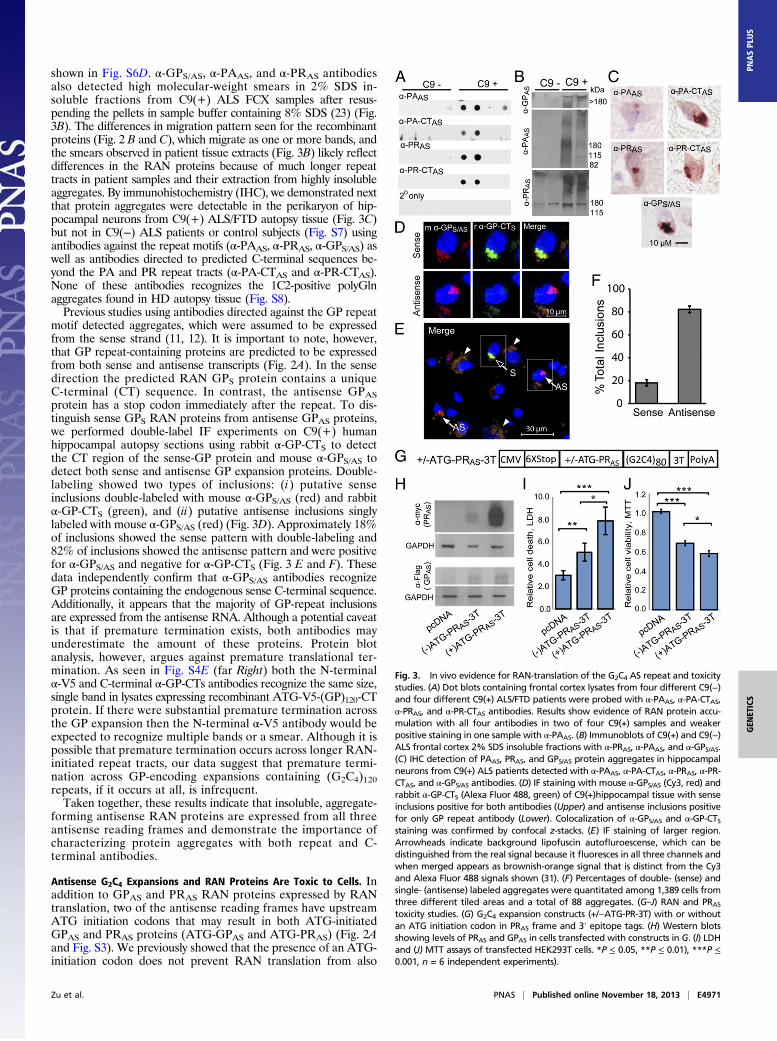
Antisense G2C4 RAN Proteins Accumulate in the Brain. BecauseC9ORF72 FTD/ALS affects the brain and spinal cord, we usedseveral approaches to determine if novel antisense proteins areexpressed in C9ORF72 expansion-positive autopsy tissue. Aggre-gated proteins are difficult to isolate from the human brain;therefore, we used a sequential protein-extraction protocol (23) onfrozen C9(+) and C9(−) ALS FCX autopsy samples. AntisensePAAS and PRAS proteins were detected with α-PAAS, α-PA-CTAS,α-PRAS, and α-PR-CTAS on immuno-dot blots of 1% (vol/vol)Triton-X100 insoluble, 2% SDS soluble extracts from a subset ofC9(+) but not C9(−) ALS patients (Fig. 3A). Additionalimmuno-dot blots showing evidence for sense-RAN protein(GPS, GRS, GAS) accumulation in C9(+) ALS/FTD FCX are
Fig. 2. In vitro evidence for RAN translation of antisense G2C4 expansionand dual immunological detection strategy. (A) Diagram of putative pro-teins translated from sense and antisense transcripts. Sequences highlightedin red were used to generate antibodies. CT, C-terminal; f1–3, readingframes 1–3; *, stop codon. (B) Abbreviated example of validation of α-PAAS
rabbit polyclonal antibody. Immunoblots (Lower Left) and IF staining (LowerRight) of HEK293T cells transfected with Flag-PAAS construct (Upper) andprobed with α-Flag and α-PAAS antibodies. See Figs. S4 and S5 for additionalcontrols and validation of eight additional antibodies generated againstRAN repeat motifs and CT regions. (C) Immunoblots (Lower Left) and IFstaining (Lower Right) of HEK293T cells 48 h posttransfection with the AS-(G2C4)EXP-3T construct (Upper). PRAS and GPAS expansion proteins weredetected by Western blot and PAAS, PRAS and GPAS proteins were detectedby IF in transfected cells.
E4970 | www.pnas.org/cgi/doi/10.1073/pnas.1315438110 Zu et al.
shown in Fig. S6D. α-GPS/AS, α-PAAS, and α-PRAS antibodiesalso detected high molecular-weight smears in 2% SDS in-soluble fractions from C9(+) ALS FCX samples after resus-pending the pellets in sample buffer containing 8% SDS (23) (Fig.3B). The differences in migration pattern seen for the recombinantproteins (Fig. 2 B and C), which migrate as one or more bands, andthe smears observed in patient tissue extracts (Fig. 3B) likely reflectdifferences in the RAN proteins because of much longer repeattracts in patient samples and their extraction from highly insolubleaggregates. By immunohistochemistry (IHC), we demonstrated nextthat protein aggregates were detectable in the perikaryon of hip-pocampal neurons from C9(+) ALS/FTD autopsy tissue (Fig. 3C)but not in C9(−) ALS patients or control subjects (Fig. S7) usingantibodies against the repeat motifs (α-PAAS, α-PRAS, α-GPS/AS) aswell as antibodies directed to predicted C-terminal sequences be-yond the PA and PR repeat tracts (α-PA-CTAS and α-PR-CTAS).None of these antibodies recognizes the 1C2-positive polyGlnaggregates found in HD autopsy tissue (Fig. S8).Previous studies using antibodies directed against the GP repeat
motif detected aggregates, which were assumed to be expressedfrom the sense strand (11, 12). It is important to note, however,that GP repeat-containing proteins are predicted to be expressedfrom both sense and antisense transcripts (Fig. 2A). In the sensedirection the predicted RAN GPS protein contains a uniqueC-terminal (CT) sequence. In contrast, the antisense GPASprotein has a stop codon immediately after the repeat. To dis-tinguish sense GPS RAN proteins from antisense GPAS proteins,we performed double-label IF experiments on C9(+) humanhippocampal autopsy sections using rabbit α-GP-CTS to detectthe CT region of the sense-GP protein and mouse α-GPS/AS todetect both sense and antisense GP expansion proteins. Double-labeling showed two types of inclusions: (i) putative senseinclusions double-labeled with mouse α-GPS/AS (red) and rabbitα-GP-CTS (green), and (ii) putative antisense inclusions singlylabeled with mouse α-GPS/AS (red) (Fig. 3D). Approximately 18%of inclusions showed the sense pattern with double-labeling and82% of inclusions showed the antisense pattern and were positivefor α-GPS/AS and negative for α-GP-CTS (Fig. 3 E and F). Thesedata independently confirm that α-GPS/AS antibodies recognizeGP proteins containing the endogenous sense C-terminal sequence.Additionally, it appears that the majority of GP-repeat inclusionsare expressed from the antisense RNA. Although a potential caveatis that if premature termination exists, both antibodies mayunderestimate the amount of these proteins. Protein blotanalysis, however, argues against premature translational ter-mination. As seen in Fig. S4E (far Right) both the N-terminalα-V5 and C-terminal α-GP-CTs antibodies recognize the same size,single band in lysates expressing recombinant ATG-V5-(GP)120-CTprotein. If there were substantial premature termination acrossthe GP expansion then the N-terminal α-V5 antibody would beexpected to recognize multiple bands or a smear. Although it ispossible that premature termination occurs across longer RAN-initiated repeat tracts, our data suggest that premature termi-nation across GP-encoding expansions containing (G2C4)120repeats, if it occurs at all, is infrequent.Taken together, these results indicate that insoluble, aggregate-
forming antisense RAN proteins are expressed from all threeantisense reading frames and demonstrate the importance ofcharacterizing protein aggregates with both repeat and C-terminal antibodies.
Antisense G2C4 Expansions and RAN Proteins Are Toxic to Cells. Inaddition to GPAS and PRAS RAN proteins expressed by RANtranslation, two of the antisense reading frames have upstreamATG initiation codons that may result in both ATG-initiatedGPAS and PRAS proteins (ATG-GPAS and ATG-PRAS) (Fig. 2Aand Fig. S3). We previously showed that the presence of an ATG-initiation codon does not prevent RAN translation from also
Fig. 3. In vivo evidence for RAN-translation of the G2C4 AS repeat and toxicitystudies. (A) Dot blots containing frontal cortex lysates from four different C9(−)and four different C9(+) ALS/FTD patients were probed with α-PAAS, α-PA-CTAS,α-PRAS, and α-PR-CTAS antibodies. Results show evidence of RAN protein accu-mulation with all four antibodies in two of four C9(+) samples and weakerpositive staining in one sample with α-PAAS. (B) Immunoblots of C9(+) and C9(−)ALS frontal cortex 2% SDS insoluble fractions with α-PRAS, α-PAAS, and α-GPS/AS.(C) IHC detection of PAAS, PRAS, and GPS/AS protein aggregates in hippocampalneurons from C9(+) ALS patients detected with α-PAAS, α-PA-CTAS, α-PRAS, α-PR-CTAS, and α-GPS/AS antibodies. (D) IF staining with mouse α-GPS/AS (Cy3, red) andrabbit α-GP-CTS (Alexa Fluor 488, green) of C9(+)hippocampal tissue with senseinclusions positive for both antibodies (Upper) and antisense inclusions positivefor only GP repeat antibody (Lower). Colocalization of α-GPS/AS and α-GP-CTSstaining was confirmed by confocal z-stacks. (E) IF staining of larger region.Arrowheads indicate background lipofuscin autofluroescense, which can bedistinguished from the real signal because it fluoresces in all three channels andwhen merged appears as brownish-orange signal that is distinct from the Cy3and Alexa Fluor 488 signals shown (31). (F) Percentages of double- (sense) andsingle- (antisense) labeled aggregates were quantitated among 1,389 cells fromthree different tiled areas and a total of 88 aggregates. (G–J) RAN and PRAStoxicity studies. (G) G2C4 expansion constructs (+/−ATG-PR-3T) with or withoutan ATG initiation codon in PRAS frame and 3′ epitope tags. (H) Western blotsshowing levels of PRAS and GPAS in cells transfected with constructs in G. (I) LDHand (J) MTT assays of transfected HEK293T cells. *P ≤ 0.05, **P ≤ 0.01), ***P ≤0.001, n = 6 independent experiments).
Zu et al. PNAS | Published online November 18, 2013 | E4971
occurring in all three reading frames (10). Therefore, GPAS andPRAS proteins may be expressed by both AUG-initiated or RANtranslation. To explore the effects that an ATG-initiation codonhas on RAN protein expression for the G2C4 expansion, wegenerated an additional minigene construct by placing an ATG-initiation codon in front of the AS-G2C4 repeat (Fig. 3G). Weselected the PRAS frame for analysis because an ATG initiationcodon naturally occurs in this reading frame. Western blottingshows that HEK293T cells transfected with (+)ATG-PR-3T ex-press substantially higher levels of PRAS protein compared with(−)ATG-PRAS-3T–transfected cells (Fig. 3H). In contrast, qRT-PCR and protein blotting showed transcript levels (Fig. S9A) andlevels of RAN-translated GPAS (Fig. 3H) were comparable.Similar to Fig. 2C, RAN-translated PAAS was not detectable byprotein blot. We then tested the effects of these constructs on cellviability using complementary assays; lactate dehydrogenase(LDH) detection and methylthiazol tetrazolium (MTT). TheLDH assay measures the amount of LDH released into the me-dium and is an indicator of lost membrane integrity and celldeath. For the LDH assay, cells transfected with the (−)ATG-PRAS-3T or (+)ATG-PRAS-3T constructs showed 1.9- and 2.9-fold increases in cell death compared with vector control cells(P = 0.008 and 0.001), respectively (Fig. 3I). Additionally,(+)ATG-PRAS-3T–transfected cells, which express elevated levelsof PRAS protein, show a 1.5-fold increase in cell death comparedwith cells transfected with the (–)ATG-PRAS-3T construct (P =0.034) (Fig. 3I). The MTT assay, which measures cellular met-abolic activity of NAD(P)H-dependent oxidoreductase enzymesand reflects the number of viable cells present, showed similarresults. Cells transfected with (–)ATG-PRAS-3T and (+)ATG-PRAS-3T constructs showed dramatic decreases in the number ofmetabolically active cells [33% (P < 0.00001) and 43% (P <0.00001), respectively], compared with untreated cells or emptyvector controls (Fig. 3J). Additionally, elevated PRAS expressionin cells transfected with (+)ATG-PRAS-3T had significantly lowerlevels of metabolic activity compared with (–)ATG-PRAS-3T cells(P < 0.05). By light microscopy, cell detachment and changes incell morphology were evident in (–)ATG-PRAS-3T compared withcontrol cells. These phenotypes were worse in (+)ATG-PRAS-3Tcells, which express elevated levels of PRAS (Fig. S9 B–D). Asimilar study using (+/−)ATG-GPs-3T–transfected cells demon-strates that GPs proteins are also toxic to cells (Fig. S9 E–I).Taken together, these data demonstrate that: (i) the G2C4
and G4C2 expansion mutations are toxic to cells; (ii) increasedPRAS and GPS proteins expressed in cells transfected with the(+)ATG-PRAS-3T or (+)ATG-GPS-3T constructs increase celltoxicity and death above levels caused by any DNA, RNA, andRAN protein effects of the (−)ATG-PRAS-3T or (−)ATG-GPS-3T constructs. Therefore, the PRAS and GPS proteins are in-trinsically toxic to cells.
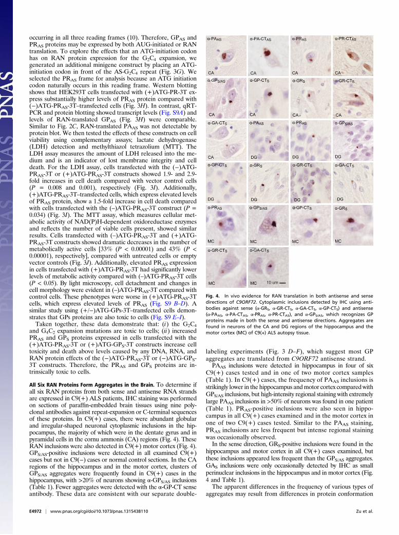
All Six RAN Proteins Form Aggregates in the Brain. To determine ifall six RAN proteins from both sense and antisense RNA strandsare expressed in C9(+) ALS patients, IHC staining was performedon sections of paraffin-embedded brain tissues using nine poly-clonal antibodies against repeat-expansion or C-terminal sequencesof these proteins. In C9(+) cases, there were abundant globularand irregular-shaped neuronal cytoplasmic inclusions in the hip-pocampus, the majority of which were in the dentate gyrus and inpyramidal cells in the cornu ammonis (CA) regions (Fig. 4). TheseRAN inclusions were also detected in C9(+) motor cortex (Fig. 4).GPS/AS-positive inclusions were detected in all examined C9(+)cases but not in C9(−) cases or normal control sections. In the CAregions of the hippocampus and in the motor cortex, clusters ofGPS/AS aggregates were frequently found in C9(+) cases in thehippocampus, with >20% of neurons showing α-GPS/AS inclusions(Table 1). Fewer aggregates were detected with the α-GP-CT senseantibody. These data are consistent with our separate double-
labeling experiments (Fig. 3 D–F), which suggest most GPaggregates are translated from C9ORF72 antisense strand.PAAS inclusions were detected in hippocampus in four of six
C9(+) cases tested and in one of two motor cortex samples(Table 1). In C9(+) cases, the frequency of PAAS inclusions isstrikingly lower in the hippocampus andmotor cortex comparedwithGPS/AS inclusions, but high-intensity regional staining with extremelylarge PAAS inclusions in >50% of neurons was found in one patient(Table 1). PRAS-positive inclusions were also seen in hippo-campus in all C9(+) cases examined and in the motor cortex inone of two C9(+) cases tested. Similar to the PAAS staining,PRAS inclusions are less frequent but intense regional stainingwas occasionally observed.In the sense direction, GRS-positive inclusions were found in the
hippocampus and motor cortex in all C9(+) cases examined, butthese inclusions appeared less frequent than the GPS/AS aggregates.GAS inclusions were only occasionally detected by IHC as smallperinuclear inclusions in the hippocampus and in motor cortex (Fig.4 and Table 1).The apparent differences in the frequency of various types of
aggregates may result from differences in protein conformation
Fig. 4. In vivo evidence for RAN translation in both antisense and sensedirections of C9ORF72. Cytoplasmic inclusions detected by IHC using anti-bodies against sense (α-GRS, α-GR-CTS, α-GA-CTS, α-GP-CTS) and antisense(α-PAAS, α-PA-CTAS, α-PRAS, α-PR-CTAS), and α-GPS/AS, which recognizes GPproteins made in both the sense and antisense directions. Aggregates arefound in neurons of the CA and DG regions of the hippocampus and themotor cortex (MC) of C9(+) ALS autopsy tissue.
E4972 | www.pnas.org/cgi/doi/10.1073/pnas.1315438110 Zu et al.
and epitope availability or differences in the affinities of theseantibodies, which were designed to different epitopes. Differencesin methods, antibody affinity, and epitope availability may alsoexplain why previously reported GA aggregates detected withα-GA antibodies (12) were the most frequent type of sense ag-gregate, in contrast to our findings that α-GPS/AS aggregates areabundant and α-GA-CTS aggregates are rare. Taken together,our data show that all six RAN proteins form aggregates in theC9(+) autopsy brains.
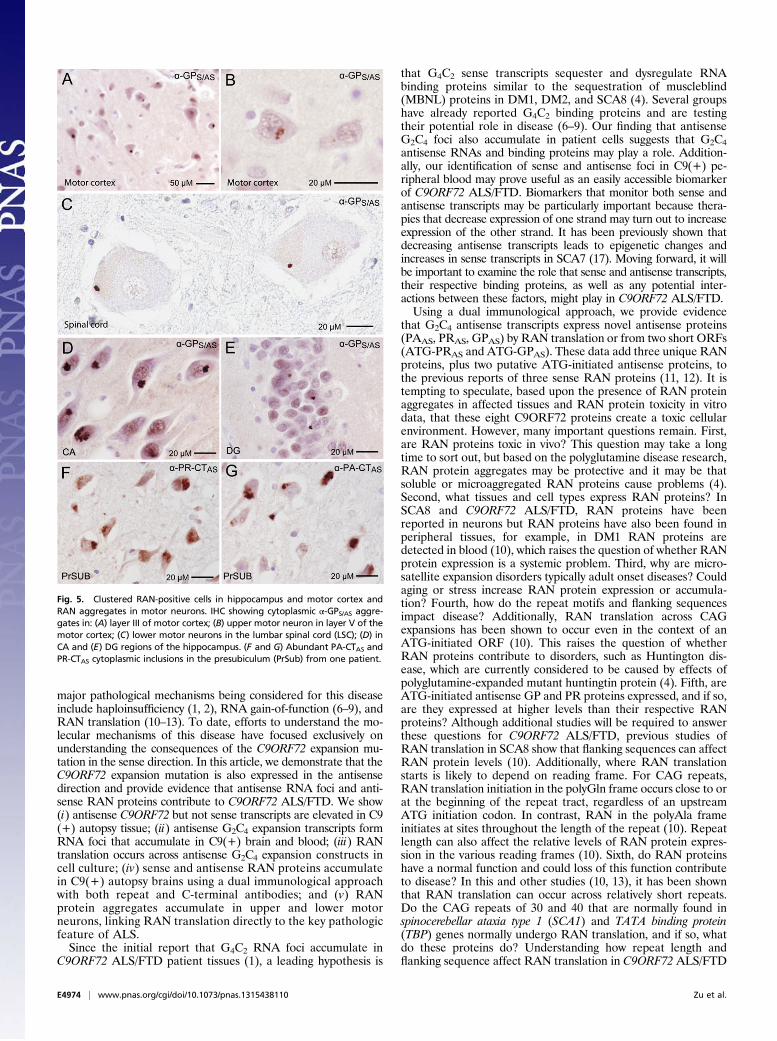
Inclusions of RAN Proteins in Upper and Lower Motor Neurons. Acentral feature of ALS is the gradual degeneration and death ofupper motor neurons in the motor cortex and lower motorneurons in the brainstem and spinal cord. To test if RAN pro-teins accumulate in upper and lower motor neurons, we per-formed IHC using all nine antibodies against predicted proteinsin both sense and antisense directions. In C9(+) cases, abundantGPS/AS neuronal cytoplasmic inclusions were seen in all layers ofthe motor cortex, with frequent GPS/AS aggregates in pyramidalneurons of layer III and throughout layer V (Fig. 5A and Fig.S10). Although cell death and atrophy made motor neurons inlayer V difficult to identify, GPS/AS inclusions in he remainingupper motor neurons were found (Fig. 5B). Additionally, PAAS-,PRAS-, GRS-, and GAS-positive inclusions were also found in themotor cortex (Fig. 4 and Table 1). In a similar series of experi-
ments performed in spinal cord sections, we detected GPS/ASaggregates in all three cases examined and aggregates in lowermotor neurons in two of three C9(+) patients but not in C9(−)ALS cases or normal controls (Fig. 5C and Table 1). AlthoughGPS/AS aggregates seen in most cells have a perinuclar location,the cytoplasmic aggregates in the lower motor neurons werelocated away from the nucleus in the periphery. Surprisingly,PAAS, PRAS, GRS, and GAS proteins were not detected in thethree C9(+) spinal cord samples we examined; this may resultfrom lower overall RAN protein expression or less-frequentaggregation in these cells. Alternatively, it is possible that RANprotein aggregates are less-frequently detected in spinal cordneurons because they are more vulnerable and RAN proteintoxicity causes the substantial motor neuron loss in C9(+) ALSpatients. This report of RAN protein accumulation in lowermotor neurons is unique. The identification of GP-aggregates inboth upper and lower motor neurons links C9 RAN-protein accu-mulation to the neurons that are selectively vulnerable in ALS.
High-Density Clustering of Cells with RAN-Protein Aggregates. Bothsense and antisense proteins accumulate in neurons of C9ORF72autopsy brains. In general, two types of aggregation patternswere observed: (i) isolated cells with cytoplasmic aggregates and(ii) high-density clusters of cells with cytoplasmic aggregates inwhich ∼10 to more than 50% of neurons are positive. Clusteredaggregates were most frequently detected for GP and were foundin the CA1–4 and dentate gyrus (DG) of the hippocampus (Fig. 5D and E). The clustered GP aggregates in the DG were smallerand less frequent than the large cytoplasmic aggregates in CAregions. Additional clustered GP aggregates were frequentlyfound in subiculum and presubiculum of the hippocampus as wellas the motor cortex. Immunostaining of serial sections showed thatmultiple RAN proteins are often found in the same region. Forexample, intense clustered staining for PA, PR, GP, GA, and GRproteins is found in the same region of the presubiculum in serialsections from one C9(+) patient (Fig. 5 F and G).Immunostaining for PA showed that some brain regions have
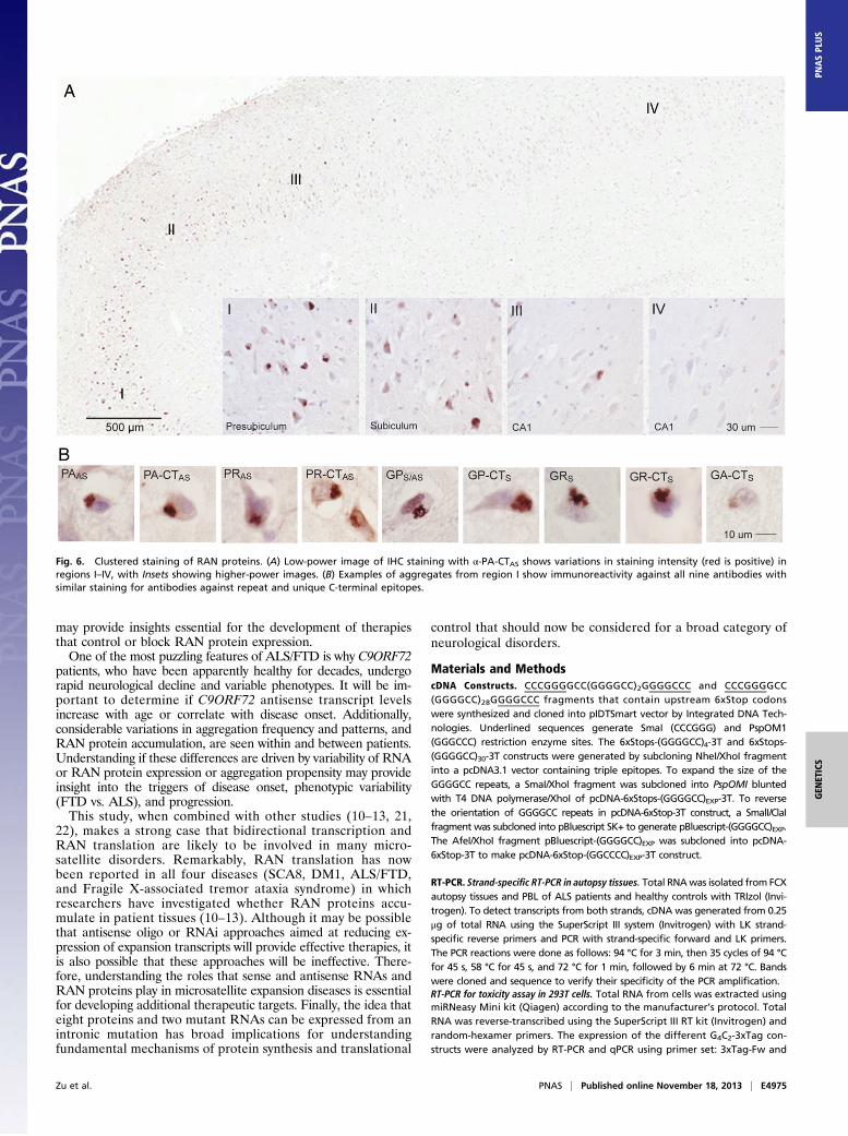
abundant aggregates, whereas other regions in the same section arerelatively spared. For example, Fig. 6A illustrates a gradient of PAinclusions (presubiculum > subiculum > CA1) across hippocampalregions in a single section in one patient. PA inclusions in thispatient were numerous (>50% of neurons) in the presubiculum (I),moderate in subiculum (II), and rare in CA1 hippocampal regions(III and IV). Consistent with the focal regional staining seen in thissection, PA staining was not detected in sections from a separateblock of hippocampal tissue taken from the same patient. Thesedata suggest expression of the PA RAN protein is variable fromcell to cell or that aggregation of PA in one cell triggers aggregationin neighboring cells, as has been proposed in Parkinson andAlzheimer’s disease (24–30).Next, we used serial sections from this C9(+) case to show that
antibodies directed against both the repeat motifs (α-PA, α-PR,α-GP, α-GR) and corresponding C-terminal regions (α-PA-CT,α-PR-CT, α-GP-CT, α-GR-CT, α-GA-CT) detect aggregates inthe same densely staining region of the presubiculum (region I)(Fig. 6B). These results show both sense and antisense RANprotein aggregates accumulate in this region. The detection ofsimilar aggregates using antibodies that recognize either the re-peat motifs or specific C-terminal regions adds substantial con-fidence that these antibodies are truly recognizing proteinsexpressed across both the antisense G2C4 and sense G4C2 ex-pansion transcripts, and provides new tools to understand thebiological impact of RAN translation in C9ORF72 ALS/FTD.
DiscussionThere has been much excitement about the finding that an intronicmicrosatellite expansion mutation in C9ORF72 causes a commonform of both familial and sporadic ALS/FTD (1, 2). The three
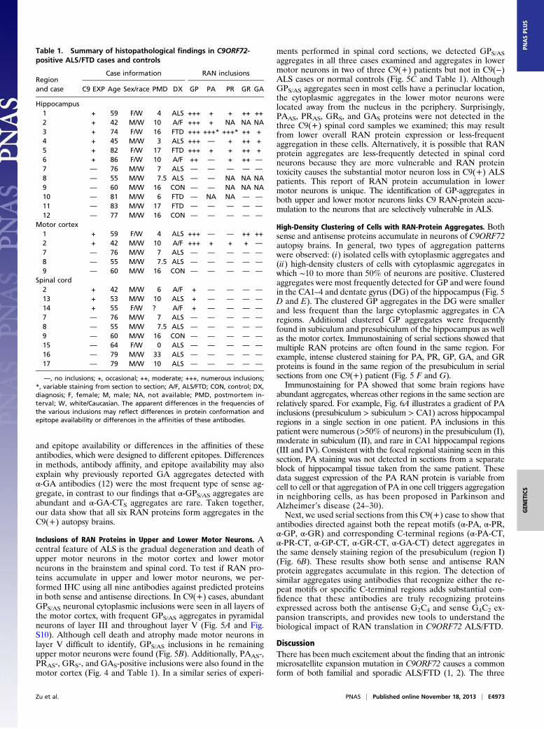
Table 1. Summary of histopathological findings in C9ORF72-positive ALS/FTD cases and controls
—, no inclusions; +, occasional; ++, moderate; +++, numerous inclusions;*, variable staining from section to section; A/F, ALS/FTD; CON, control; DX,diagnosis; F, female; M, male; NA, not available; PMD, postmortem in-terval; W, white/Caucasian. The apparent differences in the frequencies ofthe various inclusions may reflect differences in protein conformation andepitope availability or differences in the affinities of these antibodies.
Zu et al. PNAS | Published online November 18, 2013 | E4973
major pathological mechanisms being considered for this diseaseinclude haploinsufficiency (1, 2), RNA gain-of-function (6–9), andRAN translation (10–13). To date, efforts to understand the mo-lecular mechanisms of this disease have focused exclusively onunderstanding the consequences of the C9ORF72 expansion mu-tation in the sense direction. In this article, we demonstrate that theC9ORF72 expansion mutation is also expressed in the antisensedirection and provide evidence that antisense RNA foci and anti-sense RAN proteins contribute to C9ORF72 ALS/FTD. We show(i) antisense C9ORF72 but not sense transcripts are elevated in C9(+) autopsy tissue; (ii) antisense G2C4 expansion transcripts formRNA foci that accumulate in C9(+) brain and blood; (iii) RANtranslation occurs across antisense G2C4 expansion constructs incell culture; (iv) sense and antisense RAN proteins accumulatein C9(+) autopsy brains using a dual immunological approachwith both repeat and C-terminal antibodies; and (v) RANprotein aggregates accumulate in upper and lower motorneurons, linking RAN translation directly to the key pathologicfeature of ALS.Since the initial report that G4C2 RNA foci accumulate in
C9ORF72 ALS/FTD patient tissues (1), a leading hypothesis is
that G4C2 sense transcripts sequester and dysregulate RNAbinding proteins similar to the sequestration of muscleblind(MBNL) proteins in DM1, DM2, and SCA8 (4). Several groupshave already reported G4C2 binding proteins and are testingtheir potential role in disease (6–9). Our finding that antisenseG2C4 foci also accumulate in patient cells suggests that G2C4antisense RNAs and binding proteins may play a role. Addition-ally, our identification of sense and antisense foci in C9(+) pe-ripheral blood may prove useful as an easily accessible biomarkerof C9ORF72 ALS/FTD. Biomarkers that monitor both sense andantisense transcripts may be particularly important because thera-pies that decrease expression of one strand may turn out to increaseexpression of the other strand. It has been previously shown thatdecreasing antisense transcripts leads to epigenetic changes andincreases in sense transcripts in SCA7 (17). Moving forward, it willbe important to examine the role that sense and antisense transcripts,their respective binding proteins, as well as any potential inter-actions between these factors, might play in C9ORF72 ALS/FTD.Using a dual immunological approach, we provide evidence
that G2C4 antisense transcripts express novel antisense proteins(PAAS, PRAS, GPAS) by RAN translation or from two short ORFs(ATG-PRAS and ATG-GPAS). These data add three unique RANproteins, plus two putative ATG-initiated antisense proteins, tothe previous reports of three sense RAN proteins (11, 12). It istempting to speculate, based upon the presence of RAN proteinaggregates in affected tissues and RAN protein toxicity in vitrodata, that these eight C9ORF72 proteins create a toxic cellularenvironment. However, many important questions remain. First,are RAN proteins toxic in vivo? This question may take a longtime to sort out, but based on the polyglutamine disease research,RAN protein aggregates may be protective and it may be thatsoluble or microaggregated RAN proteins cause problems (4).Second, what tissues and cell types express RAN proteins? InSCA8 and C9ORF72 ALS/FTD, RAN proteins have beenreported in neurons but RAN proteins have also been found inperipheral tissues, for example, in DM1 RAN proteins aredetected in blood (10), which raises the question of whether RANprotein expression is a systemic problem. Third, why are micro-satellite expansion disorders typically adult onset diseases? Couldaging or stress increase RAN protein expression or accumula-tion? Fourth, how do the repeat motifs and flanking sequencesimpact disease? Additionally, RAN translation across CAGexpansions has been shown to occur even in the context of anATG-initiated ORF (10). This raises the question of whetherRAN proteins contribute to disorders, such as Huntington dis-ease, which are currently considered to be caused by effects ofpolyglutamine-expanded mutant huntingtin protein (4). Fifth, areATG-initiated antisense GP and PR proteins expressed, and if so,are they expressed at higher levels than their respective RANproteins? Although additional studies will be required to answerthese questions for C9ORF72 ALS/FTD, previous studies ofRAN translation in SCA8 show that flanking sequences can affectRAN protein levels (10). Additionally, where RAN translationstarts is likely to depend on reading frame. For CAG repeats,RAN translation initiation in the polyGln frame occurs close to orat the beginning of the repeat tract, regardless of an upstreamATG initiation codon. In contrast, RAN in the polyAla frameinitiates at sites throughout the length of the repeat (10). Repeatlength can also affect the relative levels of RAN protein expres-sion in the various reading frames (10). Sixth, do RAN proteinshave a normal function and could loss of this function contributeto disease? In this and other studies (10, 13), it has been shownthat RAN translation can occur across relatively short repeats.Do the CAG repeats of 30 and 40 that are normally found inspinocerebellar ataxia type 1 (SCA1) and TATA binding protein(TBP) genes normally undergo RAN translation, and if so, whatdo these proteins do? Understanding how repeat length andflanking sequence affect RAN translation in C9ORF72 ALS/FTD
Fig. 5. Clustered RAN-positive cells in hippocampus and motor cortex andRAN aggregates in motor neurons. IHC showing cytoplasmic α-GPS/AS aggre-gates in: (A) layer III of motor cortex; (B) upper motor neuron in layer V of themotor cortex; (C) lower motor neurons in the lumbar spinal cord (LSC); (D) inCA and (E) DG regions of the hippocampus. (F and G) Abundant PA-CTAS andPR-CTAS cytoplasmic inclusions in the presubiculum (PrSub) from one patient.
E4974 | www.pnas.org/cgi/doi/10.1073/pnas.1315438110 Zu et al.
may provide insights essential for the development of therapiesthat control or block RAN protein expression.One of the most puzzling features of ALS/FTD is why C9ORF72
patients, who have been apparently healthy for decades, undergorapid neurological decline and variable phenotypes. It will be im-portant to determine if C9ORF72 antisense transcript levelsincrease with age or correlate with disease onset. Additionally,considerable variations in aggregation frequency and patterns, andRAN protein accumulation, are seen within and between patients.Understanding if these differences are driven by variability of RNAor RAN protein expression or aggregation propensity may provideinsight into the triggers of disease onset, phenotypic variability(FTD vs. ALS), and progression.This study, when combined with other studies (10–13, 21,
22), makes a strong case that bidirectional transcription andRAN translation are likely to be involved in many micro-satellite disorders. Remarkably, RAN translation has nowbeen reported in all four diseases (SCA8, DM1, ALS/FTD,and Fragile X-associated tremor ataxia syndrome) in whichresearchers have investigated whether RAN proteins accu-mulate in patient tissues (10–13). Although it may be possiblethat antisense oligo or RNAi approaches aimed at reducing ex-pression of expansion transcripts will provide effective therapies, itis also possible that these approaches will be ineffective. There-fore, understanding the roles that sense and antisense RNAs andRAN proteins play in microsatellite expansion diseases is essentialfor developing additional therapeutic targets. Finally, the idea thateight proteins and two mutant RNAs can be expressed from anintronic mutation has broad implications for understandingfundamental mechanisms of protein synthesis and translational
control that should now be considered for a broad category ofneurological disorders.
Materials and MethodscDNA Constructs. CCCGGGGCC(GGGGCC)2GGGGCCC and CCCGGGGCC(GGGGCC)28GGGGCCC fragments that contain upstream 6xStop codonswere synthesized and cloned into pIDTSmart vector by Integrated DNA Tech-nologies. Underlined sequences generate SmaI (CCCGGG) and PspOM1(GGGCCC) restriction enzyme sites. The 6xStops-(GGGGCC)4-3T and 6xStops-(GGGGCC)30-3T constructs were generated by subcloning NheI/XhoI fragmentinto a pcDNA3.1 vector containing triple epitopes. To expand the size of theGGGGCC repeats, a SmaI/XhoI fragment was subcloned into PspOMI bluntedwith T4 DNA polymerase/XhoI of pcDNA-6xStops-(GGGGCC)EXP-3T. To reversethe orientation of GGGGCC repeats in pcDNA-6xStop-3T construct, a SmalI/ClaIfragment was subcloned into pBluescript SK+ to generate pBluescript-(GGGGCC)EXP.The AfeI/XhoI fragment pBluescript-(GGGGCC)EXP was subcloned into pcDNA-6xStop-3T to make pcDNA-6xStop-(GGCCCC)EXP-3T construct.
RT-PCR. Strand-specific RT-PCR in autopsy tissues. Total RNAwas isolated from FCXautopsy tissues and PBL of ALS patients and healthy controls with TRIzol (Invi-trogen). To detect transcripts from both strands, cDNA was generated from 0.25μg of total RNA using the SuperScript III system (Invitrogen) with LK strand-specific reverse primers and PCR with strand-specific forward and LK primers.The PCR reactions were done as follows: 94 °C for 3 min, then 35 cycles of 94 °Cfor 45 s, 58 °C for 45 s, and 72 °C for 1 min, followed by 6 min at 72 °C. Bandswere cloned and sequence to verify their specificity of the PCR amplification.RT-PCR for toxicity assay in 293T cells. Total RNA from cells was extracted usingmiRNeasy Mini kit (Qiagen) according to the manufacturer’s protocol. TotalRNA was reverse-transcribed using the SuperScript III RT kit (Invitrogen) andrandom-hexamer primers. The expression of the different G4C2-3xTag con-structs were analyzed by RT-PCR and qPCR using primer set: 3xTag-Fw and
Fig. 6. Clustered staining of RAN proteins. (A) Low-power image of IHC staining with α-PA-CTAS shows variations in staining intensity (red is positive) inregions I–IV, with Insets showing higher-power images. (B) Examples of aggregates from region I show immunoreactivity against all nine antibodies withsimilar staining for antibodies against repeat and unique C-terminal epitopes.
Zu et al. PNAS | Published online November 18, 2013 | E4975
GEN
ETICS
PNASPL
US
3xTag-Rv. β-Actin expression was used as a reference gene amplified withprimer set ACTB3 and ACTB4. Primer sequences are listed in Table S2.
Real time RT-PCR. Two-step quantitative RT-PCRwas performed on aMyCyclerThermal Cycler system (Bio-Rad) using SYBR Green PCR Master Mix (Bio-Rad)and ASORF strand-specific cDNA and primer sets. Control reactions wereperformed with human β-actin primers ACTB3 and ACTB4 using oligo dTsynthesized total cDNA as template. Quantitative RT-PCR was performed for40 cycles (95 °C 30 s, 60 °C 30 s) in an optical 96-well plate with each samplecDNA/primer pair done in triplicate. The relative fold-changes were gener-ated by first normalizing each experimental Ct value to their β-actin Ct valueand then normalized to the healthy control antisense ΔΔCt. Primersequences are listed in Table S2.
Rapid Ampliciation of 5′ and 3′ cDNA Ends (5′ and 3′ RACE). Four micrograms oftotal RNA from 2 C9(+) ALS patients and 2 C9(−) ALS patients FCX autopsytissues were used for 5′ and 3′ RACE (5′ RACE systems and 3′ RACE; LifeTechnologies). In 5′ RACE, Primer ASORF R was used for gene-specific first-strand cDNA synthesis and nested reverse primers are 5′ GSP1 and 5′ GSP2. In3′ RACE, nested forward primers are 3′ GSP1 and 3′ GSP2. The 3′ RACE and 5′RACE products were gel-extracted, cloned with TOPO TA Cloning (Invi-trogen) and sequenced. Primer sequences are listed in Table S2.
Production of Polyclonal Antibodies. The polyclonal rabbit antibodies weregenerated by New England Peptide and the polyclonal mouse antibody wasgenerated by the Interdisciplinary Center for Biotechnology Research at theUniversity of Florida. In sense strand (GGGGCC), antisera were raised againstsynthetic poly(GP), poly(GR) peptides and C-terminal regions of predicted GP,GR, and GA RAN proteins (Fig. S3). In the antisense strand (GGCCCC), antiserawere raised against synthetic poly(PA), poly(PR) peptides and the C-terminalregions of predicted PA and PR RAN proteins. Peptides used to generateantibodies to both antisense and sense proteins and their use for Westernblot, IF, and IHC is summarized in Table S1.
Cell Culture and Transfection. HEK293T cells were cultured in DMEM sup-plemented with 10% FBS and incubated at 37 °C in a humid atmospherecontaining 5% CO2. DNA transfections were performed using Lipofectamine2000 Reagent (Invitrogen) according to the manufacturer’s instructions.
Human Samples. Frozen frontal cortex tissue samples for biochemical and his-tological analysis included samples from seven C9(+) ALS,five C9(−) ALS controls,and one normal control were used in this research. RNA quality was sufficient insix of the seven samples for qRT-PCR. Additionally, paraffin-embedded fixedtissues from eight C9(+) ALS/FTD and seven C9(−) ALS/FTD cases, as well as twonormal controls, were used for IHC analysis. Blood samples from three C9(+)cases and eight controls were used for the blood studies. PBL were isolatedfrom the buffy coat of freshly collected whole blood following brief centrifu-gation at 2,000 × g. Red blood cells were preferentially lysed and removedusing RBC Lysis Buffer (Roche); PBLs were centrifuged, washed once with PBS,and dried on slides. This study was conducted in compliance with the Decla-ration of Helsinki. Institutional review boards of the University of Florida, TheJohns Hopkins University, and Washington University in St. Louis approved thestudy. Written, informed consent was obtained from participants or relevantparties at the time of enrollment.
Immunofluorescence. The subcellular distribution of polymeric proteins wasassessed in transfected HEK293T cells by IF. Cells were plated on eight-welltissue-culture chambers and transfectedwith plasmids the next day. Forty-eighthours posttransfection, cells were fixed in 4% PFA in PBS for 30 min andpermeabilized in 0.5% triton X-100 in PBS for 15 min on ice. The cells wereblocked in 1% normal goat serum (NGS) in PBS for 30 min. After blocking, thecells were incubated for 1 h at room temperature in blocking solution con-taining the rabbit anti-Myc (Abcam), mouse anti-HA (Covance), mouse anti-Flag(Sigma), rabbit α-GR, and rabbit α-GR-CT primary antibodies at a dilution of1:400. The slides were washed three times in PBS and incubated for 1 h at roomtemperature in blocking solution containing goat anti-rabbit conjugated toCy3 (Jackson ImmunoResearch) and goat anti-mouse conjugated to Alexa Fluor488 (Invitrogen) secondary antibodies at a dilution of 1:200. The slides werewashed three times in PBS and mounted with mounting medium containingDAPI (Invitrogen). IF in patient hippocampal tissue was performed on 6-μmfresh-frozen sections. A similar protocol was used as in transfected cells except2% NGS was used as blocking buffer and higher dilution of antibodies wasused (mouse α-GP 1:1,000 and rabbit α-GP-CT 1:5,000).
RNA-FISH on Brain Sections. Slides with FCX tissue were fixed in 4% PFA in PBSfor 10 min and incubated in prechilled 70% ethanol for 30 min on ice. Fol-lowing rehydration in 40% formamide in 2× SSC for 10 min, the slides wereblocked with hybridization solution (40% formamide, 2× SSC, 20 μg/mL BSA,100 mg/mL dextran sulfate, and 10 μg/mL yeast tRNA, 2 mM Vanadyl SulfateRibonucleosides) for 10 min at 55 °C and then incubated with 400 ng/mL ofdenatured RNA probe in hybridization solution at 55 °C for 2 h. After hy-bridization the slides were washed three times with 40% formaminde in 2×SSC and briefly washed one time in PBS. If double-labeling was performed,then denatured G2C4-Cy-5 probe was added, incubated, and processed asabove. Autofluorescence of lipofuscin was quenched by 0.25% of SudanBlack B in 70% ethanol and the slides were mounted with mounting me-dium containing DAPI (Invitrogen).
Quantitation of sense and antisense RNA foci in the FCXwas done on threedifferent C9(+) and two C9(−) ALS FCX samples. Six random pictures weretaken by confocal microscopy under 60× magnification and 10 z-stacks (0.6μm between stacks) were analyzed; 300–400 cells were counted for eachpatient. Total nuclei were detected by DAPI staining and the foci weredetected by Cy3 probes.
RNA-FISH in Peripheral Blood Leukocytes. All solutions were made with DEPC-treated water. Slides were fixed in cold 4% PFA for 10 min on ice and washedthree times in 1× PBS for 5 min each wash. Slides were immersed in ice cold70% ethanol for 15 min and then rehydrated in 40% formamide/2× SSC for10 min. After being prehybridized for 30 min at 60 °C, slides were rinsedbriefly in 40% formamide/2× SSC and denatured G4C2-Cy3 probe was addedhot (75 °C) and incubated in humidified chamber at 60 °C for 90 min. Slideswere then washed at 55 °C 3 × 10 min. If double-labeling was performed,then denatured G2C4-Cy5 probe was added, incubated, and processed asabove. Prolong Antifade Gold with DAPI (Invitrogen) was added andcoverslips mounted. Slides were viewed on Leica confocal using a 100×oil objective for PBLs foci and for brain a 63× water objective. Probeswere at a concentration of 0.8 ng/μL and 50 μL were added per slide.Prehybridization and hybridizations solutions are as described above.
The specificity of the RNA foci was determined by treating cells before FISHdetection with either RNase (100 μg/mL in 2× SSC), DNase (1 U/μL in DNaseIbuffer) or Protease K (120 μg/mL in 2 mM CaCl2, 20 mM Tris, pH 7.5). Treatedcells were incubated at 37 °C for 30 min, washed three times in corre-sponding incubation buffer and rehydrated in 40% formamide/2× SSC.Subsequent FISH detection was performed as described above. Antisenseprobe specificity was tested by hybridizing slides with 10-fold excess unlabeled(G4C2)4 oligo followed by hybridization with either (G4C2)4 -Cy3 (antisenseprobe) or (G2C4)4-Cy3 (sense probe). Subsequent treatment and detection wereperformed as described above.
Western Blotting. Transfected cells in each well of a six-well tissue-cultureplate were rinsed with PBS and lysed in 300 μL RIPA buffer with proteaseinhibitor mixture for 45 min on ice. DNA was sheared by passage througha 21-gauge needle. The cell lysates were centrifuged at 16,000 × g for 15 minat 4 °C, and the supernatant was collected. The protein concentration of thecell lysate was determined using the protein assay dye reagent (Bio-Rad).Twenty micrograms of protein were separated in a 4–12% NuPAGE Bis·Trisgel (Invitrogen) and transferred to a nitrocellulose membrane (Amersham).The membrane was blocked in 5% dry milk in PBS containing 0.05% Tween-20 (PBS-T) and probed with the anti-Flag (1:2,000), anti-Myc (1:1,000), anti-HA (1:1,000), or rabbit polyclonal antibodies (1:1,000) in blocking solution.After the membrane was incubated with anti-rabbit or anti-mouse HRP-conjugated secondary antibody (Amersham), bands were visualized by theECL plus Western Blotting Detection System (Amersham).
Sequential extraction of patient FCX autopsy tissue was performed as fol-lows: tissuewas homogenized in PBS containing 1%Triton-X100, 15mMMgCl2,0.2 mg/mL DNase I and protease inhibitor mixture and centrifuged at 16,000 ×g for 15 min at 4 °C. The supernatant was collected. The pellet was resus-pended in 2% SDS and incubated at room temperature for 1 h, then centri-fuged at 16,000 × g for 15 min at 4 °C. The supernatant was collected and the2% SDS insoluble pellet was resuspended in 8% SDS, 62.5 mM Tris·HCl pH 6.8,10% glycerol and 20% 2-Mercaptoethanol for protein blotting (23).
Protein Dot Blot. For protein dot blot, 1% Triton-X100 soluble fraction and 2%SDS soluble fraction from the sequential extraction was immobilized onto ni-trocellulose membranes with Bio-Dot 96-well microfiltration system (Bio-Rad)under vacuum. The membranes were washed in PBST and blotted with eachrabbit polyclonal antibody (1:2,000) using the same protocol as Western blotting.
E4976 | www.pnas.org/cgi/doi/10.1073/pnas.1315438110 Zu et al.
Immunohistochemistry. Ten-micrometer sections were deparaffinized inxylene and rehydrated through graded alcohol, incubated with 95–100%formic acid for 5 min, and washed with distilled water for 10 min. Heat-induced epitope retrieval was performed by steaming sections in citrate buffer,pH 6.0, at 90 °C for 30 min. To block nonspecific Ig binding, a serum-free block(Biocare Medical) was applied for 30 min. Rabbit polyclonal antibodies wereapplied at a dilution of from 1:5,000–1:15,000 in serum-free block (BiocareMedical) and incubated overnight at 4 °C. Linking reagent (streptavidin oralkaline phosphatase; Covance) was applied for 30 min at room temperature.These sections were incubated in 3% H2O2 for 15 min to bleach endogenousperoxidase activity. Then labeling reagent (HRP, Covance) was applied for 30min at room temperature. Peroxidase activity was developed with NovaRedsubstrate (Vector) and sections were counterstained with hematoxylin.
Cell-Toxicity Assays. All of the transfection experiments were performed usingLipofectamine2000 (Invitrogen), according to themanufacturer’s instructions andat a 60% cell confluence. Next, 500 ng of each vector was transfected in 35-mmwells (35 ng per well in 96 well plates). Cell death was determined by measuringLDH cell release, using CytoTox 96 nonradioactive cytotoxicity assay (Promega)according to themanufacturer’s instructions. Absorbancewas recordedat 490 nmand total LDH release was measured by lysing the cells with 1% Triton X-100. Ineach experiment, determinations were performed in quintuplicates for each
experimental condition and average data calculated. Statistical signifi-cance was determined using the two-tailed unpaired Student t test forsingle comparisons (P < 0.05) and the ANOVA when multiple pair-wiseconditions were compared.
Cell Viability Assays. HeK293T cells were transfected in 96-well plates and cellviability was determined 42 h posttransfection with the 3-(4,5- dimethy-thiazol-. 2-yl)-2,5-diphenyl tetrazolium bromide (MTT) assay. MTT was addedto cell culture media at 0.5 mg/mL final concentration and incubated for45 min at 37 °C. Cells were then lysed with 100 μL of DMSO upon mediumremoval and absorbance was measured at 595 nm. In each experiment,determinations were performed in quintuplicates. Statistical significancewas determined using Student t test (P < 0.05).
ACKNOWLEDGMENTS. We thank Drs. M. Swanson and J. Cleary for helpfulsuggestions and discussions; Dr. P. Rabins for contributions to the studies offrontotemporal dementia at The Johns Hopkins University. This work wasfunded in part by the Keck Foundation (L.P.W.R.); Target ALS (L.P.W.R.); theAmyotrophic Lateral Sclerosis Association (L.P.W.R.); the Muscular DystrophyAssociation (L.P.W.R.); the University of Florida (L.P.W.R.); Instituto deSalud Carlos III (M.B.-C.); The Johns Hopkins Alzheimer’s Disease ResearchCenter Grant NIH P50AG05146 (to J.C.T.); the Samuel I. Newhouse Foun-dation (J.C.T.); and the Packard Center (J.D.R.).
1. DeJesus-Hernandez M, et al. (2011) Expanded GGGGCC hexanucleotide repeat in
noncoding region of C9ORF72 causes chromosome 9p-linked FTD and ALS. Neuron
72(2):245–256.2. Renton AE, et al.; ITALSGEN Consortium (2011) A hexanucleotide repeat expansion in
C9ORF72 is the cause of chromosome 9p21-linked ALS-FTD. Neuron 72(2):257–268.3. Majounie E, et al.; Chromosome 9-ALS/FTD Consortium; French Research Network on
FTLD/FTLD/ALS; ITALSGEN Consortium (2012) Frequency of the C9orf72 hexanucleo-
tide repeat expansion in patients with amyotrophic lateral sclerosis and fronto-
temporal dementia: A cross-sectional study. Lancet Neurol 11(4):323–330.4. Nelson DL, Orr HT, Warren ST (2013) The unstable repeats—Three evolving faces of
neurological disease. Neuron 77(5):825–843.5. Gijselinck I, et al. (2012) A C9orf72 promoter repeat expansion in a Flanders-Belgian
cohort with disorders of the frontotemporal lobar degeneration-amyotrophic lateral
sclerosis spectrum: A gene identification study. Lancet Neurol 11(1):54–65.6. Reddy K, Zamiri B, Stanley SY, Macgregor RB, Jr., Pearson CE (2013) The disease-
associated r(GGGGCC)n repeat from the C9orf72 gene forms tract length-dependent
uni- and multimolecular RNA G-quadruplex structures. J Biol Chem 288(14):9860–9866.7. Mori K, et al. (2013) hnRNP A3 binds to GGGGCC repeats and is a constituent of p62-
positive/TDP43-negative inclusions in the hippocampus of patients with C9orf72
mutations. Acta Neuropathol 125(3):413–423.8. Xu Z, et al. (2013) Expanded GGGGCC repeat RNA associated with amyotrophic lateral
sclerosis and frontotemporal dementia causes neurodegeneration. Proc Natl Acad Sci
USA 110(19):7778–7783.9. Almeida S, et al. (2013) Modeling key pathological features of frontotemporal de-
mentia with C9ORF72 repeat expansion in iPSC-derived human neurons. Acta Neu-
ropathol 126(3):385–399.10. Zu T, et al. (2011) Non-ATG-initiated translation directed by microsatellite expansions.
Proc Natl Acad Sci USA 108(1):260–265.11. Ash PE, et al. (2013) Unconventional translation of C9ORF72 GGGGCC expansion
generates insoluble polypeptides specific to c9FTD/ALS. Neuron 77(4):639–646.12. Mori K, et al. (2013) The C9orf72 GGGGCC repeat is translated into aggregating di-
peptide-repeat proteins in FTLD/ALS. Science 339(6125):1335–1338.13. Todd PK, et al. (2013) CGG repeat-associated translation mediates neurodegeneration
in Fragile X tremor ataxia syndrome. Neuron 78(3):440–455.14. Strausberg RL, et al.; Mammalian Gene Collection Program Team (2002) Generation
and initial analysis of more than 15,000 full-length human and mouse cDNA se-
quences. Proc Natl Acad Sci USA 99(26):16899–16903.15. Venter JC, et al. (2001) The sequence of the human genome. Science 291(5507):
1304–1351.
16. Beausoleil SA, Villén J, Gerber SA, Rush J, Gygi SP (2006) A probability-based approachfor high-throughput protein phosphorylation analysis and site localization. Nat Bio-technol 24(10):1285–1292.
17. Sopher BL, et al. (2011) CTCF regulates ataxin-7 expression through promotion ofa convergently transcribed, antisense noncoding RNA. Neuron 70(6):1071–1084.
18. Chung DW, Rudnicki DD, Yu L, Margolis RL (2011) A natural antisense transcript at theHuntington’s disease repeat locus regulates HTT expression. Hum Mol Genet 20(17):3467–3477.
19. Wilburn B, et al. (2011) An antisense CAG repeat transcript at JPH3 locus mediatesexpanded polyglutamine protein toxicity in Huntington’s disease-like 2 mice. Neuron70(3):427–440.
20. Ladd PD, et al. (2007) An antisense transcript spanning the CGG repeat region ofFMR1 is upregulated in premutation carriers but silenced in full mutation individuals.Hum Mol Genet 16(24):3174–3187.
21. Moseley ML, et al. (2006) Bidirectional expression of CUG and CAG expansion tran-scripts and intranuclear polyglutamine inclusions in spinocerebellar ataxia type 8. NatGenet 38(7):758–769.
22. Cho DH, et al. (2005) Antisense transcription and heterochromatin at the DM1 CTGrepeats are constrained by CTCF. Mol Cell 20(3):483–489.
23. Li H, Wyman T, Yu ZX, Li SH, Li XJ (2003) Abnormal association of mutant huntingtinwith synaptic vesicles inhibits glutamate release. Hum Mol Genet 12(16):2021–2030.
24. Luk KC, et al. (2012) Pathological α-synuclein transmission initiates Parkinson-likeneurodegeneration in nontransgenic mice. Science 338(6109):949–953.
25. Desplats P, et al. (2009) Inclusion formation and neuronal cell death throughneuron-to-neuron transmission of alpha-synuclein. Proc Natl Acad Sci USA 106(31):13010–13015.
26. Kordower JH, Chu Y, Hauser RA, Freeman TB, Olanow CW (2008) Lewy body-likepathology in long-term embryonic nigral transplants in Parkinson’s disease. Nat Med14(5):504–506.
27. Li JY, et al. (2008) Lewy bodies in grafted neurons in subjects with Parkinson’s diseasesuggest host-to-graft disease propagation. Nat Med 14(5):501–503.
28. de Calignon A, et al. (2012) Propagation of tau pathology in a model of early Alz-heimer’s disease. Neuron 73(4):685–697.
29. Liu L, et al. (2012) Trans-synaptic spread of tau pathology in vivo. PLoS ONE 7(2):e31302.
30. Polymenidou M, Cleveland DW (2012) Prion-like spread of protein aggregates inneurodegeneration. J Exp Med 209(5):889–893.
31. Jiang H, Mankodi A, Swanson MS, Moxley RT, Thornton CA (2004) Myotonic dystro-phy type 1 is associated with nuclear foci of mutant RNA, sequestration of mu-scleblind proteins and deregulated alternative splicing in neurons. Hum Mol Genet13(24):3079–3088.
Zu et al. PNAS | Published online November 18, 2013 | E4977