32
Random ECGs 1 Dr Chris Cresswell Emergency Physician 2015
| Date post: | 15-Jul-2015 |
| Category: |
Health & Medicine |
| Upload: | chricres |
| View: | 2,226 times |
| Download: | 2 times |

Random ECGs 1
Dr Chris Cresswell
Emergency Physician
2015

95 F with 10 minutes of faintness and a systolic murmur

95 F with 10 minutes of faintness and a systolic murmur
R wave in aVL > 11mm is one criterion for LVH
This ECG + murmur suggestive of critical aortic stenosis
= does not go home

13 year old with sudden onset of palpitation while playing X-Box

13 year old with sudden onset of palpitation while playing X-Box
Was thought to be an SVT
But p wave in V1
And HR variable on monitor
Sinus rhythm -> look for the cause
Turned out to be sinus tachy from rheumatic fever

13 year old female presenting with agitation after taking “party pills”

13 year old female presenting with agitation after taking “party pills”
This patient was admitted overnight due to an abnormal ECG
This is a normal paediatric ECG

Common findings on a paediatric ECG
Heart rate >100 beats/minRightward QRS axis > +90°T wave inversions in V1-3 (“juvenile T-wave pattern”)Dominant R wave in V1RSR’ pattern in V1Marked sinus arrhythmiaShort PR interval (< 120ms) and QRS duration (<80ms)Slightly peaked P waves (< 3mm in height is normal if ≤ 6
months)Slightly long QTc (≤ 490ms in infants ≤ 6 months)Q waves in the inferior and left precordial leads.

70 year old with palpitation and chest pain

Atrial flutter
• Clues– Rate around 150– Saw tooth base line or double p waves
• Manage as for AF– Search and treat cause– If no cause or compromised
• Cardiovert if < 48 hours otherwise rate control eg dilitazem

35 year with recurrent chest pain and palpitations

35 year with recurrent chest pain and palpitations
Delta wave and short PR intervalWolff Parkinson White SyndromeIf regular SVT won’t be able to tell it’s WPW from ECG
treat as normal for SVTVagal maneuvers then eg diltiazem
If irregular wide complex tachy -> electricity
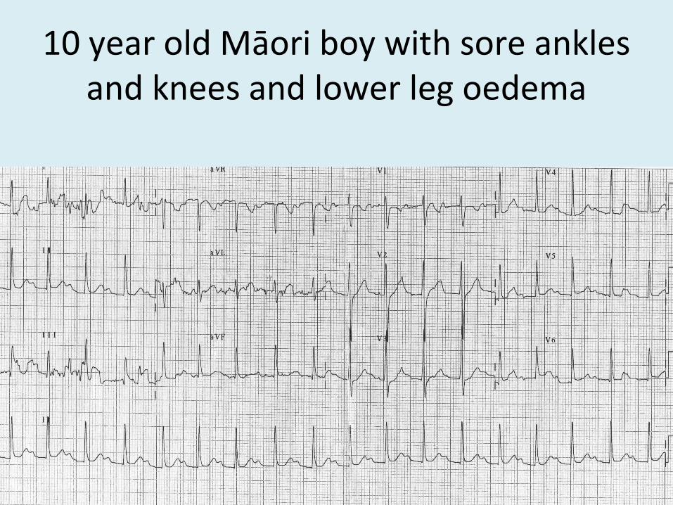
10 year old Māori boy with sore ankles and knees and lower leg oedema
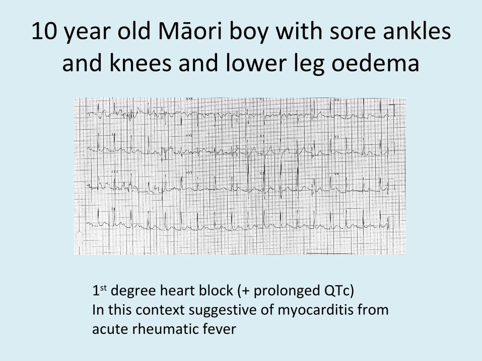
10 year old Māori boy with sore ankles and knees and lower leg oedema
1st degree heart block (+ prolonged QTc)In this context suggestive of myocarditis from acute rheumatic fever

75 M with chest pain

75 M with chest pain
Atrial flutter with variable blockTreat as for AFIn this context
no rate control neededold or new? Needs anticoagulation
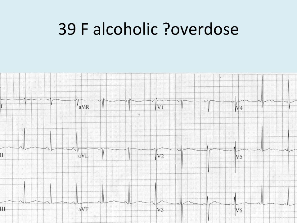
39 F alcoholic ?overdose
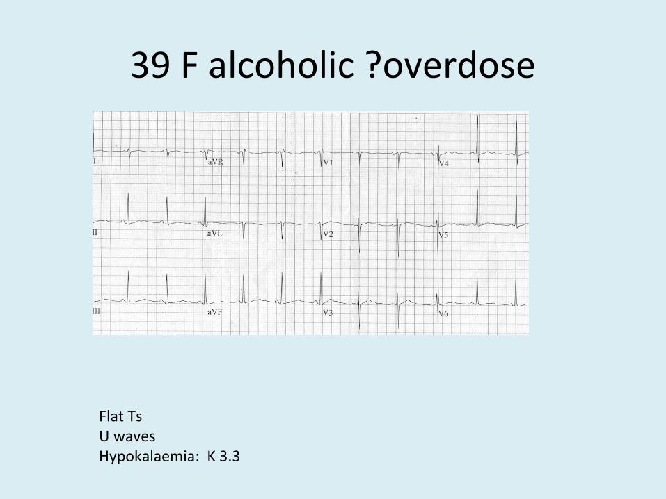
39 F alcoholic ?overdose
Flat TsU wavesHypokalaemia: K 3.3
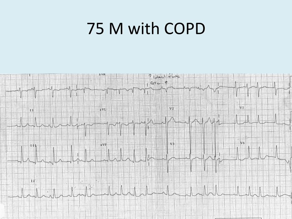
75 M with COPD

MFAT
Irregularly irregular but a p wave before each QRSP waves are different sizes and shapesP waves are coming from multiple foci in the atria= Multifocal atrial tachycardiaSeen in COPDStage before AF

70 year old SOB

Atrial flutter
With variable blockSaw tooth baseline

74 female, cough, SOB, 1/12 post THJR
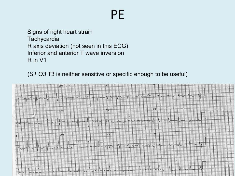
PESigns of right heart strainTachycardiaR axis deviation (not seen in this ECG)Inferior and anterior T wave inversionR in V1
(S1 Q3 T3 is neither sensitive or specific enough to be useful)
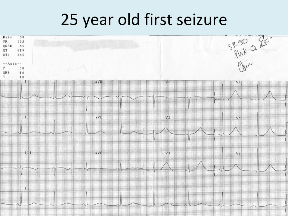
25 year old first seizure

Prolonged QT - congenital
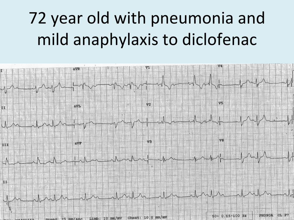
72 year old with pneumonia and mild anaphylaxis to diclofenac

Mobitz II

86 year old epigastric pain and SOB

Mobitz II again. On beta blockers

35 M with CP

PericarditisPR depression II, V2-V6PR elevation aVRSaddle shaped ST elevation V4 and V5


