RANKL-mediated cell-cell fusion in vitro Problem 2 Team work Pabel Shahrear, McGill University Peter Pivonka, Melbourne University Salwa Maria, McGill University Spencer Moran, McGill University Yongqiang Wang, Toronto University Yue Zhao, Simon Fraser University CRM August 19th-23rd 2013
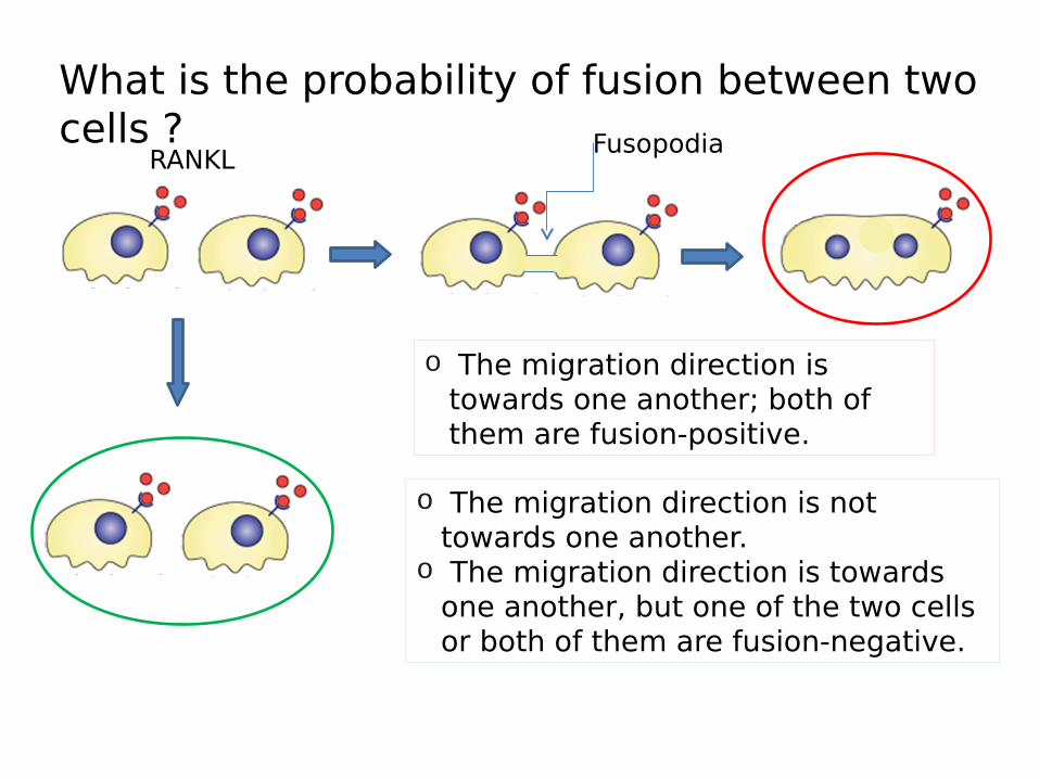
What is the probability of fusion between two cells ? Fusopodia
o The migration direction is not towards one another.
o The migration direction is towards one another, but one of the two cells or both of them are fusion-negative.
o The migration direction is towards one another; both of them are fusion-positive.
RANKL
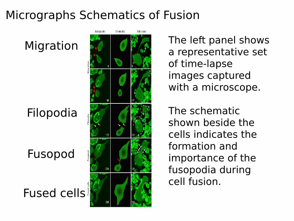
The left panel shows a representative set of time-lapse images captured with a microscope.
The schematic shown beside the cells indicates the formation and importance of the fusopodia during cell fusion.
Migration
Filopodia
Fusopod
Fused cells
Micrographs Schematics of Fusion
The Hypothesis
There are specific cytoplasmic structures that are important in RANKL-mediated cell-cell fusion in vitro.
The Objectives Compare fusion events with non-fusion
events. Characterize migrating cells: founder and
follower. Develop a mathematical model that can
characterize cells with fusion potential within the migrating ones.
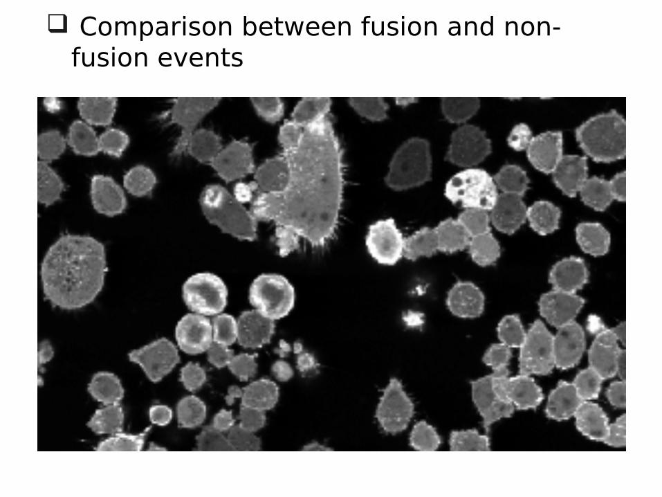
Comparison between fusion and non-fusion events
Comparison between fusion and non-fusion events
Cel
l dyn
amic
s (X
Y-c
oord
inat
e)C
ell d
ynam
ics
(XY
-coo
rdin
ate)
In a fusion event, the founder moves towards the follower forming fusopodia.
Fusion
Technical Issue in the software
Comparison between fusion and non-fusion events
In a fusion event, the founder cell body stretches to reach the follower.
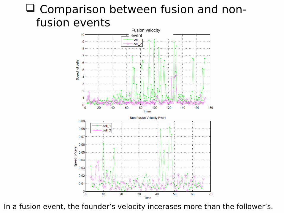
Comparison between fusion and non-fusion events
Fusion velocity event
In a fusion event, the founder’s velocity incerases more than the follower’s.
Comparison between fusion and non-fusion events
In a fusion, the founder has more dendrites endpoints closer to the fusion.
Characterizing migrating cells: founder and follower
Parameter Founder Follower
Cell DynamicsActive and forward Less active
Cell Size Larger Smaller
Velocity Higher Lower
Distance Longer Shorter
Dendrites number Higher Lower
Dendrites length Longer Shorter
The red box indicates that the two parameter values are swapped when the osteoclast reaches a certain age and size; when the cell becomes larger the founder will move more slowly than the follower.
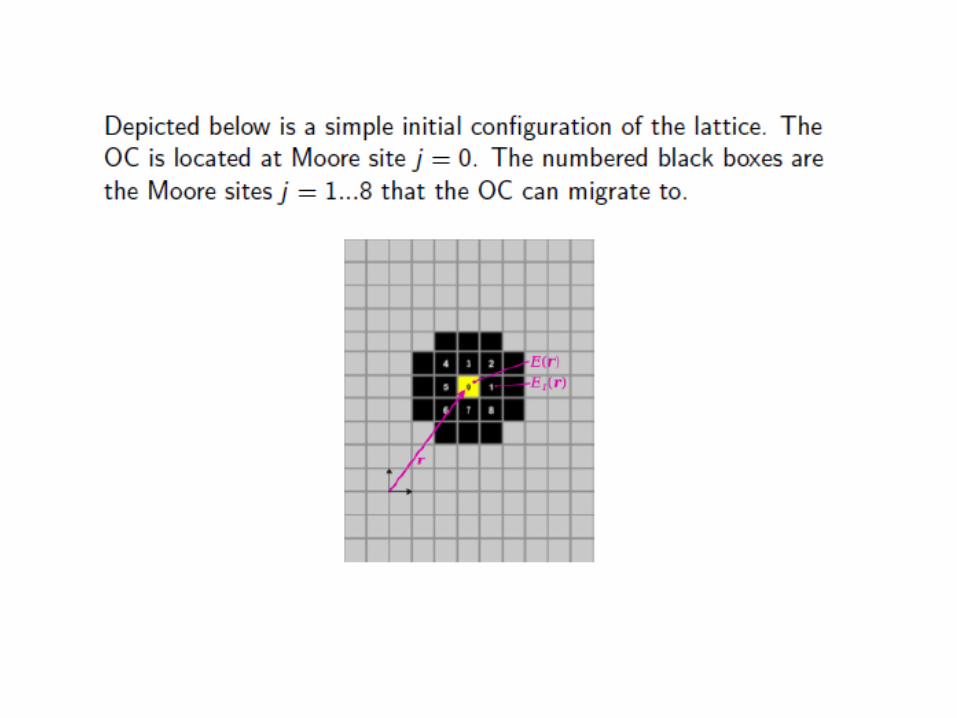
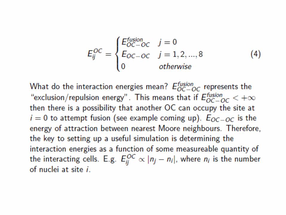
Development of a mathematical model that can characterize cells with fusion potential within the migrating ones