Rapid Detection of Bacterial Antibiotic Resistance: Preliminary Evaluation of PCR Assays Targeting Tetracycline Resistance Genes Matthias R. Dorsch Human Protection and Performance Division Defence Science and Technology Organisation DSTO-TR-2059 ABSTRACT The rapid spreading of bacterial antibiotic resistance and the lack of methods that would allow fast and accurate determination of the resistance profile of an infectious agent have become a major concern for public health. In the context of biological warfare or a bioterrorist attack, resistance against commonly used antibiotics would severely compromise the capabilities of the responsible agencies to protect large numbers of exposed individuals against a potentially lethal infection. In this study the possibility of developing polymerase chain reaction (PCR) assays for rapid detection of tetracycline resistance genes was evaluated. PCR assays were designed for seven resistance genes that frequently occur in Gram-positive and Gram-negative species and tested on eight tetracycline resistant clinical isolates. Positive results were obtained from seven of the eight species tested. RELEASE LIMITATION Approved for public release
Transcript
Rapid Detection of Bacterial Antibiotic Resistance: Preliminary Evaluation of PCR Assays Targeting
Tetracycline Resistance Genes
Matthias R. Dorsch
Human Protection and Performance Division Defence Science and Technology Organisation
DSTO-TR-2059
ABSTRACT The rapid spreading of bacterial antibiotic resistance and the lack of methods that would allow fast and accurate determination of the resistance profile of an infectious agent have become a major concern for public health. In the context of biological warfare or a bioterrorist attack, resistance against commonly used antibiotics would severely compromise the capabilities of the responsible agencies to protect large numbers of exposed individuals against a potentially lethal infection. In this study the possibility of developing polymerase chain reaction (PCR) assays for rapid detection of tetracycline resistance genes was evaluated. PCR assays were designed for seven resistance genes that frequently occur in Gram-positive and Gram-negative species and tested on eight tetracycline resistant clinical isolates. Positive results were obtained from seven of the eight species tested.
Rapid Detection of Bacterial Antibiotic Resistance: Preliminary Evaluation of PCR Assays Targeting
Tetracycline Resistance Genes
Executive Summary The link between the application of antibiotics and the development of resistance has been well documented since the discovery of penicillin in the 1940s. The excessive use of antibiotics in clinical therapy and in agriculture has caused a rapid spread of resistance in both clinical and environmental isolates. Consequently, determination of the antimicrobial susceptibility of pathogenic bacteria is of vital importance to provide the optimal chemotherapy for infected patients. Traditional methods for susceptibility testing require isolation of the pathogen from a specimen sample and culturing on appropriate media containing antibiotic(s). These methods are time consuming and labour intensive and require at least 24 to 48 hours to provide a definitive result. Considerably more time is needed for fastidious pathogens such as Coxiella burnetii, an organism considered a potential biological warfare/bioterrorism agent. C. burnetii cannot be grown in axenic medium. Susceptibility testing has to be done in animals, chicken embryos or cell culture and requires up to 9 days to determine susceptibility. The scenario of a bioterrorist attack that exposes a large number of individuals to an infectious agent necessitates an immediate response to provide adequate protection against a potentially lethal infection, and rapid determination of antibiotic resistance(s) of the infectious agent is crucial. Application of antibiotics that are ineffective is likely to have disastrous consequences. Tetracycline is a commonly used antibiotic in the management of diseases caused by a number of potential biological warfare agents such as anthrax, cholera, plague, and Q-fever. Infections like inhalation anthrax and pneumonic plague, caused by Bacillus anthracis and Yersinia pestis, have to be treated with an effective antibiotic within 24 to 48 hours post exposure. In the majority of cases antibiotic treatment is ineffective once symptoms appear. The polymerase chain reaction (PCR) is a rapid and sensitive tool for the detection of resistance genes. Seven PCR assays for the detection of frequently occurring tetracycline resistance genes in Gram-positive and Gram-negative species were designed and evaluated with eight tetracycline resistant clinical isolates. Putative resistance genes were detected in seven isolates.
Authors
Matthias R. Dorsch Human Protection and Performance Division Matthias Dorsch graduated with a diploma from Christian Albrechts University, Kiel, Germany, in 1987 and was awarded a Ph. D. in 1990 from the same University. Topic of the thesis was the phylogeny of Gram-positive eubacteria. He conducted postdoctoral research at The University of Queensland, The University of New South Wales and Macquarie University. During this time he specialised in detection and identification of bacterial and protozoan pathogens and indicator organisms. He joined the Human Protection & Performance Division in 2001. Focus of his research is the development of rapid detection and identification methods for biological warfare agents.
The discovery and successful application of penicillin, cephalosporins and streptomycin in the 1940s was closely followed by the emergence of antibiotic resistant bacteria. The rapid increase in the number of antibiotic resistant species, including strains that carry multidrug-resistance genes (Fluit et al. 1999, 2000), and the lack of methods that would allow rapid and accurate determination of the resistance profile of a pathogenic bacterium have become a severe problem for public health. Determination of antimicrobial susceptibility of a clinical isolate is in many cases crucial for providing the optimal antimicrobial therapy of infected patients (Fluit et al. 2001). The conventional methods for susceptibility testing require the isolation of the pathogen from a clinical specimen and culturing on the appropriate media that contain antibiotic(s). This is typically carried out using micro-diffusion discs that allow determination of susceptibility and the minimum inhibitory concentration (MIC) of an antibiotic that a pathogen is susceptible to. In most cases a definitive result can be obtained within 24 hours but will take longer when fastidious or slow growing pathogens such as certain Mycobacterium spp. need to be tested. Coxiella burnetii, recognised as a potential biological warfare agent, is an extreme example. The organism cannot be grown in axenic medium. For the purpose of antimicrobial susceptibility testing, three models of infection have been developed, including animals (Huebner et al. 1948), chicken embryos (Maurin & Raoult 1993), and cell cultures (Torres & Raoult 1993). The minimum time requirement of these models is between 6 and 9 days. The fact that antibiotic resistant organisms can easily be generated in the laboratory, either through selective media or by relatively simple genetic engineering techniques, has become a major concern (Courvalin 1995; Hilleman 2002; Sefton 2002). Strains of B. anthracis that are resistant to ciprofloxacin, doxycycline and rifampin have been generated by passaging the strains through media containing low levels of the antibiotics (Choe et al. 2000; Vogler et al. 2002), and Russian scientists have created a B. anthracis strain resistant to clindamycin, chloramphenicol, erythromycin, penicillin, rifampin and tetracycline (Stepanov et al. 1996). The potential exposure of a large number of individuals to an infectious agent due to an act of bioterrorism is a drastic scenario which highlights the need for methods that enable rapid determination of bacterial resistance profiles. Authorities involved in such an incident have to take immediate action to protect the victims against a potentially lethal infectious disease even if the infectious agent would not exhibit resistance against commonly used antibiotics. Inhalation anthrax is considered fatal if antibiotic treatment does not commence within 24 – 48 hours post exposure (Henderson, D.A. 1999; Franz et al. 1997). Antibiotic treatment is usually ineffective once symptoms appear. Similarly critical is the timing for treatment of pneumonic plague. Antibiotics have to be administered within 24 hours of occurrence of the first symptoms (Franz et al. 1997; Gilligan 2002). Pathogens possessing one or even multiple resistances against antibiotics that are normally effective would severely exacerbate the situation as administering ineffective antibiotics may result in mass casualties. Nucleic-acid targeted detection systems as the polymerase chain reaction (PCR) offer rapid and sensitive methods to detect the presence of resistance genes and are crucial in the
1
DSTO-TR-2059
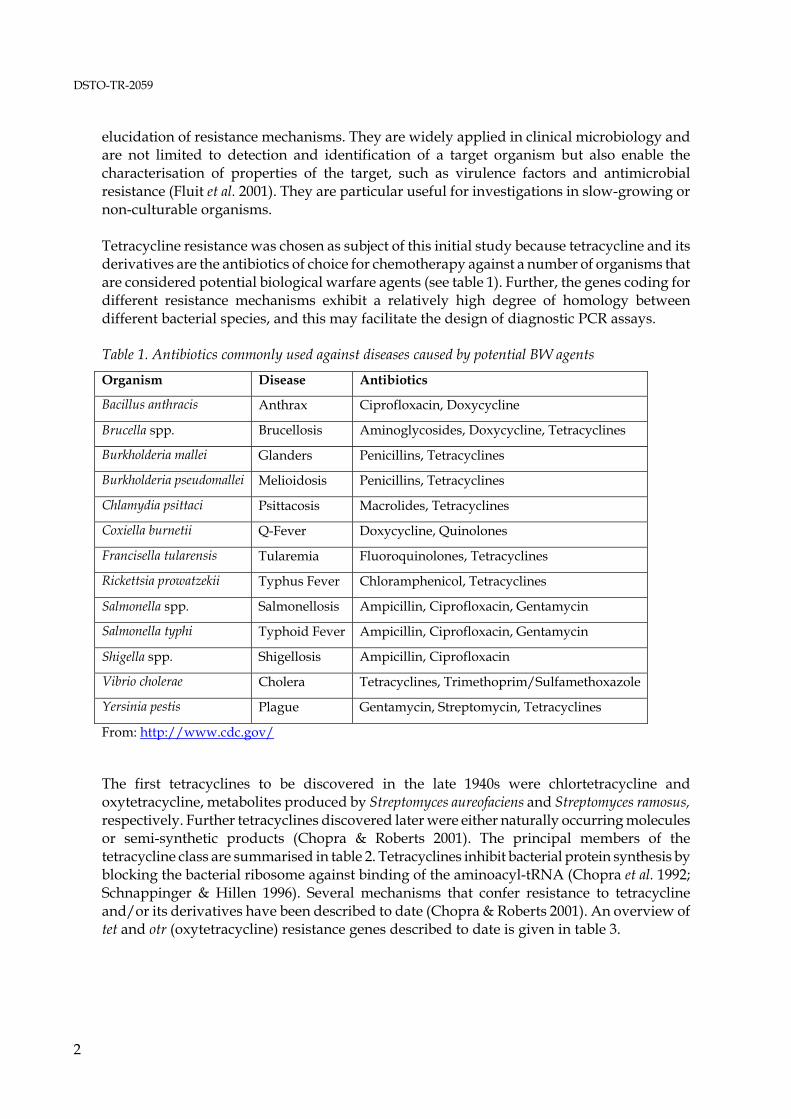
elucidation of resistance mechanisms. They are widely applied in clinical microbiology and are not limited to detection and identification of a target organism but also enable the characterisation of properties of the target, such as virulence factors and antimicrobial resistance (Fluit et al. 2001). They are particular useful for investigations in slow-growing or non-culturable organisms. Tetracycline resistance was chosen as subject of this initial study because tetracycline and its derivatives are the antibiotics of choice for chemotherapy against a number of organisms that are considered potential biological warfare agents (see table 1). Further, the genes coding for different resistance mechanisms exhibit a relatively high degree of homology between different bacterial species, and this may facilitate the design of diagnostic PCR assays. Table 1. Antibiotics commonly used against diseases caused by potential BW agents
The first tetracyclines to be discovered in the late 1940s were chlortetracycline and oxytetracycline, metabolites produced by Streptomyces aureofaciens and Streptomyces ramosus, respectively. Further tetracyclines discovered later were either naturally occurring molecules or semi-synthetic products (Chopra & Roberts 2001). The principal members of the tetracycline class are summarised in table 2. Tetracyclines inhibit bacterial protein synthesis by blocking the bacterial ribosome against binding of the aminoacyl-tRNA (Chopra et al. 1992; Schnappinger & Hillen 1996). Several mechanisms that confer resistance to tetracycline and/or its derivatives have been described to date (Chopra & Roberts 2001). An overview of tet and otr (oxytetracycline) resistance genes described to date is given in table 3.
From: Chopra & Roberts 2002 Resistance mechanisms Efflux proteins: Genes for tet efflux genes code for membrane-associated proteins that remove tetracycline from the cell. Efflux genes occur in Gram-positive and Gram-negative species. Most of the efflux proteins confer resistance to tetracycline but not minocycline or glycylcyclines. In contrast, the Gram-negative tet(B) gene confers resistance to both tetracycline and minocycline but not glycylcyclines (Chopra et al. 1992; Testa et al. 1993). Ribosomal protection proteins: These are cytoplasmic proteins that protect the ribosome against tetracycline and also confer resistance to doxycycline and minocycline. Compared to tetracycline efflux proteins they confer resistance to a wider spectrum of tetracyclines, with the exception of tet(B). Enzymatic inactivation of tetracycline: The only example described to date for inactivation of tetracycline due to enzymatic modification is the product of the tet(X) gene (Speer et al. 1991). The gene was discovered as part of two closely related Bacteroides spp. transposons and has not been found outside the genus Bacteroides. Other mechanisms of resistance: The tet(U) gene of Enterococcus faecium confers low-level tetracycline resistance (Ridenhour et al. 1996). The gene sequence does not exhibit significant homologies to efflux protein genes or ribosomal protection proteins. The otr(C) gene of Streptomyces has not undergone sequence analysis as yet, and the nature of the protection mechanism is subject to speculation (Chopra & Roberts 2001). To evaluate the feasibility of PCR-based detection of tetracycline resistance genes, all available tet resistance gene sequences were retrieved from the GenBank and EMBL databases through the Australian National Genomic Information Service (ANGIS). The efflux protein genes tet(A), tet(B), tet(D), tet(G) and tet(L), and the ribosomal protection protein genes tet(M) and tet(O) were identified as the most frequently occurring genes that have been found in a variety of Gram-positive and Gram-negative species.
3
DSTO-TR-2059
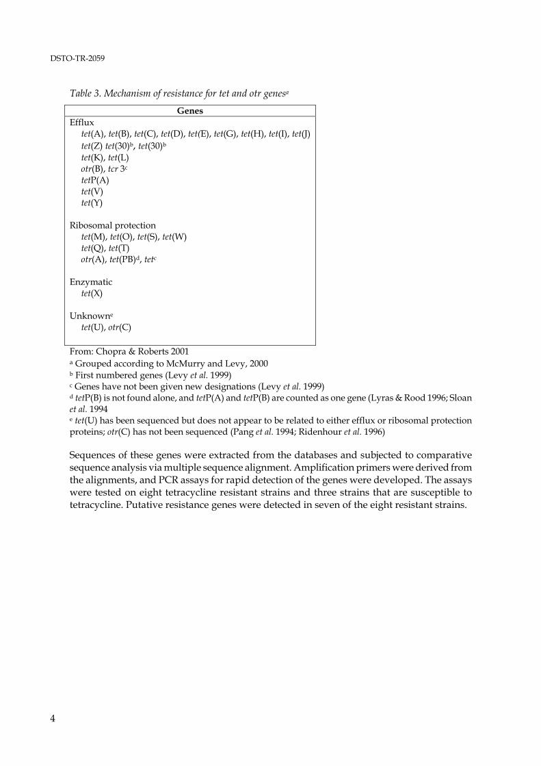
Table 3. Mechanism of resistance for tet and otr genesa
Genes Efflux tet(A), tet(B), tet(C), tet(D), tet(E), tet(G), tet(H), tet(I), tet(J) tet(Z) tet(30)b, tet(30)b tet(K), tet(L) otr(B), tcr 3c tetP(A) tet(V) tet(Y) Ribosomal protection tet(M), tet(O), tet(S), tet(W) tet(Q), tet(T) otr(A), tet(PB)d, tetc Enzymatic tet(X) Unknowne tet(U), otr(C) From: Chopra & Roberts 2001 a Grouped according to McMurry and Levy, 2000 b First numbered genes (Levy et al. 1999) c Genes have not been given new designations (Levy et al. 1999) d tetP(B) is not found alone, and tetP(A) and tetP(B) are counted as one gene (Lyras & Rood 1996; Sloan et al. 1994 e tet(U) has been sequenced but does not appear to be related to either efflux or ribosomal protection proteins; otr(C) has not been sequenced (Pang et al. 1994; Ridenhour et al. 1996) Sequences of these genes were extracted from the databases and subjected to comparative sequence analysis via multiple sequence alignment. Amplification primers were derived from the alignments, and PCR assays for rapid detection of the genes were developed. The assays were tested on eight tetracycline resistant strains and three strains that are susceptible to tetracycline. Putative resistance genes were detected in seven of the eight resistant strains.
4
DSTO-TR-2059
2. Materials and Methods
2.1 Materials
2.1.1 Amplification primers
Primers for PCR amplification were produced by GeneWorks, Thebarton, South Australia. 2.1.2 Bacterial strains and media
Ten strains were obtained from the Australian Collection of Microorganisms, Department of Microbiology, University of Queensland, Brisbane, and from Pathcentre Nedlands, Department of Microbiology and Infectious Diseases, Perth. Two additional strains were isolated from skin swabs from a healthy person. Tryptone soya agar, Mueller-Hinton agar and defibrinated sheep blood were purchased from Oxoid Australia Pty. Ltd. Tetracycline hydrochloride was from Sigma-Aldrich. 2.1.3 DNA isolation
Calcium chloride, ethylenediamine-tetraacetic acid (EDTA), glycerol, isoamylalcohol, lauryl sulfate (SDS), lysozyme, phenol, phenol-chloroform and sodium acetate were purchased from Sigma-Aldrich. Thermostable proteinase K (fungal) was from Invitrogen. 2.1.4 Real-time PCR
The LightCycler – FastStart DNA Master SYBR Green I was purchased from Roche Diagnostics. 2.1.5 Conventional PCR
Platinum Taq DNA polymerase and 1 kb ladder were from Invitrogen. PCR grade deoxy-nucleotide-triphosphates (dNTPs), molecular screening (MS) agarose and molecular size standard XIV were supplied by Roche Diagnostics. Ethidium bromide solution was purchased from Continental Lab Products. 2.1.6 Thermal cyclers
Real-time PCRs with the LightCycler – FastStart DNA Master SYBR Green I were run on the Ruggedized Advanced Pathogen Detection Device (R.A.P.I.D.™, Idaho Technology Inc., Salt Lake City, Utah, US). A Thermal Cycler PTC 200 (MJ Research, Massachusetts, US) was used for conventional PCR.
5
DSTO-TR-2059
2.2 Methods
2.2.1 Susceptibility testing of strains
Bacterial strains used in this study are listed in table 4. Cells were grown on Mueller-Hinton agar containing 10, 20, 25 and 30 μg/ml tetracycline (stock solution 5 mg/ml in ethanol). Plates were incubated at 37°C and examined after 20 h. Controls were grown on Mueller-Hinton agar without tetracycline and incubated as above. Table 4. Bacterial strains
Strain No Strain ID Species Source tet Resistancea 1 1016486 Burkholderia cepacia PathCentre Perth + 2 ATCC25416 Burkholderia cepacia ACM, University of Qld. + 3 ACM2193 Erwinia chrysanthemi ACM, University of Qld. - 4 1015932P Staphylococcus aureus PathCentre Perth + 5 1016722R Staphylococcus aureus PathCentre Perth + 6 1016674U Streptococcus Group B PathCentre Perth + 7 4553874B Streptococcus Group B PathCentre Perth + 8 ATCC9144 Staphylococcus aureus ACM, University of Qld. - 9 ATCC9290 Shigella sonnei ACM, University of Qld. - 10 SA Streptococcus sp.b CBRN DC + 11 SB Streptococcus sp.b CBRN DC + a: maximum concentration tested was 30 μg/ml b: species identity based on morphology only 2.2.2 DNA isolation
Cells were grown overnight on tryptone soya agar supplemented with 7 % defibrinated sheep blood. Approximately 10 colonies were resuspended in 500 μl TE buffer (10 mM Tris·HCl, 1 mM EDTA, pH 8.0) and washed 3 times in 500 μl TE buffer. The final cell pellet was resuspended in 500 μl TE buffer containing 1 μg/μl lysozyme (stock solution 50 mg/ml in distilled water) and incubated at 37°C for 1 h. SDS and proteinase K (stock solution 20 mg/ml in 10 mM Tris·HCl, 20 mM CaCl2, 50 % glycerol (v/v), pH 7.5) were added to a final concentration of 0.5 % (v/v) and 50 μg/ml, respectively. Cells were incubated at 65°C for 1 h and the lysate was extracted with equal volumes of phenol, phenol : chloroform (24 : 1) and chloroform : isoamylalcohol (25 : 24 : 1). Nucleic acids were precipitated with sodium acetate/ethanol and dissolved in 250 μl distilled water. Nucleic acid concentration was determined spectrophotometrically at 260 nm. 2.2.3 Design of PCR amplification primers
Sequence data were obtained and analysed using the facilities of the Australian National Genomic Information Service (ANGIS; see Littlejohn 2000). A total of 677 available tetracycline resistance gene sequences was retrieved from the databases of EMBL and GenBank. Duplicate entries were eliminated, and sequences containing more than the tetracycline resistance gene information were truncated. Sequences representing tetracycline resistance gene classes as described to date (Chopra & Roberts 2001) were subjected to comparative sequence analysis
6
DSTO-TR-2059
through the FASTA program in order to identify related sequences. Sequences of each tetracycline resistance gene class that displayed significant sequence homologies (>75 %) and occurred in three or more different bacterial species were aligned using the program clustalW. Amplification primers for the efflux genes tet(A), tet(B), tet(D), tet(G) and tet(L), and for the ribosomal protection protein genes tet(M) and tet(O) were derived from the alignments. Primers for amplification of a portion of the 16S ribosomal DNA (Stackebrandt & Charfreitag 1990) were used in positive control PCRs to ensure that negative results were not due to insufficient DNA quality. Primer sequences, their target positions and melting points (Tm) are shown in table 5. Table 5. PCR amplification primers
Primer Sequencea Tm (°C) Positionb
tet(A) forward GTCGCGCTCGACGCTGTCGGCATCGGCC 72 969-996 tet(A) reverse CCGGTGATGCCGGCCACGATCCGCCCG 72 1232-1258 tet(B) forward GGCCAAATTCCCGCAACGGTGTGGGTGC 67 1188-1215 tet(B) reverse GCCAATAACACCGGTTGCATTGGTAAGGC 63 1537-1564 tet(D) forward GCTGGCGCTGTATGCGGTGATGCAGGTC 67 105-132 tet(D) reverse GCCGGCCCGGCAATCAGCCCGGCACC 72 382-407 tet(G) forward CGGCCAAGTGCCTGC[GA]GCCCTATGGGTC 69 1772-1979 tet(G) reverse CGGGAACACCATCCATCCCTGCGTGGC 67 1986-2012 tet(L) forward GGAAC[AC]ATGAGTGT[GT]ATT[AG]TTTT[CT]GG 58 2584-2609 tet(L) reverse CCTACAATTGC[TA]ATACC[CT]GTTCCCTCTG 61 2866-2894 tet(M) forward CC[TA]AC[AT]GTCATTTATATGGA[GA]AGACC 56 1258-1283 tet(M) reverse CGAAAATCTGCTGG[CGA]GTACT[GA]ACAGGGC 64 1535-1562 tet(O) forward CCGCCAAATCCTTTCTGGGCTTCTGTCGG 66 1515-1544 tet(O) reverse CGCCCGTGAGAGATATTCCTGCGGTGC 66 1842-1868 16S forward GAGTTTGATC[AC]GGCTCAG 50 11-29 16S reverse G[AT]ATTACCGCGGC[GT]GCTG 54 519-536 a: bases in [ ] indicate degenerate primer positions b: position numbers for tetracycline resistance gene primers are according to the core sequence of the alignment (see appendix); position numbers for 16S rDNA primers are according to Brosius et al. 1978 2.2.4 Real-time PCR
Reactions were run on a R.A.P.I.D.™ with the LightCycler – FastStart DNA Master SYBR Green I (Roche Diagnostics) in volumes of 20 μl. Positive control reactions for amplification of a 526 base pair (bp) fragment of the 16S rDNA contained 0.05 μM forward and reverse primer, 4 mM MgCl2, 10 ng DNA and 2 μl 10x reaction buffer. The temperature profile comprised initial denaturation at 95°C for 10 min followed by 40 cycles of primer annealing at 50°C for 10 sec, primer extension at 72°C for 20 sec and denaturation at 95°C for 10 sec at the maximum temperature transition rate of 20°C/sec, with acquisition of the fluorescence signal at the end of each primer extension. Completed reactions were subjected to melting curve analysis in order to enable discrimination of specific products and primer-dimers. The temperature profile for melting curve analysis was 95°C for 0 sec (instrument goes to next target temperature once set temperature is reached), 50°C for 45 sec and heating to 94°C at a temperature transition rate of 0.1°C/sec under constant acquisition of the fluorescence signal. Upon completion of the melting curve samples were cooled to 40°C, and melting curve data were analysed. Duplicates of each reaction were analysed on ethidium bromide stained
7
DSTO-TR-2059
agarose gels with the 1 kb ladder (Invitrogen) and molecular size standard XIX (Roche Diagnostics). Reactions for amplification of tetracycline resistance gene fragments contained 0.2 μM forward and reverse primer, 4 mM MgCl2, 10 ng DNA and 2 μl 10x reaction buffer. After initial denaturation at 95°C for 10 min, the following temperature profiles were applied for different target genes over 40 amplification cycles: tet(A) – Primer annealing at 67°C for 5 sec, primer extension at 72°C for 10 sec and denaturation at 95°C for 10 sec. tet(B) – Primer annealing at 63°C for 5 sec, primer extension at 72°C for 15 sec and denaturation at 95°C for 10 sec. tet(D) – Primer annealing at 65°C for 5 sec, primer extension at 72°C for 20 sec and denaturation at 95°C for 10 sec. tet(G) – Primer annealing at 63°C for 5 sec, primer extension at 72 °C for 15 sec and denaturation at 95°C for 10 sec. tet(L) – Primer annealing at 52°C for 5 sec, primer extension at 72°C for 15 sec and denaturation at 95°C for 10 sec. tet(M) – Primer annealing at 52°C for 5 sec, primer extension at 72°C for 15 sec and denaturation at 95°C for 10 sec. tet(O) – Primer annealing at 62°C for 5 sec, primer extension at 72°C for 15 sec and denaturation at 95°C for 10 sec. Melting curve analysis and agarose gel electrophoresis was done as above. 2.2.5 Conventional PCR
Reactions were run on a Thermal Cycler PTC 200 (MJ Research) in thin walled PCR tubes with reaction volumes of 50 μl. Both positive control reactions for 16S rDNA amplification and PCRs for amplification of tetracycline resistance gene fragments contained 0.5 μM forward and reverse amplification primer, 3 mM MgCl2, 200 μM of each dNTP, 1 unit Platinum® Taq DNA polymerase, 100 ng DNA and 5 μl 10x reaction buffer. The temperature profile for 16S rDNA amplification comprised initial denaturation at 94°C for 2 min followed by 5 cycles of primer annealing at 53°C for 30 sec, primer extension at 72°C for 40 sec and denaturation at 94°C for 20 sec, 25 cycles of annealing at 48°C for 20 sec, primer extension at 72°C for 40 sec and denaturation at 94°C for 20 sec and a final extension step with annealing at 48°C for 20 sec and primer extension at 72°C for 2 min. Reactions were cooled to 4°C prior to analysing 20 μl aliquots on ethidium bromide stained 1.5% molecular screening (MS) agarose (Roche Diagnostics) gels. Size standards were the 1 kb ladder (Invitrogen) and molecular size standard XIV (Roche Diagnostics). The size of the amplification products as determined by comparison to the standards was estimated to be correct within ± 25 bp. After initial denaturation at 94°C for 2 min the following temperature profiles for amplification of tetracycline resistance gene fragments were applied over 40 cycles: tet(A) – Primer annealing at 63°C for 30 sec, primer extension at 72°C for 30 sec and denaturation at 94°C for 30 sec.
8
DSTO-TR-2059
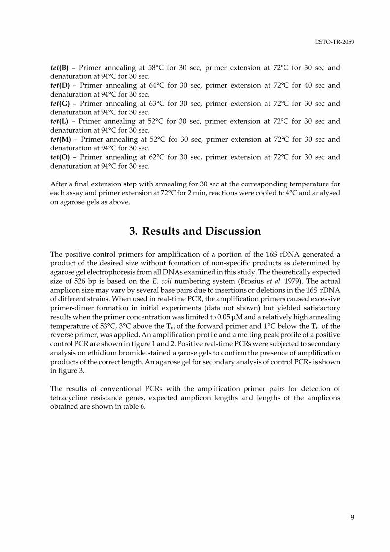
tet(B) – Primer annealing at 58°C for 30 sec, primer extension at 72°C for 30 sec and denaturation at 94°C for 30 sec. tet(D) – Primer annealing at 64°C for 30 sec, primer extension at 72°C for 40 sec and denaturation at 94°C for 30 sec. tet(G) – Primer annealing at 63°C for 30 sec, primer extension at 72°C for 30 sec and denaturation at 94°C for 30 sec. tet(L) – Primer annealing at 52°C for 30 sec, primer extension at 72°C for 30 sec and denaturation at 94°C for 30 sec. tet(M) – Primer annealing at 52°C for 30 sec, primer extension at 72°C for 30 sec and denaturation at 94°C for 30 sec. tet(O) – Primer annealing at 62°C for 30 sec, primer extension at 72°C for 30 sec and denaturation at 94°C for 30 sec. After a final extension step with annealing for 30 sec at the corresponding temperature for each assay and primer extension at 72°C for 2 min, reactions were cooled to 4°C and analysed on agarose gels as above.
3. Results and Discussion
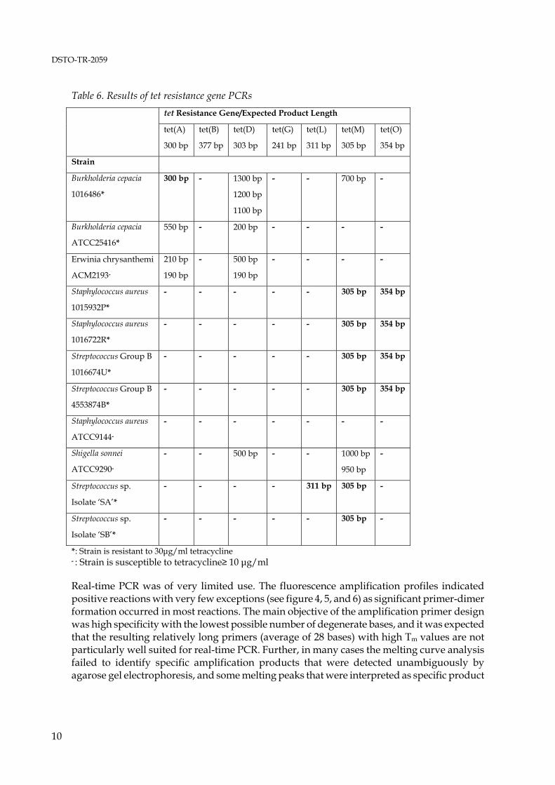
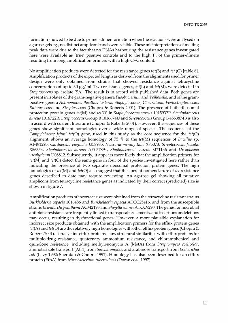
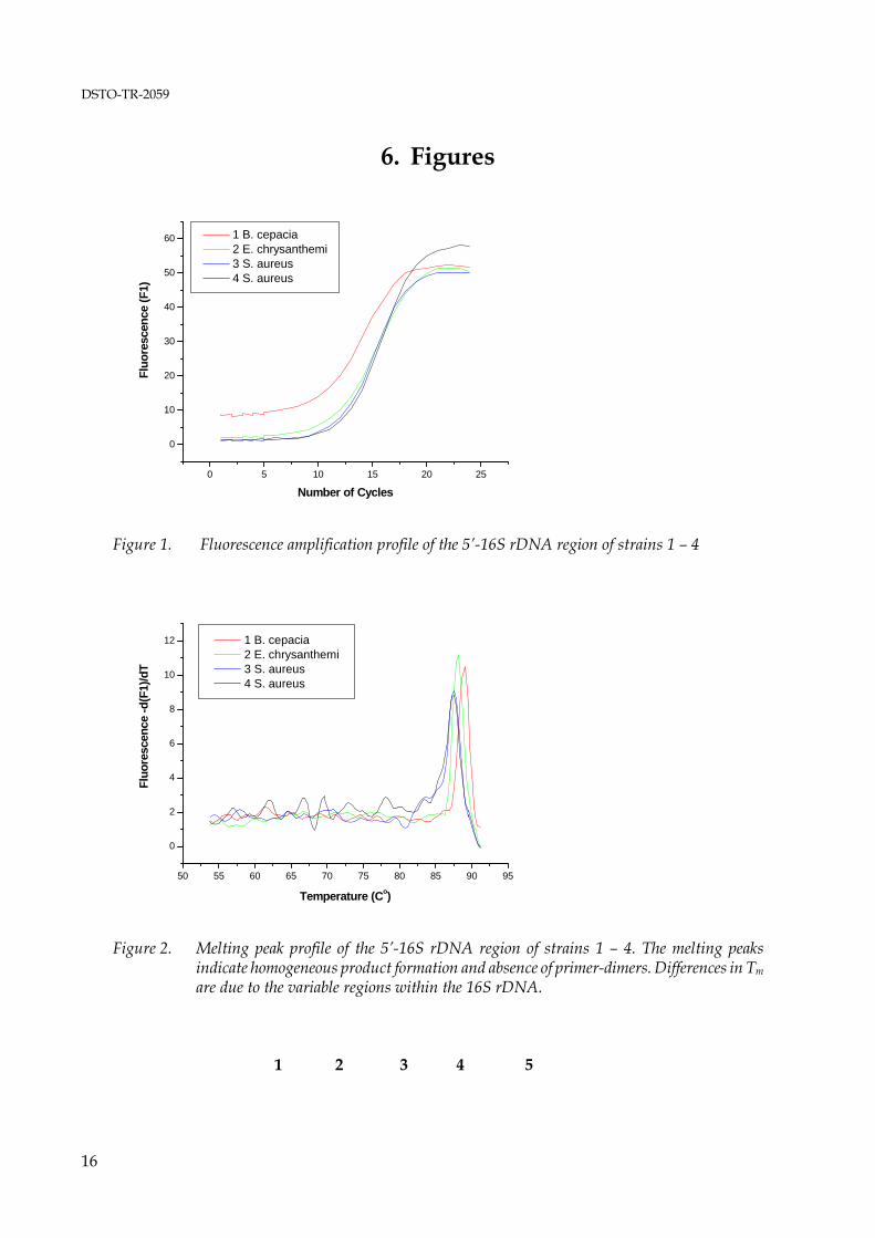
The positive control primers for amplification of a portion of the 16S rDNA generated a product of the desired size without formation of non-specific products as determined by agarose gel electrophoresis from all DNAs examined in this study. The theoretically expected size of 526 bp is based on the E. coli numbering system (Brosius et al. 1979). The actual amplicon size may vary by several base pairs due to insertions or deletions in the 16S rDNA of different strains. When used in real-time PCR, the amplification primers caused excessive primer-dimer formation in initial experiments (data not shown) but yielded satisfactory results when the primer concentration was limited to 0.05 μM and a relatively high annealing temperature of 53°C, 3°C above the Tm of the forward primer and 1°C below the Tm of the reverse primer, was applied. An amplification profile and a melting peak profile of a positive control PCR are shown in figure 1 and 2. Positive real-time PCRs were subjected to secondary analysis on ethidium bromide stained agarose gels to confirm the presence of amplification products of the correct length. An agarose gel for secondary analysis of control PCRs is shown in figure 3. The results of conventional PCRs with the amplification primer pairs for detection of tetracycline resistance genes, expected amplicon lengths and lengths of the amplicons obtained are shown in table 6.
*: Strain is resistant to 30μg/ml tetracycline - : Strain is susceptible to tetracycline≥ 10 μg/ml Real-time PCR was of very limited use. The fluorescence amplification profiles indicated positive reactions with very few exceptions (see figure 4, 5, and 6) as significant primer-dimer formation occurred in most reactions. The main objective of the amplification primer design was high specificity with the lowest possible number of degenerate bases, and it was expected that the resulting relatively long primers (average of 28 bases) with high Tm values are not particularly well suited for real-time PCR. Further, in many cases the melting curve analysis failed to identify specific amplification products that were detected unambiguously by agarose gel electrophoresis, and some melting peaks that were interpreted as specific product
10
DSTO-TR-2059
formation showed to be due to primer-dimer formation when the reactions were analysed on agarose gels eg., no distinct amplicon bands were visible. These misinterpretations of melting peak data were due to the fact that no DNAs harbouring the resistance genes investigated here were available as ‘true’ positive controls and to the high Tm of the primer-dimers resulting from long amplification primers with a high G+C content. No amplification products were detected for the resistance genes tet(B) and tet (G) [table 6]. Amplification products of the expected length as derived from the alignments used for primer design were only obtained from strains that showed resistance against tetracycline concentrations of up to 30 μg/ml. Two resistance genes, tet(L) and tet(M), were detected in Streptococcus sp. isolate ‘SA’. The result is in accord with published data. Both genes are present in isolates of the gram-negative genera Fusobacterium and Veillonella, and of the gram-positive genera Actinomyces, Bacillus, Listeria, Staphylococcus, Clostridium, Peptostreptococcus, Enterococcus and Streptococcus (Chopra & Roberts 2001). The presence of both ribosomal protection protein genes tet(M) and tet(O) in Staphylococcus aureus 1015932P, Staphylococcus aureus 1016722R, Streptococcus Group B 1016674U and Streptococcus Group B 4553874B is also in accord with current literature (Chopra & Roberts 2001). However, the sequences of these genes show significant homologies over a wide range of species. The sequence of the Campylobacter jejuni tet(O) gene, used in this study as the core sequence for the tet(O) alignment, shows an average homology of 75 % to the tet(M) sequences of Bacillus sp. AF491293, Gardnerella vaginalis U58985, Neisseria meningitidis X75073, Streptococcus faecalis X56353, Staphylococcus aureus AY057894, Staphylococcus aureus M21136 and Ureaplasma urealyticum U08812. Subsequently, it appears more likely that the amplification primers for tet(M) and tet(O) detect the same gene in four of the species investigated here rather than indicating the presence of two separate ribosomal protection protein genes. The high homologies of tet(M) and tet(O) also suggest that the current nomenclature of tet resistance genes described to date may require reviewing. An agarose gel showing all putative amplicons from tetracycline resistance genes as indicated by their correct (predicted) size is shown in figure 7. Amplification products of incorrect size were obtained from the tetracycline resistant strains Burkholderia cepacia 1016486 and Burkholderia cepacia ATCC25416, and from the susceptible strains Erwinia chrysanthemi ACM2193 and Shigella sonnei ATCC9290. The genes for microbial antibiotic resistance are frequently linked to transposable elements, and insertions or deletions may occur, resulting in dysfunctional genes. However, a more plausible explanation for incorrect size products obtained with the amplification primers for the efflux protein genes tet(A) and tet(D) are the relatively high homologies with other efflux protein genes (Chopra & Roberts 2001). Tetracycline efflux proteins show structural similarities with efflux proteins for multiple-drug resistance, quaternary ammonium resistance, and chloramphenicol and quinolone resistance, including methylenomycin A (MetA) from Streptomyces coelicolor, aminotriazole transport (Atr1) from Saccharomyces, and arabinose transport from Escherichia coli (Levy 1992; Sheridan & Chopra 1991). Homology has also been described for an efflux protein (EfpA) from Mycobacterium tuberculosis (Doran et al. 1997).
11
DSTO-TR-2059
4. Conclusions
The seven assays under evaluation detected putative tetracycline resistance genes in seven out of eight resistant isolates. None of the tetracycline susceptible strains yielded amplification products of the expected size as predicted from the multiple sequence alignments for design of the amplification primers. The occurrence of incorrect size products is likely to be due to the homology of tetracycline efflux pump protein genes with other efflux pump protein genes as discussed above. Detection assays derived from this study will utilise either hybridisation probes or TaqMan™ probes. These probes essentially enable amplicon confirmation and provide increased assay specificity that should suffice to eliminate false positive reactions caused by the presence of significantly homologous efflux pump genes. Future research will include sequence analysis to confirm the identity of the putative tetracycline resistance genes and of the incorrect size amplification products obtained from the assays for tet(A), tet(D) and tet(M). Further, the assays require extensive validation prior to routine application. This will include testing on an extended collection of tetracycline resistant and susceptible isolates and on background organisms that are present in the majority of samples analysed for the presence of potential BW agents. As an operational requirement of the assays it has to be ensured that positive results are not due to the presence of resistance genes in background organisms. As demonstrated in this study, the considerable diversity of tetracycline resistance genes necessitates the design of a number of assays to provide coverage of the most frequently occurring genes. The assays cannot be combined as a ‘one tube’ multiplex assay with the existing assays for specific agent detection due to limitations of the detection platforms and the number of amplification primer pairs that can be used in one assay. They are intended for secondary analysis eg., for samples that tested positive for a potential BW agent. The experimental work described here is a ‘proof of principle’ regarding the feasibility of assay development for the rapid detection of tetracycline resistance genes. Successful transformation of conventional PCR assays into real-time format will require some modifications. Amplification primers and fluorescently labelled detection probes will be designed utilising the ‘Beacon Designer’ software package from Premier Biosoft (Palo Alto, CA, US), with TaqMan probes as the preferred detection chemistry. The amplicon length of the conventional assays varied from 241 to 377 bp. These relatively long amplicons were generated to gain sufficient sequence information that allows assessment of the variations in tet resistance genes for the successful design of real-time assays. The amplicon length for real-time assays will be restricted to a maximum of 100 bp to reduce the time required for the amplification profile to approximately 40 min. Real-time assays will be run on the SmartCycler (Cepheid; Sunnyvale, CA, US). In contrast to the R.A.P.I.D. the SmartCycler allows the detection of up to four TaqMan probes conjugated to different fluorescent labels in the same reaction and can be applied for multiplex assays that detect three tet resistance genes and contain an internal control.
12
DSTO-TR-2059
5. Bibliography
1. Brosius, J., Palmer, J.L., Kennedy, J.P. and Noller, H.F. (1978) Complete nucleotide sequence of a 16S ribosomal RNA gene from Escherichia coli. Proceedings of the National Academy of Sciences of the USA 75: 4801-4805
2. Choe, C., Bouhaouala, S., Brook, I., Elliott, T. and Knudson, G. (2000) In vitro development
of resistance to ofloxacin and doxycycline in Bacillus anthracis Sterne. Antimicrobial Agents and Chemotherapy 44: 1766
3. Chopra, I., Hawkey, P.M. and Hinton, M. (1992) Tetracyclines, molecular and clinical
aspects. The Journal of Antimicrobial Chemotherapy 29: 245-277 4. Chopra, I. and Roberts, M. (2001) Tetracycline antibiotics: Mode of action, applications,
molecular biology, and epidemiology of bacterial resistance. Molecular Biology Reviews 65: 232-260
5. Courvalin, P. (1995) Impact of molecular biology on antibiotic susceptibility: testing and
therapy. The American Journal of Medicine Supplement 99(6A): 21S-5S 6. Doran, J.L., Pang, Y., Mdluli, K.E., Moran, A.J, Victor, T.C., Stokes, R.W., van Helden, E.,
Roberts, M.C. and Nano, F.E. (1997) Mycobacterium tuberculosis efpA encodes an efflux protein of the QacA transporter family. Clinical and Diagnostic Laboratory Immunology 4: 23-32
7. Fluit, A.C., Schmitz, F.-J., Jones, M.E., Acar, J., Gupta, R. and Verhoef, J. for the SENTRY
Participants Group (1999) Antimicrobial resistance ammong community-acquired isolates in Europe: first results from the SENTRY antimicrobial surveillance program. International Journal of Infectious Diseases 3: 153-156
8. Fluit, A.C., Jones, M.E., Schmitz, F.-J., Acar, J., Gupta, R. and Verhoef, J. for the SENTRY
Participants Group (2000) Bacteremia in European hospitals, incidence and antimicrobial susceptibility. Clinical Infectious Diseases 30: 454-460
9. Fluit, A.C., Visser, M.R. and Schmitz, F.-J. (2001) Molecular detection of antimicrobial
Pavlin, J.A., Christopher, G.W. and Eitzen, E.M.J. (1997) Clinical recognition and management of patients exposed to biological warfare agents. The Journal of The American Medical Association 278: 399-411
11. Gilligan, P. (2002) Therapeutic challenges posed by bacterial bioterrorism threats. Current
Opinion in Microbiology 5: 489-495
13
DSTO-TR-2059
12. Hilleman, M.R. (2002) Overview: cause and prevention in biowarfare and bioterrorism. Vaccine 20: 3055-3067
13. Huebner, R.J., Hottle, G.A. and Robinson, E.B. (1948) Action of streptomycin in
experimental infection with Q fever. Public Health Reports 63: 357-362 14. Levy, S.B. (1992) Active efflux mechanisms for antimicrobial resistance. Antimicrobial
Agents and Chemotherapy 36: 695-703 15. Levy, S.B., McMurry, L.M., Barbosa, T.M., Burdett, V., Courvalin, P., Hillen, W., Roberts,
M.C., Rood, J.I. and Taylor, D.E. (1999) Nomenclature for new tetracycline resistance determinants. Antimicrobial Agents and Chemotherapy 43: 1523-1524
16. Littlejohn, T. (2000) Bioinformatics in Australia. Bioinformatics 16: 849-850 17. Lyras, D. and Rood, J.I. (1996) Genetic organization and distribution of tetracycline
resistance determinants in Clostridium perfringens. Antimicrobial Agents and Chemotherapy 40: 2500-2504
18. Maurin, M. and Raoult, D. (1993) In vitro susceptibilities of spotted fever group rickettsiae
and Coxiella burnetii to clarithromycin. Antimicrobial Agents and Chemotherapy 37: 2633-2637 19. McMurry, L.M. and Levy, S.B. (2000) Tetracycline resistance in gram-positive bacteria, p.
660-677. In V.A. Fischetti, R.P. Novick, J.J. Ferretti, D.A. Portnoy, and J.I. Rood (ed.) Gram-positive pathogens. American Society for Microbiology, Washington, D.C.
Acquisition of gram-positive tetracycline resistance genes in Mycobacterium and Streptomyces species. Antimicrobial Agents and Chemotherapy 38: 1408-1412
21. Ridenhour, M.B., Fletcher, H.M., Mortensen, J.E. and Daneo-Moore, L. (1996) A novel
tetracycline-resistant determinant, tet (U), is encoded on the plasmid pKQ 10 in Enterococcus faecium. Plasmid 35: 71-80
22. Schnappinger, D. and Hillen, W. (1996) Tetracyclines: antibiotic action, uptake, and
resistance mechanisms. Archives of Microbiology 165: 359-369 23. Sefton, A.M. (2002) Mechanisms of antimicrobial resistance: their clinical relevance in the
new millennium. Drugs 62: 557-566 24. Sheridan, R.P. and Chopra, I. (1991) Origin of tetracycline efflux proteins: conclusions from
nucleotide sequence analysis. Molecular Microbiology 5: 895-900 25. Sloan, J., McMurry, L.M., Lyras, D., Levy, S.B. and Rood, J.I. (1994) The Clostridium
perfringens Tet P determinant comprises two overlapping genes: tetA(P), which mediates active tetracycline efflux, and tetB(P), which is related to the ribosomal protection family of tetracycline-resistance determinants. Molecular Microbiology 11: 403-415
14
DSTO-TR-2059
26. Speer, B.S., Bedzyk, L. and Salyers, A.A. (1991) Evidence that a novel tetracycline resistance
gene found on two Bacteroides transposons encodes an NADP-requiring oxidoreductase. Journal of Bacteriology 173: 176-183
27. Stackebrandt, E. and Charfreitag, O. (1990) Partial 16S rRNA primary structure of five
Actinomyces species: phylogenetic implications and development of an Actinomyces israelii-specific oligonucleotide probe. Journal of General Microbiology 136: 37-43
28. Stepanov, A., Marinin, L., Pomerantsev, A. and Staritsin, N. (1996) Development of novel
vaccines against anthrax in man. Journal of Biotechnology 44: 155-160 29. Testa, R.T., Petersen, P.J., Jacobus, N.L., Sum, P.-E., Lee, V.J. and Tally, F.P. (1993) In vitro
and in vivo antibacterial activities of the glycylcyclines, a new class of semisynthetic tetracyclines. Antimicrobial Agents and Chemotherapy 37: 2270-2277
30. Torres, H. and Raoult, D. (1993) In vitro activities of ceftriaxone and fusidic acid against 13
isolates of Coxiella burnetii determined using the shell vial assay. Antimicrobial Agents and Chemotherapy 37: 491-494
31. Vogler, A., Busch, J., Percy-Fine, S., Tipton-Hunton, C., Smith, K. and Keim, P. (2002)
Molecular analysis of rifampin resistance in Bacillus anthracis and Bacillus cereus. Antimicrobial Agents and Chemotherapy 46: 511-513
15
DSTO-TR-2059
6. Figures
0 5 10 15 20 25
0
10
20
30
40
50
60
Fluo
resc
ence
(F1)
Number of Cycles
1 B. cepacia 2 E. chrysanthemi 3 S. aureus 4 S. aureus
Figure 1. Fluorescence amplification profile of the 5’-16S rDNA region of strains 1 – 4
50 55 60 65 70 75 80 85 90 95
0
2
4
6
8
10
12
Fluo
resc
ence
-d(F
1)/d
T
Temperature (Co)
1 B. cepacia 2 E. chrysanthemi 3 S. aureus 4 S. aureus
Figure 2. Melting peak profile of the 5’-16S rDNA region of strains 1 – 4. The melting peaks
indicate homogeneous product formation and absence of primer-dimers. Differences in Tm
are due to the variable regions within the 16S rDNA.
Figure 3. Agarose gel for secondary analysis of real-time PCRs shown in fig. 1 and 2. Lane 1-kb standard; lane 2-Burkholderia cepacia; lane 3-Erwinia chrysanthemi; lane 4-Staphylococcus aureus; Lane 5-Staphylococcus aureus
0 10 20 30 40 50-505
10152025303540455055606570
Fluo
resc
ence
(F1)
Number of Cycles
1 B. cepacia 2 B. cepacia 3 E. chrysanthemi 4 S. aureus 5 S. aureus 6 Streptococcus Group B
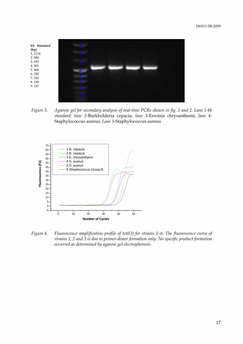
Figure 4. Fluorescence amplification profile of tet(O) for strains 1–6. The fluorescence curve of
strains 1, 2 and 3 is due to primer-dimer formation only. No specific product formation occurred as determined by agarose gel electrophoresis.
17
DSTO-TR-2059
50 55 60 65 70 75 80 85 90 95
-10123456789
1011121314
Fluo
resc
ence
-d(F
1)/d
T
Temperature (Co)
1 B. cepacia 2 B. cepacia 3 E. chrysanthemi 4 S. aureus 5 S. aureus 6 Streptococcus Group B
Figure 5. Melting peak profile of tet(O) amplification from strains 1 – 6. Agarose gel electrophoresis
showed that specific amplicons were only generated from strains 4 – 6. In contrast, melting peaks indicate specific amplicons from strains 1, 4 and 5.
1 2 3 4 5 6 7
Figure 6. Agarose gel for secondary analysis of tet(O) amplification showing that specific amplicon was only generated in reactions 4, 5, and 6. Lane 1-kb standard (see fig. 3); lane 2-Burkholderia cepacia; lane 3-Burkholderia cepacia; lane 4-Erwinia chrysanthemi; lane 5-Staphylococcus aureus; lane 6-Staphylococcus aureus; lane 7-Streptococcus Group B
1 2 3 4 5 6 7 8 9 10 11 12 13
18
DSTO-TR-2059
Figure 7. Agarose gel showing all amplicons displaying the expected legth. Lane 1-kb standard (see fig. 3); lane 2-tetA, Burkholderia cepacia; lane 3-tetL, Streptococcus sp.; lane 4-tetM, Staphylococcus aureus; lane 5-tetM, Staphylococcus aureus; lane 6- tetM, Streptococcus Group B; lane 7-tetM, Streptococcus Group B; lane 8-tetM, Streptococcus sp.; lane 9-tetM, Streptococcus sp.; lane 10-tetO, Staphylococcus aureus; lane 11-tetO, Staphylococcus aureus; lane 12-tetO, Streptococcus sp.; lane 13-tetO, Streptococcus sp. The relative weak bands obtained from tet(O) amplification (lanes 10-13) indicate mismatches between target and amplification primers.
DOCUMENT CONTROL DATA 1. PRIVACY MARKING/CAVEAT (OF DOCUMENT)
2. TITLE Rapid Detection of Bacterial Antibiotic Resistance: Preliminary Evaluation of PCR Assays Targeting Tetracycline Resistance Genes
3. SECURITY CLASSIFICATION (FOR UNCLASSIFIED REPORTS THAT ARE LIMITED RELEASE USE (L) NEXT TO DOCUMENT CLASSIFICATION) Document (U) Title (U) Abstract (U)
4. AUTHOR(S) Matthias R. Dorsch
5. CORPORATE AUTHOR DSTO Defence Science and Technology Organisation 506 Lorimer St Fishermans Bend Victoria 3207 Australia
6a. DSTO NUMBER DSTO-TR-2059
6b. AR NUMBER AR-014-034
6c. TYPE OF REPORT Technical Report
7. DOCUMENT DATE August 2007
8. FILE NUMBER 2004/1014097/1
9. TASK NUMBER 06/041
10. TASK SPONSOR DGDHS
11. NO. OF PAGES 21
12. NO. OF REFERENCES 31
13. URL on the World Wide Web http://www.dsto.defence.gov.au/corporate/reports/DSTO-TR-2059.pdf
14. RELEASE AUTHORITY Chief, Human Protection and Performance Division
15. SECONDARY RELEASE STATEMENT OF THIS DOCUMENT
Approved for public release OVERSEAS ENQUIRIES OUTSIDE STATED LIMITATIONS SHOULD BE REFERRED THROUGH DOCUMENT EXCHANGE, PO BOX 1500, EDINBURGH, SA 5111 16. DELIBERATE ANNOUNCEMENT No Limitations 17. CITATION IN OTHER DOCUMENTS Yes 18. DSTO RESEARCH LIBRARY THESAURUS http://web-vic.dsto.defence.gov.au/workareas/library/resources/dsto_thesaurus.htm Polymerase Chain Reacrtion, Rapid Diagnostic, Biological Warfare Agents, Antibiotic Resistance, Tetracycline, Genetically Engineered Resistance 19. ABSTRACT The rapid spreading of bacterial antibiotic resistance and the lack of methods that would allow fast and accurate determination of the resistance profile of an infectious agent have become a major concern for public health. In the context of biological warfare or a bioterrorist attack, resistance against commonly used antibiotics would severely compromise the capabilities of the responsible agencies to protect large numbers of exposed individuals against a potentially lethal infection. In this study the possibility of developing polymerase chain reaction (PCR) assays for rapid detection of tetracycline resistance genes was evaluated. PCR assays were designed for seven resistance genes that frequently occur in Gram-positive and Gram-negative species and tested on eight tetracycline resistant clinical isolates. Positive results were obtained from seven of the eight species tested.